clarity - ge healthcare/media/downloads/uk/product/computed... · clarity ct ge healthcare ......
TRANSCRIPT
the imprint.
clarityC T
GE Healthcare
Pediatric CT: Striking a Delicate BalancePage 32
Stroke Imaging Solutions: Shuttle-based Whole Brain PerfusionPage 50
Clinical Advantages of Gemstone™ Spectral Imaging and ASiR Page 14
t h e m a g a z i n e o f C t • S p r i n g 2 0 1 0

2
70 keV nicely demonstrates a right renal lesion.
2
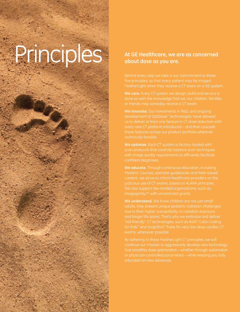
GE’s FeatherLight CT Principles
33
At GE Healthcare, we are as concerned about dose as you are.
Behind every step we take is our commitment to these five principles, so that every patient may be imaged FeatherLight when they receive a CT exam on a GE system:
We care. Every CT system we design, build and service is done so with the knowledge that we, our children, families or friends may someday receive a CT exam.
We innovate. Our investments in R&D, and ongoing development of OptiDose™ technologies, have allowed us to deliver at least one feature in CT dose reduction with every new CT platform introduced – and then cascade those features across our product portfolio wherever technically feasible.
We optimize. Each CT system is factory-loaded with scan protocols that carefully balance scan techniques with image quality requirements to efficiently facilitate confident diagnoses.
We educate. Through continuous education, including Masters’ Courses, operator guidebooks and Web-based content, we strive to inform healthcare providers on the judicious use of CT exams, based on ALARA principles. We also support like-minded organizations, such as imagegently,sm with unrestricted grants.
We understand. We know children are not just small adults; they present unique pediatric radiation challenges due to their higher susceptibility to radiation exposure and longer life spans. That’s why we embrace and deliver “kid-friendly” CT technologies, such as ASiR™ Color-Coding for Kids™ and SnapShot™ Pulse for very low-dose cardiac CT exams, whenever possible.
By adhering to these FeatherLight CT principles, we will continue our mission to aggressively develop new technology that simplifies dose optimization – whether through automation or physician-controlled parameters – while keeping you fully educated on new advances.
Principles
4 A GE Healthcare CT publication • Spring 2010
C o n T E n T St a b l e o f
GE Healthcare News
Welcome . . . . . . . . . . . . . . . . . . . . . . . . . . . . . . . . . . . . . . . . . . . . . . . . .6
Calendar of events . . . . . . . . . . . . . . . . . . . . . . . . . . . . . . . . . . . . . . . .7
ASiR Receives High Marks at european Congress of Radiology . . . . . . . . . . . . . . . . . . . . . . . . . . . . . . . . . . . .8
Sports Medicine of olympic Proportions . . . . . . . . . . . . . . . . . . . .9
42,000 year-old Baby Mammoth Scanned at GE Healthcare . . . . . . . . . . . . . . . . . . . . . . . . . . . . . . . 10
GE Healthcare News: 42,000 year-old Baby Mammoth Scanned at GE Healthcare page 10
Clinical Value
Tuning In to Lower Dose . . . . . . . . . . . . . . . . . . . . . . . . . . . . . . . . . 11
the next Generation of CT Imaging . . . . . . . . . . . . . . . . . . . . . . 14
Realizing the Vision with the Help of Low-dose, High-definition CT . . . . . . . . . . . . . . . . . . . . . . . . . . 18
new Frontiers in Image-Guided Procedures . . . . . . . . . . . . . . 22
Promises Kept: RSnA 2009 ASiR Abstracts . . . . . . . . . . . . . . . . . . . . . . . . . . . . . . . 28
Pediatric CT: Striking a Delicate Balance . . . . . . . . . . . . . . . . . 32
Pediatric CT Imaging Using ASiR Dialing Down Dose Across All Anatomies . . . . . . . . . . . . . . . . . 36
Clinical Value: Pediatric CT: Striking a Delicate Balance page 32
to receive future issues of CT Clarity, please subscribe at: www.gehealthcare.com/CTclarity
© 2010 General Electric Company, doing business as GE Healthcare. All rights reserved. The copyright, trademarks, trade names and other intellectual property rights subsisting in or used in connection with and related to this publication are the property of GE Healthcare unless otherwise specified. Reproduction in any form is forbidden without prior written permission from GE Healthcare.
LIMITATION OF LIABILITY: The information in this magazine is intended as a general presentation of the content included herein. While every effort is made by the publishers and editorial board to see that no inaccurate or misleading data, opinion or statements occur, GE cannot accept responsibility for the completeness, currency or accuracy of the information supplied or for any opinion expressed. nothing in this magazine should be used to diagnose or treat any disease or condition. Readers are advised to consult a healthcare professional with any questions. Products mentioned in the magazine may be subject to government regulation and may not be available in all locations. nothing in this magazine constitutes an offer to sell any product or service.
Technical Innovation
Under the Microscope – Examining the Approaches to Dual-energy CT . . . . . . . . . . . . 38
CT Perfusion with VolumeShuttle and Volume Helical Shuttle Used in a Successful Diagnosis and Treatment of Moyamoya neurological Disease . . . . . . 44
Removing the Barriers – ASiR Demonstrates Advantages over Filtered Back Projection . . . . . . . . . . . . . . . . . 47
Stroke Imaging Solutions: Shuttle-based Whole Brain Perfusion . . . . . . . . . . . . . . . . . . . . . 50
A new Standard in PET/CT . . . . . . . . . . . . . . . . . . . . . . . . . . . . . . . 55
Technical Innovation: Stroke Imaging Solutions: Shuttle-based Whole Brain Perfusion page 50
Publications Team:
Andrew Ackerman CT Clarity Editor CT Pediatric and Dose Segment Manager
John Allenstein Marketing Communications Manager CT and Advantage Workstation
Mike Grennier, APR Writer/Editorial Consultant
Scott Miller General Manager americas Ct
Nilesh Shah General Manager Global CT Marketing
J. Eric Stahre General Manager Global Computed Tomography
Integré Design/Production
GE Contributors:
Chelsea Beeler Communications Manager
Mark Bowman Product Manager
Amy Burris PET/CT Global Product Marketing Manager
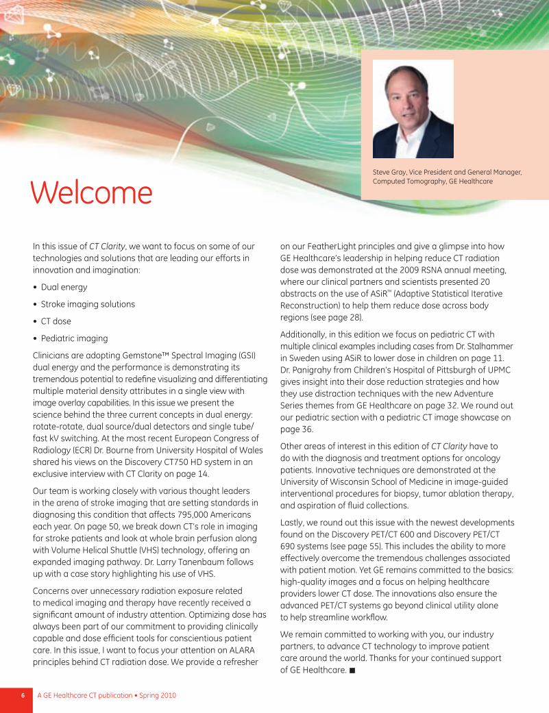
Steve Gray Vice President and General Manager Computed Tomography
Jiang Hsieh CT Chief Scientist
Kelley Knutson CT Education Manager
Paul Licato Global CT Research Manager
Dusty Majumdar Marketing Manager Premium Computed Tomography
Daniel Morris CT Global Marketing Manager
Souma Sengupta Clinical Services Manager
Christine Sickinger Marketing Communications Leader Ct europe
Saad Sirohey Global Product Manager neurology
Mary Toler TiP CT Image Database Manager
Jodi Young CT Education Manager
5A GE Healthcare CT publication • Spring 2010
C o n T E n T St a b l e o f
Beyond the Scan
Making an Informed Decision . . . . . . . . . . . . . . . . . . . . . . . . . . . . 59
Tuning In to the needs of Kids – GE Healthcare Adventure Series . . . . . . . . . . . . . . . . . . . . . . . . . . 62
learn from the experts: Ge Healthcare’s CT Masters Series . . . . . . . . . . . . . . . . . . . . . . . 66
orchestrate Your Training Clinical educational opportunities for europe . . . . . . . . . . . . 67
Beyond the Scan: Tuning In to the needs of Kids – GE Healthcare Adventure Series page 62
6 A GE Healthcare CT publication • Spring 2010
WelcomeIn this issue of CT Clarity, we want to focus on some of our technologies and solutions that are leading our efforts in innovation and imagination:
Dual energy •
Stroke imaging solutions •
CT dose•
Pediatric imaging •
Clinicians are adopting Gemstone™ Spectral Imaging (GSI) dual energy and the performance is demonstrating its tremendous potential to redefine visualizing and differentiating multiple material density attributes in a single view with image overlay capabilities. In this issue we present the science behind the three current concepts in dual energy: rotate-rotate, dual source/dual detectors and single tube/fast kV switching. At the most recent European Congress of Radiology (ECR) Dr. Bourne from University Hospital of Wales shared his views on the Discovery CT750 HD system in an exclusive interview with CT Clarity on page 14.
Our team is working closely with various thought leaders in the arena of stroke imaging that are setting standards in diagnosing this condition that affects 795,000 Americans each year. On page 50, we break down CT’s role in imaging for stroke patients and look at whole brain perfusion along with Volume Helical Shuttle (VHS) technology, offering an expanded imaging pathway. Dr. Larry Tanenbaum follows up with a case story highlighting his use of VHS.
Concerns over unnecessary radiation exposure related to medical imaging and therapy have recently received a significant amount of industry attention. Optimizing dose has always been part of our commitment to providing clinically capable and dose efficient tools for conscientious patient care. In this issue, I want to focus your attention on ALARA principles behind CT radiation dose. We provide a refresher
on our FeatherLight principles and give a glimpse into how GE Healthcare’s leadership in helping reduce CT radiation dose was demonstrated at the 2009 RSNA annual meeting, where our clinical partners and scientists presented 20 abstracts on the use of ASiR™ (Adaptive Statistical Iterative Reconstruction) to help them reduce dose across body regions (see page 28).
Additionally, in this edition we focus on pediatric CT with multiple clinical examples including cases from Dr. Stalhammer in Sweden using ASiR to lower dose in children on page 11. Dr. Panigrahy from Children’s Hospital of Pittsburgh of UPMC gives insight into their dose reduction strategies and how they use distraction techniques with the new Adventure Series themes from GE Healthcare on page 32. We round out our pediatric section with a pediatric CT image showcase on page 36.
Other areas of interest in this edition of CT Clarity have to do with the diagnosis and treatment options for oncology patients. Innovative techniques are demonstrated at the University of Wisconsin School of Medicine in image-guided interventional procedures for biopsy, tumor ablation therapy, and aspiration of fluid collections.
Lastly, we round out this issue with the newest developments found on the Discovery PET/CT 600 and Discovery PET/CT 690 systems (see page 55). This includes the ability to more effectively overcome the tremendous challenges associated with patient motion. Yet GE remains committed to the basics: high-quality images and a focus on helping healthcare providers lower CT dose. The innovations also ensure the advanced PET/CT systems go beyond clinical utility alone to help streamline workflow.
We remain committed to working with you, our industry partners, to advance CT technology to improve patient care around the world. Thanks for your continued support of GE Healthcare. n
Steve Gray, Vice President and General Manager, Computed Tomography, GE Healthcare
7A GE Healthcare CT publication • Spring 2010
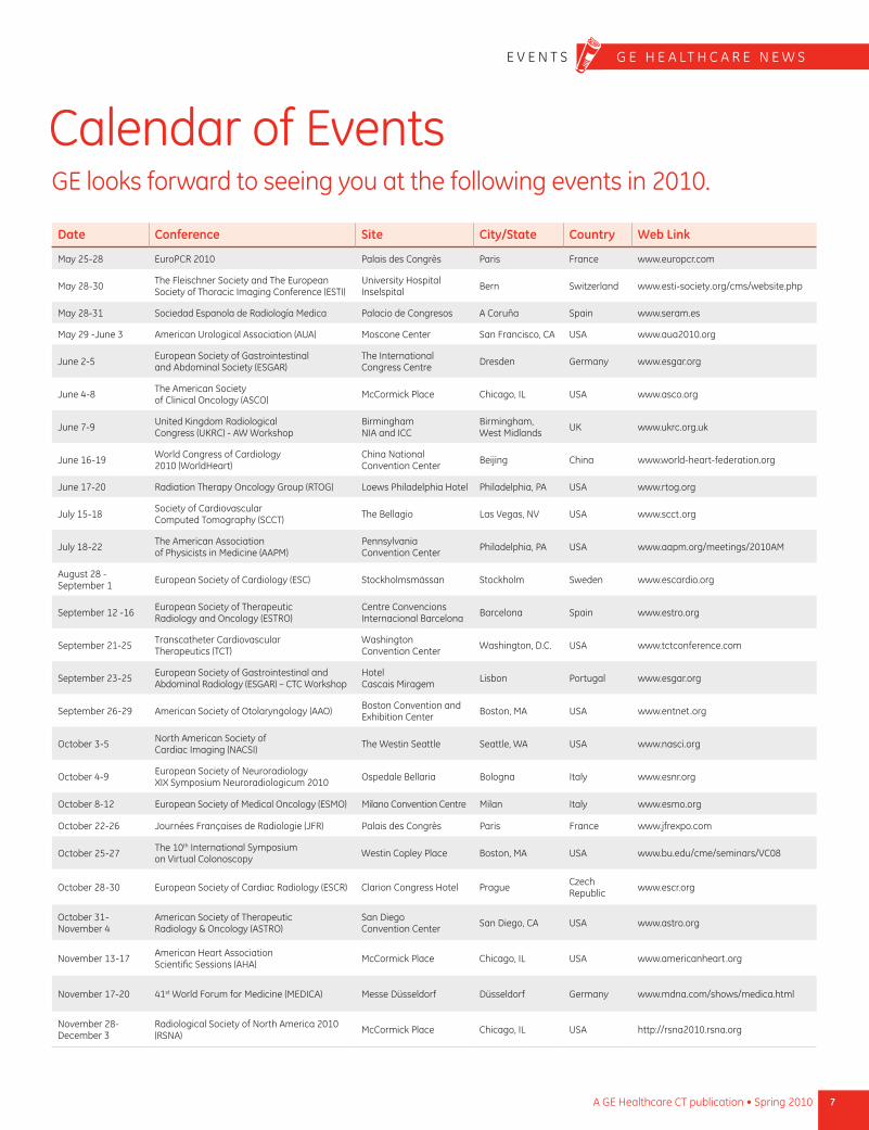
E v E n T S G E H E A lT H C A r E n E w S
GE looks forward to seeing you at the following events in 2010.
Calendar of Events
Date Conference Site City/State Country Web Link
May 25-28 EuroPCr 2010 Palais des Congrès Paris France www.europcr.com
May 28-30 The Fleischner Society and The European Society of Thoracic Imaging Conference (ESTI)
University Hospital Inselspital Bern Switzerland www.esti-society.org/cms/website.php
May 28-31 Sociedad Espanola de radiología Medica Palacio de Congresos A Coruña Spain www.seram.es
May 29 -June 3 American Urological Association (AUA) Moscone Center San Francisco, CA USA www.aua2010.org
June 2-5 European Society of Gastrointestinal and Abdominal Society (ESGAr)
The International Congress Centre Dresden Germany www.esgar.org
June 4-8 The American Society of Clinical Oncology (ASCO) McCormick Place Chicago, Il USA www.asco.org
June 7-9 United Kingdom radiological Congress (UKrC) - Aw workshop
Birmingham nIA and ICC
Birmingham, West Midlands UK www.ukrc.org.uk
June 16-19 world Congress of Cardiology 2010 (worldHeart)
China National Convention Center Beijing China www.world-heart-federation.org
June 17-20 radiation Therapy Oncology Group (rTOG) Loews Philadelphia Hotel Philadelphia, PA USA www.rtog.org
July 15-18 Society of Cardiovascular Computed Tomography (SCCT) The Bellagio las vegas, nv USA www.scct.org
July 18-22 The American Association of Physicists in Medicine (AAPM)
Pennsylvania Convention Center Philadelphia, PA USA www.aapm.org/meetings/2010AM
August 28 - September 1 European Society of Cardiology (ESC) Stockholmsmässan Stockholm Sweden www.escardio.org
September 12 -16 European Society of Therapeutic radiology and Oncology (ESTrO)
Centre Convencions Internacional Barcelona Barcelona Spain www.estro.org
September 21-25 Transcatheter Cardiovascular Therapeutics (TCT)
washington Convention Center washington, D.C. USA www.tctconference.com
September 23-25 European Society of Gastrointestinal and Abdominal radiology (ESGAr) – CTC workshop
Hotel Cascais Miragem Lisbon Portugal www.esgar.org
September 26-29 American Society of Otolaryngology (AAO) Boston Convention and Exhibition Center Boston, MA USA www.entnet.org
October 3-5 north American Society of Cardiac Imaging (nACSI) The westin Seattle Seattle, wA USA www.nasci.org
October 4-9 European Society of neuroradiology XIX Symposium neuroradiologicum 2010 Ospedale Bellaria Bologna Italy www.esnr.org
October 8-12 European Society of Medical Oncology (ESMO) Milano Convention Centre Milan Italy www.esmo.org
October 22-26 Journées Françaises de radiologie (JFr) Palais des Congrès Paris France www.jfrexpo.com
October 25-27 The 10th International Symposium on virtual Colonoscopy westin Copley Place Boston, MA USA www.bu.edu/cme/seminars/vC08
October 28-30 European Society of Cardiac radiology (ESCr) Clarion Congress Hotel Prague Czech Republic www.escr.org
October 31- november 4
American Society of Therapeutic radiology & Oncology (ASTrO)
San Diego Convention Center San Diego, CA USA www.astro.org
november 13-17 American Heart Association Scientific Sessions (AHA) McCormick Place Chicago, Il USA www.americanheart.org
november 17-20 41st world Forum for Medicine (MEDICA) Messe Düsseldorf Düsseldorf Germany www.mdna.com/shows/medica.html
november 28- December 3
radiological Society of north America 2010 (rSnA) McCormick Place Chicago, Il USA http://rsna2010.rsna.org
8 A GE Healthcare CT publication • Spring 2010
G E H E A lT H C A r E n E w S A n n o u n C E m E n T S
ASir receives High marks at European Congress of radiology
radiologists who presented their first-year findings on the functionality of ASir™ (Adaptive Statistical Iterative reconstruction) at the 2010 European Congress of radiology (ECr) in Vienna this spring report the ability to obtain high-quality images, yet at the same time, lower radiation dose by as much as 50% depending on the targeted region of interest.
leading healthcare providers who participated at ECr and shared their impressions of ASir in clinical practice include massachusetts General Hospital, the Hong Kong Sanatorium & Hospital, the Sydney Adventist Hospital, the Centre Cardiologique du nord, university Hospital of wales, Queen Silvia Children’s Hospital, and the university Hospital of rouen.
Developed by GE Healthcare and initially introduced on the Discovery™ CT750 HD scanner in the fall of 2008, ASir uses sophisticated modeling to remove noise in images while
preserving anatomical detail. with ASir, clinicians are able to effectively reduce dose and improve low-contrast detectability (lCD). Additionally, the use of ASir typically results in better contrast resolution across different patient sizes and anatomic regions.
michael Bourne, mD, a clinical radiologist from the university Hospital of wales, in Cardiff, wales, told ECr attendees the use of ASir meets the hospital’s high expectations for diagnostic performance, while also helping clinicians reduce dose in CT exams.
“Given the sensitivity to radiation dose and the increasing awareness of risks among patient and doctors, ASir becomes an obvious choice for us,” Bourne says. “Given this opportunity to reduce dose while maintaining image quality, we are excited to be making broad use of it in our facility.”
Fredrik Stälhammar, mD, who specializes in pediatric radiology at the Queen Silvia Children’s Hospital, Göteborg, Sweden, told attendees the hospital was able to reduce dose by as much as 50% using ASir – adding that the quality of the acquired images was identical to scans obtained using routine dose without ASir. Queen Silvia Children’s Hospital was the first children’s hospital to receive the advanced Discovery CT750 HD scanner with ASir.
“This is particularly beneficial for young patients with conditions which cannot be diagnosed conclusively using other techniques,” Stälhammar says. “ASir also benefits patients requiring repeat examinations, as the total radiation dose can be reduced substantially.” n
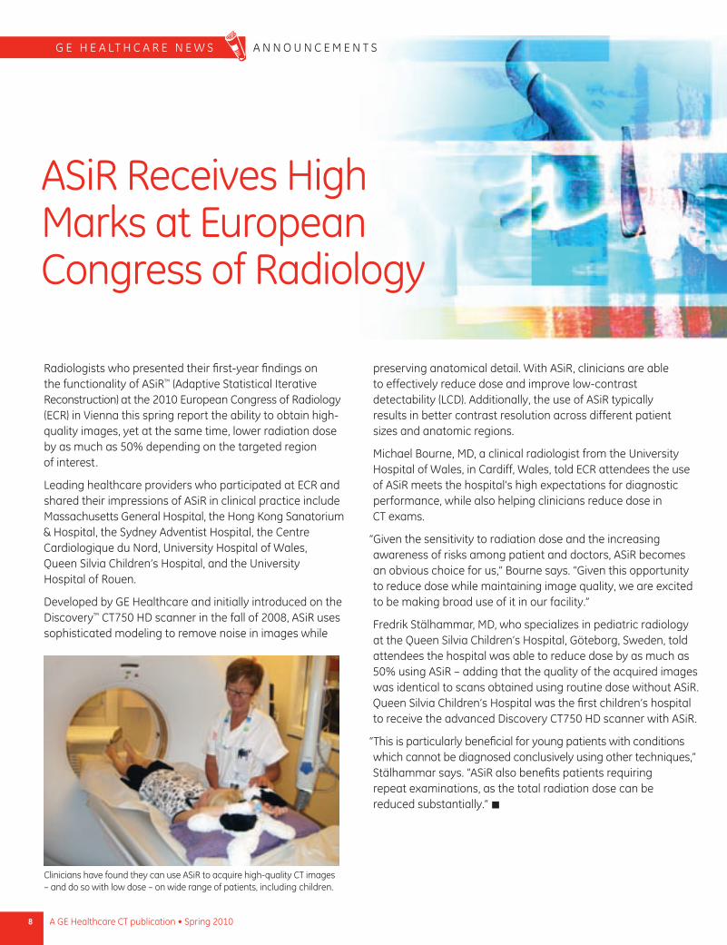
Clinicians have found they can use ASir to acquire high-quality CT images – and do so with low dose – on wide range of patients, including children.
Sports Medicine of Olympic ProportionsThanks to GE Healthcare imaging equipment, radiologists successfully performed 950 exams at the Vancouver 2010 Olympic and Paralympic Winter Games – and did so with utmost diagnostic confidence.
At the Games, two polyclinics were each outfitted with the LightSpeed™ VCT scanner, a GE Signa® HDi 1.5MR MRI system, and GE portable ultrasound equipment. Diagnostic imaging at Olympic events has taken on increased importance says Bruce Forster, MD, who managed a team of 20 radiologists at the Games.
“We performed around 950 imaging exams – compared to 567 at the 2006 Olympic Winter Games in Torino, which is almost a 70% increase,” Dr. Forster says. “It goes to show you how extremely critical and relevant the role of radiology plays in athletes. We don’t want injuries, but they’re going to happen and the fact that we’re being called upon to assist in this critical moment is very gratifying.”
In Olympic parlance, a medical visit is called an encounter. There were about 7,500 encounters at the Games and 34%
of them were athletes. The other patients were from the workforce or people “inside the fence.” Nearly twice as many CT scans were performed at the 2010 event when compared with the 2008 Olympic Games in Beijing.
Dr. Forster says the 2010 Games exemplified the concept of teamwork.
“It’s very much the technologists, radiologists, sports medicine, and orthopedic team all working together – and it’s absolutely critical. We would have physical therapists, trainers, you name it , involved in these meetings and they were inspired by the athletes,” Dr. Forster says, adding that diagnostic confidence was equally crucial.
“So much is riding on the clinical images, as they helped us assess whether an athlete could return to play, if they were out for a day or a week, or more,” Dr. Forster says. “These athletes are the best in the world. We needed to make sure we were the best in the world at what we do – with skill and equipment – so we could be there for them when they needed us.” n
GE donates LightSpeed VCT for use in 2010 Olympic Winter Games and beyond
In celebration of the Vancouver 2010 Olympic Winter Games, GE Healthcare donated a LightSpeed™ VCT scanner to the Whistler Health Care Centre in the Canadian province of British Columbia. This is the first CT scanner located in Whistler, BC, Canada, providing residents of the region with improved access to healthcare services closer to home.
The 64-slice scanner was installed at the center in advance of the Olympic Games to diagnose and treat athletes who participated in the event. It was also donated for ongoing use in Canada’s Sea to Sky region.
“Residents and visitors will no longer have to travel long distances for CT scans,” says Peter Foss, president, Olympic Sponsorship, GE. “It’s gratifying to know that one of GE’s innovative healthcare products will deliver substantial benefit within the region.”
As part of the Olympic event, GE also conducted ongoing cardiac and musculoskeletal research with some or several National Olympic Teams in order to improve technology in sports medicine. Following the Olympic Games, the LightSpeed VCT scanner became a popular imaging tool at the Whistler Health Care Centre, which is part of Vancouver Coastal Health (VCH).
“Physicians and clinicians are increasingly turning to diagnostic technology to better address the health needs of their patients and the community,” says Ida Goodreau, president and CEO of VCH.
“This CT scanner is an important legacy from the 2010 Winter Games, and demonstrates the strength of different organizations and foundations working in partnership to meet the needs of the people we serve.”
9A GE Healthcare CT publication • Spring 2010
A N N O u N C E M E N T S G E H E A LT H C A R E N E W S
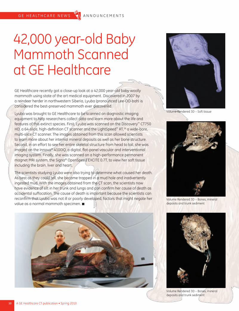
42,000 year-old Baby Mammoth Scanned at GE HealthcareGE Healthcare recently got a close-up look at a 42,000 year-old baby woolly mammoth using state of the art medical equipment. Discovered in 2007 by a reindeer herder in northwestern Siberia, Lyuba (pronounced Lee-OO-bah) is considered the best-preserved mammoth ever discovered.
Lyuba was brought to GE Healthcare to be scanned on diagnostic imaging equipment to help researchers collect data and learn more about the life and features of this extinct species. First, Lyuba was scanned on the Discovery™ CT750 HD, a 64-slice, high-definition CT scanner and the LightSpeed™ RT,16 a wide-bore, multi-slice CT scanner. The images obtained from this scan allowed scientists to learn more about her internal mineral deposits as well as her bone structure. Second, in an effort to see her entire skeletal structure from head to tail, she was imaged on the Innova® 4100IQ, a digital, flat-panel vascular and interventional imaging system. Finally, she was scanned on a high-performance permanent magnet MRI system, the Signa® OpenSpeed EXCITE 0.7T, to view her soft tissue including the brain, liver and heart.
The scientists studying Lyuba were also trying to determine what caused her death. As best as they could tell, she became trapped in a mud hole and inadvertently ingested mud. With the images obtained from the CT scan, the scientists now have evidence of silt in her trunk and lungs and can confirm her cause of death as accidental suffocation. The cause of death is important because the scientists can reconfirm that Lyuba was not ill or poorly developed, factors that might negate her value as a normal mammoth specimen. n
Volume Rendered 3D – Soft tissue
Volume Rendered 3D – Bones, mineral deposits and trunk sediment
Volume Rendered 3D – Bones, mineral deposits and trunk sediment
10 A GE Healthcare CT publication • Spring 2010
G E H E A LT H C A R E n E W S A n n O u n C E M E n T S
11A GE Healthcare CT publication • Spring 2010
Tuning In to Lower DoseCT Dose Reduction for Pediatrics
Advances in CT have increased dramatically during the past 10 years, offering a non-invasive technique for examining patients without having to resort to exploratory surgeries that were once routine clinical practice. Significant strides have also been made in the effort to minimize radiation exposure associated with CT.
ASiR helps lowers dose at Queen Silvia Children’s Hospital
An advanced technology for helping clinicians lower CT dose is ASiR,™ GE Healthcare’s proprietary adaptive statistical iterative image reconstruction technique. As shown by clinical use over the past year at major medical centers, ASiR has enabled dose reductions of up to 40-50% – often with image quality at that is equivalent or better than images obtained using conventional reconstruction methods. Some clinicians, including Fredrik Stälhammar, MD, of Queen Silvia Children’s Hospital, Göteborg, Sweden, have seen dramatically lower dose reductions with ASiR.
Presenting his findings at the 2010 European Congress of Radiology (ECR) in Vienna, Dr. Stälhammar reported significant dose reductions using ASiR, adding that image quality when using ASiR was identical to images obtained using the routine dose without ASIR.
“This is particularly beneficial for young patients with conditions that cannot be diagnosed conclusively using other techniques,” says Stälhammar, who specializes in pediatric radiology at the Queen Silvia Children’s Hospital. “ASiR also benefits patients requiring follow up examinations, as the total radiation dose can be reduced substantially.”
Queen Silvia Children’s Hospital was the first children’s hospital to receive the advanced Discovery™ CT750 HD scanner equipped with ASiR. The scanner was immediately put to use as part of the hospital’s efforts to minimize the CT radiation in adherence to ALARA principles. “A child with a medical condition is likely to have a greater exposure over a longer life expectancy than an adult with a greater risk for a larger accumulated lifetime dose,” Stälhammar says. “With the Discovery CT750 HD and ASiR we are more comfortable using CT to help in diagnosing our little patients’ conditions.”
12 A GE Healthcare CT publication • Spring 2010
C L I n I C A L V A L u E P E D I A T R I C C T & L o W D o S E
About the facility
The Queen Silvia Children’s Hospital in Göteborg is one of the leading neuromuscular and pediatric centers in Sweden. It plays an active role in research, diagnostic work-up and yearly follow-up programs for children with various chronic disorders.
“ With the Discovery CT750 HD and ASiR we are more comfortable using CT to help in diagnosing our little patients’ conditions.”
Dr. Fredrik Stälhammar
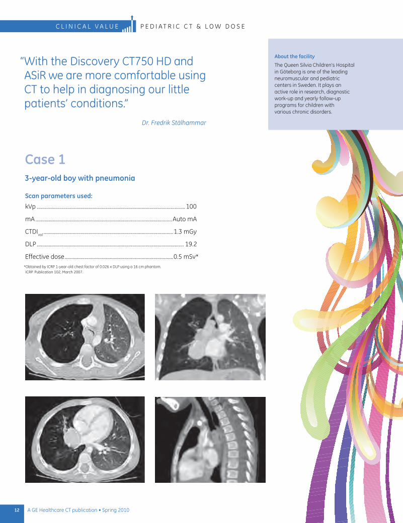
Case 13-year-old boy with pneumonia
Scan parameters used:
kVp ................................................................................................................100
mA .......................................................................................................Auto mA
CTDIvol ..................................................................................................1.3 mGy
DLP ............................................................................................................... 19.2
Effective dose ..................................................................................0.5 mSv* *obtained by ICRP 1-year-old chest factor of 0.026 x DLP using a 16 cm phantom. ICRP. Publication 102, March 2007.
13A GE Healthcare CT publication • Spring 2010
P E D I A T R I C C T & L o W D o S E C L I n I C A L V A L u E
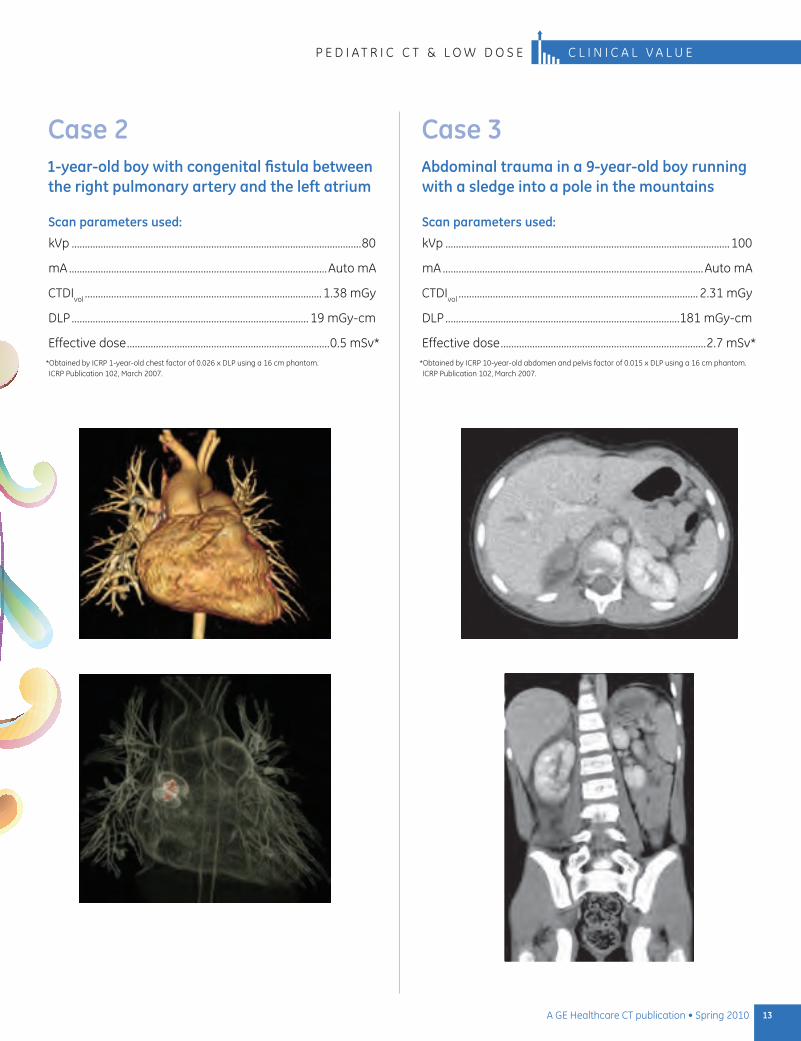
Case 21-year-old boy with congenital fistula between the right pulmonary artery and the left atrium
Scan parameters used:
kVp ..............................................................................................................80
mA ..................................................................................................Auto mA
CTDIvol ..........................................................................................1.38 mGy
DLP .......................................................................................... 19 mGy-cm
Effective dose .............................................................................0.5 mSv* *obtained by ICRP 1-year-old chest factor of 0.026 x DLP using a 16 cm phantom. ICRP Publication 102, March 2007.
Case 3Abdominal trauma in a 9-year-old boy running with a sledge into a pole in the mountains
Scan parameters used:
kVp ............................................................................................................100
mA ...................................................................................................Auto mA
CTDIvol ........................................................................................... 2.31 mGy
DLP .........................................................................................181 mGy-cm
Effective dose ..............................................................................2.7 mSv* *obtained by ICRP 10-year-old abdomen and pelvis factor of 0.015 x DLP using a 16 cm phantom. ICRP Publication 102, March 2007.
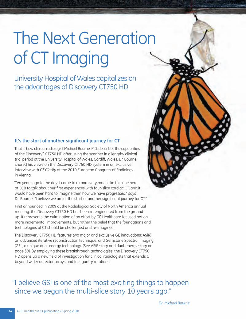
The Next Generation of CT ImagingUniversity Hospital of Wales capitalizes on the advantages of Discovery CT750 HD
It’s the start of another significant journey for CT
That is how clinical radiologist Michael Bourne, MD, describes the capabilities of the Discovery™ CT750 HD after using the scanner in a lengthy clinical trial period at the University Hospital of Wales, Cardiff, Wales. Dr. Bourne shared his views on the Discovery CT750 HD system in an exclusive interview with CT Clarity at the 2010 European Congress of Radiology in Vienna.
“Ten years ago to the day, I came to a room very much like this one here at ECR to talk about our first experiences with four-slice cardiac CT, and it would have been hard to imagine then how we have progressed,” says Dr. Bourne. “I believe we are at the start of another significant journey for CT.”
First announced in 2009 at the Radiological Society of North America annual meeting, the Discovery CT750 HD has been re-engineered from the ground up. It represents the culmination of an effort by GE Healthcare focused not on more incremental improvements, but rather the belief that the foundations and technologies of CT should be challenged and re-imagined.
The Discovery CT750 HD features two major and exclusive GE innovations: ASiR,™ an advanced iterative reconstruction technique; and Gemstone Spectral Imaging (GSI), a unique dual-energy technology. (See ASiR story and dual-energy story on page 38). By employing these breakthrough technologies, the Discovery CT750 HD opens up a new field of investigation for clinical radiologists that extends CT beyond wider detector arrays and fast gantry rotations.
“ I believe GSI is one of the most exciting things to happen since we began the multi-slice story 10 years ago.”
Dr. Michael Bourne
14 A GE Healthcare CT publication • Spring 2010
15A GE Healthcare CT publication • Spring 2010
Michael Bourne, MD, is a clinical radiologist at the University Hospital of Wales, Cardiff, Wales.
ASiR: Lowering dose without compromises
“I cannot recall any CT engineering in the last 25 years that has enabled such a significant reduction in dose,” says Dr. Bourne after using ASiR at the University Hospital of Wales. “We have done thousands of scans using ASiR, we are using it routinely, and we find there is no detrimental effect on the diagnostic performance.”
ASiR uses sophisticated statistical modeling to reduce noise in images while preserving anatomical detail. As such, it enables the same quality image to be produced with lower tube current or tube voltage, thus lower dose, for all CT applications in which it is used.
Using ASiR, clinicians are now able to effectively reduce dose by up to 50%, or improve low-contrast detectability (LCD) by as much as 40%. The use of ASiR may result in better contrast resolution across different patient sizes and anatomic regions.
“In our studies, we’ve found that ASiR can be used to improve the diagnostic quality of what is already a low-dose study, or it can be used to significantly reduce the dose of a previously high dose exam with the same background noise,” says Dr. Bourne.
New possibilities with GSI
Dr. Bourne says GSI offers the capability to explore new clinical applications for diagnosis using CT. He adds that it can even be used to revisit areas where CT might have been considered insufficient for achieving diagnostic confidence.
“I believe GSI is one of the most exciting things to happen since we began the multi-slice story 10 years ago,” Dr. Bourne says.
GSI is a dual-energy scan mode that acquires data of an object by alternating quickly between low kVp and high kVp spectrums through a particular density. By so doing, it generates data with different attenuation values based on the corresponding energy levels in terms of water and iodine, water and calcium, and iodine and calcium basis-pair images. The result is a near-perfect, simultaneous dual-energy acquisition for axial, helical, or cine at the full 50 cm Scan Field of View (SFOV) providing incredible temporal registration.
Projection-based reconstruction is used to process the data. Based on known attenuation curves, the process mathematically transforms low and high kVp attenuation measurements into effective material densities. This is also known as material decomposition. GSI provides unique CT images, termed “Material Density (MD) pairs,” which are not available with conventional contrast-enhanced CT imaging. The MD pairs can be chosen based on the clinical question and materials of interest: iodine-water, iodine-calcium, or water-calcium (Figures 1a and 1b). GSI also allows for the creation of a monochromatic image, which is synthesized from the MD images and depicts how the image object would look if the X-ray source produced only X-ray photons at a single energy.
Figure 1a. Spectral 70 keV image, MD water (iodine)
Figure 1b. Spectral 70 keV image, MD iodine (water)
D I S C O V E R y C T 7 5 0 H D W I T H A S I R A N D G S I C L I N I C A L V A L U E
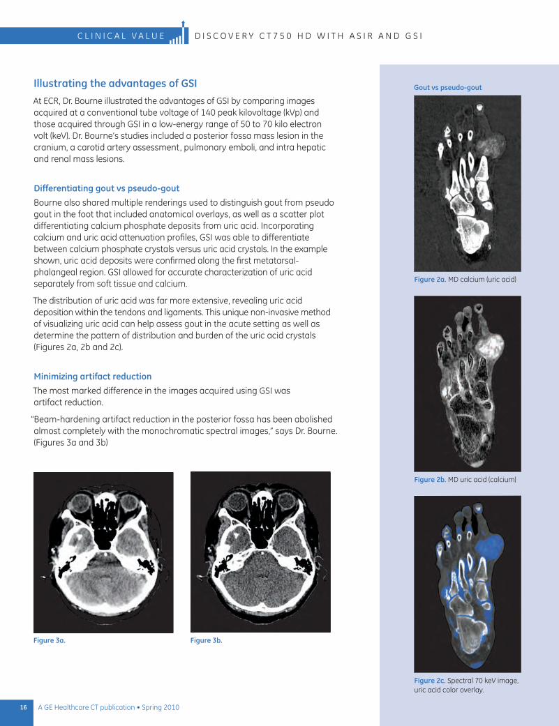
Illustrating the advantages of GSIAt ECR, Dr. Bourne illustrated the advantages of GSI by comparing images acquired at a conventional tube voltage of 140 peak kilovoltage (kVp) and those acquired through GSI in a low-energy range of 50 to 70 kilo electron volt (keV). Dr. Bourne’s studies included a posterior fossa mass lesion in the cranium, a carotid artery assessment, pulmonary emboli, and intra hepatic and renal mass lesions.
Differentiating gout vs pseudo-goutBourne also shared multiple renderings used to distinguish gout from pseudo gout in the foot that included anatomical overlays, as well as a scatter plot differentiating calcium phosphate deposits from uric acid. Incorporating calcium and uric acid attenuation profiles, GSI was able to differentiate between calcium phosphate crystals versus uric acid crystals. In the example shown, uric acid deposits were confirmed along the first metatarsal-phalangeal region. GSI allowed for accurate characterization of uric acid separately from soft tissue and calcium.
The distribution of uric acid was far more extensive, revealing uric acid deposition within the tendons and ligaments. This unique non-invasive method of visualizing uric acid can help assess gout in the acute setting as well as determine the pattern of distribution and burden of the uric acid crystals (Figures 2a, 2b and 2c).
Minimizing artifact reductionThe most marked difference in the images acquired using GSI was artifact reduction.
“Beam-hardening artifact reduction in the posterior fossa has been abolished almost completely with the monochromatic spectral images,” says Dr. Bourne. (Figures 3a and 3b)
Figure 3a. Figure 3b.
Figure 2a. MD calcium (uric acid)
Figure 2b. MD uric acid (calcium)
Figure 2c. Spectral 70 keV image, uric acid color overlay.
Gout vs pseudo-gout
16 A GE Healthcare CT publication • Spring 2010
C l I n I C A l V A l u E D I S C o V E R y C T 7 5 0 H D w I T H A S I R A n D G S I
375226_14-17_EP.indd 16 5/13/10 2:22:17 PM
17A GE Healthcare CT publication • Spring 2010
D i S C o v E r y C T 7 5 0 H D w i T H A S i r A n D G S i C l i n i C A l v A l u E
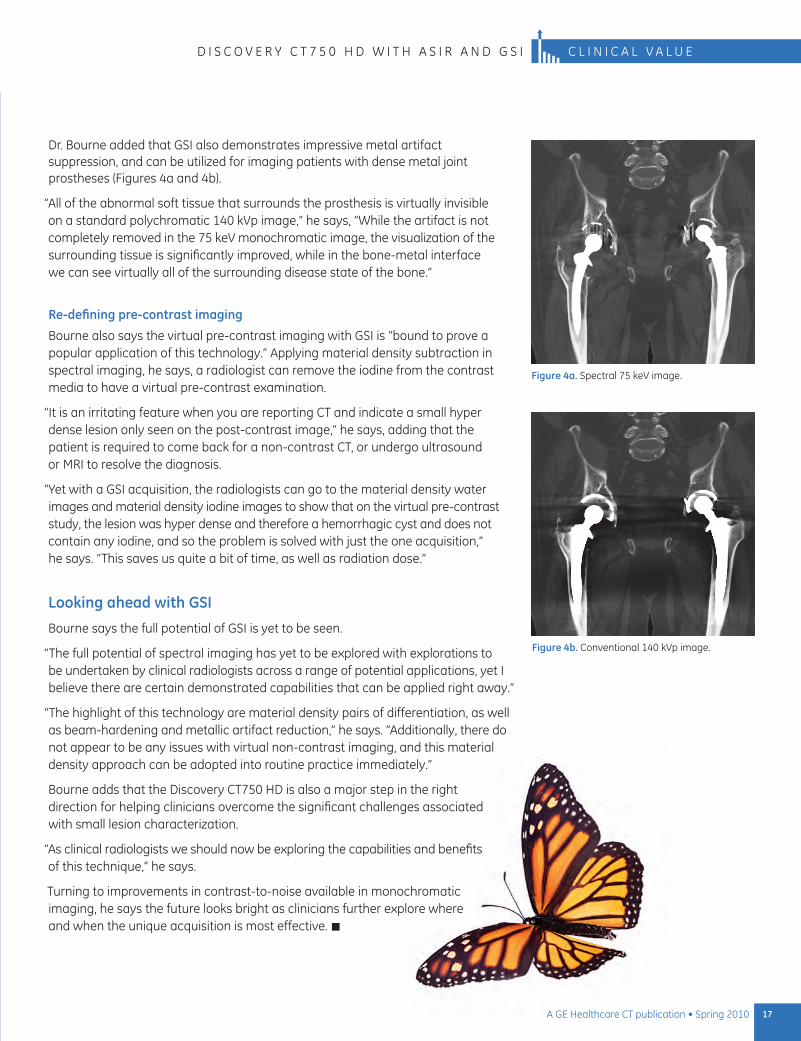
Dr. Bourne added that GSi also demonstrates impressive metal artifact suppression, and can be utilized for imaging patients with dense metal joint prostheses (Figures 4a and 4b).
“All of the abnormal soft tissue that surrounds the prosthesis is virtually invisible on a standard polychromatic 140 kvp image,” he says, “while the artifact is not completely removed in the 75 kev monochromatic image, the visualization of the surrounding tissue is significantly improved, while in the bone-metal interface we can see virtually all of the surrounding disease state of the bone.”
Re-defining pre-contrast imaging
Bourne also says the virtual pre-contrast imaging with GSi is “bound to prove a popular application of this technology.” Applying material density subtraction in spectral imaging, he says, a radiologist can remove the iodine from the contrast media to have a virtual pre-contrast examination.
“it is an irritating feature when you are reporting CT and indicate a small hyper dense lesion only seen on the post-contrast image,” he says, adding that the patient is required to come back for a non-contrast CT, or undergo ultrasound or Mri to resolve the diagnosis.
“yet with a GSi acquisition, the radiologists can go to the material density water images and material density iodine images to show that on the virtual pre-contrast study, the lesion was hyper dense and therefore a hemorrhagic cyst and does not contain any iodine, and so the problem is solved with just the one acquisition,” he says. “This saves us quite a bit of time, as well as radiation dose.”
Looking ahead with GSI
Bourne says the full potential of GSi is yet to be seen.
“The full potential of spectral imaging has yet to be explored with explorations to be undertaken by clinical radiologists across a range of potential applications, yet i believe there are certain demonstrated capabilities that can be applied right away.”
“The highlight of this technology are material density pairs of differentiation, as well as beam-hardening and metallic artifact reduction,” he says. “Additionally, there do not appear to be any issues with virtual non-contrast imaging, and this material density approach can be adopted into routine practice immediately.”
Bourne adds that the Discovery CT750 HD is also a major step in the right direction for helping clinicians overcome the significant challenges associated with small lesion characterization.
“As clinical radiologists we should now be exploring the capabilities and benefits of this technique,” he says.
Turning to improvements in contrast-to-noise available in monochromatic imaging, he says the future looks bright as clinicians further explore where and when the unique acquisition is most effective.
Figure 4a. Spectral 75 kev image.
Figure 4b. Conventional 140 kvp image.
375226_14-17_EP.indd 17 5/13/10 2:22:29 PM
18 A GE Healthcare CT publication • Spring 2010
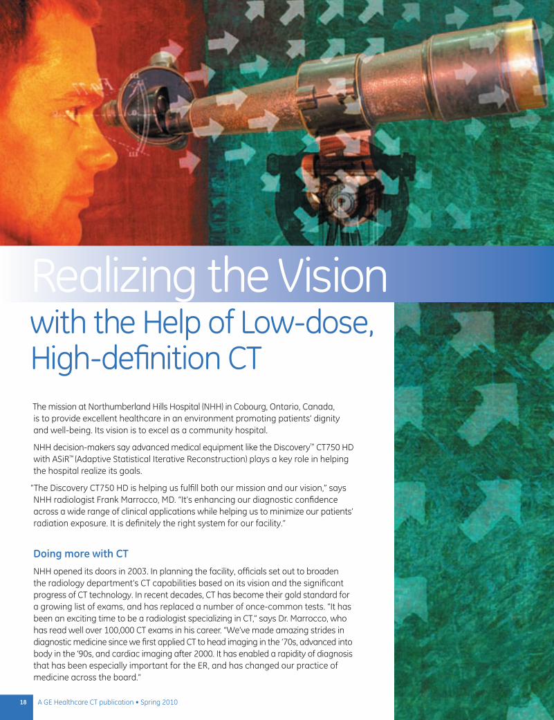
The mission at Northumberland Hills Hospital (NHH) in Cobourg, Ontario, Canada, is to provide excellent healthcare in an environment promoting patients’ dignity and well-being. Its vision is to excel as a community hospital.
NHH decision-makers say advanced medical equipment like the Discovery™ CT750 HD with ASiR™ (Adaptive Statistical Iterative Reconstruction) plays a key role in helping the hospital realize its goals.
“The Discovery CT750 HD is helping us fulfill both our mission and our vision,” says NHH radiologist Frank Marrocco, MD. “It’s enhancing our diagnostic confidence across a wide range of clinical applications while helping us to minimize our patients’ radiation exposure. It is definitely the right system for our facility.”
Doing more with CT NHH opened its doors in 2003. In planning the facility, officials set out to broaden the radiology department’s CT capabilities based on its vision and the significant progress of CT technology. In recent decades, CT has become their gold standard for a growing list of exams, and has replaced a number of once-common tests. “It has been an exciting time to be a radiologist specializing in CT,” says Dr. Marrocco, who has read well over 100,000 CT exams in his career. “We’ve made amazing strides in diagnostic medicine since we first applied CT to head imaging in the ‘70s, advanced into body in the ‘90s, and cardiac imaging after 2000. It has enabled a rapidity of diagnosis that has been especially important for the ER, and has changed our practice of medicine across the board.”
Realizing the Vision with the Help of Low-dose, High-definition CT
“ The Discovery CT750 HD is helping us fulfill both our mission and our vision. It’s enhancing our diagnostic confidence across a wide range of clinical applications while helping us to minimize our patients’ radiation exposure. It is definitely the right system for our facility.”
Dr. Frank Marrocco
19A GE Healthcare CT publication • Spring 2010
Francis Marrocco, MD, is staff radiologist and director of CT scanning at Northumberland Hills Hospital.
About the facility
The 137-bed hospital delivers a broad range of services, including medical/surgical care, complex/long term care, rehabilitation, palliative care, obstetrical care and intensive care. NHH employs over 500 people and is an active member of the Central East Local Health Integration Network (LHIN).
D I S C O V E R y C T 7 5 0 H D W I T H A S I R C L I N I C A L V A L u E
Putting dose reduction first
NHH was eager to incorporate the use of advanced CT at its facility, but only after carefully weighing the pros and cons of the technology. Chief among NHH’s concerns with any CT system is the need to minimize radiation exposure since radiation is used to obtain any CT image, says Dr. Marrocco. It’s an issue that strongly influences the approach NHH radiologists follow for imaging patients.
As CT utilizes ionizing radiation, there is a need to balance benefit against potential harm. Therefore, radiation dose is an important consideration in deciding whether to perform CT and also how to conduct the CT examination with doses as low as reasonably achievable. “It is especially important for pediatric patients and those who require repeated scans,” he says. “If it is feasible to offer an alternate test that will give us the information we need, such as ultrasound or MRI, we will always recommend that – especially for young people and children. However, as is often the case, CT is really the preferred test with which to make the correct diagnosis.”
The increased attention on CT at NHH and the industry as a whole only intensified the hospital’s scrutiny of radiation dose. It also drove the need to find a CT solution that helped reduce dose as much as possible without interfering with diagnostic utility of the ubiquitous devices.
GE and Discovery CT750 HD: a good combination
In analyzing its choices, NHH assessed a number of CT vendors and ultimately chose to install the Discovery CT750 HD at the facility in March 2009.
According to Dr. Marrocco, GE Healthcare’s leadership in dose management, combined with the capabilities of the latest high-definition scanner, were key to the decision. Perhaps the Discovery CT750 HD’s greatest contribution to dose minimization is ASiR – a breakthrough reconstruction technique that performs an adaptive statistical iterative reconstruction to the raw scan data to produce images with lower noise than the Filtered Back Projection (FBP) reconstruction approach. The end result is a system that cuts dose by up to 50% while maintaining image quality for diagnosis.
Establishing the right protocols
With the scanner up and running, NHH immediately began working on dose reduction protocols. Dr. Marrocco says the radiology team is making steady progress.
“It’s a process and we’re not likely to ever stop refining our protocols,” he says. “Every doctor I know wants to do a better job for his patients, and we’re no different.”
The overarching CT goal at NHH is to maintain image quality made possible with the hospital’s existing scanner, but at much lower dose. Achieving that goal depends at least partly on the anatomy being scanned.
According to Dr. Marrocco, the dose savings at NHH is significant when surveying anatomy such as the abdomen, pelvis, and chest and somewhat less for liver and brain. NHH uses CT extensively for imaging the lungs and large bowel (“CT colonography”). The Discovery CT750 HD constructs detailed images of these organs with an incredibly low radiation dose compared to earlier scanners.

20 A GE Healthcare CT publication • Spring 2010
C L I N I C A L V A L u E D I S C O V E R y C T 7 5 0 H D W I T H A S I R
1 Estimated Radiation Dose of Coronary CT Angiography using Adaptive Statistical Iterative Reconstruction (ERASIR 1) study, diagnosticimaging.com December 2, 2009.
The Discovery CT750 HD also offers extraordinary image detail because of its improved spatial resolution. “The bones of the inner ear and the paranasal sinus are quite striking when scanned with the Discovery CT750 HD,” Dr. Marrocco says. “And because we’re imaging bone vs. air with these studies, we can achieve significantly lower dose without sacrificing contrast.”
Dr. Marrocco says his department continues working on updating protocols to take full advantage of the Discovery CT750 HD’s low-dose potential. Protocol development is central to striking the optimum balance between clinical utility and radiation dose, he says.
“When we design protocols, we always follow ALARA (As Low As Reasonably Achievable) principles. For each protocol, we start with the clinical problem and determine how much noise we can tolerate for this particular application. Because ASiR reconstructs images with lower noise, we are able to reduce our dose while maintaining a given noise level.”
Dr. Marrocco has taken a personal interest in ensuring that everyone concerned with CT imaging is aware of radiation dose – including the technologists performing the exams and the referring physicians who order them. To underscore its importance to referring physicians, he includes a total exam dose in his reports.
“The ability to quantify exposure helps keeps me aware of it ,” Dr. Marrocco says. “And it gives us a benchmark to work from as we continue our dose-minimizing efforts.”
Low-dose cardiac studies
NHH is using the Discovery CT750 HD for angiography, including cardiac studies. Dr. Marrocco says it’s especially valuable for such applications as diagnosing atypical chest pain and stroke workups. The exams include the
use of SnapShot™ Pulse. This prospective gating technique synchronizes the acquisition to the patient’s heart rate so that X-rays are on only during the required cardiac phase, allowing clinicians to achieve dose reductions of up to 83%. A large multi-institution clinical study1 demonstrated a 1.3 mSv median dose when ASiR was used with other radiation reduction strategies. “The ability to lower dose to this level is truly amazing,” says Dr. Marrocco.
In general, Dr. Marrocco says cardiac CT is especially useful for evaluating patients with a low probability of having coronary disease.
“CT is very reliable as a negative predictive value test,” he says. “To be able to say that a patient’s chest pain is not from coronary artery disease is huge – especially when you can do it in a timely manner at low dose.”
Everyone wins
Dr. Marrocco and his staff are convinced they made the right choice in the Discovery CT750 HD. He says there’s a learning curve with the new system, adding that it’s no different than the time and investment needed to learn any other breakthrough technology.
“The images generated with ASiR look different, and our radiologists have gotten to the point where they prefer them over images produced with conventional CT.”
Although it’s only been in use for approximately one year, Dr. Marrocco says the Discovery CT750 HD benefits NHH and its patients on numerous levels.
“It’s a diagnostic tool with amazing utility,” he says, “and we’re very happy to be putting it to work on behalf of our hospital.” n
“ The images generated with ASiR look different, and our radiologists have gotten to the point where they prefer them over images produced with conventional CT.”
Dr. Frank Marrocco
21A GE Healthcare CT publication • Spring 2010
C L I N I C A L V A L u ED I S C O V E R y C T 7 5 0 H D W I T H A S I R
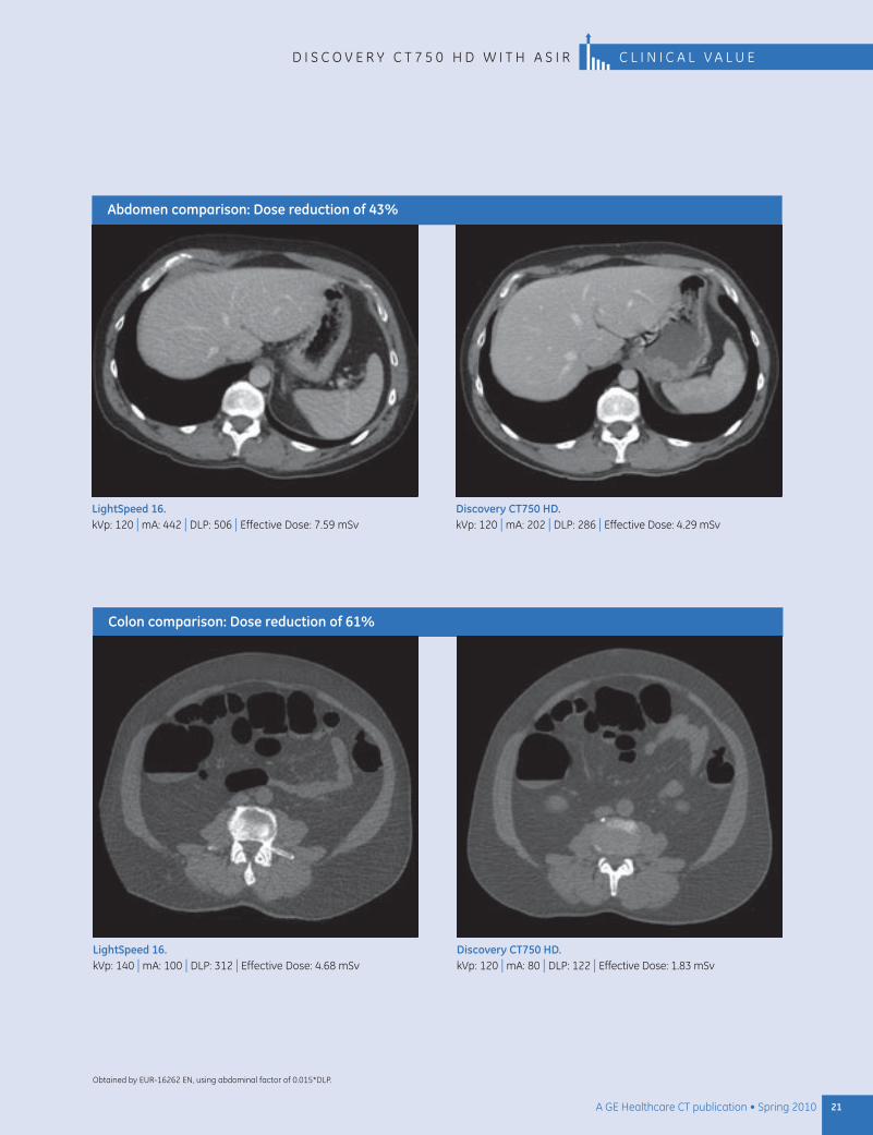
Abdomen comparison: Dose reduction of 43%
LightSpeed 16. kVp: 120 | mA: 442 | DLP: 506 | Effective Dose: 7.59 mSv
Discovery CT750 HD. kVp: 120 | mA: 202 | DLP: 286 | Effective Dose: 4.29 mSv
Colon comparison: Dose reduction of 61%
LightSpeed 16. kVp: 140 | mA: 100 | DLP: 312 | Effective Dose: 4.68 mSv
Discovery CT750 HD. kVp: 120 | mA: 80 | DLP: 122 | Effective Dose: 1.83 mSv
Obtained by EuR-16262 EN, using abdominal factor of 0.015*DLP.
New Frontiers in Image-Guided ProceduresBy Meghan G. Lubner, MD, Assistant Professor in Abdominal Imaging, University of Wisconsin School of Medicine and Public Health
22 A GE Healthcare CT publication • Spring 2010
The power of image guidance continues to expand with the increasing capability of CT, ultrasound and other imaging modalities.
Dr. Meghan G. Lubner
Advances in technology are expanding the capability of CT guidance for interventional procedures such as biopsy, aspiration, and tumor ablation.
Image-guided interventions have spared countless patients the risk and pain of more invasive surgeries, while helping clinicians complete procedures with great efficiency. The power of image guidance continues to expand with the increasing capability of CT, ultrasound and other imaging modalities.
A number of trends are driving growth in image-guided procedures. For example, cancer diagnoses are increasing as the population ages, and chemotherapeutic agents are emerging that target certain markers within tumors. This means a simple diagnosis of cancer is not enough: core biopsies are needed for testing to determine specific tumor characteristics. Meanwhile, in line with a general trend toward less invasive treatments, tumor ablation is growing almost exponentially, especially in the liver, kidney and lung.
The University of Wisconsin School of Medicine and Public Health uses CT and ultrasound guidance extensively for biopsy, tumor ablation therapy, and aspiration of fluid collections. New image-guidance tools are extending the range of applications and making procedures faster and more accurate.
Essential image-guidance tools in the UW facility include a GE LightSpeed™ Xtra wide-bore scanner and the GE LOGIQ® E9 ultrasound system.
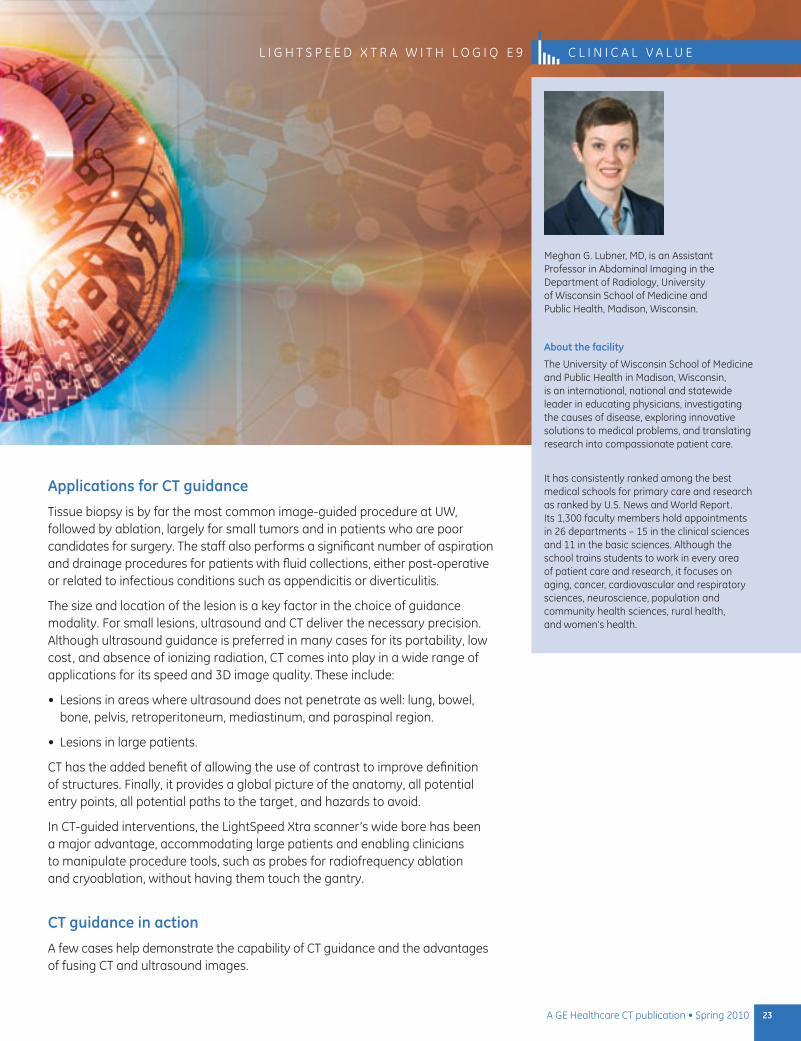
Meghan G. Lubner, MD, is an Assistant Professor in Abdominal Imaging in the Department of Radiology, University of Wisconsin School of Medicine and Public Health, Madison, Wisconsin.
About the facility
The University of Wisconsin School of Medicine and Public Health in Madison, Wisconsin, is an international, national and statewide leader in educating physicians, investigating the causes of disease, exploring innovative solutions to medical problems, and translating research into compassionate patient care.
It has consistently ranked among the best medical schools for primary care and research as ranked by U.S. News and World Report. Its 1,300 faculty members hold appointments in 26 departments – 15 in the clinical sciences and 11 in the basic sciences. Although the school trains students to work in every area of patient care and research, it focuses on aging, cancer, cardiovascular and respiratory sciences, neuroscience, population and community health sciences, rural health, and women’s health.
Applications for CT guidance
Tissue biopsy is by far the most common image-guided procedure at UW, followed by ablation, largely for small tumors and in patients who are poor candidates for surgery. The staff also performs a significant number of aspiration and drainage procedures for patients with fluid collections, either post-operative or related to infectious conditions such as appendicitis or diverticulitis.
The size and location of the lesion is a key factor in the choice of guidance modality. For small lesions, ultrasound and CT deliver the necessary precision. Although ultrasound guidance is preferred in many cases for its portability, low cost, and absence of ionizing radiation, CT comes into play in a wide range of applications for its speed and 3D image quality. These include:
Lesions in areas where ultrasound does not penetrate as well: lung, bowel, • bone, pelvis, retroperitoneum, mediastinum, and paraspinal region.
Lesions in large patients.•
CT has the added benefit of allowing the use of contrast to improve definition of structures. Finally, it provides a global picture of the anatomy, all potential entry points, all potential paths to the target, and hazards to avoid.
In CT-guided interventions, the LightSpeed Xtra scanner’s wide bore has been a major advantage, accommodating large patients and enabling clinicians to manipulate procedure tools, such as probes for radiofrequency ablation and cryoablation, without having them touch the gantry.
CT guidance in action
A few cases help demonstrate the capability of CT guidance and the advantages of fusing CT and ultrasound images.
23A GE Healthcare CT publication • Spring 2010
L I G H T S P E E D X T R A W I T H L O G I Q E 9 C L I N I C A L v A L U E
24 A GE Healthcare CT publication • Spring 2010
C L I N I C A L v A L U E L I G H T S P E E D X T R A W I T H L O G I Q E 9
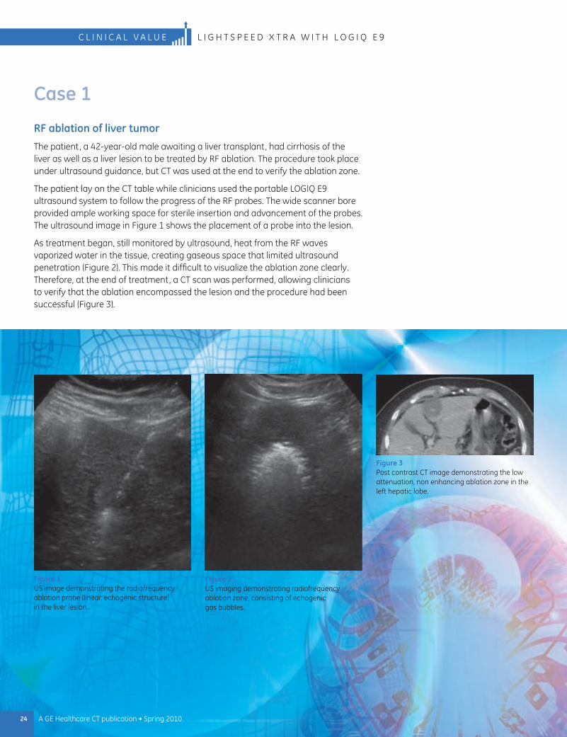
Case 1
RF ablation of liver tumor
The patient, a 42-year-old male awaiting a liver transplant, had cirrhosis of the liver as well as a liver lesion to be treated by RF ablation. The procedure took place under ultrasound guidance, but CT was used at the end to verify the ablation zone.
The patient lay on the CT table while clinicians used the portable LOGIQ E9 ultrasound system to follow the progress of the RF probes. The wide scanner bore provided ample working space for sterile insertion and advancement of the probes. The ultrasound image in Figure 1 shows the placement of a probe into the lesion.
As treatment began, still monitored by ultrasound, heat from the RF waves vaporized water in the tissue, creating gaseous space that limited ultrasound penetration (Figure 2). This made it difficult to visualize the ablation zone clearly. Therefore, at the end of treatment, a CT scan was performed, allowing clinicians to verify that the ablation encompassed the lesion and the procedure had been successful (Figure 3).
Figure 3 Post contrast CT image demonstrating the low attenuation, non enhancing ablation zone in the left hepatic lobe.
Figure 1 US image demonstrating the radiofrequency ablation probe (linear, echogenic structure) in the liver lesion.
Figure 2 US imaging demonstrating radiofrequency ablation zone, consisting of echogenic gas bubbles.
25A GE Healthcare CT publication • Spring 2010
L I G H T S P E E D X T R A W I T H L O G I Q E 9 C L I N I C A L v A L U E
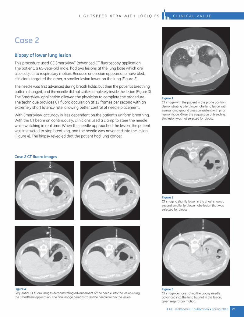
Case 2
Biopsy of lower lung lesion
This procedure used GE SmartView™ (advanced CT fluoroscopy application). The patient, a 65-year-old male, had two lesions at the lung base which are also subject to respiratory motion. Because one lesion appeared to have bled, clinicians targeted the other, a smaller lesion lower on the lung (Figure 2).
The needle was first advanced during breath holds, but then the patient’s breathing pattern changed, and the needle did not strike completely inside the lesion (Figure 3). The SmartView application allowed the physician to complete the procedure. The technique provides CT fluoro acquisition at 12 frames per second with an extremely short latency rate, allowing better control of needle placement.
With SmartView, accuracy is less dependent on the patient’s uniform breathing. With the CT beam on continuously, clinicians used a clamp to steer the needle while watching in real time. When the needle approached the lesion, the patient was instructed to stop breathing, and the needle was advanced into the lesion (Figure 4). The biopsy revealed that the patient had lung cancer.
Figure 3 CT image demonstrating the biopsy needle advanced into the lung but not in the lesion, given respiratory motion.
Figure 2 CT imaging slightly lower in the chest shows a second smaller left lower lobe lesion that was selected for biopsy.
Figure 1 CT image with the patient in the prone position demonstrating a left lower lobe lung lesion with surrounding ground glass consistent with prior hemorrhage. Given the suggestion of bleeding, this lesion was not selected for biopsy.
Figure 4 Sequential CT fluoro images demonstrating advancement of the needle into the lesion using the SmartView application. The final image demonstrates the needle within the lesion.
Case 2 CT fluoro images
26 A GE Healthcare CT publication • Spring 2010
C L I N I C A L v A L U E L I G H T S P E E D X T R A W I T H L O G I Q E 9
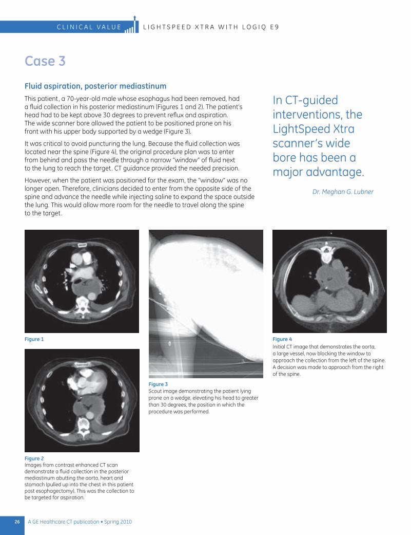
Figure 4 Initial CT image that demonstrates the aorta, a large vessel, now blocking the window to approach the collection from the left of the spine. A decision was made to approach from the right of the spine.
Figure 1
Figure 2 Images from contrast enhanced CT scan demonstrate a fluid collection in the posterior mediastinum abutting the aorta, heart and stomach (pulled up into the chest in this patient post esophagectomy). This was the collection to be targeted for aspiration.
Figure 3 Scout image demonstrating the patient lying prone on a wedge, elevating his head to greater than 30 degrees, the position in which the procedure was performed.
Case 3
Fluid aspiration, posterior mediastinumThis patient, a 70-year-old male whose esophagus had been removed, had a fluid collection in his posterior mediastinum (Figures 1 and 2). The patient’s head had to be kept above 30 degrees to prevent reflux and aspiration. The wide scanner bore allowed the patient to be positioned prone on his front with his upper body supported by a wedge (Figure 3).
It was critical to avoid puncturing the lung. Because the fluid collection was located near the spine (Figure 4), the original procedure plan was to enter from behind and pass the needle through a narrow “window” of fluid next to the lung to reach the target. CT guidance provided the needed precision.
However, when the patient was positioned for the exam, the “window” was no longer open. Therefore, clinicians decided to enter from the opposite side of the spine and advance the needle while injecting saline to expand the space outside the lung. This would allow more room for the needle to travel along the spine to the target.
In CT-guided interventions, the LightSpeed Xtra scanner’s wide bore has been a major advantage.
Dr. Meghan G. Lubner
27A GE Healthcare CT publication • Spring 2010
L I G H T S P E E D X T R A W I T H L O G I Q E 9 C L I N I C A L v A L U E
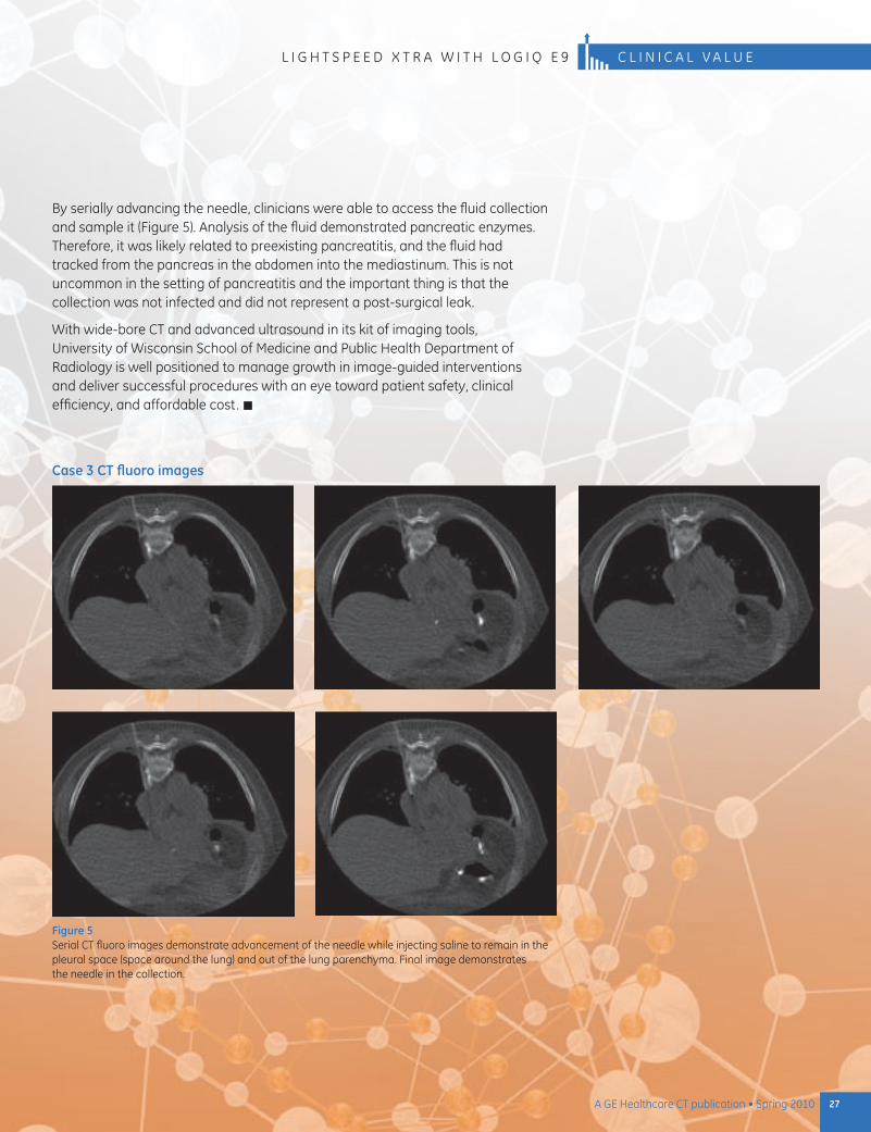
Figure 5 Serial CT fluoro images demonstrate advancement of the needle while injecting saline to remain in the pleural space (space around the lung) and out of the lung parenchyma. Final image demonstrates the needle in the collection.
Case 3 CT fluoro images
By serially advancing the needle, clinicians were able to access the fluid collection and sample it (Figure 5). Analysis of the fluid demonstrated pancreatic enzymes. Therefore, it was likely related to preexisting pancreatitis, and the fluid had tracked from the pancreas in the abdomen into the mediastinum. This is not uncommon in the setting of pancreatitis and the important thing is that the collection was not infected and did not represent a post-surgical leak.
With wide-bore CT and advanced ultrasound in its kit of imaging tools, University of Wisconsin School of Medicine and Public Health Department of Radiology is well positioned to manage growth in image-guided interventions and deliver successful procedures with an eye toward patient safety, clinical efficiency, and affordable cost. n
28 A GE Healthcare CT publication • Spring 2010
Promises KeptStudies Show ASiR Lowers Dose, Delivers Image Quality Across All Anatomies
C L I n I C A L v A L u E R S n A 2 0 0 9 A S I R A B S T R A C T S A B S T R A C T S
Based on numerous clinical studies conducted by a host of clinicians, the use of ASiR™ (Adaptive Statistical Iterative Reconstruction) on the Discovery™ CT750 HD system is living up to its promise as a breakthrough technology that allows clinicians to lower dose – and do so with high-definition image quality across all anatomies.
The Discovery CT750 HD with ASiR debuted at RSnA in 2008. ASiR is a GE Healthcare-exclusive approach to image reconstruction. It extends the capabilities of iterative reconstruction beyond the conventional Filtered Back Projection (FBP) approach to give clinicians and referring physicians the image quality they require at lower dose. (See story on ASiR vs. FBP on page 47). GE Healthcare specifically designed ASiR to remove image noise and improve low contrast detectability (LCD) for better image quality with less dose.
Since its introduction, leading medical centers have put the Discovery CT750 HD and ASiR to the test. Many practitioners also published studies to demonstrate how technology delivers value to their practices, as well as referring physicians and patients.
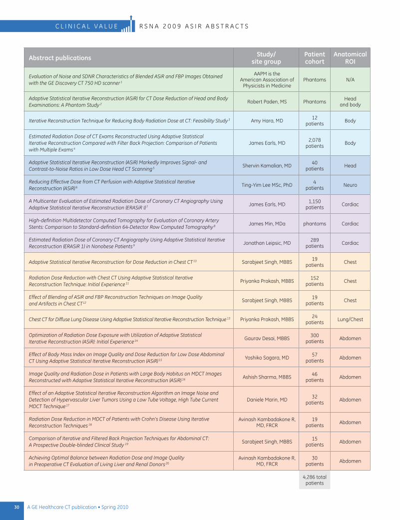
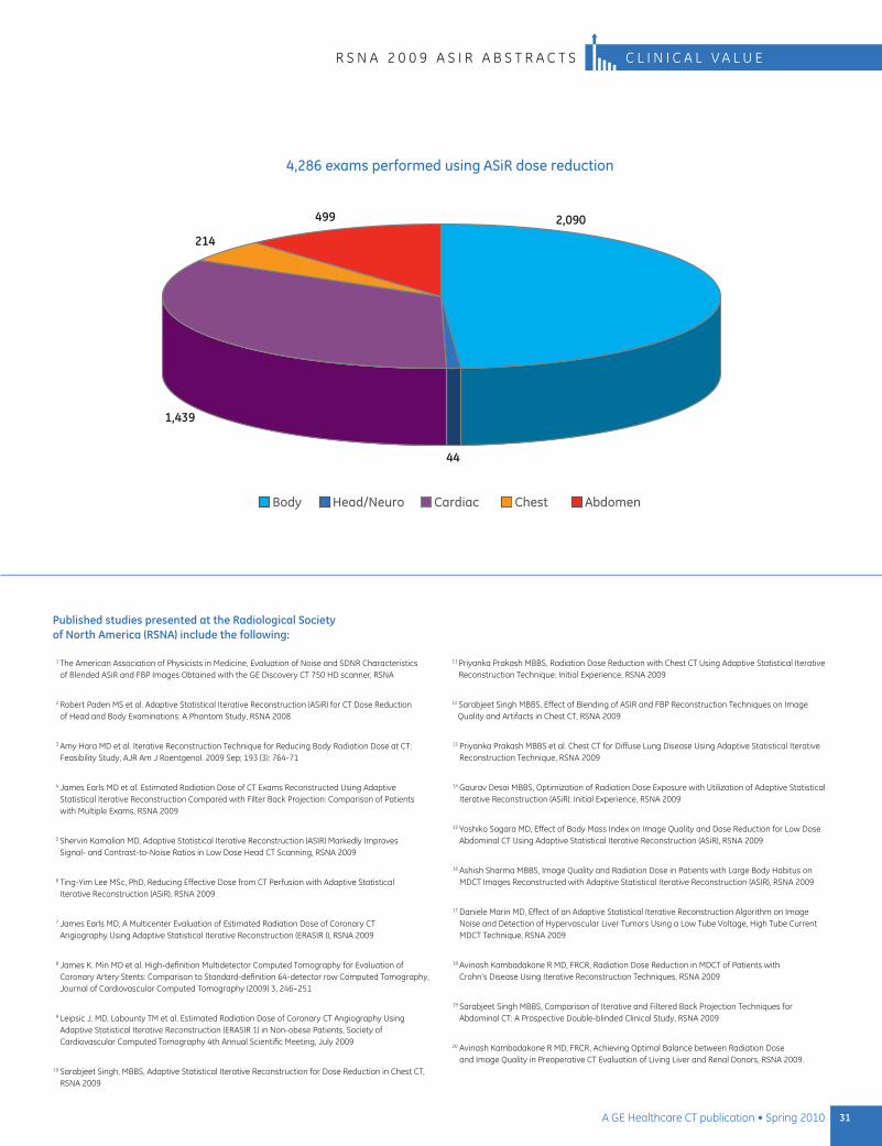
Provided here are twenty abstracts presented at the 2009 RSnA annual meeting on the use of ASiR versus FBP algorithms. Combined, the abstracts demonstrate dose reduction from 4,286 patients across five anatomical regions of interest. The largest investigation performed by Dr. James Earls and colleagues at Fairfax Radiological Consultants evaluated radiation doses in 2,078 consecutive CT examinations evaluating cardiac, chest, combined abdomen and pelvis, and combined chest, abdomen and pelvis. Studies were performed on two ASiR-equipped systems, a 64-row Discovery CT750 HD (n=1776) and a 64-row LightSpeed vCT XTe (n=302).
ASiR uses statistical modeling to remove image noise and increase the signal-to-noise ratio of CT exams. This allows the clinician to reduce tube current as compared to studies reconstructed with FBP algorithms. The purpose of the study performed by Dr. Earls and his colleagues was to directly compare studies performed using ASIR and FBP in the same patient.
29A GE Healthcare CT publication • Spring 2010
R S n A 2 0 0 9 A S I R A B S T R A C T S C L I n I C A L v A L u E
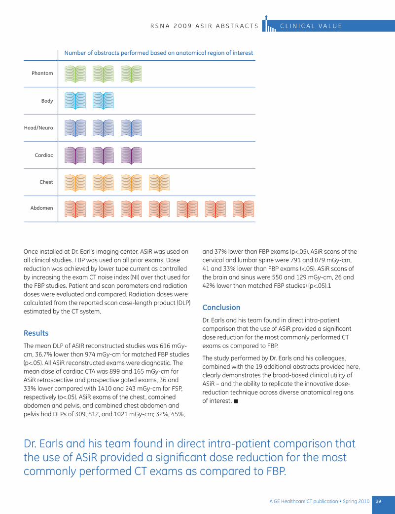
Number of abstracts performed based on anatomical region of interest
Phantom
Body
Head/Neuro
Chest
Abdomen
Cardiac
Once installed at Dr. Earl’s imaging center, ASiR was used on all clinical studies. FBP was used on all prior exams. Dose reduction was achieved by lower tube current as controlled by increasing the exam CT noise index (nI) over that used for the FBP studies. Patient and scan parameters and radiation doses were evaluated and compared. Radiation doses were calculated from the reported scan dose-length product (DLP) estimated by the CT system.
Results
The mean DLP of ASIR reconstructed studies was 616 mGy-cm, 36.7% lower than 974 mGy-cm for matched FBP studies (p<.05). All ASiR reconstructed exams were diagnostic. The mean dose of cardiac CTA was 899 and 165 mGy-cm for ASiR retrospective and prospective gated exams, 36 and 33% lower compared with 1410 and 243 mGy-cm for FSP, respectively (p<.05). ASiR exams of the chest, combined abdomen and pelvis, and combined chest abdomen and pelvis had DLPs of 309, 812, and 1021 mGy-cm; 32%, 45%,
and 37% lower than FBP exams (p<.05). ASiR scans of the cervical and lumbar spine were 791 and 879 mGy-cm, 41 and 33% lower than FBP exams (<.05). ASiR scans of the brain and sinus were 550 and 129 mGy-cm, 26 and 42% lower than matched FBP studies) (p<.05).1
Conclusion
Dr. Earls and his team found in direct intra-patient comparison that the use of ASiR provided a significant dose reduction for the most commonly performed CT exams as compared to FBP.
The study performed by Dr. Earls and his colleagues, combined with the 19 additional abstracts provided here, clearly demonstrates the broad-based clinical utility of ASiR – and the ability to replicate the innovative dose-reduction technique across diverse anatomical regions of interest. n
Dr. Earls and his team found in direct intra-patient comparison that the use of ASiR provided a significant dose reduction for the most commonly performed CT exams as compared to FBP.
30 A GE Healthcare CT publication • Spring 2010
C L I n I C A L v A L u E R S n A 2 0 0 9 A S I R A B S T R A C T S
Abstract publications Study/ site group
Patient cohort
Anatomical ROI
Evaluation of Noise and SDNR Characteristics of Blended ASiR and FBP Images Obtained with the GE Discovery CT 750 HD scanner 1
AAPM is the American Association of
Physicists in MedicinePhantoms n/A
Adaptive Statistical Iterative Reconstruction (ASiR) for CT Dose Reduction of Head and Body Examinations: A Phantom Study 2 Robert Paden, MS Phantoms Head
and body
Iterative Reconstruction Technique for Reducing Body Radiation Dose at CT: Feasibility Study 3 Amy Hara, MD 12 patients Body
Estimated Radiation Dose of CT Exams Reconstructed Using Adaptive Statistical Iterative Reconstruction Compared with Filter Back Projection: Comparison of Patients with Multiple Exams 4
James Earls, MD 2,078 patients Body
Adaptive Statistical Iterative Reconstruction (ASiR) Markedly Improves Signal- and Contrast-to-Noise Ratios in Low Dose Head CT Scanning 5
Shervin Kamalian, MD 40 patients Head
Reducing Effective Dose from CT Perfusion with Adaptive Statistical Iterative Reconstruction (ASiR) 6 Ting-Yim Lee MSc, PhD 4
patients neuro
A Multicenter Evaluation of Estimated Radiation Dose of Coronary CT Angiography Using Adaptive Statistical Iterative Reconstruction (ERASiR I) 7
James Earls, MD 1,150 patients Cardiac
High-definition Multidetector Computed Tomography for Evaluation of Coronary Artery Stents: Comparison to Standard-definition 64-Detector Row Computed Tomography 8 James Min, MDa phantoms Cardiac
Estimated Radiation Dose of Coronary CT Angiography Using Adaptive Statistical Iterative Reconstruction (ERASiR 1) in Nonobese Patients 9 Jonathan Leipsic, MD 289
patients Cardiac
Adaptive Statistical Iterative Reconstruction for Dose Reduction in Chest CT 10 Sarabjeet Singh, MBBS 19 patients Chest
Radiation Dose Reduction with Chest CT Using Adaptive Statistical Iterative Reconstruction Technique: Initial Experience 11
Priyanka Prakash, MBBS 152 patients Chest
Effect of Blending of ASiR and FBP Reconstruction Techniques on Image Quality and Artifacts in Chest CT12
Sarabjeet Singh, MBBS 19 patients Chest
Chest CT for Diffuse Lung Disease Using Adaptive Statistical Iterative Reconstruction Technique 13 Priyanka Prakash, MBBS 24 patients Lung/Chest
Optimization of Radiation Dose Exposure with Utilization of Adaptive Statistical Iterative Reconstruction (ASiR): Initial Experience 14
Gaurav Desai, MBBS 300 patients Abdomen
Effect of Body Mass Index on Image Quality and Dose Reduction for Low Dose Abdominal CT Using Adaptive Statistical Iterative Reconstruction (ASiR) 15
Yoshiko Sagara, MD 57 patients Abdomen
Image Quality and Radiation Dose in Patients with Large Body Habitus on MDCT Images Reconstructed with Adaptive Statistical Iterative Reconstruction (ASiR) 16
Ashish Sharma, MBBS 46 patients Abdomen
Effect of an Adaptive Statistical Iterative Reconstruction Algorithm on Image Noise and Detection of Hypervascular Liver Tumors Using a Low Tube Voltage, High Tube Current MDCT Technique 17
Daniele Marin, MD 32 patients Abdomen
Radiation Dose Reduction in MDCT of Patients with Crohn’s Disease Using Iterative Reconstruction Techniques 18
Avinash Kambadakone R, MD, FRCR
19 patients Abdomen
Comparison of Iterative and Filtered Back Projection Techniques for Abdominal CT: A Prospective Double-blinded Clinical Study 19
Sarabjeet Singh, MBBS 15 patients Abdomen
Achieving Optimal Balance between Radiation Dose and Image Quality in Preoperative CT Evaluation of Living Liver and Renal Donors 20
Avinash Kambadakone R, MD, FRCR
30 patients Abdomen
4,286 total patients
31A GE Healthcare CT publication • Spring 2010
R S n A 2 0 0 9 A S I R A B S T R A C T S C L I n I C A L v A L u E
Published studies presented at the Radiological Society of North America (RSNA) include the following:
1 The American Association of Physicists in Medicine, Evaluation of noise and SDnR Characteristics of Blended ASiR and FBP Images Obtained with the GE Discovery CT 750 HD scanner, RSnA
2 Robert Paden MS et al. Adaptive Statistical Iterative Reconstruction (ASiR) for CT Dose Reduction of Head and Body Examinations: A Phantom Study, RSnA 2008
3 Amy Hara MD et al. Iterative Reconstruction Technique for Reducing Body Radiation Dose at CT: Feasibility Study, AJR Am J Roentgenol. 2009 Sep; 193 (3): 764-71
4 James Earls MD et al. Estimated Radiation Dose of CT Exams Reconstructed using Adaptive Statistical Iterative Reconstruction Compared with Filter Back Projection: Comparison of Patients with Multiple Exams, RSnA 2009
5 Shervin Kamalian MD, Adaptive Statistical Iterative Reconstruction (ASIR) Markedly Improves Signal- and Contrast-to-noise Ratios in Low Dose Head CT Scanning, RSnA 2009
6 Ting-Yim Lee MSc, PhD, Reducing Effective Dose from CT Perfusion with Adaptive Statistical Iterative Reconstruction (ASiR), RSnA 2009
7 James Earls MD, A Multicenter Evaluation of Estimated Radiation Dose of Coronary CT Angiography using Adaptive Statistical Iterative Reconstruction (ERASIR I), RSnA 2009
8 James K. Min MD et al. High-definition Multidetector Computed Tomography for Evaluation of Coronary Artery Stents: Comparison to Standard-definition 64-detector row Computed Tomography, Journal of Cardiovascular Computed Tomography (2009) 3, 246–251
9 Leipsic J. MD, Labounty TM et al. Estimated Radiation Dose of Coronary CT Angiography using Adaptive Statistical Iterative Reconstruction (ERASIR 1) in non-obese Patients, Society of Cardiovascular Computed Tomography 4th Annual Scientific Meeting, July 2009
10 Sarabjeet Singh, MBBS, Adaptive Statistical Iterative Reconstruction for Dose Reduction in Chest CT, RSnA 2009
11 Priyanka Prakash MBBS, Radiation Dose Reduction with Chest CT using Adaptive Statistical Iterative Reconstruction Technique: Initial Experience, RSnA 2009
12 Sarabjeet Singh MBBS, Effect of Blending of ASIR and FBP Reconstruction Techniques on Image Quality and Artifacts in Chest CT, RSnA 2009
13 Priyanka Prakash MBBS et al. Chest CT for Diffuse Lung Disease using Adaptive Statistical Iterative Reconstruction Technique, RSnA 2009
14 Gaurav Desai MBBS, Optimization of Radiation Dose Exposure with utilization of Adaptive Statistical Iterative Reconstruction (ASiR): Initial Experience, RSnA 2009
15 Yoshiko Sagara MD, Effect of Body Mass Index on Image Quality and Dose Reduction for Low Dose Abdominal CT using Adaptive Statistical Iterative Reconstruction (ASiR), RSnA 2009
16 Ashish Sharma MBBS, Image Quality and Radiation Dose in Patients with Large Body Habitus on MDCT Images Reconstructed with Adaptive Statistical Iterative Reconstruction (ASIR), RSnA 2009
17 Daniele Marin MD, Effect of an Adaptive Statistical Iterative Reconstruction Algorithm on Image noise and Detection of Hypervascular Liver Tumors using a Low Tube voltage, High Tube Current MDCT Technique, RSnA 2009
18 Avinash Kambadakone R MD, FRCR, Radiation Dose Reduction in MDCT of Patients with Crohn’s Disease using Iterative Reconstruction Techniques, RSnA 2009
19 Sarabjeet Singh MBBS, Comparison of Iterative and Filtered Back Projection Techniques for Abdominal CT: A Prospective Double-blinded Clinical Study, RSnA 2009
20 Avinash Kambadakone R MD, FRCR, Achieving Optimal Balance between Radiation Dose and Image Quality in Preoperative CT Evaluation of Living Liver and Renal Donors, RSnA 2009.
4,286 exams performed using ASiR dose reduction
2,090
44
1,439
214
499
Body Head/Neuro Cardiac Chest Abdomen
32 A GE Healthcare CT publication • Spring 2010
c l i n i c a l v a l u e L i G H T S p E E d V C T w i T H S n A p S H o T p u L S E A n d A S i R
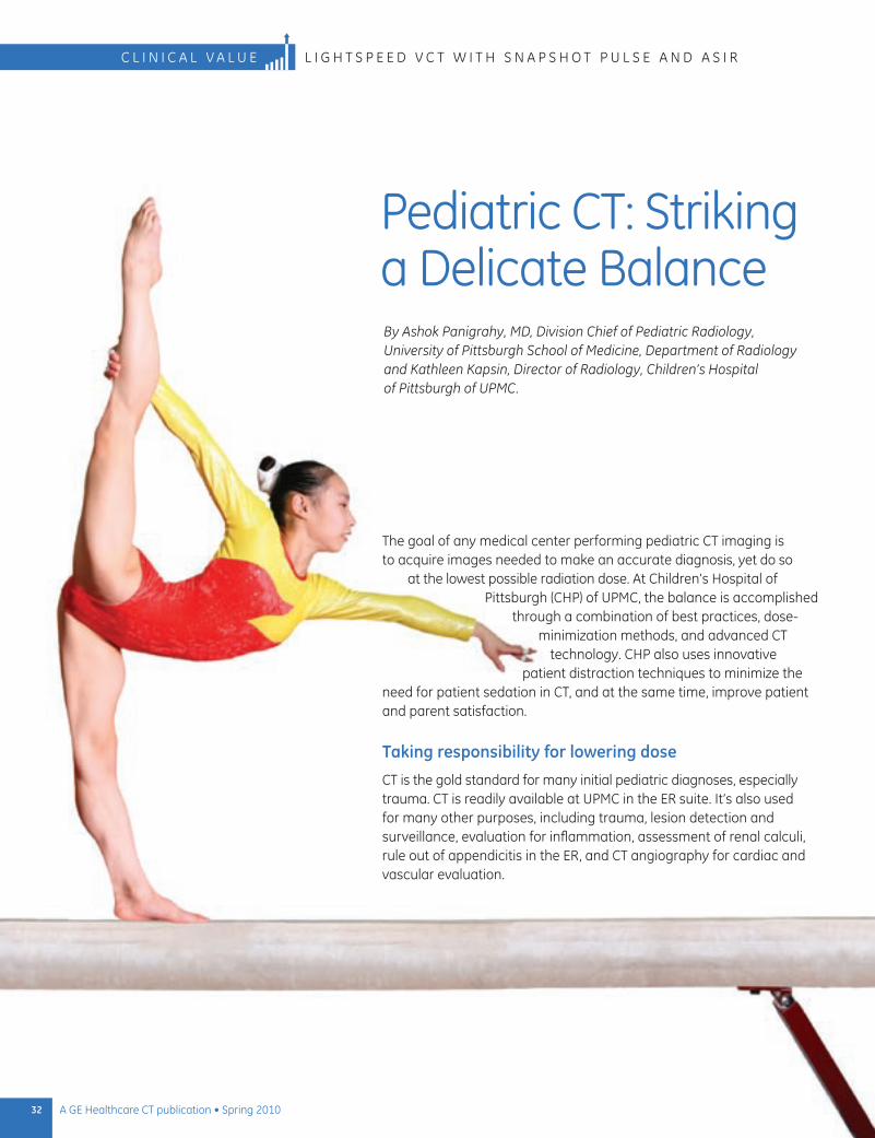
pediatric CT: Striking a delicate BalanceBy Ashok Panigrahy, MD, Division Chief of Pediatric Radiology, University of Pittsburgh School of Medicine, Department of Radiology and Kathleen Kapsin, Director of Radiology, Children’s Hospital of Pittsburgh of UPMC.
Taking responsibility for lowering dose
CT is the gold standard for many initial pediatric diagnoses, especially trauma. CT is readily available at upMC in the ER suite. it’s also used for many other purposes, including trauma, lesion detection and surveillance, evaluation for inflammation, assessment of renal calculi, rule out of appendicitis in the ER, and CT angiography for cardiac and vascular evaluation.
The goal of any medical center performing pediatric CT imaging is to acquire images needed to make an accurate diagnosis, yet do so
at the lowest possible radiation dose. At Children’s Hospital of pittsburgh (CHp) of upMC, the balance is accomplished
through a combination of best practices, dose-minimization methods, and advanced CT
technology. CHp also uses innovative patient distraction techniques to minimize the
need for patient sedation in CT, and at the same time, improve patient and parent satisfaction.
33A GE Healthcare CT publication • Spring 2010
Ashok panigrahy, Md, division Chief of pediatric Radiology, university of pittsburgh School of Medicine, department of Radiology
Kathleen Kapsin, director of Radiology, Children’s Hospital of pittsburgh of upMC
About the facility
Renowned for its outstanding clinical services, research programs and medical education, Children’s Hospital of pittsburgh of upMC has helped establish the standards of excellence in pediatric care. From Ambulatory Care to Transplantation and Cardiac Care, talented and committed pediatric experts care for infants, children and adolescents who make more than 500,000 visits to Children’s and its many neighborhood locations each year.
Children’s also has been named consistently to several elite lists of pediatric health care facilities, including ranking 10th in total dollars and seventh in number of awards (FY 2008) provided by the national institutes of Health, and consistently is named among the top pediatric hospitals in the united States by U.S. News & World Report.
L i G H T S p E E d V C T w i T H S n A p S H o T p u L S E A n d A S i R c l i n i c a l v a l u e
A key reason why CT is regularly performed in pediatrics is because MdCT offers many advantages over other imaging modalities. one major plus is that it involves relatively short scanning times, which minimizes – and in some cases eliminates – challenges with motion and respiratory mis-registration artifacts. our experience has shown that short scan times decrease our need for sedation. Additionally, technical advances in CT have allowed optimization of imaging for peak vascular enhancement. image quality is superb, offering the ability to retrospectively reconstruct overlapping images and create 2d and 3d images for enhanced procedural roadmapping.
The first step in deciding to use CT versus another imaging modality at CHp is to determine the clinical need. it is the responsibility of the radiology team to conform medical testing to As Low As Reasonably Achievable (ALARA) principles and American College of Radiology (ACR) guidelines. Continued advances in CT technology are key to helping us achieve ALARA.
Dose reduction methods at CHP
A key question we ask ourselves at CHp is this: “what are the lowest mAs needed for this patient to satisfy the diagnostic objective and maintain acceptable diagnostic image quality?” The answer requires a number of adjustments in CT techniques.
Two fundamental methods are to reduce tube current (mA) and peak kilovoltage (kVp) based on the size of the patient (weight), their age and the image contrast needed to answer the clinical question.
other dose-lowering techniques could include:
increased slice thickness (= or > 5 mm), especially on follow up.•
Minimizing the use of pitch below 1.0. (For general body scanning, • a pitch of 1.0 to 1.5 should be sufficient).
Smaller Scan Field of View (SFoV) filters when possible.•
in-plane shielding whenever possible, e.g., thyroid, breast, etc.•
properly centering patients to iso-center.•
using newer scanner technology, such as GE’s Smart mA, which makes • automatic regional adjustments in radiation dose during scanning, e.g., tube current modulation.
Minimizing use of multiple scans for each examination, e.g., pre- and post-• contrast scans. if multiphase exams are necessary, consider lower dose protocols for select phases depending on the objective, e.g., calcification or excretory phase.
Limiting the scan coverage for answering the clinical questions.•
There are also many innovative ways to achieve dose reduction when performing cardiac CT. one in particular is GE’s Snapshot™ pulse, which uses prospectively triggered axial step-and-shoot scans in which X-rays are turned on only during
34 A GE Healthcare CT publication • Spring 2010
c l i n i c a l v a l u e L i G H T S p E E d V C T w i T H S n A p S H o T p u L S E A n d A S i R
the required heart phase and turned off completely at all other times to significantly lower dose. The radiology team at CHp also advocates FeatherLight imaging dose optimization. These include using procedure- based zone protocols that follow ALARA principles and provide a unified, collaborative approach to dose reduction.
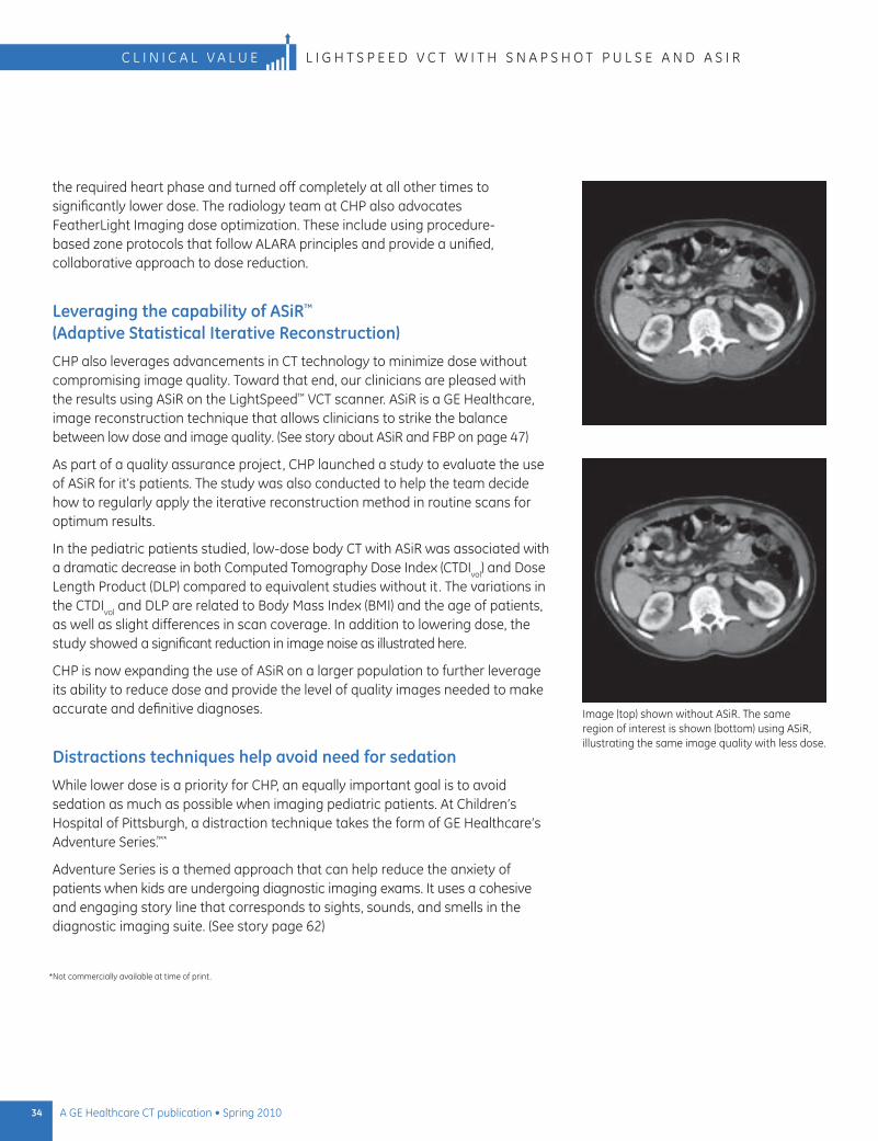
Leveraging the capability of ASiR™ (Adaptive Statistical Iterative Reconstruction)
CHp also leverages advancements in CT technology to minimize dose without compromising image quality. Toward that end, our clinicians are pleased with the results using ASiR on the LightSpeed™ VCT scanner. ASiR is a GE Healthcare, image reconstruction technique that allows clinicians to strike the balance between low dose and image quality. (See story about ASiR and FBp on page 47)
As part of a quality assurance project, CHp launched a study to evaluate the use of ASiR for it’s patients. The study was also conducted to help the team decide how to regularly apply the iterative reconstruction method in routine scans for optimum results.
in the pediatric patients studied, low-dose body CT with ASiR was associated with a dramatic decrease in both Computed Tomography dose index (CTdivol) and dose Length product (dLp) compared to equivalent studies without it . The variations in the CTdivol and dLp are related to Body Mass index (BMi) and the age of patients, as well as slight differences in scan coverage. in addition to lowering dose, the study showed a significant reduction in image noise as illustrated here.
CHp is now expanding the use of ASiR on a larger population to further leverage its ability to reduce dose and provide the level of quality images needed to make accurate and definitive diagnoses.
Distractions techniques help avoid need for sedation
while lower dose is a priority for CHp, an equally important goal is to avoid sedation as much as possible when imaging pediatric patients. At Children’s Hospital of pittsburgh, a distraction technique takes the form of GE Healthcare’s Adventure Series.™*
Adventure Series is a themed approach that can help reduce the anxiety of patients when kids are undergoing diagnostic imaging exams. it uses a cohesive and engaging story line that corresponds to sights, sounds, and smells in the diagnostic imaging suite. (See story page 62)
image (top) shown without ASiR. The same region of interest is shown (bottom) using ASiR, illustrating the same image quality with less dose.
*not commercially available at time of print.

35A GE Healthcare CT publication • Spring 2010
L i G H T S p E E d V C T w i T H S n A p S H o T p u L S E A n d A S i R c l i n i c a l v a l u e
CHp incorporated Adventure Series into CT, MR, pET/CT, and nuclear medicine imaging rooms in 2009 when it built a new 900,000-square-foot hospital. when comparing variables between the “old” hospital model and the “new” hospital model, CHp was able to reduce the number of sedations, despite an increase in the volume of scans during the same time period.
in addition to the Adventure Series theme, the reduction in sedation is attributable to other factors as well, such as fast scanning times made possible with the LightSpeed VCT scanner as well as staff training in distraction techniques. CHp customer surveys and testimonials also show that distraction techniques like Adventure Series improve parent and patient satisfaction.
The right blend for pediatrics
At CHp, the ability to minimize unnecessary exposure to CT scanning radiation is achieved through a blend of best practices, proven dose-reduction methods, and the use of advanced CT technology. Thanks to innovative distraction techniques, fast scanners, and staff training, there is also very little need to sedate pediatric patients for CT exams. n
when comparing variables between the “old” hospital model and the “new” hospital model, CHp was able to reduce the number of sedations, despite an increase in the volume of scans during the same time period.
The Adventure Series pirate island theme welcomes patients who undergo CT exams at CHp.
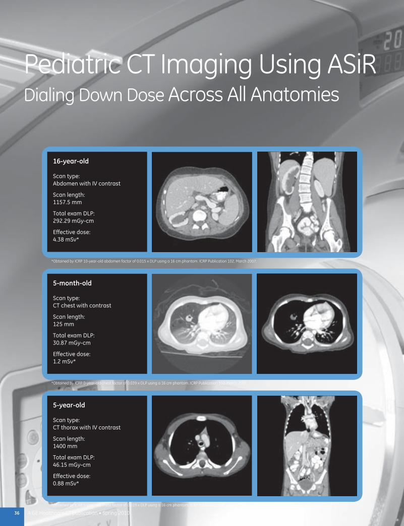
36 A GE Healthcare CT publication • Spring 2010
Pediatric CT Imaging Using ASiR Dialing Down Dose Across All Anatomies
16-year-old
Scan type: Abdomen with IV contrast
Scan length: 1157.5 mm
Total exam DLP: 292.29 mGy-cm
Effective dose: 4.38 mSv*
5-year-old
Scan type: CT thorax with IV contrast
Scan length: 1400 mm
Total exam DLP: 46.15 mGy-cm
Effective dose: 0.88 mSv*
5-month-old
Scan type: CT chest with contrast
Scan length: 125 mm
Total exam DLP: 30.87 mGy-cm
Effective dose: 1.2 mSv*
*Obtained by ICRP 10-year-old abdomen factor of 0.015 x DLP using a 16 cm phantom. ICRP Publication 102, March 2007.
*Obtained by ICRP 0-year-old chest factor of 0.039 x DLP using a 16 cm phantom. ICRP Publication 102, March 2007.
*Obtained by ICRP 5-year-old chest factor of 0.019 x DLP using a 16 cm phantom. ICRP Publication 102, March 2007.
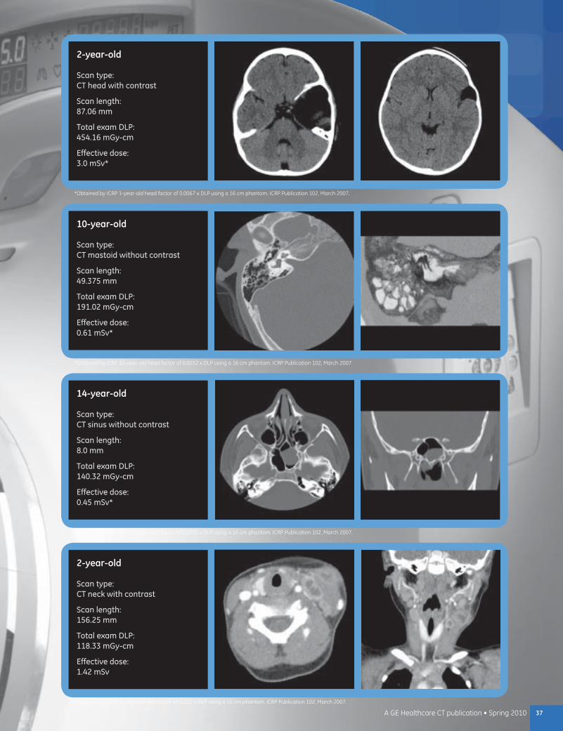
37A GE Healthcare CT publication • Spring 2010
2-year-old
Scan type: CT head with contrast
Scan length: 87.06 mm
Total exam DLP: 454.16 mGy-cm
Effective dose: 3.0 mSv*
2-year-old
Scan type: CT neck with contrast
Scan length: 156.25 mm
Total exam DLP: 118.33 mGy-cm
Effective dose: 1.42 mSv
14-year-old
Scan type: CT sinus without contrast
Scan length: 8.0 mm
Total exam DLP: 140.32 mGy-cm
Effective dose: 0.45 mSv*
10-year-old
Scan type: CT mastoid without contrast
Scan length: 49.375 mm
Total exam DLP: 191.02 mGy-cm
Effective dose: 0.61 mSv*
*Obtained by ICRP 10-year-old head factor of 0.0032 x DLP using a 16 cm phantom. ICRP Publication 102, March 2007
*Obtained by ICRP 1-year-old head factor of 0.0067 x DLP using a 16 cm phantom. ICRP Publication 102, March 2007.
*Obtained by ICRP 10-year-old head factor of 0.0032 x DLP using a 16 cm phantom. ICRP Publication 102, March 2007.
*Obtained by ICRP 1-year-old head factor of 0.012 x DLP using a 16 cm phantom. ICRP Publication 102, March 2007.
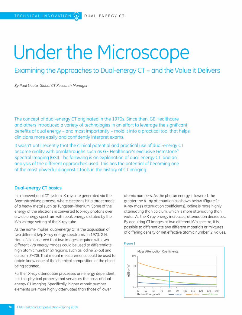
38 A GE Healthcare CT publication • Spring 2010
Under the Microscope
Dual-energy CT basics
In a conventional CT system, X-rays are generated via the Bremsstrahlung process, where electrons hit a target made of a heavy metal such as Tungsten-Rhenium. Some of the energy of the electrons is converted to X-ray photons over a wide energy spectrum with peak energy dictated by the kVp voltage setting of the X-ray tube.
As the name implies, dual-energy CT is the acquisition of two different kVp X-ray energy spectrums. In 1973, G.N. Hounsfield observed that two images acquired with two different kVp energy ranges could be used to differentiate high atomic number (Z) regions, such as iodine (Z=53) and calcium (Z=20). That meant measurements could be used to obtain knowledge of the chemical composition of the object being scanned.
Further, X-ray attenuation processes are energy dependent. It is this physical property that serves as the basis of dual- energy CT imaging. Specifically, higher atomic number elements are more highly attenuated than those of lower
T E C H N I C A l I N N o V A T I o N D u A l - E N E R G y C T
By Paul Licato, Global CT Research Manager
Examining the Approaches to Dual-energy CT – and the Value it Delivers
atomic numbers. As the photon energy is lowered, the greater the X-ray attenuation as shown below. (Figure 1: X-ray mass attenuation coefficients). Iodine is more highly attenuating than calcium, which is more attenuating than water. As the X-ray energy increases, attenuation decreases. By acquiring CT images at two different kVp spectra, it is possible to differentiate two different materials or mixtures of differing density or net effective atomic number (Z) values.
The concept of dual-energy CT originated in the 1970s. Since then, GE Healthcare and others introduced a variety of technologies in an effort to leverage the significant benefits of dual energy – and most importantly – mold it into a practical tool that helps clinicians more easily and confidently interpret exams.
It wasn’t until recently that the clinical potential and practical use of dual-energy CT became reality with breakthroughs such as GE Healthcare’s exclusive Gemstone™ Spectral Imaging (GSI). The following is an explanation of dual-energy CT, and an analysis of the different approaches used. This has the potential of becoming one of the most powerful diagnostic tools in the history of CT imaging.
Figure 1
Mass Attenuation Coefficients
40
0.1
1
10
100
50 60 70 80 90 100 110 120 130 140Photon Energy keV
u(E)
cm
2 g-1
IodineWater Calcium
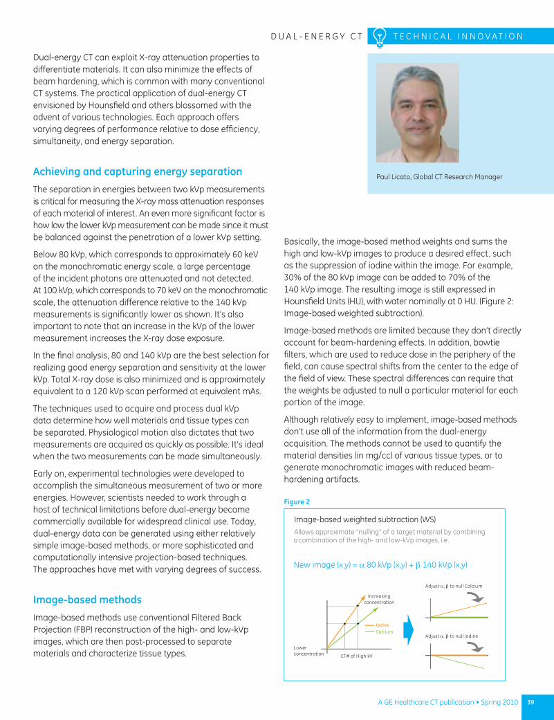
39A GE Healthcare CT publication • Spring 2010
D u A l - E N E R G y C T T E C H N I C A l I N N o V A T I o N
Paul licato, Global CT Research Manager
Dual-energy CT can exploit X-ray attenuation properties to differentiate materials. It can also minimize the effects of beam hardening, which is common with many conventional CT systems. The practical application of dual-energy CT envisioned by Hounsfield and others blossomed with the advent of various technologies. Each approach offers varying degrees of performance relative to dose efficiency, simultaneity, and energy separation.
Achieving and capturing energy separation
The separation in energies between two kVp measurements is critical for measuring the X-ray mass attenuation responses of each material of interest. An even more significant factor is how low the lower kVp measurement can be made since it must be balanced against the penetration of a lower kVp setting.
Below 80 kVp, which corresponds to approximately 60 keV on the monochromatic energy scale, a large percentage of the incident photons are attenuated and not detected. At 100 kVp, which corresponds to 70 keV on the monochromatic scale, the attenuation difference relative to the 140 kVp measurements is significantly lower as shown. It’s also important to note that an increase in the kVp of the lower measurement increases the X-ray dose exposure.
In the final analysis, 80 and 140 kVp are the best selection for realizing good energy separation and sensitivity at the lower kVp. Total X-ray dose is also minimized and is approximately equivalent to a 120 kVp scan performed at equivalent mAs.
The techniques used to acquire and process dual kVp data determine how well materials and tissue types can be separated. Physiological motion also dictates that two measurements are acquired as quickly as possible. It’s ideal when the two measurements can be made simultaneously.
Early on, experimental technologies were developed to accomplish the simultaneous measurement of two or more energies. However, scientists needed to work through a host of technical limitations before dual-energy became commercially available for widespread clinical use. Today, dual-energy data can be generated using either relatively simple image-based methods, or more sophisticated and computationally intensive projection-based techniques. The approaches have met with varying degrees of success.
Image-based methods
Image-based methods use conventional Filtered Back Projection (FBP) reconstruction of the high- and low-kVp images, which are then post-processed to separate materials and characterize tissue types.
Basically, the image-based method weights and sums the high and low-kVp images to produce a desired effect, such as the suppression of iodine within the image. For example, 30% of the 80 kVp image can be added to 70% of the 140 kVp image. The resulting image is still expressed in Hounsfield units (Hu), with water nominally at 0 Hu. (Figure 2: Image-based weighted subtraction).
Image-based methods are limited because they don’t directly account for beam-hardening effects. In addition, bowtie filters, which are used to reduce dose in the periphery of the field, can cause spectral shifts from the center to the edge of the field of view. These spectral differences can require that the weights be adjusted to null a particular material for each portion of the image.
Although relatively easy to implement, image-based methods don’t use all of the information from the dual-energy acquisition. The methods cannot be used to quantify the material densities (in mg/cc) of various tissue types, or to generate monochromatic images with reduced beam-hardening artifacts.
Image-based weighted subtraction (WS)
combinaa tion of the high- and low-kVp images, i.e.Allows approximate “nulling” of a target material by combining
New image (x,y) = α 80 kVp (x,y) + β 140 kVp (x,y)
CT# of High kV
IodineCalcium
Adjust α, β to null Calcium
Adjust α, β to null Iodine
Increasingconcentration
Lowerconcentration
Figure 2
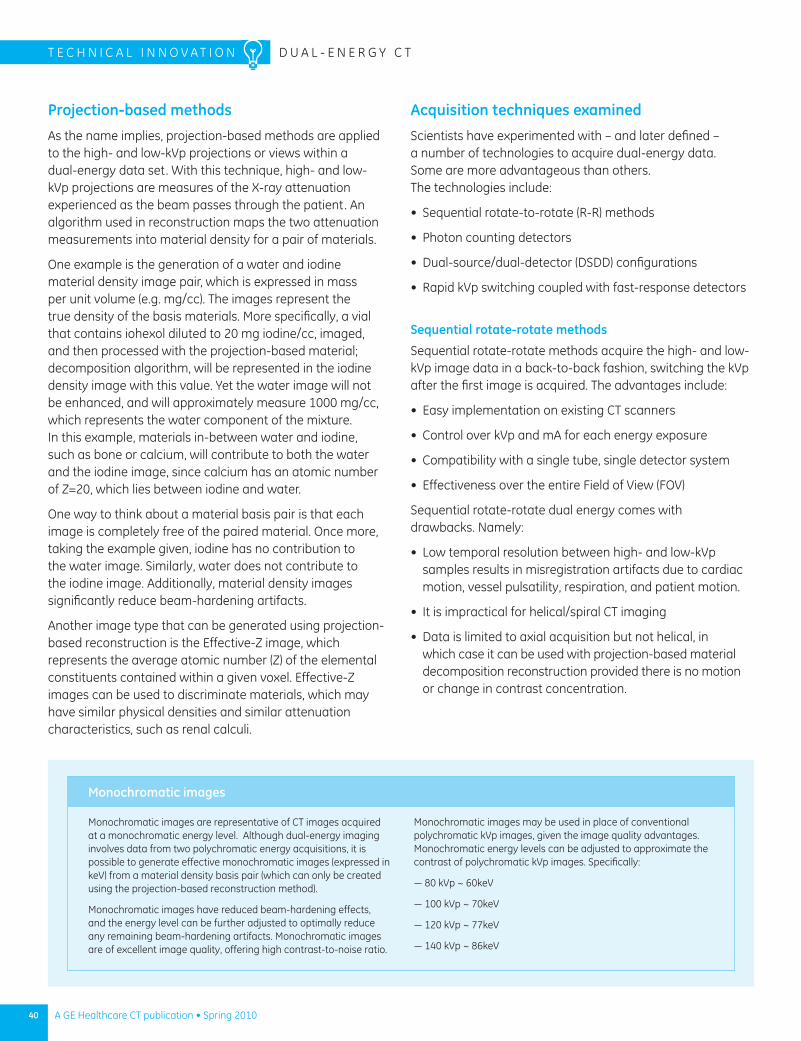
40 A GE Healthcare CT publication • Spring 2010
T E C H N I C A l I N N o V A T I o N D u A l - E N E R G y C T
Projection-based methods
As the name implies, projection-based methods are applied to the high- and low-kVp projections or views within a dual-energy data set. With this technique, high- and low-kVp projections are measures of the X-ray attenuation experienced as the beam passes through the patient. An algorithm used in reconstruction maps the two attenuation measurements into material density for a pair of materials.
one example is the generation of a water and iodine material density image pair, which is expressed in mass per unit volume (e.g. mg/cc). The images represent the true density of the basis materials. More specifically, a vial that contains iohexol diluted to 20 mg iodine/cc, imaged, and then processed with the projection-based material; decomposition algorithm, will be represented in the iodine density image with this value. yet the water image will not be enhanced, and will approximately measure 1000 mg/cc, which represents the water component of the mixture. In this example, materials in-between water and iodine, such as bone or calcium, will contribute to both the water and the iodine image, since calcium has an atomic number of Z=20, which lies between iodine and water.
one way to think about a material basis pair is that each image is completely free of the paired material. once more, taking the example given, iodine has no contribution to the water image. Similarly, water does not contribute to the iodine image. Additionally, material density images significantly reduce beam-hardening artifacts.
Another image type that can be generated using projection-based reconstruction is the Effective-Z image, which represents the average atomic number (Z) of the elemental constituents contained within a given voxel. Effective-Z images can be used to discriminate materials, which may have similar physical densities and similar attenuation characteristics, such as renal calculi.
Acquisition techniques examined
Scientists have experimented with – and later defined – a number of technologies to acquire dual-energy data. Some are more advantageous than others. The technologies include:
Sequential rotate-to-rotate (R-R) methods•
Photon counting detectors•
Dual-source/dual-detector (DSDD) configurations•
Rapid kVp switching coupled with fast-response detectors •
Sequential rotate-rotate methods
Sequential rotate-rotate methods acquire the high- and low-kVp image data in a back-to-back fashion, switching the kVp after the first image is acquired. The advantages include:
Easy implementation on existing CT scanners•
Control over kVp and mA for each energy exposure•
Compatibility with a single tube, single detector system•
Effectiveness over the entire Field of View (FoV)•
Sequential rotate-rotate dual energy comes with drawbacks. Namely:
low temporal resolution between high- and low-kVp • samples results in misregistration artifacts due to cardiac motion, vessel pulsatility, respiration, and patient motion.
It is impractical for helical/spiral CT imaging•
Data is limited to axial acquisition but not helical, in • which case it can be used with projection-based material decomposition reconstruction provided there is no motion or change in contrast concentration.
Monochromatic images
Monochromatic images are representative of CT images acquired at a monochromatic energy level. Although dual-energy imaging involves data from two polychromatic energy acquisitions, it is possible to generate effective monochromatic images (expressed in keV) from a material density basis pair (which can only be created using the projection-based reconstruction method).
Monochromatic images have reduced beam-hardening effects, and the energy level can be further adjusted to optimally reduce any remaining beam-hardening artifacts. Monochromatic images are of excellent image quality, offering high contrast-to-noise ratio.
Monochromatic images may be used in place of conventional polychromatic kVp images, given the image quality advantages. Monochromatic energy levels can be adjusted to approximate the contrast of polychromatic kVp images. Specifically:
— 80 kVp ~ 60keV
— 100 kVp ~ 70keV
— 120 kVp ~ 77keV
— 140 kVp ~ 86keV
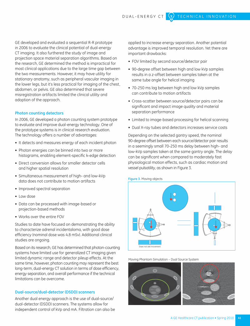
41A GE Healthcare CT publication • Spring 2010
D u A l - E N E R G y C T T E C H N I C A l I N N o V A T I o N
GE developed and evaluated a sequential R-R prototype in 2006 to evaluate the clinical potential of dual-energy CT imaging. It also furthered the study of image and projection space material separation algorithms. Based on the research, GE determined the method is impractical for most clinical applications due to the large time gap between the two measurements. However, it may have utility for stationary anatomy, such as peripheral vascular imaging in the lower legs, but it’s less practical for imaging of the chest, abdomen, or pelvis. GE also determined that severe misregistration artifacts limited the clinical utility and adoption of the approach.
Photon counting detectorsIn 2006, GE developed a photon counting system prototype to evaluate and improve dual-energy technology. one of the prototype systems is in clinical research evaluation. The technology offers a number of advantages:
It detects and measures energy of each incident photon•
Photon energies can be binned into two or more • histograms, enabling element-specific k-edge detection
Direct conversion allows for smaller detector cells • and higher spatial resolution
Simultaneous measurement of high- and low-kVp • data does not contribute to motion artifacts
Improved spectral separation•
low dose •
Data can be processed with image-based or • projection-based methods
Works over the entire FoV•
Studies to date have focused on demonstrating the ability to characterize adrenal incidentaloma, with good dose efficiency (nominal dose was 4.8 mSv). Additional clinical studies are ongoing.
Based on its research, GE has determined that photon-counting systems have limited use for generalized CT imaging given limited dynamic range and detector pileup effects. At the same time, however, photon counting may represent the best long-term, dual-energy CT solution in terms of dose efficiency, energy separation, and overall performance if the technical limitations can be overcome.
Dual-source/dual-detector (DSDD) scanners
Another dual energy approach is the use of dual-source/dual-detector (DSDD) scanners. The systems allow for independent control of kVp and mA. Filtration can also be
applied to increase energy separation. Another potential advantage is improved temporal resolution. yet there are important drawbacks:
FoV limited by second source/detector pair•
90-degree offset between high and low kVp samples • results in a z-offset between samples taken at the same tube angle for helical imaging
70-250 ms lag between high and low kVp samples • can contribute to motion artifacts
Cross-scatter between source/detector pairs can be • significant and impact image quality and material separation performance
limited to image-based processing for helical scanning•
Dual X-ray tubes and detectors increases service costs•
Depending on the selected gantry speed, the nominal 90-degree offset between each source/detector pair results in a seemingly small 70-250 ms delay between high- and low-kVp samples taken at the same gantry angle. The delay can be significant when compared to moderately fast physiological motion effects, such as cardiac motion and vessel pulsatility, as shown in Figure 3.
3538
3538
Does not see movement
Figure 3. Moving objects
Moving Phantom Simulation – Dual Source System
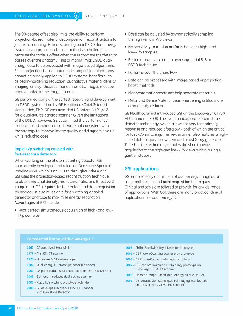
42 A GE Healthcare CT publication • Spring 2010
T E C H N I C A l I N N o V A T I o N D u A l - E N E R G y C T
The 90-degree offset also limits the ability to perform projection-based material decomposition reconstructions to just axial scanning. Helical scanning on a DSDD dual-energy system using projection-based methods is challenging because the table is offset when the second source/detector passes over the anatomy. This primarily limits DSDD dual-energy data to be processed with image-based algorithms. Since projection-based material decomposition algorithms cannot be readily applied to DSDD systems, benefits such as beam-hardening reduction, quantitative material density imaging, and synthesized monochromatic images must be approximated in the image domain.
GE performed some of the earliest research and development on DSDD systems. led by GE Healthcare Chief Scientist Jiang Hsieh, PhD, GE was awarded uS patent 6,421,412 for a dual-source cardiac scanner. Given the limitations of the DSDD, however, GE determined the performance trade-offs and increased costs were not consistent with the strategy to improve image quality and diagnostic value while reducing dose.
Rapid kVp switching coupled with fast-response detectors
When working on the photon-counting detector, GE concurrently developed and released Gemstone Spectral Imaging (GSI), which is now used throughout the world. GSI uses the projection-based reconstruction technique to obtain material density, monochromatic, and Effective-Z image data. GSI requires fast detectors and data acquisition technology. It also relies on a fast switching-enabled generator and tube to maximize energy separation. Advantages of GSI include:
Near perfect simultaneous acquisition of high- and low- • kVp samples
Dose can be adjusted by asymmetrically sampling • the high vs. low kVp views
No sensitivity to motion artifacts between high- and • low-kVp samples
Better immunity to motion over sequential R-R or • DSDD techniques
Performs over the entire FoV•
Data can be processed with image-based or projection-• based methods
Monochromatic spectrums help separate materials•
Metal and Dense Material beam-hardening artifacts are • dramatically reduced
GE Healthcare first introduced GSI on the Discovery™ CT750 HD scanner in 2008. The system incorporates Gemstone detector technology, which allows for very fast primary response and reduced afterglow – both of which are critical for fast kVp switching. The new scanner also features a high-speed data acquisition system and a fast X-ray generator. Together, the technology enables the simultaneous acquisition of the high-and low-kVp views within a single gantry rotation.
GSI applications
GSI enables easy acquisition of dual-energy image data using both helical and axial acquisition techniques. Clinical protocols are tailored to provide for a wide range of applications. With GSI, there are many practical clinical applications for dual-energy CT.
Commercial history of dual-energy CT
1967 – CT conceived (Hounsfield)
1972 – First EMI CT scanner
1973 – Hounsfield’s CT system paper
1982 – Dual-energy CT prototype paper (Kalendar)
2002 – GE patents dual-source cardiac scanner (uS 6,421,412)
2005 – Siemens introduces dual-source scanner
2005 – Rapid kV switching prototype (Kalender)
2006 – GE develops Discovery CT750 HD scanner with Gemstone Detector
2006 – Philips Sandwich layer Detector prototype
2006 – GE Photon Counting dual-energy prototype
2006 – GE Rotate/Rotate dual-energy prototype
2007 – GE Fast kVp switching dual-energy prototype on Discovery CT750 HD scanner
2008 – Siemens Image-Based, dual-energy on dual source
2009 – GE releases Gemstone Spectral Imaging (GSI) feature on the Discovery CT750 HD scanner
Iodine/calcium separation
Dual-energy CT is well suited for separating calcified atherosclerotic lesions from iodinated contrast in blood. The large attenuation differences between iodine (Z=53) and calcium (Z=20) can readily be seen in a pair of 80 and 140 kVp images. A weighted sum of these images provides a rudimentary way to separate iodine from calcium. Alternatively, the 80 and 140 kVp projection data can be processed to produce quantitative material density images, which can then be used for more advanced post-processing and tissue characterization. The projection-based reconstruction technique used in GSI is the only known method for producing material density images.
Small lesion characterization
Small lesion characterization in the liver and kidney is often difficult to diagnose with conventional CT because of pseudo-enhancement within the lesion. Beam-hardening artifacts cause this CT number shift, which results from the uptake of iodinated contrast in the surrounding organ parenchyma. Dual-energy CT data can be processed to reduce or eliminate beam-hardening and thereby eliminate pseudo- enhancement and aid in the separation of cystic from metastatic-enhancing solid lesions. Image-based reconstruction techniques do not correct for beam-hardening artifacts, which can only be done using projection-based reconstruction.
The use of GSI on the Discovery CT750 HD offers incremental information to help distinguish the lesions as enhanced or non-enhanced. This information assists with the diagnosis and is not available on a conventional CT exam. In these cases, a single-phase routine CT may obviate the need for any additional workup.
Virtual non-contrast imaging
Virtual non-contrast images offer images with the appearance and properties of pre-contrast images. GSI provides unique CT images such as “virtual non-contrast” like images (material decomposition water) which are not available with conventional contrast-enhanced CT imaging. The ability to differentiate the iodine material allowed for “iodine subtraction” or “iodine removal ”to derive non-contrast-like images from the contrast-enhanced exam. With GSI, diagnostic virtual like images were generated from a single post-contrast scan.
43A GE Healthcare CT publication • Spring 2010
D u A l - E N E R G y C T T E C H N I C A l I N N o V A T I o N
1 G.N. Hounsfield, BJR 46, 1016-22, 1973. Computerized transverse axial scanning (tomography). 1. Description of system. Hounsfield GN - Br J Radiol - 01-DEC-1973; 46(552): 1016-22
2 R. Alvarez and A. Macovski, “Energy–selective reconstructions in x–ray CT,” Phys. Med. Biol., vol. 21, no. 5, pp. 733–744,(1976);
3 Kalendar W.A., Perman W.H., Vetter J.R., Klotz E., “Evaluation of a prototype dual-energy computed tomographic apparatus. I. Phantom studies”, Med. Phys. 13(3), pp334-339 May/June 1986
1. lehmann et al: Med Phys 8, 659-67, 1981. 2. Kalender et al, Med Phys 13, 334, 1986.
Beam-hardening reduction
Beam-hardening may be reduced or eliminated with projection-based dual-energy image reconstruction. Artifacts from metal and other dense objects in the body can be more effectively managed with dual-energy CT imaging. only GSI uses projection-based reconstruction algorithms.
Characterization of renal calculi
Material density images can aid in the classification of renal calculi. Although the majority of lesions are easily characterized, a significant number do not meet established criteria for a simple cyst and are indeterminate, i.e., have relative increased attenuation with Hu measurements.
In addition to these baseline capabilities, several promising applications are being evaluated to assess pulmonary emboli, stent patency, aneurysm leaks, gout, lung nodules, brain hemorrhage, tendons and ligaments, and to reduce contrast volumes. n
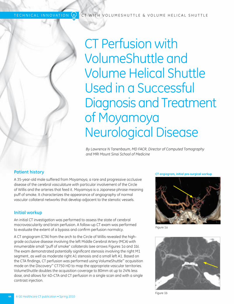
44 A GE Healthcare CT publication • Spring 2010
Patient history
A 35-year-old male suffered from Moyamoya, a rare and progressive occlusive disease of the cerebral vasculature with particular involvement of the Circle of Willis and the arteries that feed it. Moyamoya is a Japanese phrase meaning puff of smoke. It characterizes the appearance of angiography of normal vascular collateral networks that develop adjacent to the stenotic vessels.
Initial workupAn initial CT investigation was performed to assess the state of cerebral macrovascularity and brain perfusion. A follow-up CT exam was performed to evaluate the extent of a bypass and confirm perfusion normalcy.
A CT angiogram (CTA) from the arch to the Circle of Willis revealed the high- grade occlusive disease involving the left Middle Cerebral Artery (MCA) with innumerable small “puff of smoke” collaterals (see arrows Figures 1a and 1b). The exam demonstrated potentially significant stenosis involving the right M1 segment, as well as moderate right A1 stenosis and a small left A1. Based on the CTA findings, CT perfusion was performed using VolumeShuttle™ acquisition mode on the Discovery™ CT750 HD to map the appropriate vascular territories. VolumeShuttle doubles the acquisition coverage to 80mm at up to 24% less dose, and allows for 4D-CTA and CT perfusion in a single scan and with a single contrast injection.
T E C H n I C A l I n n o V A T I o n C T W I T H V o l u M E S H u T T l E & V o l u M E H E l I C A l S H u T T l E
By Lawrence N Tanenbaum, MD FACR, Director of Computed Tomography and MRI Mount Sinai School of Medicine
CT angiogram, initial pre-surgical workup
Figure 1a
Figure 1b
CT Perfusion with VolumeShuttle and Volume Helical Shuttle used in a Successful Diagnosis and Treatment of Moyamoya neurological Disease
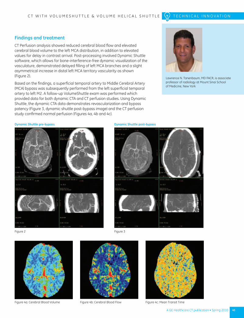
45A GE Healthcare CT publication • Spring 2010
C T W I T H V o l u M E S H u T T l E & V o l u M E H E l I C A l S H u T T l E T E C H n I C A l I n n o V A T I o n
lawrence n. Tanenbaum, MD FACR, is associate professor of radiology at Mount Sinai School of Medicine, new York
Findings and treatmentCT Perfusion analysis showed reduced cerebral blood flow and elevated cerebral blood volume to the left MCA distribution, in addition to elevated values for delay in contrast arrival. Post-processing involved Dynamic Shuttle software, which allows for bone-interference-free dynamic visualization of the vasculature, demonstrated delayed filling of left MCA branches and a slight asymmetrical increase in distal left MCA territory vascularity as shown (Figure 2).
Based on the findings, a superficial temporal artery to Middle Cerebral Artery (MCA) bypass was subsequently performed from the left superficial temporal artery to left M2. A follow-up VolumeShuttle exam was performed which provided data for both dynamic CTA and CT perfusion studies. using Dynamic Shuttle, the dynamic CTA data demonstrates revascularization and bypass patency (Figure 3, dynamic shuttle post-bypass image) and the CT perfusion study confirmed normal perfusion (Figures 4a, 4b and 4c).
Figure 4a: Cerebral Blood Volume Figure 4b: Cerebral Blood Flow Figure 4c: Mean Transit Time
Dynamic Shuttle pre-bypass
Figure 2
Dynamic Shuttle post-bypass
Figure 3
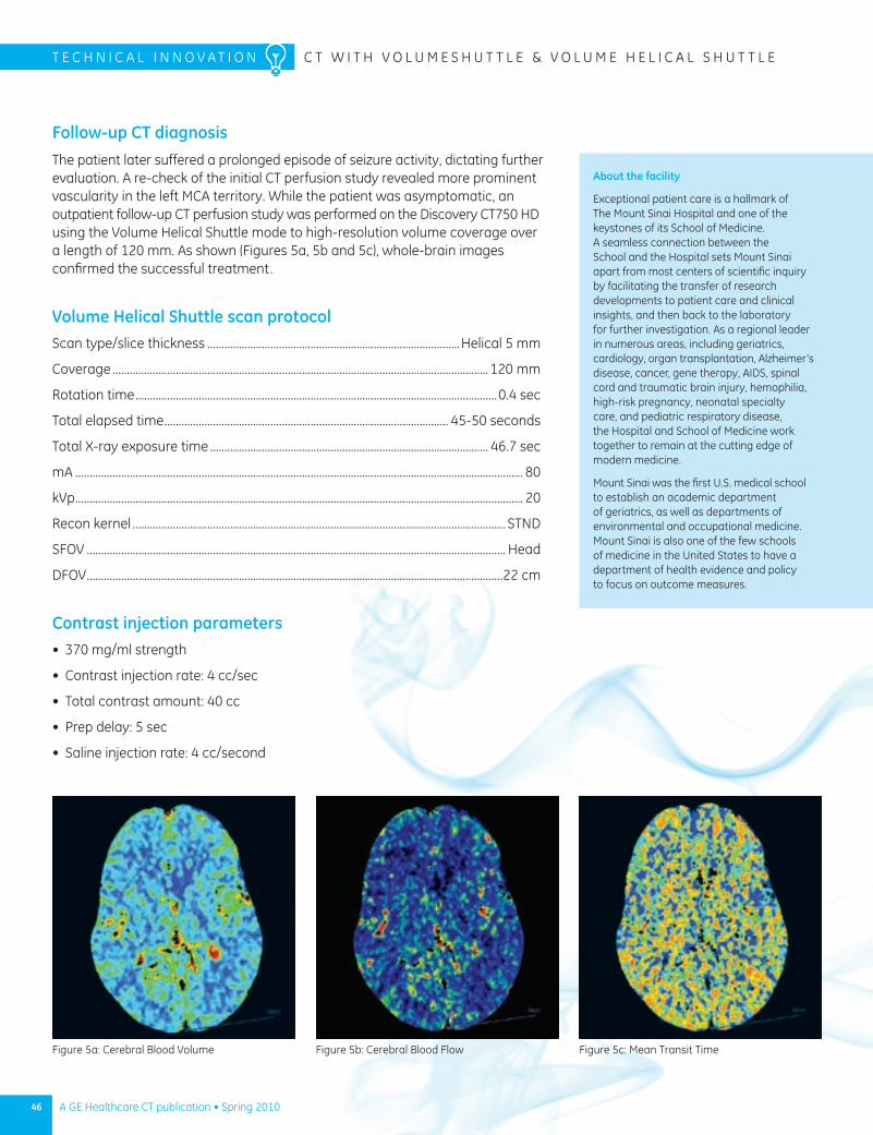
46 A GE Healthcare CT publication • Spring 2010
T E C H n I C A l I n n o V A T I o n C T W I T H V o l u M E S H u T T l E & V o l u M E H E l I C A l S H u T T l E
Follow-up CT diagnosisThe patient later suffered a prolonged episode of seizure activity, dictating further evaluation. A re-check of the initial CT perfusion study revealed more prominent vascularity in the left MCA territory. While the patient was asymptomatic, an outpatient follow-up CT perfusion study was performed on the Discovery CT750 HD using the Volume Helical Shuttle mode to high-resolution volume coverage over a length of 120 mm. As shown (Figures 5a, 5b and 5c), whole-brain images confirmed the successful treatment.
Volume Helical Shuttle scan protocol Scan type/slice thickness ........................................................................................Helical 5 mm
Coverage ...................................................................................................................................120 mm
Rotation time ..............................................................................................................................0.4 sec
Total elapsed time ................................................................................................... 45-50 seconds
Total X-ray exposure time ................................................................................................. 46.7 sec
mA ............................................................................................................................................................ 80
kVp ............................................................................................................................................................ 20
Recon kernel .................................................................................................................................. STnD
SFoV .................................................................................................................................................. Head
DFoV .................................................................................................................................................22 cm
Contrast injection parameters370 mg/ml strength•
Contrast injection rate: 4 cc/sec•
Total contrast amount: 40 cc•
Prep delay: 5 sec•
Saline injection rate: 4 cc/second•
Figure 5a: Cerebral Blood Volume Figure 5b: Cerebral Blood Flow Figure 5c: Mean Transit Time
About the facility
Exceptional patient care is a hallmark of The Mount Sinai Hospital and one of the keystones of its School of Medicine. A seamless connection between the School and the Hospital sets Mount Sinai apart from most centers of scientific inquiry by facilitating the transfer of research developments to patient care and clinical insights, and then back to the laboratory for further investigation. As a regional leader in numerous areas, including geriatrics, cardiology, organ transplantation, Alzheimer’s disease, cancer, gene therapy, AIDS, spinal cord and traumatic brain injury, hemophilia, high-risk pregnancy, neonatal specialty care, and pediatric respiratory disease, the Hospital and School of Medicine work together to remain at the cutting edge of modern medicine.
Mount Sinai was the first u.S. medical school to establish an academic department of geriatrics, as well as departments of environmental and occupational medicine. Mount Sinai is also one of the few schools of medicine in the united States to have a department of health evidence and policy to focus on outcome measures.
47A GE Healthcare CT publication • Spring 2010
For years, noise and artifacts stood in the way of producing high-quality CT images in low-dose exams that reduce risk to patients. Advanced image reconstruction techniques provide the breakthrough, enabling high image quality in multi-slice CT exams at significantly less dose than before.
The new techniques use iterative reconstruction, which differs substantially from filtered back projection (FBP), traditionally used to reduce CT image noise. In particular, clinical studies have begun to document the dose-reduction advantages of ASiR,™ a proprietary iterative reconstruction technique developed by GE Healthcare.
By Jiang Hsieh, CT Chief Scientist, GE Healthcare
ASiR Demonstrates Advantages over Filtered Back Projection in Producing High-quality CT Images at Reduced Dose
the BarriersRemoving
48 A GE Healthcare CT publication • Spring 2010
T E C H n I C A l I n n o v A T I o n A S I R A n D l o w D o S E
Jiang Hsieh, CT Chief Scientist, GE Healthcare
one study, looking at 1,150 patients at three centers, determined that ASiR, when combined with other dose-reducing strategies, cut the median dose in 64-slice coronary CTA by up to 50%, when compared with the median dose reported in a large international multi-site investigation.1 Already, all three of a major U.S. clinic’s sites have replaced FBP with ASiR in a growing range of CT applications performed by nearly 200 radiologists.2
The bottom line: There is a distinct, clinical advantage to ASiR when compared with FBP reconstruction.
Basic difference
In the simplest terms, ASiR uses sophisticated statistical modeling to remove noise in images while preserving anatomical detail. This enables a same- or better-quality image to be produced with lower tube current or tube voltage, thus lower dose, for all CT applications in which it is used.
FBP algorithms are fast, and they have served clinicians well over the past 30 years, but they do have drawbacks. For one, the CT system model that forms the basis for the FBP algorithms is idealized and does not represent reality. In particular, the system statistics are poorly modeled with FBP. The simplification in the modeling has an effect on the quality of reconstructed images. on the other hand, the FBP process tends to create image noise, which then must be overpowered (or masked) by increasing the radiation dosage.
In FBP, projection data is calibrated, filtered, weighted, and backprojected. when the last projection view has been processed, the reconstruction is complete, and the reconstructed images are generated. However, with the advent of ASiR, clinicians are now able to effectively reduce dose by up to 50%, or improve low-contrast detectability (lCD) by as much as 30%. Additionally, the use of ASiR typically results in better contrast resolution across different patient sizes and anatomic regions.
Statistical method
In essence, the projection data used with FBP is filtered and then pushed back along ray paths to form image pixels. In other words, a number of the assumptions are made to derive the FBP algorithm.
one way of overcoming the overly simplified assumptions of the FBP algorithm is to use a technique called iterative reconstruction (IR). To do so, the software first calculates
a synthesized projection at a particular view angle by performing “forward projection” on images of the estimated object. This estimate mimics, as much as possible, the process in real CT scanning in which X-ray photons traverse through the object and reach the detector. It mimics the finite (known) focal spot size by placing the initial location of the X-ray photons over a small area instead of a single point. During the modeling of X-ray photon interaction with the object, the size and shape of the image voxel (not an assumed point) is considered by calculating different path lengths for X-ray photons entering the voxel at slightly different orientations and locations. In a similar fashion, the detector shape and size are considered through the modeling of the detector response function. Thus the system optics are modeled with an IR reconstruction while optics are not modeled with FBP.
The synthesized projection is then compared to the actual measurement and the difference between the two shows the amount of adjustment or update needed for the current estimation of the object (image). one of the goals of the image update (AKA modification) is to minimize this difference. Fluctuations in the projection measurement due to limited photon statistics can also be taken into consideration during the image update process. Photon statistics of each individual measurement is estimated and this information is used in the reconstruction update process. In addition, should the value of an image voxel be significantly different from all of its neighbors, it is highly unlikely that such a difference represents true anatomical structure of the patient. It is more likely that this difference is due to statistical fluctuation or noise in the image. when all information is taken into consideration, the currently reconstructed image is updated. This image is then fed through the entire synthesizing and updating process again to obtain a newly updated image. This process is illustrated in Figure 1.

49A GE Healthcare CT publication • Spring 2010
A S I R A n D l o w D o S E T E C H n I C A l I n n o v A T I o n
1 Estimated Radiation Dose of Coronary CT Angiography Using Adaptive Statistical Iterative Reconstruction (ERASIR 1) study, DiagnosticImaging.com December 2, 2009
2 “ ASiR reconstruction sharpens images, slices abdominal CT dose,” auntminnie.com, May 21, 2009
3 Estimated Radiation Dose of Coronary CT Angiography Using Adaptive Statistical Iterative Reconstruction (ERASIR 1) study, DiagnosticImaging.com December 2, 2009
4 “ Filtered back projection: A mathematical technique used in magnetic resonance imaging and computed tomography to create images from a set of multiple projection profiles,” ImagingBz newsletter, Aug. 17, 2009.
ASiR reduces computation time
Although iterative reconstruction is excellent from an image quality standpoint, a disadvantage is that the modeling process is computationally intense and the reconstruction time is much longer than desired. ASiR address the issue by eliminating system optics modeling in the iterative algorithm.
It turns out the most time-consuming portion of the reconstruction process is the modeling of the system optics. That’s because the computational intensity on the modeling of the noise portion of the system is not nearly as big as the modeling of the system optics. Therefore, by focusing first on the modeling of the noise properties and the scanned object, ASiR provides significant benefit for examinations that may experience limitations due to noise in the reconstructed images, as a result of lower dose examinations, large patients, thinner slices, etc. Phrased differently, clinicians are able to gain the dose benefit by removing the noise in the reconstructed images so the scanning technique can be reduced with equivalent noise.
Gaining acceptance
The largest clinical study of ASiR to date is the Estimated Radiation Dose of Coronary CT Angiography Using Adaptive Statistical Iterative Reconstruction (ERASIR 1) study.3 The investigation found that patients received median doses of:
3.8 mSv when the hospitals used FBP in a standard • battery of radiation reduction strategies
2.6 mSv in procedures using ASiR alone•
1.3 mSv when ASiR was used with other radiation • reduction strategies
In a study known as the International PRoTECTIon I trial, which involved 2,000 patients from more than 50 sites, the median dose was 14 mSv.
More power to come
As promising as it is now, ASiR may be a prelude to even more powerful iterative reconstruction techniques in development with GE Healthcare. At the Medical College of wisconsin, w. Dennis Foley, Chief of Digital Imaging and a Professor of Radiology, tested a more advanced iterative reconstruction technique in a limited study in 2008. Foley and his research team ranked image quality in 29 low-dose abdominal exams, comparing FBP images and iterative reconstructions side by side.4
The research team looked at image-quality factors including spatial resolution, noise, high and low contrast, uniformity parameters, and artifact level, such as streaks and beam hardening. The viewers perceived this iterative reconstruction as superior in spatial resolution, noise suppression, and high-contrast and low-contrast detail.
ASiR and other advanced forms of iterative reconstruction that are in the R&D phase at GE Healthcare are pointing the way toward a future of lower-dose CT exams with high diagnostic value and clinical confidence. If you would like a full white paper on ASiR and how it differs from FBP, contact your GE Healthcare sales representative.
Fig. 2: Flow diagram of iterative reconstruction process
system statistics
statisticsmodel
objectmodel
current estimationof the object
synthesizedprojection
measuredprojection
system optics
x-ray source model
image voxel model
detector model
object update candidate
Figure 1

50 A GE Healthcare CT publication • Spring 2010
Stroke Imaging Solutions: Shuttle-based
Whole Brain Perfusion
CT’s role in imaging of stroke
A comprehensive assessment of stroke involves non-contrast CT, CTA from the arch to the vertex and perfusion CT.1 More formally this protocol typically consists of the following diagnostic imaging scans:
1. Non-contrast CT
2. CT Angiography of head, neck and arch
3. Whole territory CT Perfusion
As mentioned above, the non-contrast scan is performed to help physicians rule out any bleeds that may indicate a hemorrhagic stroke or some alternative diagnosis that is going to preclude use of thrombolytic treatment. Non-contrast CT is also useful in detecting a completing ischemic stroke, which shows up as a hypo-dense region darker than the surrounding tissue. In some instances it is possible to observe a disturbance in the gray matter-white matter boundary as well as to determine the size of the infarcted tissue using non-contrast CT. However, it is not possible to determine tissue at risk using this technique.1 The CTA is performed to provide a comprehensive evaluation of intracranial and cervical arteries that supply blood to the various parts of the brain. Of particular interest is the presence and location of vessel occlusions. CTA imaging
By Souma Sengupta, Clinical Services Manager; B. Dusty Majumdar, PhD, Marketing Manager, Premium CT; Mark Bowman, Product Manager, CT Advanced Applications; and Saad Sirohey, PhD, Global Product Manager
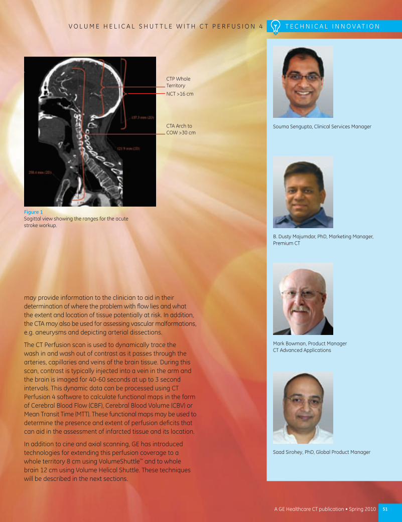
51A GE Healthcare CT publication • Spring 2010
T E C H N I C A l I N N O v A T I O N
Souma Sengupta, Clinical Services Manager
B. Dusty Majumdar, PhD, Marketing Manager, Premium CT
Mark Bowman, Product Manager CT Advanced Applications
Saad Sirohey, PhD, Global Product Manager
may provide information to the clinician to aid in their determination of where the problem with flow lies and what the extent and location of tissue potentially at risk. In addition, the CTA may also be used for assessing vascular malformations, e.g. aneurysms and depicting arterial dissections.
The CT Perfusion scan is used to dynamically trace the wash in and wash out of contrast as it passes through the arteries, capillaries and veins of the brain tissue. During this scan, contrast is typically injected into a vein in the arm and the brain is imaged for 40-60 seconds at up to 3 second intervals. This dynamic data can be processed using CT Perfusion 4 software to calculate functional maps in the form of Cerebral Blood Flow (CBF), Cerebral Blood volume (CBv) or Mean Transit Time (MTT). These functional maps may be used to determine the presence and extent of perfusion deficits that can aid in the assessment of infarcted tissue and its location.
In addition to cine and axial scanning, GE has introduced technologies for extending this perfusion coverage to a whole territory 8 cm using volumeShuttle™ and to whole brain 12 cm using volume Helical Shuttle. These techniques will be described in the next sections.
v O l u M E H E l I C A l S H u T T l E W I T H C T P E r F u S I O N 4
CTA Arch to COW >30 cm
CTP Whole Territory
Figure 1 Sagittal view showing the ranges for the acute stroke workup.
NCT >16 cm
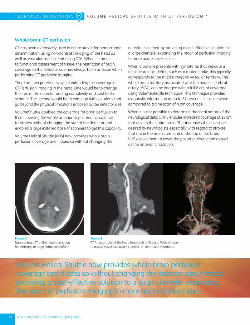
52 A GE Healthcare CT publication • Spring 2010
T E C H N I C A l I N N O v A T I O N v O l u M E H E l I C A l S H u T T l E W I T H C T P E r F u S I O N 4
Whole brain CT perfusion
CT has been extensively used in acute stroke for hemorrhage determination using non-contrast imaging of the head as well as vascular assessment using CTA. When it comes to functional assessment of tissue, the restriction of brain coverage to the detector size has always been an issue when performing CT perfusion imaging.
There are two potential ways of extending the coverage of CT Perfusion imaging in the head. One would be to change the size of the detector adding complexity and cost to the scanner. The second would be to come up with solutions that go beyond the physical limitations imposed by the detector size.
volumeShuttle doubled the coverage for brain perfusion to 8 cm covering the whole anterior or posterior circulation territories without changing the size of the detector and enabled a large installed base of scanners to get this capability.
volume Helical Shuttle (vHS) now provides whole brain perfusion coverage and it does so without changing the
detector size thereby providing a cost effective solution to a large clientele, expanding the reach of perfusion imaging to more acute stroke cases.
When a patient presents with symptoms that indicate a focal neurologic deficit, such as a motor stroke, this typically corresponds to the middle cerebral vascular territory. The whole brain territory associated with the middle cerebral artery (MCA), can be imaged with a full 8 cm of coverage using volumeShuttle technique. This technique provides diagnostic information at up to 24 percent less dose when compared to a cine scan of 4 cm coverage.
When it is not possible to determine the focal nature of the neurological deficit, vHS enables increased coverage of 12 cm that covers the entire brain. This increases the coverage desired by neurologists especially with regard to strokes that are in the brain stem and at the top of the brain. vHS allows them to cover the posterior circulation as well as the anterior circulation.
Figure 2 Non-contrast CT of the head to exclude hemorrhage or large completed infarct.
Figure 3 CT Angiography of the head from arch to Circle of Willis in order to assess vessel occlusion, stenosis, or ventricular thrombus.
volume Helical Shuttle now provides whole brain perfusion coverage and it does so without changing the detector size, thereby providing a cost-effective solution to a large clientele, expanding the reach of perfusion imaging to more acute stroke cases.
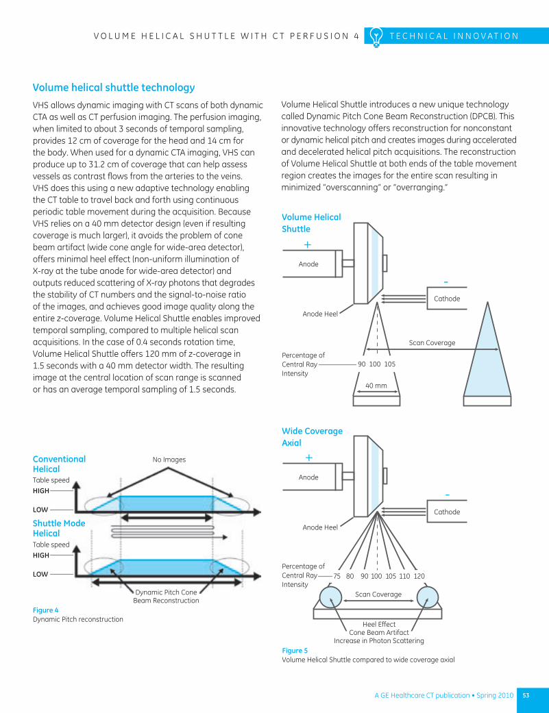
53A GE Healthcare CT publication • Spring 2010
v O l u M E H E l I C A l S H u T T l E W I T H C T P E r F u S I O N 4 T E C H N I C A l I N N O v A T I O N
Figure 4 Dynamic Pitch reconstruction
Table speed
HIGH
LOW
Table speed
HIGH
LOW
Dynamic Pitch Cone Beam reconstruction
Shuttle Mode Helical
No ImagesConventional Helical
Volume helical shuttle technology
vHS allows dynamic imaging with CT scans of both dynamic CTA as well as CT perfusion imaging. The perfusion imaging, when limited to about 3 seconds of temporal sampling, provides 12 cm of coverage for the head and 14 cm for the body. When used for a dynamic CTA imaging, vHS can produce up to 31.2 cm of coverage that can help assess vessels as contrast flows from the arteries to the veins. vHS does this using a new adaptive technology enabling the CT table to travel back and forth using continuous periodic table movement during the acquisition. Because vHS relies on a 40 mm detector design (even if resulting coverage is much larger), it avoids the problem of cone beam artifact (wide cone angle for wide-area detector), offers minimal heel effect (non-uniform illumination of X-ray at the tube anode for wide-area detector) and outputs reduced scattering of X-ray photons that degrades the stability of CT numbers and the signal-to-noise ratio of the images, and achieves good image quality along the entire z-coverage. volume Helical Shuttle enables improved temporal sampling, compared to multiple helical scan acquisitions. In the case of 0.4 seconds rotation time, volume Helical Shuttle offers 120 mm of z-coverage in 1.5 seconds with a 40 mm detector width. The resulting image at the central location of scan range is scanned or has an average temporal sampling of 1.5 seconds.
volume Helical Shuttle introduces a new unique technology called Dynamic Pitch Cone Beam reconstruction (DPCB). This innovative technology offers reconstruction for nonconstant or dynamic helical pitch and creates images during accelerated and decelerated helical pitch acquisitions. The reconstruction of volume Helical Shuttle at both ends of the table movement region creates the images for the entire scan resulting in minimized “overscanning” or “overranging.”
Anode
Cathode
Scan Coverage
Scan Coverage
40 mm
Volume Helical Shuttle
Anode Heel
Percentage of Central ray Intensity
90 100 105
Figure 5 volume Helical Shuttle compared to wide coverage axial
Cathode
Anode
Wide Coverage Axial
Heel Effect Cone Beam Artifact
Increase in Photon Scattering
Anode Heel
Percentage of Central ray Intensity
75 80 90 100 105 110 120
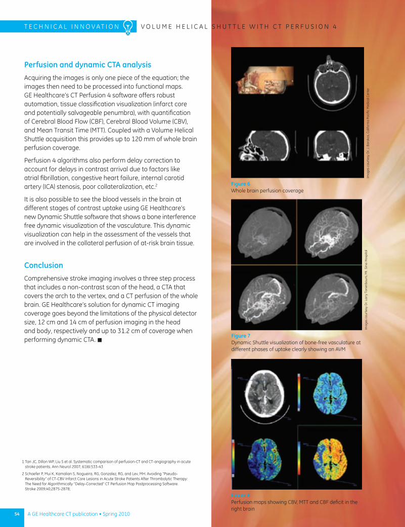
54 A GE Healthcare CT publication • Spring 2010
T E C H N I C A l I N N O v A T I O N v O l u M E H E l I C A l S H u T T l E W I T H C T P E r F u S I O N 4
Perfusion and dynamic CTA analysis
Acquiring the images is only one piece of the equation; the images then need to be processed into functional maps. GE Healthcare’s CT Perfusion 4 software offers robust automation, tissue classification visualization (infarct core and potentially salvageable penumbra), with quantification of Cerebral Blood Flow (CBF), Cerebral Blood volume (CBv), and Mean Transit Time (MTT). Coupled with a volume Helical Shuttle acquisition this provides up to 120 mm of whole brain perfusion coverage.
Perfusion 4 algorithms also perform delay correction to account for delays in contrast arrival due to factors like atrial fibrillation, congestive heart failure, internal carotid artery (ICA) stenosis, poor collateralization, etc.2
It is also possible to see the blood vessels in the brain at different stages of contrast uptake using GE Healthcare’s new Dynamic Shuttle software that shows a bone interference free dynamic visualization of the vasculature. This dynamic visualization can help in the assessment of the vessels that are involved in the collateral perfusion of at-risk brain tissue.
Conclusion
Comprehensive stroke imaging involves a three step process that includes a non-contrast scan of the head, a CTA that covers the arch to the vertex, and a CT perfusion of the whole brain. GE Healthcare’s solution for dynamic CT imaging coverage goes beyond the limitations of the physical detector size, 12 cm and 14 cm of perfusion imaging in the head and body, respectively and up to 31.2 cm of coverage when performing dynamic CTA. n
Figure 8 Perfusion maps showing CBv, MTT and CBF deficit in the right brain
Figure 6 Whole brain perfusion coverage
Imag
es c
ourt
esy
Dr.
J. B
arak
os, C
alifo
rnia
Pac
ific
Med
ical
Cen
ter
Figure 7 Dynamic Shuttle visualization of bone-free vasculature at different phases of uptake clearly showing an AvM
Imag
es c
ourt
esy
Dr.
larr
y Ta
nenb
aum
, Mt.
Sin
ai H
ospi
tal
1 Tan JC, Dillon WP, liu S et al. Systematic comparison of perfusion-CT and CT-angiography in acute stroke patients. Ann Neurol 2007; 61(6):533-43
2 Schaefer P, Mui K, Kamalian S, Nogueira, rG, Gonzalez, rG, and lev, MH. Avoiding “Pseudo-reversibility” of CT-CBv Infarct Core lesions in Acute Stroke Patients After Thrombolytic Therapy: The Need for Algorithmically “Delay-Corrected” CT Perfusion Map Postprocessing Software. Stroke 2009;40;2875-2878;
55
A New Standard in PET/CT
Gain Confidence, Better Manage Dose, and Streamline Workflow with the Discovery PET/CT 600 Series
A GE Healthcare CT publication • Spring 2010
By Amy Burris, PET/CT Global Product Marketing Manager
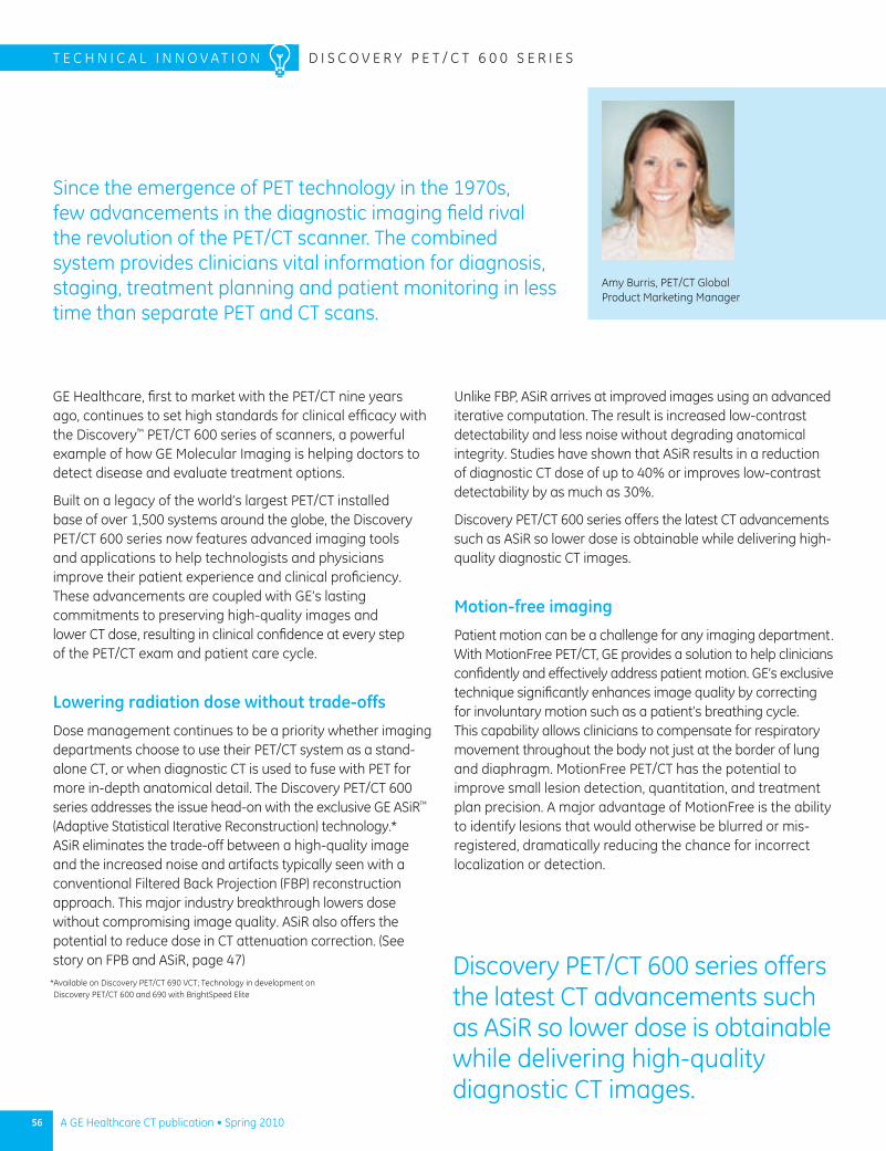
Discovery PET/CT 600 series offers the latest CT advancements such as ASiR so lower dose is obtainable while delivering high-quality diagnostic CT images.
Amy Burris, PET/CT Global Product Marketing Manager
Unlike FBP, ASiR arrives at improved images using an advanced iterative computation. The result is increased low-contrast detectability and less noise without degrading anatomical integrity. Studies have shown that ASiR results in a reduction of diagnostic CT dose of up to 40% or improves low-contrast detectability by as much as 30%.
Discovery PET/CT 600 series offers the latest CT advancements such as ASiR so lower dose is obtainable while delivering high-quality diagnostic CT images.
Motion-free imaging
Patient motion can be a challenge for any imaging department. With MotionFree PET/CT, GE provides a solution to help clinicians confidently and effectively address patient motion. GE’s exclusive technique significantly enhances image quality by correcting for involuntary motion such as a patient’s breathing cycle. This capability allows clinicians to compensate for respiratory movement throughout the body not just at the border of lung and diaphragm. MotionFree PET/CT has the potential to improve small lesion detection, quantitation, and treatment plan precision. A major advantage of MotionFree is the ability to identify lesions that would otherwise be blurred or mis-registered, dramatically reducing the chance for incorrect localization or detection.
GE Healthcare, first to market with the PET/CT nine years ago, continues to set high standards for clinical efficacy with the Discovery™ PET/CT 600 series of scanners, a powerful example of how GE Molecular Imaging is helping doctors to detect disease and evaluate treatment options.
Built on a legacy of the world’s largest PET/CT installed base of over 1,500 systems around the globe, the Discovery PET/CT 600 series now features advanced imaging tools and applications to help technologists and physicians improve their patient experience and clinical proficiency. These advancements are coupled with GE’s lasting commitments to preserving high-quality images and lower CT dose, resulting in clinical confidence at every step of the PET/CT exam and patient care cycle.
Lowering radiation dose without trade-offs
Dose management continues to be a priority whether imaging departments choose to use their PET/CT system as a stand-alone CT, or when diagnostic CT is used to fuse with PET for more in-depth anatomical detail. The Discovery PET/CT 600 series addresses the issue head-on with the exclusive GE ASiR™ (Adaptive Statistical Iterative Reconstruction) technology.* ASiR eliminates the trade-off between a high-quality image and the increased noise and artifacts typically seen with a conventional Filtered Back Projection (FBP) reconstruction approach. This major industry breakthrough lowers dose without compromising image quality. ASiR also offers the potential to reduce dose in CT attenuation correction. (See story on FPB and ASiR, page 47)
* Available on Discovery PET/CT 690 VCT; Technology in development on Discovery PET/CT 600 and 690 with BrightSpeed Elite
56 A GE Healthcare CT publication • Spring 2010
T E C H N I C A l I N N o V A T I o N D I S C o V E R y P E T / C T 6 0 0 S E R I E S
Since the emergence of PET technology in the 1970s, few advancements in the diagnostic imaging field rival the revolution of the PET/CT scanner. The combined system provides clinicians vital information for diagnosis, staging, treatment planning and patient monitoring in less time than separate PET and CT scans.
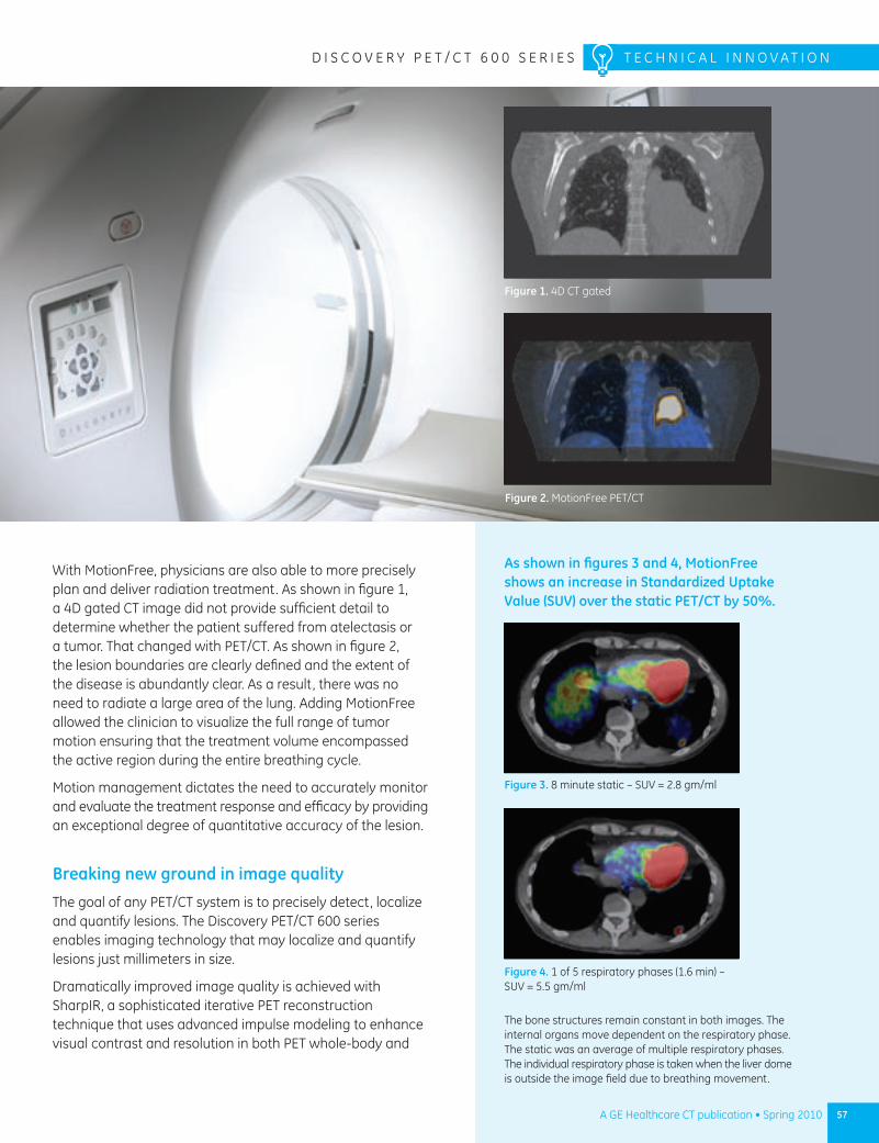
57A GE Healthcare CT publication • Spring 2010
D I S C o V E R y P E T / C T 6 0 0 S E R I E S T E C H N I C A l I N N o V A T I o N
With MotionFree, physicians are also able to more precisely plan and deliver radiation treatment. As shown in figure 1, a 4D gated CT image did not provide sufficient detail to determine whether the patient suffered from atelectasis or a tumor. That changed with PET/CT. As shown in figure 2, the lesion boundaries are clearly defined and the extent of the disease is abundantly clear. As a result, there was no need to radiate a large area of the lung. Adding MotionFree allowed the clinician to visualize the full range of tumor motion ensuring that the treatment volume encompassed the active region during the entire breathing cycle.
Motion management dictates the need to accurately monitor and evaluate the treatment response and efficacy by providing an exceptional degree of quantitative accuracy of the lesion.
Breaking new ground in image quality
The goal of any PET/CT system is to precisely detect, localize and quantify lesions. The Discovery PET/CT 600 series enables imaging technology that may localize and quantify lesions just millimeters in size.
Dramatically improved image quality is achieved with SharpIR, a sophisticated iterative PET reconstruction technique that uses advanced impulse modeling to enhance visual contrast and resolution in both PET whole-body and
Figure 2. MotionFree PET/CT
Figure 4. 1 of 5 respiratory phases (1.6 min) – SUV = 5.5 gm/ml
Figure 3. 8 minute static – SUV = 2.8 gm/ml
As shown in figures 3 and 4, MotionFree shows an increase in Standardized Uptake Value (SUV) over the static PET/CT by 50%.
The bone structures remain constant in both images. The internal organs move dependent on the respiratory phase. The static was an average of multiple respiratory phases. The individual respiratory phase is taken when the liver dome is outside the image field due to breathing movement.
Figure 1. 4D CT gated
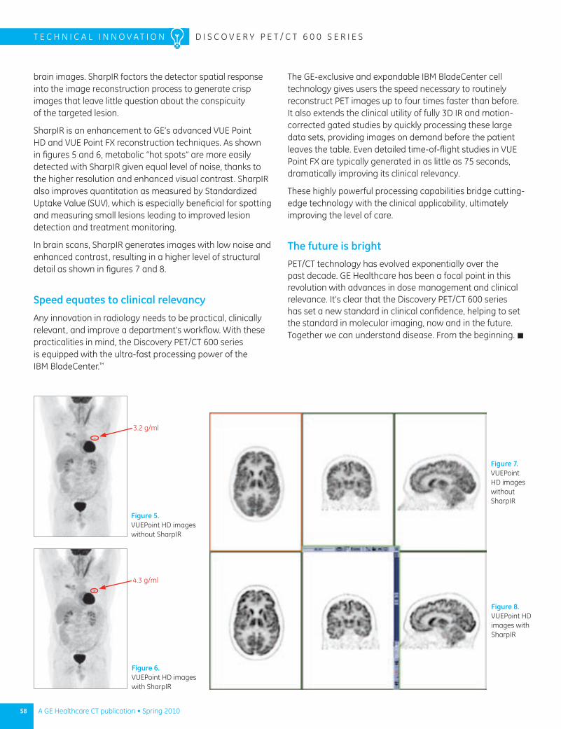
Figure 5. VUEPoint HD images without SharpIR
Figure 7. VUEPoint HD images without SharpIR
Figure 6. VUEPoint HD images with SharpIR
Figure 8. VUEPoint HD images with SharpIR
3.2 g/ml
4.3 g/ml
brain images. SharpIR factors the detector spatial response into the image reconstruction process to generate crisp images that leave little question about the conspicuity of the targeted lesion.
SharpIR is an enhancement to GE’s advanced VUE Point HD and VUE Point FX reconstruction techniques. As shown in figures 5 and 6, metabolic “hot spots” are more easily detected with SharpIR given equal level of noise, thanks to the higher resolution and enhanced visual contrast. SharpIR also improves quantitation as measured by Standardized Uptake Value (SUV), which is especially beneficial for spotting and measuring small lesions leading to improved lesion detection and treatment monitoring.
In brain scans, SharpIR generates images with low noise and enhanced contrast, resulting in a higher level of structural detail as shown in figures 7 and 8.
Speed equates to clinical relevancy
Any innovation in radiology needs to be practical, clinically relevant, and improve a department’s workflow. With these practicalities in mind, the Discovery PET/CT 600 series is equipped with the ultra-fast processing power of the IBM BladeCenter.™
The GE-exclusive and expandable IBM BladeCenter cell technology gives users the speed necessary to routinely reconstruct PET images up to four times faster than before. It also extends the clinical utility of fully 3D IR and motion-corrected gated studies by quickly processing these large data sets, providing images on demand before the patient leaves the table. Even detailed time-of-flight studies in VUE Point FX are typically generated in as little as 75 seconds, dramatically improving its clinical relevancy.
These highly powerful processing capabilities bridge cutting-edge technology with the clinical applicability, ultimately improving the level of care.
The future is brightPET/CT technology has evolved exponentially over the past decade. GE Healthcare has been a focal point in this revolution with advances in dose management and clinical relevance. It’s clear that the Discovery PET/CT 600 series has set a new standard in clinical confidence, helping to set the standard in molecular imaging, now and in the future. Together we can understand disease. From the beginning. n
58 A GE Healthcare CT publication • Spring 2010
T E C H N I C A l I N N o V A T I o N D I S C o V E R y P E T / C T 6 0 0 S E R I E S
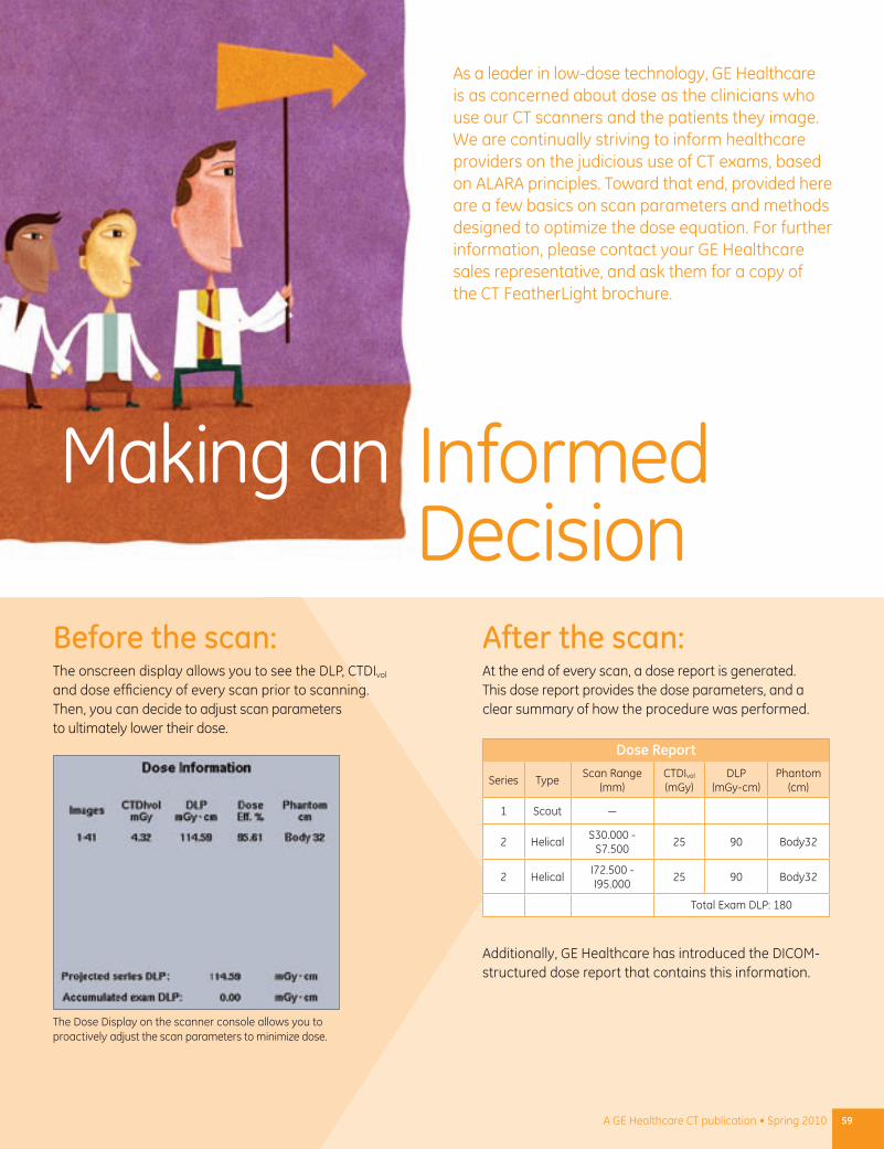
Making an Informed DecisionBefore the scan:The onscreen display allows you to see the DLP, CTDIvol and dose efficiency of every scan prior to scanning. Then, you can decide to adjust scan parameters to ultimately lower their dose.
The Dose Display on the scanner console allows you to proactively adjust the scan parameters to minimize dose.
After the scan:At the end of every scan, a dose report is generated. This dose report provides the dose parameters, and a clear summary of how the procedure was performed.
Additionally, GE Healthcare has introduced the DICOM-structured dose report that contains this information.
Dose Report
Series TypeScan Range
(mm)CTDIvol (mGy)
DLP (mGy-cm)
Phantom (cm)
1 Scout —
2 HelicalS30.000 -
S7.50025 90 Body32
2 HelicalI72.500 - I95.000
25 90 Body32
Total Exam DLP: 180
As a leader in low-dose technology, GE Healthcare is as concerned about dose as the clinicians who use our CT scanners and the patients they image. We are continually striving to inform healthcare providers on the judicious use of CT exams, based on ALARA principles. Toward that end, provided here are a few basics on scan parameters and methods designed to optimize the dose equation. For further information, please contact your GE Healthcare sales representative, and ask them for a copy of the CT FeatherLight brochure.
59A GE Healthcare CT publication • Spring 2010
60 A GE Healthcare CT publication • Spring 2010
B E y O n D T H E S C A n C T & L O W D O S E
Understanding the dose equation
The decision to adjust scan parameters involves understanding the method for estimating dose. Three factors are used to estimate effective dose: (1) Phantom, which represents the size of the body part being scanned; (2) Computed Tomography Dose Index (CTDIvol); and (3) Dose Length Product (DLP).
Phantom
The size of phantom is either small (16 cm diameter) or large (32 cm diameter) depending on the size of the body part. The actual dose to the patient is dependent on body habitus as compared to that of the reference phantom.
The average absorbed energy per kg is higher in the smaller object.
DLPDose Length Product
DLP is representative of the total dose delivered, and is also used for comparing different protocols.
DLP equals the CTDIvol times the exposure length. In order to decrease DLP, the CTDIvol can be decreased and/or the scan length can be decreased.
CTDIvolCT Dose Index
CTDIvol is the dose index used for CT scanning to represent the average dose for the area scanned. This dose index allows a comparison for different acquisition parameters for a given CT system.
If all CT conditions of operation remain constant, CTDIvol will be lowered if:
mA is reduced. The relationship is directly proportional, • meaning if mA is reduced by half, the corresponding dose is reduced by half.
kV is reduced. The relationship is non-linear. For instance, • if you reduce kV from 120kV to 100kV (16.5% reduction) the result is a dose reduction of almost 40%.
Gantry rotation speed is doubled. The relationship is • directly proportional, meaning that if speed is reduced by half, the dose is reduced by half.
Pitch is increased. This relationship is inversely • proportional. When pitch is increased, the table moves faster and the dose over the area scanned is decreased.
noise Index is increased. This relationship is non-linear so • when noise is increased, mA is decreased. GE’s 3D Dose Modulation allows the user to set the level of image noise for the exam.
Different scans produce different levels of dose. Scan parameters can be changed to affect dose.
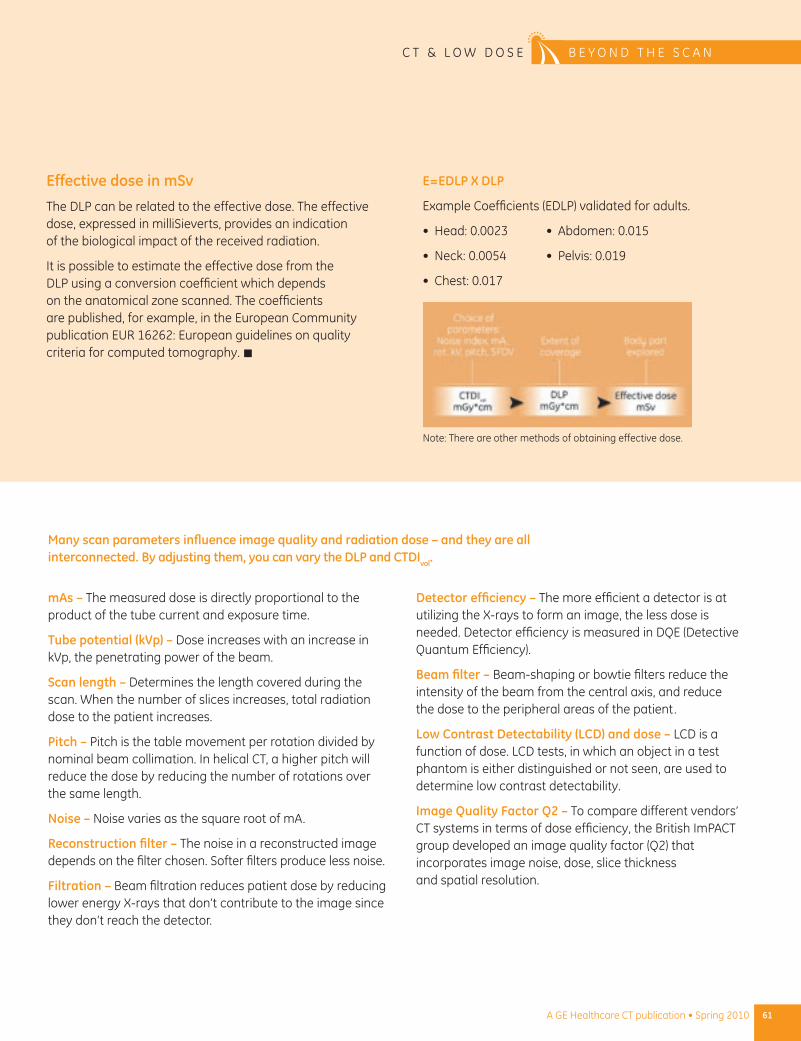
Effective dose in mSv
The DLP can be related to the effective dose. The effective dose, expressed in milliSieverts, provides an indication of the biological impact of the received radiation.
It is possible to estimate the effective dose from the DLP using a conversion coefficient which depends on the anatomical zone scanned. The coefficients are published, for example, in the European Community publication EUR 16262: European guidelines on quality criteria for computed tomography. n
note: There are other methods of obtaining effective dose.
E=EDLP X DLP
Example Coefficients (EDLP) validated for adults.
Head: 0.0023 •
neck: 0.0054 •
Chest: 0.017•
Abdomen: 0.015 •
Pelvis: 0.019•
mAs – The measured dose is directly proportional to the product of the tube current and exposure time.
Tube potential (kVp) – Dose increases with an increase in kVp, the penetrating power of the beam.
Scan length – Determines the length covered during the scan. When the number of slices increases, total radiation dose to the patient increases.
Pitch – Pitch is the table movement per rotation divided by nominal beam collimation. In helical CT, a higher pitch will reduce the dose by reducing the number of rotations over the same length.
Noise – noise varies as the square root of mA.
Reconstruction filter – The noise in a reconstructed image depends on the filter chosen. Softer filters produce less noise.
Filtration – Beam filtration reduces patient dose by reducing lower energy X-rays that don’t contribute to the image since they don’t reach the detector.
Detector efficiency – The more efficient a detector is at utilizing the X-rays to form an image, the less dose is needed. Detector efficiency is measured in DQE (Detective Quantum Efficiency).
Beam filter – Beam-shaping or bowtie filters reduce the intensity of the beam from the central axis, and reduce the dose to the peripheral areas of the patient.
Low Contrast Detectability (LCD) and dose – LCD is a function of dose. LCD tests, in which an object in a test phantom is either distinguished or not seen, are used to determine low contrast detectability.
Image Quality Factor Q2 – To compare different vendors’ CT systems in terms of dose efficiency, the British ImPACT group developed an image quality factor (Q2) that incorporates image noise, dose, slice thickness and spatial resolution.
Many scan parameters influence image quality and radiation dose – and they are all interconnected. By adjusting them, you can vary the DLP and CTDIvol.
61A GE Healthcare CT publication • Spring 2010
C T & L O W D O S E B E y O n D T H E S C A n
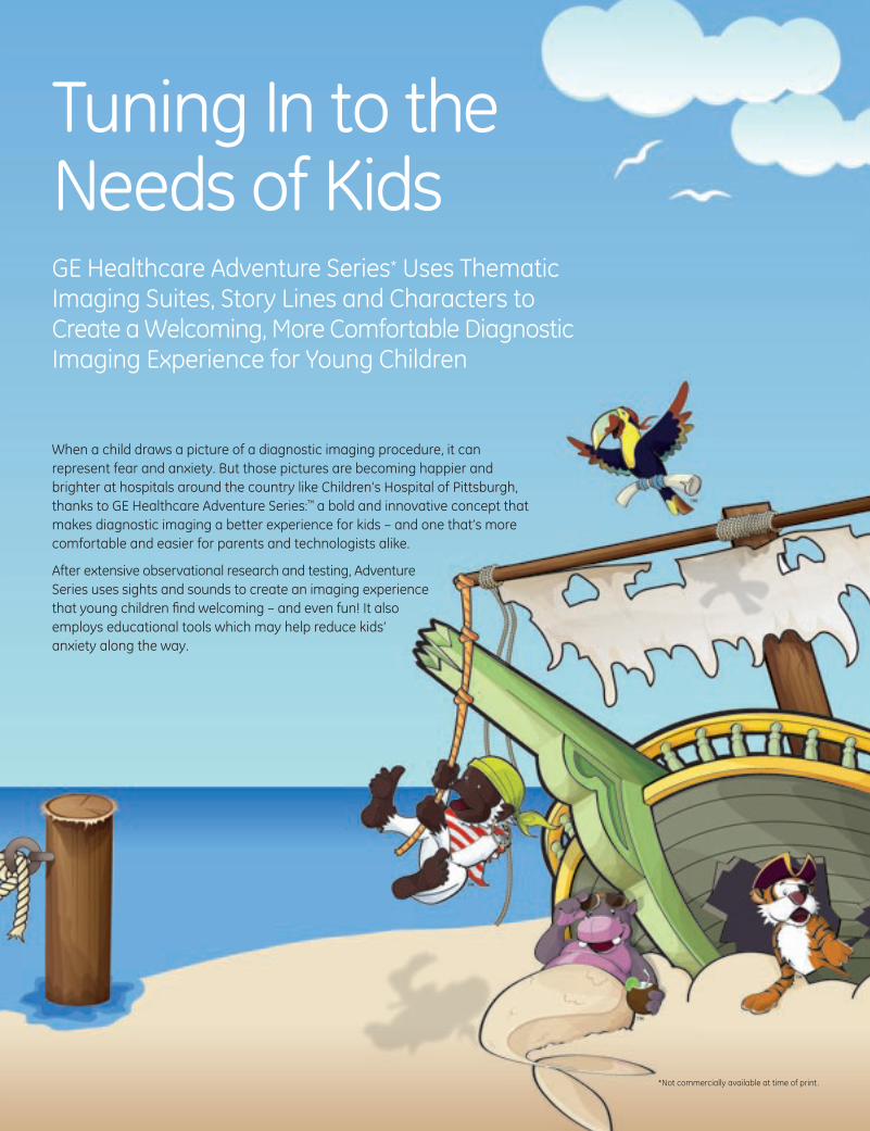
GE Healthcare Adventure Series* Uses Thematic Imaging Suites, Story Lines and Characters to Create a Welcoming, More Comfortable Diagnostic Imaging Experience for Young Children
Tuning In to the Needs of Kids
When a child draws a picture of a diagnostic imaging procedure, it can represent fear and anxiety. But those pictures are becoming happier and brighter at hospitals around the country like Children’s Hospital of Pittsburgh, thanks to GE Healthcare Adventure Series:™ a bold and innovative concept that makes diagnostic imaging a better experience for kids – and one that’s more comfortable and easier for parents and technologists alike.
After extensive observational research and testing, Adventure Series uses sights and sounds to create an imaging experience that young children find welcoming – and even fun! It also employs educational tools which may help reduce kids’ anxiety along the way.
*Not commercially available at time of print.
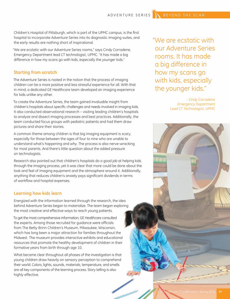
Children’s Hospital of Pittsburgh, which is part of the UPMC campus, is the first hospital to incorporate Adventure Series into its diagnostic imaging suites, and the early results are nothing short of inspirational.
“We are ecstatic with our Adventure Series rooms,” says Cindy Corradene, Emergency Department lead CT technologist, UPMC. “It has made a big difference in how my scans go with kids, especially the younger kids.”
Starting from scratch
The Adventure Series is rooted in the notion that the process of imaging children can be a more positive and less stressful experience for all. With that in mind, a dedicated GE Healthcare team developed an imaging experience for kids unlike any other.
To create the Adventure Series, the team gained invaluable insight from children’s hospitals about specific challenges and needs involved in imaging kids. It also conducted observational research – visiting leading children’s hospitals to analyze and dissect imaging processes and best practices. Additionally, the team conducted focus groups with pediatric patients and had them draw pictures and share their stories.
A common theme among children is that big imaging equipment is scary, especially for those between the ages of four to nine who are unable to understand what’s happening and why. The process is also nerve-wracking for most parents. And there’s little question about the added pressure on technologists.
Research also pointed out that children’s hospitals do a good job at helping kids through the imaging process, yet it was clear that more could be done about the look and feel of imaging equipment and the atmosphere around it. Additionally, anything that reduces children’s anxiety pays significant dividends in terms of workflow and hospital expenses.
Learning how kids learn
Energized with the information learned through the research, the idea behind Adventure Series began to materialize. The team began exploring the most creative and effective ways to reach young patients.
To get the most comprehensive information, GE Healthcare consulted the experts. Among those recruited for guidance were officials from The Betty Brinn Children’s Museum, Milwaukee, Wisconsin, which has long been a major attraction for families throughout the Midwest. The museum provides interactive exhibits and educational resources that promote the healthy development of children in their formative years from birth through age 10.
What became clear throughout all phases of the investigation is that young children draw heavily on sensory perception to comprehend their world. Colors, lights, sounds, materials, temperature, and smells are all key components of the learning process. Story telling is also highly effective.
“ We are ecstatic with our Adventure Series rooms. It has made a big difference in how my scans go with kids, especially the younger kids.”
– Cindy Corradene Emergency Department
Lead CT Technologist, UPMC
63A GE Healthcare CT publication • Spring 2010
A D v E N T U R E S E R I E S B E Y o N D T H E S C A N
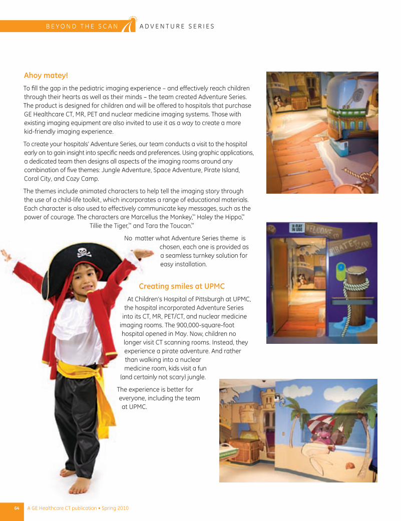
Ahoy matey!
To fill the gap in the pediatric imaging experience – and effectively reach children through their hearts as well as their minds – the team created Adventure Series. The product is designed for children and will be offered to hospitals that purchase GE Healthcare CT, MR, PET and nuclear medicine imaging systems. Those with existing imaging equipment are also invited to use it as a way to create a more kid-friendly imaging experience.
To create your hospitals’ Adventure Series, our team conducts a visit to the hospital early on to gain insight into specific needs and preferences. Using graphic applications, a dedicated team then designs all aspects of the imaging rooms around any combination of five themes: Jungle Adventure, Space Adventure, Pirate Island, Coral City, and Cozy Camp.
The themes include animated characters to help tell the imaging story through the use of a child-life toolkit, which incorporates a range of educational materials. Each character is also used to effectively communicate key messages, such as the power of courage. The characters are Marcellus the Monkey,™ Haley the Hippo,™
Tillie the Tiger,™ and Tara the Toucan.™
No matter what Adventure Series theme is chosen, each one is provided as a seamless turnkey solution for easy installation.
Creating smiles at UPMC
At Children’s Hospital of Pittsburgh at UPMC, the hospital incorporated Adventure Series
into its CT, MR, PET/CT, and nuclear medicine imaging rooms. The 900,000-square-foot hospital opened in May. Now, children no longer visit CT scanning rooms. Instead, they experience a pirate adventure. And rather than walking into a nuclear medicine room, kids visit a fun
(and certainly not scary) jungle.
The experience is better for everyone, including the team at UPMC.
64 A GE Healthcare CT publication • Spring 2010
B E Y o N D T H E S C A N A D v E N T U R E S E R I E S
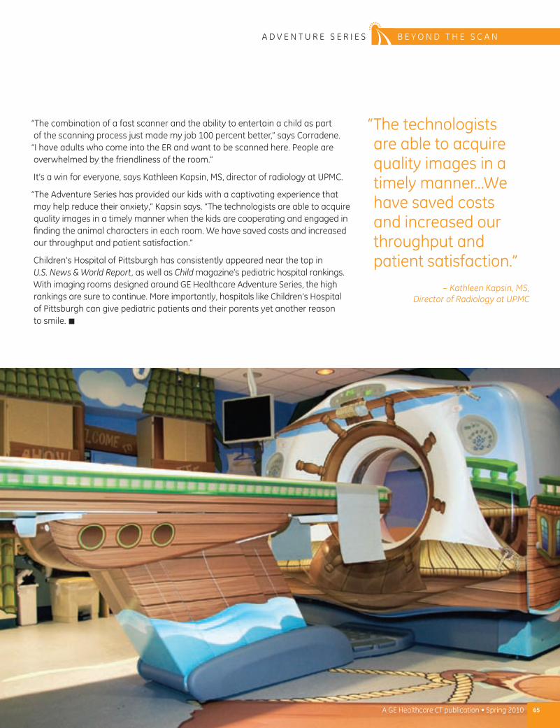
65A GE Healthcare CT publication • Spring 2010
A D v E N T U R E S E R I E S B E Y o N D T H E S C A N
“The combination of a fast scanner and the ability to entertain a child as part of the scanning process just made my job 100 percent better,” says Corradene.
“I have adults who come into the ER and want to be scanned here. People are overwhelmed by the friendliness of the room.”
It’s a win for everyone, says Kathleen Kapsin, MS, director of radiology at UPMC.
“The Adventure Series has provided our kids with a captivating experience that may help reduce their anxiety,” Kapsin says. “The technologists are able to acquire quality images in a timely manner when the kids are cooperating and engaged in finding the animal characters in each room. We have saved costs and increased our throughput and patient satisfaction.”
Children’s Hospital of Pittsburgh has consistently appeared near the top in U.S. News & World Report, as well as Child magazine’s pediatric hospital rankings. With imaging rooms designed around GE Healthcare Adventure Series, the high rankings are sure to continue. More importantly, hospitals like Children’s Hospital of Pittsburgh can give pediatric patients and their parents yet another reason to smile. n
“ The technologists are able to acquire quality images in a timely manner…We have saved costs and increased our throughput and patient satisfaction.”
– Kathleen Kapsin, MS, Director of Radiology at UPMC
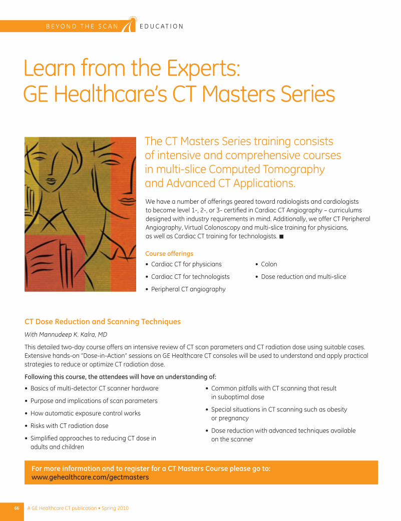
66 A GE Healthcare CT publication • Spring 2010
b E y o n d T H E S C A n e d u c a t i o n
Learn from the experts: GE Healthcare’s CT Masters Series
We have a number of offerings geared toward radiologists and cardiologists to become level 1-, 2-, or 3- certified in Cardiac CT Angiography – curriculums designed with industry requirements in mind. Additionally, we offer CT Peripheral Angiography, Virtual Colonoscopy and multi-slice training for physicians, as well as Cardiac CT training for technologists. n
The CT Masters Series training consists of intensive and comprehensive courses in multi-slice Computed Tomography and Advanced CT Applications.
Course offerings
Cardiac CT for physicians•
Cardiac CT for technologists•
Peripheral CT angiography•
colon•
dose reduction and multi-slice•
For more information and to register for a CT Masters Course please go to: www.gehealthcare.com/gectmasters
CT Dose Reduction and Scanning Techniques
With Mannudeep K. Kalra, MD
This detailed two-day course offers an intensive review of CT scan parameters and CT radiation dose using suitable cases. Extensive hands-on “dose-in-Action” sessions on GE Healthcare CT consoles will be used to understand and apply practical strategies to reduce or optimize CT radiation dose.
Following this course, the attendees will have an understanding of:
basics of multi-detector CT scanner hardware •
Purpose and implications of scan parameters •
How automatic exposure control works •
Risks with CT radiation dose •
Simplified approaches to reducing CT dose in • adults and children
Common pitfalls with CT scanning that result • in suboptimal dose
Special situations in CT scanning such as obesity • or pregnancy
dose reduction with advanced techniques available • on the scanner
67A GE Healthcare CT publication • Spring 2010
e d u c a t i o n b E y o n d T H E S C A n
We want you to join us in the company of other professionals; you can share your clinical experiences and develop your own clinical expertise with the support of GE Clinical Education. Do not hesitate to visit us at http://www.gehealthcare.com/euen/clinical-education/clinical-education.html.
Here you will find the description of each course, the calendar of training events and regular updates.
CT Master SeriesMaster Series is a new, combined offering in the European portfolio that adds to the full range of GE continuous education courses. It is designed for clients at an advanced level, who want to further improve their clinical imaging expertise with specific GE equipment.
Master Series combines several training formats varying from doctor-to-doctor (d2d), TVA (training through remote connection) and on-site sessions to gain maximum confidence in a specific topic.
With the new GE Master Series, you will learn at a structured pace and, upon return, will have the reassurance of GE remote support.
Master Series is the perfect tool to help you get the best out of your GE equipment and ensure that you stay at the forefront of healthcare imaging technology.
orchestrate your Training
Cardiac CT Master Series1x2 hours TVA session•
Cardiac CT d2d training•
2 days on-site training•
2x1 hour TVA sessions•
CT Colonography Master Series
1x2 hours TVA •
CT Colonography d2d training•
2 days on-site training•
2x1 hour TVA sessions•
CT Neurovascular Master Series
1x2 hours TVA session•
CT neurovascular d2d training•
2 days on-site training •
2x1 hour TVA sessions•
@Live Expert for CT@Live Expert - Cardiac CT •
@Live Expert - CT Colonography•
@Live Expert - CT neurovascular•
@Live Expert - CT Vascular•
Acquisition courses for CTCT Acquisition for Radiographers•
CT Cardiac Acquisition • for Radiographers
CT Vascular Acquisition • for Radiographers
Doctor-to-Doctor for CTCT Cardiac/Advanced • Cardiac workshops
CT Colonography workshop•
CT neurovascular workshop•
CT Vascular workshop•
Clinical Educational opportunities for europe
Integrating Dose Reduction Techniques across Clinical Education Curriculum
GE Healthcare’s CT Clinical Education team is integrating dose reduction techniques across its range of different customer training offerings.
This starts right from the initial on-site applications visit through to the various customer support re-visits. during these sessions the CT Clinical Education Specialist will work with the customer to optimize their protocols using the latest ASiR™ technology where appropriate to deliver high image quality at optimized dose.
dose reduction is fast becoming one of the key modules within our classroom-based and doctor-to-doctor offerings targeting the educational needs of both the radiographers and radiologists/cardiologists. n
A healthy dose of freedom.
GE Healthcare
Today, you have the freedom to lower patient dose dramatically without compromising image quality. Our dedication to you and your patients drives our mission to develop technology, training and education, so that every patient may be imaged FeatherLight when they receive a CT exam.
Learn more at gehealthcare.com/lowdoseCT
CT-0436-04.10-EN-US©2009 General Electric Company – All rights reserved.
To receive future issues of CT Clarity, please subscribe at: www.gehealthcare.com/CTclarity