clh report - europa · clh report for formaldehyde 1 clh report proposal for harmonised...
TRANSCRIPT
CLH REPORT FOR FORMALDEHYDE
1
CLH report
Proposal for Harmonised Classification and Labelling
Based on Regulation (EC) No 1272/2008 (CLP Regulation),
Annex VI, Part 2
Substance Name: FORMALDEHYDE … %
EC Number: 200-001-8
CAS Number: 50-00-0
Index Number: 605-001-00-5
Contact details for dossier submitter: ANSES (on behalf of the French MSCA)
253 avenue du General Leclerc
F-94701 Maisons-Alfort Cedex
+33 1 56 29 19 30
Version number: 2 Date: 28 September 2011
CLH REPORT FOR FORMALDEHYDE
2
CONTENTS
Part A.
1 PROPOSAL FOR HARMONISED CLASSIFICATION AND LABELLING ................................................. 5
1.1 SUBSTANCE ........................................................................................................................................................... 5 1.2 HARMONISED CLASSIFICATION AND LABELLING PROPOSAL .................................................................................. 5 1.3 PROPOSED HARMONISED CLASSIFICATION AND LABELLING BASED ON CLP REGULATION AND/OR DSD CRITERIA
7
2 BACKGROUND TO THE CLH PROPOSAL ................................................................................................... 10
2.1 HISTORY OF THE PREVIOUS CLASSIFICATION AND LABELLING ............................................................................ 10 2.2 SHORT SUMMARY OF THE SCIENTIFIC JUSTIFICATION FOR THE CLH PROPOSAL .................................................. 10 2.3 CURRENT HARMONISED CLASSIFICATION AND LABELLING .................................................................................. 12
2.3.1 Current classification and labelling in Annex VI, Table 3.1 in the CLP Regulation ................................ 12 2.3.2 Current classification and labelling in Annex VI, Table 3.2 in the CLP Regulation ................................ 12
2.4 CURRENT SELF-CLASSIFICATION AND LABELLING ............................................................................................... 12
3 JUSTIFICATION THAT ACTION IS NEEDED AT COMMUNITY LEVEL .............................................. 13
SCIENTIFIC EVALUATION OF THE DATA ........................................................................................................... 14
1 IDENTITY OF THE SUBSTANCE .................................................................................................................... 14
1.1 NAME AND OTHER IDENTIFIERS OF THE SUBSTANCE ............................................................................................ 14 1.2 COMPOSITION OF THE SUBSTANCE ...................................................................................................................... 15
1.2.1 Composition of test material ..................................................................................................................... 16 1.3 PHYSICO-CHEMICAL PROPERTIES ........................................................................................................................ 16
2 MANUFACTURE AND USES ............................................................................................................................ 18
2.1 MANUFACTURE ................................................................................................................................................... 18 2.2 IDENTIFIED USES ................................................................................................................................................. 18
3 CLASSIFICATION FOR PHYSICO-CHEMICAL PROPERTIES ................................................................ 19
4 HUMAN HEALTH HAZARD ASSESSMENT .................................................................................................. 19
4.1 TOXICOKINETICS (ABSORPTION, METABOLISM, DISTRIBUTION AND ELIMINATION) (OECD 2002) ...................... 19 4.2 ACUTE TOXICITY ................................................................................................................................................. 20 4.3 SPECIFIC TARGET ORGAN TOXICITY – SINGLE EXPOSURE (STOT SE).................................................................. 20 4.4 IRRITATION ......................................................................................................................................................... 21 4.5 CORROSIVITY ...................................................................................................................................................... 21 4.6 SENSITISATION .................................................................................................................................................... 21 4.7 REPEATED DOSE TOXICITY .................................................................................................................................. 21 4.8 SPECIFIC TARGET ORGAN TOXICITY (CLP REGULATION) – REPEATED EXPOSURE (STOT RE) ............................ 21 4.9 GERM CELL MUTAGENICITY (MUTAGENICITY) .................................................................................................... 21
4.9.1 Non-human information ............................................................................................................................ 21 4.9.1.1 In vitro data ............................................................................................................................................................ 21 4.9.1.2 In vivo data ............................................................................................................................................................ 44
4.9.2 Human information ................................................................................................................................... 58 4.9.2.1 Studies performed at the site of contact ................................................................................................................. 58 4.9.2.2 Studies performed at distant sites ........................................................................................................................... 62
4.9.3 Summary and discussion of mutagenicity ................................................................................................. 81 4.9.4 Comparison with criteria .......................................................................................................................... 85 4.9.5 Conclusions on classification and labelling ............................................................................................. 86
4.10 CARCINOGENICITY ......................................................................................................................................... 87 4.10.1 Non-human information ....................................................................................................................... 87
4.10.1.1 Carcinogenicity: oral ........................................................................................................................................ 87 Table 16: Experimental data on carcinogenicity by oral route .......................................................................................... 87
CLH REPORT FOR FORMALDEHYDE
3
4.10.1.2 Carcinogenicity: inhalation ............................................................................................................................... 90 Table 17: Experimental data on carcinogenicity by inhalation ......................................................................................... 90 4.10.1.3 Carcinogenicity: dermal .................................................................................................................................... 96 Table 18: Experimental data on carcinogenicity by dermal route ..................................................................................... 96
4.10.2 Human information .............................................................................................................................. 98 4.10.2.1 Industrial cohort studies .................................................................................................................................... 98 Table 19: Industrial cohort studies .................................................................................................................................... 98 4.10.2.2 Professional cohort studies ............................................................................................................................. 111 Table 20: Professional cohort studies ............................................................................................................................. 111 4.10.2.3 Case-control studies ........................................................................................................................................ 114 4.10.2.4 Meta-analysis .................................................................................................................................................. 145
4.10.3 Other relevant information ................................................................................................................ 155 4.10.4 Summary and discussion of carcinogenicity ...................................................................................... 157 4.10.5 Comparison with criteria ................................................................................................................... 171 4.10.6 Conclusions on classification and labelling ....................................................................................... 176
4.11 TOXICITY FOR REPRODUCTION ..................................................................................................................... 177 4.12 OTHER EFFECTS ............................................................................................................................................ 177
5 ENVIRONMENTAL HAZARD ASSESSMENT ............................................................................................. 177
6 OTHER INFORMATION .................................................................................................................................. 178
7 REFERENCES .................................................................................................................................................... 180
CLH REPORT FOR FORMALDEHYDE
4
List of abbreviations
AML: acute myeloid leukaemia
ALL: acute lymphocytic leukaemia
BAL: Broncho-alveolar lavage
BrdUrd: 5-bromodeoxyuirdine
CA : chromosomal aberration
CI: confidence interval
CML: chronic myeloid leukaemia
CLL: chronic lymphocytic leukaemia
CPA: cyclophosphamide
DPX : DNA-protein crosslink
dAdo: deoxyadenosine
FA: formaldehyde
HNEC: Human Nasal Epithelial Cells
IP: intra-peritoneal
LM: lateral meatus
ML: myeloid leukaemia
MN: micronucleus
M:PM: medial and posterior meatus
MRR: meta-relative risk
NALT: nasal-associated lymphoid tissue
NHL: Non-Hodgkin lymphoma
NOAEL: No observable adverse effect level
4-NOQ: 4-nitroquinoline 1-oxide
NPC: nasopharynx carcinoma
OR: odd ratio
PMR: proportionate mortality ratio
RCP: regenerative cell proliferation
ROS: reactive oxygen species
RR: relative risk
SCE: sister chromatid exchange
SCL: specific concentration limit
SIR: standardised incidence ratio
SMR: standardised mortality ratio
SPICR: standardised proportionate incidence cancer ratios
TWA: time-weighted average concentration
CLH REPORT FOR FORMALDEHYDE
5
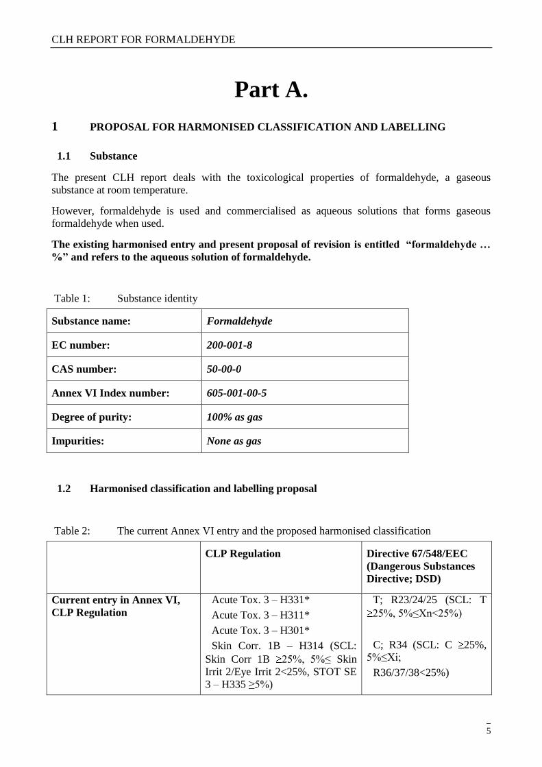
Part A.
1 PROPOSAL FOR HARMONISED CLASSIFICATION AND LABELLING
1.1 Substance
The present CLH report deals with the toxicological properties of formaldehyde, a gaseous
substance at room temperature.
However, formaldehyde is used and commercialised as aqueous solutions that forms gaseous
formaldehyde when used.
The existing harmonised entry and present proposal of revision is entitled “formaldehyde …
%” and refers to the aqueous solution of formaldehyde.
Table 1: Substance identity
Substance name: Formaldehyde
EC number: 200-001-8
CAS number: 50-00-0
Annex VI Index number: 605-001-00-5
Degree of purity: 100% as gas
Impurities: None as gas
1.2 Harmonised classification and labelling proposal
Table 2: The current Annex VI entry and the proposed harmonised classification
CLP Regulation Directive 67/548/EEC
(Dangerous Substances
Directive; DSD)
Current entry in Annex VI,
CLP Regulation
Acute Tox. 3 – H331*
Acute Tox. 3 – H311*
Acute Tox. 3 – H301*
Skin Corr. 1B – H314 (SCL:
Skin Corr 1B 25%, 5%≤ Skin
Irrit 2/Eye Irrit 2<25%, STOT SE
3 – H335 ≥5%)
T; R23/24/25 (SCL: T
25%, 5%≤Xn<25%)
C; R34 (SCL: C 25%,
5%≤Xi;
R36/37/38<25%)
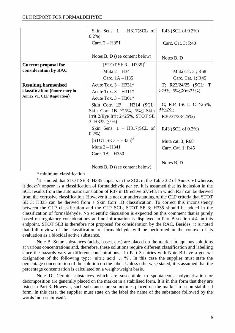
CLH REPORT FOR FORMALDEHYDE
6
Skin Sens. 1 – H317(SCL of
0.2%)
Carc. 2 – H351
Notes B, D (see content below)
R43 (SCL of 0.2%)
Carc. Cat. 3; R40
Notes B, D
Current proposal for
consideration by RAC
[STOT SE 3 – H335]#
Muta 2 – H341
Carc. 1A – H35
Muta cat. 3 ; R68
Carc. Cat. 1; R45
Resulting harmonised
classification (future entry in
Annex VI, CLP Regulation)
Acute Tox. 3 – H331*
Acute Tox. 3 – H311*
Acute Tox. 3 – H301*
Skin Corr. 1B – H314 (SCL:
Skin Corr 1B 25%, 5%≤ Skin
Irrit 2/Eye Irrit 2<25%, STOT SE
3- H335 ≥5%)
Skin Sens. 1 – H317(SCL of
0.2%)
[STOT SE 3 – H335]#
Muta 2 – H341
Carc. 1A – H350
Notes B, D (see content below)
T; R23/24/25 (SCL: T
25%, 5%≤Xn<25%)
C; R34 (SCL: C 25%,
5%≤Xi;
R36/37/38<25%)
R43 (SCL of 0.2%)
Muta cat. 3; R68
Carc. Cat. 1; R45
Notes B, D
* minimum classification
#It is noted that STOT SE 3- H335 appears in the SCL in the Table 3.2 of Annex VI whereas
it doesn‘t appear as a classification of formaldehyde per se. It is assumed that its inclusion in the
SCL results from the automatic translation of R37 in Directive 67/548, in which R37 can be derived
from the corrosive classification. However it is not our understanding of the CLP criteria that STOT
SE 3; H335 can be derived from a Skin Corr 1B classification. To correct this inconsistency
between the CLP classification and the CLP SCL, STOT SE 3; H335 should be added in the
classification of formaldehyde. No scientific discussion is expected on this comment that is purely
based on regulatory considerations and no information is displayed in Part B section 4.4 on this
endpoint. STOT SE3 is therefore not proposed for consideration by the RAC. Besides, it is noted
that full review of the classification of formaldehyde will be performed in the context of its
evaluation as a biocidal active substance.
Note B: Some substances (acids, bases, etc.) are placed on the market in aqueous solutions
at various concentrations and, therefore, these solutions require different classification and labelling
since the hazards vary at different concentrations. In Part 3 entries with Note B have a general
designation of the following type: ‗nitric acid … %‘. In this case the supplier must state the
percentage concentration of the solution on the label. Unless otherwise stated, it is assumed that the
percentage concentration is calculated on a weight/weight basis.
Note D: Certain substances which are susceptible to spontaneous polymerisation or
decomposition are generally placed on the market in a stabilised form. It is in this form that they are
listed in Part 3. However, such substances are sometimes placed on the market in a non-stabilised
form. In this case, the supplier must state on the label the name of the substance followed by the
words ‗non-stabilised‘.
CLH REPORT FOR FORMALDEHYDE
7
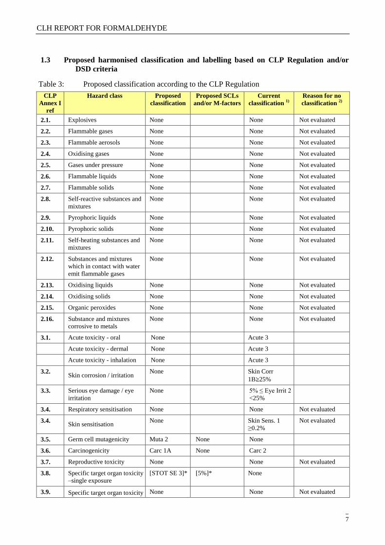
1.3 Proposed harmonised classification and labelling based on CLP Regulation and/or
DSD criteria
Table 3: Proposed classification according to the CLP Regulation
CLP
Annex I
ref
Hazard class Proposed
classification
Proposed SCLs
and/or M-factors
Current
classification 1)
Reason for no
classification 2)
2.1. Explosives None None Not evaluated
2.2. Flammable gases None None Not evaluated
2.3. Flammable aerosols None None Not evaluated
2.4. Oxidising gases None None Not evaluated
2.5. Gases under pressure None None Not evaluated
2.6. Flammable liquids None None Not evaluated
2.7. Flammable solids None None Not evaluated
2.8. Self-reactive substances and
mixtures
None None Not evaluated
2.9. Pyrophoric liquids None None Not evaluated
2.10. Pyrophoric solids None None Not evaluated
2.11. Self-heating substances and
mixtures
None None Not evaluated
2.12. Substances and mixtures
which in contact with water
emit flammable gases
None None Not evaluated
2.13. Oxidising liquids None None Not evaluated
2.14. Oxidising solids None None Not evaluated
2.15. Organic peroxides None None Not evaluated
2.16. Substance and mixtures
corrosive to metals
None None Not evaluated
3.1. Acute toxicity - oral None Acute 3
Acute toxicity - dermal None Acute 3
Acute toxicity - inhalation None Acute 3
3.2. Skin corrosion / irritation
None Skin Corr
1B 25%
3.3. Serious eye damage / eye
irritation
None 5% ≤ Eye Irrit 2
<25%
3.4. Respiratory sensitisation None None Not evaluated
3.4. Skin sensitisation
None Skin Sens. 1
≥0.2%
Not evaluated
3.5. Germ cell mutagenicity Muta 2 None None
3.6. Carcinogenicity Carc 1A None Carc 2
3.7. Reproductive toxicity None None Not evaluated
3.8. Specific target organ toxicity
–single exposure
[STOT SE 3]* [5%]* None
3.9. Specific target organ toxicity None None Not evaluated
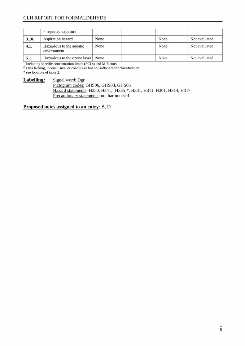
CLH REPORT FOR FORMALDEHYDE
8
– repeated exposure
3.10. Aspiration hazard None None Not evaluated
4.1. Hazardous to the aquatic
environment
None None Not evaluated
5.1. Hazardous to the ozone layer None None Not evaluated 1) Including specific concentration limits (SCLs) and M-factors
2) Data lacking, inconclusive, or conclusive but not sufficient for classification
* see footnote of table 2.
Labelling: Signal word: Dgr
Pictogram codes: GHS06, GHS08, GHS05
Hazard statements: H350, H341, [H335]*, H331, H311, H301, H314, H317
Precautionary statements: not harmonised
Proposed notes assigned to an entry: B, D
CLH REPORT FOR FORMALDEHYDE
9
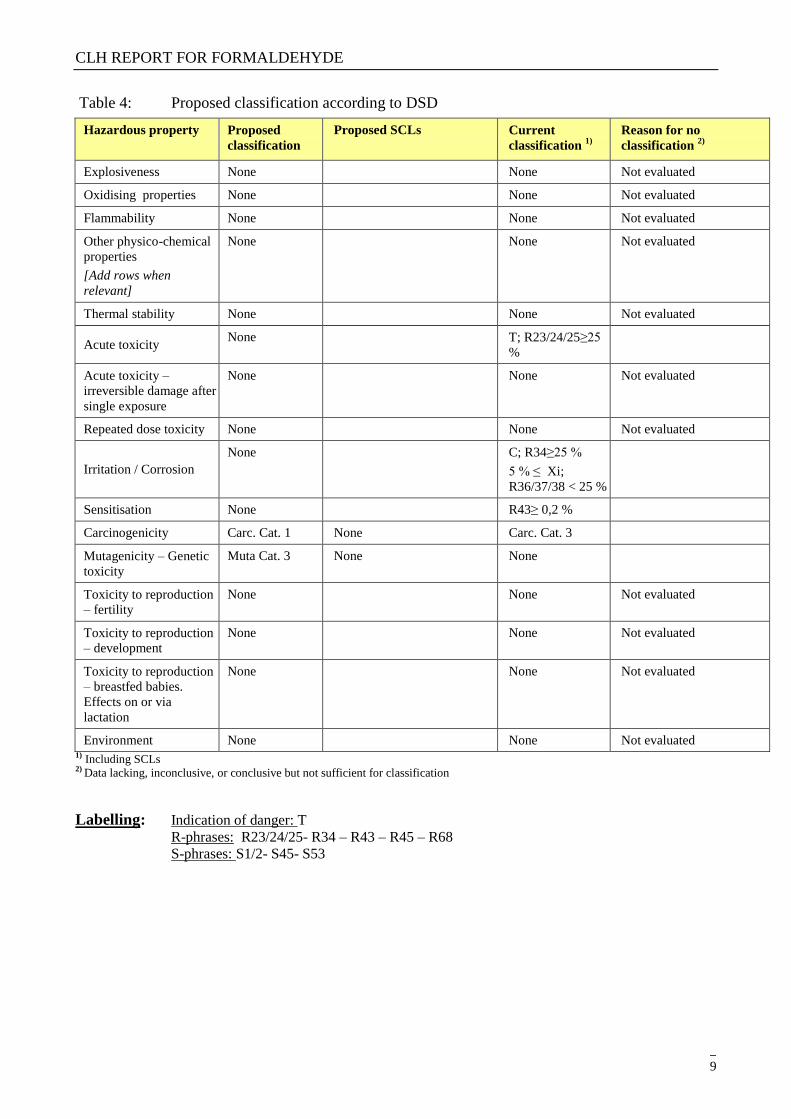
Table 4: Proposed classification according to DSD
Hazardous property
Proposed
classification
Proposed SCLs Current
classification 1)
Reason for no
classification 2)
Explosiveness None None Not evaluated
Oxidising properties None None Not evaluated
Flammability None None Not evaluated
Other physico-chemical
properties
[Add rows when
relevant]
None None Not evaluated
Thermal stability None None Not evaluated
Acute toxicity None T; R23/24/25≥25
%
Acute toxicity –
irreversible damage after
single exposure
None None Not evaluated
Repeated dose toxicity None None Not evaluated
Irritation / Corrosion
None C; R34≥25 %
5 % ≤ Xi;
R36/37/38 < 25 %
Sensitisation None R43≥ 0,2 %
Carcinogenicity Carc. Cat. 1 None Carc. Cat. 3
Mutagenicity – Genetic
toxicity
Muta Cat. 3 None None
Toxicity to reproduction
– fertility
None None Not evaluated
Toxicity to reproduction
– development
None None Not evaluated
Toxicity to reproduction
– breastfed babies.
Effects on or via
lactation
None None Not evaluated
Environment None None Not evaluated 1) Including SCLs 2) Data lacking, inconclusive, or conclusive but not sufficient for classification
Labelling: Indication of danger: T
R-phrases: R23/24/25- R34 – R43 – R45 – R68
S-phrases: S1/2- S45- S53
CLH REPORT FOR FORMALDEHYDE
10
2 BACKGROUND TO THE CLH PROPOSAL
2.1 History of the previous classification and labelling
The classification of aqueous solutions of formaldehyde (…%) is harmonised in Annex VI of CLP
under the index number 605-001-00-5 as follows:
Carc. Cat. 3; R40
T; R23/24/25 (SCL: T 25%, 5%≤Xn<25%)
C; R34 (SCL: C 25%, 5%≤Xi; R36/37/38<25%)
R43 (SCL of 0.2%)
Note B, D
Classification of formaldehyde was inserted in the 1st ATP (1976) of Annexe I of Directive
67/548/EEC. Carcinogenicity classification was inserted in the 8th
ATP in 1987 and has not been
modified since then. The last update of formaldehyde classification was included in the 22nd ATP
of Directive 67/548/EEC (1996) and focused on the adoption of SCL for skin irritation.
It is not known whether discussions on the carcinogenicity and mutagenicity of formaldehyde have
taken place since the first insertion of carcinogenic classification in Annexe I. However, no
discussion on these endpoints has taken place at least from the 22nd
ATP to our knowledge.
A classification proposal was submitted by the French CA at the TC C&L and was presented at the
TC C&L of November 2005. No discussion took place as several Members States were not ready
for discussion. The substance was removed from the agenda of TC C&L of March 2006 and
October 2006, as it was decided that the update of the NCI cohort and national positions of the MS
should be awaited. No further discussion took place at the TC C&L.
2.2 Short summary of the scientific justification for the CLH proposal
The International Agency for Research on Cancer (IARC) has evaluated the carcinogenity of
formaldehyde several times. In 2006, IARC concluded that formaldehyde is a known human
carcinogen (group 1) on the basis of induction of nasopharyngeal cancers (IARC 2006). It was
reaffirmed in its re-evaluation of 2009 and extended to the induction of leukaemia and particularly
myeloid leukaemia (Baan 2010).
A large amount of new relevant data on carcinogenicity and mutagenicity of formaldehyde has been
published in the past 15 years that has not been evaluated by the TC C&L (see history of
formaldehyde classification in 2.1) and the French Competent Authorities considers that the
classification for carcinogenicity and mutagenicity needs to be revised on the basis of the new
studies available. Several reviews of the toxicological properties of formaldehyde have also been
published by international or national organisations as discussed in section 6 of this report.
On mutagenicity, positive evidence are available in vivo at the site of contact in somatic cells. They
consist in induction of chromosomal aberrations in rats by inhalation at high dose (Dallas 1992) and
of micronuclei in rats in the gastrointestinal tract by oral route (Migliore 1989). These positive data
are further supported by in vitro positive results in numerous genotoxicity and mutagenicity tests,
in vivo induction of DNA adducts and DNA-protein crosslinks (DPX) at the site of contact and
indications of consistent increases in micronuclei frequency in humans at the site of contact. Based
CLH REPORT FOR FORMALDEHYDE
11
on induction of genotoxic and mutagenic effects of formaldehyde on somatic cells at the site of
contact, classification in Category 2 is warranted.
On carcinogenicity, experimental data clearly provide evidence of a carcinogenic effect at the site
of contact in rats by inhalation. Although this finding is restricted to a single species (rat), consistent
results were obtained from several independent studies and in both females and males. Tumours
consists in both benign and malignant tumours but were induced at a single site (nasal cavity). Data
investigating the mode of action support the existence of a threshold type mode of action for its
carcinogenic properties based on the cytotoxic effect of formaldehyde. Genotoxicity is also
expected to play a role above this threshold. Overall the level of experimental evidence is judged
as sufficient evidence in agreement with induction of tumours (b) [in] two or more
independent studies in one species carried out at different times or in different laboratories or
under different protocols.
At the site of contact, positive epidemiological evidence of association from both cohort studies and
case-control studies were identified for nasopharynx. Results were statistically significant and
supported by trends with exposure in both types of studies. However, the existence of a grouping of
cases in plant 1 of the National Cancer Institute (NCI) cohort raises a doubt on potential cofounder
and lowers the level of evidence. But the grouping of cases but it can also be explained by the
largest number of subjects exposed to high peaks in this specific plant. Several factors however
support the existence of a carcinogenic potential of formaldehyde at the site of contact:
- Induction of tumours in the nasal cavity in rats with a proposed mode of action based on
chronic irritation of the respiratory tract and local genotoxicity at doses inducing an
increased proliferation
- Indication of local genotoxicity in exposed humans as evidenced by increases in
micronuclei frequency in buccal and nasal mucosa cells in several studies
- Human sensitivity to FA-induced irritation, with irritation of the eye and of the
nose/throat being consistently reported after exposure to formaldehyde (IARC 2006).
No species-specific mechanism is evident and human data denote human sensitivity to FA effects
(genotoxicity and irritation). The mode of action of carcinogenicity in the rat nasal cavity is
therefore considered relevant to humans, as reviewed in the context of the IPCS framework
(McGregor 2007).
The induction of nasopharyngeal carcinomas in human exposed to formaldehyde is therefore
strongly plausible.
The biological plausibility of the induction of nasopharyngeal carcinomas in humans exposed to
formaldehyde highly supports the consistent epidemiological evidence obtained from the NCI
cohort and from several case-control studies. It is considered that the doubt of a potential cofounder
is raised by the grouping of cases in the plant 1 of the NCI cohort. But considering the overall
database and more specifically the fact that the grouping of cases in plant 1 can also be explained by
the largest number of subjects exposed to high peaks in this specific plant, correlation of NPC with
the level of peak exposure to formaldehyde, the evidence provided by case-control studies and the
biological plausibility, the doubt that the observed induction of NPC may be due to confounder can
be ruled out with reasonable confidence.
Altogether, the data support a causal relationship between formaldehyde exposure and
induction of NPC and corresponds to a sufficient evidence of carcinogenicity in humans.

CLH REPORT FOR FORMALDEHYDE
12
2.3 Current harmonised classification and labelling
2.3.1 Current classification and labelling in Annex VI, Table 3.1 in the CLP Regulation
The classification of formaldehyde is harmonised in Annex VI of CLP under the index number
605-001-00-5 as follows:
Table 3.1 (CLP)
Acute Tox. 3 – H331*
Acute Tox. 3 – H311*
Acute Tox. 3 – H301*
Skin Corr. 1B – H314 (SCL: Skin Corr 1B 25%,
5%≤ Skin Irrit 2/Eye Irrit 2<25%, STOT SE 3 – H335
≥5%)
Skin Sens. 1 – H317(SCL of 0.2%)
Carc. 2 – H351
Notes B, D
* minimum classification
2.3.2 Current classification and labelling in Annex VI, Table 3.2 in the CLP Regulation
The classification of formaldehyde is harmonised in Annex VI of CLP under the index number 605-
001-00-5 as follows:
Table 3.2 (67/548/EEC)
T; R23/24/25 (SCL: T 25%, 5%≤Xn<25%)
C; R34 (SCL: C 25%, 5%≤Xi; R36/37/38<25%)
R43 (SCL of 0.2%)
Carc. Cat. 3; R40
Notes B, D
2.4 Current self-classification and labelling
Not relevant
CLH REPORT FOR FORMALDEHYDE
13
3 JUSTIFICATION THAT ACTION IS NEEDED AT COMMUNITY LEVEL
Formaldehyde has a harmonised classification and labelling (as aqueous solution) in Annex VI of
CLP that includes classification for carcinogenicity.
A large amount of new relevant data on carcinogenicity and on mutagenicity of formaldehyde has
been published in the past 15 years that has not been evaluated by the TC C&L (see history of
formaldehyde classification in 2.1).
The French Competent Authorities considers that the classification for carcinogenicity and
mutagenicity needs to be revised on the basis of the new studies available.
Carcinogenicity and mutagenicity as other CMR properties justifies a harmonised classification and
labelling according to article 36 of CLP.
Regulatory considerations are added on STOT SE3 –H335 (see footnote of table 2) but this
endpoint is not proposed for consideration by the RAC. Besides, it is noted that full review of the
classification of formaldehyde will be performed in the context of its evaluation as a biocidal active
substance.
CLH REPORT FOR FORMALDEHYDE
14
Part B.
SCIENTIFIC EVALUATION OF THE DATA
1 IDENTITY OF THE SUBSTANCE
1.1 Name and other identifiers of the substance
Table 5: Substance identity
EC number: 200-001-8
EC name: Formaldehyde
Synonyms: formaldehyde gas, formaldehyde
solution, methanal, formic aldehyde,
methylene oxide, oxymethylene,
methylaldehyde, oxomethane, formol,
formalin, formalith, méthylaldehyde,
morbicid, oxomethane, paraform.
CAS number (EC inventory): 50-00-0
CAS number: 50-00-0
CAS name: Formaldehyde
IUPAC name: Formaldehyde
CLP Annex VI Index number: 605-001-00-5
Molecular formula: CH2O
Molecular weight range: 30.026 g/mol
CLH REPORT FOR FORMALDEHYDE
15
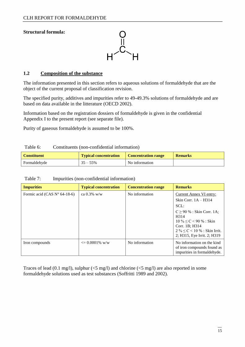
Structural formula:
1.2 Composition of the substance
The information presented in this section refers to aqueous solutions of formaldehyde that are the
object of the current proposal of classification revision.
The specified purity, additives and impurities refer to 49-49.3% solutions of formaldehyde and are
based on data available in the litterature (OECD 2002).
Information based on the registration dossiers of formaldehyde is given in the confidential
Appendix I to the present report (see separate file).
Purity of gaseous formaldehyde is assumed to be 100%.
Table 6: Constituents (non-confidential information)
Constituent Typical concentration Concentration range Remarks
Formaldehyde 35 – 55% No information
Table 7: Impurities (non-confidential information)
Impurities Typical concentration Concentration range Remarks
Formic acid (CAS N° 64-18-6) ca 0.3% w/w No information Current Annex VI entry:
Skin Corr. 1A – H314
SCL:
C ≥ 90 % : Skin Corr. 1A;
H314
10 % ≤ C < 90 % : Skin
Corr. 1B; H314
2 % ≤ C < 10 % : Skin Irrit.
2; H315, Eye Irrit. 2; H319
Iron compounds <= 0.0001% w/w No information No information on the kind
of iron compounds found as
impurities in formaldehyde.
Traces of lead (0.1 mg/l), sulphur (<5 mg/l) and chlorine (<5 mg/l) are also reported in some
formaldehyde solutions used as test substances (Soffritti 1989 and 2002).
CLH REPORT FOR FORMALDEHYDE
16
Table 8: Additives (non-confidential information)
Additives Typical concentration Concentration range Remarks
Methanol (CAS N° 67-56-1) ca 2% w/w No information Used as a stabiliser
Current Annex VI entry:
Flam. Liq. 2 - H225
Acute Tox. 3 * - H331
Acute Tox. 3 * - H311
Acute Tox. 3 * - H301
STOT SE 1 – 370**
SCL:
C ≥ 10 % : STOT SE 1;
H370
3 % ≤ C <10 % :
STOT SE 2; H371
6,6‘-(m-phenylene) bis (1,3,5-triazine-2,4-diamine) (CAS N° 5118-80-9) is also mentionned as an additives (OCDE
2002) but this statement cannot be checked in absence of any information on its function as an additive and it is not
known whether it is an additive or an impurity in the meaning of REACH.
1.2.1 Composition of test material
Relevant information is given in the respective study summaries when available.
1.3 Physico-chemical properties
Formaldehyde is a very volatile gas at room temperature (high vapour pressure), very soluble in
water but not stable.
When dissolved into water, formaldehyde converts to methanediol H2C(OH)2, a diol. Aqueous
solutions of formaldehyde are referred to as formalin. A typical commercial grade formalin may
contain 10–12% methanol in addition to various metallic impurities. The diol also exists in
equilibrium with a series of short polymers (called oligomers), depending on the concentration and
temperature.
The infinite polymer formed from formaldehyde is called paraformaldehyde.
CLH REPORT FOR FORMALDEHYDE
17
Table 9: Summary of physico - chemical properties
Property Value Reference Comment (e.g. measured or
estimated)
State of the substance at
20°C and 101,3 kPa
Nearly colourless
pungent, suffocating
gas
HSDB (interrogation
2010)
Formaldehyde solution is a
clear, colorless or nearly
colorless liquid having a
pungent, irritating odor
Melting/freezing point melting point: - 92°C
freezing point: -117°C
(formaldehyde 37%
inhibited)
CRC Handbook of
chemistry and
Physics, 2006
HSDB (interrogation
2010)
Boiling point -19.1 °C CRC Handbook of
chemistry and
Physics, 2006
Relative density 1.067 (Air = 1)
Density:
0.815 g/cm3 at –20°C
HSDB (interrogation
2010)
CRC Handbook of
chemistry and
Physics, 2006
Vapour pressure 88 556 Pa at – 22,29°C
101 325 Pa at – 19,5°C
CRC Handbook of
chemistry and
Physics, 2006
Measured
Summary of literature
Surface tension No data
Water solubility Very soluble in water
(up to 55% at 25°C)
1220 g/L at 25°C
CRC Handbook of
chemistry and
Physics, 2006
Tends to polymerise and
precipitate in aqueous solution
from 30% at room temperature
if not stabilised.
Partition coefficient n-
octanol/water
0.35 at 25°C CRC Handbook of
chemistry and
Physics, 2006
Experimental
Flash point 85°C (gas)
50°C (Formaldehyde
37%, 15% methanol,
solution)
HSDB (interrogation
2010)
Closed cup
Flammability Flammable liquid when
exposed to heat or
flame; can react
vigorously with
oxidizers.
The gas is a more
dangerous fire hazard
than the vapor.
HSDB (interrogation
2010)
Explosive properties Not explosive because
of chemical structure.
Forms explosive
mixture with air.
Explosivity limits:
lower: 7%
upper: 73%
Flammable liquid when
exposed to heat or
HSDB (interrogation
2010)
CLH REPORT FOR FORMALDEHYDE
18
flame.
When aqueous
formaldehyde solutions
are heated above their
flash points, a potential
for an explosion hazard
exists
Self-ignition temperature Auto-ignition
temperature: 424°C
HSDB (interrogation
2010)
Oxidising properties Readily polymerize at
room temperature when
not inhibited.
Granulometry Not relevant
Stability in organic solvents
and identity of relevant
degradation products
Formaldehyde reacts
violently with 90%
performic acid.
Reactions with
peroxide, nitrogen
dioxide, and performic
acid, cause explosions.
Decomposition
products: carbon
monoxide and carbon
dioxide
HSDB (interrogation
2010)
Dissociation constant pKa = 13,27 at 25°C HSDB (interrogation
2010)
Viscosity Not relevant for the gas
To convert concentrations in air (at 25°C) 1 ppm = 1.23 mg/m3and 1 mg/m
3 = 0.81 ppm
2 MANUFACTURE AND USES
2.1 Manufacture
Formaldehyde is produced industrially by the catalytic oxidation of methanol.
2.2 Identified uses
Industrial/occupational : starting material in chemical synthesis, intermediate in the
chemical industry for the production of condensed resins for the wood, paper and textile processing
industry, reagent used for tissue preservation and in embalming fluids in autopsy rooms and
pathology departments, disinfectant in operating rooms.
General public: detergents, disinfectants and cleaning agents, building and insulating
material, paints and lacquers, adhesives, preservative in cosmetics.
CLH REPORT FOR FORMALDEHYDE
19
3 CLASSIFICATION FOR PHYSICO-CHEMICAL PROPERTIES
Not evaluated in this dossier.
4 HUMAN HEALTH HAZARD ASSESSMENT
4.1 Toxicokinetics (absorption, metabolism, distribution and elimination) (OECD 2002)
Formaldehyde (FA) is a highly water-soluble gas and under normal conditions, it is expected that
formaldehyde in ambient air is absorbed through inhalation in the upper respiratory tract. In rats,
93% of the dose is retained in the nasal passage regardless of airborne concentrations. Differences
in breathing patterns across species may lead to differences in absorption and distribution. In rats,
almost all inhaled formaldehyde is absorbed in the nasal passage, whereas in primates, some
absorption occurs in the trachea and proximal regions of the major bronchi (Monticello 1989).
From in vitro experiments using human skin, it is estimated that the absorption of a concentrated
solution of formalin through the skin amounted to 319 µg/cm2 per hour.
After inhalation of radioactive formaldehyde by the rat, the radioactivity is distributed in the tissues,
with the highest concentration in the oesophagus, followed by the kidney, liver, intestines, and lung
and was due to metabolic incorporation of formaldehyde.
Formaldehyde is an endogenous metabolite with measurable levels in body fluids and tissues in
mammalian systems. Although formaldehyde is a gas at room temperature, it hydrates rapidly and is
in equilibrium with its hydrated form methanediol. Formaldehyde is rapidly metabolised to formate
mainly subsequently to formation of a FA–glutathione conjugate. Formate is metabolised and either
incorporated via normal metabolic pathways into the one-carbon pool or further oxidised to carbon
dioxide and exhaled.
Formaldehyde may also react with biological macromolecules at the site of contact if detoxification
pathways are overwhelmed and produce DNA-protein and probably protein-protein cross-links. In
rats, depletion of glutathione in the nasal cavity was associated with an increase of covalently bound
formaldehyde in the nasal mucosa.
Several studies have measured by GC-MS blood concentration of formaldehyde further to
inhalation exposure:
In F-344 rats (n=8/group) exposed to 14.4 ppm (17.3 mg/m3) for 2 hours, a blood
concentration of 2.25±0.07 µg/g was measured immediately after the end of exposure in
exposed animals vs 2.24±0.07 µg/g in controls (not significant) (Heck 1985).
In Rhesus monkeys (n=3) exposed to 6 ppm (7.2 mg/m3) for 6 h/d, 5d/week for 4 weeks,
formaldehyde blood concentration was measured 7 minutes and 45 h after the last exposure.
There was no statistical difference between the two measures: 1.84±0.15 µg/g after 7 min
and 2.04±0.40 in µg/g after 45 h (p=0.33) (Casanova 1988).
CLH REPORT FOR FORMALDEHYDE
20
In humans, 6 volunteers (2 women and 4 men) were exposed to 1.9 ppm formaldehyde (2.3
mg/m3) for 40 minutes under controlled conditions. No difference was found between blood
concentration of formaldehyde before exposure (2.61±0.14 µg/g) and immediately after
exposure (2.77±0.28 µg/g). For some individuals, blood concentration of formaldehyde
raised after exposure while it decreased in others suggesting that formaldehyde blood
concentration may vary with time (Heck 1985).
It is noted that GC-MS actually measured both formaldehyde as such and in its solubilised form
methanediol (Heck 1982). Absence of an increase in blood concentration further to inhalation is
probably due to its deposition principally within the respiratory tract and its rapid metabolism in the
nasal mucosa. In animal species, the half-life of formaldehyde administered intravenously ranges
from approximately 1 to 1.5 min in the circulation.
After inhalation of radioactive formaldehyde in the rat, radioactivity is mainly exhaled as carbon
dioxide during the 70-h post-exposure period (40%) and excreted in the urine (17%). 35-39%
remained in the tissues presumably as products of metabolic incorporation in macromolecules
(Heck, 1985). It was further demonstrated that the radioactivity incorporated in the blood and bone
marrow further to inhalation of [14
C] FA was due to metabolic incorporation and not to covalent
binding (Casanova-Schmitz 1984).
A mathematical model for the absorption and metabolism of formaldehyde in humans (Franks
2005) have determined that at inhaled concentration of 1.9 ppm, the flux of formaldehyde to the
blood increases rapidly at the beginning of exposure, reaching a constant magnitude within a few
seconds. The predicted amount of inhaled formaldehyde entering the blood is relatively small, i.e.
0.00044 mg/l, with the remainder having been removed by other processes such as enzymatic and
non-enzymatic reactions. This is calculated to correspond to 2.42 x 10⁻⁷
mg/l of free formaldehyde,
the remaining being methanediol. These results are consistent with the absence of variation of blood
endogenous concentrations being around 2.74±0.14 mg/l further to exposure to 1.9 ppm for 40 min
in 6 volunteers (Heck 1985). The predicted increase represents only 0.016% of this pre-exposure
value. The simulation of exposure to 1.9 ppm for 8 hr/day, 5 days/week predicted a constant
maximum concentration in the blood at the same level, with a quick removal (probably few
minutes, value not given in the publication) from the blood post-exposure.
Considering an exposure range of 0.1-10 ppm, the concentration in the blood was found to obey a
linear relationship with the inhaled concentration of formaldehyde. Even at the highest exposure
concentration, the amount entering the blood was extremely small and insignificant compared to
pre-exposure endogenous levels (data not shown in the publication).
4.2 Acute toxicity
Not evaluated in this dossier.
4.3 Specific target organ toxicity – single exposure (STOT SE)
Not evaluated in this dossier.
CLH REPORT FOR FORMALDEHYDE
21
4.4 Irritation
Not evaluated in this dossier.
4.5 Corrosivity
Not evaluated in this dossier.
4.6 Sensitisation
Not evaluated in this dossier.
4.7 Repeated dose toxicity
Not evaluated in this dossier.
4.8 Specific target organ toxicity (CLP Regulation) – repeated exposure (STOT RE)
Not evaluated in this dossier.
4.9 Germ cell mutagenicity (Mutagenicity)
A very large database of studies investigating mutagenicity of formaldehyde is available. The most
recent and critical studies were reviewed based on the publications. However, the inclusion of
others studies in the present dossier relies on the information evaluated and quoted in the OECD
SIDS (2002). These latter studies are identified in the reference column with an asterisk (*). Some
studies are also industry studies that are described on the basis of the information given in the
robust study summary in the registration dossier. They are identified in the table below with the
sign # .
4.9.1 Non-human information
4.9.1.1 In vitro data
CLH REPORT FOR FORMALDEHYDE
22
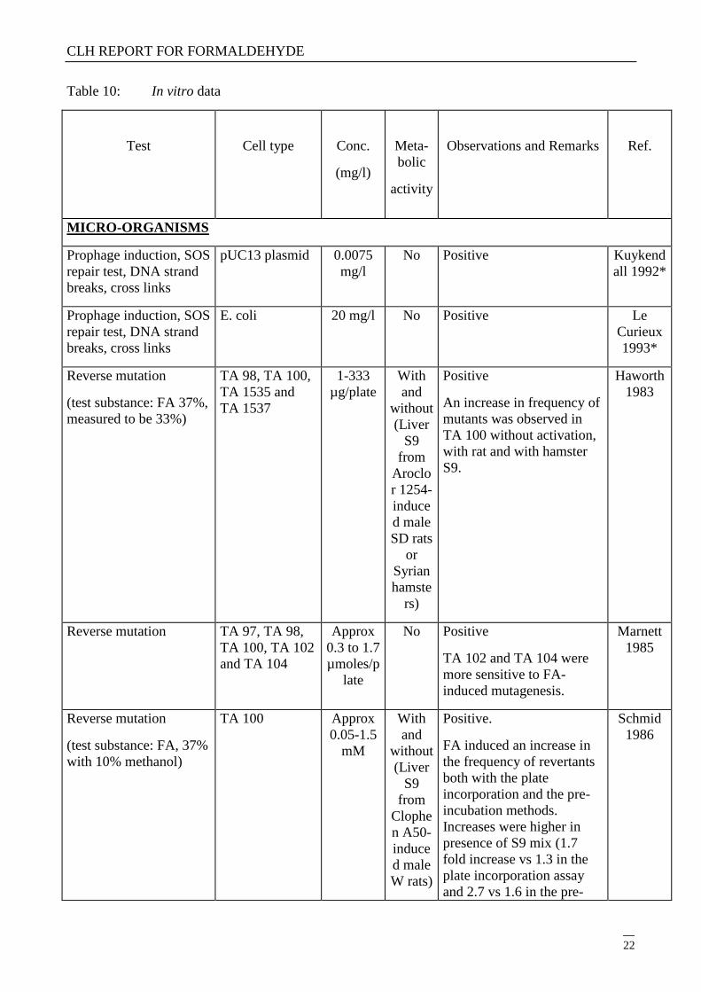
Table 10: In vitro data
Test
Cell type
Conc.
(mg/l)
Meta-
bolic
activity
Observations and Remarks
Ref.
MICRO-ORGANISMS
Prophage induction, SOS
repair test, DNA strand
breaks, cross links
pUC13 plasmid 0.0075
mg/l
No Positive Kuykend
all 1992*
Prophage induction, SOS
repair test, DNA strand
breaks, cross links
E. coli 20 mg/l No Positive Le
Curieux
1993*
Reverse mutation
(test substance: FA 37%,
measured to be 33%)
TA 98, TA 100,
TA 1535 and
TA 1537
1-333
µg/plate
With
and
without
(Liver
S9
from
Aroclo
r 1254-
induce
d male
SD rats
or
Syrian
hamste
rs)
Positive
An increase in frequency of
mutants was observed in
TA 100 without activation,
with rat and with hamster
S9.
Haworth
1983
Reverse mutation TA 97, TA 98,
TA 100, TA 102
and TA 104
Approx
0.3 to 1.7
µmoles/p
late
No Positive
TA 102 and TA 104 were
more sensitive to FA-
induced mutagenesis.
Marnett
1985
Reverse mutation
(test substance: FA, 37%
with 10% methanol)
TA 100 Approx
0.05-1.5
mM
With
and
without
(Liver
S9
from
Clophe
n A50-
induce
d male
W rats)
Positive.
FA induced an increase in
the frequency of revertants
both with the plate
incorporation and the pre-
incubation methods.
Increases were higher in
presence of S9 mix (1.7
fold increase vs 1.3 in the
plate incorporation assay
and 2.7 vs 1.6 in the pre-
Schmid
1986
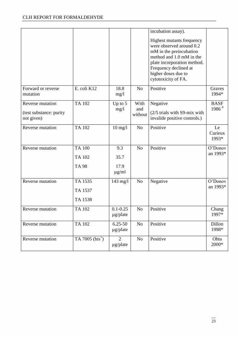
CLH REPORT FOR FORMALDEHYDE
23
incubation assay).
Highest mutants frequency
were observed around 0.2
mM in the preincubation
method and 1.0 mM in the
plate incorporation method.
Frequency declined at
higher doses due to
cytotoxicity of FA.
Forward or reverse
mutation
E. coli K12 18.8
mg/l
No Positive Graves
1994*
Reverse mutation
(test substance: purity
not given)
TA 102 Up to 5
mg/l
With
and
without
Negative
(2/5 trials with S9-mix with
invalide positive controls.)
BASF
1986 #
Reverse mutation TA 102 10 mg/l No Positive Le
Curieux
1993*
Reverse mutation TA 100
TA 102
TA 98
9.3
35.7
17.9
µg/ml
No Positive O‘Donov
an 1993*
Reverse mutation TA 1535
TA 1537
TA 1538
143 mg/l No Negative O‘Donov
an 1993*
Reverse mutation TA 102 0.1-0.25
µg/plate
No Positive Chang
1997*
Reverse mutation TA 102 6.25-50
µg/plate
No Positive Dillon
1998*
Reverse mutation TA 7005 (his+) 2
µg/plate
No Positive Ohta
2000*
CLH REPORT FOR FORMALDEHYDE
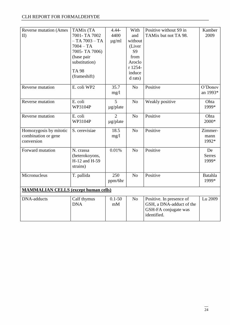
24
Reverse mutation (Ames
II)
TAMix (TA
7001- TA 7002
– TA 7003 – TA
7004 – TA
7005- TA 7006)
(base pair
substitution)
TA 98
(frameshift)
4.44-
4400
µg/ml
With
and
without
(Liver
S9
from
Aroclo
r 1254-
induce
d rats)
Positive without S9 in
TAMix but not TA 98.
Kamber
2009
Reverse mutation E. coli WP2 35.7
mg/l
No Positive O‘Donov
an 1993*
Reverse mutation E. coli
WP3104P
5
µg/plate
No Weakly positive Ohta
1999*
Reverse mutation E. coli
WP3104P
2
µg/plate
No Positive Ohta
2000*
Homozygosis by mitotic
combination or gene
conversion
S. cerevisiae 18.5
mg/l
No Positive Zimmer-
mann
1992*
Forward mutation N. crassa
(heterokoyons,
H-12 and H-59
strains)
0.01% No Positive De
Serres
1999*
Micronucleus T. pallida 250
ppm/6hr
No Positive Batahla
1999*
MAMMALIAN CELLS (except human cells)
DNA-adducts Calf thymus
DNA
0.1-50
mM
No Positive. In presence of
GSH, a DNA-adduct of the
GSH-FA conjugate was
identified.
Lu 2009
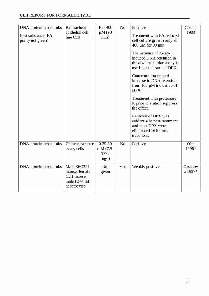
CLH REPORT FOR FORMALDEHYDE
25
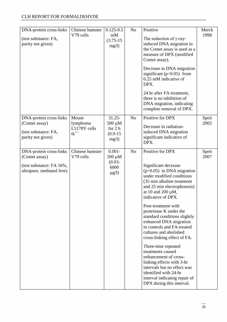
DNA-protein cross-links
(test substance: FA,
purity not given)
Rat tracheal
epithelial cell
line C18
100-400
µM (90
min)
No Positive
Treatment with FA reduced
cell culture growth only at
400 µM for 90 min.
The increase of X-ray-
induced DNA retention in
the alkaline elution assay is
used as a measure of DPX.
Concentration-related
increase in DNA retention
from 100 µM indicative of
DPX.
Treatment with proteinase
K prior to elution suppress
the effect.
Removal of DPX was
evident 4 hr post-treatment
and most DPX were
eliminated 16 hr post-
treatment.
Cosma
1988
DNA-protein cross-links Chinese hamster
ovary cells
0.25-59
mM (7.5-
1770
mg/l)
No Positive Olin
1996*
DNA-protein cross-links Male B6C3F1
mouse, female
CD1 mouse,
male F344 rat
hepatocytes
Not
given
Yes Weakly positive Casanov
a 1997*
CLH REPORT FOR FORMALDEHYDE
26
DNA-protein cross-links
(test substance: FA,
purity not given)
Chinese hamster
V79 cells
0.125-0.5
mM
(3.75-15
mg/l)
No Positive
The reduction of γ-ray-
induced DNA migration in
the Comet assay is used as a
measure of DPX (modified
Comet assay).
Decrease in DNA migration
significant (p<0.05) from
0.25 mM indicative of
DPX.
24 hr after FA treatment,
there is no inhibition of
DNA migration, indicating
complete removal of DPX.
Merck
1998
DNA-protein cross-links
(Comet assay)
(test substance: FA,
purity not given)
Mouse
lymphoma
L5178Y cells
tk+/-
31.25-
500 µM
for 2 h
(0.9-15
mg/l)
No Positive for DPX
Decrease in radiation-
induced DNA migration
significant indicative of
DPX.
Speit
2002
DNA-protein cross-links
(Comet assay)
(test substance: FA 16%,
ultrapure, methanol free)
Chinese hamster
V79 cells
0.001-
200 µM
(0.03-
6000
µg/l)
No Positive for DPX
Significant decrease
(p<0.05) in DNA migration
under modified conditions
(35 min alkaline treatment
and 25 min electrophoresis)
at 10 and 200 µM,
indicative of DPX.
Post-treatment with
proteinase K under the
standard conditions slightly
enhanced DNA migration
in controls and FA-treated
cultures and abolished
cross-linking effect of FA.
Three-time repeated
treatments caused
enhancement of cross-
linking effects with 3-hr
intervals but no effect was
identified with 24-hr
interval indicating repair of
DPX during this interval.
Speit
2007
CLH REPORT FOR FORMALDEHYDE
27
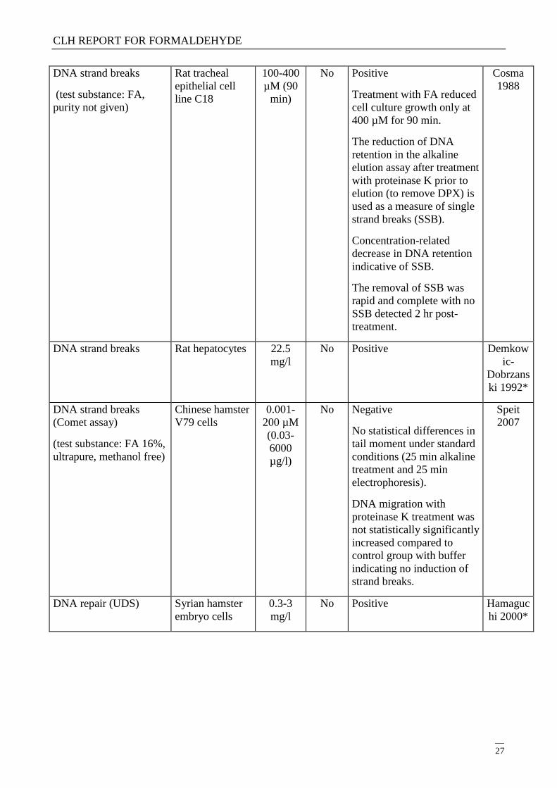
DNA strand breaks
(test substance: FA,
purity not given)
Rat tracheal
epithelial cell
line C18
100-400
µM (90
min)
No Positive
Treatment with FA reduced
cell culture growth only at
400 µM for 90 min.
The reduction of DNA
retention in the alkaline
elution assay after treatment
with proteinase K prior to
elution (to remove DPX) is
used as a measure of single
strand breaks (SSB).
Concentration-related
decrease in DNA retention
indicative of SSB.
The removal of SSB was
rapid and complete with no
SSB detected 2 hr post-
treatment.
Cosma
1988
DNA strand breaks Rat hepatocytes 22.5
mg/l
No Positive Demkow
ic-
Dobrzans
ki 1992*
DNA strand breaks
(Comet assay)
(test substance: FA 16%,
ultrapure, methanol free)
Chinese hamster
V79 cells
0.001-
200 µM
(0.03-
6000
µg/l)
No Negative
No statistical differences in
tail moment under standard
conditions (25 min alkaline
treatment and 25 min
electrophoresis).
DNA migration with
proteinase K treatment was
not statistically significantly
increased compared to
control group with buffer
indicating no induction of
strand breaks.
Speit
2007
DNA repair (UDS) Syrian hamster
embryo cells
0.3-3
mg/l
No Positive Hamaguc
hi 2000*
CLH REPORT FOR FORMALDEHYDE
28
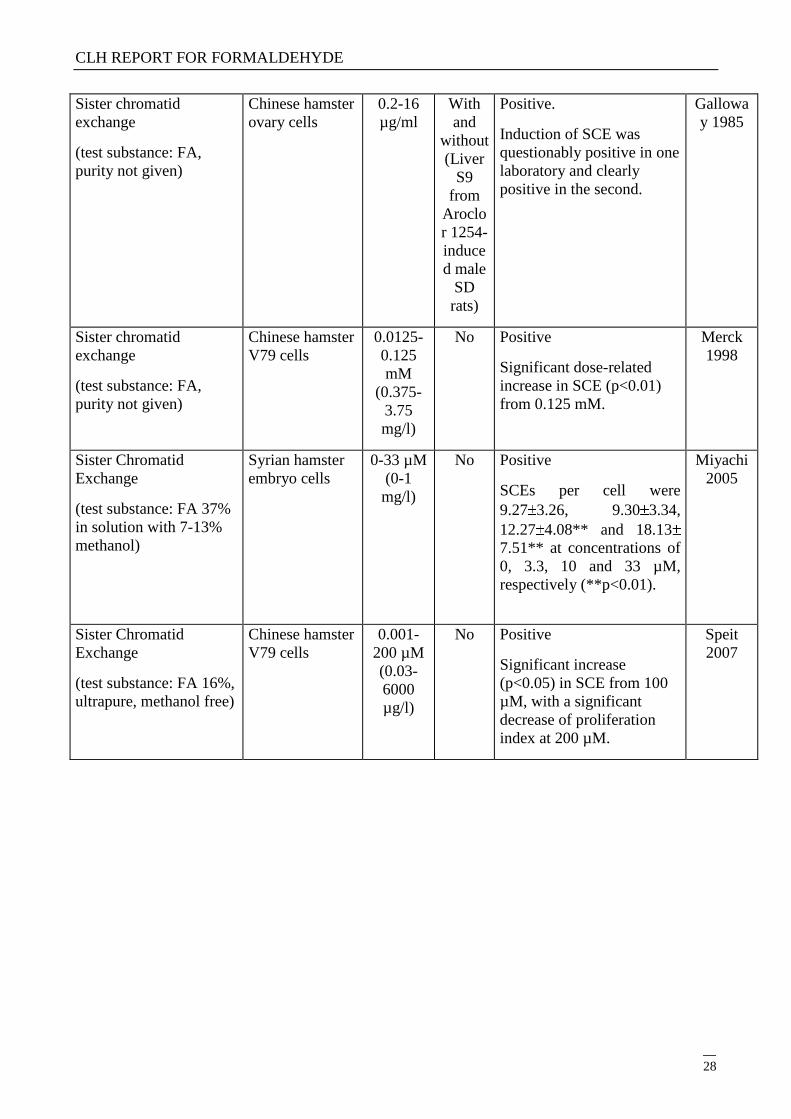
Sister chromatid
exchange
(test substance: FA,
purity not given)
Chinese hamster
ovary cells
0.2-16
µg/ml
With
and
without
(Liver
S9
from
Aroclo
r 1254-
induce
d male
SD
rats)
Positive.
Induction of SCE was
questionably positive in one
laboratory and clearly
positive in the second.
Gallowa
y 1985
Sister chromatid
exchange
(test substance: FA,
purity not given)
Chinese hamster
V79 cells
0.0125-
0.125
mM
(0.375-
3.75
mg/l)
No Positive
Significant dose-related
increase in SCE (p<0.01)
from 0.125 mM.
Merck
1998
Sister Chromatid
Exchange
(test substance: FA 37%
in solution with 7-13%
methanol)
Syrian hamster
embryo cells
0-33 µM
(0-1
mg/l)
No Positive
SCEs per cell were
9.27 3.26, 9.30 3.34,
12.27 4.08** and 18.13
7.51** at concentrations of
0, 3.3, 10 and 33 µM,
respectively (**p<0.01).
Miyachi
2005
Sister Chromatid
Exchange
(test substance: FA 16%,
ultrapure, methanol free)
Chinese hamster
V79 cells
0.001-
200 µM
(0.03-
6000
µg/l)
No Positive
Significant increase
(p<0.05) in SCE from 100
µM, with a significant
decrease of proliferation
index at 200 µM.
Speit
2007
CLH REPORT FOR FORMALDEHYDE
29
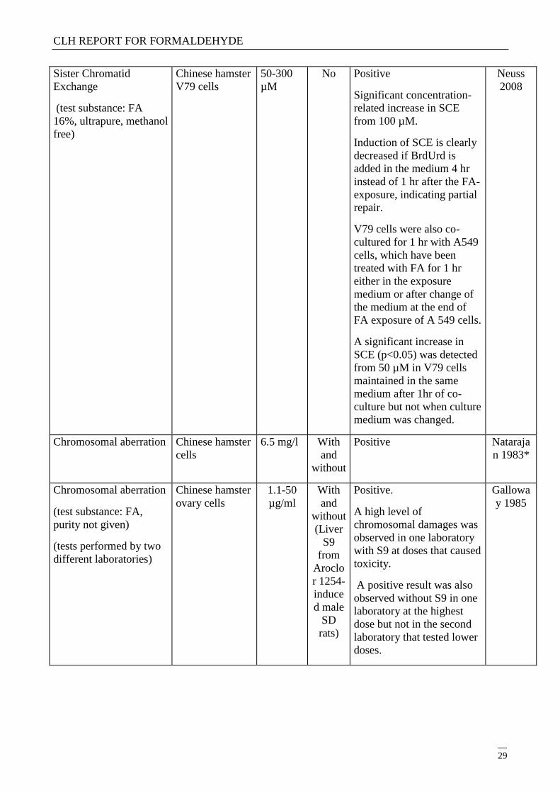
Sister Chromatid
Exchange
(test substance: FA
16%, ultrapure, methanol
free)
Chinese hamster
V79 cells
50-300
µM
No Positive
Significant concentration-
related increase in SCE
from 100 µM.
Induction of SCE is clearly
decreased if BrdUrd is
added in the medium 4 hr
instead of 1 hr after the FA-
exposure, indicating partial
repair.
V79 cells were also co-
cultured for 1 hr with A549
cells, which have been
treated with FA for 1 hr
either in the exposure
medium or after change of
the medium at the end of
FA exposure of A 549 cells.
A significant increase in
SCE (p<0.05) was detected
from 50 µM in V79 cells
maintained in the same
medium after 1hr of co-
culture but not when culture
medium was changed.
Neuss
2008
Chromosomal aberration Chinese hamster
cells
6.5 mg/l With
and
without
Positive Nataraja
n 1983*
Chromosomal aberration
(test substance: FA,
purity not given)
(tests performed by two
different laboratories)
Chinese hamster
ovary cells
1.1-50
µg/ml
With
and
without
(Liver
S9
from
Aroclo
r 1254-
induce
d male
SD
rats)
Positive.
A high level of
chromosomal damages was
observed in one laboratory
with S9 at doses that caused
toxicity.
A positive result was also
observed without S9 in one
laboratory at the highest
dose but not in the second
laboratory that tested lower
doses.
Gallowa
y 1985
CLH REPORT FOR FORMALDEHYDE
30
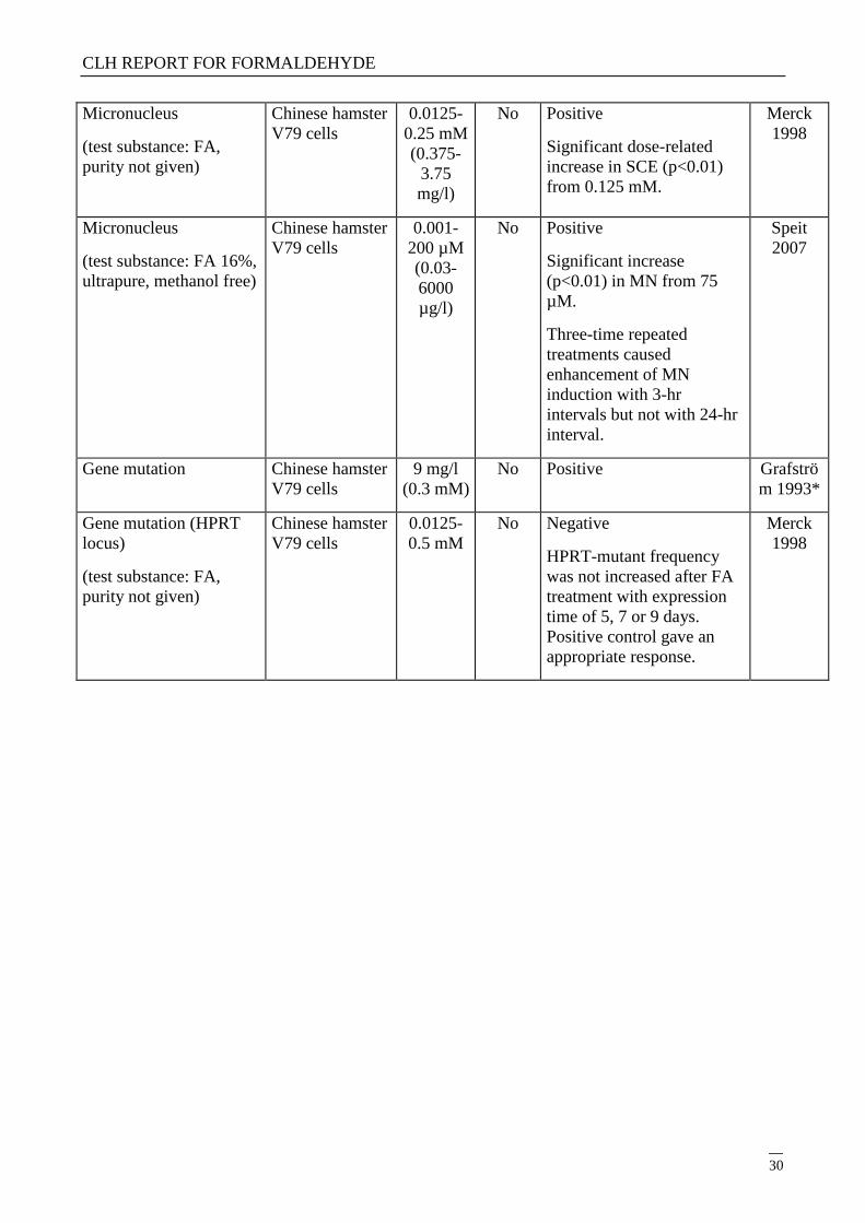
Micronucleus
(test substance: FA,
purity not given)
Chinese hamster
V79 cells
0.0125-
0.25 mM
(0.375-
3.75
mg/l)
No Positive
Significant dose-related
increase in SCE (p<0.01)
from 0.125 mM.
Merck
1998
Micronucleus
(test substance: FA 16%,
ultrapure, methanol free)
Chinese hamster
V79 cells
0.001-
200 µM
(0.03-
6000
µg/l)
No Positive
Significant increase
(p<0.01) in MN from 75
µM.
Three-time repeated
treatments caused
enhancement of MN
induction with 3-hr
intervals but not with 24-hr
interval.
Speit
2007
Gene mutation Chinese hamster
V79 cells
9 mg/l
(0.3 mM)
No Positive Grafströ
m 1993*
Gene mutation (HPRT
locus)
(test substance: FA,
purity not given)
Chinese hamster
V79 cells
0.0125-
0.5 mM
No Negative
HPRT-mutant frequency
was not increased after FA
treatment with expression
time of 5, 7 or 9 days.
Positive control gave an
appropriate response.
Merck
1998
CLH REPORT FOR FORMALDEHYDE
31
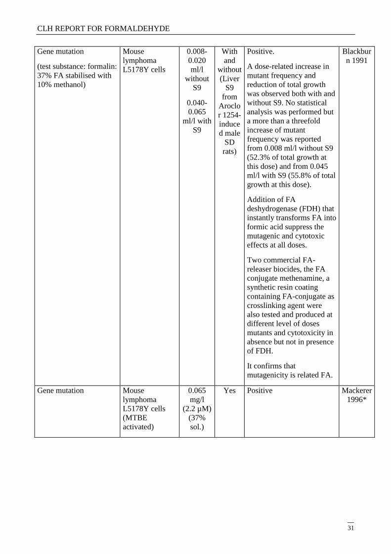
Gene mutation
(test substance: formalin:
37% FA stabilised with
10% methanol)
Mouse
lymphoma
L5178Y cells
0.008-
0.020
ml/l
without
S9
0.040-
0.065
ml/l with
S9
With
and
without
(Liver
S9
from
Aroclo
r 1254-
induce
d male
SD
rats)
Positive.
A dose-related increase in
mutant frequency and
reduction of total growth
was observed both with and
without S9. No statistical
analysis was performed but
a more than a threefold
increase of mutant
frequency was reported
from 0.008 ml/l without S9
(52.3% of total growth at
this dose) and from 0.045
ml/l with S9 (55.8% of total
growth at this dose).
Addition of FA
deshydrogenase (FDH) that
instantly transforms FA into
formic acid suppress the
mutagenic and cytotoxic
effects at all doses.
Two commercial FA-
releaser biocides, the FA
conjugate methenamine, a
synthetic resin coating
containing FA-conjugate as
crosslinking agent were
also tested and produced at
different level of doses
mutants and cytotoxicity in
absence but not in presence
of FDH.
It confirms that
mutagenicity is related FA.
Blackbur
n 1991
Gene mutation Mouse
lymphoma
L5178Y cells
(MTBE
activated)
0.065
mg/l
(2.2 µM)
(37%
sol.)
Yes Positive Mackerer
1996*
CLH REPORT FOR FORMALDEHYDE
32
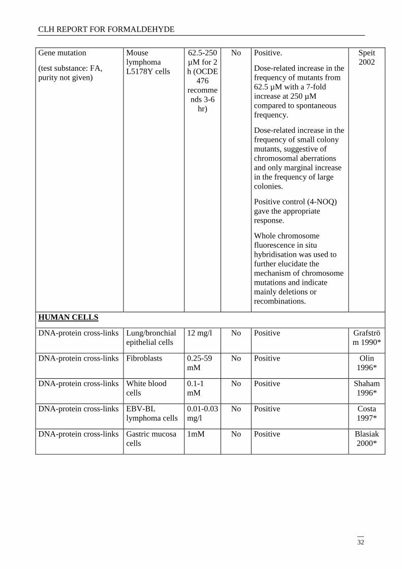
Gene mutation
(test substance: FA,
purity not given)
Mouse
lymphoma
L5178Y cells
62.5-250
µM for 2
h (OCDE
476
recomme
nds 3-6
hr)
No Positive.
Dose-related increase in the
frequency of mutants from
62.5 µM with a 7-fold
increase at 250 µM
compared to spontaneous
frequency.
Dose-related increase in the
frequency of small colony
mutants, suggestive of
chromosomal aberrations
and only marginal increase
in the frequency of large
colonies.
Positive control (4-NOQ)
gave the appropriate
response.
Whole chromosome
fluorescence in situ
hybridisation was used to
further elucidate the
mechanism of chromosome
mutations and indicate
mainly deletions or
recombinations.
Speit
2002
HUMAN CELLS
DNA-protein cross-links Lung/bronchial
epithelial cells
12 mg/l No Positive Grafströ
m 1990*
DNA-protein cross-links Fibroblasts 0.25-59
mM
No Positive Olin
1996*
DNA-protein cross-links White blood
cells
0.1-1
mM
No Positive Shaham
1996*
DNA-protein cross-links EBV-BL
lymphoma cells
0.01-0.03
mg/l
No Positive Costa
1997*
DNA-protein cross-links Gastric mucosa
cells
1mM No Positive Blasiak
2000*
CLH REPORT FOR FORMALDEHYDE
33
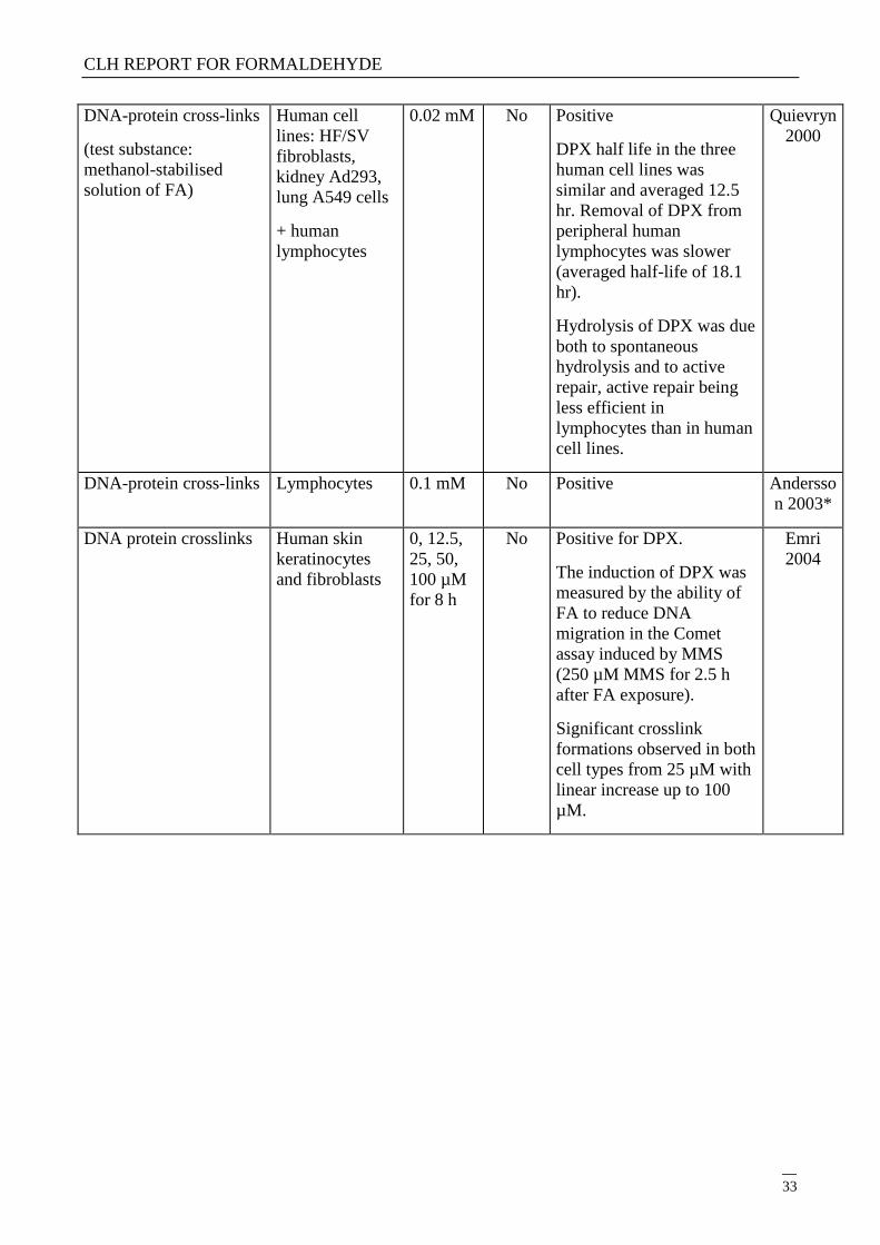
DNA-protein cross-links
(test substance:
methanol-stabilised
solution of FA)
Human cell
lines: HF/SV
fibroblasts,
kidney Ad293,
lung A549 cells
+ human
lymphocytes
0.02 mM No Positive
DPX half life in the three
human cell lines was
similar and averaged 12.5
hr. Removal of DPX from
peripheral human
lymphocytes was slower
(averaged half-life of 18.1
hr).
Hydrolysis of DPX was due
both to spontaneous
hydrolysis and to active
repair, active repair being
less efficient in
lymphocytes than in human
cell lines.
Quievryn
2000
DNA-protein cross-links Lymphocytes 0.1 mM No Positive Andersso
n 2003*
DNA protein crosslinks Human skin
keratinocytes
and fibroblasts
0, 12.5,
25, 50,
100 µM
for 8 h
No Positive for DPX.
The induction of DPX was
measured by the ability of
FA to reduce DNA
migration in the Comet
assay induced by MMS
(250 µM MMS for 2.5 h
after FA exposure).
Significant crosslink
formations observed in both
cell types from 25 µM with
linear increase up to 100
µM.
Emri
2004
CLH REPORT FOR FORMALDEHYDE
34
DNA-protein cross-links,
repair (Comet)
Test substance: 10%
formalin
Human
peripheral blood
lymphocytes (1
sample) and
Hela cell lines
5-625
µM
No Positive
No significant increase in
DPX coefficient at 5 and 25
µM but significant dose-
related increase at
concentration 50 µM in
both human peripheral
lymphocytes and Hela cell
lines.
In Hela cell lines at the non
cytotoxic concentration of
50 µM, a statistically
significant decrease in DPX
coefficient was observed
when FA was removed
from cell culture for 18
hr, indicating progressive
repair of DPX.
Liu 2006
DNA-protein cross-links
(Comet assay)
(test substance: FA
16%, ultrapure, methanol
free)
Human blood
samples
25-300
µM
No Positive
Significant concentration-
related decrease (p<0.05) in
gamma ray ( 2 Gy) induced
DNA migration from 25
µM, indicating induction of
DPX.
When cells are irradiated at
different time points after
treatments, reduction of
gamma ray induced DNA
migration decreased with
time. At 100µM, DPX are
completely removed after 8
hr, while a portion of DPX
still persists after 24 hr at
200 and 300 µM.
Schmid
2007
CLH REPORT FOR FORMALDEHYDE
35
DNA-protein cross-links
(Comet assay)
(test substance: FA
16%, ultrapure, methanol
free)
A549 epithelia-
like human lung
cell lines and
human nasal
epithelial cells
100-300
µM
No Positive
A concentration-related
induction of DPX was
induced in A549 cells after
treatment for 1 or 4 hr.
After 4 hr incubation in
fresh medium, a reduction
of the crosslinking effect is
was seen and complete
removal after 8 hr.
A concentration-related
induction of DPX was
induced in human nasal
epithelium cells after
treatment for 1 hr. After 4
hr incubation in fresh
medium, a reduction of the
crosslinking effect was seen
and DNA migration was not
significantly decreased after
8 hr in fresh medium.
Speit
2008
DNA-protein cross-links,
repair
Test substance: 10%
formalin
HepG2 cells
(human liver
carcinoma cell
line)
25-50-
75-100
µM for 1
hr
Repair
experime
nt: 75
µM for 1
hr (+0, 6,
12, 18,
24h of
incubatio
n after
removal
of FA)
No Positive
Significant dose-related
increase of the DPX
coefficient at concentration
75 µM.
In the repair experiment,
the DPX coefficient was
significantly decreased and
similar to control after 18 hr
or more.
DPX coefficient was
determined as the ratio of
the percentage of the DNA
involved in DPX over the
percentage of the DNA
involved in DPX +
unbound fraction of DNA
Zhao
2009
CLH REPORT FOR FORMALDEHYDE
36
DNA-protein cross-links
(Comet assay)
(test substance: FA
16%, ultrapure, methanol
free)
A549 epithelia-
like human lung
cell lines
50-300
µM
No Positive
A concentration-related
induction of DPX was
induced in A549 cells after
treatment for 1 hr,
significant at 200 µM and
above. With three repeated
1-hr exposures with 24-hr
or 48-hr intervals, the
crosslinking effect of FA
was clearly enhanced at 200
and 300 µM.
Preexposure to low level of
FA-concentrations (50 µM)
does not influence the
crosslinking effect of a high
FA-concentration or DPX
removal.
Speit
2010
DNA-protein cross-links
(Comet assay)
(test substance: FA
16%, ultrapure, methanol
free)
Primary human
nasal epithelial
cells (HNEC)
from 3 women
100-200
µM
No Positive for DPX
A concentration-related
induction of DPX was
induced in HNEC cells after
treatment for 1 hr,
significant from 100 µM.
After 4 hr incubation in
fresh medium, a reduction
of the crosslinking effect
was seen and DNA
migration was not
significantly decreased after
8 hr in fresh medium.
Neuss,
2010a
CLH REPORT FOR FORMALDEHYDE
37
DNA-protein cross-links
(Comet assay)
(test substance: FA
16%, ultrapure, methanol
free)
Primary human
nasal epithelial
cells (HNEC)
and human
lymphocytes
100-300
µM
No Positive in lymphocytes and
HNEC directly exposed to
FA, negative in
lymphocytes co-cultured
with exposed HNEC in
absence of FA in the
medium.
In lymphocytes treated for 1
hr, significant
concentration-related
decrease (p<0.05) in
gamma ray (2 Gy) induced
DNA migration from 100
µM, indicating induction of
DPX.
Lymphocytes were co-
cultured for 1 or 4 hr with
HNEC, which have been
treated with FA for 1 hr,
either in the exposure
medium or after change of
the medium at the end of
FA exposure of HNEC.
A significant concentration-
related decrease (p<0.05) in
gamma ray induced DNA
migration was detected
from 100 µM in HNEC
exposed for 1 hr and
maintained in the same
medium after 4 hr of co-
culture. Only a slight cross-
linking effect was detected
when the exposure medium
was removed for co-
cultivation for 4 hr.
A significant concentration-
related decrease (p<0.05) in
gamma ray induced DNA
migration was detected
from 100 µM in
lymphocytes maintained in
the same medium after both
1hr or 4 hr of co-culture.
FA concentration was
measured to decrease with
time in the presence of cells
with around 75% of the
initial concentration
measured after 4 hr at 100
µM.
Neuss
2010b
CLH REPORT FOR FORMALDEHYDE
38
No significant effect was
detected in lymphocytes co-
cultured with HNEC when
the medium was changed
before co-cultivation. No
significantly increased
amounts of FA were
detectable in the new
medium after 5, 15, 30 min,
and 1, 4 or 8 hr
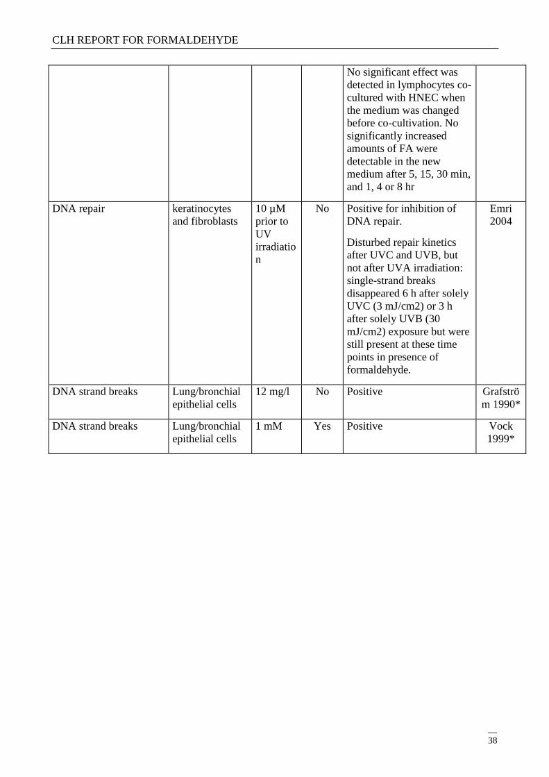
DNA repair keratinocytes
and fibroblasts
10 µM
prior to
UV
irradiatio
n
No Positive for inhibition of
DNA repair.
Disturbed repair kinetics
after UVC and UVB, but
not after UVA irradiation:
single-strand breaks
disappeared 6 h after solely
UVC (3 mJ/cm2) or 3 h
after solely UVB (30
mJ/cm2) exposure but were
still present at these time
points in presence of
formaldehyde.
Emri
2004
DNA strand breaks Lung/bronchial
epithelial cells
12 mg/l No Positive Grafströ
m 1990*
DNA strand breaks Lung/bronchial
epithelial cells
1 mM Yes Positive Vock
1999*
CLH REPORT FOR FORMALDEHYDE
39
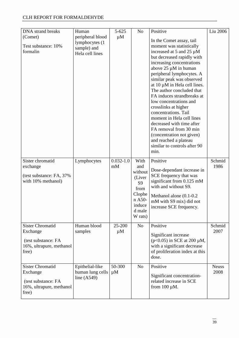
DNA strand breaks
(Comet)
Test substance: 10%
formalin
Human
peripheral blood
lymphocytes (1
sample) and
Hela cell lines
5-625
µM
No Positive
In the Comet assay, tail
moment was statistically
increased at 5 and 25 µM
but decreased rapidly with
increasing concentrations
above 25 µM in human
peripheral lymphocytes. A
similar peak was observed
at 10 µM in Hela cell lines.
The author concluded that
FA induces strandbreaks at
low concentrations and
crosslinks at higher
concentrations. Tail
moment in Hela cell lines
decreased with time after
FA removal from 30 min
(concentration not given)
and reached a plateau
similar to controls after 90
min.
Liu 2006
Sister chromatid
exchange
(test substance: FA, 37%
with 10% methanol)
Lymphocytes 0.032-1.0
mM
With
and
without
(Liver
S9
from
Clophe
n A50-
induce
d male
W rats)
Positive
Dose-dependant increase in
SCE frequency that was
significant from 0.125 mM
with and without S9.
Methanol alone (0.1-0.2
mM with S9 mix) did not
increase SCE frequency.
Schmid
1986
Sister Chromatid
Exchange
(test substance: FA
16%, ultrapure, methanol
free)
Human blood
samples
25-200
µM
No Positive
Significant increase
(p<0.05) in SCE at 200 µM,
with a significant decrease
of proliferation index at this
dose.
Schmid
2007
Sister Chromatid
Exchange
(test substance: FA
16%, ultrapure, methanol
free)
Epithelial-like
human lung cells
line (A549)
50-300
µM
No Positive
Significant concentration-
related increase in SCE
from 100 µM.
Neuss
2008
CLH REPORT FOR FORMALDEHYDE
40
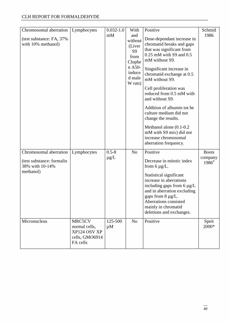
Chromosomal aberration
(test substance: FA, 37%
with 10% methanol)
Lymphocytes 0.032-1.0
mM
With
and
without
(Liver
S9
from
Clophe
n A50-
induce
d male
W rats)
Positive
Dose-dependant increase in
chromatid breaks and gaps
that was significant from
0.25 mM with S9 and 0.5
mM without S9.
Singnificant increase in
chromatid exchange at 0.5
mM without S9.
Cell proliferation was
reduced from 0.5 mM with
and without S9.
Addition of albumin tot he
culture medium did not
change the results.
Methanol alone (0.1-0.2
mM with S9 mix) did not
increase chromosomal
aberration frequency.
Schmid
1986
Chromosomal aberration
(test substance: formalin
38% with 10-14%
methanol)
Lymphocytes 0.5-8
µg/L
No Positive
Decrease in mitotic index
from 6 µg/L.
Statistical significant
increase in aberrations
including gaps from 6 µg/L
and in aberration excluding
gaps from 8 µg/L.
Aberrations consisted
mainly in chromatid
deletions and exchanges.
Boots
company
1986#
Micronucleus MRC5CV
normal cells,
XP124 OSV XP
cells, GMO6914
FA cells
125-500
µM
No Positive Speit
2000*
CLH REPORT FOR FORMALDEHYDE
41
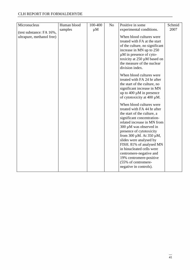
Micronucleus
(test substance: FA 16%,
ultrapure, methanol free)
Human blood
samples
100-400
µM
No Positive in some
experimental conditions.
When blood cultures were
treated with FA at the start
of the culture, no significant
increase in MN up to 250
µM in presence of cyto-
toxicity at 250 µM based on
the measure of the nuclear
division index.
When blood cultures were
treated with FA 24 hr after
the start of the culture, no
significant increase in MN
up to 400 µM in presence
of cytotoxicity at 400 µM.
When blood cultures were
treated with FA 44 hr after
the start of the culture, a
significant concentration-
related increase in MN from
300 µM was observed in
presence of cytotoxicity
from 300 µM. At 350 µM,
slides were analysed by
FISH. 81% of analysed MN
in binucleated cells were
centromere-negative and
19% centromere-positive
(55% of centromere-
negative in controls).
Schmid
2007
CLH REPORT FOR FORMALDEHYDE
42
Gene mutation (HPRT
locus)
TK6 human
lymphoblast
150 µM
(8
sequentia
l
exposure
s of 2 hr)
No Positive
Treatment with FA induced
a mutant frequency of
23x10-6
(12-fold higher
than controls).
30 mutants were analysed
by Northen and Southern
blot. 6/30 mutants had
completely lost the hprt
gene. 8/30 had partial
deletion of the gene DNA.
None of these mutants
produced RNAm. 16/30
mutants had point mutation
(no visible alteration with
southern blot). RNAm of 6
of these mutants contained
a single base-pair
substitution at AT base
pairs and 4 at the same site.
The remaining mutant was
lacking exon 8. In
comparison with
spontaneous mutations FA
lead to a shift from point
mutations in favour of
complete deletion.
Liber
1989
Gene mutation (HPRT
locus)
Bronchial
fibroblast/epithel
ial cells
3 mg/l No Positive Grafströ
m 1990*
CLH REPORT FOR FORMALDEHYDE
43
Microarray analyses
(test substance: FA
16%, ultrapure, methanol
free)
Primary human
nasal epithelial
cells (HNEC)
from 3 women
50-100
µM for
2h
50-200
µM for 4
h
100-200
µM for
24 h
4 x 20-50
µM with
24 h
intervals
No A two-fold variation in the
expression of 153 and 887
genes was observed at 100
µM and 200 µM for 4 h,
respectively. No significant
effect was seen with
treatment for 2 h or for 24
h. Repeated treatments with
50 µM changed gene
expression of 143 genes.
Genes up-regulated
involved most frequently
the biological processes of
―transcription‖,
―translation‖, ―nucleosome
assembly‖ and ―negative
regulation of transcription
from RNA polymerase II
promoter‖. The expression
of genes involved in FA
detoxification and DNA
repair were not significantly
altered.
Neuss,
2010a
CLH REPORT FOR FORMALDEHYDE
44
4.9.1.2 In vivo data
4.9.1.2.1 Somatic cells at sites of contact
Table 11: Experimental in vivo data at the site of contact
Test Species Tissue Exposure route &
Harvest time
Observations and remarks Ref
DNA adducts
(test substance:
heated
radiolabelled
paraformalde-
hyde – purity not
specified)
Fischer
344 rats
(male)
(n=5/8)
Nasal
respiratory
epithelium
Inhalation: 10 ppm
for 6 hr or 5 days
(6hr/d)
(nose-only)
Positive
Detection of N2-hydroxy-
methyl-dG adducts:
- Endogenous: detected
after both 1 or 5 days
of exposure (2.84±
1.13 at 5 days)
- Exogenous: detected
after both 1 or 5 days
of exposure (1.28±
0.49 at 1 day and
2.43± 0.78 at 5 days)
Detection of N6-hydroxy-
methyl-dA adducts:
- Endogenous: detected
after both 1 or 5 days
of exposure (3.61±
0.95 at 5 days)
- Exogenous: not
detected.
Detection of dG-CH2-dG
crosslinks:
- Endogenous: detected
after both 1 or 5 days
of exposure (0.18±
0.06 at 5 days)
- Exogenous: detected
after both 1 or 5 days
of exposure (0.14±
0.06 at 1 day and
0.26± 0.07 at 5 days)
Lu 2010
CLH REPORT FOR FORMALDEHYDE
45
Test Species Tissue Exposure route &
Harvest time
Observations and remarks Ref
DNA adducts
(test substance:
radiolabelled
formaldehyde –
purity not
specified)
Rats
(n=3-
5/group)
Bone
marrow
Inhalation: 0.7,
2.0, 5.8, 9.1 or
15.2 ppm for 6hr
Positive
Detection of N2-hydroxy-
methyl-dG adducts:
- Endogenous: detected
(similar levels across
groups; mean: 4.7± 1.8
adducts/107 dG)
- Exogenous: detected at
all concentrations:
0.04±0.02, 0.19±0.08,
1.04±0.24, 2.03±0.43,
and 11.15±3.01
adducts/ 107 dG at 0.7,
2.0, 5.8, 9.1, and 15.2
ppm)
Lu 2011
DNA adducts
(test substance:
radiolabelled
formaldehyde –
purity not
specified)
Cynomol
gus
macaque
(n=4/grou
p)
Nasal
maxilloturb
inate
Inhalation: 1.9 or
6.1 ppm for 2 days
(6hr/d)
(whole body)
Positive
Detection of N2-hydroxy-
methyl-dG adducts:
- Endogenous: detected
(2.05±0.54
adducts/107 dG at 6.1
ppm)
- Exogenous: detected at
both concentrations
(0.26± 0.04 at 1.9 ppm
and 0.41± 0.05 at 6.1
ppm)
Moeller
2011
DNA-protein
cross-links
Rats Nasal
respiratory
mucosa
Inhalation: 0.3,
0.7, 2, 6, or 10
ppm for 6 hr
Positive
At 6 ppm, 14
C radioactivity
was detected in DNA.
Approximately 91% was
attributed to metabolic
incorporation (by analysis of
the 3H/
14C ratio). Additional
radioactivity was attributed to
the formation of DPX.
In the dosimetry experiment,
DPX were detected at all
concentrations from 0.3 ppm.
Casanova
1989
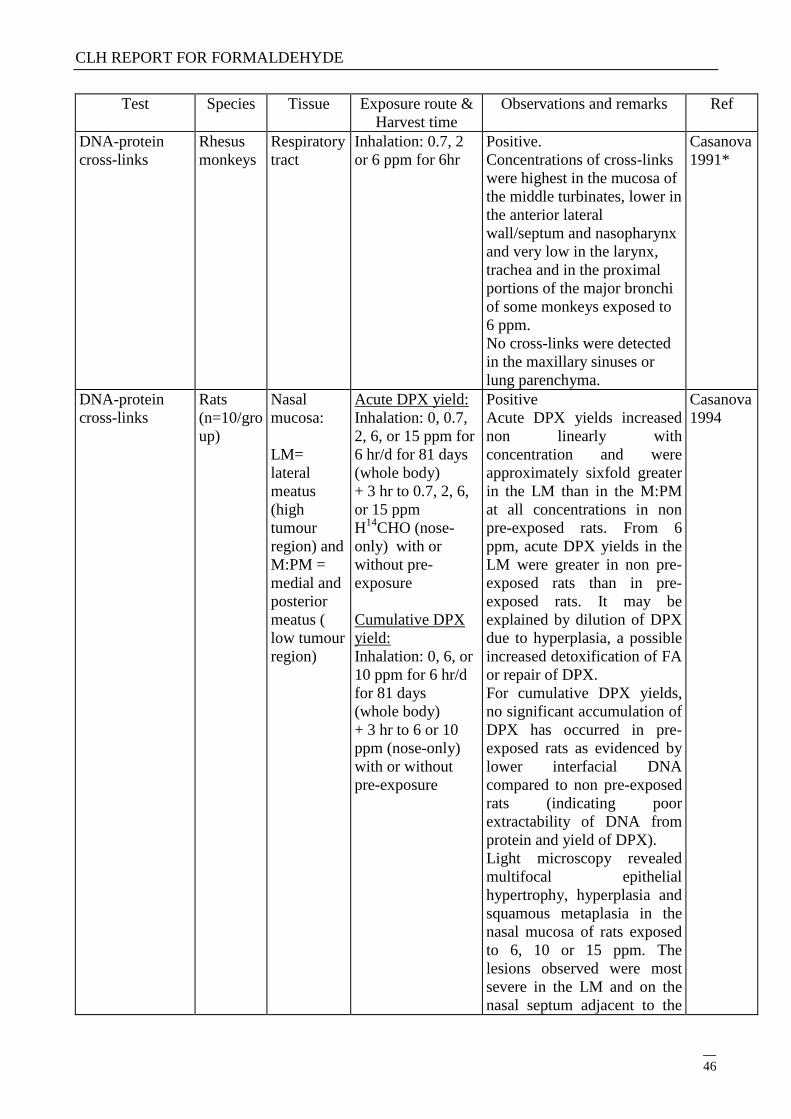
CLH REPORT FOR FORMALDEHYDE
46
Test Species Tissue Exposure route &
Harvest time
Observations and remarks Ref
DNA-protein
cross-links
Rhesus
monkeys
Respiratory
tract
Inhalation: 0.7, 2
or 6 ppm for 6hr
Positive.
Concentrations of cross-links
were highest in the mucosa of
the middle turbinates, lower in
the anterior lateral
wall/septum and nasopharynx
and very low in the larynx,
trachea and in the proximal
portions of the major bronchi
of some monkeys exposed to
6 ppm.
No cross-links were detected
in the maxillary sinuses or
lung parenchyma.
Casanova
1991*
DNA-protein
cross-links
Rats
(n=10/gro
up)
Nasal
mucosa:
LM=
lateral
meatus
(high
tumour
region) and
M:PM =
medial and
posterior
meatus (
low tumour
region)
Acute DPX yield:
Inhalation: 0, 0.7,
2, 6, or 15 ppm for
6 hr/d for 81 days
(whole body)
+ 3 hr to 0.7, 2, 6,
or 15 ppm
H14
CHO (nose-
only) with or
without pre-
exposure
Cumulative DPX
yield:
Inhalation: 0, 6, or
10 ppm for 6 hr/d
for 81 days
(whole body)
+ 3 hr to 6 or 10
ppm (nose-only)
with or without
pre-exposure
Positive
Acute DPX yields increased
non linearly with
concentration and were
approximately sixfold greater
in the LM than in the M:PM
at all concentrations in non
pre-exposed rats. From 6
ppm, acute DPX yields in the
LM were greater in non pre-
exposed rats than in pre-
exposed rats. It may be
explained by dilution of DPX
due to hyperplasia, a possible
increased detoxification of FA
or repair of DPX.
For cumulative DPX yields,
no significant accumulation of
DPX has occurred in pre-
exposed rats as evidenced by
lower interfacial DNA
compared to non pre-exposed
rats (indicating poor
extractability of DNA from
protein and yield of DPX).
Light microscopy revealed
multifocal epithelial
hypertrophy, hyperplasia and
squamous metaplasia in the
nasal mucosa of rats exposed
to 6, 10 or 15 ppm. The
lesions observed were most
severe in the LM and on the
nasal septum adjacent to the
Casanova
1994
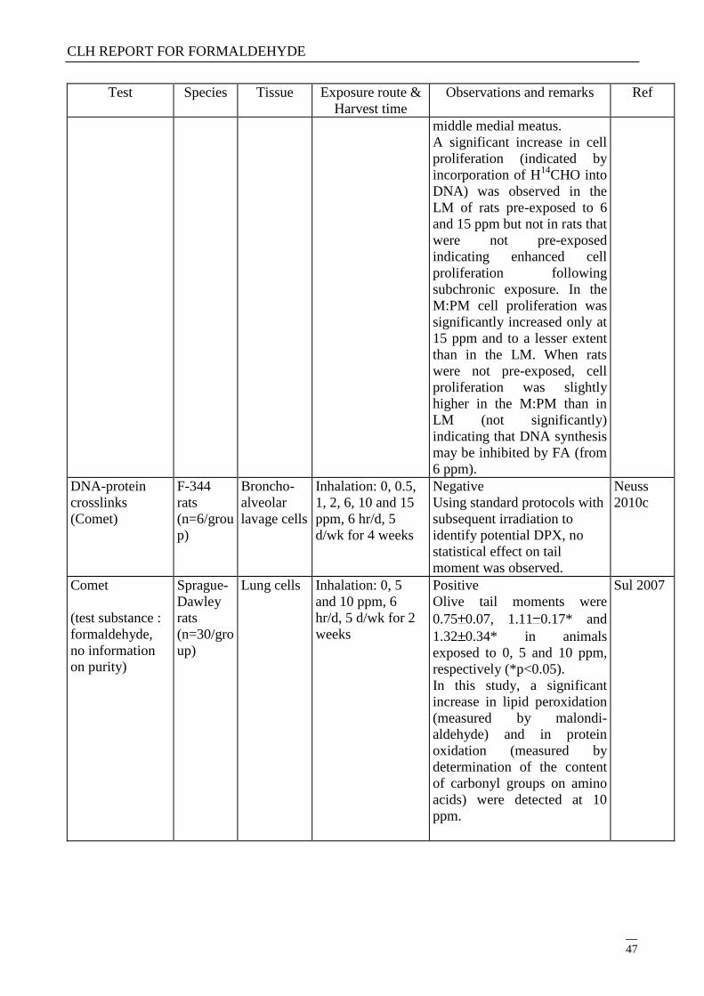
CLH REPORT FOR FORMALDEHYDE
47
Test Species Tissue Exposure route &
Harvest time
Observations and remarks Ref
middle medial meatus.
A significant increase in cell
proliferation (indicated by
incorporation of H14
CHO into
DNA) was observed in the
LM of rats pre-exposed to 6
and 15 ppm but not in rats that
were not pre-exposed
indicating enhanced cell
proliferation following
subchronic exposure. In the
M:PM cell proliferation was
significantly increased only at
15 ppm and to a lesser extent
than in the LM. When rats
were not pre-exposed, cell
proliferation was slightly
higher in the M:PM than in
LM (not significantly)
indicating that DNA synthesis
may be inhibited by FA (from
6 ppm).
DNA-protein
crosslinks
(Comet)
F-344
rats
(n=6/grou
p)
Broncho-
alveolar
lavage cells
Inhalation: 0, 0.5,
1, 2, 6, 10 and 15
ppm, 6 hr/d, 5
d/wk for 4 weeks
Negative
Using standard protocols with
subsequent irradiation to
identify potential DPX, no
statistical effect on tail
moment was observed.
Neuss
2010c
Comet
(test substance :
formaldehyde,
no information
on purity)
Sprague-
Dawley
rats
(n=30/gro
up)
Lung cells Inhalation: 0, 5
and 10 ppm, 6
hr/d, 5 d/wk for 2
weeks
Positive
Olive tail moments were
0.75 0.07, 1.11 0.17* and
1.32 0.34* in animals
exposed to 0, 5 and 10 ppm,
respectively (*p<0.05).
In this study, a significant
increase in lipid peroxidation
(measured by malondi-
aldehyde) and in protein
oxidation (measured by
determination of the content
of carbonyl groups on amino
acids) were detected at 10
ppm.
Sul 2007
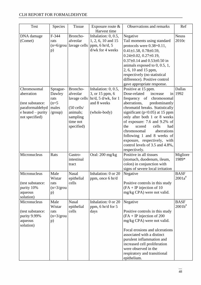
CLH REPORT FOR FORMALDEHYDE
48
Test Species Tissue Exposure route &
Harvest time
Observations and remarks Ref
DNA damage
(Comet)
F-344
rats
(n=6/grou
p)
Broncho-
alveolar
lavage cells
Inhalation: 0, 0.5,
1, 2, 6, 10 and 15
ppm, 6 hr/d, 5
d/wk for 4 weeks
Negative
Tail moments using standard
protocols were 0.38 0.11,
0.41 1.58, 0.78 0.59,
0.24 0.02, 0.27 0.19,
0.37 0.14 and 0.53 0.50 in
animals exposed to 0, 0.5, 1,
2, 6, 10 and 15 ppm,
respectively (no statistical
difference). Positive control
gave appropriate response.
Neuss
2010c
Chromosomal
aberration
(test substance:
paraformaldehyd
e heated – purity
not specified)
Sprague-
Dawley
rats
(n=5
males
/group)
Broncho-
alveolar
lavage cells
(50 cells/
animals;
sampling
time not
specified)
Inhalation: 0, 0.5,
3, or 15 ppm, 6
hr/d, 5 d/wk, for 1
and 8 weeks
(whole-body)
Positive at 15 ppm.
Dose-related increase in
frequency of chromosomal
aberrations, predominantly
chromatid breaks. Statistically
significant (p<0.05) at 15 ppm
only after both 1 or 8 weeks
of exposure: 7.6 and 9.2% of
the scored cells had
chromosomal aberrations
following 1 and 8 weeks of
exposure, respectively, with
control levels of 3.5 and 4.8%,
respectively.
Dallas
1992
Micronucleus Rats Gastro-
intestinal
tract
Oral: 200 mg/kg Positive in all tissues
(stomach, duodenum, ileum,
colon) in conjunction with
signs of severe local irritation
Migliore
1989*
Micronucleus
(test substance:
purity 10%
aqueous
solution)
Male
Wistar
rats
(n=3/grou
p)
Nasal
epithelial
cells
Inhalation: 0 or 20
ppm, once 6 hr/d
Negative
Positive controls in this study
(FA + IP injection of 10
mg/kg CPA) were not valid.
BASF
2001a#
Micronucleus
(test substance:
purity 9.99%
aqueous
solution)
Male
Wistar
rats
(n=3/grou
p)
Nasal
epithelial
cells
Inhalation: 0 or 20
ppm, 6 hr/d for 5
days
Negative
Positive controls in this study
(FA + IP injection of 200
mg/kg CPA) were not valid.
Focal erosions and ulcerations
associated with a distinct
purulent inflammation and
increased cell proliferation
were observed in the
respiratory and transitional
epithelium.
BASF
2001b#
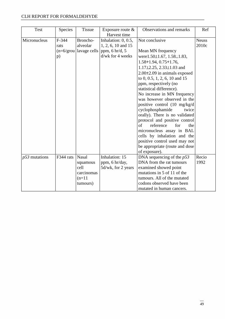
CLH REPORT FOR FORMALDEHYDE
49
Test Species Tissue Exposure route &
Harvest time
Observations and remarks Ref
Micronucleus F-344
rats
(n=6/grou
p)
Broncho-
alveolar
lavage cells
Inhalation: 0, 0.5,
1, 2, 6, 10 and 15
ppm, 6 hr/d, 5
d/wk for 4 weeks
Not conclusive
Mean MN frequency
were1.50 1.67, 1.58 1.83,
1.58 1.94, 0.75 1.76,
1.17 2.25, 2.33 1.03 and
2.00 2.09 in animals exposed
to 0, 0.5, 1, 2, 6, 10 and 15
ppm, respectively (no
statistical difference).
No increase in MN frequency
was however observed in the
positive control (10 mg/kg/d
cyclophosphamide twice
orally). There is no validated
protocol and positive control
of reference for the
micronucleus assay in BAL
cells by inhalation and the
positive control used may not
be appropriate (route and dose
of exposure).
Neuss
2010c
p53 mutations F344 rats Nasal
squamous
cell
carcinomas
(n=11
tumours)
Inhalation: 15
ppm, 6 hr/day,
5d/wk, for 2 years
DNA sequencing of the p53
DNA from the rat tumours
examined showed point
mutations in 5 of 11 of the
tumours. All of the mutated
codons observed have been
mutated in human cancers.
Recio
1992
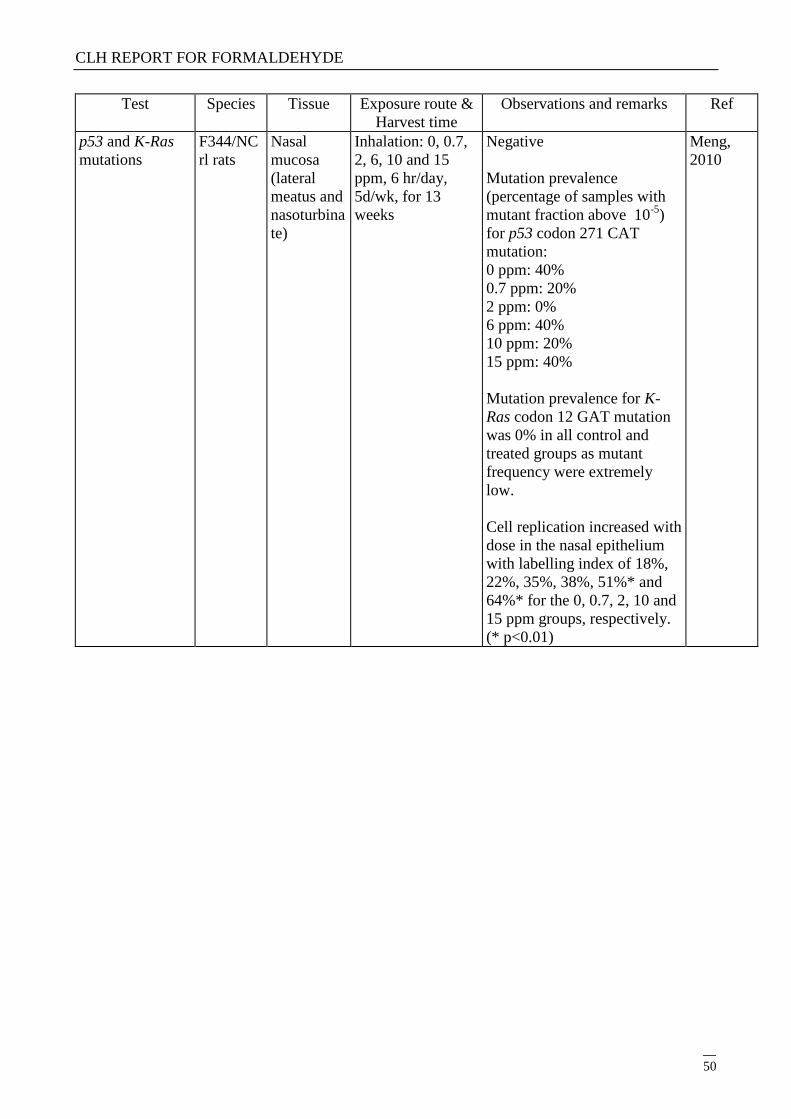
CLH REPORT FOR FORMALDEHYDE
50
Test Species Tissue Exposure route &
Harvest time
Observations and remarks Ref
p53 and K-Ras
mutations
F344/NC
rl rats
Nasal
mucosa
(lateral
meatus and
nasoturbina
te)
Inhalation: 0, 0.7,
2, 6, 10 and 15
ppm, 6 hr/day,
5d/wk, for 13
weeks
Negative
Mutation prevalence
(percentage of samples with
mutant fraction above 10-5
)
for p53 codon 271 CAT
mutation:
0 ppm: 40%
0.7 ppm: 20%
2 ppm: 0%
6 ppm: 40%
10 ppm: 20%
15 ppm: 40%
Mutation prevalence for K-
Ras codon 12 GAT mutation
was 0% in all control and
treated groups as mutant
frequency were extremely
low.
Cell replication increased with
dose in the nasal epithelium
with labelling index of 18%,
22%, 35%, 38%, 51%* and
64%* for the 0, 0.7, 2, 10 and
15 ppm groups, respectively.
(* p<0.01)
Meng,
2010
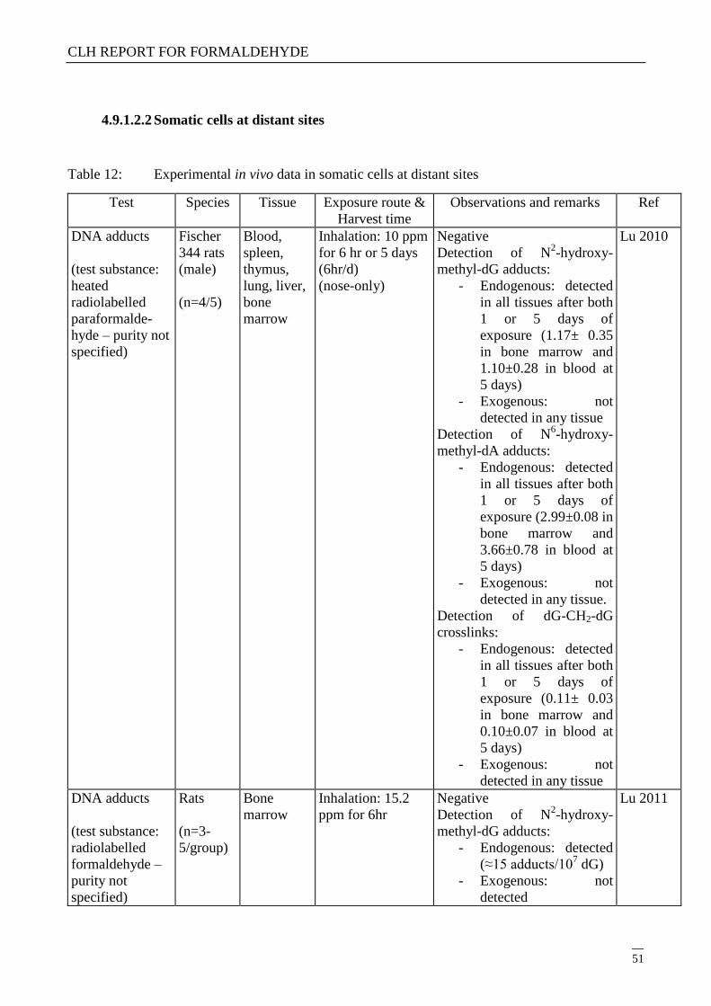
CLH REPORT FOR FORMALDEHYDE
51
4.9.1.2.2 Somatic cells at distant sites
Table 12: Experimental in vivo data in somatic cells at distant sites
Test Species Tissue Exposure route &
Harvest time
Observations and remarks Ref
DNA adducts
(test substance:
heated
radiolabelled
paraformalde-
hyde – purity not
specified)
Fischer
344 rats
(male)
(n=4/5)
Blood,
spleen,
thymus,
lung, liver,
bone
marrow
Inhalation: 10 ppm
for 6 hr or 5 days
(6hr/d)
(nose-only)
Negative
Detection of N2-hydroxy-
methyl-dG adducts:
- Endogenous: detected
in all tissues after both
1 or 5 days of
exposure (1.17± 0.35
in bone marrow and
1.10±0.28 in blood at
5 days)
- Exogenous: not
detected in any tissue
Detection of N6-hydroxy-
methyl-dA adducts:
- Endogenous: detected
in all tissues after both
1 or 5 days of
exposure (2.99±0.08 in
bone marrow and
3.66±0.78 in blood at
5 days)
- Exogenous: not
detected in any tissue.
Detection of dG-CH2-dG
crosslinks:
- Endogenous: detected
in all tissues after both
1 or 5 days of
exposure (0.11± 0.03
in bone marrow and
0.10±0.07 in blood at
5 days)
- Exogenous: not
detected in any tissue
Lu 2010
DNA adducts
(test substance:
radiolabelled
formaldehyde –
purity not
specified)
Rats
(n=3-
5/group)
Bone
marrow
Inhalation: 15.2
ppm for 6hr
Negative
Detection of N2-hydroxy-
methyl-dG adducts:
- Endogenous: detected
(≈15 adducts/107 dG)
- Exogenous: not
detected
Lu 2011
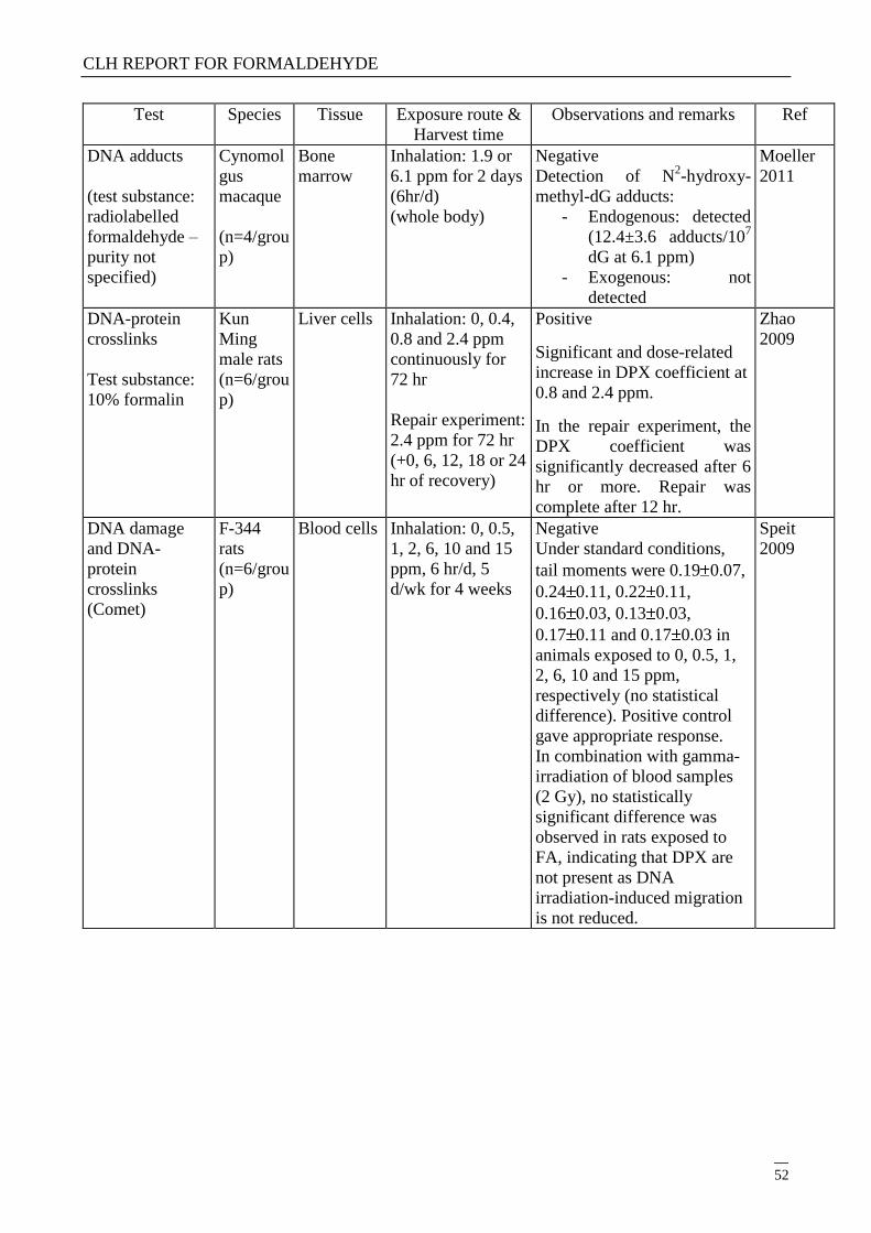
CLH REPORT FOR FORMALDEHYDE
52
Test Species Tissue Exposure route &
Harvest time
Observations and remarks Ref
DNA adducts
(test substance:
radiolabelled
formaldehyde –
purity not
specified)
Cynomol
gus
macaque
(n=4/grou
p)
Bone
marrow
Inhalation: 1.9 or
6.1 ppm for 2 days
(6hr/d)
(whole body)
Negative
Detection of N2-hydroxy-
methyl-dG adducts:
- Endogenous: detected
(12.4±3.6 adducts/107
dG at 6.1 ppm)
- Exogenous: not
detected
Moeller
2011
DNA-protein
crosslinks
Test substance:
10% formalin
Kun
Ming
male rats
(n=6/grou
p)
Liver cells Inhalation: 0, 0.4,
0.8 and 2.4 ppm
continuously for
72 hr
Repair experiment:
2.4 ppm for 72 hr
(+0, 6, 12, 18 or 24
hr of recovery)
Positive
Significant and dose-related
increase in DPX coefficient at
0.8 and 2.4 ppm.
In the repair experiment, the
DPX coefficient was
significantly decreased after 6
hr or more. Repair was
complete after 12 hr.
Zhao
2009
DNA damage
and DNA-
protein
crosslinks
(Comet)
F-344
rats
(n=6/grou
p)
Blood cells
Inhalation: 0, 0.5,
1, 2, 6, 10 and 15
ppm, 6 hr/d, 5
d/wk for 4 weeks
Negative
Under standard conditions,
tail moments were 0.19 0.07,
0.24 0.11, 0.22 0.11,
0.16 0.03, 0.13 0.03,
0.17 0.11 and 0.17 0.03 in
animals exposed to 0, 0.5, 1,
2, 6, 10 and 15 ppm,
respectively (no statistical
difference). Positive control
gave appropriate response.
In combination with gamma-
irradiation of blood samples
(2 Gy), no statistically
significant difference was
observed in rats exposed to
FA, indicating that DPX are
not present as DNA
irradiation-induced migration
is not reduced.
Speit
2009

CLH REPORT FOR FORMALDEHYDE
53
Test Species Tissue Exposure route &
Harvest time
Observations and remarks Ref
DNA damage
(Comet)
Test substance:
FA (purity not
specified)
Sprague
Dawley
male rats
( n=10/
group)
Lymphocyt
es and liver
Inhalation: 0, 5 or
10 ppm, 6 hr/d,
5d/wk for 2 weeks
Positive
Olive tail moment in
lymphocytes:
Controls: 1.24±0.04
5 ppm: 1.72±0.11, p=0.0019)
10 ppm: 2.16±0.14, p=0.0001)
Olive tail moment in liver
cells:
Controls: 1.19±0.08
5 ppm: 1.73±0.10, p=0.0001)
10 ppm: 2.49±0.20, p=0.0001)
In this assay, peroxidation of
lipids and oxidation of
proteins was observed at 10
ppm in lymphocytes and liver
cells. Expression of 32 plasma
proteins was up or down
regulated. Analysis of the
expression of plasma
cytokines showed a dose
related upregulation of IL-4
and down regulation of IFN-
gamma suggesting an
inflammatory effect.
Im 2006
Sister Chromatid
Exchange
Mice
(n=10/
sex in 1st
exp. and
5/sex in
the 2nd)
Bone
marrow
Inhalation:
1st experiment : 0,
6, 12 or 25 ppm, 6
hr/d for 5 days
2nd
experiment: 0,
5, 10, 15 or 25
ppm, 6 hr/d for 5
days
Equivocal
Positive in females et 12 and
25 ppm but not in males in the
1st experiment.
Negative in males and females
in the second experiment but
SCE frequency in controls
was unusually high.
Only 20 cells per animal
analysed.
Formalde
hyde
Institute
1982#
Sister Chromatid
Exchange
Mice
(n=5/
sex)
Bone
marrow
Inhalation:
1st experiment : 0,
6, 12 or 25 ppm, 6
hr/d for 4 days
Negative
Positive in females et 12 and
25 ppm but not in males in the
1st experiment.
Negative in males and females
in the second experiment but
SCE frequency in controls
was unusually high.
Only 50 cells per animal
analysed.
Formalde
hyde
Institute
1982#
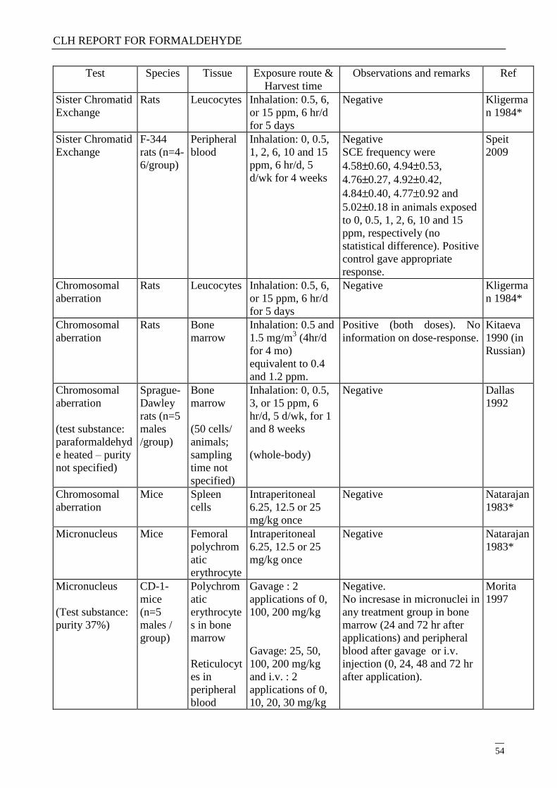
CLH REPORT FOR FORMALDEHYDE
54
Test Species Tissue Exposure route &
Harvest time
Observations and remarks Ref
Sister Chromatid
Exchange
Rats Leucocytes Inhalation: 0.5, 6,
or 15 ppm, 6 hr/d
for 5 days
Negative Kligerma
n 1984*
Sister Chromatid
Exchange
F-344
rats (n=4-
6/group)
Peripheral
blood
Inhalation: 0, 0.5,
1, 2, 6, 10 and 15
ppm, 6 hr/d, 5
d/wk for 4 weeks
Negative
SCE frequency were
4.58 0.60, 4.94 0.53,
4.76 0.27, 4.92 0.42,
4.84 0.40, 4.77 0.92 and
5.02 0.18 in animals exposed
to 0, 0.5, 1, 2, 6, 10 and 15
ppm, respectively (no
statistical difference). Positive
control gave appropriate
response.
Speit
2009
Chromosomal
aberration
Rats Leucocytes Inhalation: 0.5, 6,
or 15 ppm, 6 hr/d
for 5 days
Negative Kligerma
n 1984*
Chromosomal
aberration
Rats Bone
marrow
Inhalation: 0.5 and
1.5 mg/m3 (4hr/d
for 4 mo)
equivalent to 0.4
and 1.2 ppm.
Positive (both doses). No
information on dose-response.
Kitaeva
1990 (in
Russian)
Chromosomal
aberration
(test substance:
paraformaldehyd
e heated – purity
not specified)
Sprague-
Dawley
rats (n=5
males
/group)
Bone
marrow
(50 cells/
animals;
sampling
time not
specified)
Inhalation: 0, 0.5,
3, or 15 ppm, 6
hr/d, 5 d/wk, for 1
and 8 weeks
(whole-body)
Negative Dallas
1992
Chromosomal
aberration
Mice Spleen
cells
Intraperitoneal
6.25, 12.5 or 25
mg/kg once
Negative Natarajan
1983*
Micronucleus Mice Femoral
polychrom
atic
erythrocyte
Intraperitoneal
6.25, 12.5 or 25
mg/kg once
Negative Natarajan
1983*
Micronucleus
(Test substance:
purity 37%)
CD-1-
mice
(n=5
males /
group)
Polychrom
atic
erythrocyte
s in bone
marrow
Reticulocyt
es in
peripheral
blood
Gavage : 2
applications of 0,
100, 200 mg/kg
Gavage: 25, 50,
100, 200 mg/kg
and i.v. : 2
applications of 0,
10, 20, 30 mg/kg
Negative.
No incresase in micronuclei in
any treatment group in bone
marrow (24 and 72 hr after
applications) and peripheral
blood after gavage or i.v.
injection (0, 24, 48 and 72 hr
after application).
Morita
1997
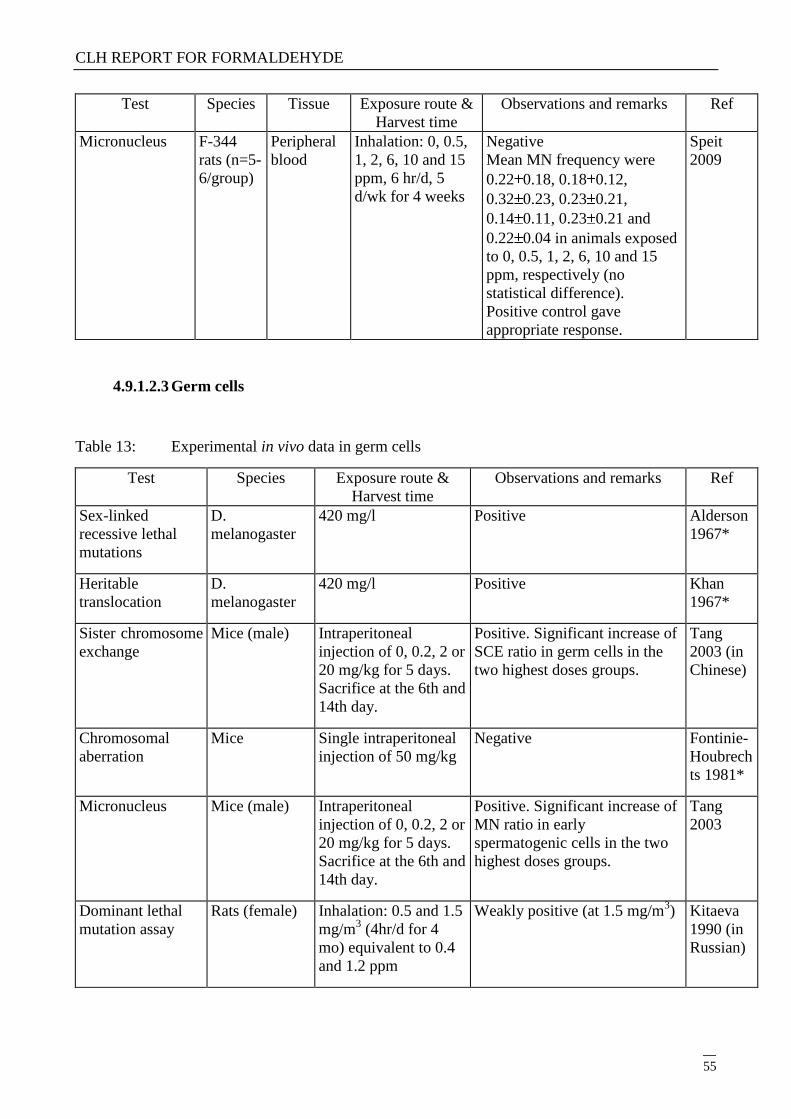
CLH REPORT FOR FORMALDEHYDE
55
Test Species Tissue Exposure route &
Harvest time
Observations and remarks Ref
Micronucleus
F-344
rats (n=5-
6/group)
Peripheral
blood
Inhalation: 0, 0.5,
1, 2, 6, 10 and 15
ppm, 6 hr/d, 5
d/wk for 4 weeks
Negative
Mean MN frequency were
0.22 0.18, 0.18 0.12,
0.32 0.23, 0.23 0.21,
0.14 0.11, 0.23 0.21 and
0.22 0.04 in animals exposed
to 0, 0.5, 1, 2, 6, 10 and 15
ppm, respectively (no
statistical difference).
Positive control gave
appropriate response.
Speit
2009
4.9.1.2.3 Germ cells
Table 13: Experimental in vivo data in germ cells
Test Species Exposure route &
Harvest time
Observations and remarks Ref
Sex-linked
recessive lethal
mutations
D.
melanogaster
420 mg/l Positive Alderson
1967*
Heritable
translocation
D.
melanogaster
420 mg/l Positive Khan
1967*
Sister chromosome
exchange
Mice (male) Intraperitoneal
injection of 0, 0.2, 2 or
20 mg/kg for 5 days.
Sacrifice at the 6th and
14th day.
Positive. Significant increase of
SCE ratio in germ cells in the
two highest doses groups.
Tang
2003 (in
Chinese)
Chromosomal
aberration
Mice Single intraperitoneal
injection of 50 mg/kg
Negative Fontinie-
Houbrech
ts 1981*
Micronucleus
Mice (male) Intraperitoneal
injection of 0, 0.2, 2 or
20 mg/kg for 5 days.
Sacrifice at the 6th and
14th day.
Positive. Significant increase of
MN ratio in early
spermatogenic cells in the two
highest doses groups.
Tang
2003
Dominant lethal
mutation assay
Rats (female) Inhalation: 0.5 and 1.5
mg/m3 (4hr/d for 4
mo) equivalent to 0.4
and 1.2 ppm
Weakly positive (at 1.5 mg/m3) Kitaeva
1990 (in
Russian)
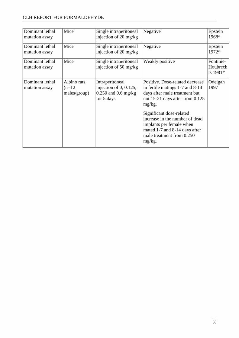
CLH REPORT FOR FORMALDEHYDE
56
Dominant lethal
mutation assay
Mice Single intraperitoneal
injection of 20 mg/kg
Negative Epstein
1968*
Dominant lethal
mutation assay
Mice Single intraperitoneal
injection of 20 mg/kg
Negative Epstein
1972*
Dominant lethal
mutation assay
Mice Single intraperitoneal
injection of 50 mg/kg
Weakly positive Fontinie-
Houbrech
ts 1981*
Dominant lethal
mutation assay
Albino rats
(n=12
males/group)
Intraperitoneal
injection of 0, 0.125,
0.250 and 0.6 mg/kg
for 5 days
Positive. Dose-related decrease
in fertile matings 1-7 and 8-14
days after male treatment but
not 15-21 days after from 0.125
mg/kg.
Significant dose-related
increase in the number of dead
implants per female when
mated 1-7 and 8-14 days after
male treatment from 0.250
mg/kg.
Odeigah
1997
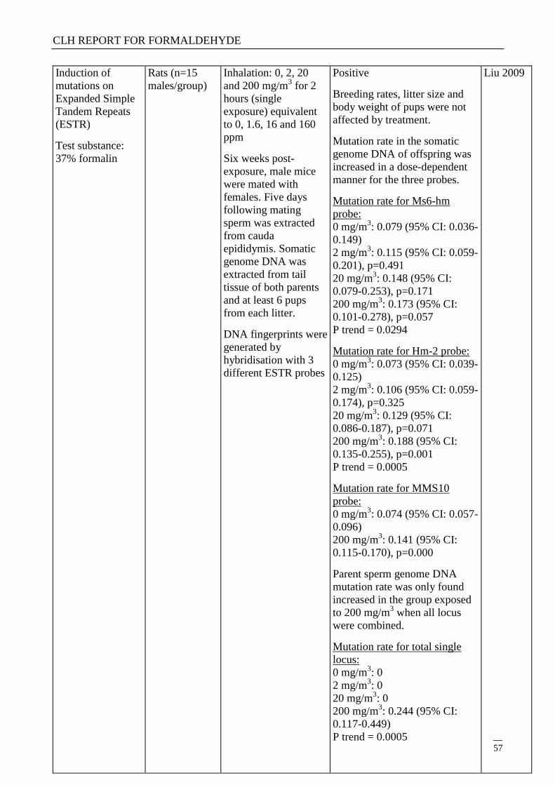
CLH REPORT FOR FORMALDEHYDE
57
Induction of
mutations on
Expanded Simple
Tandem Repeats
(ESTR)
Test substance:
37% formalin
Rats (n=15
males/group)
Inhalation: 0, 2, 20
and 200 mg/m3 for 2
hours (single
exposure) equivalent
to 0, 1.6, 16 and 160
ppm
Six weeks post-
exposure, male mice
were mated with
females. Five days
following mating
sperm was extracted
from cauda
epididymis. Somatic
genome DNA was
extracted from tail
tissue of both parents
and at least 6 pups
from each litter.
DNA fingerprints were
generated by
hybridisation with 3
different ESTR probes
Positive
Breeding rates, litter size and
body weight of pups were not
affected by treatment.
Mutation rate in the somatic
genome DNA of offspring was
increased in a dose-dependent
manner for the three probes.
Mutation rate for Ms6-hm
probe:
0 mg/m3: 0.079 (95% CI: 0.036-
0.149)
2 mg/m3: 0.115 (95% CI: 0.059-
0.201), p=0.491
20 mg/m3: 0.148 (95% CI:
0.079-0.253), p=0.171
200 mg/m3: 0.173 (95% CI:
0.101-0.278), p=0.057
P trend = 0.0294
Mutation rate for Hm-2 probe:
0 mg/m3: 0.073 (95% CI: 0.039-
0.125)
2 mg/m3: 0.106 (95% CI: 0.059-
0.174), p=0.325
20 mg/m3: 0.129 (95% CI:
0.086-0.187), p=0.071
200 mg/m3: 0.188 (95% CI:
0.135-0.255), p=0.001
P trend = 0.0005
Mutation rate for MMS10
probe:
0 mg/m3: 0.074 (95% CI: 0.057-
0.096)
200 mg/m3: 0.141 (95% CI:
0.115-0.170), p=0.000
Parent sperm genome DNA
mutation rate was only found
increased in the group exposed
to 200 mg/m3 when all locus
were combined.
Mutation rate for total single
locus:
0 mg/m3: 0
2 mg/m3: 0
20 mg/m3: 0
200 mg/m3: 0.244 (95% CI:
0.117-0.449)
P trend = 0.0005
Liu 2009
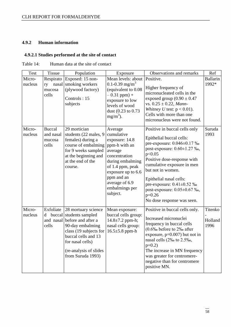
CLH REPORT FOR FORMALDEHYDE
58
4.9.2 Human information
4.9.2.1 Studies performed at the site of contact
Table 14: Human data at the site of contact
Test Tissue Population Exposure Observations and remarks Ref
Micro-
nucleus
Respirato
ry nasal
mucosa
cells
Exposed: 15 non-
smoking workers
(plywood factory)
Controls : 15
subjects
Mean levels: about
0.1-0.39 mg/m3
(equivalent to 0.08
– 0.31 ppm) +
exposure to low
levels of wood
dust (0.23 to 0.73
mg/m3).
Positive.
Higher frequency of
micronucleated cells in the
exposed group (0.90 ± 0.47
vs. 0.25 ± 0.22, Mann-
Whitney U test: p < 0.01).
Cells with more than one
micronucleus were not found.
Ballarin
1992*
Micro-
nucleus
Buccal
and nasal
mucosa
cells
29 mortician
students (22 males, 9
females) during a
course of embalming
for 9 weeks sampled
at the beginning and
at the end of the
course.
Average
cumulative
exposure: 14.8
ppm-h with an
average
concentration
during embalming
of 1.4 ppm, peak
exposure up to 6.6
ppm and an
average of 6.9
embalmings per
subject.
Positive in buccal cells only
Epithelial buccal cells:
pre-exposure: 0.046±0.17 ‰
post-exposure: 0.60±1.27 ‰,
p<0.05
Positive dose-response with
cumulative exposure in men
but not in women.
Epithelial nasal cells:
pre-exposure: 0.41±0.52 ‰
post-exposure: 0.05±0.67 ‰,
p=0.26
No dose response was seen.
Suruda
1993
Micro-
nucleus
Exfoliate
d buccal
and nasal
cells
28 mortuary science
students sampled
before and after a
90-day embalming
class (19 subjects for
buccal cells and 13
for nasal cells)
(re-analysis of slides
from Suruda 1993)
Mean exposure:
buccal cells group:
14.8±7.2 ppm-h;
nasal cells group:
16.5±5.8 ppm-h
Positive in buccal cells only.
Increased micronuclei
frequency in buccal cells
(0.6‰ before to 2‰ after
exposure, p=0.007) but not in
nasal cells (2‰ to 2.5‰,
p=0.2)
The increase in MN frequency
was greater for centromere-
negative than for centromere
positive MN.
Titenko
-
Holland
1996
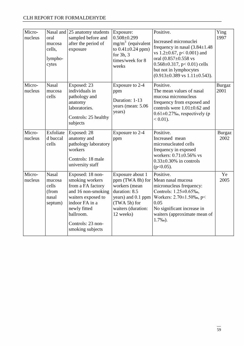
CLH REPORT FOR FORMALDEHYDE
59
Micro-
nucleus
Nasal and
oral
mucosa
cells,
lympho-
cytes
25 anatomy students
sampled before and
after the period of
exposure
Exposure:
0.508±0.299
mg/m3
(equivalent
to 0.41±0.24 ppm)
for 3h, 3
times/week for 8
weeks
Positive.
Increased micronuclei
frequency in nasal (3.84±1.48
vs 1.2±0.67, p< 0.001) and
oral (0.857±0.558 vs
0.568±0.317, p< 0.01) cells
but not in lymphocytes
(0.913±0.389 vs 1.11±0.543).
Ying
1997
Micro-
nucleus
Nasal
mucosa
cells
Exposed: 23
individuals in
pathology and
anatomy
laboratories.
Controls: 25 healthy
subjects
Exposure to 2-4
ppm
Duration: 1-13
years (mean: 5.06
years)
Positive.
The mean values of nasal
mucosa micronucleus
frequency from exposed and
controls were 1.01±0.62 and
0.61±0.27‰, respectively (p
< 0.01).
Burgaz
2001
Micro-
nucleus
Exfoliate
d buccal
cells
Exposed: 28
anatomy and
pathology laboratory
workers
Controls: 18 male
university staff
Exposure to 2-4
ppm
Positive.
Increased mean
micronucleated cells
frequency in exposed
workers: 0.71±0.56% vs
0.33±0.30% in controls
(p<0.05).
Burgaz
2002
Micro-
nucleus
Nasal
mucosa
cells
(from
nasal
septum)
Exposed: 18 non-
smoking workers
from a FA factory
and 16 non-smoking
waiters exposed to
indoor FA in a
newly fitted
ballroom.
Controls: 23 non-
smoking subjects
Exposure about 1
ppm (TWA 8h) for
workers (mean
duration: 8.5
years) and 0.1 ppm
(TWA 5h) for
waiters (duration:
12 weeks)
Positive.
Mean nasal mucosa
micronucleus frequency:
Controls: 1.25±0.65‰,
Workers: 2.70±1.50‰, p<
0.05
No significant increase in
waiters (approximate mean of
1.7‰).
Ye
2005
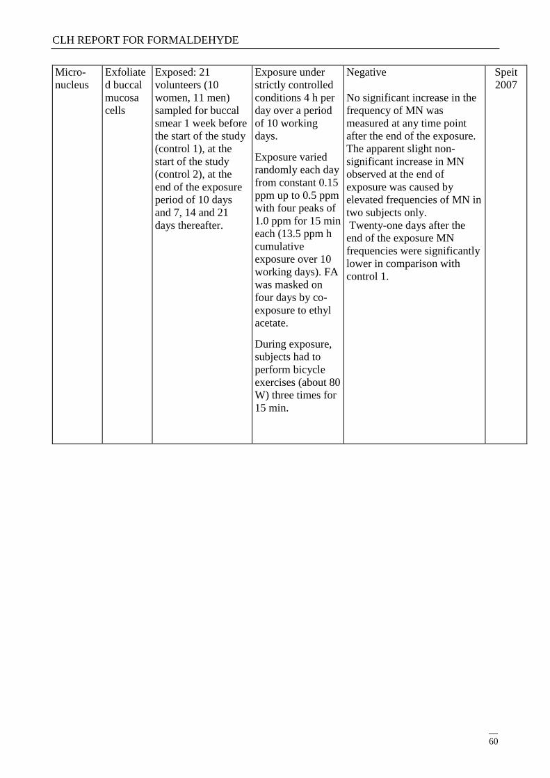
CLH REPORT FOR FORMALDEHYDE
60
Micro-
nucleus
Exfoliate
d buccal
mucosa
cells
Exposed: 21
volunteers (10
women, 11 men)
sampled for buccal
smear 1 week before
the start of the study
(control 1), at the
start of the study
(control 2), at the
end of the exposure
period of 10 days
and 7, 14 and 21
days thereafter.
Exposure under
strictly controlled
conditions 4 h per
day over a period
of 10 working
days.
Exposure varied
randomly each day
from constant 0.15
ppm up to 0.5 ppm
with four peaks of
1.0 ppm for 15 min
each (13.5 ppm h
cumulative
exposure over 10
working days). FA
was masked on
four days by co-
exposure to ethyl
acetate.
During exposure,
subjects had to
perform bicycle
exercises (about 80
W) three times for
15 min.
Negative
No significant increase in the
frequency of MN was
measured at any time point
after the end of the exposure.
The apparent slight non-
significant increase in MN
observed at the end of
exposure was caused by
elevated frequencies of MN in
two subjects only.
Twenty-one days after the
end of the exposure MN
frequencies were significantly
lower in comparison with
control 1.
Speit
2007

CLH REPORT FOR FORMALDEHYDE
61
Micro-
nucleus
Exfoliate
d buccal
mucosa
cells
Exposed: 80 workers
occupationally
exposed to FA ( 30
from FA and FA-
based resins
production factory
and 50 from
pathology and
anatomy laboratory)
Controls: 85 non-
exposed subjects
Exposure in
industrial workers:
mean TWA of
0.21 ppm with
mean ceiling
concentration of
0.52 ppm for a
mean duration of
6.74 years.
Exposure in
laboratory
workers: mean
TWA of 0.28 ppm
with mean ceiling
concentration of
2.52 ppm for a
mean duration of
9.12 years.
Positive.
Mean nasal mucosa
micronucleus frequency:
Controls: 0.13±0.48‰,
Industrial workers:
1.27±1.55‰, p<0.001
Laboratory workers:
0.64±1.74‰, p<0.005
A moderate positive
association was observed with
duration of exposure (r=0.209,
p<0.05).
Control and exposed groups
did not differ in age and
smoking habits but a larger
number of women were
included in the control group
(63.5 vs 40%). Gender was
however not found to have a
significant impact on
frequency of micronuclei.
Viegas
2010
Micro-
nucleus
Nasal
mucosa
cells
Exposed : 41 male
non-smoking
volunteers sampled
before the first
exposure, after the
last exposure and 1,
2 and 3 weeks after
the end of exposure.
Exposure under
strictly controlled
conditions 4 h per
day over a period
of 5 consecutive
days.
Exposure varied
randomly each day
from 0 ppm or 0.3
ppm with four
peaks of 0.6 ppm
for 15 min, or 0.4
with four peaks of
0.8 ppm for 15
min, or 0.5 ppm or
0.7 ppm.
During exposure,
subjects had to
perform bicycle
exercises (about 80
W) four times for
15 min.
Negative.
Samples from 33 to 36
volunteers were analysed (56
000 to 62 000 cells per data
point).
Mean micronucleus frequency
was 0.21±0.35‰ before
exposure, 0.27±0.42‰ post-
exposure, 0.24±0.43‰ one
week after, 0.24±0.45‰ two
weeks after and 0.17±0.41‰
three weeks after.
Analysis of variance did not
indicate a significant
difference between groups
(p=0.8664).
Zeller
2011
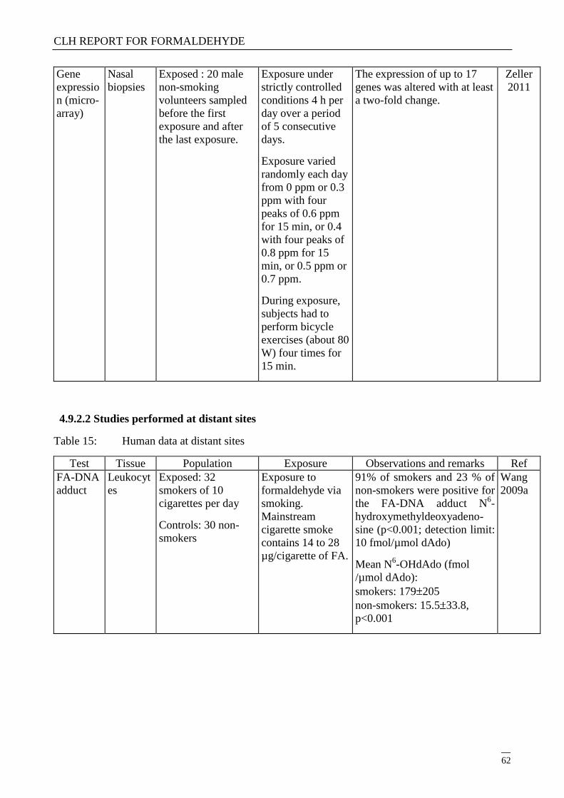
CLH REPORT FOR FORMALDEHYDE
62
Gene
expressio
n (micro-
array)
Nasal
biopsies
Exposed : 20 male
non-smoking
volunteers sampled
before the first
exposure and after
the last exposure.
Exposure under
strictly controlled
conditions 4 h per
day over a period
of 5 consecutive
days.
Exposure varied
randomly each day
from 0 ppm or 0.3
ppm with four
peaks of 0.6 ppm
for 15 min, or 0.4
with four peaks of
0.8 ppm for 15
min, or 0.5 ppm or
0.7 ppm.
During exposure,
subjects had to
perform bicycle
exercises (about 80
W) four times for
15 min.
The expression of up to 17
genes was altered with at least
a two-fold change.
Zeller
2011
4.9.2.2 Studies performed at distant sites
Table 15: Human data at distant sites
Test Tissue Population Exposure Observations and remarks Ref
FA-DNA
adduct
Leukocyt
es
Exposed: 32
smokers of 10
cigarettes per day
Controls: 30 non-
smokers
Exposure to
formaldehyde via
smoking.
Mainstream
cigarette smoke
contains 14 to 28
µg/cigarette of FA.
91% of smokers and 23 % of
non-smokers were positive for
the FA-DNA adduct N6-
hydroxymethyldeoxyadeno-
sine (p<0.001; detection limit:
10 fmol/µmol dAdo)
Mean N6-OHdAdo (fmol
/µmol dAdo):
smokers: 179 205
non-smokers: 15.5 33.8,
p<0.001
Wang
2009a
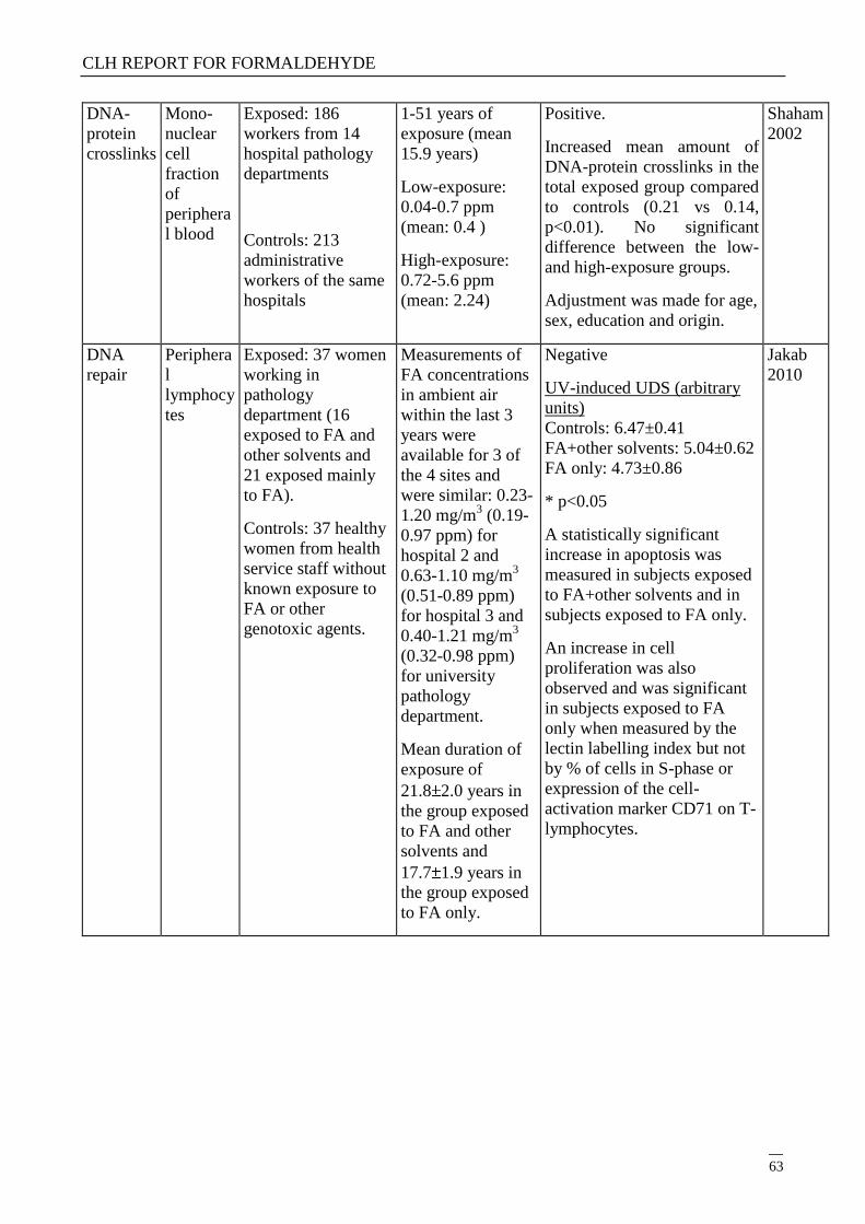
CLH REPORT FOR FORMALDEHYDE
63
DNA-
protein
crosslinks
Mono-
nuclear
cell
fraction
of
periphera
l blood
Exposed: 186
workers from 14
hospital pathology
departments
Controls: 213
administrative
workers of the same
hospitals
1-51 years of
exposure (mean
15.9 years)
Low-exposure:
0.04-0.7 ppm
(mean: 0.4 )
High-exposure:
0.72-5.6 ppm
(mean: 2.24)
Positive.
Increased mean amount of
DNA-protein crosslinks in the
total exposed group compared
to controls (0.21 vs 0.14,
p<0.01). No significant
difference between the low-
and high-exposure groups.
Adjustment was made for age,
sex, education and origin.
Shaham
2002
DNA
repair
Periphera
l
lymphocy
tes
Exposed: 37 women
working in
pathology
department (16
exposed to FA and
other solvents and
21 exposed mainly
to FA).
Controls: 37 healthy
women from health
service staff without
known exposure to
FA or other
genotoxic agents.
Measurements of
FA concentrations
in ambient air
within the last 3
years were
available for 3 of
the 4 sites and
were similar: 0.23-
1.20 mg/m3 (0.19-
0.97 ppm) for
hospital 2 and
0.63-1.10 mg/m3
(0.51-0.89 ppm)
for hospital 3 and
0.40-1.21 mg/m3
(0.32-0.98 ppm)
for university
pathology
department.
Mean duration of
exposure of
21.8 2.0 years in
the group exposed
to FA and other
solvents and
17.7 1.9 years in
the group exposed
to FA only.
Negative
UV-induced UDS (arbitrary
units)
Controls: 6.47±0.41
FA+other solvents: 5.04±0.62
FA only: 4.73±0.86
* p<0.05
A statistically significant
increase in apoptosis was
measured in subjects exposed
to FA+other solvents and in
subjects exposed to FA only.
An increase in cell
proliferation was also
observed and was significant
in subjects exposed to FA
only when measured by the
lectin labelling index but not
by % of cells in S-phase or
expression of the cell-
activation marker CD71 on T-
lymphocytes.
Jakab
2010
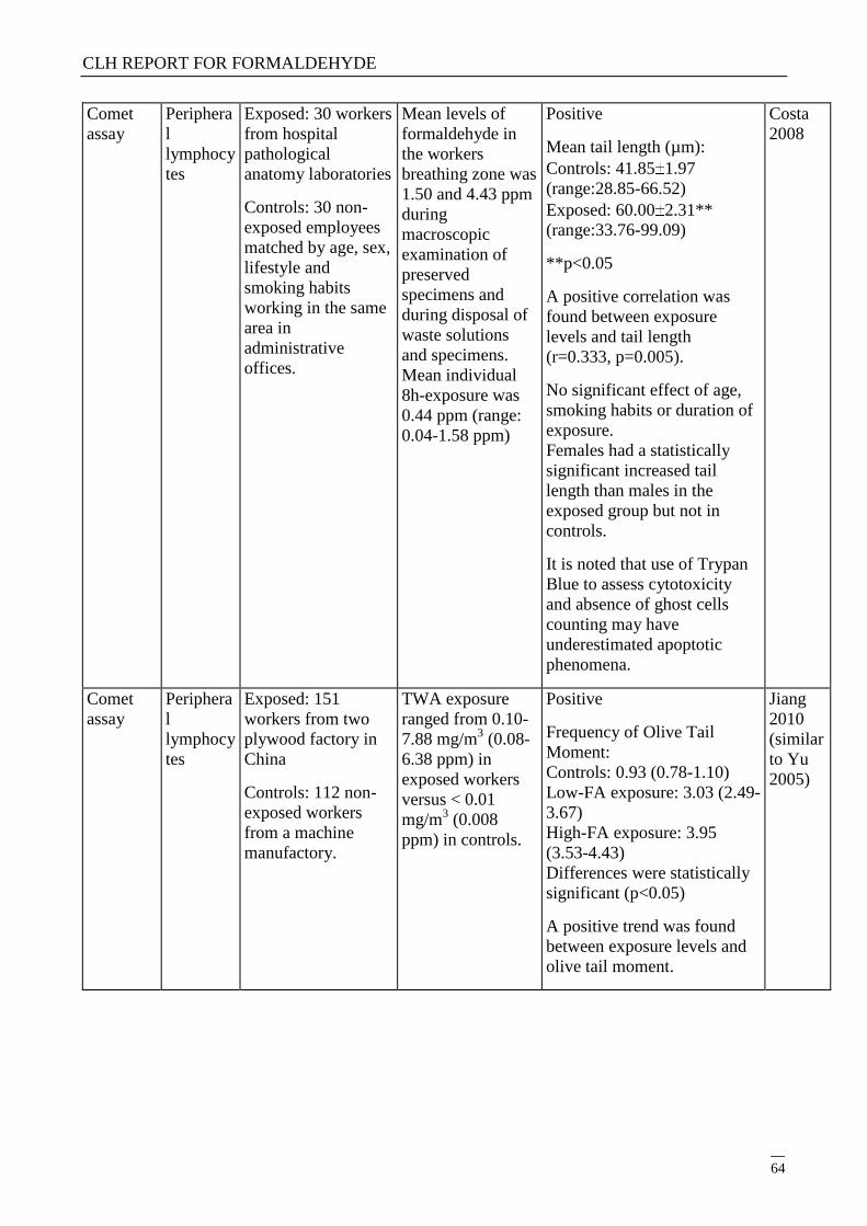
CLH REPORT FOR FORMALDEHYDE
64
Comet
assay
Periphera
l
lymphocy
tes
Exposed: 30 workers
from hospital
pathological
anatomy laboratories
Controls: 30 non-
exposed employees
matched by age, sex,
lifestyle and
smoking habits
working in the same
area in
administrative
offices.
Mean levels of
formaldehyde in
the workers
breathing zone was
1.50 and 4.43 ppm
during
macroscopic
examination of
preserved
specimens and
during disposal of
waste solutions
and specimens.
Mean individual
8h-exposure was
0.44 ppm (range:
0.04-1.58 ppm)
Positive
Mean tail length (µm):
Controls: 41.85 1.97
(range:28.85-66.52)
Exposed: 60.00 2.31**
(range:33.76-99.09)
**p<0.05
A positive correlation was
found between exposure
levels and tail length
(r=0.333, p=0.005).
No significant effect of age,
smoking habits or duration of
exposure.
Females had a statistically
significant increased tail
length than males in the
exposed group but not in
controls.
It is noted that use of Trypan
Blue to assess cytotoxicity
and absence of ghost cells
counting may have
underestimated apoptotic
phenomena.
Costa
2008
Comet
assay
Periphera
l
lymphocy
tes
Exposed: 151
workers from two
plywood factory in
China
Controls: 112 non-
exposed workers
from a machine
manufactory.
TWA exposure
ranged from 0.10-
7.88 mg/m3 (0.08-
6.38 ppm) in
exposed workers
versus < 0.01
mg/m3 (0.008
ppm) in controls.
Positive
Frequency of Olive Tail
Moment:
Controls: 0.93 (0.78-1.10)
Low-FA exposure: 3.03 (2.49-
3.67)
High-FA exposure: 3.95
(3.53-4.43)
Differences were statistically
significant (p<0.05)
A positive trend was found
between exposure levels and
olive tail moment.
Jiang
2010
(similar
to Yu
2005)
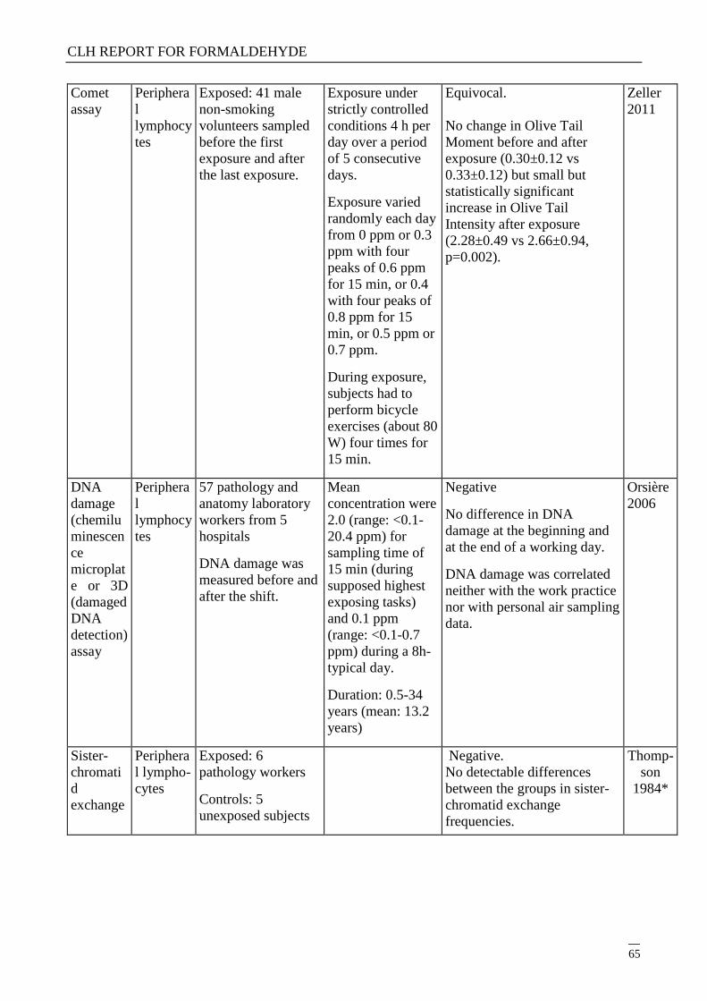
CLH REPORT FOR FORMALDEHYDE
65
Comet
assay
Periphera
l
lymphocy
tes
Exposed: 41 male
non-smoking
volunteers sampled
before the first
exposure and after
the last exposure.
Exposure under
strictly controlled
conditions 4 h per
day over a period
of 5 consecutive
days.
Exposure varied
randomly each day
from 0 ppm or 0.3
ppm with four
peaks of 0.6 ppm
for 15 min, or 0.4
with four peaks of
0.8 ppm for 15
min, or 0.5 ppm or
0.7 ppm.
During exposure,
subjects had to
perform bicycle
exercises (about 80
W) four times for
15 min.
Equivocal.
No change in Olive Tail
Moment before and after
exposure (0.30±0.12 vs
0.33±0.12) but small but
statistically significant
increase in Olive Tail
Intensity after exposure
(2.28±0.49 vs 2.66±0.94,
p=0.002).
Zeller
2011
DNA
damage
(chemilu
minescen
ce
microplat
e or 3D
(damaged
DNA
detection)
assay
Periphera
l
lymphocy
tes
57 pathology and
anatomy laboratory
workers from 5
hospitals
DNA damage was
measured before and
after the shift.
Mean
concentration were
2.0 (range: <0.1-
20.4 ppm) for
sampling time of
15 min (during
supposed highest
exposing tasks)
and 0.1 ppm
(range: <0.1-0.7
ppm) during a 8h-
typical day.
Duration: 0.5-34
years (mean: 13.2
years)
Negative
No difference in DNA
damage at the beginning and
at the end of a working day.
DNA damage was correlated
neither with the work practice
nor with personal air sampling
data.
Orsière
2006
Sister-
chromati
d
exchange
Periphera
l lympho-
cytes
Exposed: 6
pathology workers
Controls: 5
unexposed subjects
Negative.
No detectable differences
between the groups in sister-
chromatid exchange
frequencies.
Thomp-
son
1984*
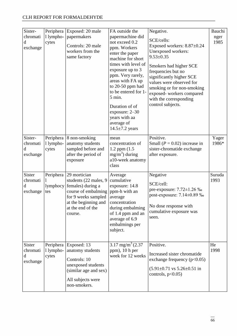
CLH REPORT FOR FORMALDEHYDE
66
Sister-
chromati
d
exchange
Periphera
l lympho-
cytes
Exposed: 20 male
papermakers
Controls: 20 male
workers from the
same factory
FA outside the
papermachine did
not exceed 0.2
ppm. Workers
enter the paper
machine for short
times with level of
exposure up to 3
ppm. Very rarely,
areas with FA up
to 20-50 ppm had
to be entered for 1-
5 min.
Duration of of
exposure: 2–30
years with aa
average of
14.5±7.2 years
Negative.
SCE/cells:
Exposed workers: 8.87±0.24
Unexposed workers:
9.53±0.35
Smokers had higher SCE
frequencies but no
significantly higher SCE
values were observed for
smoking or for non-smoking
exposed- workers compared
with the corresponding
control subjects.
Bauchi
nger
1985
Sister-
chromati
d
exchange
Periphera
l lympho-
cytes
8 non-smoking
anatomy students
sampled before and
after the period of
exposure
mean
concentration of
1.2 ppm (1.5
mg/m3) during
a10-week anatomy
class
Positive.
Small (P = 0.02) increase in
sister-chromatide exchange
after exposure.
Yager
1986*
Sister
chromati
d
exchange
Periphera
l
lymphocy
tes
29 mortician
students (22 males, 9
females) during a
course of embalming
for 9 weeks sampled
at the beginning and
at the end of the
course.
Average
cumulative
exposure: 14.8
ppm-h with an
average
concentration
during embalming
of 1.4 ppm and an
average of 6.9
embalmings per
subject.
Negative
SCE/cell:
pre-exposure: 7.72±1.26 ‰
post-exposure: 7.14±0.89 ‰
No dose response with
cumulative exposure was
seen.
Suruda
1993
Sister
chromati
d
exchange
Periphera
l lympho-
cytes
Exposed: 13
anatomy students
Controls: 10
unexposed students
(similar age and sex)
All subjects were
non-smokers.
3.17 mg/m3
(2.37
ppm), 10 h per
week for 12 weeks
Positive.
Increased sister chromatide
exchange frequency (p<0.05)
(5.91±0.71 vs 5.26±0.51 in
controls, p<0.05)
He
1998
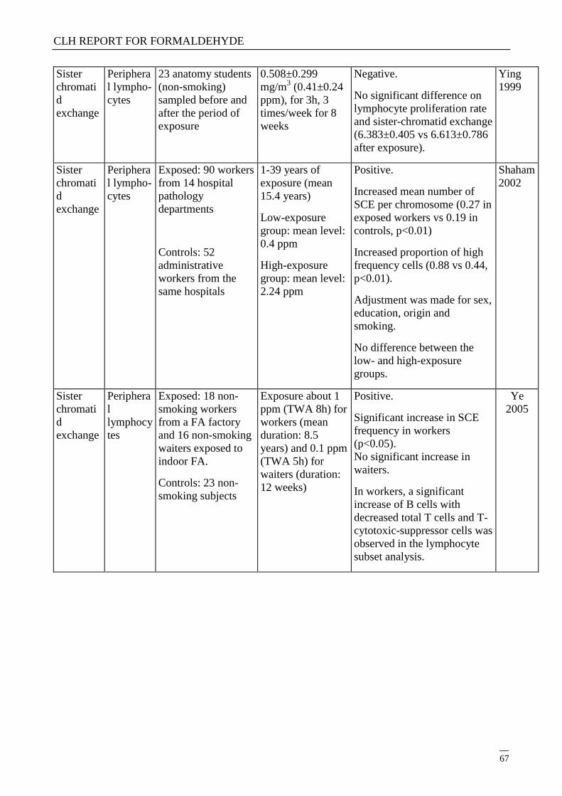
CLH REPORT FOR FORMALDEHYDE
67
Sister
chromati
d
exchange
Periphera
l lympho-
cytes
23 anatomy students
(non-smoking)
sampled before and
after the period of
exposure
0.508±0.299
mg/m3 (0.41±0.24
ppm), for 3h, 3
times/week for 8
weeks
Negative.
No significant difference on
lymphocyte proliferation rate
and sister-chromatid exchange
(6.383±0.405 vs 6.613±0.786
after exposure).
Ying
1999
Sister
chromati
d
exchange
Periphera
l lympho-
cytes
Exposed: 90 workers
from 14 hospital
pathology
departments
Controls: 52
administrative
workers from the
same hospitals
1-39 years of
exposure (mean
15.4 years)
Low-exposure
group: mean level:
0.4 ppm
High-exposure
group: mean level:
2.24 ppm
Positive.
Increased mean number of
SCE per chromosome (0.27 in
exposed workers vs 0.19 in
controls, p<0.01)
Increased proportion of high
frequency cells (0.88 vs 0.44,
p<0.01).
Adjustment was made for sex,
education, origin and
smoking.
No difference between the
low- and high-exposure
groups.
Shaham
2002
Sister
chromati
d
exchange
Periphera
l
lymphocy
tes
Exposed: 18 non-
smoking workers
from a FA factory
and 16 non-smoking
waiters exposed to
indoor FA.
Controls: 23 non-
smoking subjects
Exposure about 1
ppm (TWA 8h) for
workers (mean
duration: 8.5
years) and 0.1 ppm
(TWA 5h) for
waiters (duration:
12 weeks)
Positive.
Significant increase in SCE
frequency in workers
(p<0.05).
No significant increase in
waiters.
In workers, a significant
increase of B cells with
decreased total T cells and T-
cytotoxic-suppressor cells was
observed in the lymphocyte
subset analysis.
Ye
2005
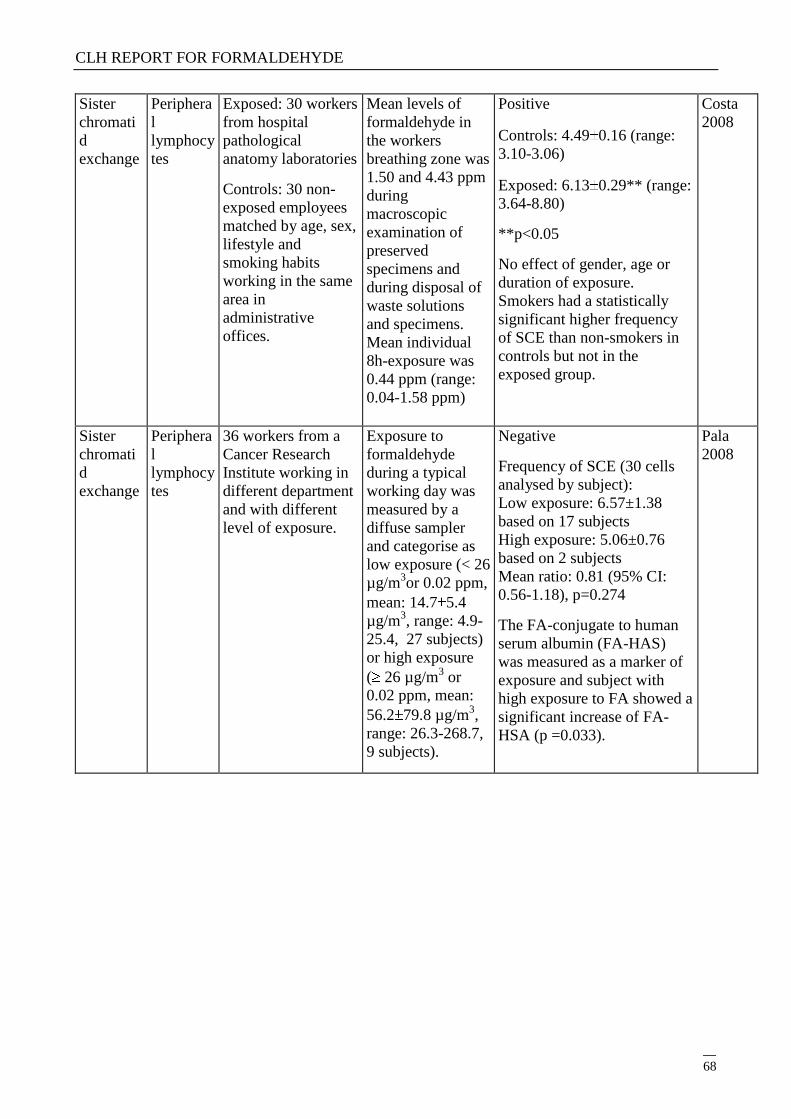
CLH REPORT FOR FORMALDEHYDE
68
Sister
chromati
d
exchange
Periphera
l
lymphocy
tes
Exposed: 30 workers
from hospital
pathological
anatomy laboratories
Controls: 30 non-
exposed employees
matched by age, sex,
lifestyle and
smoking habits
working in the same
area in
administrative
offices.
Mean levels of
formaldehyde in
the workers
breathing zone was
1.50 and 4.43 ppm
during
macroscopic
examination of
preserved
specimens and
during disposal of
waste solutions
and specimens.
Mean individual
8h-exposure was
0.44 ppm (range:
0.04-1.58 ppm)
Positive
Controls: 4.49 0.16 (range:
3.10-3.06)
Exposed: 6.13 0.29** (range:
3.64-8.80)
**p<0.05
No effect of gender, age or
duration of exposure.
Smokers had a statistically
significant higher frequency
of SCE than non-smokers in
controls but not in the
exposed group.
Costa
2008
Sister
chromati
d
exchange
Periphera
l
lymphocy
tes
36 workers from a
Cancer Research
Institute working in
different department
and with different
level of exposure.
Exposure to
formaldehyde
during a typical
working day was
measured by a
diffuse sampler
and categorise as
low exposure (< 26
µg/m3or 0.02 ppm,
mean: 14.7 5.4
µg/m3, range: 4.9-
25.4, 27 subjects)
or high exposure
( 26 µg/m3 or
0.02 ppm, mean:
56.2 79.8 µg/m3,
range: 26.3-268.7,
9 subjects).
Negative
Frequency of SCE (30 cells
analysed by subject):
Low exposure: 6.57±1.38
based on 17 subjects
High exposure: 5.06±0.76
based on 2 subjects
Mean ratio: 0.81 (95% CI:
0.56-1.18), p=0.274
The FA-conjugate to human
serum albumin (FA-HAS)
was measured as a marker of
exposure and subject with
high exposure to FA showed a
significant increase of FA-
HSA (p =0.033).
Pala
2008
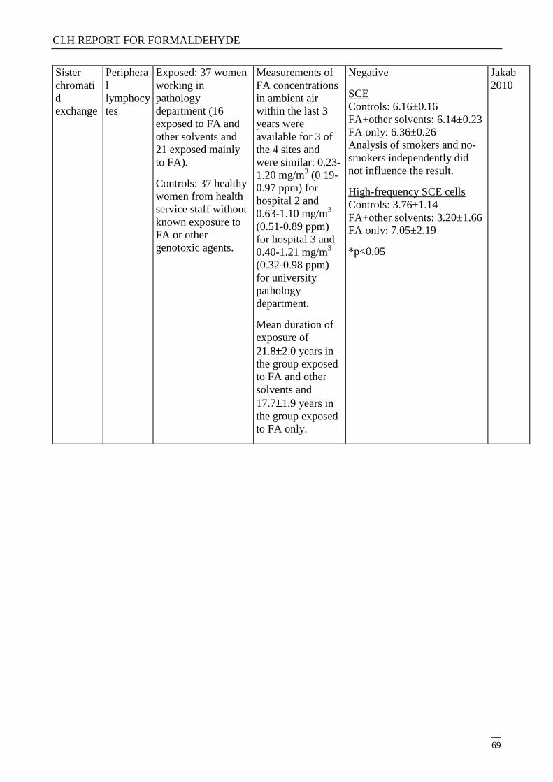
CLH REPORT FOR FORMALDEHYDE
69
Sister
chromati
d
exchange
Periphera
l
lymphocy
tes
Exposed: 37 women
working in
pathology
department (16
exposed to FA and
other solvents and
21 exposed mainly
to FA).
Controls: 37 healthy
women from health
service staff without
known exposure to
FA or other
genotoxic agents.
Measurements of
FA concentrations
in ambient air
within the last 3
years were
available for 3 of
the 4 sites and
were similar: 0.23-
1.20 mg/m3 (0.19-
0.97 ppm) for
hospital 2 and
0.63-1.10 mg/m3
(0.51-0.89 ppm)
for hospital 3 and
0.40-1.21 mg/m3
(0.32-0.98 ppm)
for university
pathology
department.
Mean duration of
exposure of
21.8 2.0 years in
the group exposed
to FA and other
solvents and
17.7 1.9 years in
the group exposed
to FA only.
Negative
SCE
Controls: 6.16±0.16
FA+other solvents: 6.14±0.23
FA only: 6.36±0.26
Analysis of smokers and no-
smokers independently did
not influence the result.
High-frequency SCE cells
Controls: 3.76±1.14
FA+other solvents: 3.20±1.66
FA only: 7.05±2.19
*p<0.05
Jakab
2010
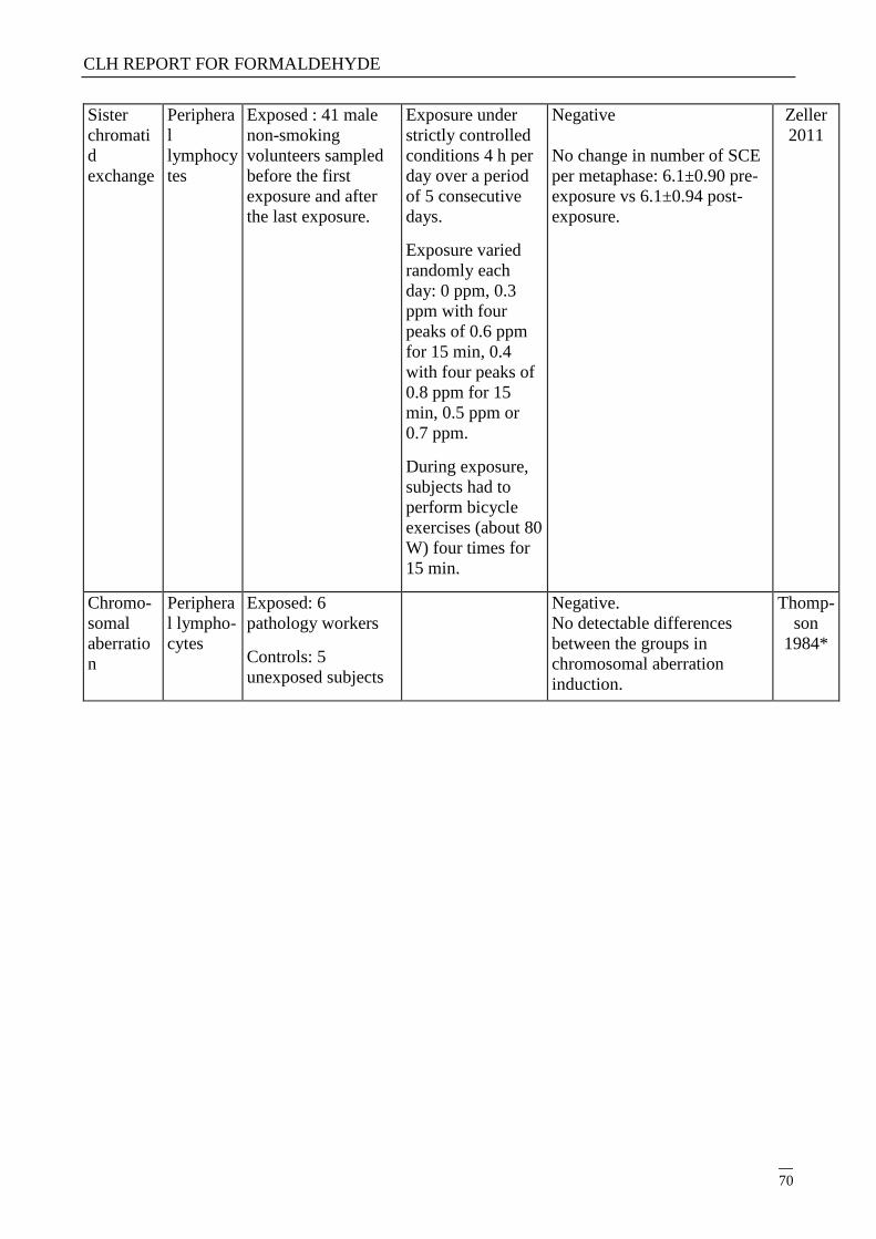
CLH REPORT FOR FORMALDEHYDE
70
Sister
chromati
d
exchange
Periphera
l
lymphocy
tes
Exposed : 41 male
non-smoking
volunteers sampled
before the first
exposure and after
the last exposure.
Exposure under
strictly controlled
conditions 4 h per
day over a period
of 5 consecutive
days.
Exposure varied
randomly each
day: 0 ppm, 0.3
ppm with four
peaks of 0.6 ppm
for 15 min, 0.4
with four peaks of
0.8 ppm for 15
min, 0.5 ppm or
0.7 ppm.
During exposure,
subjects had to
perform bicycle
exercises (about 80
W) four times for
15 min.
Negative
No change in number of SCE
per metaphase: 6.1±0.90 pre-
exposure vs 6.1±0.94 post-
exposure.
Zeller
2011
Chromo-
somal
aberratio
n
Periphera
l lympho-
cytes
Exposed: 6
pathology workers
Controls: 5
unexposed subjects
Negative.
No detectable differences
between the groups in
chromosomal aberration
induction.
Thomp-
son
1984*
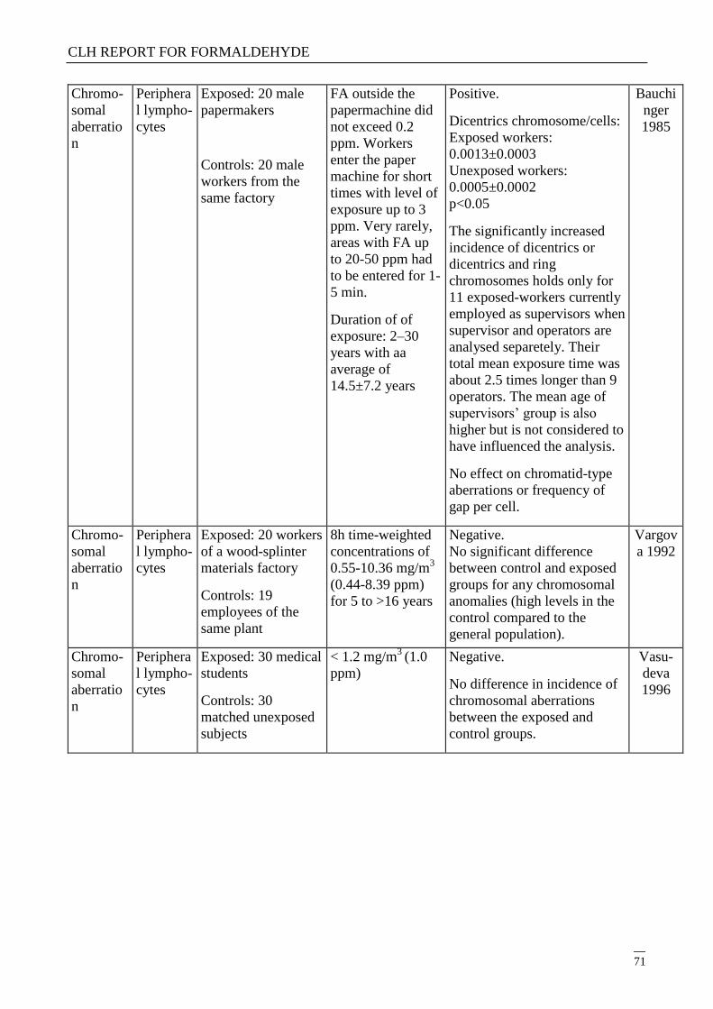
CLH REPORT FOR FORMALDEHYDE
71
Chromo-
somal
aberratio
n
Periphera
l lympho-
cytes
Exposed: 20 male
papermakers
Controls: 20 male
workers from the
same factory
FA outside the
papermachine did
not exceed 0.2
ppm. Workers
enter the paper
machine for short
times with level of
exposure up to 3
ppm. Very rarely,
areas with FA up
to 20-50 ppm had
to be entered for 1-
5 min.
Duration of of
exposure: 2–30
years with aa
average of
14.5±7.2 years
Positive.
Dicentrics chromosome/cells:
Exposed workers:
0.0013±0.0003
Unexposed workers:
0.0005±0.0002
p<0.05
The significantly increased
incidence of dicentrics or
dicentrics and ring
chromosomes holds only for
11 exposed-workers currently
employed as supervisors when
supervisor and operators are
analysed separetely. Their
total mean exposure time was
about 2.5 times longer than 9
operators. The mean age of
supervisors‘ group is also
higher but is not considered to
have influenced the analysis.
No effect on chromatid-type
aberrations or frequency of
gap per cell.
Bauchi
nger
1985
Chromo-
somal
aberratio
n
Periphera
l lympho-
cytes
Exposed: 20 workers
of a wood-splinter
materials factory
Controls: 19
employees of the
same plant
8h time-weighted
concentrations of
0.55-10.36 mg/m3
(0.44-8.39 ppm)
for 5 to >16 years
Negative.
No significant difference
between control and exposed
groups for any chromosomal
anomalies (high levels in the
control compared to the
general population).
Vargov
a 1992
Chromo-
somal
aberratio
n
Periphera
l lympho-
cytes
Exposed: 30 medical
students
Controls: 30
matched unexposed
subjects
< 1.2 mg/m3
(1.0
ppm)
Negative.
No difference in incidence of
chromosomal aberrations
between the exposed and
control groups.
Vasu-
deva
1996
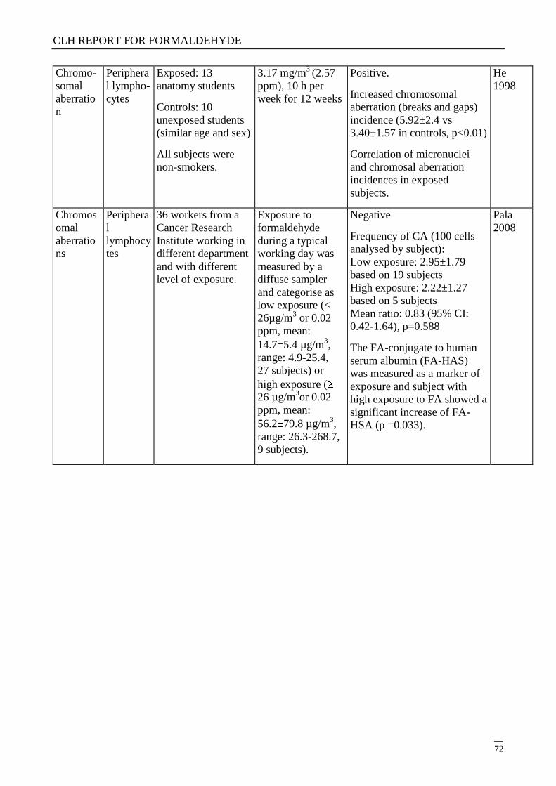
CLH REPORT FOR FORMALDEHYDE
72
Chromo-
somal
aberratio
n
Periphera
l lympho-
cytes
Exposed: 13
anatomy students
Controls: 10
unexposed students
(similar age and sex)
All subjects were
non-smokers.
3.17 mg/m3
(2.57
ppm), 10 h per
week for 12 weeks
Positive.
Increased chromosomal
aberration (breaks and gaps)
incidence (5.92±2.4 vs
3.40±1.57 in controls, p<0.01)
Correlation of micronuclei
and chromosal aberration
incidences in exposed
subjects.
He
1998
Chromos
omal
aberratio
ns
Periphera
l
lymphocy
tes
36 workers from a
Cancer Research
Institute working in
different department
and with different
level of exposure.
Exposure to
formaldehyde
during a typical
working day was
measured by a
diffuse sampler
and categorise as
low exposure (<
26µg/m3 or 0.02
ppm, mean:
14.7 5.4 µg/m3,
range: 4.9-25.4,
27 subjects) or
high exposure (
26 µg/m3or 0.02
ppm, mean:
56.2 79.8 µg/m3,
range: 26.3-268.7,
9 subjects).
Negative
Frequency of CA (100 cells
analysed by subject):
Low exposure: 2.95±1.79
based on 19 subjects
High exposure: 2.22±1.27
based on 5 subjects
Mean ratio: 0.83 (95% CI:
0.42-1.64), p=0.588
The FA-conjugate to human
serum albumin (FA-HAS)
was measured as a marker of
exposure and subject with
high exposure to FA showed a
significant increase of FA-
HSA (p =0.033).
Pala
2008
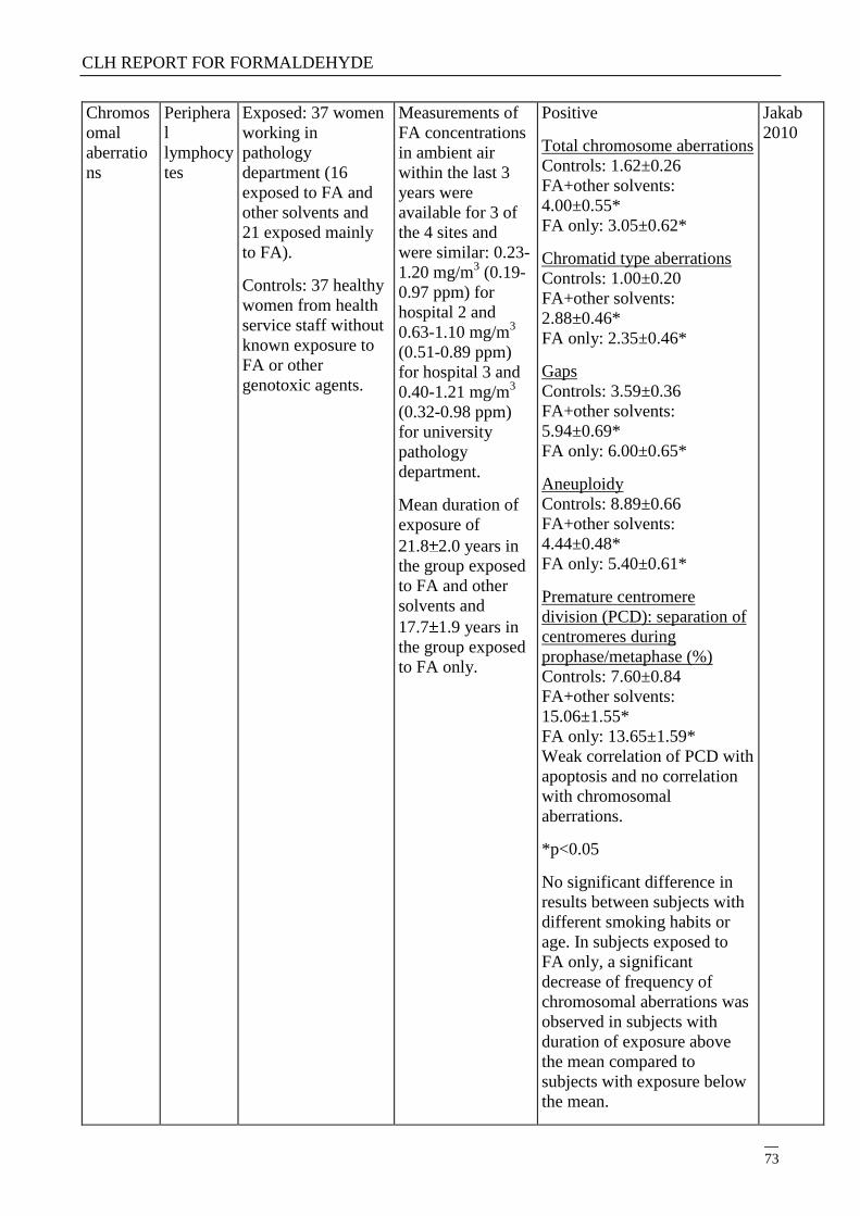
CLH REPORT FOR FORMALDEHYDE
73
Chromos
omal
aberratio
ns
Periphera
l
lymphocy
tes
Exposed: 37 women
working in
pathology
department (16
exposed to FA and
other solvents and
21 exposed mainly
to FA).
Controls: 37 healthy
women from health
service staff without
known exposure to
FA or other
genotoxic agents.
Measurements of
FA concentrations
in ambient air
within the last 3
years were
available for 3 of
the 4 sites and
were similar: 0.23-
1.20 mg/m3 (0.19-
0.97 ppm) for
hospital 2 and
0.63-1.10 mg/m3
(0.51-0.89 ppm)
for hospital 3 and
0.40-1.21 mg/m3
(0.32-0.98 ppm)
for university
pathology
department.
Mean duration of
exposure of
21.8 2.0 years in
the group exposed
to FA and other
solvents and
17.7 1.9 years in
the group exposed
to FA only.
Positive
Total chromosome aberrations
Controls: 1.62±0.26
FA+other solvents:
4.00±0.55*
FA only: 3.05±0.62*
Chromatid type aberrations
Controls: 1.00±0.20
FA+other solvents:
2.88±0.46*
FA only: 2.35±0.46*
Gaps
Controls: 3.59±0.36
FA+other solvents:
5.94±0.69*
FA only: 6.00±0.65*
Aneuploidy
Controls: 8.89±0.66
FA+other solvents:
4.44±0.48*
FA only: 5.40±0.61*
Premature centromere
division (PCD): separation of
centromeres during
prophase/metaphase (%)
Controls: 7.60±0.84
FA+other solvents:
15.06±1.55*
FA only: 13.65±1.59*
Weak correlation of PCD with
apoptosis and no correlation
with chromosomal
aberrations.
*p<0.05
No significant difference in
results between subjects with
different smoking habits or
age. In subjects exposed to
FA only, a significant
decrease of frequency of
chromosomal aberrations was
observed in subjects with
duration of exposure above
the mean compared to
subjects with exposure below
the mean.
Jakab
2010
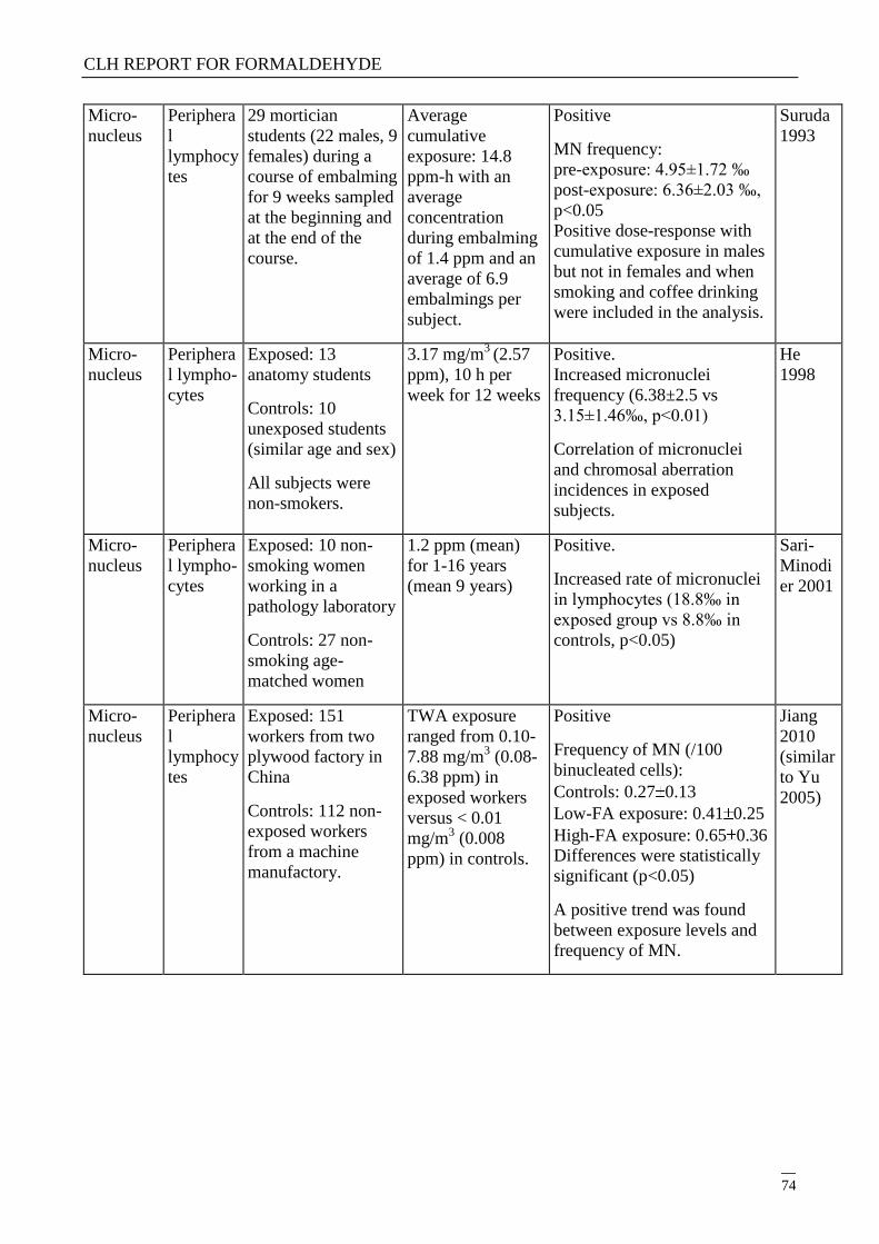
CLH REPORT FOR FORMALDEHYDE
74
Micro-
nucleus
Periphera
l
lymphocy
tes
29 mortician
students (22 males, 9
females) during a
course of embalming
for 9 weeks sampled
at the beginning and
at the end of the
course.
Average
cumulative
exposure: 14.8
ppm-h with an
average
concentration
during embalming
of 1.4 ppm and an
average of 6.9
embalmings per
subject.
Positive
MN frequency:
pre-exposure: 4.95±1.72 ‰
post-exposure: 6.36±2.03 ‰,
p<0.05
Positive dose-response with
cumulative exposure in males
but not in females and when
smoking and coffee drinking
were included in the analysis.
Suruda
1993
Micro-
nucleus
Periphera
l lympho-
cytes
Exposed: 13
anatomy students
Controls: 10
unexposed students
(similar age and sex)
All subjects were
non-smokers.
3.17 mg/m3
(2.57
ppm), 10 h per
week for 12 weeks
Positive.
Increased micronuclei
frequency (6.38±2.5 vs
3.15±1.46‰, p<0.01)
Correlation of micronuclei
and chromosal aberration
incidences in exposed
subjects.
He
1998
Micro-
nucleus
Periphera
l lympho-
cytes
Exposed: 10 non-
smoking women
working in a
pathology laboratory
Controls: 27 non-
smoking age-
matched women
1.2 ppm (mean)
for 1-16 years
(mean 9 years)
Positive.
Increased rate of micronuclei
in lymphocytes (18.8‰ in
exposed group vs 8.8‰ in
controls, p<0.05)
Sari-
Minodi
er 2001
Micro-
nucleus
Periphera
l
lymphocy
tes
Exposed: 151
workers from two
plywood factory in
China
Controls: 112 non-
exposed workers
from a machine
manufactory.
TWA exposure
ranged from 0.10-
7.88 mg/m3 (0.08-
6.38 ppm) in
exposed workers
versus < 0.01
mg/m3 (0.008
ppm) in controls.
Positive
Frequency of MN (/100
binucleated cells):
Controls: 0.27 0.13
Low-FA exposure: 0.41 0.25
High-FA exposure: 0.65 0.36
Differences were statistically
significant (p<0.05)
A positive trend was found
between exposure levels and
frequency of MN.
Jiang
2010
(similar
to Yu
2005)
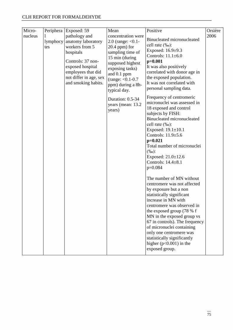
CLH REPORT FOR FORMALDEHYDE
75
Micro-
nucleus
Periphera
l
lymphocy
tes
Exposed: 59
pathology and
anatomy laboratory
workers from 5
hospitals
Controls: 37 non-
exposed hospital
employees that did
not differ in age, sex
and smoking habits.
Mean
concentration were
2.0 (range: <0.1-
20.4 ppm) for
sampling time of
15 min (during
supposed highest
exposing tasks)
and 0.1 ppm
(range: <0.1-0.7
ppm) during a 8h-
typical day.
Duration: 0.5-34
years (mean: 13.2
years)
Positive
Binucleated micronucleated
cell rate (‰):
Exposed: 16.9±9.3
Controls: 11.1±6.0
p=0.001
It was also positively
correlated with donor age in
the exposed population.
It was not correlated with
personal sampling data.
Frequency of centromeric
micronuclei was assessed in
18 exposed and control
subjects by FISH:
Binucleated micronucleated
cell rate (‰):
Exposed: 19.1±10.1
Controls: 11.9±5.6
p=0.021
Total number of micronuclei
(‰):
Exposed: 21.0±12.6
Controls: 14.4±8.1
p=0.084
The number of MN without
centromere was not affected
by exposure but a non
statistically significant
increase in MN with
centromere was observed in
the exposed group (78 % f
MN in the exposed group vs
67 in controls). The frequency
of micronuclei containing
only one centromere was
statistically significantly
higher (p<0.001) in the
exposed group.
Orsière
2006
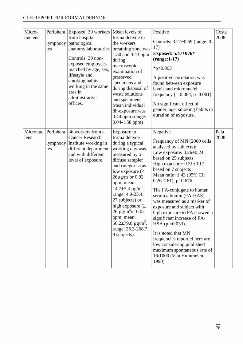
CLH REPORT FOR FORMALDEHYDE
76
Micro-
nucleus
Periphera
l
lymphocy
tes
Exposed: 30 workers
from hospital
pathological
anatomy laboratories
Controls: 30 non-
exposed employees
matched by age, sex,
lifestyle and
smoking habits
working in the same
area in
administrative
offices.
Mean levels of
formaldehyde in
the workers
breathing zone was
1.50 and 4.43 ppm
during
macroscopic
examination of
preserved
specimens and
during disposal of
waste solutions
and specimens.
Mean individual
8h-exposure was
0.44 ppm (range:
0.04-1.58 ppm)
Positive
Controls: 3.27 0.69 (range: 0-
17)
Exposed: 5.47 076*
(range:1-17)
*p<0.003
A positive correlation was
found between exposure
levels and micronuclei
frequency (r=0.384, p=0.001).
No significant effect of
gender, age, smoking habits or
duration of exposure.
Costa
2008
Micronuc
leus
Periphera
l
lymphocy
tes
36 workers from a
Cancer Research
Institute working in
different department
and with different
level of exposure.
Exposure to
formaldehyde
during a typical
working day was
measured by a
diffuse sampler
and categorise as
low exposure (<
26µg/m3or 0.02
ppm, mean:
14.7 5.4 µg/m3,
range: 4.9-25.4,
27 subjects) or
high exposure (
26 µg/m3or 0.02
ppm, mean:
56.2 79.8 µg/m3,
range: 26.3-268.7,
9 subjects).
Negative
Frequency of MN (2000 cells
analysed by subjects):
Low exposure: 0.26±0.24
based on 25 subjects
High exposure: 0.31±0.17
based on 7 subjects
Mean ratio: 1.43 (95% CI:
0.26-7.81), p=0.676
The FA-conjugate to human
serum albumin (FA-HAS)
was measured as a marker of
exposure and subject with
high exposure to FA showed a
significant increase of FA-
HSA (p =0.033).
It is noted that MN
frequencies reported here are
low considering published
maximum spontaneous rate of
16/1000 (Van Hummelen
1990)
Pala
2008
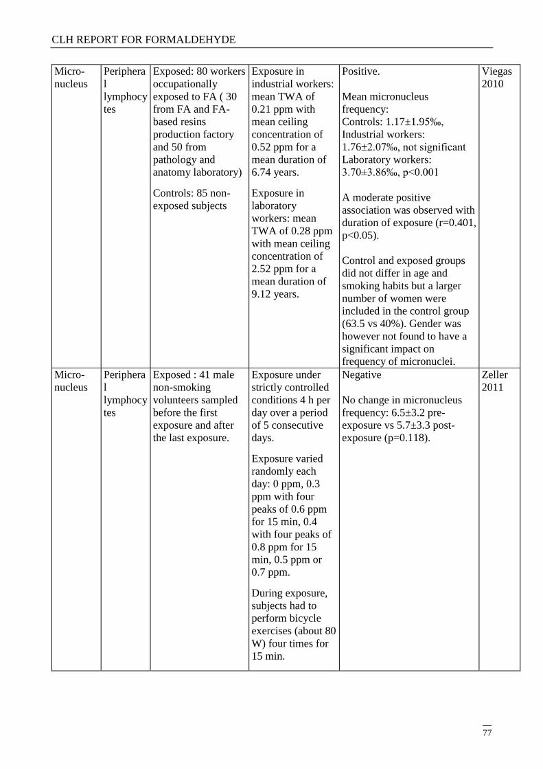
CLH REPORT FOR FORMALDEHYDE
77
Micro-
nucleus
Periphera
l
lymphocy
tes
Exposed: 80 workers
occupationally
exposed to FA ( 30
from FA and FA-
based resins
production factory
and 50 from
pathology and
anatomy laboratory)
Controls: 85 non-
exposed subjects
Exposure in
industrial workers:
mean TWA of
0.21 ppm with
mean ceiling
concentration of
0.52 ppm for a
mean duration of
6.74 years.
Exposure in
laboratory
workers: mean
TWA of 0.28 ppm
with mean ceiling
concentration of
2.52 ppm for a
mean duration of
9.12 years.
Positive.
Mean micronucleus
frequency:
Controls: 1.17±1.95‰,
Industrial workers:
1.76±2.07‰, not significant
Laboratory workers:
3.70±3.86‰, p<0.001
A moderate positive
association was observed with
duration of exposure (r=0.401,
p<0.05).
Control and exposed groups
did not differ in age and
smoking habits but a larger
number of women were
included in the control group
(63.5 vs 40%). Gender was
however not found to have a
significant impact on
frequency of micronuclei.
Viegas
2010
Micro-
nucleus
Periphera
l
lymphocy
tes
Exposed : 41 male
non-smoking
volunteers sampled
before the first
exposure and after
the last exposure.
Exposure under
strictly controlled
conditions 4 h per
day over a period
of 5 consecutive
days.
Exposure varied
randomly each
day: 0 ppm, 0.3
ppm with four
peaks of 0.6 ppm
for 15 min, 0.4
with four peaks of
0.8 ppm for 15
min, 0.5 ppm or
0.7 ppm.
During exposure,
subjects had to
perform bicycle
exercises (about 80
W) four times for
15 min.
Negative
No change in micronucleus
frequency: 6.5±3.2 pre-
exposure vs 5.7±3.3 post-
exposure (p=0.118).
Zeller
2011
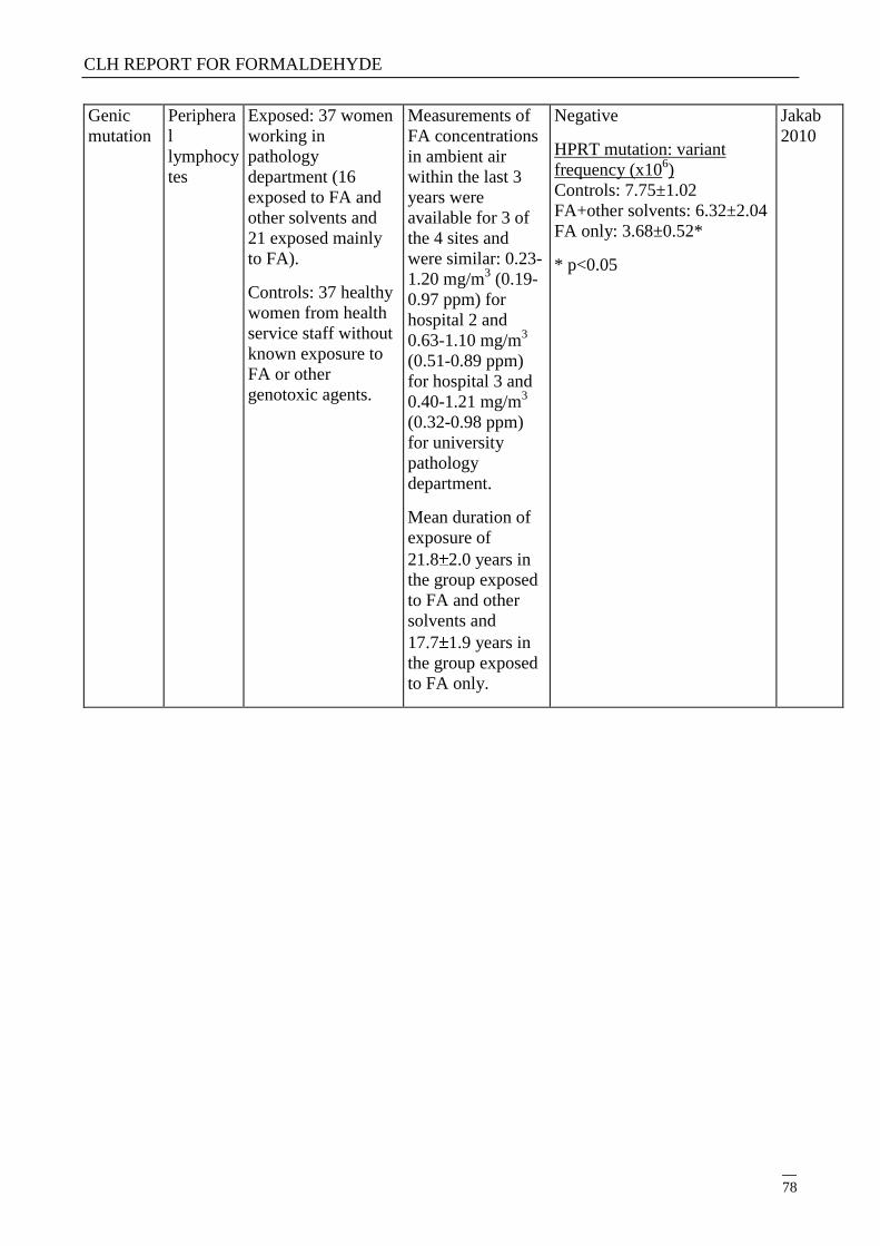
CLH REPORT FOR FORMALDEHYDE
78
Genic
mutation
Periphera
l
lymphocy
tes
Exposed: 37 women
working in
pathology
department (16
exposed to FA and
other solvents and
21 exposed mainly
to FA).
Controls: 37 healthy
women from health
service staff without
known exposure to
FA or other
genotoxic agents.
Measurements of
FA concentrations
in ambient air
within the last 3
years were
available for 3 of
the 4 sites and
were similar: 0.23-
1.20 mg/m3 (0.19-
0.97 ppm) for
hospital 2 and
0.63-1.10 mg/m3
(0.51-0.89 ppm)
for hospital 3 and
0.40-1.21 mg/m3
(0.32-0.98 ppm)
for university
pathology
department.
Mean duration of
exposure of
21.8 2.0 years in
the group exposed
to FA and other
solvents and
17.7 1.9 years in
the group exposed
to FA only.
Negative
HPRT mutation: variant
frequency (x106)
Controls: 7.75±1.02
FA+other solvents: 6.32±2.04
FA only: 3.68±0.52*
* p<0.05
Jakab
2010
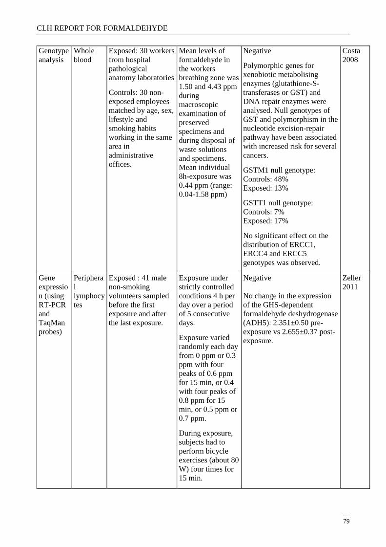
CLH REPORT FOR FORMALDEHYDE
79
Genotype
analysis
Whole
blood
Exposed: 30 workers
from hospital
pathological
anatomy laboratories
Controls: 30 non-
exposed employees
matched by age, sex,
lifestyle and
smoking habits
working in the same
area in
administrative
offices.
Mean levels of
formaldehyde in
the workers
breathing zone was
1.50 and 4.43 ppm
during
macroscopic
examination of
preserved
specimens and
during disposal of
waste solutions
and specimens.
Mean individual
8h-exposure was
0.44 ppm (range:
0.04-1.58 ppm)
Negative
Polymorphic genes for
xenobiotic metabolising
enzymes (glutathione-S-
transferases or GST) and
DNA repair enzymes were
analysed. Null genotypes of
GST and polymorphism in the
nucleotide excision-repair
pathway have been associated
with increased risk for several
cancers.
GSTM1 null genotype:
Controls: 48%
Exposed: 13%
GSTT1 null genotype:
Controls: 7%
Exposed: 17%
No significant effect on the
distribution of ERCC1,
ERCC4 and ERCC5
genotypes was observed.
Costa
2008
Gene
expressio
n (using
RT-PCR
and
TaqMan
probes)
Periphera
l
lymphocy
tes
Exposed : 41 male
non-smoking
volunteers sampled
before the first
exposure and after
the last exposure.
Exposure under
strictly controlled
conditions 4 h per
day over a period
of 5 consecutive
days.
Exposure varied
randomly each day
from 0 ppm or 0.3
ppm with four
peaks of 0.6 ppm
for 15 min, or 0.4
with four peaks of
0.8 ppm for 15
min, or 0.5 ppm or
0.7 ppm.
During exposure,
subjects had to
perform bicycle
exercises (about 80
W) four times for
15 min.
Negative
No change in the expression
of the GHS-dependent
formaldehyde deshydrogenase
(ADH5): 2.351±0.50 pre-
exposure vs 2.655±0.37 post-
exposure.
Zeller
2011
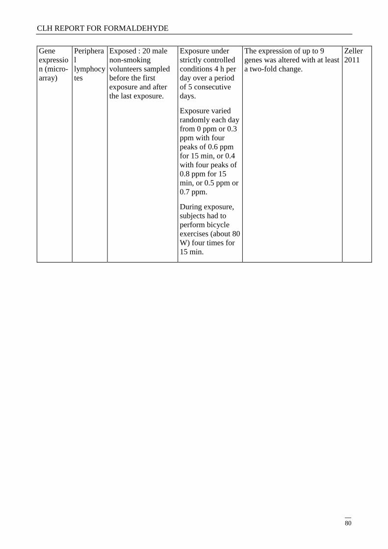
CLH REPORT FOR FORMALDEHYDE
80
Gene
expressio
n (micro-
array)
Periphera
l
lymphocy
tes
Exposed : 20 male
non-smoking
volunteers sampled
before the first
exposure and after
the last exposure.
Exposure under
strictly controlled
conditions 4 h per
day over a period
of 5 consecutive
days.
Exposure varied
randomly each day
from 0 ppm or 0.3
ppm with four
peaks of 0.6 ppm
for 15 min, or 0.4
with four peaks of
0.8 ppm for 15
min, or 0.5 ppm or
0.7 ppm.
During exposure,
subjects had to
perform bicycle
exercises (about 80
W) four times for
15 min.
The expression of up to 9
genes was altered with at least
a two-fold change.
Zeller
2011
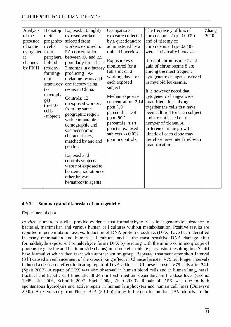
CLH REPORT FOR FORMALDEHYDE
81
Analysis
of the
presence
of some
cytogenet
ic
changes
by FISH
Hematop
oietic
progenito
r cells
from
periphera
l blood
(colony-
forming-
unit-
granulocy
te-
macropha
ge)
(n=150
cells
/subject)
Exposed: 10 highly
exposed workers
selected from
workers exposed to
FA concentration
between 0.6 and 2.5
ppm daily for at least
3 months in a factory
producing FA-
melanine resins and
one factory using
resins in China.
Controls: 12
unexposed workers
from the same
geographic region
with comparable
demographic and
socioeconomic
characteristics,
matched by age and
gender.
Exposed and
controls subjects
were not exposed to
benzene, radiation or
other known
hematotoxic agents
Occupational
exposure collected
by a questionnaire
administered by a
trained interview.
Exposure was
monitored for a
full shift on 3
working days for
each exposed
subject.
Median exposure
concentration: 2.14
ppm (10th
percentile: 1.38
ppm; 90th
percentile: 4.14
ppm) in exposed
subjects vs 0.032
ppm in controls.
The frequency of loss of
chromosome 7 (p=0.0039)
and of trisomy of
chromosome 8 (p=0.040)
were statistically increased.
Loss of chromosome 7 and
gain of chromosome 8 are
among the most frequent
cytogenetic changes observed
in myeloid leukaemia.
It is however noted that
cytogenetic changes were
quantified after mixing
together the cells that have
been cultured for each subject
and are not based on the
number of clones. A
difference in the growth
kinetic of each clone may
therefore have interfered with
quantification.
Zhang
2010
4.9.3 Summary and discussion of mutagenicity
Experimental data
In vitro, numerous studies provide evidence that formaldehyde is a direct genotoxic substance in
bacterial, mammalian and various human cell cultures without metabolisation. Positive results are
reported in gene mutation assays. Induction of DNA-protein crosslinks (DPX) have been identified
in many mammalian and human cell cultures and is the most sensitive DNA damage after
formaldehyde exposure. Formaldehyde forms DPX by reacting with the amino or imino groups of
proteins (e.g. lysine and histidine side chains) or of nucleic acids (e.g. cytosine) resulting in a Schiff
base formation which then react with another amino group. Repeated treatment after short interval
(3 h) caused an enhancement of the crosslinking effect in Chinese hamster V79 but longer intervals
induced a decreased effect indicating repair of DNA-adduct in Chinese hamster V79 cells after 24 h
(Speit 2007). A repair of DPX was also observed in human blood cells and in human lung, nasal,
tracheal and hepatic cell lines after 8-24h in fresh medium depending on the dose level (Cosma
1988, Liu 2006, Schmidt 2007, Speit 2008, Zhao 2009). Repair of DPX was due to both
spontaneous hydrolysis and active repair in human lymphocytes and human cell lines (Quievryn
2000). A recent study from Neuss et al. (2010b) comes to the conclusion that DPX adducts are the
CLH REPORT FOR FORMALDEHYDE
82
most relevant primary DNA alterations induced by formaldehyde exposure. They are repaired to a
similar extent of their induction post-incubation after repeated treatments at low exposure but
persistence of DPX has been observed in some studies for exposure to higher formaldehyde
concentrations (Schmid 2007). Under test tube conditions, formaldehyde glutathione-conjugate was
also observed to link to DNA (Lu 2009).
Positive results on strand break induction were obtained in several studies and in particular on both
human lymphocytes and Hela cell lines at low concentration but not at higher concentrations in Liu
et al. (2006), indicating that at higher concentrations DPX formation may mask the detection of
strand breaks in the Comet assay. Using a post-treatment with proteinase K, which abolishes
crosslinking effect of formaldehyde, the detection of strand breaks was observed in rat epithelial
tracheal cells (Cosma 1988) but not in in Chinese hamster V79 cells (Speit 2007). A complete
repair of strand breaks 2 hr after exposure was noted by Cosma et al (1988). It was also observed
that the repair of UV-induced single-strand breaks was delayed in presence of formaldehyde (Emri
2004).
Induction of sister chromatid exchanges (SCE) was observed in mammalian cells and in human
blood cells in several studies as well as induction of chromosomal aberrations.
Induction of micronuclei was observed in mammalian and human cells. It was detected in Schmid et
al. (2007) only under specific experimental conditions with indication of an effect on chromosome
breaks. However, it was observed under standard conditions in Merck et al. (1998), Speit et al.
(2007) and Speit et al. (2000). In Speit et al. (2007), repeated treatment with 24-hr intervals did not
show an accumulation of micronuclei. However, the meaning of this finding is unclear considering
that some micronucleated cells may be discarded by apoptosis.
Formaldehyde has also been shown to induce gene mutations in V79 cells in Grafstrom et al. (1993)
but not in Merck et al. (1998). Positive results are also reported in the MLA assay in Blackburn et
al. (1991) and in Mackerer et al. (1996) and with indications of an effect on chromosomal damage
in Speit et al. (2002). The effect was not observed in presence of FA deshydrogenase confirming
that the genotoxic effect was due to unmetabolised FA (Blackburn 1991).
Altogether, these data indicate that formaldehyde has the potential to damage DNA in vitro.
In vivo, at the site of contact, induction of DPX by inhalation was observed in rats in the nasal
mucosa and in monkeys in the nasal turbinates and to a lower extent in the respiratory tract
(Casanova 1991, Lu 2010, Lu 2011, Moeller 2011). A dose-related increase in DNA damaged as
measured by a Comet assay (Sul 2007) was also observed in rats although the detection of such an
effect by a Comet assay may be conflicting with the presence of DPX that lead to a decrease in
DNA migration. Besides, weak but positive genotoxic effects are observed such as the induction of
respectively micronuclei at irritating doses in the gastrointestinal tract via oral route (Migliore
1989) and of chromosomal aberrations in pulmonary cells at the highest dose of 15 ppm by
inhalation (Dallas 1992). Compared to the OECD guideline, this latter study display no positive
control and fewer cells were analysed than recommended (50 cells/animal instead of 100 in the
guideline). However, these limitations were not considered to affect the validity of the study
considering that a positive and statistically significant effect was observed at the highest dose in
spite of the small number of cells analysed. No increase of micronucleus frequency was found in
nasal epithelial cells by inhalation at 20 ppm but in these experimental conditions that induced
massive damages in the respiratory epithelium after repeated exposure positive controls also gave a
negative result and the study is therefore considered of poor reliability (BASF 2001b). The recent
study by Neuss et al. (2010c) also found no evidence of DPX in the modified Comet assay and did
CLH REPORT FOR FORMALDEHYDE
83
not reproduce the induction of chromosomal aberrations in its micronucleus assay under
experimental conditions comparable to Dallas et al. (1992). It should be noted that in Neuss 2010c
the positive controls did not give an appropriate response for micronuclei induction. This study was
performed according to a non-standard protocol that may explain why the standard positive control
used in this assay is not appropriate in this case.
Investigations have shown that formaldehyde induces DNA-protein crosslinks in vivo in rats and
monkeys with site-specific rate of DPX formation and a non-linear relationship with formaldehyde
concentration. A comparative investigation found that induction of SCE and micronuclei induction
is parallel to DPX formation in vitro, although subsequent induction of gene mutation remains
unclear (Merk 1998). Observed DNA damage suggests a mechanism in which DPX prevents
replication of DNA (Heck 1999). Inhibition of replication may enhance SCE formation and
incomplete repair of DNA might lead to chromosomal aberrations and micronuclei through
chromosomal breaks. DPX formation appears therefore as an essential step in the genotoxic events
induced by formaldehyde.However, the absence of DPX accumulation following repeated exposure
suggests a rapid removal, involving efficient enzymatic removal system or spontaneous dissociation
(Casanova 1994). Besides, inhibition of replication by DPX may induce a delay in replication and
therefore an inhibitory effect on cell division. Indeed, a J-shaped dose-response in regenerative cell
proliferation (RCP) is observed in rats in vivo in Monticello et al. (1996) with rates of RCP slightly
lower than control at 0.7 and 2 ppm (Conolly 2002, Gaylor 2004). A delay in cell replication at low
dose was however not confirmed by the findings of Meng et al. (2010) observing a dose-related
increase in cell proliferation from 0.7 ppm and significant from 10 ppm.
Cell division is a necessary step in mutation fixation and acceleration in cell cycle do not allow
extensive DNA repair before replication. At low dose, the incremental DNA damage may therefore
be repaired at non-elevated levels in cell proliferation. This may explain that mutagenic effects are
only observed at high doses as confirmed by the observation of chromosomal aberrations in vivo at
15 ppm only (Dallas 1992).
Besides, recent studies able to discriminate between DNA-adducts of endogenous or or exogenous
origin shows that the level of exogenous DNA-adducts in rat nasal epithelium is of similar order of
magnitude than endogenous DNA-adduct level up to 9 ppm but is dramatically increased at 15 ppm
(Lu 2011).
In vivo, on somatic cells at distant sites of exposure, no adduct to DNA were detected in different
organs of rats at 10 and 15 ppm (Lu 2010, Lu 2011) or in the bone marrow of monkeys up to 6 ppm.
Similarly, DPX were not observed in the blood of rats up to 15 ppm (Speit 2009) but DPX were
found in the liver cells of mice from 0.8 ppm (Zhao 2009). Im et al. (2006) observed DNA damage
in the Comet assay in the liver and lymphocytes from 5 ppm. Several studies show that
formaldehyde does not induce sister chromatid exchanges, chromosomal aberrations or micronuclei
in the rat by inhalation (Speit 2009, Kligerman 1984, Dallas 1992), in mice by IP (Natarajan 1983),
oral and i.v. routes (Morita 1997) or in monkeys by inhalation (Moeller 2011). However, Kitaeva et
al. (1990) observed an increased incidence of chromosomal aberrations in the bone marrow
following repeated exposure by inhalation. The reliability of the study was difficult to establish as
the complete publication is not available (in Russian) and results are challenged by the negative
findings of Dallas et al. and of Kligerman et al. at similar doses.
In vivo, on germ cells, effects in mammals were investigated in several intraperitoneal (IP) studies
that came to inconsistent results. In particular in the recent study by Tang et al. (2003), dose related
increases in SCE and micronuclei in germ cells were observed. It is consistent with fetal loss
observed further to male exposure in Odeigah et al. (1997). However, the dose used in this study
CLH REPORT FOR FORMALDEHYDE
84
were much lower than doses inducing chromosomal effects in Tang et al. (2003) introducing some
inconsistency. However, positive results obtained via intraperitoneal route are not considered as
relevant to evaluate the mutagenic potential of formaldehyde on germ cells as normal metabolic
pathways are bypassed by IP administration and the test agent is delivered close to the site of
contact where it may create a massive irritation. A single study of dominant lethal mutation assay
was performed by inhalation (Kitaeva 1990) and provides a weak positive result but as discussed
above the reliability of this study cannot be assessed. Liu et al. (2009) identified induction of
mutations in sperm cells of males exposed to a very high dose of formaldehyde (160 ppm) by
inhalation. This study was performed according to a non-standard protocol. Besides, such a high
dose is expected to induce excessive toxicity that may interfere with normal physiology of the
animal. Besides, inhalation of formaldehyde doesn‘t modify formaldehyde blood levels in rats,
monkeys and humans and due to its high reactivity, its rapid metabolism and detoxification
formaldehyde is not expected to reach distant site (Heck 2004) and the biological plausibility for
induction of germ cell mutation is therefore weak. Further positive data were obtained in non-
mammalian species but their relevance is doubtful.
Human data
In humans at the site of contact, most available studies report an increase in the number of
micronuclei in buccal cells in people exposed to formaldehyde. The same effect was observed on
nasal mucosa cells except in Suruda et al. (1993) and its re-analysis (Titenko-Holland 1996). It is
noted that baseline control levels reported in Titenko-Holland et al. (1996) were lower than the
average micronucleus frequency in a healthy population. Co-exposure to wood dust may have
influenced the positive results in nasal mucosa cells in Ballarin et al. (1992) (Speit 2006). Only the
study by Speit et al. (2007) and Zeller et al. (2011) did not detect an increase in micronuclei in the
buccal and nasal cells respectively in studies that weres performed under controlled conditions.The
exposure and in particular the exposure to peaks may however be lower (maximum of 0.7 ppm with
15 min-peak up to 1 ppm) than in professionally or industrially exposed populations. All the studies
were however performed on a small number of subjects, which makes it difficult to interpret.
However, these positive results were observed in populations exposed in different settings such as
industrial plants (Ballarin 1992 and Ye 2005) and embalming and anatomy/ pathology laboratories
(Ying 1997, Burgaz 2001 and 2002), which supports that the positive results are not likely to be due
to co-exposures or confounding factors specific to one type of exposure. Altogether indication of a
local genotoxic effect of formaldehyde at the site of contact is provided by these studies. It is
however noted that standardisation and information on the role of confounding factors is lacking for
these protocols (Knasmueller 2011).
In humans at distant sites, many studies have investigated genotoxicity of formaldehyde in
peripheral blood lymphocytes and due to the difficulty of collecting sample of bone marrow in
humans, no data have therefore investigated genotoxicity directly in the bone marrow. While
evidence of chromosomal damages in the Comet assay are provided in Yu et al. (2005) and Costa et
al. (2008), inconsistent results are reported for induction of sister chromatid exchanges (SCE). Both
positive and negative findings are also reported in the induction of chromosomal aberrations.
However, positive results were consistently reported for micronucleus induction (Suruda 1993, He
1998, Sari-Minodier 2001, Orsière 2006, Viegas 2010), in particular in recent studies showing a
positive correlation between the micronuclei frequency and formaldehyde exposure (Yu 2005 and
Costa 2008). These positive results were observed mainly in populations exposed in embalming
procedures and anatomy/pathology laboratories but also in industrial plants in one study (Ye 2005).
Viegas et al. (2010) detected an increase in micronuclei frequency in laboratory workers but not in
industrial workers. Mean exposure between both groups was similar but laboratory workers were
CLH REPORT FOR FORMALDEHYDE
85
exposed to 5-fold higher peaks (mean 2.52 ppm). Only two studies did not observe such an effect:
no increase in micronuclei was observed in Pala et al. (2008) whereas exposure was confirmed by
presence of a marker of formaldehyde exposure in the high-exposure group. Even in the high-
exposure group the level of formaldehyde was however very low in this study (mean in the high-
exposure group of 56.2 µg/m3 or 0.046 ppm) and may explain the absence of genotoxic effects.
Besides, the number of subjects in the high-exposure group was very low (n=7 for micronuclei
analysis) and limits the reliability of this result. In Zeller et al. (2011), no genotoxicity was detected
in peripheral blood of volunteers exposed under controlled conditions. The exposure and in
particular the exposure to peaks may however be lower (maximum of 0.7 ppm with 15 min-peak up
to 1 ppm) than in professionally or industrially exposed populations.
4.9.4 Comparison with criteria
Annex VI of CLP states for the hazard class germ cell mutagenicity that ―the classification in
Category 2 is based on positive evidence obtained from experiments in mammals and/or in some
cases from in vitro experiments, obtained from:
- Somatic cell mutagenicity tests in vivo, in mammals; or
- Other in vivo somatic genotoxicity tests which are supported by positive results from in vitro
mutagenicity assay‖
In vivo at the site of contact in somatic cells, positive evidence in mutagenicity tests are available
from induction of chromosomal aberrations in rats by inhalation at high dose (Dallas 1992) and of
micronuclei in rats in the GI tract by oral route (Migliore 1989).
These positive data are further supported by:
- in vitro positive results in numerous genotoxicity and mutagenicity tests
- in vivo induction of DNA adducts and DPX at the site of contact
- indications of consistent increases in micronuclei frequency in humans at the site of contact
ECHA guidance to CLP states in section 3.5.2.1.2 that ―With the exception of in vivo studies
proving ―site of contact‖ effects, genotoxicity data from such non-standard in vivo studies are not
sufficient but may offer supporting information for classification.‖ This implies that tests non
standard because they are performed on the site of contact may be sufficient for classification and
confirms that effects at the site of contact are relevant for classification.
In vivo at distant sites in somatic cells, indications of consistent increases in micronuclei frequency
in humans is available. However, it is not supported by experimental data that report an absence of
induction of either genotoxicity or mutagenicity and by inconsistent results for induction of SCE
and chromosomal aberrations in humans.
Annex VI of CLP states for the hazard class germ cell mutagenicity that ―the classification in
Category 1B is based on:
- positive result(s) from in vivo heritable germ cell mutagenicity tests in mammals; or
- positive result(s) from in vivo somatic cell mutagenicity tests, in mammals, in combination
with some evidence that the substance has potential to cause mutations to germ cell. It is
possible to derive this supporting evidence from mutagenicity/genotoxicity tests in germ
CLH REPORT FOR FORMALDEHYDE
86
cell in vivo, or by demonstrating the ability of the substance or its metabolite(s) to interact
with genetic material of germ cells; or
- positive results from test showing mutagenic effects in the germ cells of humans, without
demonstration of transmission to progeny; for example, an increase in the frequency of
aneuploidy in sperm cells of exposed people. ‖
Positive experimental results were obtained on germinal cells in vivo. However, they were mainly
performed via intra-peritoneal route and are not considered as relevant to evaluate the mutagenic
potential of formaldehyde on germ cells as normal metabolic pathways are bypassed by IP
administration and the test agent is delivered close to the site of contact where it may create a
massive irritation. A single study of dominant lethal mutation assay was performed by inhalation
(Kitaeva 1990) and provides a weak positive result but as discussed above the reliability of this
study cannot be assessed. Besides, Liu et al. (2009) identified induction of mutations in sperm cells
of males exposed to a very high dose of formaldehyde by inhalation and such a high dose is
expected to induce excessive toxicity that may interfere with normal physiology of the animal. This
study was performed according to a non-standard protocol and its significance is unclear in
particular on the heritability of the mutations induced.
No data investigating effect on formaldehyde on human germ cells has been located.
Besides, formaldehyde is very quickly metabolised and formaldehyde inhalation does not result in
measurable changes in blood levels of formaldehyde in rats and human. In this context, the positive
results of in vitro studies and the inconsistent results in IP studies are particularly of poor relevance
in the assessment of the in vivo systemic genotoxic potential via normal routes of exposure. A
systemic genotoxic effect on germ cells is therefore unlikely.
Overall, formaldehyde induces mutagenicity in vivo on somatic cells at the site of contact but no
convincing evidence of an effect on germ cells by a relevant route of exposure is available and the
overall database support a classification in category 2.
It is noted that the hazard class for mutagenicity strictly refer to germ cells, but the CLP guidance
clearly says in section 3.5.1 (p. 286) that : ―It is also warranted that where there is evidence of only
somatic cell genotoxicity, substances are classified as suspected germ cell mutagens. Classification
as a suspected germ cell mutagen may also have implications for potential carcinogenicity
classification. This holds true espially for those genotoxicants which are incapable of causing
heritable mutations because they cannot reach the germ cells (e.g. genotoxicants only acting locally,
―site of contact‖ genotoxicants).‖
The genotoxic effect of formaldehyde on somatic cells at the site of contact is therefore relevant to
warrant a classification in category 2.
4.9.5 Conclusions on classification and labelling
Based on induction of genotoxic and mutagenic effects of FA on somatic cells at the site of contact,
classification in Category 2 is warranted.

CLH REPORT FOR FORMALDEHYDE
87
4.10 Carcinogenicity
4.10.1 Non-human information
4.10.1.1 Carcinogenicity: oral
Table 16: Experimental data on carcinogenicity by oral route
Species
Dose
mg/kg/body weight
Durat°
of
treatmt
Observations and Remarks Ref.
Wistar
rats
(n=10 to
30
males/gro
up)
Initiation: 100 mg/l
MNNG in drinking
water and 10% sodium
chloride in diet for 8
weeks
Promotion: 0.5%
formalin equivalent to
0.2% FA in drinking
water (equivalent to
2000 mg/l)
32 wk
of
promo-
tion
After initiation with MNNG, significantly
increased incidence of adenocarcinoma of the
glandular stomach (4/17, 23.5% vs 1/30, 3.3%
in the concurrent control group with initiation,
p<0.05) and significantly increased incidence
of squamous cell papilloma of the forestomach
(15/17, 88.2% vs 0/30 in the control group,
p<0.01).
Without prior initiation, significantly
increased incidence of squamous cell
papilloma of the forestomach (8/10 rats
exposed to FA only and 0/10 in the control
group, p<0.01).
Takahash
i 1986
Wistar
rats
(n=50/sex
/group)
(test
substance
:
paraforma
ldehyde
95% plus
5% water)
0, 20, 260 or 1900 mg/l
FA in drinking water
(corresponding to 0, 1.2,
15 and 82 mg/kg/d in
males and 0, 1.8, 21 and
109 mg/kg/d in females,
respectively)
2 years No effect on mortality.
In the high-dose group: decreased liquid
consumption (-40%), decreased food
consumption and reduced body weight
development; lesions in the forestomach and
in the glandular stomach likely due to the
corrosive properties of FA; kidney lesions
mainly ascribed to dehydration.
No other systemic adverse effect.
No increased incidence of gastric tumours or
tumours at other sites.
One generalised histiocytic sarcoma and one
myeloid leukaemia were observed in the males
at high dose versus none in other male and
female groups but were considered incidental.
No information is available on historical
control data.
Til, 1989
CLH REPORT FOR FORMALDEHYDE
88
Wistar
rats
(n=20/sex
/group)
(test
substance:
crystalline
para-
formaldeh
yde,
purity
80%)
0, 0.02, 0.1 or 0.5% FA
in drinking-water
(approx. 0, 10, 50 or
250 mg/kg/d)
2 year In the high-dose group: significant decreases
in body weight and food and water intake;
100% mortality by 24 months; erosions and/or
ulcers in the forestomach and glandular
stomach; squamous cell hyperplasia with or
without hyperkeratosis in the forestomach.
A few signs of irritation of the GI tract in the
0.10% group.
No increase of local or systemic tumour
incidence compared to controls (incidence of
individual tumours not given in the
publication).
Tobe
1989
Sprague-
Dawley
rats
(n=50/sex
)
(7-wk
old)
(test
substance:
formaldeh
yde
stabilised
with
methanol
0.3%,
impurities
: iron 0.6
mg/l, lead
0.1 mg/l,
sulphur
<5.0 mg/l,
chlorine
<5.0
mg/l)
0, 10, 50, 100, 500,
1000 or 1500 mg/l FA
with 0.3% methanol in
drinking water
(approx. 0, 1.28, 6.44,
12.8, 64.4, 128 and 192
mg/kg/d in males and 0,
1.45, 7.24, 14.5, 72.4,
145 and 217 mg/kg/d in
females, respectively)
+ additional methanol
control group: 15 mg/l
methanol
24 mo
(+
lifetim
e obs.)
No effect on survival or body weight
Increased incidence of all
hemolymphoreticular neoplasias in the treated
group: 22% and 14% in the males and females
at highest dose compared to 4% and 3% in the
untreated control males and females and 10%
and 6% in the methanol males and females,
respectively. No analysis performed by
subtype.
Occasional increased incidence of gastro-
intestinal tumours but not dose-related. At the
highest dose 6% of females had intestine
leiomyomas vs none in controls (historical
data: 0.04%) and 4% of males had intestine
leiomyosarcomas vs none in controls
(historical data: 0.04%).
No statistical analysis provided.
Soffritti
1989
Sprague-
Dawley
rats
(n=50/sex
)
(7-wk
0, 10, 50, 100, 500,
1000 or 1500 mg/l FA
with 0.3% methanol in
drinking water
(approx. 0, 1.28, 6.44,
12.8, 64.4, 128 and 192
mg/kg/d in males and 0,
24 mo
(+
lifetim
e obs.)
Decrease in water intake in high-dose males
and females treated over 500 mg/l. No
difference in food consumption, body weight
and survival.
Increase in total malignant tumour incidence
in males and females at 1500 mg/l, in males at
500 mg/l and in females at 1000 and 100 mg/l.
Soffritti
2002
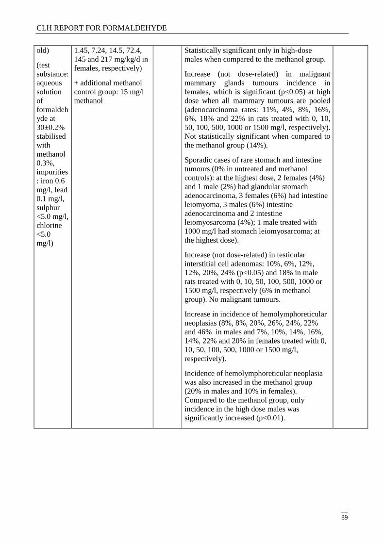
CLH REPORT FOR FORMALDEHYDE
89
old)
(test
substance:
aqueous
solution
of
formaldeh
yde at
30±0.2%
stabilised
with
methanol
0.3%,
impurities
: iron 0.6
mg/l, lead
0.1 mg/l,
sulphur
<5.0 mg/l,
chlorine
<5.0
mg/l)
1.45, 7.24, 14.5, 72.4,
145 and 217 mg/kg/d in
females, respectively)
+ additional methanol
control group: 15 mg/l
methanol
Statistically significant only in high-dose
males when compared to the methanol group.
Increase (not dose-related) in malignant
mammary glands tumours incidence in
females, which is significant (p<0.05) at high
dose when all mammary tumours are pooled
(adenocarcinoma rates: 11%, 4%, 8%, 16%,
6%, 18% and 22% in rats treated with 0, 10,
50, 100, 500, 1000 or 1500 mg/l, respectively).
Not statistically significant when compared to
the methanol group (14%).
Sporadic cases of rare stomach and intestine
tumours (0% in untreated and methanol
controls): at the highest dose, 2 females (4%)
and 1 male (2%) had glandular stomach
adenocarcinoma, 3 females (6%) had intestine
leiomyoma, 3 males (6%) intestine
adenocarcinoma and 2 intestine
leiomyosarcoma (4%); 1 male treated with
1000 mg/l had stomach leiomyosarcoma; at
the highest dose).
Increase (not dose-related) in testicular
interstitial cell adenomas: 10%, 6%, 12%,
12%, 20%, 24% (p<0.05) and 18% in male
rats treated with 0, 10, 50, 100, 500, 1000 or
1500 mg/l, respectively (6% in methanol
group). No malignant tumours.
Increase in incidence of hemolymphoreticular
neoplasias (8%, 8%, 20%, 26%, 24%, 22%
and 46% in males and 7%, 10%, 14%, 16%,
14%, 22% and 20% in females treated with 0,
10, 50, 100, 500, 1000 or 1500 mg/l,
respectively).
Incidence of hemolymphoreticular neoplasia
was also increased in the methanol group
(20% in males and 10% in females).
Compared to the methanol group, only
incidence in the high dose males was
significantly increased (p<0.01).
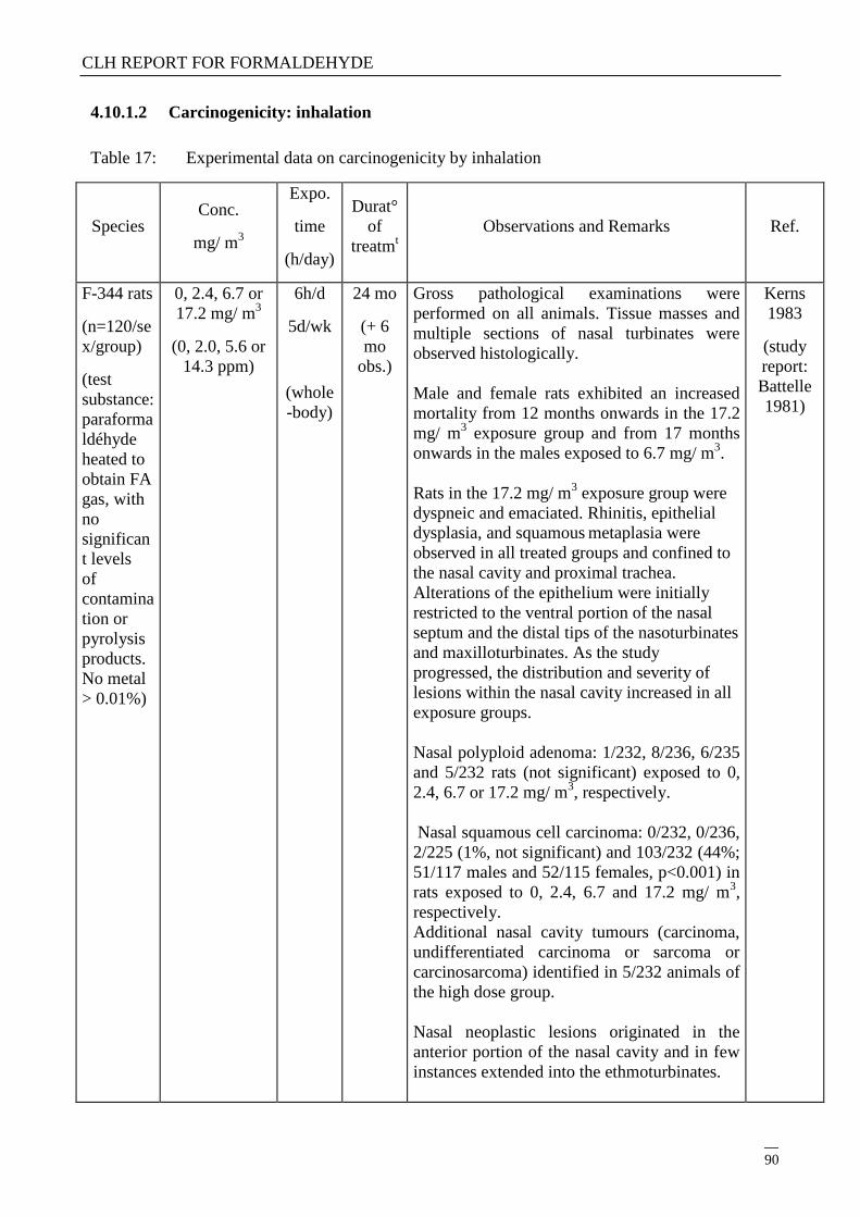
CLH REPORT FOR FORMALDEHYDE
90
4.10.1.2 Carcinogenicity: inhalation
Table 17: Experimental data on carcinogenicity by inhalation
Species Conc.
mg/ m3
Expo.
time
(h/day)
Durat°
of
treatmt
Observations and Remarks
Ref.
F-344 rats
(n=120/se
x/group)
(test
substance:
paraforma
ldéhyde
heated to
obtain FA
gas, with
no
significan
t levels
of
contamina
tion or
pyrolysis
products.
No metal
> 0.01%)
0, 2.4, 6.7 or
17.2 mg/ m3
(0, 2.0, 5.6 or
14.3 ppm)
6h/d
5d/wk
(whole
-body)
24 mo
(+ 6
mo
obs.)
Gross pathological examinations were
performed on all animals. Tissue masses and
multiple sections of nasal turbinates were
observed histologically.
Male and female rats exhibited an increased
mortality from 12 months onwards in the 17.2
mg/ m3 exposure group and from 17 months
onwards in the males exposed to 6.7 mg/ m3.
Rats in the 17.2 mg/ m3 exposure group were
dyspneic and emaciated. Rhinitis, epithelial
dysplasia, and squamous metaplasia were
observed in all treated groups and confined to
the nasal cavity and proximal trachea.
Alterations of the epithelium were initially
restricted to the ventral portion of the nasal
septum and the distal tips of the nasoturbinates
and maxilloturbinates. As the study
progressed, the distribution and severity of
lesions within the nasal cavity increased in all
exposure groups.
Nasal polyploid adenoma: 1/232, 8/236, 6/235
and 5/232 rats (not significant) exposed to 0,
2.4, 6.7 or 17.2 mg/ m3, respectively.
Nasal squamous cell carcinoma: 0/232, 0/236,
2/225 (1%, not significant) and 103/232 (44%;
51/117 males and 52/115 females, p<0.001) in
rats exposed to 0, 2.4, 6.7 and 17.2 mg/ m3,
respectively.
Additional nasal cavity tumours (carcinoma,
undifferentiated carcinoma or sarcoma or
carcinosarcoma) identified in 5/232 animals of
the high dose group.
Nasal neoplastic lesions originated in the
anterior portion of the nasal cavity and in few
instances extended into the ethmoturbinates.
Kerns
1983
(study
report:
Battelle
1981)
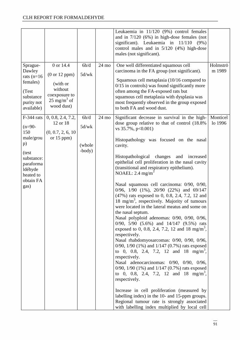
CLH REPORT FOR FORMALDEHYDE
91
Leukaemia in 11/120 (9%) control females
and in 7/120 (6%) in high-dose females (not
significant). Leukaemia in 11/110 (9%)
control males and in 5/120 (4%) high-dose
males (not significant).
Sprague-
Dawley
rats (n=16
females)
(Test
substance
purity not
available)
0 or 14.4
(0 or 12 ppm)
(with or
without
coexposure to
25 mg/m3 of
wood dust)
6h/d
5d/wk
24 mo One well differentiated squamous cell
carcinoma in the FA group (not significant).
Squamous cell metaplasia (10/16 compared to
0/15 in controls) was found significantly more
often among the FA-exposed rats but
squamous cell metaplasia with dysplasia was
most frequently observed in the group exposed
to both FA and wood dust.
Holmströ
m 1989
F-344 rats
(n=90-
150
male/grou
p)
(test
substance:
paraforma
ldéhyde
heated to
obtain FA
gas)
0, 0.8, 2.4, 7.2,
12 or 18
(0, 0.7, 2, 6, 10
or 15 ppm)
6h/d
5d/wk
(whole
-body)
24 mo
Significant decrease in survival in the high-
dose group relative to that of control (18.8%
vs 35.7%, p<0.001)
Histopathology was focused on the nasal
cavity.
Histopathological changes and increased
epithelial cell proliferation in the nasal cavity
(transitional and respiratory epithelium).
NOAEL: 2.4 mg/m3
Nasal squamous cell carcinoma: 0/90, 0/90,
0/96, 1/90 (1%), 20/90 (22%) and 69/147
(47%) rats exposed to 0, 0.8, 2.4, 7.2, 12 and
18 mg/m3, respectively. Majority of tumours
were located in the lateral meatus and some on
the nasal septum.
Nasal polyploid adenomas: 0/90, 0/90, 0/96,
0/90, 5/90 (5.6%) and 14/147 (9.5%) rats
exposed to 0, 0.8, 2.4, 7.2, 12 and 18 mg/m3,
respectively.
Nasal rhabdomyosarcomas: 0/90, 0/90, 0/96,
0/90, 1/90 (1%) and 1/147 (0.7%) rats exposed
to 0, 0.8, 2.4, 7.2, 12 and 18 mg/m3,
respectively.
Nasal adenocarcinomas: 0/90, 0/90, 0/96,
0/90, 1/90 (1%) and 1/147 (0.7%) rats exposed
to 0, 0.8, 2.4, 7.2, 12 and 18 mg/m3,
respectively.
Increase in cell proliferation (measured by
labelling index) in the 10- and 15-ppm groups.
Regional tumour rate is strongly associated
with labelling index multiplied by local cell
Monticel
lo 1996
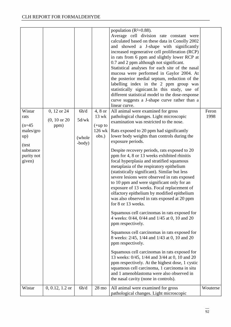
CLH REPORT FOR FORMALDEHYDE
92
population (R²=0.88).
Average cell division rate constant were
calculated based on these data in Conolly 2002
and showed a J-shape with significantly
increased regenerative cell proliferation (RCP)
in rats from 6 ppm and slightly lower RCP at
0.7 and 2 ppm although not significant.
Statistical analyses for each site of the nasal
mucosa were performed in Gaylor 2004. At
the posterior medial septum, reduction of the
labelling index in the 2 ppm group was
statistically signicant.In this study, use of
different statistical model to the dose-response
curve suggests a J-shape curve rather than a
linear curve.
Wistar
rats
(n=45
males/gro
up)
(test
substance
purity not
given)
0, 12 or 24
(0, 10 or 20
ppm)
6h/d
5d/wk
(whole
-body)
4, 8 or
13 wk
(+up to
126 wk
obs.)
All animal were examined for gross
pathological changes. Light microscopic
examination was restricted to the nose.
Rats exposed to 20 ppm had significantly
lower body weights than controls during the
exposure periods.
Despite recovery periods, rats exposed to 20
ppm for 4, 8 or 13 weeks exhibited rhinitis
focal hyperplasia and stratified squamous
metaplasia of the respiratory epithelium
(statistically significant). Similar but less
severe lesions were observed in rats exposed
to 10 ppm and were significant only for an
exposure of 13 weeks. Focal replacement of
olfactory epithelium by modified epithelium
was also observed in rats exposed at 20 ppm
for 8 or 13 weeks.
Squamous cell carcinomas in rats exposed for
4 weeks: 0/44, 0/44 and 1/45 at 0, 10 and 20
ppm respectively.
Squamous cell carcinomas in rats exposed for
8 weeks: 2/45, 1/44 and 1/43 at 0, 10 and 20
ppm respectively.
Squamous cell carcinomas in rats exposed for
13 weeks: 0/45, 1/44 and 3/44 at 0, 10 and 20
ppm respectively. At the highest dose, 1 cystic
squamous cell carcinoma, 1 carcinoma in situ
and 1 amenoblastoma were also observed in
the nasal cavity (none in controls).
Feron
1998
Wistar 0, 0.12, 1.2 or 6h/d 28 mo All animal were examined for gross
pathological changes. Light microscopic
Wouterse
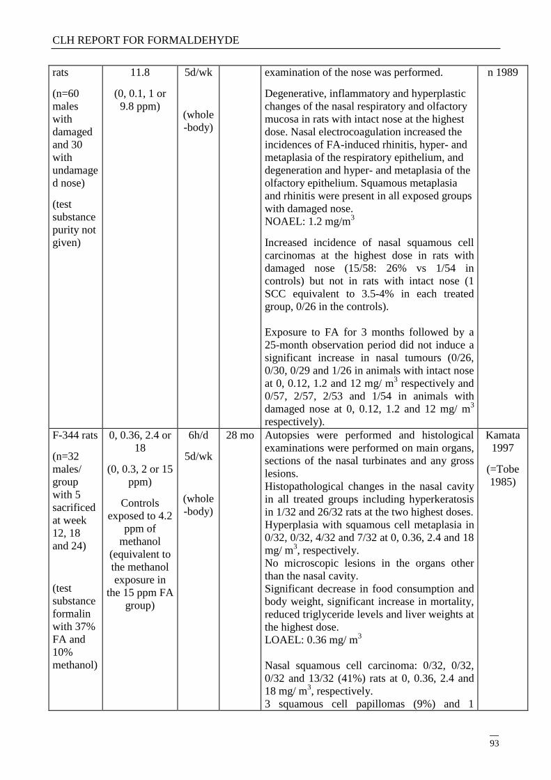
CLH REPORT FOR FORMALDEHYDE
93
rats
(n=60
males
with
damaged
and 30
with
undamage
d nose)
(test
substance
purity not
given)
11.8
(0, 0.1, 1 or
9.8 ppm)
5d/wk
(whole
-body)
examination of the nose was performed.
Degenerative, inflammatory and hyperplastic
changes of the nasal respiratory and olfactory
mucosa in rats with intact nose at the highest
dose. Nasal electrocoagulation increased the
incidences of FA-induced rhinitis, hyper- and
metaplasia of the respiratory epithelium, and
degeneration and hyper- and metaplasia of the
olfactory epithelium. Squamous metaplasia
and rhinitis were present in all exposed groups
with damaged nose.
NOAEL: 1.2 mg/m3
Increased incidence of nasal squamous cell
carcinomas at the highest dose in rats with
damaged nose (15/58: 26% vs 1/54 in
controls) but not in rats with intact nose (1
SCC equivalent to 3.5-4% in each treated
group, 0/26 in the controls).
Exposure to FA for 3 months followed by a
25-month observation period did not induce a
significant increase in nasal tumours (0/26,
0/30, 0/29 and 1/26 in animals with intact nose
at 0, 0.12, 1.2 and 12 mg/ m3 respectively and
0/57, 2/57, 2/53 and 1/54 in animals with
damaged nose at 0, 0.12, 1.2 and 12 mg/ m3
respectively).
n 1989
F-344 rats
(n=32
males/
group
with 5
sacrificed
at week
12, 18
and 24)
(test
substance
formalin
with 37%
FA and
10%
methanol)
0, 0.36, 2.4 or
18
(0, 0.3, 2 or 15
ppm)
Controls
exposed to 4.2
ppm of
methanol
(equivalent to
the methanol
exposure in
the 15 ppm FA
group)
6h/d
5d/wk
(whole
-body)
28 mo Autopsies were performed and histological
examinations were performed on main organs,
sections of the nasal turbinates and any gross
lesions.
Histopathological changes in the nasal cavity
in all treated groups including hyperkeratosis
in 1/32 and 26/32 rats at the two highest doses.
Hyperplasia with squamous cell metaplasia in
0/32, 0/32, 4/32 and 7/32 at 0, 0.36, 2.4 and 18
mg/ m3, respectively.
No microscopic lesions in the organs other
than the nasal cavity.
Significant decrease in food consumption and
body weight, significant increase in mortality,
reduced triglyceride levels and liver weights at
the highest dose.
LOAEL: 0.36 mg/ m3
Nasal squamous cell carcinoma: 0/32, 0/32,
0/32 and 13/32 (41%) rats at 0, 0.36, 2.4 and
18 mg/ m3, respectively.
3 squamous cell papillomas (9%) and 1
Kamata
1997
(=Tobe
1985)
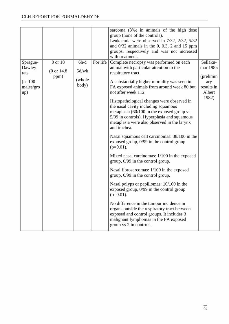
CLH REPORT FOR FORMALDEHYDE
94
sarcoma (3%) in animals of the high dose
group (none of the controls).
Leukaemia were observed in 7/32, 2/32, 5/32
and 0/32 animals in the 0, 0.3, 2 and 15 ppm
groups, respectively and was not increased
with treatment.
Sprague-
Dawley
rats
(n=100
males/gro
up)
0 or 18
(0 or 14.8
ppm)
6h/d
5d/wk
(whole
body)
For life
Complete necropsy was performed on each
animal with particular attention to the
respiratory tract.
A substantially higher mortality was seen in
FA exposed animals from around week 80 but
not after week 112.
Histopathological changes were observed in
the nasal cavity including squamous
metaplasia (60/100 in the exposed group vs
5/99 in controls). Hyperplasia and squamous
metaplasia were also observed in the larynx
and trachea.
Nasal squamous cell carcinomas: 38/100 in the
exposed group, 0/99 in the control group
(p=0.01).
Mixed nasal carcinomas: 1/100 in the exposed
group, 0/99 in the control group.
Nasal fibrosarcomas: 1/100 in the exposed
group, 0/99 in the control group.
Nasal polyps or papillomas: 10/100 in the
exposed group, 0/99 in the control group
(p=0.01).
No difference in the tumour incidence in
organs outside the respiratory tract between
exposed and control groups. It includes 3
malignant lymphomas in the FA exposed
group vs 2 in controls.
Sellaku-
mar 1985
(prelimin
ary
results in
Albert
1982)
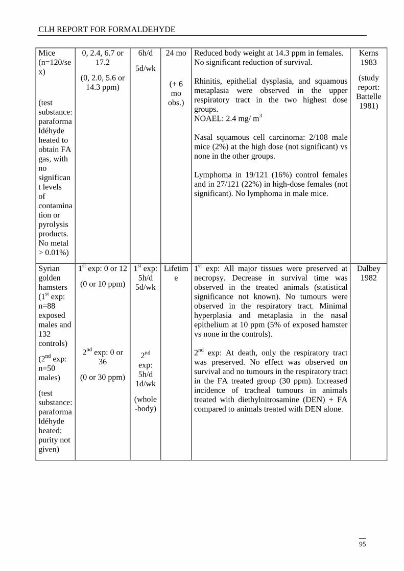
CLH REPORT FOR FORMALDEHYDE
95
Mice
(n=120/se
x)
(test
substance:
paraforma
ldéhyde
heated to
obtain FA
gas, with
no
significan
t levels
of
contamina
tion or
pyrolysis
products.
No metal
> 0.01%)
0, 2.4, 6.7 or
17.2
(0, 2.0, 5.6 or
14.3 ppm)
6h/d
5d/wk
24 mo
(+ 6
mo
obs.)
Reduced body weight at 14.3 ppm in females.
No significant reduction of survival.
Rhinitis, epithelial dysplasia, and squamous
metaplasia were observed in the upper
respiratory tract in the two highest dose
groups.
NOAEL: 2.4 mg/ m3
Nasal squamous cell carcinoma: 2/108 male
mice (2%) at the high dose (not significant) vs
none in the other groups.
Lymphoma in 19/121 (16%) control females
and in 27/121 (22%) in high-dose females (not
significant). No lymphoma in male mice.
Kerns
1983
(study
report:
Battelle
1981)
Syrian
golden
hamsters
(1st exp:
n=88
exposed
males and
132
controls)
(2nd
exp:
n=50
males)
(test
substance:
paraforma
ldéhyde
heated;
purity not
given)
1st exp: 0 or 12
(0 or 10 ppm)
2nd
exp: 0 or
36
(0 or 30 ppm)
1st exp:
5h/d
5d/wk
2nd
exp:
5h/d
1d/wk
(whole
-body)
Lifetim
e
1st exp: All major tissues were preserved at
necropsy. Decrease in survival time was
observed in the treated animals (statistical
significance not known). No tumours were
observed in the respiratory tract. Minimal
hyperplasia and metaplasia in the nasal
epithelium at 10 ppm (5% of exposed hamster
vs none in the controls).
2nd
exp: At death, only the respiratory tract
was preserved. No effect was observed on
survival and no tumours in the respiratory tract
in the FA treated group (30 ppm). Increased
incidence of tracheal tumours in animals
treated with diethylnitrosamine (DEN) + FA
compared to animals treated with DEN alone.
Dalbey
1982
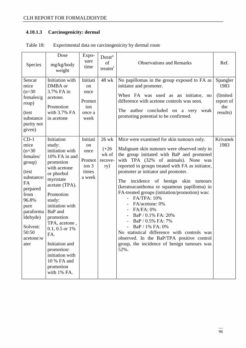
CLH REPORT FOR FORMALDEHYDE
96
4.10.1.3 Carcinogenicity: dermal
Table 18: Experimental data on carcinogenicity by dermal route
Species
Dose
mg/kg/body
weight
Expo-
sure
time
Durat°
of
treatmt
Observations and Remarks Ref.
Sencar
mice
(n=30
females/g
roup)
(test
substance
purity not
given)
Initiation with
DMBA or
3.7% FA in
acetone.
Promotion
with 3.7% FA
in acetone
Initiati
on
once
Promot
ion
once a
week
48 wk No papillomas in the group exposed to FA as
initiator and promoter.
When FA was used as an initiator, no
difference with acetone controls was seen.
The author concluded on a very weak
promoting potential to be confirmed.
Spangler
1983
(limited
report of
the
results)
CD-1
mice
(n=30
females/
group)
(test
substance:
FA
prepared
from
96.8%
pure
paraforma
ldehyde)
Solvent:
50:50
acetone:w
ater
Initiation
study:
initiation with
10% FA in and
promotion
with acetone
or phorbol
myristate
acetate (TPA).
Promotion
study:
initiation with
BaP and
promotion
TPA, acetone ,
0.1, 0.5 or 1%
FA.
Initiation and
promotion:
initiation with
10 % FA and
promotion
with 1% FA.
Initiati
on
once
Promot
ion 3
times
a week
26 wk
(+26
wk of
recove-
ry)
Mice were examined for skin tumours only.
Malignant skin tumours were observed only in
the group initiated with BaP and promoted
with TPA (32% of animals). None was
reported in groups treated with FA as initiator,
promoter ar initiator and promoter.
The incidence of benign skin tumours
(keratoacanthoma or squamous papilloma) in
FA-treated groups (initiation/promotion) was:
- FA/TPA: 10%
- FA/acetone: 0%
- FA/FA: 0%
- BaP / 0.1% FA: 20%
- BaP / 0.5% FA: 7%
- BaP / 1% FA: 0%
No statistical difference with controls was
observed. In the BaP/TPA positive control
group, the incidence of benign tumours was
52%.
Krivanek
1983
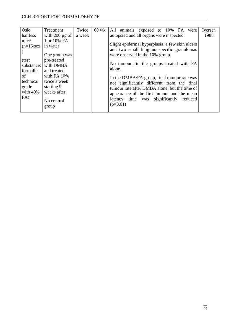
CLH REPORT FOR FORMALDEHYDE
97
Oslo
hairless
mice
(n=16/sex
)
(test
substance:
formalin
of
technical
grade
with 40%
FA)
Treatment
with 200 µg of
1 or 10% FA
in water
One group was
pre-treated
with DMBA
and treated
with FA 10%
twice a week
starting 9
weeks after.
No control
group
Twice
a week
60 wk All animals exposed to 10% FA were
autopsied and all organs were inspected.
Slight epidermal hyperplasia, a few skin ulcers
and two small lung nonspecific granulomas
were observed in the 10% group.
No tumours in the groups treated with FA
alone.
In the DMBA/FA group, final tumour rate was
not significantly different from the final
tumour rate after DMBA alone, but the time of
appearance of the first tumour and the mean
latency time was significantly reduced
(p=0.01)
Iversen
1988

CLH REPORT FOR FORMALDEHYDE
98
4.10.2 Human information
4.10.2.1 Industrial cohort studies
Table 19: Industrial cohort studies
Cohort description Estimation of
exposure
Cancer site Risk estimate Observations and remarks Ref
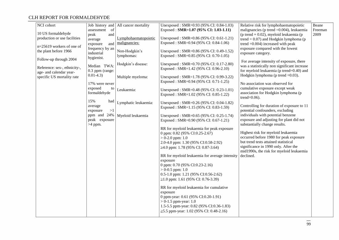
CLH REPORT FOR FORMALDEHYDE
99
NCI cohort
10 US formaldehyde
production or use facilities
n=25619 workers of one of
the plant before 1966
Follow-up through 2004
Reference: sex-, ethnicity-,
age- and calendar year-
specific US mortality rate
Job history and
assessment of
peak and
average
exposure and
frequency by an
industrial
hygienist.
Median TWA:
0.3 ppm (range:
0.01-4.3)
17% were never
exposed to
formaldehyde
15% had
average
exposure >1
ppm and 24%
peak exposure
>4 ppm.
All cancer mortality
Lymphohaematopoietic
malignancies:
Non-Hodgkin‘s
lymphomas:
Hodgkin‘s disease:
Multiple myeloma:
Leukaemia:
Lymphatic leukaemia:
Myeloid leukaemia
Unexposed : SMR=0.93 (95% CI: 0.84-1.03)
Exposed : SMR=1.07 (95% CI: 1.03-1.11)
Unexposed : SMR=0.86 (95% CI: 0.61-1.21)
Exposed : SMR=0.94 (95% CI: 0.84-1.06)
Unexposed : SMR=0.86 (95% CI: 0.49-1.52)
Exposed : SMR=0.85 (95% CI: 0.70-1.05)
Unexposed : SMR=0.70 (95% CI: 0.17-2.80)
Exposed : SMR=1.42 (95% CI: 0.96-2.10)
Unexposed : SMR=1.78 (95% CI: 0.99-3.22)
Exposed : SMR=0.94 (95% CI: 0.71-1.25)
Unexposed : SMR=0.48 (95% CI: 0.23-1.01)
Exposed : SMR=1.02 (95% CI: 0.85-1.22)
Unexposed : SMR=0.26 (95% CI: 0.04-1.82)
Exposed : SMR=1.15 (95% CI: 0.83-1.59)
Unexposed : SMR=0.65 (95% CI: 0.25-1.74)
Exposed : SMR=0.90 (95% CI: 0.67-1.21)
RR for myeloid leukaemia for peak exposure
0 ppm: 0.82 (95% CI:0.25-2.67)
> 0-2.0 ppm: 1.0
2.0-4.0 ppm: 1.30 (95% CI:0.58-2.92)
4.0 ppm: 1.78 (95% CI: 0.87-3.64)
RR for myeloid leukaemia for average intensity
exposure
0 ppm: 0.70 (95% CI:0.23-2.16)
> 0-0.5 ppm: 1.0
0.5-1.0 ppm: 1.21 (95% CI:0.56-2.62)
1.0 ppm: 1.61 (95% CI: 0.76-3.39)
RR for myeloid leukaemia for cumulative
exposure
0 ppm-year: 0.61 (95% CI:0.20-1.91)
> 0-1.5 ppm-year: 1.0
1.5-5.5 ppm-year: 0.82 (95% CI:0.36-1.83)
5.5 ppm-year: 1.02 (95% CI: 0.48-2.16)
Relative risk for lymphohaematopoietic
malignancies (p trend =0.004), leukaemia
(p trend = 0.02), myeloid leukaemia (p
trend = 0.07) and Hodgkin lymphoma (p
trend =0.004) increased with peak
exposure compared with the lowest
exposure category.
For average intensity of exposure, there
was a statistically non significant increase
for myeloid leukaemia (p trend=0.40) and
Hodgkin lymphoma (p trend =0.03).
No association was observed for
cumulative exposure except weak
association for Hodgkin lymphoma (p
trend=0.06).
Controlling for duration of exposure to 11
potential confounders, excluding
individuals with potential benzene
exposure and adjusting for plant did not
substantially change results.
Highest risk for myeloid leukaemia
occurred before 1980 for peak exposure
but trend tests attained statistical
significance in 1990 only. After the
mid1990s, the risk for myeloid leukaemia
declined.
Beane
Freeman
2009
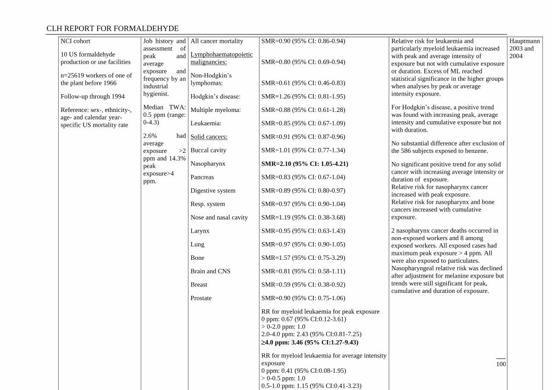
CLH REPORT FOR FORMALDEHYDE
100
NCI cohort
10 US formaldehyde
production or use facilities
n=25619 workers of one of
the plant before 1966
Follow-up through 1994
Reference: sex-, ethnicity-,
age- and calendar year-
specific US mortality rate
Job history and
assessment of
peak and
average
exposure and
frequency by an
industrial
hygienist.
Median TWA:
0.5 ppm (range:
0-4.3)
2.6% had
average
exposure >2
ppm and 14.3%
peak
exposure>4
ppm.
All cancer mortality
Lymphohaematopoietic
malignancies:
Non-Hodgkin‘s
lymphomas:
Hodgkin‘s disease:
Multiple myeloma:
Leukaemia:
Solid cancers:
Buccal cavity
Nasopharynx
Pancreas
Digestive system
Resp. system
Nose and nasal cavity
Larynx
Lung
Bone
Brain and CNS
Breast
Prostate
SMR=0.90 (95% CI: 0.86-0.94)
SMR=0.80 (95% CI: 0.69-0.94)
SMR=0.61 (95% CI: 0.46-0.83)
SMR=1.26 (95% CI: 0.81-1.95)
SMR=0.88 (95% CI: 0.61-1.28)
SMR=0.85 (95% CI: 0.67-1.09)
SMR=0.91 (95% CI: 0.87-0.96)
SMR=1.01 (95% CI: 0.77-1.34)
SMR=2.10 (95% CI: 1.05-4.21)
SMR=0.83 (95% CI: 0.67-1.04)
SMR=0.89 (95% CI: 0.80-0.97)
SMR=0.97 (95% CI: 0.90-1.04)
SMR=1.19 (95% CI: 0.38-3.68)
SMR=0.95 (95% CI: 0.63-1.43)
SMR=0.97 (95% CI: 0.90-1.05)
SMR=1.57 (95% CI: 0.75-3.29)
SMR=0.81 (95% CI: 0.58-1.11)
SMR=0.59 (95% CI: 0.38-0.92)
SMR=0.90 (95% CI: 0.75-1.06)
RR for myeloid leukaemia for peak exposure
0 ppm: 0.67 (95% CI:0.12-3.61)
> 0-2.0 ppm: 1.0
2.0-4.0 ppm: 2.43 (95% CI:0.81-7.25)
4.0 ppm: 3.46 (95% CI:1.27-9.43)
RR for myeloid leukaemia for average intensity
exposure
0 ppm: 0.41 (95% CI:0.08-1.95)
> 0-0.5 ppm: 1.0
0.5-1.0 ppm: 1.15 (95% CI:0.41-3.23)
1.0 ppm: 2.49 (95% CI:1.03-6.03)
Relative risk for leukaemia and
particularly myeloid leukaemia increased
with peak and average intensity of
exposure but not with cumulative exposure
or duration. Excess of ML reached
statistical significance in the higher groups
when analyses by peak or average
intensity exposure.
For Hodgkin‘s disease, a positive trend
was found with increasing peak, average
intensity and cumulative exposure but not
with duration.
No substantial difference after exclusion of
the 586 subjects exposed to benzene.
No significant positive trend for any solid
cancer with increasing average intensity or
duration of exposure.
Relative risk for nasopharynx cancer
increased with peak exposure.
Relative risk for nasopharynx and bone
cancers increased with cumulative
exposure.
2 nasopharynx cancer deaths occurred in
non-exposed workers and 8 among
exposed workers. All exposed cases had
maximum peak exposure > 4 ppm. All
were also exposed to particulates.
Nasopharyngeal relative risk was declined
after adjustment for melanine exposure but
trends were still significant for peak,
cumulative and duration of exposure.
Hauptmann
2003 and
2004
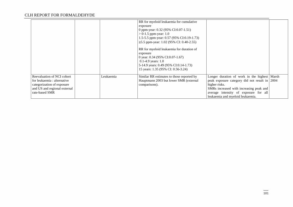
CLH REPORT FOR FORMALDEHYDE
101
RR for myeloid leukaemia for cumulative
exposure
0 ppm-year: 0.32 (95% CI:0.07-1.51)
> 0-1.5 ppm-year: 1.0
1.5-5.5 ppm-year: 0.57 (95% CI:0.19-1.73)
5.5 ppm-year: 1.02 (95% CI: 0.40-2.55)
RR for myeloid leukaemia for duration of
exposure
0 year: 0.34 (95% CI:0.07-1.67)
0.1-4.9 years: 1.0
5-14.9 years: 0.49 (95% CI:0.14-1.73)
15 years: 1.35 (95% CI: 0.56-3.24)
Reevaluation of NCI cohort
for leukaemia : alternative
categorization of exposure
and US and regional external
rate-based SMR
Leukaemia Similar RR estimates to those reported by
Hauptmann 2003 but lower SMR (external
comparisons).
Longer duration of work in the highest
peak exposure category did not result in
higher risks.
SMRs increased with increasing peak and
average intensity of exposure for all
leukaemia and myeloid leukaemia.
Marsh
2004
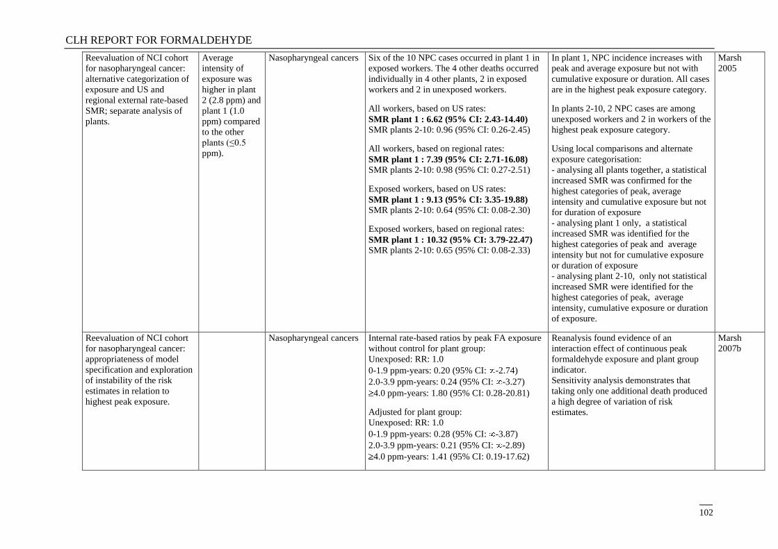
CLH REPORT FOR FORMALDEHYDE
102
Reevaluation of NCI cohort
for nasopharyngeal cancer:
alternative categorization of
exposure and US and
regional external rate-based
SMR; separate analysis of
plants.
Average
intensity of
exposure was
higher in plant
2 (2.8 ppm) and
plant 1 (1.0
ppm) compared
to the other
plants (≤0.5
ppm).
Nasopharyngeal cancers Six of the 10 NPC cases occurred in plant 1 in
exposed workers. The 4 other deaths occurred
individually in 4 other plants, 2 in exposed
workers and 2 in unexposed workers.
All workers, based on US rates:
SMR plant 1 : 6.62 (95% CI: 2.43-14.40)
SMR plants 2-10: 0.96 (95% CI: 0.26-2.45)
All workers, based on regional rates:
SMR plant 1 : 7.39 (95% CI: 2.71-16.08) SMR plants 2-10: 0.98 (95% CI: 0.27-2.51)
Exposed workers, based on US rates:
SMR plant 1 : 9.13 (95% CI: 3.35-19.88)
SMR plants 2-10: 0.64 (95% CI: 0.08-2.30)
Exposed workers, based on regional rates:
SMR plant 1 : 10.32 (95% CI: 3.79-22.47) SMR plants 2-10: 0.65 (95% CI: 0.08-2.33)
In plant 1, NPC incidence increases with
peak and average exposure but not with
cumulative exposure or duration. All cases
are in the highest peak exposure category.
In plants 2-10, 2 NPC cases are among
unexposed workers and 2 in workers of the
highest peak exposure category.
Using local comparisons and alternate
exposure categorisation:
- analysing all plants together, a statistical
increased SMR was confirmed for the
highest categories of peak, average
intensity and cumulative exposure but not
for duration of exposure
- analysing plant 1 only, a statistical
increased SMR was identified for the
highest categories of peak and average
intensity but not for cumulative exposure
or duration of exposure
- analysing plant 2-10, only not statistical
increased SMR were identified for the
highest categories of peak, average
intensity, cumulative exposure or duration
of exposure.
Marsh
2005
Reevaluation of NCI cohort
for nasopharyngeal cancer:
appropriateness of model
specification and exploration
of instability of the risk
estimates in relation to
highest peak exposure.
Nasopharyngeal cancers Internal rate-based ratios by peak FA exposure
without control for plant group:
Unexposed: RR: 1.0
0-1.9 ppm-years: 0.20 (95% CI: -2.74)
2.0-3.9 ppm-years: 0.24 (95% CI: -3.27)
4.0 ppm-years: 1.80 (95% CI: 0.28-20.81)
Adjusted for plant group:
Unexposed: RR: 1.0
0-1.9 ppm-years: 0.28 (95% CI: -3.87)
2.0-3.9 ppm-years: 0.21 (95% CI: -2.89)
4.0 ppm-years: 1.41 (95% CI: 0.19-17.62)
Reanalysis found evidence of an
interaction effect of continuous peak
formaldehyde exposure and plant group
indicator.
Sensitivity analysis demonstrates that
taking only one additional death produced
a high degree of variation of risk
estimates.
Marsh
2007b
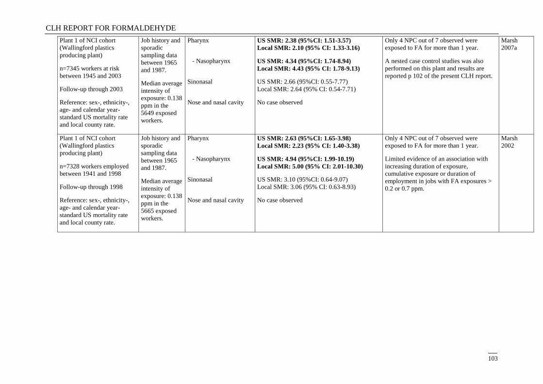
CLH REPORT FOR FORMALDEHYDE
103
Plant 1 of NCI cohort
(Wallingford plastics
producing plant)
n=7345 workers at risk
between 1945 and 2003
Follow-up through 2003
Reference: sex-, ethnicity-,
age- and calendar year-
standard US mortality rate
and local county rate.
Job history and
sporadic
sampling data
between 1965
and 1987.
Median average
intensity of
exposure: 0.138
ppm in the
5649 exposed
workers.
Pharynx
- Nasopharynx
Sinonasal
Nose and nasal cavity
US SMR: 2.38 (95%CI: 1.51-3.57)
Local SMR: 2.10 (95% CI: 1.33-3.16)
US SMR: 4.34 (95%CI: 1.74-8.94)
Local SMR: 4.43 (95% CI: 1.78-9.13)
US SMR: 2.66 (95%CI: 0.55-7.77)
Local SMR: 2.64 (95% CI: 0.54-7.71)
No case observed
Only 4 NPC out of 7 observed were
exposed to FA for more than 1 year.
A nested case control studies was also
performed on this plant and results are
reported p 102 of the present CLH report.
Marsh
2007a
Plant 1 of NCI cohort
(Wallingford plastics
producing plant)
n=7328 workers employed
between 1941 and 1998
Follow-up through 1998
Reference: sex-, ethnicity-,
age- and calendar year-
standard US mortality rate
and local county rate.
Job history and
sporadic
sampling data
between 1965
and 1987.
Median average
intensity of
exposure: 0.138
ppm in the
5665 exposed
workers.
Pharynx
- Nasopharynx
Sinonasal
Nose and nasal cavity
US SMR: 2.63 (95%CI: 1.65-3.98)
Local SMR: 2.23 (95% CI: 1.40-3.38)
US SMR: 4.94 (95%CI: 1.99-10.19)
Local SMR: 5.00 (95% CI: 2.01-10.30)
US SMR: 3.10 (95%CI: 0.64-9.07)
Local SMR: 3.06 (95% CI: 0.63-8.93)
No case observed
Only 4 NPC out of 7 observed were
exposed to FA for more than 1 year.
Limited evidence of an association with
increasing duration of exposure,
cumulative exposure or duration of
employment in jobs with FA exposures >
0.2 or 0.7 ppm.
Marsh
2002
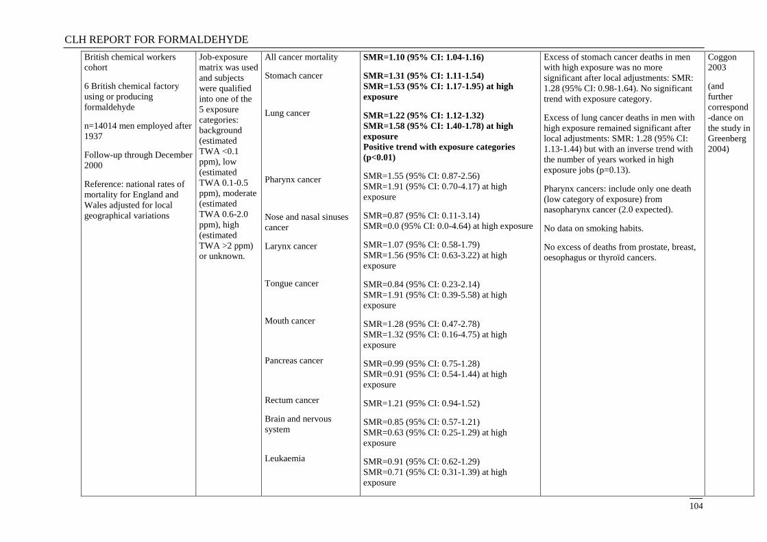
CLH REPORT FOR FORMALDEHYDE
104
British chemical workers
cohort
6 British chemical factory
using or producing
formaldehyde
n=14014 men employed after
1937
Follow-up through December
2000
Reference: national rates of
mortality for England and
Wales adjusted for local
geographical variations
Job-exposure
matrix was used
and subjects
were qualified
into one of the
5 exposure
categories:
background
(estimated
TWA <0.1
ppm), low
(estimated
TWA 0.1-0.5
ppm), moderate
(estimated
TWA 0.6-2.0
ppm), high
(estimated
TWA >2 ppm)
or unknown.
All cancer mortality
Stomach cancer
Lung cancer
Pharynx cancer
Nose and nasal sinuses
cancer
Larynx cancer
Tongue cancer
Mouth cancer
Pancreas cancer
Rectum cancer
Brain and nervous
system
Leukaemia
SMR=1.10 (95% CI: 1.04-1.16)
SMR=1.31 (95% CI: 1.11-1.54)
SMR=1.53 (95% CI: 1.17-1.95) at high
exposure
SMR=1.22 (95% CI: 1.12-1.32)
SMR=1.58 (95% CI: 1.40-1.78) at high
exposure
Positive trend with exposure categories
(p<0.01)
SMR=1.55 (95% CI: 0.87-2.56)
SMR=1.91 (95% CI: 0.70-4.17) at high
exposure
SMR=0.87 (95% CI: 0.11-3.14)
SMR=0.0 (95% CI: 0.0-4.64) at high exposure
SMR=1.07 (95% CI: 0.58-1.79)
SMR=1.56 (95% CI: 0.63-3.22) at high
exposure
SMR=0.84 (95% CI: 0.23-2.14)
SMR=1.91 (95% CI: 0.39-5.58) at high
exposure
SMR=1.28 (95% CI: 0.47-2.78)
SMR=1.32 (95% CI: 0.16-4.75) at high
exposure
SMR=0.99 (95% CI: 0.75-1.28)
SMR=0.91 (95% CI: 0.54-1.44) at high
exposure
SMR=1.21 (95% CI: 0.94-1.52)
SMR=0.85 (95% CI: 0.57-1.21)
SMR=0.63 (95% CI: 0.25-1.29) at high
exposure
SMR=0.91 (95% CI: 0.62-1.29)
SMR=0.71 (95% CI: 0.31-1.39) at high
exposure
Excess of stomach cancer deaths in men
with high exposure was no more
significant after local adjustments: SMR:
1.28 (95% CI: 0.98-1.64). No significant
trend with exposure category.
Excess of lung cancer deaths in men with
high exposure remained significant after
local adjustments: SMR: 1.28 (95% CI:
1.13-1.44) but with an inverse trend with
the number of years worked in high
exposure jobs (p=0.13).
Pharynx cancers: include only one death
(low category of exposure) from
nasopharynx cancer (2.0 expected).
No data on smoking habits.
No excess of deaths from prostate, breast,
oesophagus or thyroïd cancers.
Coggon
2003
(and
further
correspond
-dance on
the study in
Greenberg
2004)
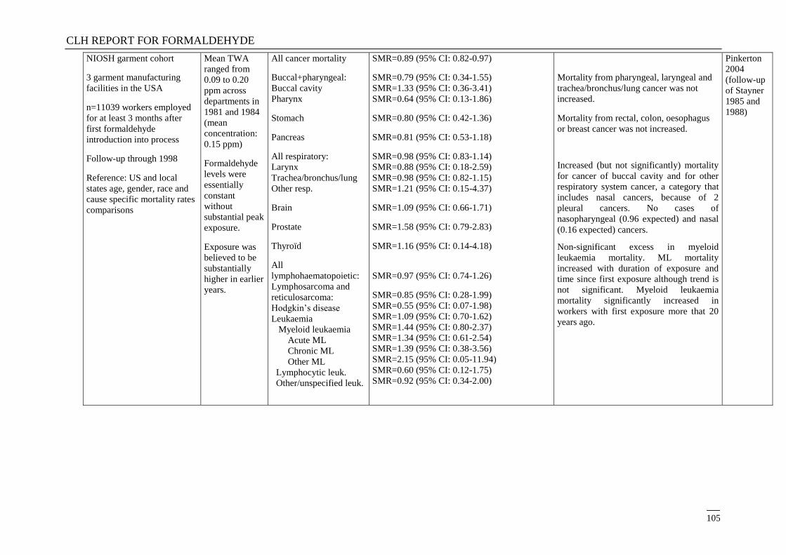
CLH REPORT FOR FORMALDEHYDE
105
NIOSH garment cohort
3 garment manufacturing
facilities in the USA
n=11039 workers employed
for at least 3 months after
first formaldehyde
introduction into process
Follow-up through 1998
Reference: US and local
states age, gender, race and
cause specific mortality rates
comparisons
Mean TWA
ranged from
0.09 to 0.20
ppm across
departments in
1981 and 1984
(mean
concentration:
0.15 ppm)
Formaldehyde
levels were
essentially
constant
without
substantial peak
exposure.
Exposure was
believed to be
substantially
higher in earlier
years.
All cancer mortality
Buccal+pharyngeal:
Buccal cavity
Pharynx
Stomach
Pancreas
All respiratory:
Larynx
Trachea/bronchus/lung
Other resp.
Brain
Prostate
Thyroïd
All
lymphohaematopoietic:
Lymphosarcoma and
reticulosarcoma:
Hodgkin‘s disease
Leukaemia
Myeloid leukaemia
Acute ML
Chronic ML
Other ML
Lymphocytic leuk.
Other/unspecified leuk.
SMR=0.89 (95% CI: 0.82-0.97)
SMR=0.79 (95% CI: 0.34-1.55)
SMR=1.33 (95% CI: 0.36-3.41)
SMR=0.64 (95% CI: 0.13-1.86)
SMR=0.80 (95% CI: 0.42-1.36)
SMR=0.81 (95% CI: 0.53-1.18)
SMR=0.98 (95% CI: 0.83-1.14)
SMR=0.88 (95% CI: 0.18-2.59)
SMR=0.98 (95% CI: 0.82-1.15)
SMR=1.21 (95% CI: 0.15-4.37)
SMR=1.09 (95% CI: 0.66-1.71)
SMR=1.58 (95% CI: 0.79-2.83)
SMR=1.16 (95% CI: 0.14-4.18)
SMR=0.97 (95% CI: 0.74-1.26)
SMR=0.85 (95% CI: 0.28-1.99)
SMR=0.55 (95% CI: 0.07-1.98)
SMR=1.09 (95% CI: 0.70-1.62)
SMR=1.44 (95% CI: 0.80-2.37)
SMR=1.34 (95% CI: 0.61-2.54)
SMR=1.39 (95% CI: 0.38-3.56)
SMR=2.15 (95% CI: 0.05-11.94)
SMR=0.60 (95% CI: 0.12-1.75)
SMR=0.92 (95% CI: 0.34-2.00)
Mortality from pharyngeal, laryngeal and
trachea/bronchus/lung cancer was not
increased.
Mortality from rectal, colon, oesophagus
or breast cancer was not increased.
Increased (but not significantly) mortality
for cancer of buccal cavity and for other
respiratory system cancer, a category that
includes nasal cancers, because of 2
pleural cancers. No cases of
nasopharyngeal (0.96 expected) and nasal
(0.16 expected) cancers.
Non-significant excess in myeloid
leukaemia mortality. ML mortality
increased with duration of exposure and
time since first exposure although trend is
not significant. Myeloid leukaemia
mortality significantly increased in
workers with first exposure more that 20
years ago.
Pinkerton
2004
(follow-up
of Stayner
1985 and
1988)
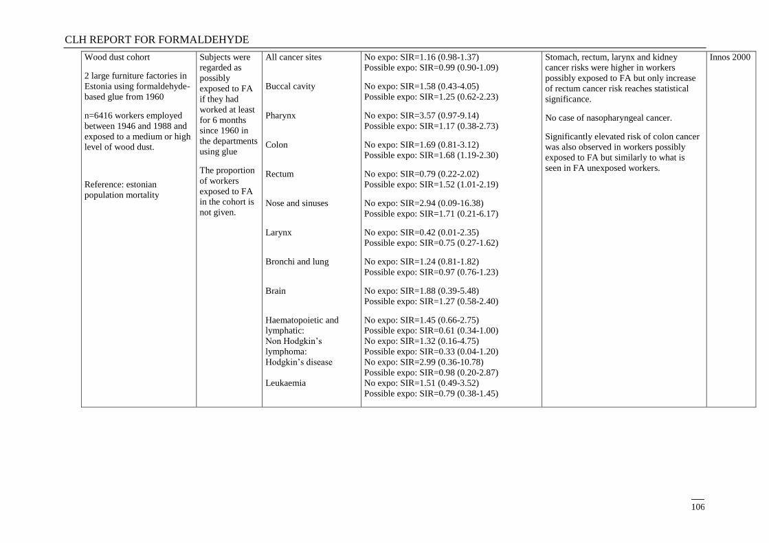
CLH REPORT FOR FORMALDEHYDE
106
Wood dust cohort
2 large furniture factories in
Estonia using formaldehyde-
based glue from 1960
n=6416 workers employed
between 1946 and 1988 and
exposed to a medium or high
level of wood dust.
Reference: estonian
population mortality
Subjects were
regarded as
possibly
exposed to FA
if they had
worked at least
for 6 months
since 1960 in
the departments
using glue
The proportion
of workers
exposed to FA
in the cohort is
not given.
All cancer sites
Buccal cavity
Pharynx
Colon
Rectum
Nose and sinuses
Larynx
Bronchi and lung
Brain
Haematopoietic and
lymphatic:
Non Hodgkin‘s
lymphoma:
Hodgkin‘s disease
Leukaemia
No expo: SIR=1.16 (0.98-1.37)
Possible expo: SIR=0.99 (0.90-1.09)
No expo: SIR=1.58 (0.43-4.05)
Possible expo: SIR=1.25 (0.62-2.23)
No expo: SIR=3.57 (0.97-9.14)
Possible expo: SIR=1.17 (0.38-2.73)
No expo: SIR=1.69 (0.81-3.12)
Possible expo: SIR=1.68 (1.19-2.30)
No expo: SIR=0.79 (0.22-2.02)
Possible expo: SIR=1.52 (1.01-2.19)
No expo: SIR=2.94 (0.09-16.38)
Possible expo: SIR=1.71 (0.21-6.17)
No expo: SIR=0.42 (0.01-2.35)
Possible expo: SIR=0.75 (0.27-1.62)
No expo: SIR=1.24 (0.81-1.82)
Possible expo: SIR=0.97 (0.76-1.23)
No expo: SIR=1.88 (0.39-5.48)
Possible expo: SIR=1.27 (0.58-2.40)
No expo: SIR=1.45 (0.66-2.75)
Possible expo: SIR=0.61 (0.34-1.00)
No expo: SIR=1.32 (0.16-4.75)
Possible expo: SIR=0.33 (0.04-1.20)
No expo: SIR=2.99 (0.36-10.78)
Possible expo: SIR=0.98 (0.20-2.87)
No expo: SIR=1.51 (0.49-3.52)
Possible expo: SIR=0.79 (0.38-1.45)
Stomach, rectum, larynx and kidney
cancer risks were higher in workers
possibly exposed to FA but only increase
of rectum cancer risk reaches statistical
significance.
No case of nasopharyngeal cancer.
Significantly elevated risk of colon cancer
was also observed in workers possibly
exposed to FA but similarly to what is
seen in FA unexposed workers.
Innos 2000
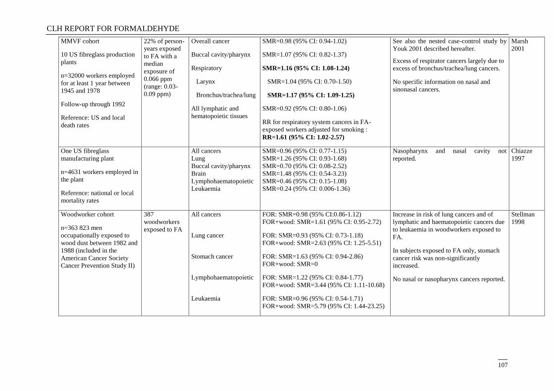
CLH REPORT FOR FORMALDEHYDE
107
MMVF cohort
10 US fibreglass production
plants
n=32000 workers employed
for at least 1 year between
1945 and 1978
Follow-up through 1992
Reference: US and local
death rates
22% of person-
years exposed
to FA with a
median
exposure of
0.066 ppm
(range: 0.03-
0.09 ppm)
Overall cancer
Buccal cavity/pharynx
Respiratory
Larynx
Bronchus/trachea/lung
All lymphatic and
hematopoietic tissues
SMR=0.98 (95% CI: 0.94-1.02)
SMR=1.07 (95% CI: 0.82-1.37)
SMR=1.16 (95% CI: 1.08-1.24)
SMR=1.04 (95% CI: 0.70-1.50)
SMR=1.17 (95% CI: 1.09-1.25)
SMR=0.92 (95% CI: 0.80-1.06)
RR for respiratory system cancers in FA-
exposed workers adjusted for smoking :
RR=1.61 (95% CI: 1.02-2.57)
See also the nested case-control study by
Youk 2001 described hereafter.
Excess of respirator cancers largely due to
excess of bronchus/trachea/lung cancers.
No specific information on nasal and
sinonasal cancers.
Marsh
2001
One US fibreglass
manufacturing plant
n=4631 workers employed in
the plant
Reference: national or local
mortality rates
All cancers
Lung
Buccal cavity/pharynx
Brain
Lymphohaematopoietic
Leukaemia
SMR=0.96 (95% CI: 0.77-1.15)
SMR=1.26 (95% CI: 0.93-1.68)
SMR=0.70 (95% CI: 0.08-2.52)
SMR=1.48 (95% CI: 0.54-3.23)
SMR=0.46 (95% CI: 0.15-1.08)
SMR=0.24 (95% CI: 0.006-1.36)
Nasopharynx and nasal cavity not
reported.
Chiazze
1997
Woodworker cohort
n=363 823 men
occupationally exposed to
wood dust between 1982 and
1988 (included in the
American Cancer Society
Cancer Prevention Study II)
387
woodworkers
exposed to FA
All cancers
Lung cancer
Stomach cancer
Lymphohaematopoietic
Leukaemia
FOR: SMR=0.98 (95% CI:0.86-1.12)
FOR+wood: SMR=1.61 (95% CI: 0.95-2.72)
FOR: SMR=0.93 (95% CI: 0.73-1.18)
FOR+wood: SMR=2.63 (95% CI: 1.25-5.51)
FOR: SMR=1.63 (95% CI: 0.94-2.86)
FOR+wood: SMR=0
FOR: SMR=1.22 (95% CI: 0.84-1.77)
FOR+wood: SMR=3.44 (95% CI: 1.11-10.68)
FOR: SMR=0.96 (95% CI: 0.54-1.71)
FOR+wood: SMR=5.79 (95% CI: 1.44-23.25)
Increase in risk of lung cancers and of
lymphatic and haematopoietic cancers due
to leukaemia in woodworkers exposed to
FA.
In subjects exposed to FA only, stomach
cancer risk was non-significantly
increased.
No nasal or nasopharynx cancers reported.
Stellman
1998
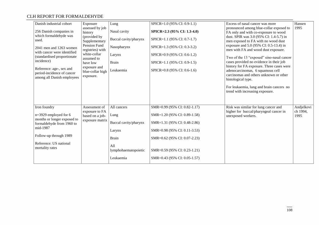
CLH REPORT FOR FORMALDEHYDE
108
Danish industrial cohort
256 Danish companies in
which formaldehyde was
used.
2041 men and 1263 women
with cancer were identified
(standardised proportionate
incidence)
Reference: age-, sex and
period-incidence of cancer
among all Danish employees
Exposure
assessed by job
history
(provided by
Supplementary
Pension Fund
registries) with
white-collar
assumed to
have low
exposure and
blue-collar high
exposure.
Lung
Nasal cavity
Buccal cavity/pharynx
Nasopharynx
Larynx
Brain
Leukaemia
SPICR=1.0 (95% CI: 0.9-1.1)
SPICR=2.3 (95% CI: 1.3-4.0)
SPICR=1.1 (95% CI: 0.7-1.7)
SPICR=1.3 (95% CI: 0.3-3.2)
SPICR=0.9 (95% CI: 0.6-1.2)
SPICR=1.1 (95% CI: 0.9-1.5)
SPICR=0.8 (95% CI: 0.6-1.6)
Excess of nasal cancer was more
pronounced among blue-collar exposed to
FA only and with co-exposure to wood
dust. SPIR was 3.0 (95% CI: 1.4-5.7) in
men exposed to FA with no wood dust
exposure and 5.0 (95% CI: 0.5-13.4) in
men with FA and wood dust exposure.
Two of the 13 ―exposed‖ sino-nasal cancer
cases provided no evidence in their job
history for FA exposure. Three cases were
adenocarcinomas, 6 squamous cell
carcinomas and others unknown or other
histological type.
For leukaemia, lung and brain cancers no
trend with increasing exposure.
Hansen
1995
Iron foundry
n=3929 employed for 6
months or longer exposed to
formaldehyde from 1960 to
mid-1987
Follow-up through 1989
Reference: US national
mortality rates
Assessment of
exposure to FA
based on a job-
exposure matrix
All cancers
Lung
Buccal cavity/pharynx
Larynx
Brain
All
lymphohaematopoietic
Leukaemia
SMR=0.99 (95% CI: 0.82-1.17)
SMR=1.20 (95% CI: 0.89-1.58)
SMR=1.31 (95% CI: 0.48-2.86)
SMR=0.98 (95% CI: 0.11-3.53)
SMR=0.62 (95% CI: 0.07-2.23)
SMR=0.59 (95% CI: 0.23-1.21)
SMR=0.43 (95% CI: 0.05-1.57)
Risk was similar for lung cancer and
higher for buccal/pharyngeal cancer in
unexposed workers.
Andjelkovi
ch 1994,
1995
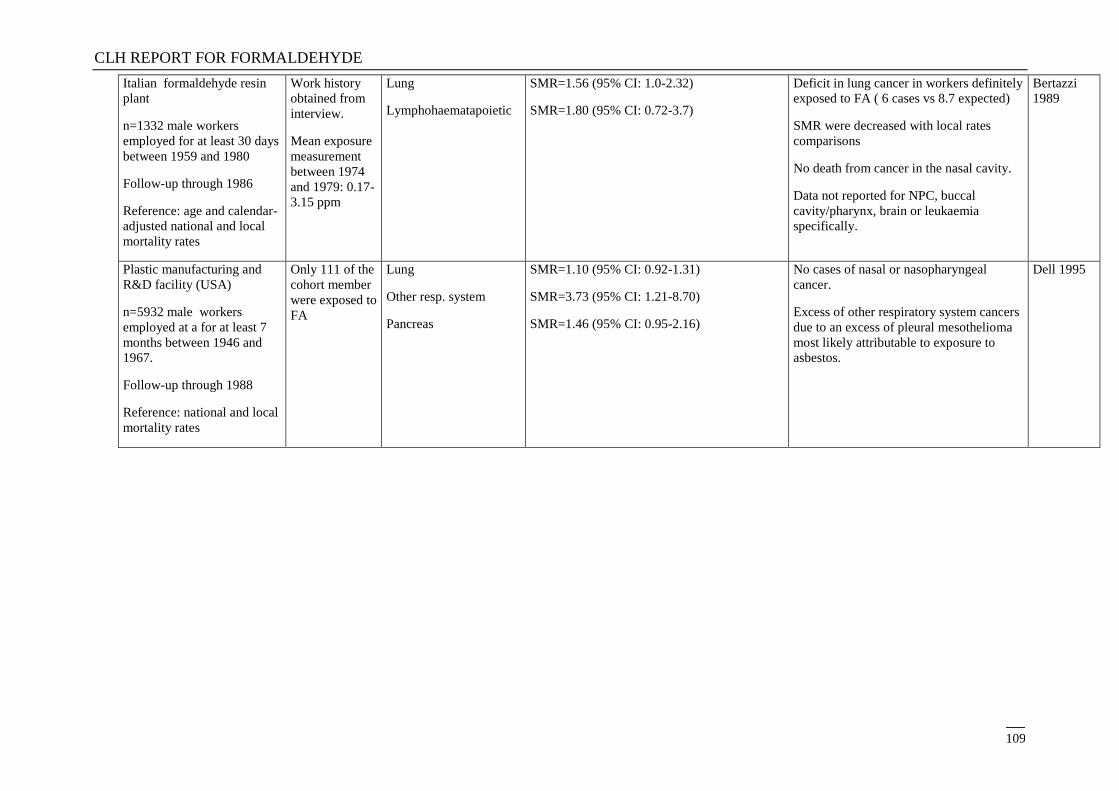
CLH REPORT FOR FORMALDEHYDE
109
Italian formaldehyde resin
plant
n=1332 male workers
employed for at least 30 days
between 1959 and 1980
Follow-up through 1986
Reference: age and calendar-
adjusted national and local
mortality rates
Work history
obtained from
interview.
Mean exposure
measurement
between 1974
and 1979: 0.17-
3.15 ppm
Lung
Lymphohaematapoietic
SMR=1.56 (95% CI: 1.0-2.32)
SMR=1.80 (95% CI: 0.72-3.7)
Deficit in lung cancer in workers definitely
exposed to FA ( 6 cases vs 8.7 expected)
SMR were decreased with local rates
comparisons
No death from cancer in the nasal cavity.
Data not reported for NPC, buccal
cavity/pharynx, brain or leukaemia
specifically.
Bertazzi
1989
Plastic manufacturing and
R&D facility (USA)
n=5932 male workers
employed at a for at least 7
months between 1946 and
1967.
Follow-up through 1988
Reference: national and local
mortality rates
Only 111 of the
cohort member
were exposed to
FA
Lung
Other resp. system
Pancreas
SMR=1.10 (95% CI: 0.92-1.31)
SMR=3.73 (95% CI: 1.21-8.70)
SMR=1.46 (95% CI: 0.95-2.16)
No cases of nasal or nasopharyngeal
cancer.
Excess of other respiratory system cancers
due to an excess of pleural mesothelioma
most likely attributable to exposure to
asbestos.
Dell 1995
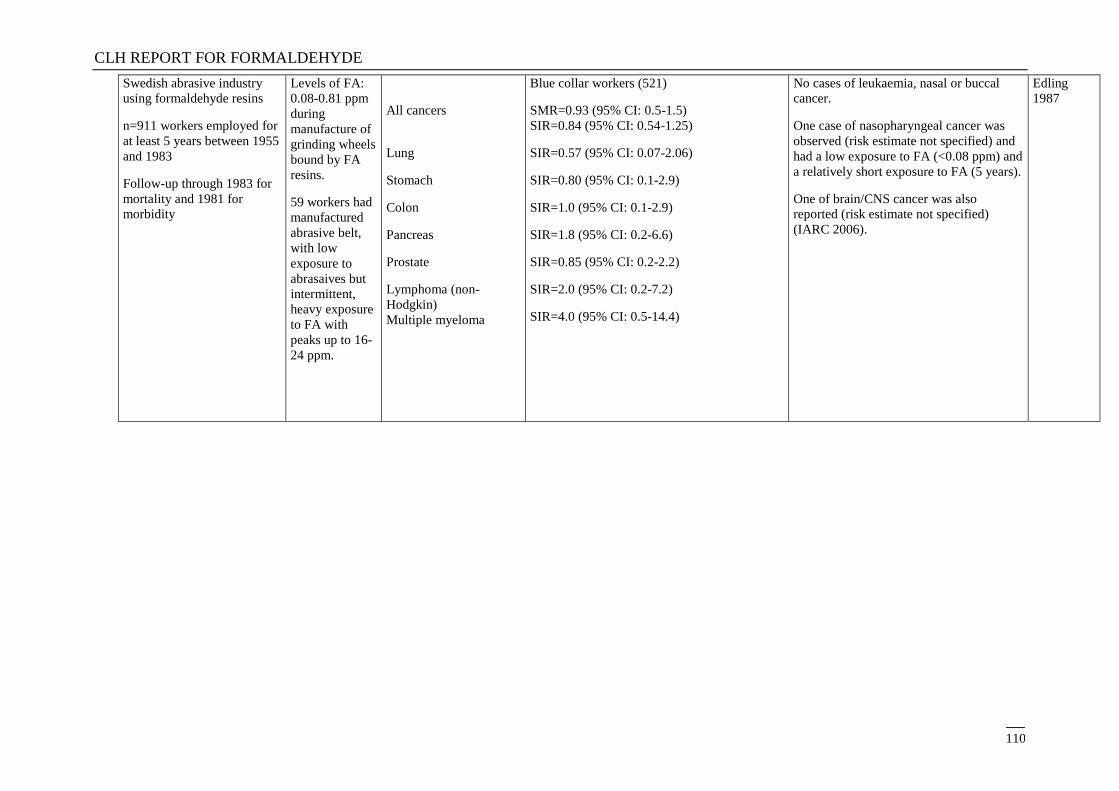
CLH REPORT FOR FORMALDEHYDE
110
Swedish abrasive industry
using formaldehyde resins
n=911 workers employed for
at least 5 years between 1955
and 1983
Follow-up through 1983 for
mortality and 1981 for
morbidity
Levels of FA:
0.08-0.81 ppm
during
manufacture of
grinding wheels
bound by FA
resins.
59 workers had
manufactured
abrasive belt,
with low
exposure to
abrasaives but
intermittent,
heavy exposure
to FA with
peaks up to 16-
24 ppm.
All cancers
Lung
Stomach
Colon
Pancreas
Prostate
Lymphoma (non-
Hodgkin)
Multiple myeloma
Blue collar workers (521)
SMR=0.93 (95% CI: 0.5-1.5)
SIR=0.84 (95% CI: 0.54-1.25)
SIR=0.57 (95% CI: 0.07-2.06)
SIR=0.80 (95% CI: 0.1-2.9)
SIR=1.0 (95% CI: 0.1-2.9)
SIR=1.8 (95% CI: 0.2-6.6)
SIR=0.85 (95% CI: 0.2-2.2)
SIR=2.0 (95% CI: 0.2-7.2)
SIR=4.0 (95% CI: 0.5-14.4)
No cases of leukaemia, nasal or buccal
cancer.
One case of nasopharyngeal cancer was
observed (risk estimate not specified) and
had a low exposure to FA (<0.08 ppm) and
a relatively short exposure to FA (5 years).
One of brain/CNS cancer was also
reported (risk estimate not specified)
(IARC 2006).
Edling
1987
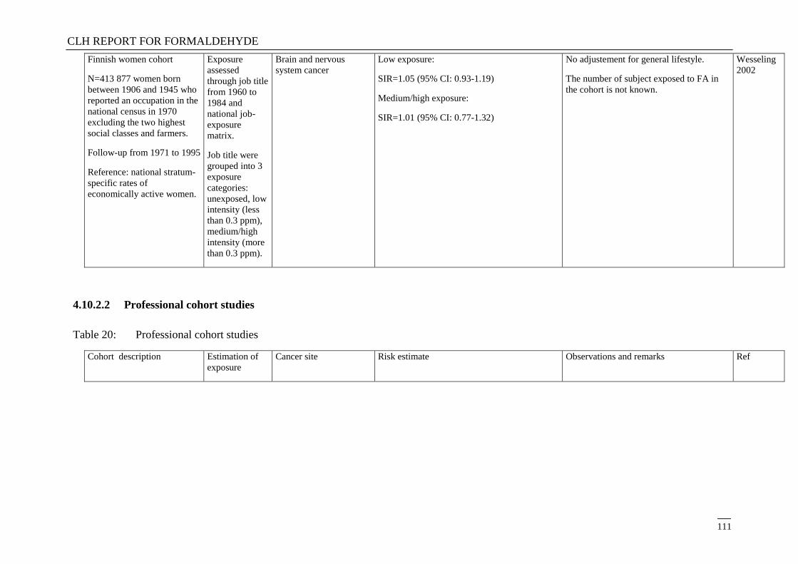
CLH REPORT FOR FORMALDEHYDE
111
Finnish women cohort
N=413 877 women born
between 1906 and 1945 who
reported an occupation in the
national census in 1970
excluding the two highest
social classes and farmers.
Follow-up from 1971 to 1995
Reference: national stratum-
specific rates of
economically active women.
Exposure
assessed
through job title
from 1960 to
1984 and
national job-
exposure
matrix.
Job title were
grouped into 3
exposure
categories:
unexposed, low
intensity (less
than 0.3 ppm),
medium/high
intensity (more
than 0.3 ppm).
Brain and nervous
system cancer
Low exposure:
SIR=1.05 (95% CI: 0.93-1.19)
Medium/high exposure:
SIR=1.01 (95% CI: 0.77-1.32)
No adjustement for general lifestyle.
The number of subject exposed to FA in
the cohort is not known.
Wesseling
2002
4.10.2.2 Professional cohort studies
Table 20: Professional cohort studies
Cohort description Estimation of
exposure
Cancer site Risk estimate Observations and remarks Ref
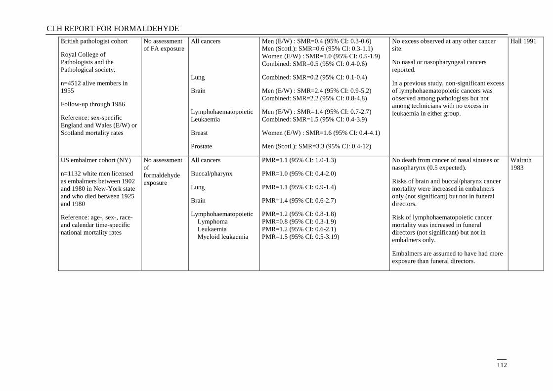
CLH REPORT FOR FORMALDEHYDE
112
British pathologist cohort
Royal College of
Pathologists and the
Pathological society.
n=4512 alive members in
1955
Follow-up through 1986
Reference: sex-specific
England and Wales (E/W) or
Scotland mortality rates
No assessment
of FA exposure
All cancers
Lung
Brain
Lymphohaematopoietic
Leukaemia
Breast
Prostate
Men (E/W) : SMR=0.4 (95% CI: 0.3-0.6)
Men (Scotl.): SMR=0.6 (95% CI: 0.3-1.1)
Women (E/W) : SMR=1.0 (95% CI: 0.5-1.9)
Combined: SMR=0.5 (95% CI: 0.4-0.6)
Combined: SMR=0.2 (95% CI: 0.1-0.4)
Men (E/W) : SMR=2.4 (95% CI: 0.9-5.2)
Combined: SMR=2.2 (95% CI: 0.8-4.8)
Men (E/W) : SMR=1.4 (95% CI: 0.7-2.7)
Combined: SMR=1.5 (95% CI: 0.4-3.9)
Women (E/W) : SMR=1.6 (95% CI: 0.4-4.1)
Men (Scotl.): SMR=3.3 (95% CI: 0.4-12)
No excess observed at any other cancer
site.
No nasal or nasopharyngeal cancers
reported.
In a previous study, non-significant excess
of lymphohaematopoietic cancers was
observed among pathologists but not
among technicians with no excess in
leukaemia in either group.
Hall 1991
US embalmer cohort (NY)
n=1132 white men licensed
as embalmers between 1902
and 1980 in New-York state
and who died between 1925
and 1980
Reference: age-, sex-, race-
and calendar time-specific
national mortality rates
No assessment
of
formaldehyde
exposure
All cancers
Buccal/pharynx
Lung
Brain
Lymphohaematopoietic
Lymphoma
Leukaemia
Myeloid leukaemia
PMR=1.1 (95% CI: 1.0-1.3)
PMR=1.0 (95% CI: 0.4-2.0)
PMR=1.1 (95% CI: 0.9-1.4)
PMR=1.4 (95% CI: 0.6-2.7)
PMR=1.2 (95% CI: 0.8-1.8)
PMR=0.8 (95% CI: 0.3-1.9)
PMR=1.2 (95% CI: 0.6-2.1)
PMR=1.5 (95% CI: 0.5-3.19)
No death from cancer of nasal sinuses or
nasopharynx (0.5 expected).
Risks of brain and buccal/pharynx cancer
mortality were increased in embalmers
only (not significant) but not in funeral
directors.
Risk of lymphohaematopoietic cancer
mortality was increased in funeral
directors (not significant) but not in
embalmers only.
Embalmers are assumed to have had more
exposure than funeral directors.
Walrath
1983
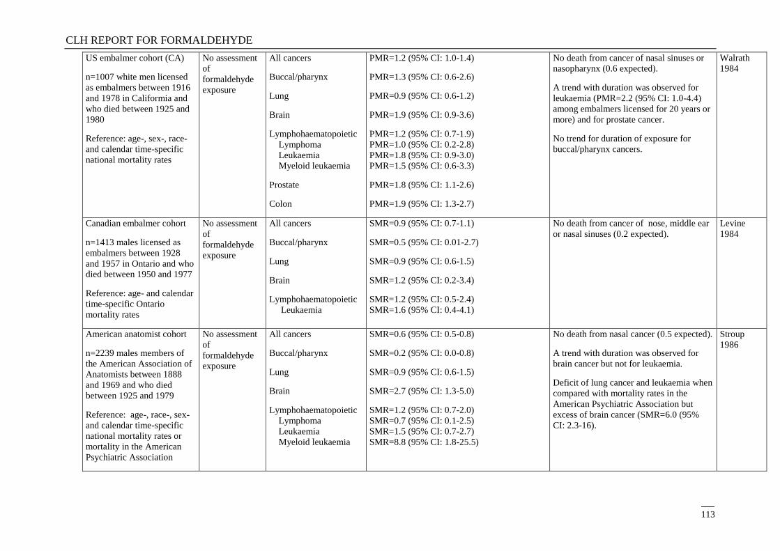
CLH REPORT FOR FORMALDEHYDE
113
US embalmer cohort (CA)
n=1007 white men licensed
as embalmers between 1916
and 1978 in Califormia and
who died between 1925 and
1980
Reference: age-, sex-, race-
and calendar time-specific
national mortality rates
No assessment
of
formaldehyde
exposure
All cancers
Buccal/pharynx
Lung
Brain
Lymphohaematopoietic
Lymphoma
Leukaemia
Myeloid leukaemia
Prostate
Colon
PMR=1.2 (95% CI: 1.0-1.4)
PMR=1.3 (95% CI: 0.6-2.6)
PMR=0.9 (95% CI: 0.6-1.2)
PMR=1.9 (95% CI: 0.9-3.6)
PMR=1.2 (95% CI: 0.7-1.9)
PMR=1.0 (95% CI: 0.2-2.8)
PMR=1.8 (95% CI: 0.9-3.0)
PMR=1.5 (95% CI: 0.6-3.3)
PMR=1.8 (95% CI: 1.1-2.6)
PMR=1.9 (95% CI: 1.3-2.7)
No death from cancer of nasal sinuses or
nasopharynx (0.6 expected).
A trend with duration was observed for
leukaemia (PMR=2.2 (95% CI: 1.0-4.4)
among embalmers licensed for 20 years or
more) and for prostate cancer.
No trend for duration of exposure for
buccal/pharynx cancers.
Walrath
1984
Canadian embalmer cohort
n=1413 males licensed as
embalmers between 1928
and 1957 in Ontario and who
died between 1950 and 1977
Reference: age- and calendar
time-specific Ontario
mortality rates
No assessment
of
formaldehyde
exposure
All cancers
Buccal/pharynx
Lung
Brain
Lymphohaematopoietic
Leukaemia
SMR=0.9 (95% CI: 0.7-1.1)
SMR=0.5 (95% CI: 0.01-2.7)
SMR=0.9 (95% CI: 0.6-1.5)
SMR=1.2 (95% CI: 0.2-3.4)
SMR=1.2 (95% CI: 0.5-2.4)
SMR=1.6 (95% CI: 0.4-4.1)
No death from cancer of nose, middle ear
or nasal sinuses (0.2 expected).
Levine
1984
American anatomist cohort
n=2239 males members of
the American Association of
Anatomists between 1888
and 1969 and who died
between 1925 and 1979
Reference: age-, race-, sex-
and calendar time-specific
national mortality rates or
mortality in the American
Psychiatric Association
No assessment
of
formaldehyde
exposure
All cancers
Buccal/pharynx
Lung
Brain
Lymphohaematopoietic
Lymphoma
Leukaemia
Myeloid leukaemia
SMR=0.6 (95% CI: 0.5-0.8)
SMR=0.2 (95% CI: 0.0-0.8)
SMR=0.9 (95% CI: 0.6-1.5)
SMR=2.7 (95% CI: 1.3-5.0)
SMR=1.2 (95% CI: 0.7-2.0)
SMR=0.7 (95% CI: 0.1-2.5)
SMR=1.5 (95% CI: 0.7-2.7)
SMR=8.8 (95% CI: 1.8-25.5)
No death from nasal cancer (0.5 expected).
A trend with duration was observed for
brain cancer but not for leukaemia.
Deficit of lung cancer and leukaemia when
compared with mortality rates in the
American Psychiatric Association but
excess of brain cancer (SMR=6.0 (95%
CI: 2.3-16).
Stroup
1986
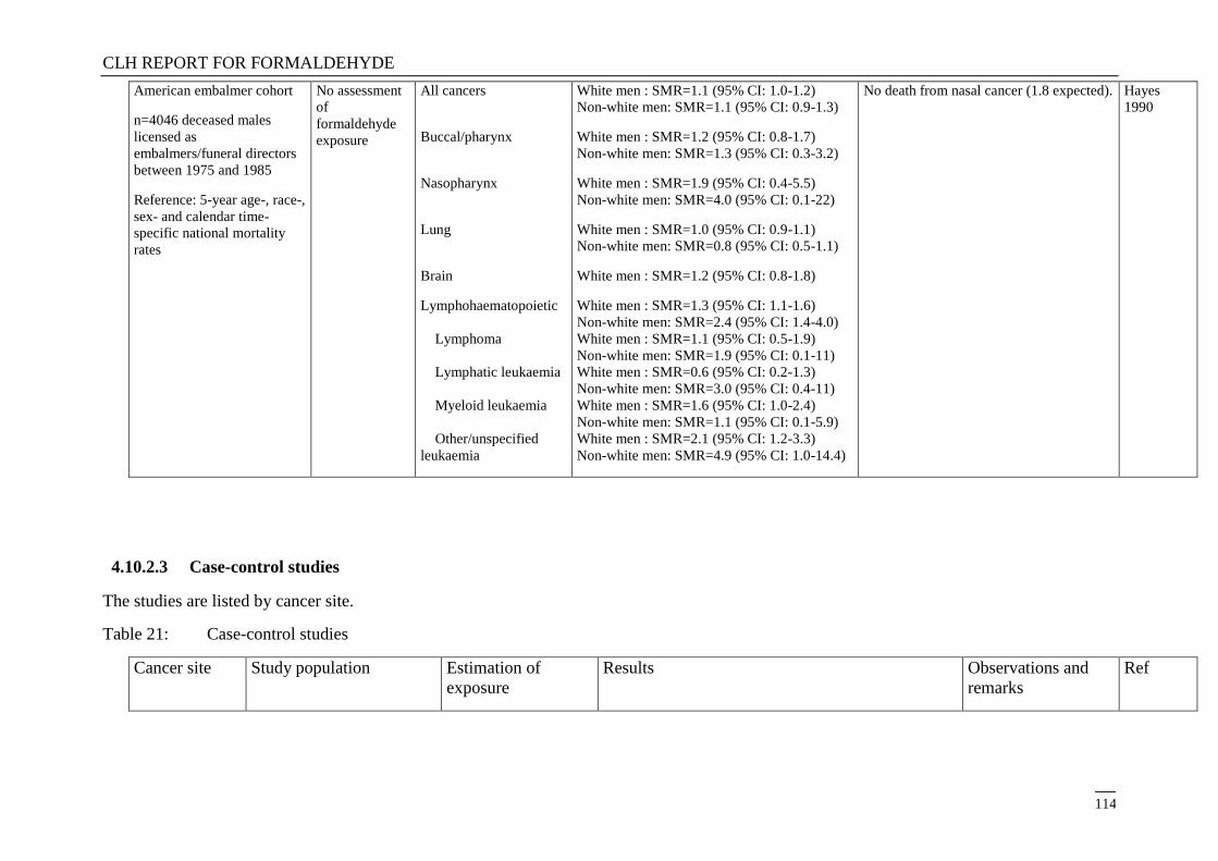
CLH REPORT FOR FORMALDEHYDE
114
American embalmer cohort
n=4046 deceased males
licensed as
embalmers/funeral directors
between 1975 and 1985
Reference: 5-year age-, race-,
sex- and calendar time-
specific national mortality
rates
No assessment
of
formaldehyde
exposure
All cancers
Buccal/pharynx
Nasopharynx
Lung
Brain
Lymphohaematopoietic
Lymphoma
Lymphatic leukaemia
Myeloid leukaemia
Other/unspecified
leukaemia
White men : SMR=1.1 (95% CI: 1.0-1.2)
Non-white men: SMR=1.1 (95% CI: 0.9-1.3)
White men : SMR=1.2 (95% CI: 0.8-1.7)
Non-white men: SMR=1.3 (95% CI: 0.3-3.2)
White men : SMR=1.9 (95% CI: 0.4-5.5)
Non-white men: SMR=4.0 (95% CI: 0.1-22)
White men : SMR=1.0 (95% CI: 0.9-1.1)
Non-white men: SMR=0.8 (95% CI: 0.5-1.1)
White men : SMR=1.2 (95% CI: 0.8-1.8)
White men : SMR=1.3 (95% CI: 1.1-1.6)
Non-white men: SMR=2.4 (95% CI: 1.4-4.0)
White men : SMR=1.1 (95% CI: 0.5-1.9)
Non-white men: SMR=1.9 (95% CI: 0.1-11)
White men : SMR=0.6 (95% CI: 0.2-1.3)
Non-white men: SMR=3.0 (95% CI: 0.4-11)
White men : SMR=1.6 (95% CI: 1.0-2.4)
Non-white men: SMR=1.1 (95% CI: 0.1-5.9)
White men : SMR=2.1 (95% CI: 1.2-3.3)
Non-white men: SMR=4.9 (95% CI: 1.0-14.4)
No death from nasal cancer (1.8 expected).
Hayes
1990
4.10.2.3 Case-control studies
The studies are listed by cancer site.
Table 21: Case-control studies
Cancer site Study population Estimation of
exposure
Results Observations and
remarks
Ref
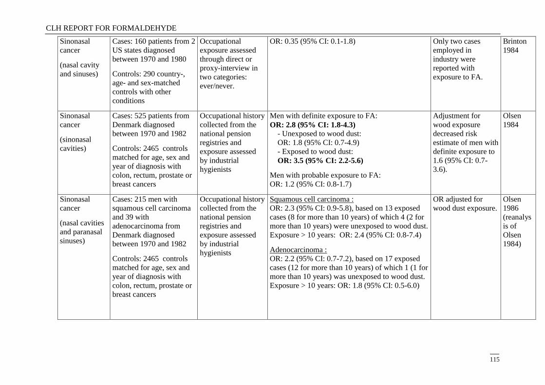
CLH REPORT FOR FORMALDEHYDE
115
Sinonasal
cancer
(nasal cavity
and sinuses)
Cases: 160 patients from 2
US states diagnosed
between 1970 and 1980
Controls: 290 country-,
age- and sex-matched
controls with other
conditions
Occupational
exposure assessed
through direct or
proxy-interview in
two categories:
ever/never.
OR: 0.35 (95% CI: 0.1-1.8) Only two cases
employed in
industry were
reported with
exposure to FA.
Brinton
1984
Sinonasal
cancer
(sinonasal
cavities)
Cases: 525 patients from
Denmark diagnosed
between 1970 and 1982
Controls: 2465 controls
matched for age, sex and
year of diagnosis with
colon, rectum, prostate or
breast cancers
Occupational history
collected from the
national pension
registries and
exposure assessed
by industrial
hygienists
Men with definite exposure to FA:
OR: 2.8 (95% CI: 1.8-4.3) - Unexposed to wood dust:
OR: 1.8 (95% CI: 0.7-4.9)
- Exposed to wood dust:
OR: 3.5 (95% CI: 2.2-5.6)
Men with probable exposure to FA:
OR: 1.2 (95% CI: 0.8-1.7)
Adjustment for
wood exposure
decreased risk
estimate of men with
definite exposure to
1.6 (95% CI: 0.7-
3.6).
Olsen
1984
Sinonasal
cancer
(nasal cavities
and paranasal
sinuses)
Cases: 215 men with
squamous cell carcinoma
and 39 with
adenocarcinoma from
Denmark diagnosed
between 1970 and 1982
Controls: 2465 controls
matched for age, sex and
year of diagnosis with
colon, rectum, prostate or
breast cancers
Occupational history
collected from the
national pension
registries and
exposure assessed
by industrial
hygienists
Squamous cell carcinoma :
OR: 2.3 (95% CI: 0.9-5.8), based on 13 exposed
cases (8 for more than 10 years) of which 4 (2 for
more than 10 years) were unexposed to wood dust.
Exposure > 10 years: OR: 2.4 (95% CI: 0.8-7.4)
Adenocarcinoma :
OR: 2.2 (95% CI: 0.7-7.2), based on 17 exposed
cases (12 for more than 10 years) of which 1 (1 for
more than 10 years) was unexposed to wood dust.
Exposure > 10 years: OR: 1.8 (95% CI: 0.5-6.0)
OR adjusted for
wood dust exposure.
Olsen
1986
(reanalys
is of
Olsen
1984)
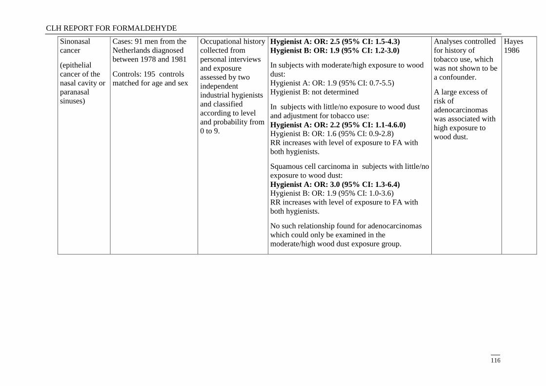
CLH REPORT FOR FORMALDEHYDE
116
Sinonasal
cancer
(epithelial
cancer of the
nasal cavity or
paranasal
sinuses)
Cases: 91 men from the
Netherlands diagnosed
between 1978 and 1981
Controls: 195 controls
matched for age and sex
Occupational history
collected from
personal interviews
and exposure
assessed by two
independent
industrial hygienists
and classified
according to level
and probability from
0 to 9.
Hygienist A: OR: 2.5 (95% CI: 1.5-4.3)
Hygienist B: OR: 1.9 (95% CI: 1.2-3.0)
In subjects with moderate/high exposure to wood
dust:
Hygienist A: OR: 1.9 (95% CI: 0.7-5.5)
Hygienist B: not determined
In subjects with little/no exposure to wood dust
and adjustment for tobacco use:
Hygienist A: OR: 2.2 (95% CI: 1.1-4.6.0) Hygienist B: OR: 1.6 (95% CI: 0.9-2.8)
RR increases with level of exposure to FA with
both hygienists.
Squamous cell carcinoma in subjects with little/no
exposure to wood dust:
Hygienist A: OR: 3.0 (95% CI: 1.3-6.4) Hygienist B: OR: 1.9 (95% CI: 1.0-3.6)
RR increases with level of exposure to FA with
both hygienists.
No such relationship found for adenocarcinomas
which could only be examined in the
moderate/high wood dust exposure group.
Analyses controlled
for history of
tobacco use, which
was not shown to be
a confounder.
A large excess of
risk of
adenocarcinomas
was associated with
high exposure to
wood dust.
Hayes
1986
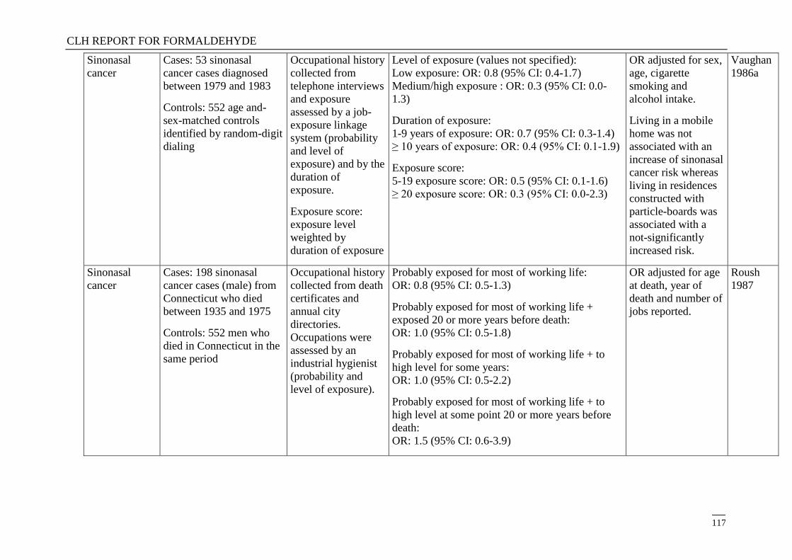
CLH REPORT FOR FORMALDEHYDE
117
Sinonasal
cancer
Cases: 53 sinonasal
cancer cases diagnosed
between 1979 and 1983
Controls: 552 age and-
sex-matched controls
identified by random-digit
dialing
Occupational history
collected from
telephone interviews
and exposure
assessed by a job-
exposure linkage
system (probability
and level of
exposure) and by the
duration of
exposure.
Exposure score:
exposure level
weighted by
duration of exposure
Level of exposure (values not specified):
Low exposure: OR: 0.8 (95% CI: 0.4-1.7)
Medium/high exposure : OR: 0.3 (95% CI: 0.0-
1.3)
Duration of exposure:
1-9 years of exposure: OR: 0.7 (95% CI: 0.3-1.4)
≥ 10 years of exposure: OR: 0.4 (95% CI: 0.1-1.9)
Exposure score:
5-19 exposure score: OR: 0.5 (95% CI: 0.1-1.6)
≥ 20 exposure score: OR: 0.3 (95% CI: 0.0-2.3)
OR adjusted for sex,
age, cigarette
smoking and
alcohol intake.
Living in a mobile
home was not
associated with an
increase of sinonasal
cancer risk whereas
living in residences
constructed with
particle-boards was
associated with a
not-significantly
increased risk.
Vaughan
1986a
Sinonasal
cancer
Cases: 198 sinonasal
cancer cases (male) from
Connecticut who died
between 1935 and 1975
Controls: 552 men who
died in Connecticut in the
same period
Occupational history
collected from death
certificates and
annual city
directories.
Occupations were
assessed by an
industrial hygienist
(probability and
level of exposure).
Probably exposed for most of working life:
OR: 0.8 (95% CI: 0.5-1.3)
Probably exposed for most of working life +
exposed 20 or more years before death:
OR: 1.0 (95% CI: 0.5-1.8)
Probably exposed for most of working life + to
high level for some years:
OR: 1.0 (95% CI: 0.5-2.2)
Probably exposed for most of working life + to
high level at some point 20 or more years before
death:
OR: 1.5 (95% CI: 0.6-3.9)
OR adjusted for age
at death, year of
death and number of
jobs reported.
Roush
1987
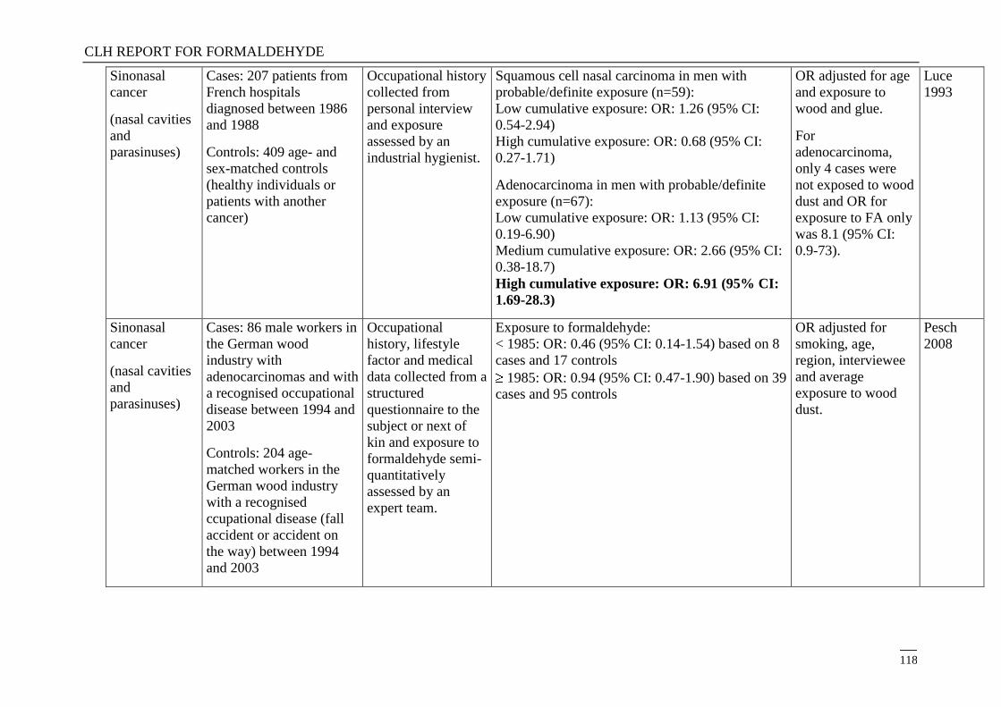
CLH REPORT FOR FORMALDEHYDE
118
Sinonasal
cancer
(nasal cavities
and
parasinuses)
Cases: 207 patients from
French hospitals
diagnosed between 1986
and 1988
Controls: 409 age- and
sex-matched controls
(healthy individuals or
patients with another
cancer)
Occupational history
collected from
personal interview
and exposure
assessed by an
industrial hygienist.
Squamous cell nasal carcinoma in men with
probable/definite exposure (n=59):
Low cumulative exposure: OR: 1.26 (95% CI:
0.54-2.94)
High cumulative exposure: OR: 0.68 (95% CI:
0.27-1.71)
Adenocarcinoma in men with probable/definite
exposure (n=67):
Low cumulative exposure: OR: 1.13 (95% CI:
0.19-6.90)
Medium cumulative exposure: OR: 2.66 (95% CI:
0.38-18.7)
High cumulative exposure: OR: 6.91 (95% CI:
1.69-28.3)
OR adjusted for age
and exposure to
wood and glue.
For
adenocarcinoma,
only 4 cases were
not exposed to wood
dust and OR for
exposure to FA only
was 8.1 (95% CI:
0.9-73).
Luce
1993
Sinonasal
cancer
(nasal cavities
and
parasinuses)
Cases: 86 male workers in
the German wood
industry with
adenocarcinomas and with
a recognised occupational
disease between 1994 and
2003
Controls: 204 age-
matched workers in the
German wood industry
with a recognised
ccupational disease (fall
accident or accident on
the way) between 1994
and 2003
Occupational
history, lifestyle
factor and medical
data collected from a
structured
questionnaire to the
subject or next of
kin and exposure to
formaldehyde semi-
quantitatively
assessed by an
expert team.
Exposure to formaldehyde:
< 1985: OR: 0.46 (95% CI: 0.14-1.54) based on 8
cases and 17 controls
1985: OR: 0.94 (95% CI: 0.47-1.90) based on 39
cases and 95 controls
OR adjusted for
smoking, age,
region, interviewee
and average
exposure to wood
dust.
Pesch
2008
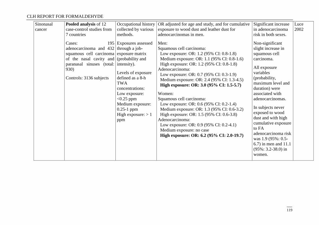
CLH REPORT FOR FORMALDEHYDE
119
Sinonasal
cancer
Pooled analysis of 12
case-control studies from
7 countries
Cases: 195
adenocarcinoma and 432
squamous cell carcinoma
of the nasal cavity and
paranasal sinuses (total:
930)
Controls: 3136 subjects
Occupational history
collected by various
methods.
Exposures assessed
through a job-
exposure matrix
(probability and
intensity).
Levels of exposure
defined as a 8-h
TWA
concentrations:
Low exposure:
<0.25 ppm
Medium exposure:
0.25-1 ppm
High exposure: > 1
ppm
OR adjusted for age and study, and for cumulative
exposure to wood dust and leather dust for
adenocarcinomas in men.
Men:
Squamous cell carcinoma:
Low exposure: OR: 1.2 (95% CI: 0.8-1.8)
Medium exposure: OR: 1.1 (95% CI: 0.8-1.6)
High exposure: OR: 1.2 (95% CI: 0.8-1.8)
Adenocarcinoma:
Low exposure: OR: 0.7 (95% CI: 0.3-1.9)
Medium exposure: OR: 2.4 (95% CI: 1.3-4.5)
High exposure: OR: 3.0 (95% CI: 1.5-5.7)
Women:
Squamous cell carcinoma:
Low exposure: OR: 0.6 (95% CI: 0.2-1.4)
Medium exposure: OR: 1.3 (95% CI: 0.6-3.2)
High exposure: OR: 1.5 (95% CI: 0.6-3.8)
Adenocarcinoma:
Low exposure: OR: 0.9 (95% CI: 0.2-4.1)
Medium exposure: no case
High exposure: OR: 6.2 (95% CI: 2.0-19.7)
Significant increase
in adenocarcinoma
risk in both sexes.
Non-significant
slight increase in
squamous cell
carcinoma.
All exposure
variables
(probability,
maximum level and
duration) were
associated with
adenocarcinomas.
In subjects never
exposed to wood
dust and with high
cumulative exposure
to FA
adenocarcinoma risk
was 1.9 (95%: 0.5-
6.7) in men and 11.1
(95%: 3.2-38.0) in
women.
Luce
2002
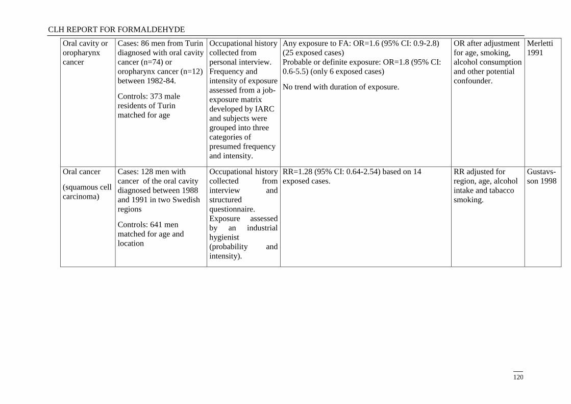
CLH REPORT FOR FORMALDEHYDE
120
Oral cavity or
oropharynx
cancer
Cases: 86 men from Turin
diagnosed with oral cavity
cancer (n=74) or
oropharynx cancer (n=12)
between 1982-84.
Controls: 373 male
residents of Turin
matched for age
Occupational history
collected from
personal interview.
Frequency and
intensity of exposure
assessed from a job-
exposure matrix
developed by IARC
and subjects were
grouped into three
categories of
presumed frequency
and intensity.
Any exposure to FA: OR=1.6 (95% CI: 0.9-2.8)
(25 exposed cases)
Probable or definite exposure: OR=1.8 (95% CI:
0.6-5.5) (only 6 exposed cases)
No trend with duration of exposure.
OR after adjustment
for age, smoking,
alcohol consumption
and other potential
confounder.
Merletti
1991
Oral cancer
(squamous cell
carcinoma)
Cases: 128 men with
cancer of the oral cavity
diagnosed between 1988
and 1991 in two Swedish
regions
Controls: 641 men
matched for age and
location
Occupational history
collected from
interview and
structured
questionnaire.
Exposure assessed
by an industrial
hygienist
(probability and
intensity).
RR=1.28 (95% CI: 0.64-2.54) based on 14
exposed cases.
RR adjusted for
region, age, alcohol
intake and tabacco
smoking.
Gustavs-
son 1998
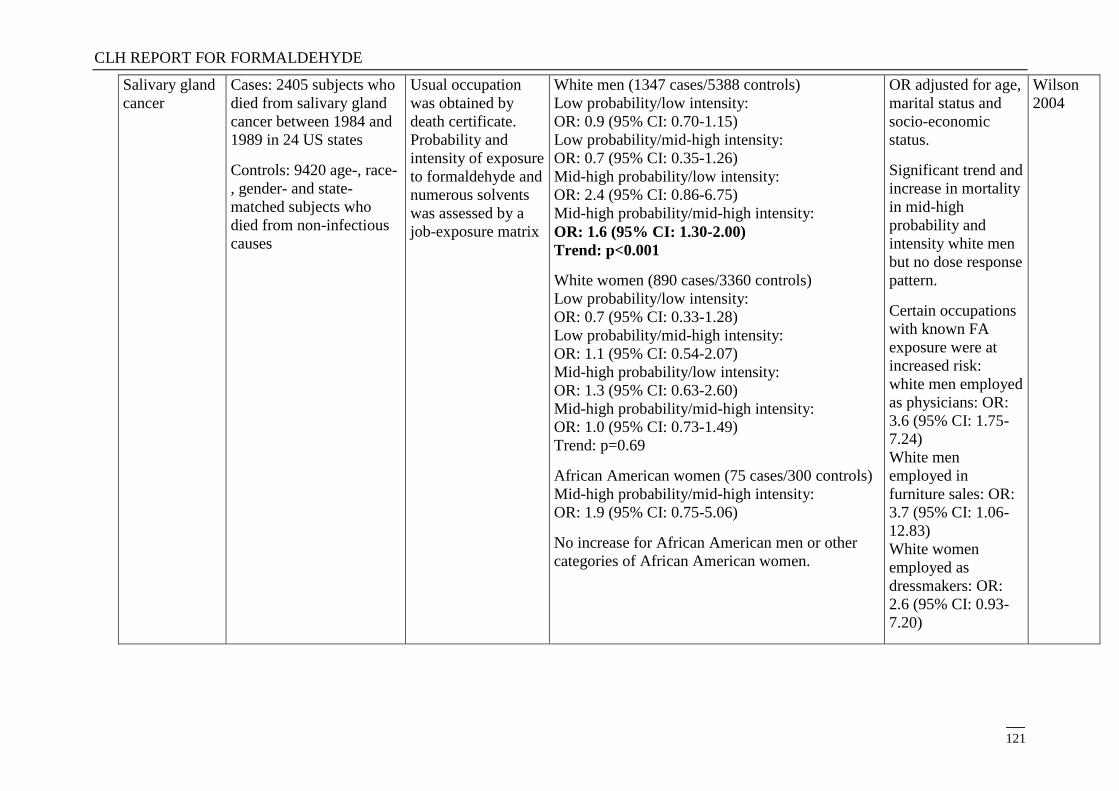
CLH REPORT FOR FORMALDEHYDE
121
Salivary gland
cancer
Cases: 2405 subjects who
died from salivary gland
cancer between 1984 and
1989 in 24 US states
Controls: 9420 age-, race-
, gender- and state-
matched subjects who
died from non-infectious
causes
Usual occupation
was obtained by
death certificate.
Probability and
intensity of exposure
to formaldehyde and
numerous solvents
was assessed by a
job-exposure matrix
White men (1347 cases/5388 controls)
Low probability/low intensity:
OR: 0.9 (95% CI: 0.70-1.15)
Low probability/mid-high intensity:
OR: 0.7 (95% CI: 0.35-1.26)
Mid-high probability/low intensity:
OR: 2.4 (95% CI: 0.86-6.75)
Mid-high probability/mid-high intensity:
OR: 1.6 (95% CI: 1.30-2.00)
Trend: p<0.001
White women (890 cases/3360 controls)
Low probability/low intensity:
OR: 0.7 (95% CI: 0.33-1.28)
Low probability/mid-high intensity:
OR: 1.1 (95% CI: 0.54-2.07)
Mid-high probability/low intensity:
OR: 1.3 (95% CI: 0.63-2.60)
Mid-high probability/mid-high intensity:
OR: 1.0 (95% CI: 0.73-1.49)
Trend: p=0.69
African American women (75 cases/300 controls)
Mid-high probability/mid-high intensity:
OR: 1.9 (95% CI: 0.75-5.06)
No increase for African American men or other
categories of African American women.
OR adjusted for age,
marital status and
socio-economic
status.
Significant trend and
increase in mortality
in mid-high
probability and
intensity white men
but no dose response
pattern.
Certain occupations
with known FA
exposure were at
increased risk:
white men employed
as physicians: OR:
3.6 (95% CI: 1.75-
7.24)
White men
employed in
furniture sales: OR:
3.7 (95% CI: 1.06-
12.83)
White women
employed as
dressmakers: OR:
2.6 (95% CI: 0.93-
7.20)
Wilson
2004
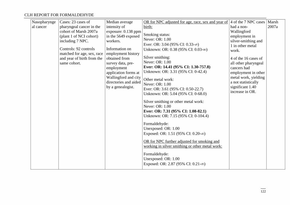
CLH REPORT FOR FORMALDEHYDE
122
Nasopharynge
al cancer
Cases: 23 cases of
pharyngeal cancer in the
cohort of Marsh 2007a
(plant 1 of NCI cohort)
including 7 NPC.
Controls: 92 controls
matched for age, sex, race
and year of birth from the
same cohort.
Median average
intensity of
exposure: 0.138 ppm
in the 5649 exposed
workers.
Information on
employment history
obtained from
survey data, pre-
employment
application forms at
Wallingford and city
directories and aided
by a genealogist.
OR for NPC adjusted for age, race, sex and year of
birth:
Smoking status:
Never: OR: 1.00
Ever: OR: 3.04 (95% CI: 0.33- )
Unknown: OR: 0.38 (95% CI: 0.03- )
Silver smithing:
Never: OR: 1.00
Ever: OR: 14.41 (95% CI: 1.30-757.8) Unknown: OR: 3.31 (95% CI: 0-42.4)
Other metal work:
Never: OR: 1.00
Ever: OR: 3.61 (95% CI: 0.50-22.7)
Unknown: OR: 5.04 (95% CI: 0-68.0)
Silver smithing or other metal work:
Never: OR: 1.00
Ever: OR: 7.31 (95% CI: 1.08-82.1) Unknown: OR: 7.15 (95% CI: 0-104.4)
Formaldehyde:
Unexposed: OR: 1.00
Exposed: OR: 1.51 (95% CI: 0.20- )
OR for NPC further adjusted for smoking and
working in silver smithing or other metal work:
Formaldehyde:
Unexposed: OR: 1.00
Exposed: OR: 2.87 (95% CI: 0.21- )
4 of the 7 NPC cases
had a non-
Wallingford
employment in
silver-smithing and
1 in other metal
work.
4 of the 16 cases of
all other pharyngeal
cancers had
employment in other
metal work, yielding
a not statistically
significant 1.40
increase in OR.
Marsh
2007a
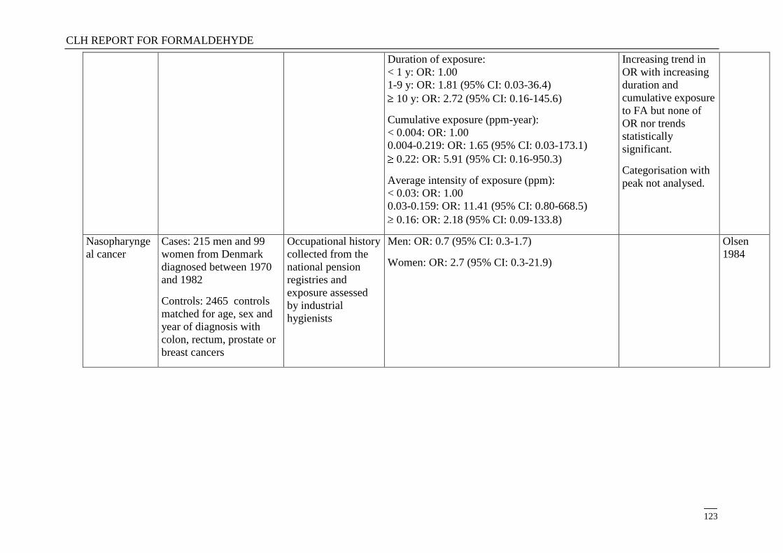
CLH REPORT FOR FORMALDEHYDE
123
Duration of exposure:
< 1 y: OR: 1.00
1-9 y: OR: 1.81 (95% CI: 0.03-36.4)
10 y: OR: 2.72 (95% CI: 0.16-145.6)
Cumulative exposure (ppm-year):
< 0.004: OR: 1.00
0.004-0.219: OR: 1.65 (95% CI: 0.03-173.1)
0.22: OR: 5.91 (95% CI: 0.16-950.3)
Average intensity of exposure (ppm):
< 0.03: OR: 1.00
0.03-0.159: OR: 11.41 (95% CI: 0.80-668.5)
0.16: OR: 2.18 (95% CI: 0.09-133.8)
Increasing trend in
OR with increasing
duration and
cumulative exposure
to FA but none of
OR nor trends
statistically
significant.
Categorisation with
peak not analysed.
Nasopharynge
al cancer
Cases: 215 men and 99
women from Denmark
diagnosed between 1970
and 1982
Controls: 2465 controls
matched for age, sex and
year of diagnosis with
colon, rectum, prostate or
breast cancers
Occupational history
collected from the
national pension
registries and
exposure assessed
by industrial
hygienists
Men: OR: 0.7 (95% CI: 0.3-1.7)
Women: OR: 2.7 (95% CI: 0.3-21.9)
Olsen
1984
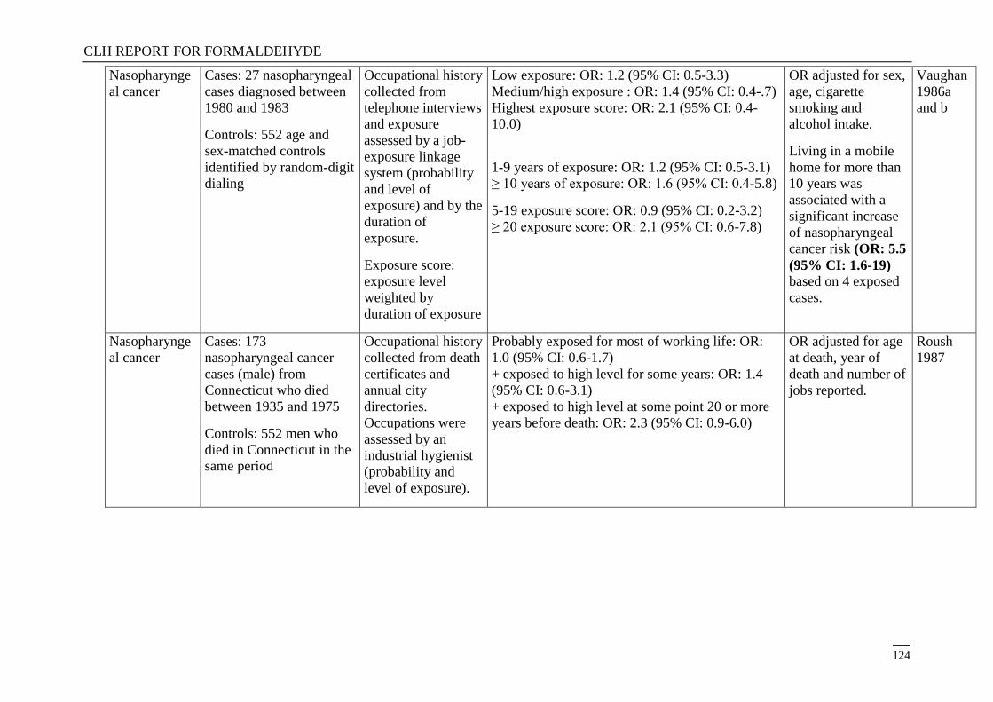
CLH REPORT FOR FORMALDEHYDE
124
Nasopharynge
al cancer
Cases: 27 nasopharyngeal
cases diagnosed between
1980 and 1983
Controls: 552 age and
sex-matched controls
identified by random-digit
dialing
Occupational history
collected from
telephone interviews
and exposure
assessed by a job-
exposure linkage
system (probability
and level of
exposure) and by the
duration of
exposure.
Exposure score:
exposure level
weighted by
duration of exposure
Low exposure: OR: 1.2 (95% CI: 0.5-3.3)
Medium/high exposure : OR: 1.4 (95% CI: 0.4-.7)
Highest exposure score: OR: 2.1 (95% CI: 0.4-
10.0)
1-9 years of exposure: OR: 1.2 (95% CI: 0.5-3.1)
≥ 10 years of exposure: OR: 1.6 (95% CI: 0.4-5.8)
5-19 exposure score: OR: 0.9 (95% CI: 0.2-3.2)
≥ 20 exposure score: OR: 2.1 (95% CI: 0.6-7.8)
OR adjusted for sex,
age, cigarette
smoking and
alcohol intake.
Living in a mobile
home for more than
10 years was
associated with a
significant increase
of nasopharyngeal
cancer risk (OR: 5.5
(95% CI: 1.6-19) based on 4 exposed
cases.
Vaughan
1986a
and b
Nasopharynge
al cancer
Cases: 173
nasopharyngeal cancer
cases (male) from
Connecticut who died
between 1935 and 1975
Controls: 552 men who
died in Connecticut in the
same period
Occupational history
collected from death
certificates and
annual city
directories.
Occupations were
assessed by an
industrial hygienist
(probability and
level of exposure).
Probably exposed for most of working life: OR:
1.0 (95% CI: 0.6-1.7)
+ exposed to high level for some years: OR: 1.4
(95% CI: 0.6-3.1)
+ exposed to high level at some point 20 or more
years before death: OR: 2.3 (95% CI: 0.9-6.0)
OR adjusted for age
at death, year of
death and number of
jobs reported.
Roush
1987
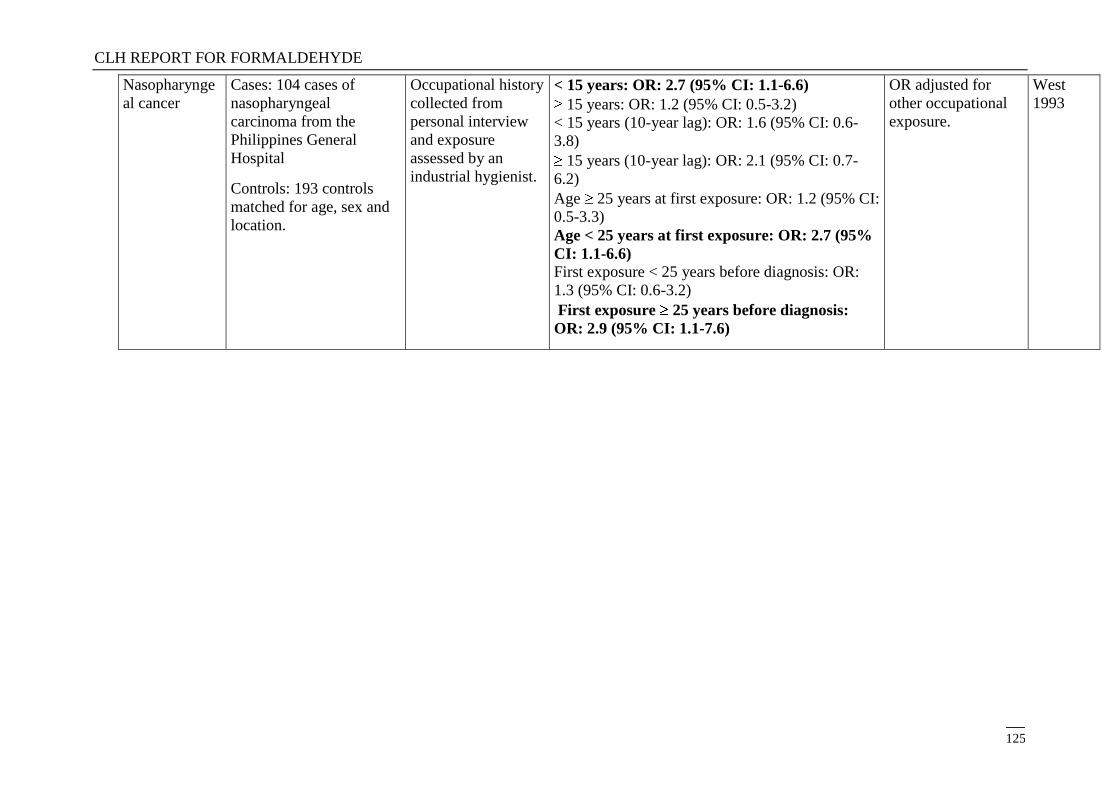
CLH REPORT FOR FORMALDEHYDE
125
Nasopharynge
al cancer
Cases: 104 cases of
nasopharyngeal
carcinoma from the
Philippines General
Hospital
Controls: 193 controls
matched for age, sex and
location.
Occupational history
collected from
personal interview
and exposure
assessed by an
industrial hygienist.
< 15 years: OR: 2.7 (95% CI: 1.1-6.6)
15 years: OR: 1.2 (95% CI: 0.5-3.2)
< 15 years (10-year lag): OR: 1.6 (95% CI: 0.6-
3.8)
15 years (10-year lag): OR: 2.1 (95% CI: 0.7-
6.2)
Age 25 years at first exposure: OR: 1.2 (95% CI:
0.5-3.3)
Age < 25 years at first exposure: OR: 2.7 (95%
CI: 1.1-6.6) First exposure < 25 years before diagnosis: OR:
1.3 (95% CI: 0.6-3.2)
First exposure 25 years before diagnosis:
OR: 2.9 (95% CI: 1.1-7.6)
OR adjusted for
other occupational
exposure.
West
1993
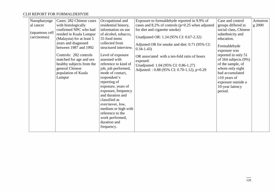
CLH REPORT FOR FORMALDEHYDE
126
Nasopharynge
al cancer
(squamous cell
carcinomas)
Cases: 282 Chinese cases
with histologically
confirmed NPC who had
resided in Kuala Lumpur
(Malaysia) for at least 5
years and diagnosed
between 1987 and 1992
Controls: 282 controls
matched for age and sex
healthy subjects from the
general Chinese
population of Kuala
Lumpur
Occupational and
residential history,
information on use
of alcohol, tobacco,
55 food items
collected from
structured interview.
Level of exposure
assessed with
reference to kind of
job, job performed,
mode of contact,
respondent‘s
reporting of
exposure, years of
exposure, frequency
and duration and
classified as
ever/never, low,
medium or high with
reference to the
work performed,
duration and
frequency.
Exposure to formaldehyde reported in 9.9% of
cases and 8.2% of controls (p=0.25 when adjusted
for diet and cigarette smoke)
Unadjusted OR: 1.24 (95% CI: 0.67-2.32)
Adjusted OR for smoke and diet: 0.71 (95% CI:
0.34-1.43)
OR associated with a ten-fold ratio of hours
exposed:
Unadjusted: 1.04 (95% CI: 0.86-1.27)
Adjusted: : 0.88 (95% CI: 0.70-1.12), p=0.29
Case and control
groups differed in
social class, Chinese
subethnicity and
education.
Formaldehyde
exposure was
reported in only 51
of 564 subjects (9%)
of the sample, of
whom only eight
had accumulated
10 years of
exposure outside a
10-year latency
period.
Armstron
g 2000
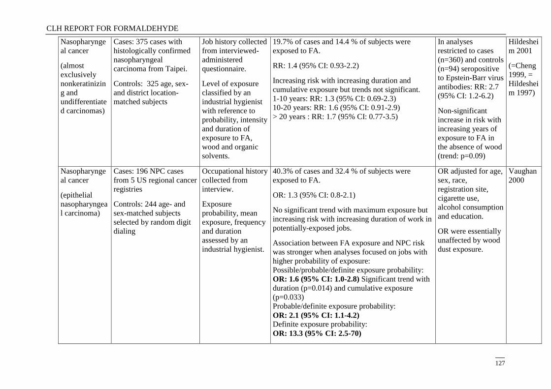
CLH REPORT FOR FORMALDEHYDE
127
Nasopharynge
al cancer
(almost
exclusively
nonkeratinizin
g and
undifferentiate
d carcinomas)
Cases: 375 cases with
histologically confirmed
nasopharyngeal
carcinoma from Taipei.
Controls: 325 age, sex-
and district location-
matched subjects
Job history collected
from interviewed-
administered
questionnaire.
Level of exposure
classified by an
industrial hygienist
with reference to
probability, intensity
and duration of
exposure to FA,
wood and organic
solvents.
19.7% of cases and 14.4 % of subjects were
exposed to FA.
RR: 1.4 (95% CI: 0.93-2.2)
Increasing risk with increasing duration and
cumulative exposure but trends not significant.
1-10 years: RR: 1.3 (95% CI: 0.69-2.3)
10-20 years: RR: 1.6 (95% CI: 0.91-2.9)
> 20 years : RR: 1.7 (95% CI: 0.77-3.5)
In analyses
restricted to cases
(n=360) and controls
(n=94) seropositive
to Epstein-Barr virus
antibodies: RR: 2.7
(95% CI: 1.2-6.2)
Non-significant
increase in risk with
increasing years of
exposure to FA in
the absence of wood
(trend: p=0.09)
Hildeshei
m 2001
(=Cheng
1999, =
Hildeshei
m 1997)
Nasopharynge
al cancer
(epithelial
nasopharyngea
l carcinoma)
Cases: 196 NPC cases
from 5 US regional cancer
registries
Controls: 244 age- and
sex-matched subjects
selected by random digit
dialing
Occupational history
collected from
interview.
Exposure
probability, mean
exposure, frequency
and duration
assessed by an
industrial hygienist.
40.3% of cases and 32.4 % of subjects were
exposed to FA.
OR: 1.3 (95% CI: 0.8-2.1)
No significant trend with maximum exposure but
increasing risk with increasing duration of work in
potentially-exposed jobs.
Association between FA exposure and NPC risk
was stronger when analyses focused on jobs with
higher probability of exposure:
Possible/probable/definite exposure probability:
OR: 1.6 (95% CI: 1.0-2.8) Significant trend with
duration (p=0.014) and cumulative exposure
(p=0.033)
Probable/definite exposure probability:
OR: 2.1 (95% CI: 1.1-4.2) Definite exposure probability:
OR: 13.3 (95% CI: 2.5-70)
OR adjusted for age,
sex, race,
registration site,
cigarette use,
alcohol consumption
and education.
OR were essentially
unaffected by wood
dust exposure.
Vaughan
2000
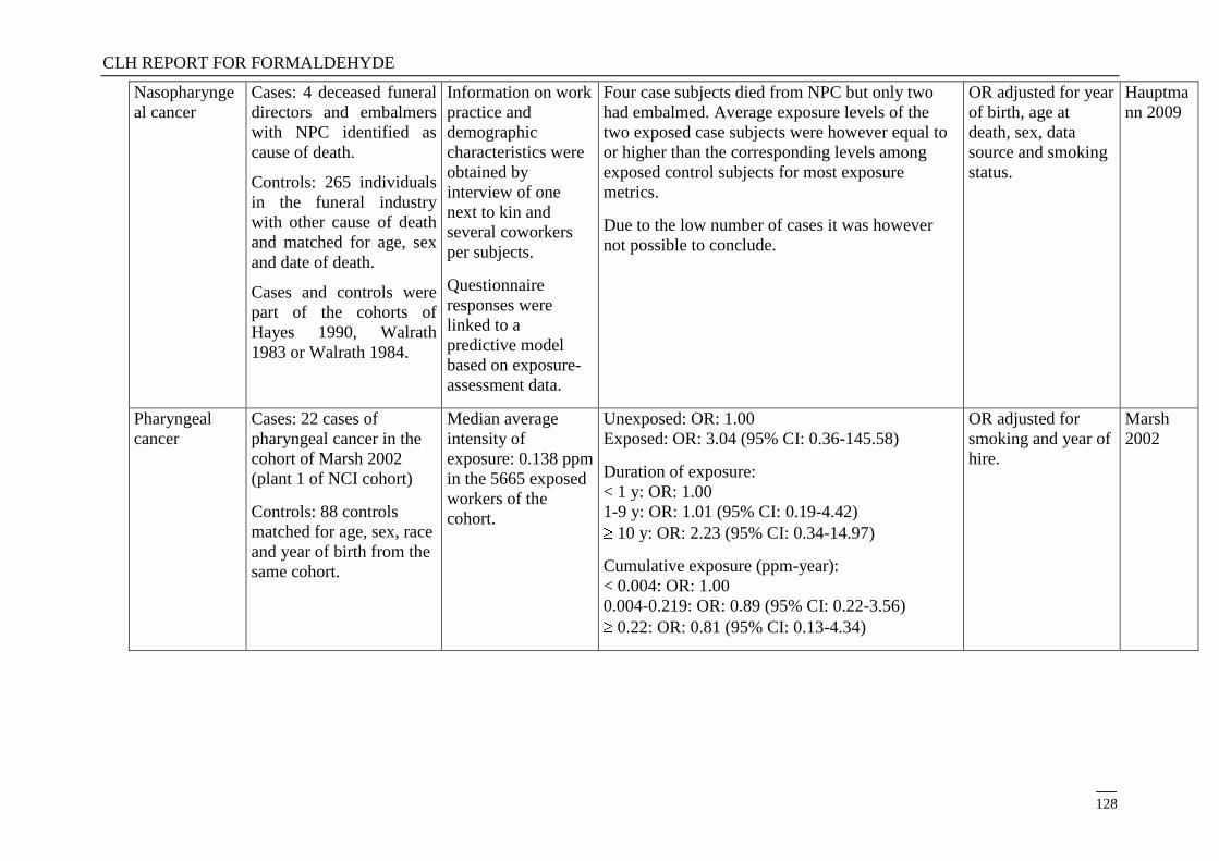
CLH REPORT FOR FORMALDEHYDE
128
Nasopharynge
al cancer
Cases: 4 deceased funeral
directors and embalmers
with NPC identified as
cause of death.
Controls: 265 individuals
in the funeral industry
with other cause of death
and matched for age, sex
and date of death.
Cases and controls were
part of the cohorts of
Hayes 1990, Walrath
1983 or Walrath 1984.
Information on work
practice and
demographic
characteristics were
obtained by
interview of one
next to kin and
several coworkers
per subjects.
Questionnaire
responses were
linked to a
predictive model
based on exposure-
assessment data.
Four case subjects died from NPC but only two
had embalmed. Average exposure levels of the
two exposed case subjects were however equal to
or higher than the corresponding levels among
exposed control subjects for most exposure
metrics.
Due to the low number of cases it was however
not possible to conclude.
OR adjusted for year
of birth, age at
death, sex, data
source and smoking
status.
Hauptma
nn 2009
Pharyngeal
cancer
Cases: 22 cases of
pharyngeal cancer in the
cohort of Marsh 2002
(plant 1 of NCI cohort)
Controls: 88 controls
matched for age, sex, race
and year of birth from the
same cohort.
Median average
intensity of
exposure: 0.138 ppm
in the 5665 exposed
workers of the
cohort.
Unexposed: OR: 1.00
Exposed: OR: 3.04 (95% CI: 0.36-145.58)
Duration of exposure:
< 1 y: OR: 1.00
1-9 y: OR: 1.01 (95% CI: 0.19-4.42)
10 y: OR: 2.23 (95% CI: 0.34-14.97)
Cumulative exposure (ppm-year):
< 0.004: OR: 1.00
0.004-0.219: OR: 0.89 (95% CI: 0.22-3.56)
0.22: OR: 0.81 (95% CI: 0.13-4.34)
OR adjusted for
smoking and year of
hire.
Marsh
2002
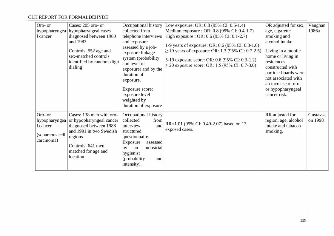
CLH REPORT FOR FORMALDEHYDE
129
Oro- or
hypopharyngea
l cancer
Cases: 205 oro- or
hypopharyngeal cases
diagnosed between 1980
and 1983
Controls: 552 age and
sex-matched controls
identified by random-digit
dialing
Occupational history
collected from
telephone interviews
and exposure
assessed by a job-
exposure linkage
system (probability
and level of
exposure) and by the
duration of
exposure.
Exposure score:
exposure level
weighted by
duration of exposure
Low exposure: OR: 0.8 (95% CI: 0.5-1.4)
Medium exposure : OR: 0.8 (95% CI: 0.4-1.7)
High exposure : OR: 0.6 (95% CI: 0.1-2.7)
1-9 years of exposure: OR: 0.6 (95% CI: 0.3-1.0)
≥ 10 years of exposure: OR: 1.3 (95% CI: 0.7-2.5)
5-19 exposure score: OR: 0.6 (95% CI: 0.3-1.2)
≥ 20 exposure score: OR: 1.5 (95% CI: 0.7-3.0)
OR adjusted for sex,
age, cigarette
smoking and
alcohol intake.
Living in a mobile
home or living in
residences
constructed with
particle-boards were
not associated with
an increase of oro-
or hypopharyngeal
cancer risk.
Vaughan
1986a
Oro- or
hypopharyngea
l cancer
(squamous cell
carcinoma)
Cases: 138 men with oro-
or hypopharyngeal cancer
diagnosed between 1988
and 1991 in two Swedish
regions
Controls: 641 men
matched for age and
location
Occupational history
collected from
interview and
structured
questionnaire.
Exposure assessed
by an industrial
hygienist
(probability and
intensity).
RR=1.01 (95% CI: 0.49-2.07) based on 13
exposed cases.
RR adjusted for
region, age, alcohol
intake and tabacco
smoking.
Gustavss
on 1998
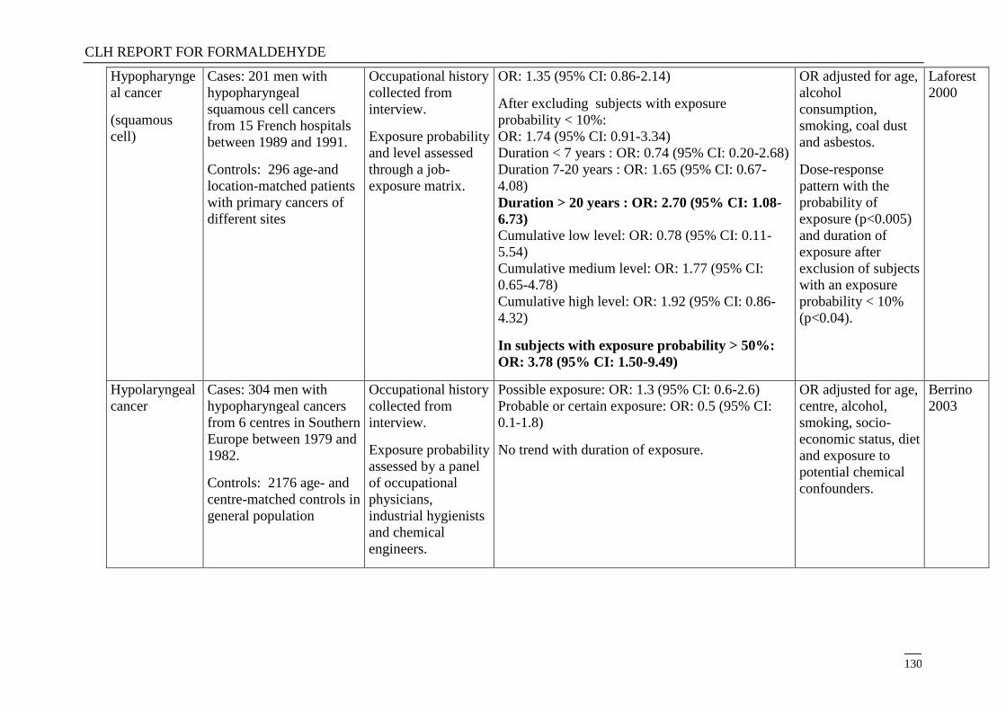
CLH REPORT FOR FORMALDEHYDE
130
Hypopharynge
al cancer
(squamous
cell)
Cases: 201 men with
hypopharyngeal
squamous cell cancers
from 15 French hospitals
between 1989 and 1991.
Controls: 296 age-and
location-matched patients
with primary cancers of
different sites
Occupational history
collected from
interview.
Exposure probability
and level assessed
through a job-
exposure matrix.
OR: 1.35 (95% CI: 0.86-2.14)
After excluding subjects with exposure
probability < 10%:
OR: 1.74 (95% CI: 0.91-3.34)
Duration < 7 years : OR: 0.74 (95% CI: 0.20-2.68)
Duration 7-20 years : OR: 1.65 (95% CI: 0.67-
4.08)
Duration > 20 years : OR: 2.70 (95% CI: 1.08-
6.73)
Cumulative low level: OR: 0.78 (95% CI: 0.11-
5.54)
Cumulative medium level: OR: 1.77 (95% CI:
0.65-4.78)
Cumulative high level: OR: 1.92 (95% CI: 0.86-
4.32)
In subjects with exposure probability > 50%:
OR: 3.78 (95% CI: 1.50-9.49)
OR adjusted for age,
alcohol
consumption,
smoking, coal dust
and asbestos.
Dose-response
pattern with the
probability of
exposure (p<0.005)
and duration of
exposure after
exclusion of subjects
with an exposure
probability < 10%
(p<0.04).
Laforest
2000
Hypolaryngeal
cancer
Cases: 304 men with
hypopharyngeal cancers
from 6 centres in Southern
Europe between 1979 and
1982.
Controls: 2176 age- and
centre-matched controls in
general population
Occupational history
collected from
interview.
Exposure probability
assessed by a panel
of occupational
physicians,
industrial hygienists
and chemical
engineers.
Possible exposure: OR: 1.3 (95% CI: 0.6-2.6)
Probable or certain exposure: OR: 0.5 (95% CI:
0.1-1.8)
No trend with duration of exposure.
OR adjusted for age,
centre, alcohol,
smoking, socio-
economic status, diet
and exposure to
potential chemical
confounders.
Berrino
2003
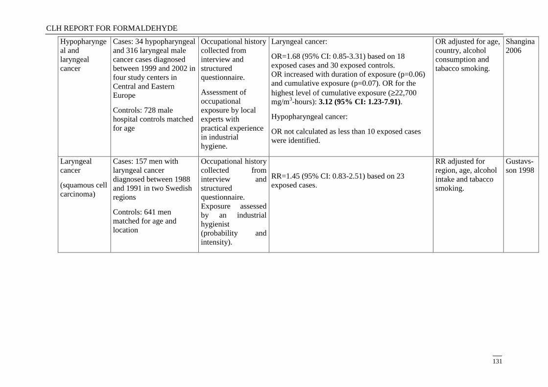
CLH REPORT FOR FORMALDEHYDE
131
Hypopharynge
al and
laryngeal
cancer
Cases: 34 hypopharyngeal
and 316 laryngeal male
cancer cases diagnosed
between 1999 and 2002 in
four study centers in
Central and Eastern
Europe
Controls: 728 male
hospital controls matched
for age
Occupational history
collected from
interview and
structured
questionnaire.
Assessment of
occupational
exposure by local
experts with
practical experience
in industrial
hygiene.
Laryngeal cancer:
OR=1.68 (95% CI: 0.85-3.31) based on 18
exposed cases and 30 exposed controls.
OR increased with duration of exposure (p=0.06)
and cumulative exposure (p=0.07). OR for the
highest level of cumulative exposure ( 22,700
mg/m3-hours): 3.12 (95% CI: 1.23-7.91).
Hypopharyngeal cancer:
OR not calculated as less than 10 exposed cases
were identified.
OR adjusted for age,
country, alcohol
consumption and
tabacco smoking.
Shangina
2006
Laryngeal
cancer
(squamous cell
carcinoma)
Cases: 157 men with
laryngeal cancer
diagnosed between 1988
and 1991 in two Swedish
regions
Controls: 641 men
matched for age and
location
Occupational history
collected from
interview and
structured
questionnaire.
Exposure assessed
by an industrial
hygienist
(probability and
intensity).
RR=1.45 (95% CI: 0.83-2.51) based on 23
exposed cases.
RR adjusted for
region, age, alcohol
intake and tabacco
smoking.
Gustavs-
son 1998
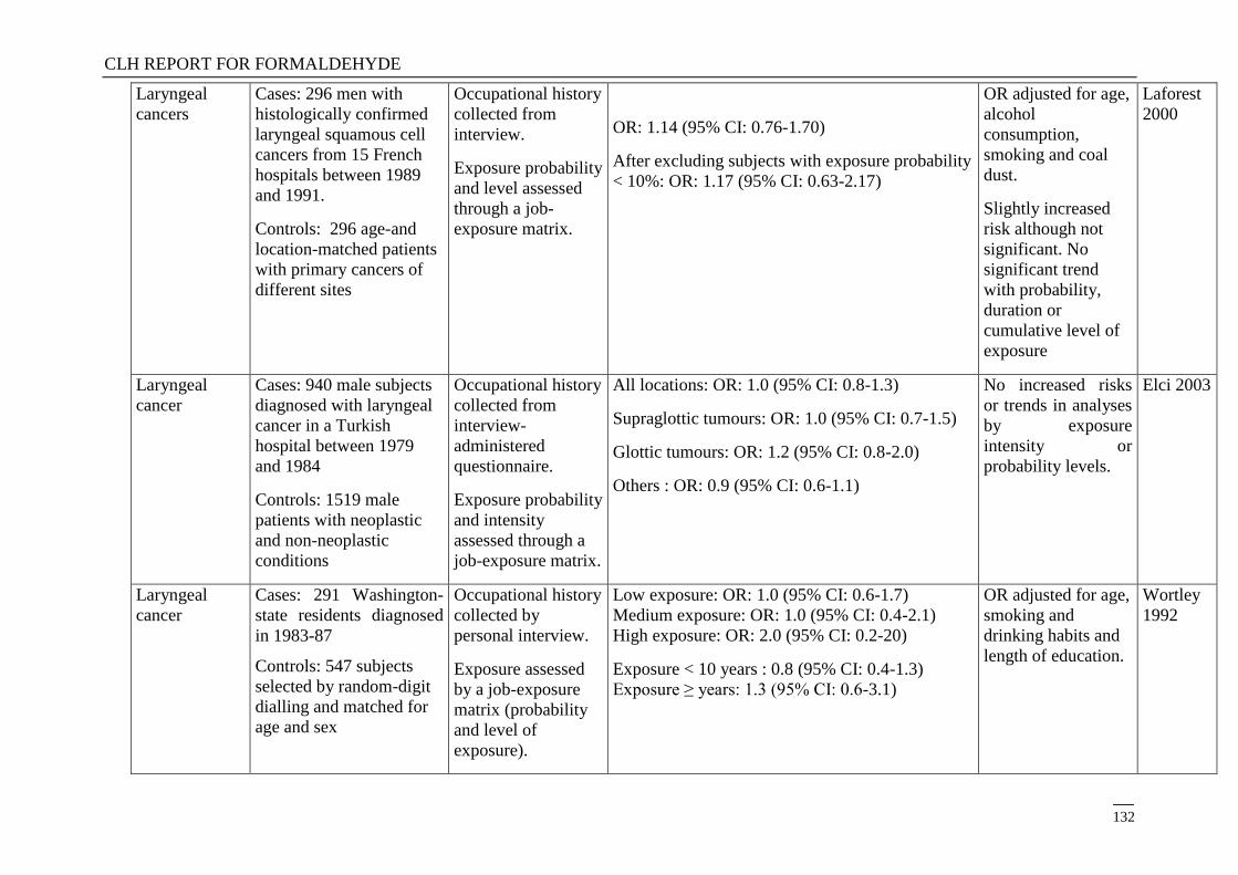
CLH REPORT FOR FORMALDEHYDE
132
Laryngeal
cancers
Cases: 296 men with
histologically confirmed
laryngeal squamous cell
cancers from 15 French
hospitals between 1989
and 1991.
Controls: 296 age-and
location-matched patients
with primary cancers of
different sites
Occupational history
collected from
interview.
Exposure probability
and level assessed
through a job-
exposure matrix.
OR: 1.14 (95% CI: 0.76-1.70)
After excluding subjects with exposure probability
< 10%: OR: 1.17 (95% CI: 0.63-2.17)
OR adjusted for age,
alcohol
consumption,
smoking and coal
dust.
Slightly increased
risk although not
significant. No
significant trend
with probability,
duration or
cumulative level of
exposure
Laforest
2000
Laryngeal
cancer
Cases: 940 male subjects
diagnosed with laryngeal
cancer in a Turkish
hospital between 1979
and 1984
Controls: 1519 male
patients with neoplastic
and non-neoplastic
conditions
Occupational history
collected from
interview-
administered
questionnaire.
Exposure probability
and intensity
assessed through a
job-exposure matrix.
All locations: OR: 1.0 (95% CI: 0.8-1.3)
Supraglottic tumours: OR: 1.0 (95% CI: 0.7-1.5)
Glottic tumours: OR: 1.2 (95% CI: 0.8-2.0)
Others : OR: 0.9 (95% CI: 0.6-1.1)
No increased risks
or trends in analyses
by exposure
intensity or
probability levels.
Elci 2003
Laryngeal
cancer
Cases: 291 Washington-
state residents diagnosed
in 1983-87
Controls: 547 subjects
selected by random-digit
dialling and matched for
age and sex
Occupational history
collected by
personal interview.
Exposure assessed
by a job-exposure
matrix (probability
and level of
exposure).
Low exposure: OR: 1.0 (95% CI: 0.6-1.7)
Medium exposure: OR: 1.0 (95% CI: 0.4-2.1)
High exposure: OR: 2.0 (95% CI: 0.2-20)
Exposure < 10 years : 0.8 (95% CI: 0.4-1.3)
Exposure ≥ years: 1.3 (95% CI: 0.6-3.1)
OR adjusted for age,
smoking and
drinking habits and
length of education.
Wortley
1992
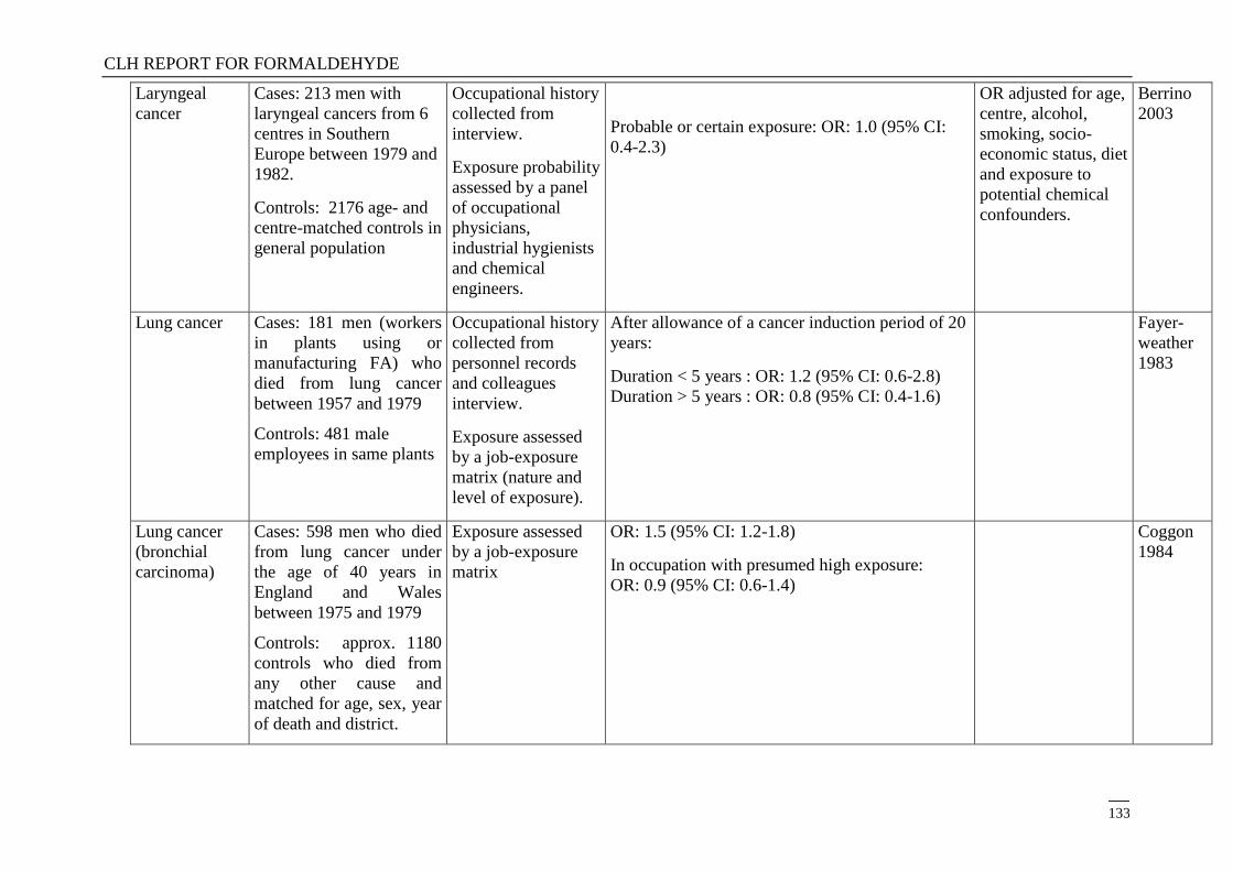
CLH REPORT FOR FORMALDEHYDE
133
Laryngeal
cancer
Cases: 213 men with
laryngeal cancers from 6
centres in Southern
Europe between 1979 and
1982.
Controls: 2176 age- and
centre-matched controls in
general population
Occupational history
collected from
interview.
Exposure probability
assessed by a panel
of occupational
physicians,
industrial hygienists
and chemical
engineers.
Probable or certain exposure: OR: 1.0 (95% CI:
0.4-2.3)
OR adjusted for age,
centre, alcohol,
smoking, socio-
economic status, diet
and exposure to
potential chemical
confounders.
Berrino
2003
Lung cancer Cases: 181 men (workers
in plants using or
manufacturing FA) who
died from lung cancer
between 1957 and 1979
Controls: 481 male
employees in same plants
Occupational history
collected from
personnel records
and colleagues
interview.
Exposure assessed
by a job-exposure
matrix (nature and
level of exposure).
After allowance of a cancer induction period of 20
years:
Duration < 5 years : OR: 1.2 (95% CI: 0.6-2.8)
Duration > 5 years : OR: 0.8 (95% CI: 0.4-1.6)
Fayer-
weather
1983
Lung cancer
(bronchial
carcinoma)
Cases: 598 men who died
from lung cancer under
the age of 40 years in
England and Wales
between 1975 and 1979
Controls: approx. 1180
controls who died from
any other cause and
matched for age, sex, year
of death and district.
Exposure assessed
by a job-exposure
matrix
OR: 1.5 (95% CI: 1.2-1.8)
In occupation with presumed high exposure:
OR: 0.9 (95% CI: 0.6-1.4)
Coggon
1984
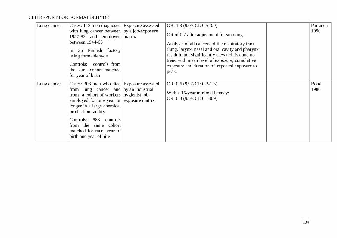
CLH REPORT FOR FORMALDEHYDE
134
Lung cancer Cases: 118 men diagnosed
with lung cancer between
1957-82 and employed
between 1944-65
in 35 Finnish factory
using formaldehyde
Controls: controls from
the same cohort matched
for year of birth
Exposure assessed
by a job-exposure
matrix
OR: 1.3 (95% CI: 0.5-3.0)
OR of 0.7 after adjustment for smoking.
Analysis of all cancers of the respiratory tract
(lung, larynx, nasal and oral cavity and pharynx)
result in not significantly elevated risk and no
trend with mean level of exposure, cumulative
exposure and duration of repeated exposure to
peak.
Partanen
1990
Lung cancer Cases: 308 men who died
from lung cancer and
from a cohort of workers
employed for one year or
longer in a large chemical
production facility
Controls: 588 controls
from the same cohort
matched for race, year of
birth and year of hire
Exposure assessed
by an industrial
hygienist job-
exposure matrix
OR: 0.6 (95% CI: 0.3-1.3)
With a 15-year minimal latency:
OR: 0.3 (95% CI: 0.1-0.9)
Bond
1986
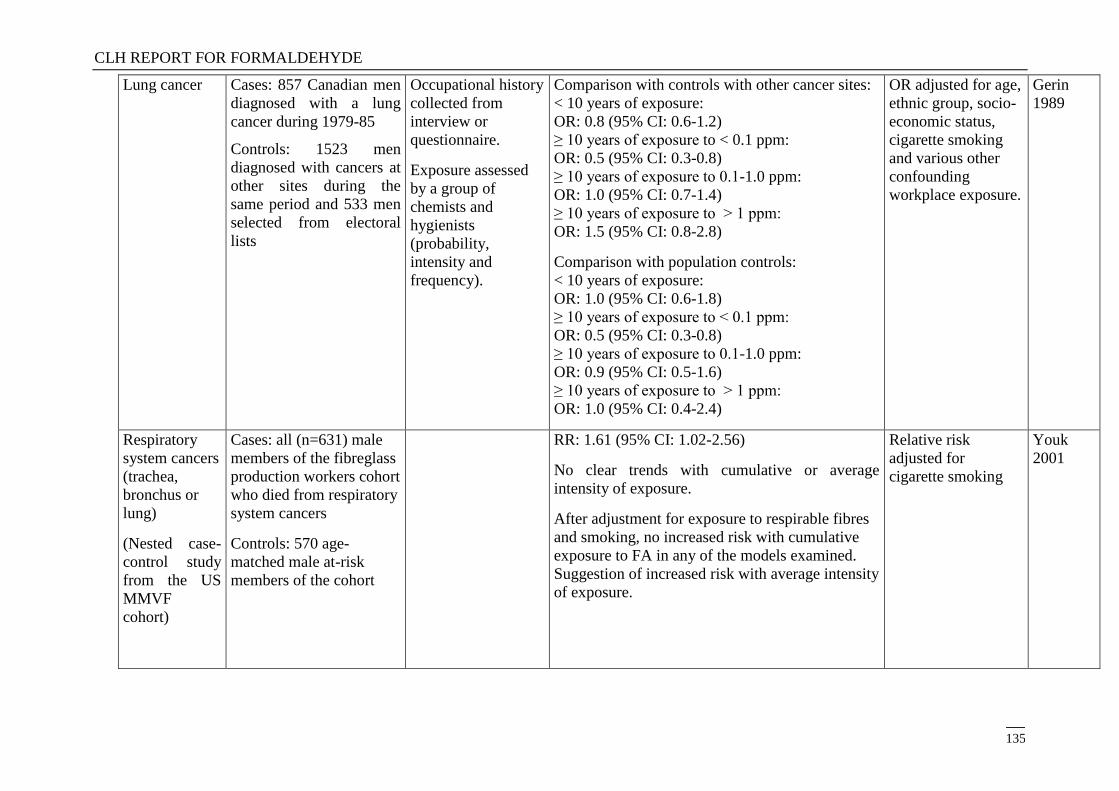
CLH REPORT FOR FORMALDEHYDE
135
Lung cancer Cases: 857 Canadian men
diagnosed with a lung
cancer during 1979-85
Controls: 1523 men
diagnosed with cancers at
other sites during the
same period and 533 men
selected from electoral
lists
Occupational history
collected from
interview or
questionnaire.
Exposure assessed
by a group of
chemists and
hygienists
(probability,
intensity and
frequency).
Comparison with controls with other cancer sites:
< 10 years of exposure:
OR: 0.8 (95% CI: 0.6-1.2)
≥ 10 years of exposure to < 0.1 ppm:
OR: 0.5 (95% CI: 0.3-0.8)
≥ 10 years of exposure to 0.1-1.0 ppm:
OR: 1.0 (95% CI: 0.7-1.4)
≥ 10 years of exposure to > 1 ppm:
OR: 1.5 (95% CI: 0.8-2.8)
Comparison with population controls:
< 10 years of exposure:
OR: 1.0 (95% CI: 0.6-1.8)
≥ 10 years of exposure to < 0.1 ppm:
OR: 0.5 (95% CI: 0.3-0.8)
≥ 10 years of exposure to 0.1-1.0 ppm:
OR: 0.9 (95% CI: 0.5-1.6)
≥ 10 years of exposure to > 1 ppm:
OR: 1.0 (95% CI: 0.4-2.4)
OR adjusted for age,
ethnic group, socio-
economic status,
cigarette smoking
and various other
confounding
workplace exposure.
Gerin
1989
Respiratory
system cancers
(trachea,
bronchus or
lung)
(Nested case-
control study
from the US
MMVF
cohort)
Cases: all (n=631) male
members of the fibreglass
production workers cohort
who died from respiratory
system cancers
Controls: 570 age-
matched male at-risk
members of the cohort
RR: 1.61 (95% CI: 1.02-2.56)
No clear trends with cumulative or average
intensity of exposure.
After adjustment for exposure to respirable fibres
and smoking, no increased risk with cumulative
exposure to FA in any of the models examined.
Suggestion of increased risk with average intensity
of exposure.
Relative risk
adjusted for
cigarette smoking
Youk
2001
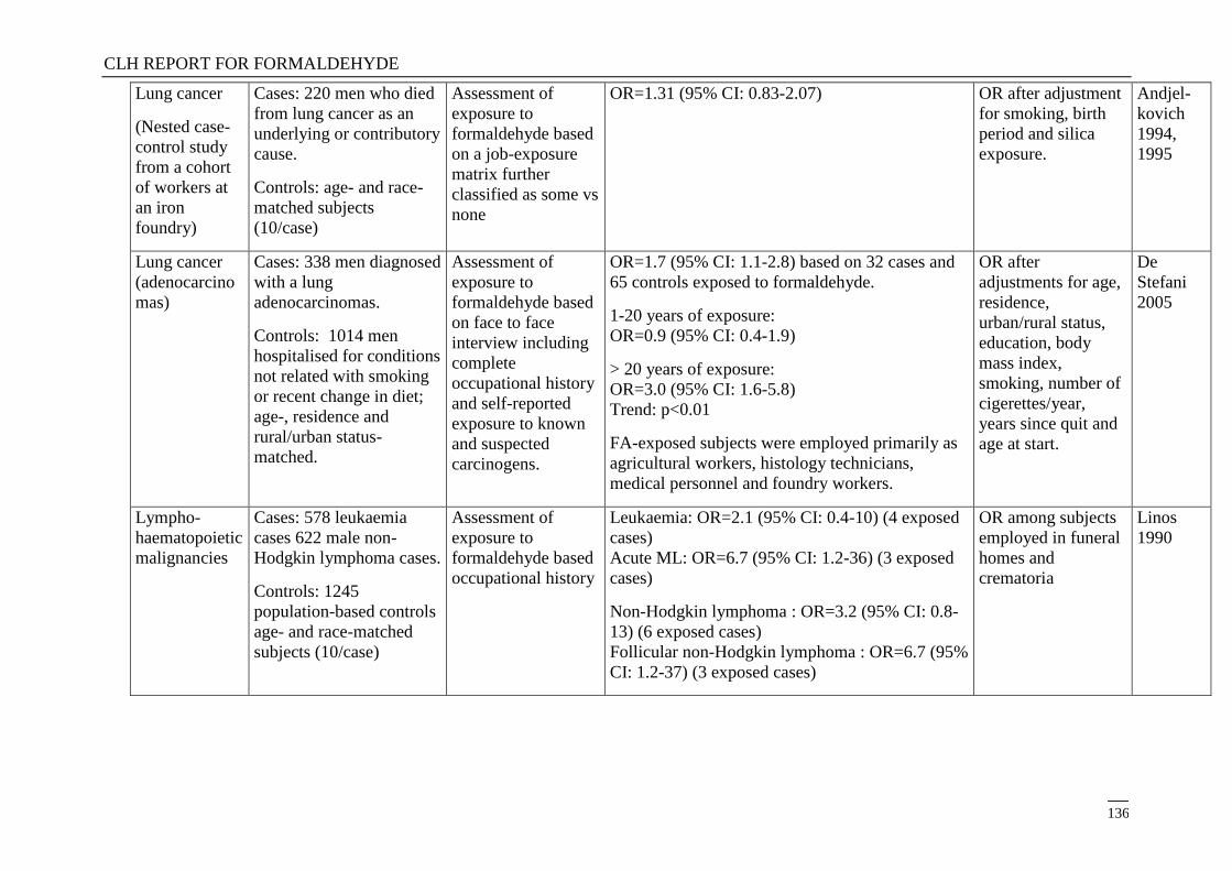
CLH REPORT FOR FORMALDEHYDE
136
Lung cancer
(Nested case-
control study
from a cohort
of workers at
an iron
foundry)
Cases: 220 men who died
from lung cancer as an
underlying or contributory
cause.
Controls: age- and race-
matched subjects
(10/case)
Assessment of
exposure to
formaldehyde based
on a job-exposure
matrix further
classified as some vs
none
OR=1.31 (95% CI: 0.83-2.07) OR after adjustment
for smoking, birth
period and silica
exposure.
Andjel-
kovich
1994,
1995
Lung cancer
(adenocarcino
mas)
Cases: 338 men diagnosed
with a lung
adenocarcinomas.
Controls: 1014 men
hospitalised for conditions
not related with smoking
or recent change in diet;
age-, residence and
rural/urban status-
matched.
Assessment of
exposure to
formaldehyde based
on face to face
interview including
complete
occupational history
and self-reported
exposure to known
and suspected
carcinogens.
OR=1.7 (95% CI: 1.1-2.8) based on 32 cases and
65 controls exposed to formaldehyde.
1-20 years of exposure:
OR=0.9 (95% CI: 0.4-1.9)
> 20 years of exposure:
OR=3.0 (95% CI: 1.6-5.8)
Trend: p<0.01
FA-exposed subjects were employed primarily as
agricultural workers, histology technicians,
medical personnel and foundry workers.
OR after
adjustments for age,
residence,
urban/rural status,
education, body
mass index,
smoking, number of
cigerettes/year,
years since quit and
age at start.
De
Stefani
2005
Lympho-
haematopoietic
malignancies
Cases: 578 leukaemia
cases 622 male non-
Hodgkin lymphoma cases.
Controls: 1245
population-based controls
age- and race-matched
subjects (10/case)
Assessment of
exposure to
formaldehyde based
occupational history
Leukaemia: OR=2.1 (95% CI: 0.4-10) (4 exposed
cases)
Acute ML: OR=6.7 (95% CI: 1.2-36) (3 exposed
cases)
Non-Hodgkin lymphoma : OR=3.2 (95% CI: 0.8-
13) (6 exposed cases)
Follicular non-Hodgkin lymphoma : OR=6.7 (95%
CI: 1.2-37) (3 exposed cases)
OR among subjects
employed in funeral
homes and
crematoria
Linos
1990
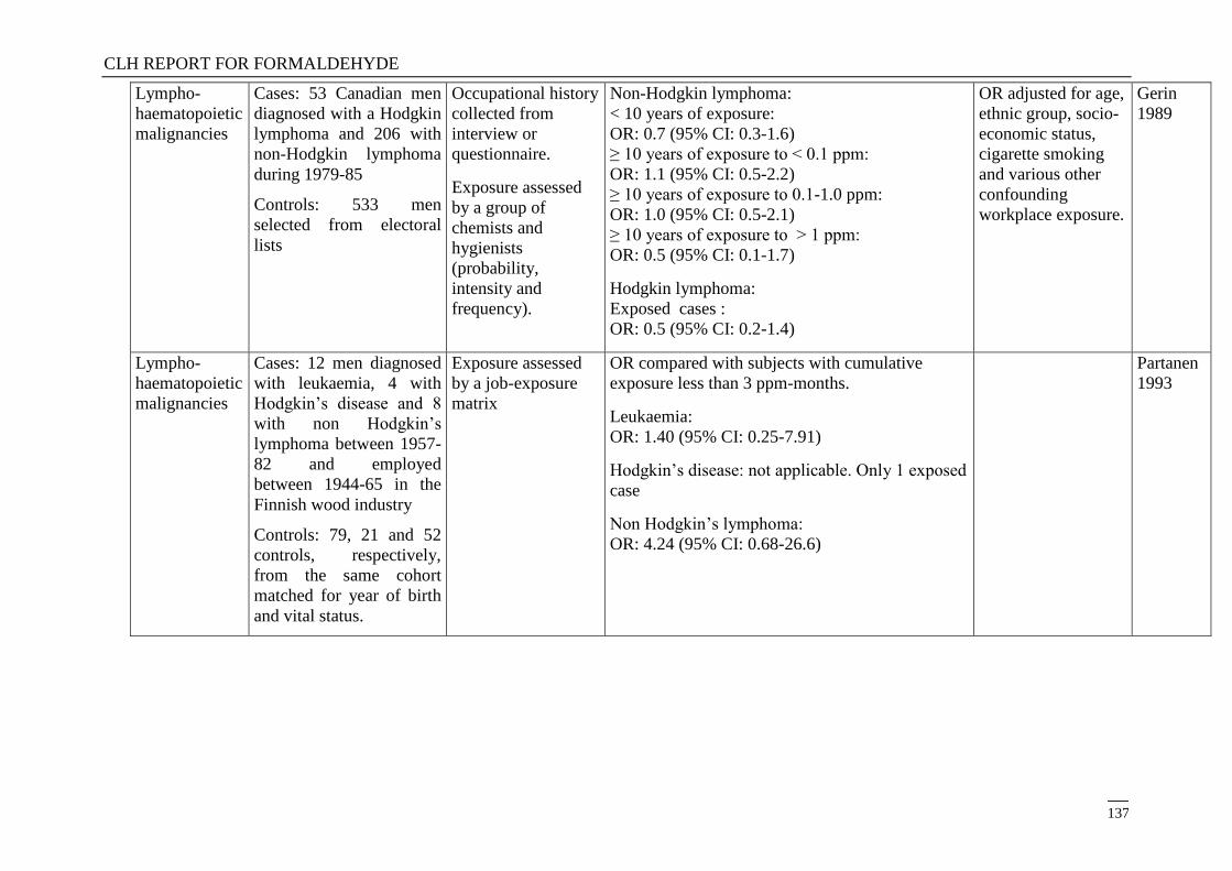
CLH REPORT FOR FORMALDEHYDE
137
Lympho-
haematopoietic
malignancies
Cases: 53 Canadian men
diagnosed with a Hodgkin
lymphoma and 206 with
non-Hodgkin lymphoma
during 1979-85
Controls: 533 men
selected from electoral
lists
Occupational history
collected from
interview or
questionnaire.
Exposure assessed
by a group of
chemists and
hygienists
(probability,
intensity and
frequency).
Non-Hodgkin lymphoma:
< 10 years of exposure:
OR: 0.7 (95% CI: 0.3-1.6)
≥ 10 years of exposure to < 0.1 ppm:
OR: 1.1 (95% CI: 0.5-2.2)
≥ 10 years of exposure to 0.1-1.0 ppm:
OR: 1.0 (95% CI: 0.5-2.1)
≥ 10 years of exposure to > 1 ppm:
OR: 0.5 (95% CI: 0.1-1.7)
Hodgkin lymphoma:
Exposed cases :
OR: 0.5 (95% CI: 0.2-1.4)
OR adjusted for age,
ethnic group, socio-
economic status,
cigarette smoking
and various other
confounding
workplace exposure.
Gerin
1989
Lympho-
haematopoietic
malignancies
Cases: 12 men diagnosed
with leukaemia, 4 with
Hodgkin‘s disease and 8
with non Hodgkin‘s
lymphoma between 1957-
82 and employed
between 1944-65 in the
Finnish wood industry
Controls: 79, 21 and 52
controls, respectively,
from the same cohort
matched for year of birth
and vital status.
Exposure assessed
by a job-exposure
matrix
OR compared with subjects with cumulative
exposure less than 3 ppm-months.
Leukaemia:
OR: 1.40 (95% CI: 0.25-7.91)
Hodgkin‘s disease: not applicable. Only 1 exposed
case
Non Hodgkin‘s lymphoma:
OR: 4.24 (95% CI: 0.68-26.6)
Partanen
1993
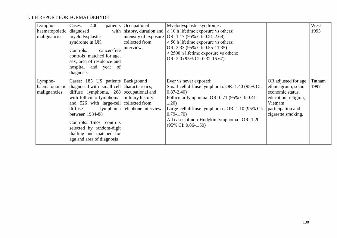
CLH REPORT FOR FORMALDEHYDE
138
Lympho-
haematopoietic
malignancies
Cases: 400 patients
diagnosed with
myelodysplastic
syndrome in UK
Controls: cancer-free
controls matched for age,
sex, area of residence and
hospital and year of
diagnosis
Occupational
history, duration and
intensity of exposure
collected from
interview.
Myelodysplastic syndrome :
≥ 10 h lifetime exposure vs others:
OR: 1.17 (95% CI: 0.51-2.68)
≥ 50 h lifetime exposure vs others:
OR: 2.33 (95% CI: 0.55-11.35)
≥ 2500 h lifetime exposure vs others:
OR: 2.0 (95% CI: 0.32-15.67)
West
1995
Lympho-
haematopoietic
malignancies
Cases: 185 US patients
diagnosed with small-cell
diffuse lymphoma, 268
with follicular lymphoma,
and 526 with large-cell
diffuse lymphoma
between 1984-88
Controls: 1659 controls
selected by random-digit
dialling and matched for
age and area of diagnosis
Background
characteristics,
occupational and
military history
collected from
telephone interview.
Ever vs never exposed:
Small-cell diffuse lymphoma: OR: 1.40 (95% CI:
0.87-2.40)
Follicular lymphoma: OR: 0.71 (95% CI: 0.41-
1.20)
Large-cell diffuse lymphoma : OR: 1.10 (95% CI:
0.79-1.70)
All cases of non-Hodgkin lymphoma : OR: 1.20
(95% CI: 0.86-1.50)
OR adjusted for age,
ethnic group, socio-
economic status,
education, religion,
Vietnam
participation and
cigarette smoking.
Tatham
1997
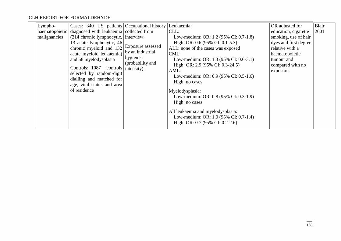
CLH REPORT FOR FORMALDEHYDE
139
Lympho-
haematopoietic
malignancies
Cases: 340 US patients
diagnosed with leukaemia
(214 chronic lymphocytic,
13 acute lymphocytic, 46
chronic myeloid and 132
acute myeloid leukaemia)
and 58 myelodysplasia
Controls: 1087 controls
selected by random-digit
dialling and matched for
age, vital status and area
of residence
Occupational history
collected from
interview.
Exposure assessed
by an industrial
hygienist
(probability and
intensity).
Leukaemia:
CLL:
Low-medium: OR: 1.2 (95% CI: 0.7-1.8)
High: OR: 0.6 (95% CI: 0.1-5.3)
ALL: none of the cases was exposed
CML:
Low-medium: OR: 1.3 (95% CI: 0.6-3.1)
High: OR: 2.9 (95% CI: 0.3-24.5)
AML:
Low-medium: OR: 0.9 (95% CI: 0.5-1.6)
High: no cases
Myelodysplasia:
Low-medium: OR: 0.8 (95% CI: 0.3-1.9)
High: no cases
All leukaemia and myelodysplasia:
Low-medium: OR: 1.0 (95% CI: 0.7-1.4)
High: OR: 0.7 (95% CI: 0.2-2.6)
OR adjusted for
education, cigarette
smoking, use of hair
dyes and first degree
relative with a
haematopoietic
tumour and
compared with no
exposure.
Blair
2001
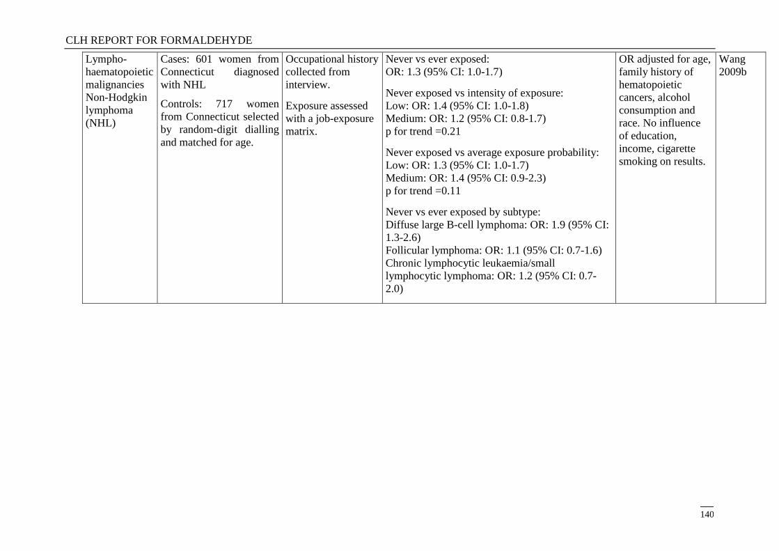
CLH REPORT FOR FORMALDEHYDE
140
Lympho-
haematopoietic
malignancies
Non-Hodgkin
lymphoma
(NHL)
Cases: 601 women from
Connecticut diagnosed
with NHL
Controls: 717 women
from Connecticut selected
by random-digit dialling
and matched for age.
Occupational history
collected from
interview.
Exposure assessed
with a job-exposure
matrix.
Never vs ever exposed:
OR: 1.3 (95% CI: 1.0-1.7)
Never exposed vs intensity of exposure:
Low: OR: 1.4 (95% CI: 1.0-1.8)
Medium: OR: 1.2 (95% CI: 0.8-1.7)
p for trend =0.21
Never exposed vs average exposure probability:
Low: OR: 1.3 (95% CI: 1.0-1.7)
Medium: OR: 1.4 (95% CI: 0.9-2.3)
p for trend =0.11
Never vs ever exposed by subtype:
Diffuse large B-cell lymphoma: OR: 1.9 (95% CI:
1.3-2.6)
Follicular lymphoma: OR: 1.1 (95% CI: 0.7-1.6)
Chronic lymphocytic leukaemia/small
lymphocytic lymphoma: OR: 1.2 (95% CI: 0.7-
2.0)
OR adjusted for age,
family history of
hematopoietic
cancers, alcohol
consumption and
race. No influence
of education,
income, cigarette
smoking on results.
Wang
2009b
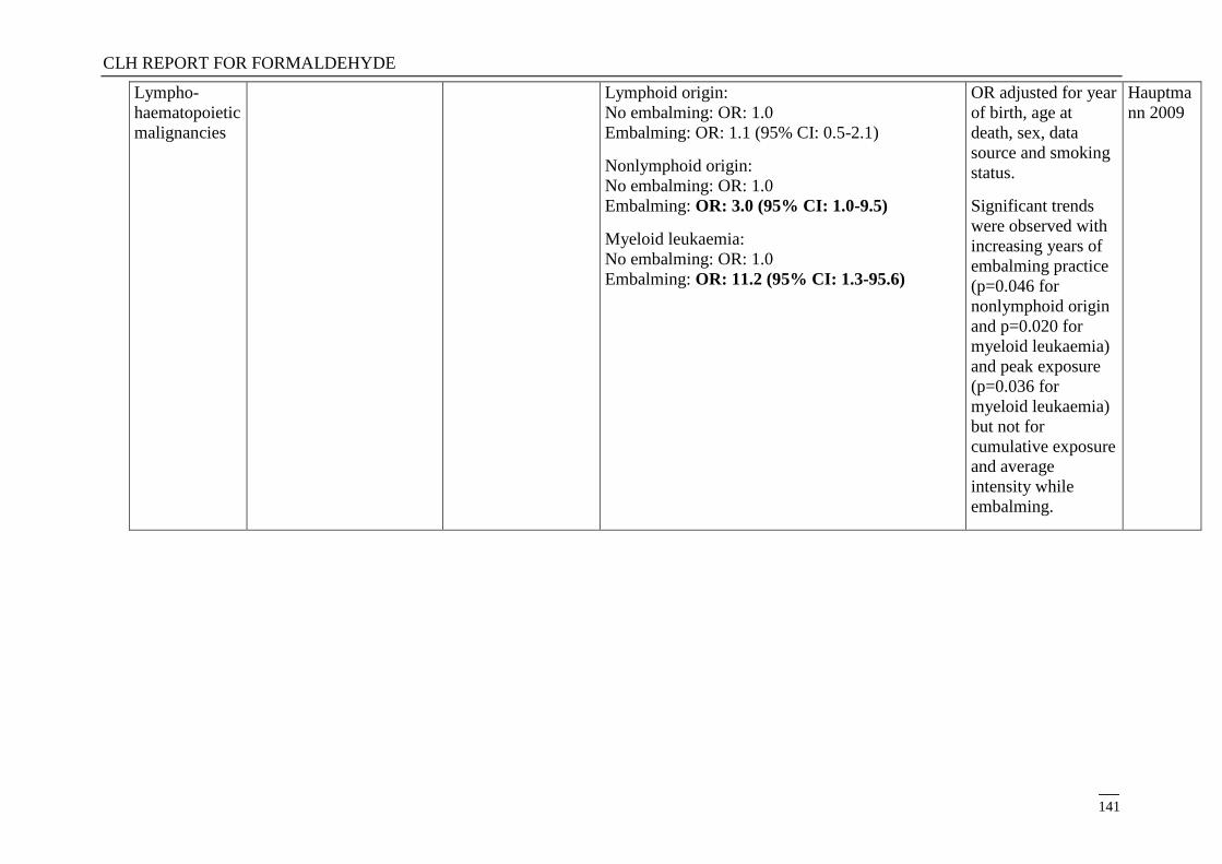
CLH REPORT FOR FORMALDEHYDE
141
Lympho-
haematopoietic
malignancies
Lymphoid origin:
No embalming: OR: 1.0
Embalming: OR: 1.1 (95% CI: 0.5-2.1)
Nonlymphoid origin:
No embalming: OR: 1.0
Embalming: OR: 3.0 (95% CI: 1.0-9.5)
Myeloid leukaemia:
No embalming: OR: 1.0
Embalming: OR: 11.2 (95% CI: 1.3-95.6)
OR adjusted for year
of birth, age at
death, sex, data
source and smoking
status.
Significant trends
were observed with
increasing years of
embalming practice
(p=0.046 for
nonlymphoid origin
and p=0.020 for
myeloid leukaemia)
and peak exposure
(p=0.036 for
myeloid leukaemia)
but not for
cumulative exposure
and average
intensity while
embalming.
Hauptma
nn 2009
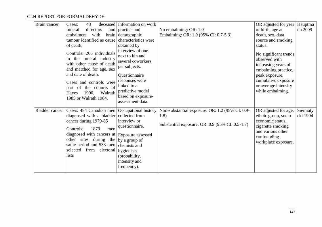
CLH REPORT FOR FORMALDEHYDE
142
Brain cancer Cases: 48 deceased
funeral directors and
embalmers with brain
tumour identified as cause
of death.
Controls: 265 individuals
in the funeral industry
with other cause of death
and matched for age, sex
and date of death.
Cases and controls were
part of the cohorts of
Hayes 1990, Walrath
1983 or Walrath 1984.
Information on work
practice and
demographic
characteristics were
obtained by
interview of one
next to kin and
several coworkers
per subjects.
Questionnaire
responses were
linked to a
predictive model
based on exposure-
assessment data.
No embalming: OR: 1.0
Embalming: OR: 1.9 (95% CI: 0.7-5.3)
OR adjusted for year
of birth, age at
death, sex, data
source and smoking
status.
No significant trends
observed with
increasing years of
embalming practice,
peak exposure,
cumulative exposure
or average intensity
while embalming.
Hauptma
nn 2009
Bladder cancer Cases: 484 Canadian men
diagnosed with a bladder
cancer during 1979-85
Controls: 1879 men
diagnosed with cancers at
other sites during the
same period and 533 men
selected from electoral
lists
Occupational history
collected from
interview or
questionnaire.
Exposure assessed
by a group of
chemists and
hygienists
(probability,
intensity and
frequency).
Non-substantial exposure: OR: 1.2 (95% CI: 0.9-
1.8)
Substantial exposure: OR: 0.9 (95% CI: 0.5-1.7)
OR adjusted for age,
ethnic group, socio-
economic status,
cigarette smoking
and various other
confounding
workplace exposure.
Siemiaty
cki 1994
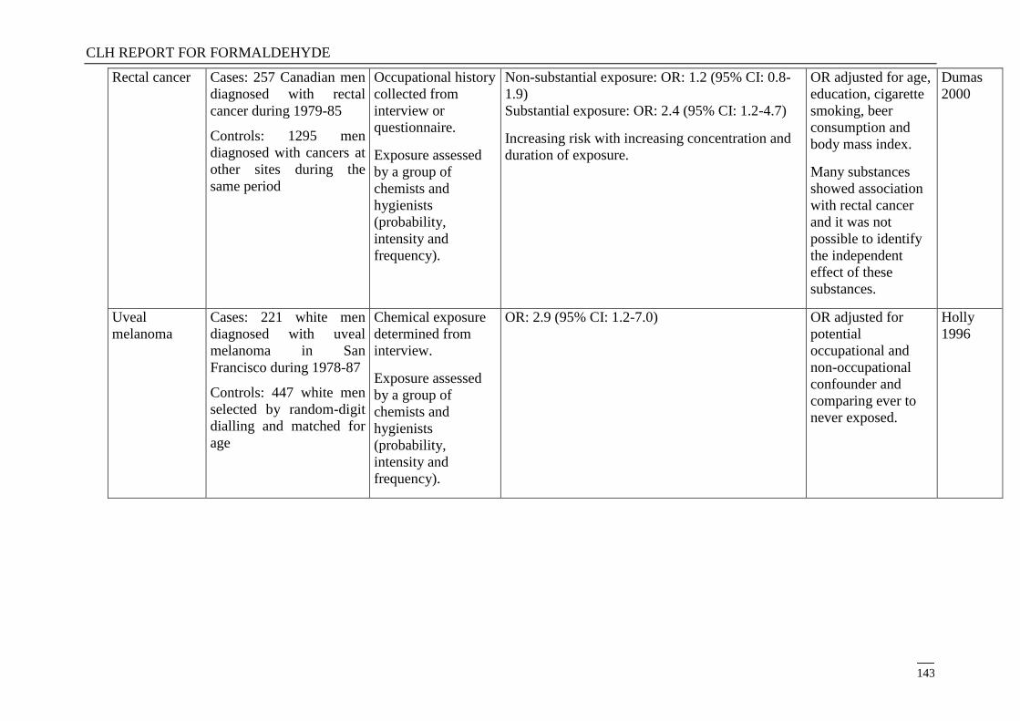
CLH REPORT FOR FORMALDEHYDE
143
Rectal cancer Cases: 257 Canadian men
diagnosed with rectal
cancer during 1979-85
Controls: 1295 men
diagnosed with cancers at
other sites during the
same period
Occupational history
collected from
interview or
questionnaire.
Exposure assessed
by a group of
chemists and
hygienists
(probability,
intensity and
frequency).
Non-substantial exposure: OR: 1.2 (95% CI: 0.8-
1.9)
Substantial exposure: OR: 2.4 (95% CI: 1.2-4.7)
Increasing risk with increasing concentration and
duration of exposure.
OR adjusted for age,
education, cigarette
smoking, beer
consumption and
body mass index.
Many substances
showed association
with rectal cancer
and it was not
possible to identify
the independent
effect of these
substances.
Dumas
2000
Uveal
melanoma
Cases: 221 white men
diagnosed with uveal
melanoma in San
Francisco during 1978-87
Controls: 447 white men
selected by random-digit
dialling and matched for
age
Chemical exposure
determined from
interview.
Exposure assessed
by a group of
chemists and
hygienists
(probability,
intensity and
frequency).
OR: 2.9 (95% CI: 1.2-7.0)
OR adjusted for
potential
occupational and
non-occupational
confounder and
comparing ever to
never exposed.
Holly
1996
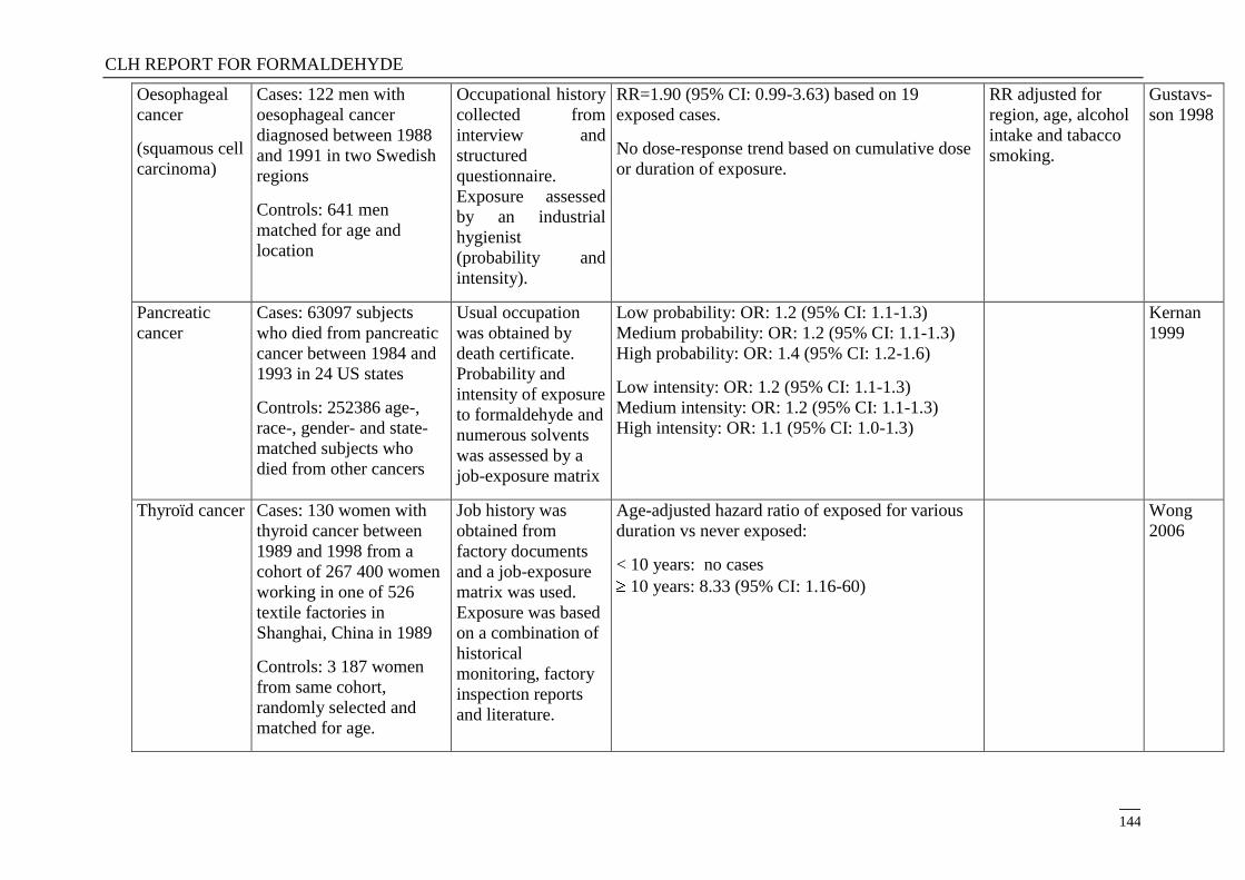
CLH REPORT FOR FORMALDEHYDE
144
Oesophageal
cancer
(squamous cell
carcinoma)
Cases: 122 men with
oesophageal cancer
diagnosed between 1988
and 1991 in two Swedish
regions
Controls: 641 men
matched for age and
location
Occupational history
collected from
interview and
structured
questionnaire.
Exposure assessed
by an industrial
hygienist
(probability and
intensity).
RR=1.90 (95% CI: 0.99-3.63) based on 19
exposed cases.
No dose-response trend based on cumulative dose
or duration of exposure.
RR adjusted for
region, age, alcohol
intake and tabacco
smoking.
Gustavs-
son 1998
Pancreatic
cancer
Cases: 63097 subjects
who died from pancreatic
cancer between 1984 and
1993 in 24 US states
Controls: 252386 age-,
race-, gender- and state-
matched subjects who
died from other cancers
Usual occupation
was obtained by
death certificate.
Probability and
intensity of exposure
to formaldehyde and
numerous solvents
was assessed by a
job-exposure matrix
Low probability: OR: 1.2 (95% CI: 1.1-1.3)
Medium probability: OR: 1.2 (95% CI: 1.1-1.3)
High probability: OR: 1.4 (95% CI: 1.2-1.6)
Low intensity: OR: 1.2 (95% CI: 1.1-1.3)
Medium intensity: OR: 1.2 (95% CI: 1.1-1.3)
High intensity: OR: 1.1 (95% CI: 1.0-1.3)
Kernan
1999
Thyroïd cancer Cases: 130 women with
thyroid cancer between
1989 and 1998 from a
cohort of 267 400 women
working in one of 526
textile factories in
Shanghai, China in 1989
Controls: 3 187 women
from same cohort,
randomly selected and
matched for age.
Job history was
obtained from
factory documents
and a job-exposure
matrix was used.
Exposure was based
on a combination of
historical
monitoring, factory
inspection reports
and literature.
Age-adjusted hazard ratio of exposed for various
duration vs never exposed:
< 10 years: no cases
10 years: 8.33 (95% CI: 1.16-60)
Wong
2006
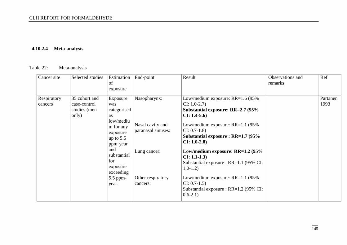
CLH REPORT FOR FORMALDEHYDE
145
4.10.2.4 Meta-analysis
Table 22: Meta-analysis
Cancer site Selected studies Estimation
of
exposure
End-point Result Observations and
remarks
Ref
Respiratory
cancers
35 cohort and
case-control
studies (men
only)
Exposure
was
categorised
as
low/mediu
m for any
exposure
up to 5.5
ppm-year
and
substantial
for
exposure
exceeding
5.5 ppm-
year.
Nasopharynx:
Nasal cavity and
paranasal sinuses:
Lung cancer:
Other respiratory
cancers:
Low/medium exposure: RR=1.6 (95%
CI: 1.0-2.7)
Substantial exposure: RR=2.7 (95%
CI: 1.4-5.6)
Low/medium exposure: RR=1.1 (95%
CI: 0.7-1.8)
Substantial exposure : RR=1.7 (95%
CI: 1.0-2.8)
Low/medium exposure: RR=1.2 (95%
CI: 1.1-1.3) Substantial exposure : RR=1.1 (95% CI:
1.0-1.2)
Low/medium exposure: RR=1.1 (95%
CI: 0.7-1.5)
Substantial exposure : RR=1.2 (95% CI:
0.6-2.1)
Partanen
1993
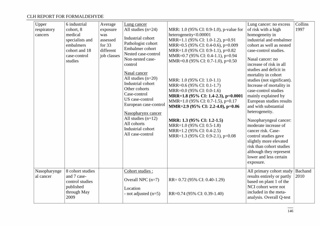
CLH REPORT FOR FORMALDEHYDE
146
Upper
respiratory
cancers
6 industrial
cohort, 8
medical
specialists and
embalmers
cohort and 18
case-control
studies
Average
exposure
was
assessed
for 33
different
job classes
Lung cancer
All studies (n=24)
Industrial cohort
Pathologist cohort
Embalmer cohort
Nested case-control
Non-nested case-
control
Nasal cancer
All studies (n=20)
Industrial cohort
Other cohorts
Case-control
US case-control
European case-control
Nasopharynx cancer
All studies (n=12)
All cohorts
Industrial cohort
All case-control
MRR: 1.0 (95% CI: 0.9-1.0), p-value for
heterogeneity<0.00001
MRR=1.1 (95% CI: 1.0-1.2), p=0.91
MRR=0.5 (95% CI: 0.4-0.6), p=0.009
MRR=1.0 (95% CI: 0.9-1.1), p=0.82
MMR=0.7 (95% CI: 0.4-1.1), p=0.94
MMR=0.8 (95% CI: 0.7-1.0), p=0.50
MRR: 1.0 (95% CI: 1.0-1.1)
MRR=0.6 (95% CI: 0.1-1.7)
MRR=0.0 (95% CI: 0.0-1.6)
MRR=1.8 (95% CI: 1.4-2.3), p=0.0001 MMR=1.0 (95% CI: 0.7-1.5), p=0.17
MMR=2.9 (95% CI: 2.2-4.0), p=0.06
MRR: 1.3 (95% CI: 1.2-1.5) MRR=1.0 (95% CI: 0.5-1.8)
MRR=1.2 (95% CI: 0.4-2.5)
MRR=1.3 (95% CI: 0.9-2.1), p=0.08
Lung cancer: no excess
of risk with a high
homogeneity in
industrial and embalmer
cohort as well as nested
case-control studies.
Nasal cancer: no
increase of risk in all
studies and deficit in
mortality in cohort
studies (not significant).
Increase of mortality in
case-control studies
mainly explained by
European studies results
and with substantial
heterogeneity.
Nasopharyngeal cancer:
moderate increase of
cancer risk. Case-
control studies gave
slightly more elevated
risk than cohort studies
although they represent
lower and less certain
exposure.
Collins
1997
Nasopharynge
al cancer
8 cohort studies
and 7 case-
control studies
published
through May
2009
Cohort studies :
Overall NPC (n=7)
Location
- not adjusted (n=5)
RR= 0.72 (95% CI: 0.40-1.29)
RR=0.74 (95% CI: 0.39-1.40)
All primary cohort study
results entirely or partly
based on plant 1 of the
NCI cohort were not
included in the meta-
analysis. Overall Q-test
Bachand
2010
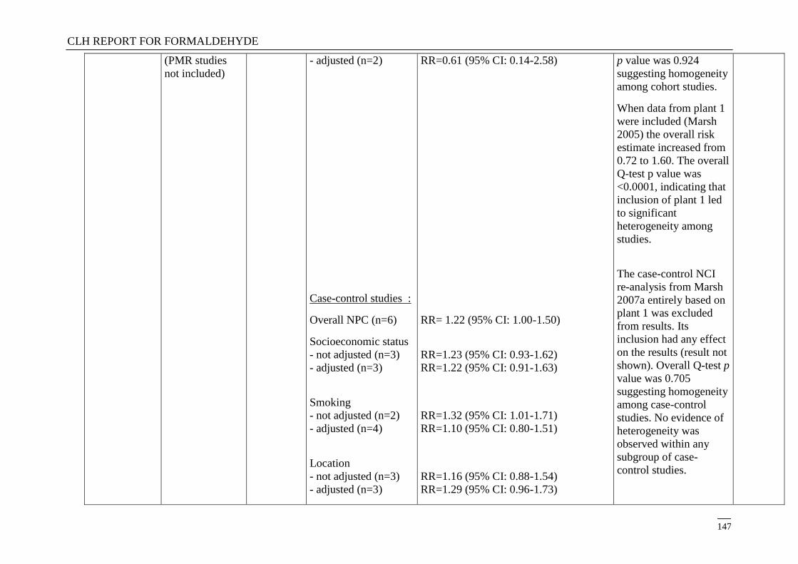
CLH REPORT FOR FORMALDEHYDE
147
(PMR studies
not included)
- adjusted (n=2)
Case-control studies :
Overall NPC (n=6)
Socioeconomic status
- not adjusted (n=3)
- adjusted (n=3)
Smoking
- not adjusted (n=2)
- adjusted (n=4)
Location
- not adjusted (n=3)
- adjusted (n=3)
RR=0.61 (95% CI: 0.14-2.58)
RR= 1.22 (95% CI: 1.00-1.50)
RR=1.23 (95% CI: 0.93-1.62)
RR=1.22 (95% CI: 0.91-1.63)
RR=1.32 (95% CI: 1.01-1.71)
RR=1.10 (95% CI: 0.80-1.51)
RR=1.16 (95% CI: 0.88-1.54)
RR=1.29 (95% CI: 0.96-1.73)
p value was 0.924
suggesting homogeneity
among cohort studies.
When data from plant 1
were included (Marsh
2005) the overall risk
estimate increased from
0.72 to 1.60. The overall
Q-test p value was
<0.0001, indicating that
inclusion of plant 1 led
to significant
heterogeneity among
studies.
The case-control NCI
re-analysis from Marsh
2007a entirely based on
plant 1 was excluded
from results. Its
inclusion had any effect
on the results (result not
shown). Overall Q-test p
value was 0.705
suggesting homogeneity
among case-control
studies. No evidence of
heterogeneity was
observed within any
subgroup of case-
control studies.
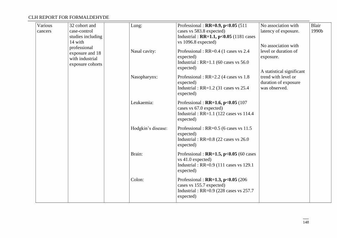
CLH REPORT FOR FORMALDEHYDE
148
Various
cancers
32 cohort and
case-control
studies including
14 with
professional
exposure and 18
with industrial
exposure cohorts
Lung:
Nasal cavity:
Nasopharynx:
Leukaemia:
Hodgkin‘s disease:
Brain:
Colon:
Professional : RR=0.9, p<0.05 (511
cases vs 583.8 expected)
Industrial : RR=1.1, p<0.05 (1181 cases
vs 1096.8 expected)
Professional : RR=0.4 (1 cases vs 2.4
expected)
Industrial : RR=1.1 (60 cases vs 56.0
expected)
Professional : RR=2.2 (4 cases vs 1.8
expected)
Industrial : RR=1.2 (31 cases vs 25.4
expected)
Professional : RR=1.6, p<0.05 (107
cases vs 67.0 expected)
Industrial : RR=1.1 (122 cases vs 114.4
expected)
Professional : RR=0.5 (6 cases vs 11.5
expected)
Industrial : RR=0.8 (22 cases vs 26.0
expected)
Professional : RR=1.5, p<0.05 (60 cases
vs 41.0 expected)
Industrial : RR=0.9 (111 cases vs 129.1
expected)
Professional : RR=1.3, p<0.05 (206
cases vs 155.7 expected)
Industrial : RR=0.9 (228 cases vs 257.7
expected)
No association with
latency of exposure.
No association with
level or duration of
exposure.
A statistical significant
trend with level or
duration of exposure
was observed.
Blair
1990b

CLH REPORT FOR FORMALDEHYDE
149
Various
cancers
12 cohorts
published
through
February 2007
Oral cavity and
pharynx:
Lung:
Brain:
Lymphatic and
hemopoietic:
Leukaemia:
Ind. workers: RR=1.09 (95% CI: 0.88-
1.34)
Professionals: RR= 0.96 (95% CI : 0.75-
1.24)
Ind. workers: RR=1.06 (95% CI: 0.92-
1.23)
Professionals: RR= 0.63 (95% CI : 0.47-
0.84)
Ind. workers: RR=0.92 (95% CI: 0.75-
1.13)
Professionals: RR= 1.56 (95% CI :
1.24-1.96)
Ind. workers: RR=0.85 (95% CI: 0.74-
0.96)
Professionals: RR= 1.31 (95% CI :
1.16-1.48)
Ind. workers: RR=0.90 (95% CI: 0.75-
1.07)
Professionals: RR= 1.39 (95% CI :
1.15-1.68)
For nasopharynx a SMR
of 1.33 (0.61-2.53) is
calculated in 3 cohorts
of industrial workers.
Excluding a cluster of 6
deaths from a single
plant of the NCI study,
the pooled RRamong
industry declined to
0.49 based on 3 deaths.
For the sinus and nasal
cavity, a SMR of 1.01
(0.33-2.35) is calculated
in 3 cohorts of industrial
workers. No death was
observed in
professionals.
Bosetti
2008
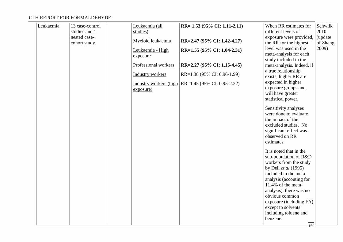
CLH REPORT FOR FORMALDEHYDE
150
Leukaemia 13 case-control
studies and 1
nested case-
cohort study
Leukaemia (all
studies)
Myeloid leukaemia
Leukaemia - High
exposure
Professional workers
Industry workers
Industry workers (high
exposure)
RR= 1.53 (95% CI: 1.11-2.11)
RR=2.47 (95% CI: 1.42-4.27)
RR=1.55 (95% CI: 1.04-2.31)
RR=2.27 (95% CI: 1.15-4.45)
RR=1.38 (95% CI: 0.96-1.99)
RR=1.45 (95% CI: 0.95-2.22)
When RR estimates for
different levels of
exposure were provided,
the RR for the highest
level was used in the
meta-analysis for each
study included in the
meta-analysis. Indeed, if
a true relationship
exists, higher RR are
expected in higher
exposure groups and
will have greater
statistical power.
Sensitivity analyses
were done to evaluate
the impact of the
excluded studies. No
significant effect was
observed on RR
estimates.
It is noted that in the
sub-population of R&D
workers from the study
by Dell et al (1995)
included in the meta-
analysis (accouting for
11.4% of the meta-
analysis), there was no
obvious common
exposure (including FA)
except to solvents
including toluene and
benzene.
Schwilk
2010
(update
of Zhang
2009)
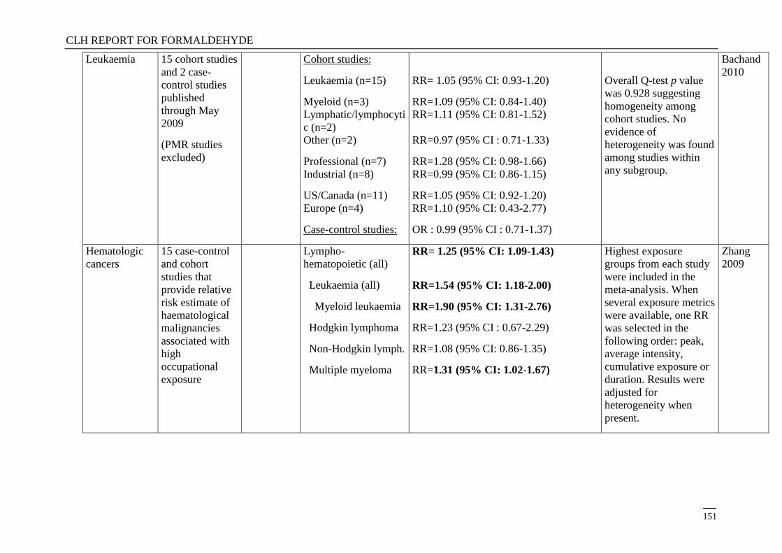
CLH REPORT FOR FORMALDEHYDE
151
Leukaemia 15 cohort studies
and 2 case-
control studies
published
through May
2009
(PMR studies
excluded)
Cohort studies:
Leukaemia (n=15)
Myeloid (n=3)
Lymphatic/lymphocyti
c (n=2)
Other (n=2)
Professional (n=7)
Industrial (n=8)
US/Canada (n=11)
Europe (n=4)
Case-control studies:
RR= 1.05 (95% CI: 0.93-1.20)
RR=1.09 (95% CI: 0.84-1.40)
RR=1.11 (95% CI: 0.81-1.52)
RR=0.97 (95% CI : 0.71-1.33)
RR=1.28 (95% CI: 0.98-1.66)
RR=0.99 (95% CI: 0.86-1.15)
RR=1.05 (95% CI: 0.92-1.20)
RR=1.10 (95% CI: 0.43-2.77)
OR : 0.99 (95% CI : 0.71-1.37)
Overall Q-test p value
was 0.928 suggesting
homogeneity among
cohort studies. No
evidence of
heterogeneity was found
among studies within
any subgroup.
Bachand
2010
Hematologic
cancers
15 case-control
and cohort
studies that
provide relative
risk estimate of
haematological
malignancies
associated with
high
occupational
exposure
Lympho-
hematopoietic (all)
Leukaemia (all)
Myeloid leukaemia
Hodgkin lymphoma
Non-Hodgkin lymph.
Multiple myeloma
RR= 1.25 (95% CI: 1.09-1.43)
RR=1.54 (95% CI: 1.18-2.00)
RR=1.90 (95% CI: 1.31-2.76)
RR=1.23 (95% CI : 0.67-2.29)
RR=1.08 (95% CI: 0.86-1.35)
RR=1.31 (95% CI: 1.02-1.67)
Highest exposure
groups from each study
were included in the
meta-analysis. When
several exposure metrics
were available, one RR
was selected in the
following order: peak,
average intensity,
cumulative exposure or
duration. Results were
adjusted for
heterogeneity when
present.
Zhang
2009
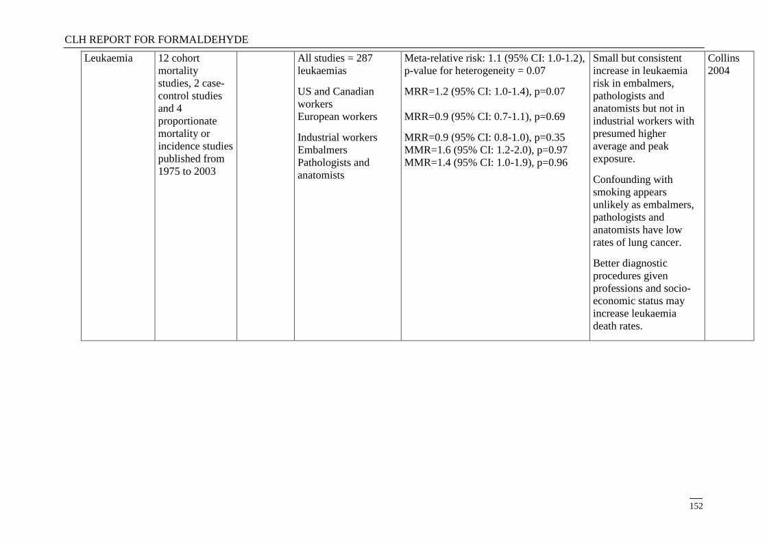
CLH REPORT FOR FORMALDEHYDE
152
Leukaemia 12 cohort
mortality
studies, 2 case-
control studies
and 4
proportionate
mortality or
incidence studies
published from
1975 to 2003
All studies = 287
leukaemias
US and Canadian
workers
European workers
Industrial workers
Embalmers
Pathologists and
anatomists
Meta-relative risk: 1.1 (95% CI: 1.0-1.2),
p-value for heterogeneity = 0.07
MRR=1.2 (95% CI: 1.0-1.4), p=0.07
MRR=0.9 (95% CI: 0.7-1.1), p=0.69
MRR=0.9 (95% CI: 0.8-1.0), p=0.35
MMR=1.6 (95% CI: 1.2-2.0), p=0.97
MMR=1.4 (95% CI: 1.0-1.9), p=0.96
Small but consistent
increase in leukaemia
risk in embalmers,
pathologists and
anatomists but not in
industrial workers with
presumed higher
average and peak
exposure.
Confounding with
smoking appears
unlikely as embalmers,
pathologists and
anatomists have low
rates of lung cancer.
Better diagnostic
procedures given
professions and socio-
economic status may
increase leukaemia
death rates.
Collins
2004
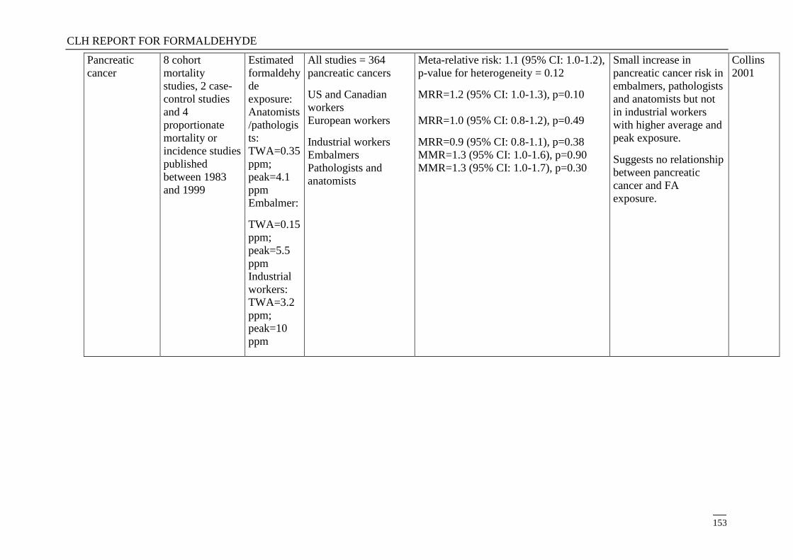
CLH REPORT FOR FORMALDEHYDE
153
Pancreatic
cancer
8 cohort
mortality
studies, 2 case-
control studies
and 4
proportionate
mortality or
incidence studies
published
between 1983
and 1999
Estimated
formaldehy
de
exposure:
Anatomists
/pathologis
ts:
TWA=0.35
ppm;
peak=4.1
ppm
Embalmer:
TWA=0.15
ppm;
peak=5.5
ppm
Industrial
workers:
TWA=3.2
ppm;
peak=10
ppm
All studies = 364
pancreatic cancers
US and Canadian
workers
European workers
Industrial workers
Embalmers
Pathologists and
anatomists
Meta-relative risk: 1.1 (95% CI: 1.0-1.2),
p-value for heterogeneity = 0.12
MRR=1.2 (95% CI: 1.0-1.3), p=0.10
MRR=1.0 (95% CI: 0.8-1.2), p=0.49
MRR=0.9 (95% CI: 0.8-1.1), p=0.38
MMR=1.3 (95% CI: 1.0-1.6), p=0.90
MMR=1.3 (95% CI: 1.0-1.7), p=0.30
Small increase in
pancreatic cancer risk in
embalmers, pathologists
and anatomists but not
in industrial workers
with higher average and
peak exposure.
Suggests no relationship
between pancreatic
cancer and FA
exposure.
Collins
2001
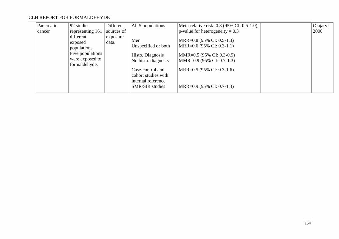
CLH REPORT FOR FORMALDEHYDE
154
Pancreatic
cancer
92 studies
representing 161
different
exposed
populations.
Five populations
were exposed to
formaldehyde.
Different
sources of
exposure
data.
All 5 populations
Men
Unspecified or both
Histo. Diagnosis
No histo. diagnosis
Case-control and
cohort studies with
internal reference
SMR/SIR studies
Meta-relative risk: 0.8 (95% CI: 0.5-1.0),
p-value for heterogeneity = 0.3
MRR=0.8 (95% CI: 0.5-1.3)
MRR=0.6 (95% CI: 0.3-1.1)
MMR=0.5 (95% CI: 0.3-0.9)
MMR=0.9 (95% CI: 0.7-1.3)
MRR=0.5 (95% CI: 0.3-1.6)
MRR=0.9 (95% CI: 0.7-1.3)
Ojajarvi
2000
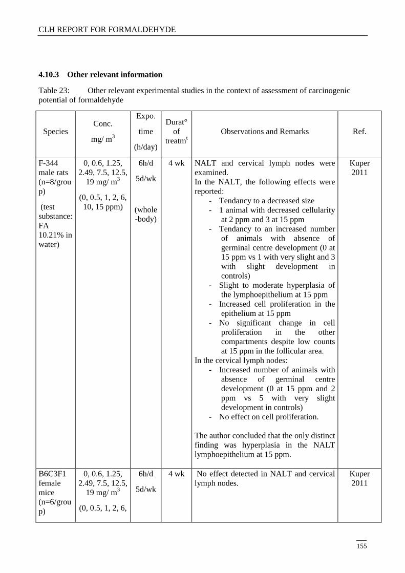
CLH REPORT FOR FORMALDEHYDE
155
4.10.3 Other relevant information
Table 23: Other relevant experimental studies in the context of assessment of carcinogenic
potential of formaldehyde
Species Conc.
mg/ m3
Expo.
time
(h/day)
Durat°
of
treatmt
Observations and Remarks
Ref.
F-344
male rats
(n=8/grou
p)
(test
substance:
FA
10.21% in
water)
0, 0.6, 1.25,
2.49, 7.5, 12.5,
19 mg/ m3
(0, 0.5, 1, 2, 6,
10, 15 ppm)
6h/d
5d/wk
(whole
-body)
4 wk NALT and cervical lymph nodes were
examined.
In the NALT, the following effects were
reported:
- Tendancy to a decreased size
- 1 animal with decreased cellularity
at 2 ppm and 3 at 15 ppm
- Tendancy to an increased number
of animals with absence of
germinal centre development (0 at
15 ppm vs 1 with very slight and 3
with slight development in
controls)
- Slight to moderate hyperplasia of
the lymphoepithelium at 15 ppm
- Increased cell proliferation in the
epithelium at 15 ppm
- No significant change in cell
proliferation in the other
compartments despite low counts
at 15 ppm in the follicular area.
In the cervical lymph nodes:
- Increased number of animals with
absence of germinal centre
development (0 at 15 ppm and 2
ppm vs 5 with very slight
development in controls)
- No effect on cell proliferation.
The author concluded that the only distinct
finding was hyperplasia in the NALT
lymphoepithelium at 15 ppm.
Kuper
2011
B6C3F1
female
mice
(n=6/grou
p)
0, 0.6, 1.25,
2.49, 7.5, 12.5,
19 mg/ m3
(0, 0.5, 1, 2, 6,
6h/d
5d/wk
4 wk No effect detected in NALT and cervical
lymph nodes.
Kuper
2011
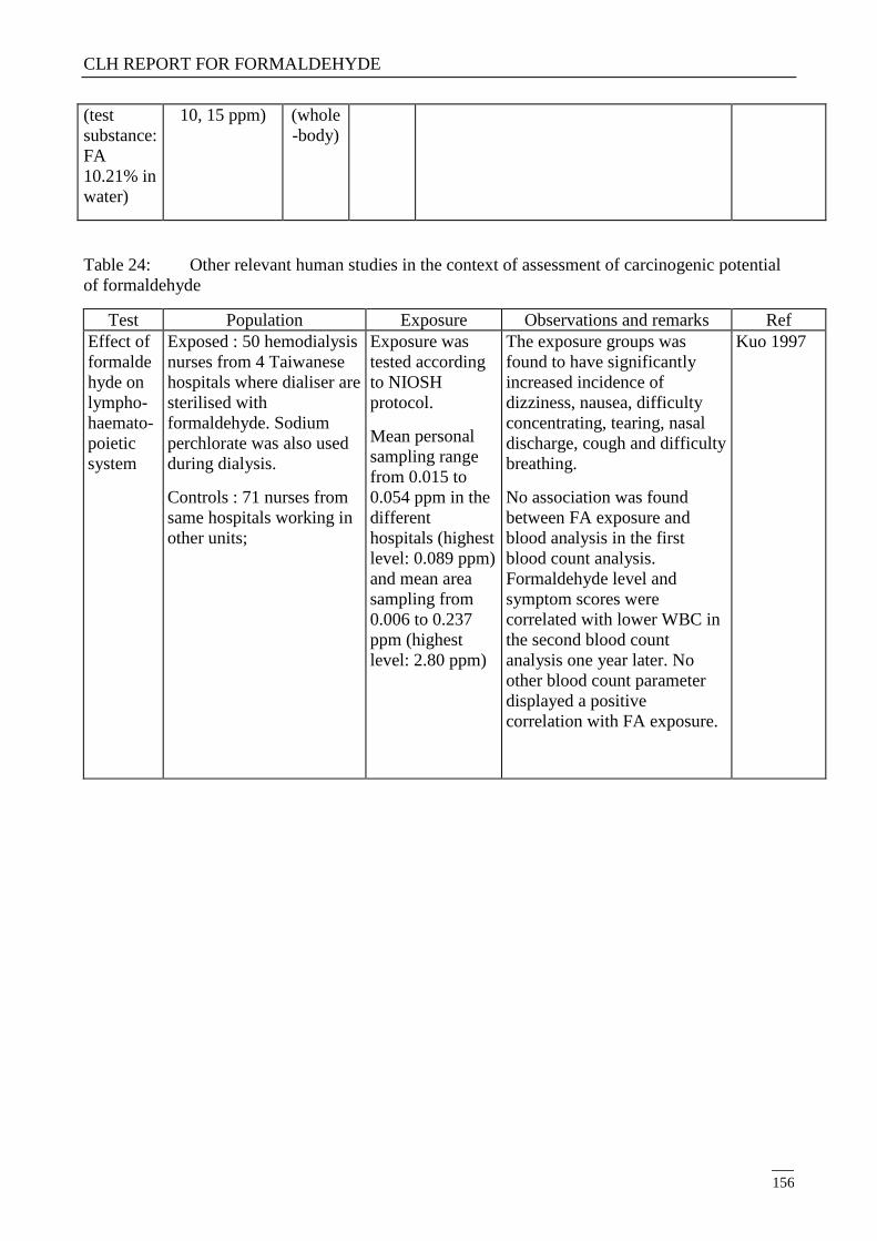
CLH REPORT FOR FORMALDEHYDE
156
(test
substance:
FA
10.21% in
water)
10, 15 ppm) (whole
-body)
Table 24: Other relevant human studies in the context of assessment of carcinogenic potential
of formaldehyde
Test Population Exposure Observations and remarks Ref
Effect of
formalde
hyde on
lympho-
haemato-
poietic
system
Exposed : 50 hemodialysis
nurses from 4 Taiwanese
hospitals where dialiser are
sterilised with
formaldehyde. Sodium
perchlorate was also used
during dialysis.
Controls : 71 nurses from
same hospitals working in
other units;
Exposure was
tested according
to NIOSH
protocol.
Mean personal
sampling range
from 0.015 to
0.054 ppm in the
different
hospitals (highest
level: 0.089 ppm)
and mean area
sampling from
0.006 to 0.237
ppm (highest
level: 2.80 ppm)
The exposure groups was
found to have significantly
increased incidence of
dizziness, nausea, difficulty
concentrating, tearing, nasal
discharge, cough and difficulty
breathing.
No association was found
between FA exposure and
blood analysis in the first
blood count analysis.
Formaldehyde level and
symptom scores were
correlated with lower WBC in
the second blood count
analysis one year later. No
other blood count parameter
displayed a positive
correlation with FA exposure.
Kuo 1997
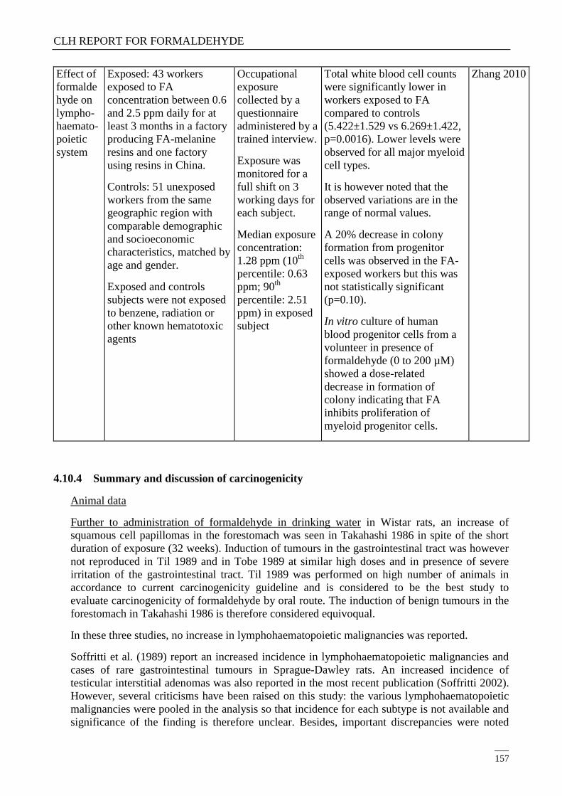
CLH REPORT FOR FORMALDEHYDE
157
Effect of
formalde
hyde on
lympho-
haemato-
poietic
system
Exposed: 43 workers
exposed to FA
concentration between 0.6
and 2.5 ppm daily for at
least 3 months in a factory
producing FA-melanine
resins and one factory
using resins in China.
Controls: 51 unexposed
workers from the same
geographic region with
comparable demographic
and socioeconomic
characteristics, matched by
age and gender.
Exposed and controls
subjects were not exposed
to benzene, radiation or
other known hematotoxic
agents
Occupational
exposure
collected by a
questionnaire
administered by a
trained interview.
Exposure was
monitored for a
full shift on 3
working days for
each subject.
Median exposure
concentration:
1.28 ppm (10th
percentile: 0.63
ppm; 90th
percentile: 2.51
ppm) in exposed
subject
Total white blood cell counts
were significantly lower in
workers exposed to FA
compared to controls
(5.422±1.529 vs 6.269±1.422,
p=0.0016). Lower levels were
observed for all major myeloid
cell types.
It is however noted that the
observed variations are in the
range of normal values.
A 20% decrease in colony
formation from progenitor
cells was observed in the FA-
exposed workers but this was
not statistically significant
(p=0.10).
In vitro culture of human
blood progenitor cells from a
volunteer in presence of
formaldehyde (0 to 200 µM)
showed a dose-related
decrease in formation of
colony indicating that FA
inhibits proliferation of
myeloid progenitor cells.
Zhang 2010
4.10.4 Summary and discussion of carcinogenicity
Animal data
Further to administration of formaldehyde in drinking water in Wistar rats, an increase of
squamous cell papillomas in the forestomach was seen in Takahashi 1986 in spite of the short
duration of exposure (32 weeks). Induction of tumours in the gastrointestinal tract was however
not reproduced in Til 1989 and in Tobe 1989 at similar high doses and in presence of severe
irritation of the gastrointestinal tract. Til 1989 was performed on high number of animals in
accordance to current carcinogenicity guideline and is considered to be the best study to
evaluate carcinogenicity of formaldehyde by oral route. The induction of benign tumours in the
forestomach in Takahashi 1986 is therefore considered equivoqual.
In these three studies, no increase in lymphohaematopoietic malignancies was reported.
Soffritti et al. (1989) report an increased incidence in lymphohaematopoietic malignancies and
cases of rare gastrointestinal tumours in Sprague-Dawley rats. An increased incidence of
testicular interstitial adenomas was also reported in the most recent publication (Soffritti 2002).
However, several criticisms have been raised on this study: the various lymphohaematopoietic
malignancies were pooled in the analysis so that incidence for each subtype is not available and
significance of the finding is therefore unclear. Besides, important discrepancies were noted
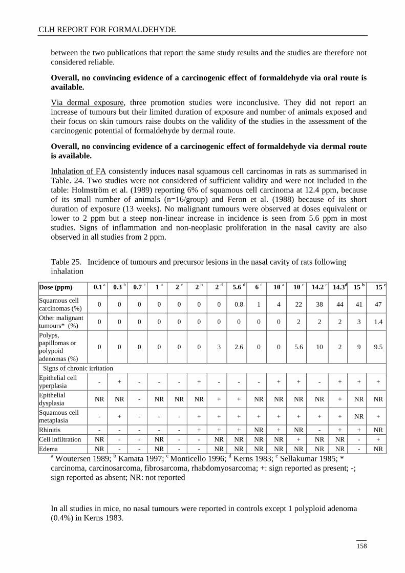
CLH REPORT FOR FORMALDEHYDE
158
between the two publications that report the same study results and the studies are therefore not
considered reliable.
Overall, no convincing evidence of a carcinogenic effect of formaldehyde via oral route is
available.
Via dermal exposure, three promotion studies were inconclusive. They did not report an
increase of tumours but their limited duration of exposure and number of animals exposed and
their focus on skin tumours raise doubts on the validity of the studies in the assessment of the
carcinogenic potential of formaldehyde by dermal route.
Overall, no convincing evidence of a carcinogenic effect of formaldehyde via dermal route
is available.
Inhalation of FA consistently induces nasal squamous cell carcinomas in rats as summarised in
Table. 24. Two studies were not considered of sufficient validity and were not included in the
table: Holmström et al. (1989) reporting 6% of squamous cell carcinoma at 12.4 ppm, because
of its small number of animals (n=16/group) and Feron et al. (1988) because of its short
duration of exposure (13 weeks). No malignant tumours were observed at doses equivalent or
lower to 2 ppm but a steep non-linear increase in incidence is seen from 5.6 ppm in most
studies. Signs of inflammation and non-neoplasic proliferation in the nasal cavity are also
observed in all studies from 2 ppm.
Table 25. Incidence of tumours and precursor lesions in the nasal cavity of rats following
inhalation
Dose (ppm) 0.1 a 0.3
b 0.7
c 1
a 2
c 2
b 2
d 5.6
d 6
c 10
a 10
c 14.2
e 14.3
d 15
b 15
c
Squamous cell
carcinomas (%) 0 0 0 0 0 0 0 0.8 1 4 22 38 44 41 47
Other malignant
tumours* (%) 0 0 0 0 0 0 0 0 0 0 2 2 2 3 1.4
Polyps,
papillomas or
polypoid
adenomas (%)
0 0 0 0 0 0 3 2.6 0 0 5.6 10 2 9 9.5
Signs of chronic irritation
Epithelial cell
yperplasia - + - - - + - - - + + - + + +
Epithelial
dysplasia NR NR - NR NR NR + + NR NR NR NR + NR NR
Squamous cell
metaplasia - + - - - + + + + + + + + NR +
Rhinitis - - - - - + + + NR + NR - + + NR
Cell infiltration NR - - NR - - NR NR NR NR + NR NR - +
Edema NR - - NR - - NR NR NR NR NR NR NR - NR a Woutersen 1989;
b Kamata 1997;
c Monticello 1996;
d Kerns 1983;
e Sellakumar 1985; *
carcinoma, carcinosarcoma, fibrosarcoma, rhabdomyosarcoma; +: sign reported as present; -;
sign reported as absent; NR: not reported
In all studies in mice, no nasal tumours were reported in controls except 1 polyploid adenoma
(0.4%) in Kerns 1983.
CLH REPORT FOR FORMALDEHYDE
159
In this study (Kerns 1983) reports a small non-significant increase in nasal squamous cell
carcinomas (2%) at the highest dose in males only (14.3 ppm). This tumour was however not
observed in any other control or treated animals. Inflammation of the nasal mucosa including
squamous metaplasia was also observed from 5.6 ppm and this study suggests a lower
sensitivity to FA-induced irritation and nasal tumour induction in this species.
In hamsters, no tumours of the respiratory tract were produced up to 10 ppm and only minimal
hyperplasia and metaplasia were observed.
No evidence of induction of tumours at distant sites and in particular in the
lymphohaematopoietic system was obtained by inhalation.
Overall, the carcinogenicity of formaldehyde is well established in rats by inhalation with
induction of tumours at the site of contact. Formaldehyde is highly cytotoxic and irritant and
nasal tumours are observed only at doses producing chronic irritation as evidenced by the
accompanying inflammatory, hyperplastic and metaplastic responses. Among species, the
degree of sensitivity to nasal irritation is associated with the degree of sensitivity to nasal
tumour induction. Localisation of damage to the nasal epithelium also corresponds with tumour
site and distribution is attributable to regional dosimetry and/or local tissue susceptibility.
A consistent database provides evidence that regenerative cell on (RCP) secondary to
cytolethality highly correlates with tumour incidence and regional distribution. RCP is observed
at 10 and 15 ppm with 6 ppm being a borderline concentration (Monticello 1996, Casanova
1994, Meng 2010). Besides, Woutersen et al. (1989) have demonstrated that nasal mucosa
damage induced by preexposure electrocoagulation treatment contributes to tumour induction.
Modeling studies (Conolly 2004) have discussed induction of proliferation in response to
cytotoxicity and formation of DPX to explain the mechanism of nasal tumour induction and its
particular dose-response relationship.
At low dose, a delay in replication by DPX formation may induce a decrease in cellular
proliferation as supported by the observed J-shape dose-response (Conolly 2004) and is it may
allow the repair of DNA damages. A delay in cell replication at low dose was however not
confirmed by the findings of Meng et al. (2010) observing a dose-related increase in cell
proliferation significant from 10 ppm. As discussed in the mutagenicity part, at low dose the
incremental DNA damage may therefore be repaired due to non-elevated levels in cell
proliferation and the genotoxic potential of formaldehyde is not expected to give rise to
mutagenicity at low doses.
At higher dose, cytolethality is followed by a RCP. An increased rate in cell proliferation is
associated with a larger probability of fixing a primary DNA lesion as a mutation and a decrease
in the time available for DNA repair. Observation of hyperplastic and metaplastic changes
strongly support the hypothesis of a mechanism driven by regenerative proliferation
accompanied by an inflammatory response that may also secondary amplify the high-dose
genotoxic effects of formaldehyde. A steep increase in tumour induction is therefore expected at
doses exerting cytotoxicity and RCP as observed experimentally. It is also consistent with the
induction of chromosomal aberrations at the site of contact at high dose in Dallas et al. (1992).
Besides, saturation of the glutathione mediated detoxification of FA may contribute to the non
linearity of the dose response (2007)
CLH REPORT FOR FORMALDEHYDE
160
Experimental results and mechanistic data therefore support the existence of a threshold
type dose-response for induction of nasal tumours with regenerative cell proliferation
being the predominant feature in the carcinogenic process. The genotoxicity of
formaldehyde is also expected to play a role above this threshold.
Overall, there is no convincing evidence of a carcinogenic effect at distant sites or via other
routes of exposure than inhalation.
Human data
Numerous studies investigate the association of formaldehyde exposure with cancer incidence.
They consist of cohorts, case-control studies and meta-analyses. In all these studies, human
exposure was by inhalation.
Cohorts report mortality or incidence of cancers in two types of exposed workers: industrial
cohorts from formaldehyde production plants, resin plants or other industries using
formaldehyde or professional cohorts of embalmers or anatomo-pathologists. Three large,
recently-updated industrial cohorts are considered as the most informative: the NCI cohort
(Beane-Freeman 2009 and Hauptmann 2004), the British cohort (Coggon 2003) and the NIOSH
cohort (Pinkerton 2004) include large populations and provide detailed assessments of the levels
of exposure. It should be noted that among these cohorts, exposure was lower in the NIOSH
cohort with limited exposure to peaks. Exposure characteristics are summarised in Table 26
below.
Table 26 Exposure characteristics of the three main occupational cohorts
NCI cohort1
British cohort (Coggon
2003)2
NIOSH cohort3
Size of the cohort n=25619 n=14014 n= 11039
Average exposure Median TWA-8hr =
0.3 ppm (range: 0.01-
4.3 ppm)
3927 subjects (15%)
with TWA ≥ 1 ppm
3872 subjects (28%
with exposure < 0.1
ppm; 3815 subjects
(27%) with exposure
0.1-0.5 ppm; 1362
(10%) with exposure
0.6-2 ppm; 3993 (28%)
with exposure > 2 ppm;
975 (7%) with
unknown exposure.
Mean TWA-8hr = 0.15
ppm (range: 0.09-2.0
ppm)
Peak exposure 6255 subjects (24%)
exposed to peaks ≥ 4
ppm
No data Continuous air
monitoring suggested
no substantial peaks. 1 Based on data from Beane-Freeman 2009; ² Based on data from Gardner 1993;
3 Based on data from Pinkerton
2004
The other industrial cohorts available are generally not focused on formaldehyde except
Bertazzi et al. (1989) and Hansen et al. (1995). They consist of smaller cohorts with fewer or
unknown (Wesseling 2002) number of people exposed to formaldehyde. Exposure to
formaldehyde was also generally lower and/or less adequately characterised.
CLH REPORT FOR FORMALDEHYDE
161
None of the professional cohorts available investigate characterisation and analysis of levels of
exposure. The mean concentrations of formaldehyde in the workroom of mortuaries, hospitals
and laboratories reported in the IARC review (2006) range from 0.05 to 4.2 ppm and embalmers
and anatomists are expected to be exposed to higher peaks than in industrial settings. Among the
professional cohorts, the British pathologist cohort (Hall 1991) and the US embalmer cohort
(Hayes 1990) include the largest population.
Epidemiological data showing a positive association are summarised in table 27 below.
Epidemiological data are then discussed below for each potential site of cancer.
In the overall weight of evidence, it is considered that studies showing a statistically significant
excess of risk supported by statistically significant trends with one exposure metrics (when
evaluated) provide the strongest level of evidence that the observed carcinogenic effects is
related to formaldehyde exposure. In addition to the studies reporting statistically significant
excess of risk (with or without trends with exposure) the studies with a non-statistical excess of
risk but with a positive trend for exposure levels are also considered as supportive evidence.
Data were also analysed for consistency in the results in different types of populations as
positive associations in different populations strengthen the evidence that the effects are not due
to confounders specific to one population (e.g. occupational co-exposures, socioeconomic
factors). Each type of epidemiological study provides different information and consistency in
the results from different epidemiological approaches (cohort or case-control studies) is also
considered to strengthen the evidence. When relevant, the reasons for apparent inconsistencies
were sought. The overall consistency of the available studies considering their respective
strengths and limitations is also discussed.
In the overall conclusion, biological plausibility was also considered as an important element to
evaluate the weight of evidence for causality.
Table 27 Synthesis of epidemiological data showing a positive association by site
Cancer site and
type of studies
Statistically
significant
increase in risk
supported by a
statistical
significant trend
with at least one
FA-exposure
metrics
Statistically
significant
increase in risk
with negative or
not reported
trend with FA-
exposure metrics
Not statistically
significant
increasea in risk
supported by a
statistically
significant trend
with at least one
FA-exposure
metrics
Not statistically
significant
increasea in risk
with negative or
not reported
trend with FA-
exposure metrics
Overall
appreciation based
on:
- significant
evidence available
from different type
of populations
(industrial workers
vs professionals)
- significant
evidence available
from different types
of studies (cohorts
vs case-control
studies)
Sinonasal cancer
Large industrial
cohorts
- - - NCI
Type of
populationb : -
Type of studies c: +
Other industrial
cohorts
- Hansen 1995 - -
Professional
cohorts
- - - -
Case-control
studies
Luce 1993
(adeno), Luce
2002 (adeno),
Hayes 1986
(SCC)
Olsen 1984,
Olsen 1986,
Roush 1987,
Luce 2000
(SCC)
-
CLH REPORT FOR FORMALDEHYDE
162
Oral cavity
Large industrial
cohorts
- - Coggon 2003 NIOSH
Type of
populationb : -
Type of studies c : +
Other industrial
cohorts
- - - Marsh 2001 d,
Andjelkovic
1994, 1995 d
Professional
cohorts
- Walrath 1984 d,
Hayes 1990 d
- -
Case-control
studies
Wilson 2004e - - Merletti 1991
d,
Gustavsson 1998
Nasopharynx
Large industrial
cohorts
NCI - - -
Type of
populationb : -
Type of studies c : +
Other industrial
cohorts
- - - Hansen 1995
Professional
cohorts
- - - Hayes 1990
Case-control
studies
West 1993,
Vaughan 2000
- Marsh 2007a,
Vaughan 1986,
Roush 1987
Hildesheim 2001
Olsen 1984
Pharynx
Large industrial
cohorts
- - Coggon 2003 -
Type of
populationb : -
Type of studies c : +
Other industrial
cohorts
- - - Marsh 2001 d,
Andjelkovic
1994, 1995 d
Professional
cohorts
- Walrath 1984 d,
Hayes 1990 d
- -
Case-control
studies
Laforest 2000 - Marsh 2002 -
Larynx
Large industrial
cohorts
- - Coggon 2003 -
Type of
populationb : -
Type of studies c : -
Other industrial
cohorts
- - - -
Professional
cohorts
- - - -
Case-control
studies
Shangina 2006 - - Gustavsson 1998,
Laforest 2000,
Wortley 1992
Lung
Large industrial
cohorts
Coggon 2003 - - -
Type of
populationb : -
Type of studies c : +
Other industrial
cohorts
- Marsh 2001,
Bertazzi 1989
- Chiazze 1997,
Andjelkovic
1994, 1995, Dell
1995
Professional
cohorts
- - - -
Case-control
studies
De Stefani 2005 Coggon 1984,
Youk 2001
Gerin 1989 Andjelkovic
1994, 1995
Brain
Large industrial
cohorts
- - - -
Type of
populationb : -
Type of studies c : -
Other industrial
cohorts
- - - Innos 2000,
Chiazze 1997,
Wesseling 2002
Professional Strout 1986 - - Hall 1991,
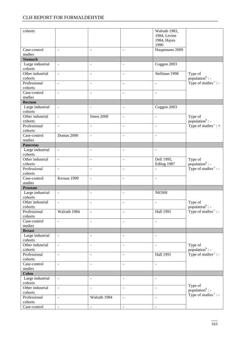
CLH REPORT FOR FORMALDEHYDE
163
cohorts Walrath 1983,
1984, Levine
1984, Hayes
1990
Case-control
studies
- - - Hauptmann 2009
Stomach
Large industrial
cohorts
- - - Coggon 2003
Type of
populationb : -
Type of studies c : -
Other industrial
cohorts
- - - Stellman 1998
Professional
cohorts
- - - -
Case-control
studies
- - - -
Rectum
Large industrial
cohorts
- - - Coggon 2003
Type of
populationb : -
Type of studies c : +
Other industrial
cohorts
- Innos 2000 - -
Professional
cohorts
- - - -
Case-control
studies
Dumas 2000 - - -
Pancreas
Large industrial
cohorts
- - - -
Type of
populationb : -
Type of studies c : -
Other industrial
cohorts
- - - Dell 1995,
Edling 1987
Professional
cohorts
- - - -
Case-control
studies
Kernan 1999 - - -
Prostate
Large industrial
cohorts
- - - NIOSH
Type of
populationb : -
Type of studies c : -
Other industrial
cohorts
- - - -
Professional
cohorts
Walrath 1984 - - Hall 1991
Case-control
studies
- - - -
Breast
Large industrial
cohorts
- - - -
Type of
populationb : -
Type of studies c : -
Other industrial
cohorts
- - - -
Professional
cohorts
- - - Hall 1991
Case-control
studies
- - - -
Colon
Large industrial
cohorts
- - - -
Type of
populationb : -
Type of studies c : -
Other industrial
cohorts
- - - -
Professional
cohorts
- Walrath 1984 - -
Case-control - - - -
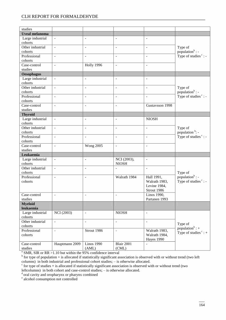
CLH REPORT FOR FORMALDEHYDE
164
studies
Uveal melanoma
Large industrial
cohorts
- - - -
Type of
populationb : -
Type of studies c : -
Other industrial
cohorts
- - - -
Professional
cohorts
- - - -
Case-control
studies
- Holly 1996 - -
Oesophagus
Large industrial
cohorts
- - - -
Type of
populationb : -
Type of studies c : -
Other industrial
cohorts
- - - -
Professional
cohorts
- - - -
Case-control
studies
- - - Gustavsson 1998
Thyroïd
Large industrial
cohorts
- - - NIOSH
Type of
population b: -
Type of studies c : -
Other industrial
cohorts
- - - -
Professional
cohorts
- - - -
Case-control
studies
- Wong 2005 - -
Leukaemia
Large industrial
cohorts
- - NCI (2003),
NIOSH
-
Type of
populationb : -
Type of studies c : -
Other industrial
cohorts
- - - -
Professional
cohorts
- - Walrath 1984 Hall 1991,
Walrath 1983,
Levine 1984,
Strout 1986
Case-control
studies
- - - Linos 1990,
Partanen 1993
Myeloid
leukaemia
Large industrial
cohorts
NCI (2003) - NIOSH -
Type of
populationb : +
Type of studies c : +
Other industrial
cohorts
- - - -
Professional
cohorts
- Strout 1986 - Walrath 1983,
Walrath 1984,
Hayes 1990
Case-control
studies
Hauptmann 2009 Linos 1990
(AML)
Blair 2001
(CML)
-
a SMR, SIR or RR >1.10 but within the 95% confidence interval
b for type of population + is allocated if statistically significant association is observed with or without trend (two left
columns) in both industrial and professional cohort studies; – is otherwise allocated. c for type of studies + is allocated if statistically significant association is observed with or without trend (two
leftcolumns) in both cohort and case-control studies; – is otherwise allocated. d oral cavity and oropharynx or pharynx combined
e alcohol consumption not controlled
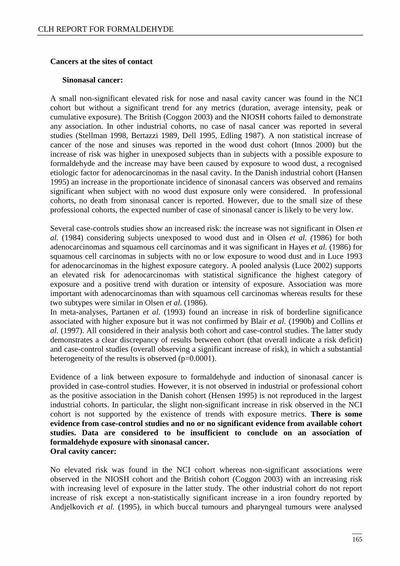
CLH REPORT FOR FORMALDEHYDE
165
Cancers at the sites of contact
Sinonasal cancer:
A small non-significant elevated risk for nose and nasal cavity cancer was found in the NCI
cohort but without a significant trend for any metrics (duration, average intensity, peak or
cumulative exposure). The British (Coggon 2003) and the NIOSH cohorts failed to demonstrate
any association. In other industrial cohorts, no case of nasal cancer was reported in several
studies (Stellman 1998, Bertazzi 1989, Dell 1995, Edling 1987). A non statistical increase of
cancer of the nose and sinuses was reported in the wood dust cohort (Innos 2000) but the
increase of risk was higher in unexposed subjects than in subjects with a possible exposure to
formaldehyde and the increase may have been caused by exposure to wood dust, a recognised
etiologic factor for adenocarcinomas in the nasal cavity. In the Danish industrial cohort (Hansen
1995) an increase in the proportionate incidence of sinonasal cancers was observed and remains
significant when subject with no wood dust exposure only were considered. In professional
cohorts, no death from sinonasal cancer is reported. However, due to the small size of these
professional cohorts, the expected number of case of sinonasal cancer is likely to be very low.
Several case-controls studies show an increased risk: the increase was not significant in Olsen et
al. (1984) considering subjects unexposed to wood dust and in Olsen et al. (1986) for both
adenocarcinomas and squamous cell carcinomas and it was significant in Hayes et al. (1986) for
squamous cell carcinomas in subjects with no or low exposure to wood dust and in Luce 1993
for adenocarcinomas in the highest exposure category. A pooled analysis (Luce 2002) supports
an elevated risk for adenocarcinomas with statistical significance the highest category of
exposure and a positive trend with duration or intensity of exposure. Association was more
important with adenocarcinomas than with squamous cell carcinomas whereas results for these
two subtypes were similar in Olsen et al. (1986).
In meta-analyses, Partanen et al. (1993) found an increase in risk of borderline significance
associated with higher exposure but it was not confirmed by Blair et al. (1990b) and Collins et
al. (1997). All considered in their analysis both cohort and case-control studies. The latter study
demonstrates a clear discrepancy of results between cohort (that overall indicate a risk deficit)
and case-control studies (overall observing a significant increase of risk), in which a substantial
heterogeneity of the results is observed (p=0.0001).
Evidence of a link between exposure to formaldehyde and induction of sinonasal cancer is
provided in case-control studies. However, it is not observed in industrial or professional cohort
as the positive association in the Danish cohort (Hensen 1995) is not reproduced in the largest
industrial cohorts. In particular, the slight non-significant increase in risk observed in the NCI
cohort is not supported by the existence of trends with exposure metrics. There is some
evidence from case-control studies and no or no significant evidence from available cohort
studies. Data are considered to be insufficient to conclude on an association of
formaldehyde exposure with sinonasal cancer.
Oral cavity cancer:
No elevated risk was found in the NCI cohort whereas non-significant associations were
observed in the NIOSH cohort and the British cohort (Coggon 2003) with an increasing risk
with increasing level of exposure in the latter study. The other industrial cohort do not report
increase of risk except a non-statistically significant increase in a iron foundry reported by
Andjelkovich et al. (1995), in which buccal tumours and pharyngeal tumours were analysed
CLH REPORT FOR FORMALDEHYDE
166
together. In professional cohorts, buccal cancers were also pooled with pharyngeal cancers and
results were largely inconsistent with some studies showing a decreased risk (Levine 1984,
Strout 1986) while some others report a small non-significant increase (Walrath 1984, Hayes
1990).
Only three case-control studies are available and show a non-significant increase in cancer risk
with no evidence of trend with duration in an analysis grouping oral cavity and oropharyngeal
cancers (Merletti 1991) or no evidence of trend with intensity (Wilson 2004). A statistical
increase in salivary gland cancers was observed in Wilson et al. (2004) in white men only but
the analysis in this study was not controlled for alcohol consumption and link with
formaldehyde exposure in these conditions is therefore uncertain.
Data from cohorts are inconsistent and no result from any reliable study attained
statistical significance and data are not considered as sufficient to provide a causality
relationship between formaldehyde and cancers of the oral cavity.
Nasopharyngeal cancer:
In the NCI cohort (Hauptmann 2004) which is the most important industrial cohort available in
term of size and duration of follow-up, a twofold increase in the risk of nasopharyngeal cancer
(statistically significant) was found. The increase is supported by positive trends with peak
exposure (p trend < 0.001) and with cumulative exposure (p trend = 0.03). These results were
confirmed when comparing the NPC mortality with local rates to take into account regional
environmental factors (Marsh 2005). It however highlights that most NPC cases occured in the
same plant (plant 1). Marsh et al. (2007b) also shows that risk estimates for NPC in the NCI
cohort are unstable but this problem is linked with the rarity of NPC and the difficulty to
provide evidence of association for small increases of rare cancers. In this study, a non-
significant increase in the relative risk for NPC in the highest exposure category was however
observed even after adjustment for plant group. Marsh et al. (2007a) also further investigate
plant 1 of the NCI cohort in a nested case-control study, with the hypothesis that excess of NPC
in plant 1 can be due to external employment in the ferrous and non-ferrous metal industries that
entailed possible exposure to several suspected risk factor for upper respiratory system cancer
(e.g., sulfuric acid mists, mineral acid, metal dust and fumes). A statistical association between
NPC and working in silver smithing or other metal work has been identified. However, a non-
statistically significant association between NPC and formaldehyde was still observed after
adjustment for this factor as well as positive trends with duration of employment and with
cumulative exposure but not with average intensity. Stratification by peak exposure, which was
identified as the most significant metrics in Hauptman et al. (2004), was however not
performed. Besides, a history of working in silver smithing or other metal work was not found
in all NPC cases and cannot entirely explain the increase of NPC in the plant. Detailed
information on types of jobs and exposures in metal work was also not available and it has not
been possible to confirm that cases were actually exposed to any of the chemical agents that are
suspected risk factors in this industry. These data were therefore not considered to be sufficient
to explain the increase in NPC risk identified in the NCI cohort but raise a doubt on the
existence of a cofounder in plant 1 of the NCI cohort. The analysis of the number of workers
and level of exposure in the different plants included in the NCI cohort shows that plant 1
includes the largest number of subjects in the highest category of exposure to peaks (calculated
on the basis of the data reported in Marsh et al. 2005): they are 1964 subjects exposed to the
highest category of exposure to peaks in plant 1, 1864 in plant 10, 1233 in plant 4, 718 in plant
CLH REPORT FOR FORMALDEHYDE
167
2 and less than 200 in other plants. Plant 1 is therefore the plant in which an excess of risk is the
more likely to be identified.
Several other industrial cohorts investigate exposure to formaldehyde and found no evidence of
an increased risk of pharyngeal cancer with no or very few cases reported. Given that
nasopharyngeal tumours are rare (world incidence of 1.2 per 100 000 and mortality of 0.8 per
100 000 reported in GLOBOCAN 2008) and that these studies include a smaller number of
subjects, the absence of an increased incidence is inconclusive. For example, the power to detect
a twofold or greater increase in mortality from nasopharyngeal cancer was 13% in the NIOSH
cohort and 44% in the British cohort (BfR 2006).
Small number of subjects is also a major weakness in the professional cohorts of pathologists
and embalmers. In the two larger cohorts (around 4000 subjects), no nasopharyngeal cancers
were observed in Hall et al. (1991) whereas a non-significant increase of tumours was reported
in Hayes et al. (1990). Such cohort size that may have sufficient statistical power to detect an
increase in common tumours but not for very rare tumours such as NPC
Several case-control studies investigate the association between exposure to formaldehyde and
nasopharyngeal carcinoma (NPC). Although not statistically significant, formaldehyde exposure
was associated with an increased risk of nasopharyngeal carcinoma, with supportive indications
of higher risk with higher level of exposure (Vaughan 1986 and Roush 1987), duration of
exposure (Vaughan 1986, West 1993 Hildesheim 2001 and Marsh 2007a), latency (Roush 1987
and West 1993) and cumulative exposure (Marsh 2007a). In West et al. (1993), the increase
reached statistical significant with longer latency. Besides, Vaughan et al. (2000) reports an
increase in risk with formaldehyde exposure unaffected by wood dust and that gained statistical
significance when restricted to higher probabilities of exposure. In Olsen et al. (1984), an
increase in risk was associated with formaldehyde exposure in women but not in men. In
Amstrong et al. (2000), the risk was not increased after adjustment.
In the meta-analysis by Partanen et al. (1993), NPC risk was elevated with statistical
significance in the substantial exposure category (exposure exceeding 5.5 ppm-year). NPC risk
was also significantly elevated in Blair et al. (1990) and in Collins et al. (1997). Two recent
meta-analyses (Bosetti 2008 and Bachand 2010) have highlighted the role of the NCI cohort and
in particular of its plant 1 in the overall increase in risk. An overall increase in risk of borderline
significance in pooled case-control studies was however observed in Collins et al. (1997) and in
Bachand et al. (2010).
Significant evidence of an association between formaldehyde exposure and NPC is therefore
provided from the most informative cohort study and from several case-control studies and
meta-analyses. The NCI cohort is the most important in terms of cohort size and length of
follow-up and is based on a detailed assessment of exposure. The increase in risk is supported
by trends for several metrics of exposure. However, although the increase in risk may not be
entirely explained by co-exposures investigated by Marsh et al. (2007a), the existence of a
grouping of cases in NCI plant 1 can be explained by the largest number of subjects exposed to
high peaks but also raise a doubt on potential cofounder.
It should also be noted that the majority of available studies are based on mortality and not on
incidence. Because of its location, NPC may not cause symptoms at early stages, remains
undetected and most NPC are diagnosed at an advanced stage with metastases typically in the
cervical lymph nodes. Distant metastases may also occur in the bone, lung, mediastinum and
more rarely, in the liver (Brennan 2006) with up to 80-90% of lymph node metastasis for the
undifferentiated type (CHU-PS 2010). Due to the high rate of metastasis, it is expected in some
cases that NPC may not be identified as the primary cause of deaths, resulting in an under-
estimation of its incidence in cohorts. In addition, NPC is a rare tumour (Chang 2006), for
which very large cohorts and statistical power are needed to be able to identify an excess of risk,
CLH REPORT FOR FORMALDEHYDE
168
Case-control studies are therefore considered as a critical source of information for NPC and
predominantly indicate an increase of risk of NPC.
Overall, there is consistent evidence from the NCI cohort and from several case-control
studies that formaldehyde may induce NPC. The existence of a grouping of cases in plant 1
of the NCI cohort raises a doubt on potential cofounder and lowers the level of evidence
but it can also be explained by the largest number of subjects exposed to high peaks in this
specific plant.
Pharyngeal cancers (other than nasopharyngeal or combined):
In industrial cohorts, a non-significant increased risk is observed in the British cohort (Coggon
2003) but not in the NIOSH cohort (no data on pharynx available in the NCI cohort). In
professional cohorts and most other industrial cohorts, pharyngeal cancers were pooled with
buccal cancers and results were largely inconsistent with some studies showing a decreased risk
(Levine 1984, Strout 1986) while some others small non-significant increase (Walrath 1984,
Hayes 1990). Four case-control studies are available and whereas Vaughan et al. (1983),
Gustavsson et al. (1998) and Berrino et al. (2003) show no elevated risk, Laforest et al. (2000)
observed a significant increase in cancer risk with evidence of trend with duration and
cumulative dose.
Evidence of a link between exposure to formaldehyde and induction of pharyngeal cancer
is provided in case-control studies and in particular in Laforest et al (2000). Data from
cohorts are inconsistent and overall provide no clear evidence of an increased risk of
pharyngeal cancer other than nasopharyngeal.
Laryngeal cancer:
A non significant elevated risk was reported in the British cohort study (Coggon 2003) only in
the high exposure category. No elevated risk was observed in other industrial cohorts and no
results for laryngeal cancers were reported in professional cohorts. Non-significant increases in
the case-control studies by Wortley et al. (1992) at high dose only and in Gustavsson et al.
(1998). The increase was however statistically significant in the highest category of cumulative
exposure in Shangina et al. (2006).
Data from cohort studies therefore provide no evidence of an increased risk of laryngeal
cancer to support the slight increase identified in some case-control studies.
Lung cancer:
In the British industrial cohort (Coggon 2003), an elevated risk of lung cancer was associated
with higher intensity but not with longer duration of exposure. Results in the two other large
cohort studies do not confirm this result. In other industrial cohorts, positive results are reported
in cohorts co-exposed to MMVF (Marsh 2001, Chiazze 1997), asbestos (Dell 1995) or silica
(Adjelkovic 1994). No increased risk was detected in any professional cohorts. In case-control
studies, Andjelkovich et al. (1994) showed a non-significant increased risk. The increase
reached statistical significance in two case-control studies (Coggon 1984, Youk 2001) but with
negative trends with exposure. An excess of risk in workers exposed to formaldehyde with a
significant trend with duration of exposure was observed in a third case-control study that
investigate specifically lung adenocarcinomas (De Stephani 2005). In meta-analyses, whereas
Collins et al. (1997) found no increased risk, Partanen et al. (1993) found a weak positive effect
CLH REPORT FOR FORMALDEHYDE
169
of borderline significance but risk was not increased with higher exposure category. Finally, a
positive association was found in industrial workers and a negative in professional workers
(Blair 1990).
Overall, the inconsistency of the results in the large industrial cohorts, the discrepancy
between results in industrial and professional workers and the potential cofounders in
small industrial cohorts does not allow to identify an association between formaldehyde
exposure and lung cancer.
Cancer at distant sites
Lymphohaematopoietic malignancies:
An excess of lymphohaematopoietic cancers is reported most specifically for leukaemia. A non-
statistically significant increase was reported in two large industrial cohorts with support of
positive trends for peak and average intensity (NCI cohort in Hauptmann 2003) and for duration
and time since first exposure (NIOSH cohort). Non-statistically significant increases in risk
were reported in several professional cohorts that were supported with trend for duration in
Walrath et al. (1984) but not in Strout et al. (1986) as well as in two case-control studies.
Statistical significance was however attained in several studies investigating more specifically a
potential increase in risk for myeloid leukaemia. In the NCI cohort (Hauptmann 2003), excess in
relative risks for myeloid leukaemia were statistically significant in the categories of highest
peak and average intensity exposure, with statistically significant trends for these two metrics
but not for duration or cumulative exposure. A re-analysis of the NCI cohort (Marsh 2004)
found no significant excess of risk for external comparison (SMR) but confirmed a statistically
significant excess of risk in the highest peak exposure categories based on internal comparison
(RR). These high RR were explained by very low incidence of ML in unexposed and control
groups (low exposure group). Risk estimates however declined in the more recent update of the
NCI cohort (Beane-Freeman 2009). Considering a relatively short period of latency for myeloid
leukaemia, the reduction of association after the 1990s could however reflect a reduction in
levels of exposure with time. A statistical excess of risk was also observed in a professional
cohort (Strout 1986) and in two case-control studies: Linos et al. (1990) was focused on acute
myeloid leukaemia more specifically. Hauptmann et al. (2009) also investigate trends with
different metrics of exposure and found positive association with duration of practice and peak
exposure.
Finally, results of two meta-analyses show significant increases in leukaemia only in
professionals (Blair 1990b, Bosetti 2008). The study by Collins et al (2004) confirms the
discrepancy in the results from different high exposure occupations, with an absence of
increased risk in industrial workers. A recent meta-analysis however found overall significant
results for leukaemia and myeloid leukaemia but this study focused on highest exposure group
from each study considered in the meta-analysis (Zhang 2009). Consideration of higher levels of
exposure is expected to generate a greater statistical power to detect a relationship if a true
effect exists. The study by Zhang (2009) was updated in Schwilk et al. (2010) using the same
methodology and including the latest updates and epidemiology studies (in particular
Hauptmann 2009 nd Beane-Freeman 2009). An excess of risk of 2.47 was found for myeloid
leukaemia. Based on a similar set of study but taking into account RR estimates for all levels of
exposure, Bachand et al. (2010) did not identify a statistical increase in RR estimates.
CLH REPORT FOR FORMALDEHYDE
170
Overall, some positive observations have emerged in industrial populations but meta-
analyses generally show a discrepancy in the results between industrial and professional
populations in which several studies indicate an increased risk of leukaemia and especially
myeloid leukaemia. Therefore, it is considered that available data does not provide causal
evidence for formaldehyde as the aetiological factor as a bias specific to professionals
cannot be ruled out.
Brain cancers:
Brain cancer risk was similar to expected in the NIOSH cohort and lower than expected in the
NCI and British cohorts. A non-significant increase in risk was observed in two small industrial
cohorts but with no trend with exposure (Innos 2000) and higher risk in unexposed subjects
(Chiazze 1997). Several non-significant (Hall 1191, Walrath 1983, Walrath 1984, Levine 1984
and Hayes 1990) and significant (Strout 1986) increases in risk were consistently reported in
professional cohorts. The discrepancy between industrial and professional cohorts is highlighted
by meta-analyses showing significant increases in professional cohort but not in industrial
workers (Blair 1990, Bosetti 2008). The only case-control study investigating this cancer type
reports a non statistical increase (Hauptmann 2009). However, it was not supported by trends
with duration of practice, peak, cumulative or average intensity exposures and was considered
not conclusive for brain cancer. In absence of other evidence from industrial cohort or case-
control studies, the effect observed in professionals is more likely to be due to confounding
factors.
Other cancers:
Isolated results across studies suggest an elevated risk of cancers at other sites such as:
- Stomach: non-significant increase in risk in the Bristish cohort (Coggon 2003) and in
another industrial cohort (Stellman 1998), more likely to be due to confounding factors
as it was not confirmed in the other large industrial cohorts or in professionals.
- Rectum: increase in risk of borderline significance in a small industrial cohort (Innos
2000) and significant increase in the only case-control study investigating this cancer
type with positive trends with concentration and duration of exposure (Dumas 2000). In
this study, many substances showed associations with rectal cancer and it was not
possible to clearly assign the observed effect to formaldehyde or to another substance
independently. Besides, the absence of increases in risk in large industrial cohorts and in
professionals does not support an association of formaldehyde with rectum cancer.
- Pancreas: non-significant increase in risk in two small industrial cohorts (Dell 1995 and
Edling 1987) and significant increase in the only case-control study investigating this
cancer type with trends with probability but not with intensity of exposure (Kernan
1999). However, the absence of increases in risk in large industrial cohorts and in
professionals does not support an association of formaldehyde with pancreas cancer.
- Prostate: non-significant increase in risk in one professional cohort (Hall 1991) and
significant increase in another professional cohort with a trend with duration of
exposure (Walrath 1984). However, the absence of increases in risk in industrial cohorts
does not support an association of formaldehyde with prostate cancer. No relevant case-
control study is available on prostate cancer.
- Breast: isolated non-significant increase in risk in one professional cohort (Hall 1991)
- Colon: isolated significant increase in risk in one professional cohort (Walrath 1984)
CLH REPORT FOR FORMALDEHYDE
171
- Uveal melanoma: isolated significant increase in risk in the only case-control study
investigating this cancer type (Holly 1996)
- Oesophagus: isolated non-significant increase in risk in the only case-control study
investigating this cancer type (Gustavsson 1998) not supported by trend for cumulative
or duration of exposure.
- Thyroid: isolated significant increase in risk in the only case-control study investigating
this cancer type (Wong 2005).
However, these results were highly inconsistent for stomach, brain, colon, pancreas and prostate
with excess of cancers limited to either industrial workers or professionals and not identified in
the largest industrial cohorts. Other results were isolated and it cannot be excluded that they are
due to confounding factors.
4.10.5 Comparison with criteria
For experimental data, the CLP criteria for classification establish different levels of evidence:
— “sufficient evidence of carcinogenicity: a causal relationship has been established between
the agent and an increased incidence of malignant neoplasms or of an appropriate
combination of benign and malignant neoplasms in (a) two or more species of animals or
(b) two or more independent studies in one species carried out at different times or in
different laboratories or under different protocols. An increased incidence of tumours in
both sexes of a single species in a well-conducted study, ideally conducted under Good
Laboratory Practices, can also provide sufficient evidence. A single study in one species and
sex might be considered to provide sufficient evidence of carcinogenicity when malignant
neoplasms occur to an unusual degree with regard to incidence, site, type of tumour or age
at onset, or when there are strong findings of tumours at multiple sites;
— limited evidence of carcinogenicity: the data suggest a carcinogenic effect but are limited
for making a definitive evaluation because, e.g. (a) the evidence of carcinogenicity is
restricted to a single experiment; (b) there are unresolved questions regarding the adequacy
of the design, conduct or interpretation of the studies; (c) the agent increases the incidence
only of benign neoplasms or lesions of uncertain neoplastic potential; or (d) the evidence of
carcinogenicity is restricted to studies that demonstrate only promoting activity in a narrow
range of tissues or organs.”
Experimental data clearly provide evidence of a carcinogenic effect at the site of contact in rats by
inhalation. Although this finding is restricted to a single species (rat), consistent results were
obtained from several independent studies and in both females and males. Tumours consists in both
benign and malignant tumours but were induced at a single site (nasal cavity). Data investigating
the mode of action support the existence of a threshold type mode of action for its carcinogenic
properties based on the cytotoxic effect of formaldehyde. Genotoxicity is also expected to play a
role above this threshold.
Overall the level of experimental evidence is judged as sufficient evidence in agreement with
induction of tumours (b) [in] two or more independent studies in one species carried out at
different times or in different laboratories or under different protocols.
CLH REPORT FOR FORMALDEHYDE
172
For epidemiological data, the CLP criteria for classification establish different levels of evidence:
— “sufficient evidence of carcinogenicity: a causal relationship has been established between
exposure to the agent and human cancer. That is, a positive relationship has been observed
between the exposure and cancer in studies in which chance, bias and confounding could be
ruled out with reasonable confidence;
— limited evidence of carcinogenicity: a positive association has been observed between
exposure to the agent and cancer for which a causal interpretation is considered to be
credible, but chance, bias or confounding could not be ruled out with reasonable
confidence.”
At the site of contact, positive associations between exposure to formaldehyde and cancer were
identified from both cohort studies and case-control studies for cancers of the sinonasal cavity, oral
cavity, nasopharynx, pharynx and lung. Results were statistically significant and supported by
trends with exposure in both types of studies for nasopharynx, which provide a high level of
evidence of an association. However, the existence of a grouping of cases in plant 1 of the NCI
cohort raises a doubt on potential cofounder and lowers the level of evidence but the grouping of
cases can also be explained by the largest number of subjects exposed to high peaks in this specific
plant. .
Several factors support the existence of a carcinogenic potential of formaldehyde at the site of
contact:
- Induction of tumours in the nasal cavity in rats with a proposed mode of action based on
chronic irritation of the respiratory tract and local genotoxicity at doses inducing
cytotoxicity and increased proliferation
- Indication of local genotoxicity in exposed humans as evidenced by increases in
micronuclei frequency in buccal and nasal mucosa cells in several studies
- Human sensitivity to FA-induced irritation, with irritation of the eye and of the
nose/throat consistently reported after exposure to formaldehyde (IARC 2006).
No species-specific mechanism is evident and human data denote human sensitivity to FA effects
(genotoxicity and irritation). The mode of action of carcinogenicity in the rat nasal cavity is
therefore considered relevant to humans, as reviewed in the context of the IPCS framework
(McGregor 2007).
It is noted that the site of local tumours in rats (nasal cavity) and in humans (nasopharynx) differs.
Humans, unlike rats, are oronasal breathers and dosimetry in the different parts of the respiratory
tract is expected to be different. In rats, lesions and DPX formation were mainly observed in the
lateral meatus of the nasal cavity. In rhesus monkeys, DPX are also formed in proximal portions of
the lower respiratory tract in rhesus monkeys (Casanova 1991). Modeling of FA dosimetry in the
respiratory tract indicates that when the switch to oronasal breathing occurs, cells in the upper
segments of the lower respiratory tract receive a considerably higher flux of formaldehyde from oral
intake (Conolly 2004). Difference in the site of tumours in the respiratory tract is therefore not in
contradiction with the relevance of the rat data for humans.
The induction of nasopharyngeal carcinomas in human exposed to formaldehyde is therefore
strongly plausible.
CLH REPORT FOR FORMALDEHYDE
173
The biological plausibility of the induction of nasopharyngeal carcinomas in humans exposed
to formaldehyde highly supports the consistent epidemiological evidence obtained from the
NCI cohort and from several case-control studies. It is considered that the doubt of a potential
confounder is raised by the grouping of cases in the plant 1 of the NCI cohort. But considering
the overall database and more specifically the fact that the grouping of cases in plant 1 can
also be explained by the largest number of subjects exposed to high peaks in this specific
plant, correlation of NPC with the level of peak exposure to formaldehyde, the evidence
provided by case-control studies and the biological plausibility, the doubt that the observed
induction of NPC may be due to chance, bias or confounding can be ruled out with reasonable
confidence.
Altogether, the data support a causal relationship between formaldehyde exposure and
induction of NPC and corresponds to a sufficient evidence of carcinogenicity in humans.
At distant site, excess of risk are reported for myeloid leukaemia. Some positive observations have
emerged in industrial populations but meta-analysis show a discrepancy in the results between
industrial and professional populations in which several studies indicate an increased risk of
leukaemia and especially myeloid leukaemia. Therefore, it is considered that available data does
not provide causal evidence for formaldehyde as the aetiological factor as a bias specific to
professionals cannot be ruled out.
Besides, inhalation of formaldehyde doesn‘t modify formaldehyde blood levels in rats, monkeys
and humans and due to its high reactivity, its rapid metabolism and detoxification formaldehyde is
not expected to reach distant site and the biological plausibility for induction of leukaemia is
therefore weak (Heck 2004). Finally, no convincing evidence for induction of tumours in the
lympho-haematopoietic system is identified in experimental animals whereas haemopathies are
observed in rodents with known leukemogens. This potential mode of action was discussed in
several reviews funded by the FA industry (Casanova 2004, Golden 2006, Pyatt 2008, Rhomberg
2011) or in a recent ECETOC/ILSI/HESI workshop (Carmichael 2011) that concluded that no
convincing mechanism has been indentified si far.
However, several observations have emerged recently and tend to indicate that formaldehyde may
have systemic effects, in particular on the lympho-haematopoietic system:
- Evidence for induction of genotoxicity (micronuclei) in peripheral lymphocytes in
humans. Inconsistent results are however reported for induction of SCE and
chromosomal aberrations. Besides, negative results on bone marrow and blood cells
were obtained in rats by inhalation under controlled conditions.
- Report in a recent study (Zhang 2010) of an increase in the frequency in the myeloid
progenitor cells from peripheral blood of exposed workers of loss of chromosome 7
(p=0.0039) and of trisomy of chromosome 8 (p=0.040), which are among the most
frequent cytogenetic changes observed in myeloid leukaemia. Cytogenetic anomalies
were however analysed on a very limited number of cells (150/subjects) and subjects
(10 exposed and 12 controls) and it cannot be excluded that the observed effect may
reflect individual heterogeneity considering that these anomalies are also found in non-
exposed subjects. Besides, the meaning of these cytogenetic anomalies is not known in
terms of molecular oncogenesis. They are not known to be sufficient to induce the
apparition of a leukemic phenotype and are also present in control subjects at a
substantial frequency
CLH REPORT FOR FORMALDEHYDE
174
It is regrettable that additional cytogenetic anomalies characteristic of myeloid
leukaemia and which have a clear biological significance as they re-arrange genes
involved in proliferation or differentiation (e.g. translocations t(8;21), t(9;22) or
t(15;17)) have not been investigated. Due to the very small number of subjects the study
therefore need to be replicated.
- Recent formulation of hypothesis for potential modes of action (Zhang 2009):
- transport in the blood in the hydrated form methanediol and damage of stem
cells in the bone marrow.
Considering the chemistry of formaldehyde in solution, the equilibrium between
formaldehyde and methanediol is largely in favour of methanediol under
physiological conditions (37°C and pH 5-7) but a proportion of 1% of the
substance is also present as formaldehyde. As formaldehyde is highly reactive
and is likely to quickly disappear by linking to macromolecules where it is
produced, spontaneous release of formaldehyde from methanediol may take
place to maintain the equilibrium between methanediol and formaldehyde. A
small but continuous production of formaldehyde can therefore take place at
distant sites where methanediol is present. However, the level of methanediol in
blood (reported as formaldehyde in the publications by misuse of language but
GC-MS actually measures methanediol and formaldehyde together) further to
inhalation exposure to formaldehyde did not raise (Heck 2004). A mathematical
model also predicted that the increase of the formaldehyde concentration
(reflecting both free and hydrated forms) in blood further to inhalation exposure
is insignificant compared to endogenous levels of formaldehyde (Franks 2005).
Besides, the radioactivity incorporated in the blood and bone marrow further to
inhalation of [14
C] FA was due to metabolic incorporation and not to covalent
binding (Casanova-Schmitz 1984). Lu et al (2010, 2011) recently showed in rats
that N2-hydroxymethyl-dG adducts and dG-dG crosslinks from exogenous
origin were detected further to inhalation of radiolabelled formaldehyde in nasal
respiratory epithelium but not in bone marrow, spleen, lung, liver, thymus
tissues or blood in rats. N2-hydroxymethyl-dG adducts was also not detected in
the bone marrow of monkeys up to 6 ppm. N2-hydroxymethyl-dG adducts was
found to be a suitable biomarker for formaldehyde exposure in preliminary cell
culture experiments.
Besides, a recent studies (Neuss 2008 and Neuss 2010b) has shown that
formaldehyde is not released from exposed cells and DPX and SCE are observed
only in cells in direct contact with formaldehyde.
The hypothesis that formaldehyde may be transported to the bone marrow by
damaged cells or as active forms is therefore considered unlikely.
- damage to stem cells circulating in the blood at the site of contact and re-
incorporation of damaged stem cells in the bone marrow.
Hematopoietic stem cells present in blood have however a short half life in
circulation estimated around 1-2 hours (Papayannopoulou 2008) and they are
100 less numerous in blood than in bone marrow where they represent only 1-
3% of normal cells. Besides, they have a very intermittent and brief exposure
considering the number of passage of each stem cell in the nasal tracts and the
short duration of transit of cells in this area. It would therefore be expected that a
similar mode of actions would occur with other factors such as UV radiations
CLH REPORT FOR FORMALDEHYDE
175
that may reach blood cells by cutaneous exposure. Such effects has however not
been identified.
The possibility for haematopoietic stem cells to go from the bone marrow to
blood and inversely is known (homing). However, it has never been observed in
the case of damaged circulating progenitor cells giving rise to leukaemia either
with formaldehyde or other leukemogen factors.
- damage to primitive pluripotent stem cells present in the oral and nasal
mucosa and re-incorporation of damaged stem cells in the bone marrow.
All flat bones are haematopoietic in adults and haematopoietic stem cells are
expected to be present in the ethmoid and nasal bones. But the penetration of
formaldehyde to the marrow of these bones seems not compatible with its
reactivity. Murrell et al. (2005) has demonstrated in rats that cells able to
differentiate into haematopoietic stem cells were present in the nasal mucosa as
they repopulate the bone marrow of irradiated hosts. Additional experiments
indicate that this effect was not attributable to the presence of hematopoietic
stem cells in the olfactory mucosa sample but to other stem-like cells. The
presence of such stem cells able to differentiate in vitro and in vivo into multiple
cell types was also found in the olfactory mucosa of mice and humans but they
were not shown to differentiate into haematopoietic cells.
The presence of haematopoietic stem cells in the nasal mucosa has been
demonstrated (Sergejeva 2005). But a genotoxic and leukemogenic effect of
formaldehyde on these cells would induce an increase in the frequency of
chloromas (accumulation of leukemic cells in tissues) in the nasal mucosa of
exposed subjects but this has not been reported.
This hypothesis is also not supported by the fact that many nasal carcinogens are
not identified as leukemogens. It is the case of chromium, nickel or arsenic
compounds. Only sulphur mustard is proved to be a nasal carcinogen an induced
pancytopenia in heavily exposed subjects (Goldstein 2010).
As in the previous hypothesis, the possibility for damaged circulating progenitor
cells to go back to the bone marrow and to give rise to leukaemia is also not
demonstrated.
Besides, a recent study (Kuper 2011) has investigated in animals the effect of
FA on nasal lymphoid tissue further to inhalation. The 28-day study revealed
hyperplasia of the lymphoepithelium in the NALT at 15 ppm in rats but no
significant effect on the NALT lymphoid tissue or in the cervical lymph nodes
(decreased NALT activity in some animals but no significant effect compared to
controls). No effects were detected at similar doses in mice that are less sensitive
than rats to FA damage in the nasal mucosa. This tends to show that FA does no
induce a proliferative effect in the nasal lymphoid tissues that could participate
in haematological malignancies..
- Indications that formaldehyde may produce toxicity to white blood cells in humans
(Zhang 2010). A decrease in white blood cell counts was observed in exposed workers
but the values remains in the normal range. A decrease in colony formation from
exposed human progenitor blood cells was also observed but the effect was not
statistically significant and the meaning of this finding in terms of toxicity or inhibition
is not clear. Indeed, a dose-related decrease in the number colony formed by progenitor
cells was observed in vitro but this is not surprising considering the cytotoxic effect of
CLH REPORT FOR FORMALDEHYDE
176
formaldehyde. Besides, pancytopenic effects are not found in long-term studies in
rodents to the maximally tolerated doses (Goldstein 2010). No effect on blood count
related to FA exposure was detected in Kuo et al. (1997) but exposure in this study was
very low. A higher sensitivity of humans may be hypothesised to explain this difference
but it has not been further explored and demonstrated by any element up to now.
These elements are therefore considered as preliminary evidence. Besides, the study by Zhang et al
(2010) tends to show an effect on blood cells and progenitor cells in peripheral blood but it provides
no evidence of a direct effect in the bone marrow.
Altogether, in absence of convincing evidence for a biologically plausible mechanism and
considering the discrepancy of results in epidemiological studies, a causal relationship
between formaldehyde exposure and induction of myeloid leukaemia cannot be concluded.
Overall, CLP criteria for classification states:
“The classification in Category 1A and 1B is based on strength of evidence together with additional
considerations (see section 3.6.2.2). Such evidence may be derived from:
— [1A:] human studies that establish a causal relationship between human exposure to a
substance and the development of cancer (known human carcinogen); or
— [1B:] animal experiments for which there is sufficient (1) evidence to demonstrate animal
carcinogenicity (presumed human carcinogen).
In addition, on a case-by-case basis, scientific judgment may warrant a decision of presumed
human carcinogenicity derived from studies showing limited evidence of carcinogenicity in
humans together with limited evidence of carcinogenicity in experimental animals.”
A category Carc 1A is therefore warranted for formaldehyde for carcinogenicity at the site of
contact and more specifically induction of NPC. Sufficient evidence in humans is concluded
based on consistent evidence from the NCI cohort and from several case-control studies
supported by animal data and biological plausibility.
4.10.6 Conclusions on classification and labelling
A classification Carc 1A – H350 is warranted (carc . cat. 1 ; R45 according to Directive
67/548/EEC).
The proposed carcinogenic classification is entirely based on data obtained by the inhalation route
either in humans or in experimental animals. The route of exposure can be specified in the hazard
statement ―if it is conclusively proven that no other routes of exposure cause the hazard‖. Reliable
studies are available in experimental animals by the oral route but not by dermal route. In humans,
it is expected that due to formaldehyde uses and physical properties only data resulting from
respiratory exposure will be obtained. However, the present database does not allow proving that
formaldehyde does not have a carcinogenic effect by dermal route and the route of exposure cannot
be specified in the hazard statement.
CLH REPORT FOR FORMALDEHYDE
177
The relevance of setting specific concentration limits was assessed based on the recommended
guidance (EC 1999). It is based on the evaluation of potency, which is defined as ―the magnitude,
with respect to dose, of the carcinogenic activity of a chemical in the species under consideration‖.
The proposed classification Carc 1A is based on nasopharyngeal cancers in humans. Evaluation of
potency in humans is however difficult as specified in the guidance. The lack of precise exposure
measurement do not allow establishing a reliable dose-response curve and EC guidelines
recommend to assess the potency calculation on the dose that produces a tumour incidence of 25%
(T25) in experimental studies. However, it also mention in section 2.5 that determination of T25
value is not appropriate in the case of a non-systemic contact carcinogen, as in the case of
formaldehyde. A SCL cannot therefore be derived.
4.11 Toxicity for reproduction
Not evaluated in this dossier.
4.12 Other effects
Not evaluated in this dossier.
5 ENVIRONMENTAL HAZARD ASSESSMENT
Not evaluated in this dossier.
CLH REPORT FOR FORMALDEHYDE
178
6 OTHER INFORMATION
The information included in this report is based on a bibliographic search perfomed in April 2010
and supplemented by articles identified by a search alert up to the date of submission of the report.
Registration dossiers available in May 2011 were rewieved. Information in part 7.6 (Genetic
toxicity), 7.7 (Carcinogenicity) and 7.10 (Exposure related observations in humans) that was not
already present in the CLH report (version 1) was included in the revised version when relevant in
the discussion of carcinogenic or mutagenic effects, performed through a relevant route of
exposure, and available in english language.
A discussion with the formaldehyde industry was organised in the preparation of this dossier in the
form of a meeting with Formacare on July 18th
2011. Their position on carcinogenic classification
of formaldehyde is included in the IUCLID 5 dossier (Formacare position paper).
Formaldehyde has been studied for a long time and reviews of the toxicological properties of
formaldehyde were performed by several international or national organisation. The main recent
reviews (issued after 2005) that discuss mutagenicity and/or carcinogenicity of formaldehyde are:
Carcinogenicity of formaldehyde was evaluated in 2006 by the BfR that concluded
that there is sufficient evidence to assume a causal relationship between
formaldehyde exposure and induction of nasopharyngeal cancer in humans (BfR
2006).
IARC evaluated carcinogenicity of formaldehyde in a monograph published in 2006
(IARC 2006). Formaldehyde IARC classification has been revised in 2009 (Baan
2010). Although the resulting monograph is not published yet, the IARC Working
Group unanimously reaffirmed the classification of formaldehyde in Group 1, based
on sufficient evidence in humans of nasopharyngeal cancer. The Working Group
concluded that, overall, there is sufficient evidence for leukaemia, particularly
myeloid leukaemia.Formaldehyde is under discussion at NTP to revise its listing
status under the 12th
Report of Carcinogen (ROC). A background document on
carcinogenicity of formaldehyde has been published in 2010 (NTP 2010a). A
DRAFT recommendation to list formaldehyde as a known to be a human carcinogen
based on evidence of causality for nasopharyngeal cancer, sinonasal cancer, and myeloid
leukemia in June 2010 (NTP 2010b) but is still under discussion.
The US EPA has published in June 2010 a DRAFT toxicological review of inhalation
toxicity of formaldehyde (EPA 2010) concluding that ―Human epidemiological
evidence is sufficient to conclude a causal association between formaldehyde
exposure and nasopharyngeal cancer, nasal and paranasal cancer, all leukemias,
myeloid leukemia and lymphohematopoietic cancers as a group.‖ The National
Research Council (NRC) reviewed this draft assessment and concluded (NRC 2011)
that on respiratory tract cancers, ―the committee agrees that there is sufficient

CLH REPORT FOR FORMALDEHYDE
179
evidence […] of a causal association between formaldehyde and cancers of the nose,
nasal cavity, and nasopharnyx. It disagrees that the evidence regarding other sites in
the respiratory tract is sufficient. […] Accordingly, the committee recommends that
EPA revisit arguments that support determinations of causality for specific LHP
cancers and in so doing include detailed descriptions of the criteria that were used to
weigh evidence and assess causality.‖
These reviews are attached for information in the IUCLID dossier.
CLH REPORT FOR FORMALDEHYDE
180
7 REFERENCES
Albert RE, Sellakumar AR, Laskin S, Kuschner M, Nelson N, Snyder CA (1982) Gaseous
formaldehyde and hydrogen chloride induction of nasal cancer in the rat. J Natl Cancer Inst. 68,
597-603.
Andjelkovich DA, Janszen DB, Brown MH, Richardson RB, Miller FJ (1995) Mortality of iron
foundry workers: IV. Analysis of a subcohort exposed to formaldehyde. J Occup Environ Med 37,
826-837.
Andjelkovich DA, Shy CM, Brown MH, Janszen DB, Levine RJ, Richardson RB (1994) Mortality
of iron foundry workers. III. Lung cancer case-control study. J Occup Med 36, 1301-1309.
Armstrong RW, Imrey PB, Lye MS, Armstrong MJ, Yu MC, Sani S (2000) Nasopharyngeal
carcinoma in Malaysian Chinese: occupational exposures to particles, formaldehyde and heat. Int J
Epidemiol. 29, 991-998.
Baan R, Grosse Y, Straif K, Secretan B, El GF, Bouvard V, brahim-Tallaa L, Guha N, Freeman C,
Galichet L, Cogliano V (2009) A review of human carcinogens--Part F: chemical agents and related
occupations. Lancet Oncol 10, 1143-1144.
Bachand AM, Mundt KA, Mundt DJ, Montgomery RR (2010) Epidemiological studies of
formaldehyde exposure and risk of leukemia and nasopharyngeal cancer: a meta-analysis. Crit Rev
Toxicol 40, 85-100.
Ballarin C, Sarto F, Giacomelli L, Bartolucci GB, Clonfero E (1992) Micronucleated cells in nasal
mucosa of formaldehyde-exposed workers. Mutat.Res 280, 1-7.
Battelle Columbus Laboratories. Final report on a chronic inhalation toxicology study in rats and
mice exposed to formaldehyde. Prepared by Battelle Columbus Laboratories and CIIT Docket #
10922. 1981.
Bauchinger M, Schmid E (1985) Cytogenetic effects in lymphocytes of formaldehyde workers of a
paper factory. Mutat.Res 158, 195-199.
Beane Freeman LE, Blair A, Lubin JH, Stewart PA, Hayes RB, Hoover RN, Hauptmann M (2009)
Mortality From Lymphohematopoietic Malignancies Among Workers in Formaldehyde Industries:
The National Cancer Institute Cohort. J Natl Cancer Inst 101, 751-761.
Berrino F, Richiardi L, Boffetta P, Esteve J, Belletti I, Raymond L, Troschel L, Pisani P, Zubiri L,
Ascunce N, Guberan E, Tuyns A, Terracini B, Merletti F (2003) Occupation and larynx and
hypopharynx cancer: a job-exposure matrix approach in an international case-control study in
France, Italy, Spain and Switzerland. Cancer Causes and Control 14, 213-223.
Bertazzi PA, Pesatori A, Guercilena S, Consonni D, Zocchetti C (1989) [Carcinogenic risk for resin
producers exposed to formaldehyde: extension of follow-up]. Med Lav. 80, 111-122.
CLH REPORT FOR FORMALDEHYDE
181
BfR (Bundesinstitut für Risikobewertung). Assessment of the Carcinogenicity of Formaldehyde
[CAS No 50-00-0].
http://www.bfr.bund.de/cm/349/assessment_of_the_carcinogenicity_of_formaldehyde.pdf . 2006.
Blackburn GR, Dooley JF, Schreiner CA, and Mackerer CR. Specific Identification of
Formaldehyde-Mediated Mutagenicity Using the Mouse Lymphoma L5178Y TK+/- Assay
Supplemented with Formaldehyde Dehydrogenase. In Vitro Toxicol 4, 121-132. 1991.
Ref Type: Generic
Blair A, Saracci R, Stewart PA, Hayes RB, Shy C (1990) Epidemiologic evidence on the
relationship between formaldehyde exposure and cancer. Scand.J Work Environ Health 16, 381-
393.
Blair A, Zheng T, Linos A, Stewart PA, Zhang YW, Cantor KP Occupation and leukemia: a
population-based case-control study in Iowa and Minnesota. Am J Ind Med.%2001., Jul. 40(1):3-
14., American.
Bond GG, Flores GH, Shellenberger RJ, Cartmill JB, Fishbeck WA, Cook RR Nested case-control
study of lung cancer among chemical workers. Am J Epidemiol.%1986., Jul. 124(1):53-66.,
American.
Bosetti C, McLaughlin JK, Tarone RE, Pira E, La VC (2008) Formaldehyde and cancer risk: a
quantitative review of cohort studies through 2006. Ann Oncol. 19, 29-43.
Brennan B (2006) Nasopharyngeal carcinoma. Orphanet.J Rare.Dis 1, 23.
Brinton LA, Blot WJ, Becker JA, Winn DM, Browder JP, Farmer JC, Jr., Fraumeni JF, Jr. A case-
control study of cancers of the nasal cavity and paranasal sinuses. Am J Epidemiol. 1984., Jun.
119(6):896-906., American.
Burgaz S, Cakmak G, Erdem O, Yilmaz M, Karakaya AE (2001) Micronuclei frequencies in
exfoliated nasal mucosa cells from pathology and anatomy laboratory workers exposed to
formaldehyde. Neoplasma 48, 144-147.
Burgaz S, Erdem O, Cakmak G, Erdem N, Karakaya A, Karakaya AE (2002) Cytogenetic analysis
of buccal cells from shoe-workers and pathology and anatomy laboratory workers exposed to n-
hexane, toluene, methyl ethyl ketone and formaldehyde. Biomarkers 7, 151-161.
Carmichael N, Bausen M, Boobis AR, Cohen SM, Embry M, Fruijtier-Palloth C, Greim H, Lewis
R, Meek ME, Mellor H, Vickers C, Doe J (2011) Using mode of action information to improve
regulatory decision-making: An ECETOC/ILSI RF/HESI workshop overview. Critical Reviews in
Toxicology 41, 175-186.
Casanova M, Cole P, Collins JJ, Conolly R, Delzell E, Heck Hd HA, Leonard R, Lewis R, Marsh
GM, Ott MG, Sorahan T, Axten CW (2004) Re: Mortality from lymphohematopoietic malignancies
among workers in formaldehyde industries. J Natl Cancer Inst 96, 966-967.
Casanova M, Deyo DF, Heck HD (1989) Covalent binding of inhaled formaldehyde to DNA in the
nasal mucosa of Fischer 344 rats: analysis of formaldehyde and DNA by high-performance liquid
chromatography and provisional pharmacokinetic interpretation. Fundam Appl Toxicol 12, 397-417.
CLH REPORT FOR FORMALDEHYDE
182
Casanova M, Heck HD, Everitt JI, Harrington WW, Jr., Popp JA (1988) Formaldehyde
concentrations in the blood of rhesus monkeys after inhalation exposure. Food Chem Toxicol 26,
715-716.
Casanova M, Morgan KT, Gross EA, Moss OR, Heck HA (1994a) DNA-protein cross-links and
cell replication at specific sites in the nose of F344 rats exposed subchronically to formaldehyde.
Fundam Appl Toxicol 23, 525-536.
Casanova M, Morgan KT, Steinhagen WH, Everitt JI, Popp JA, Heck HD (1991) Covalent binding
of inhaled formaldehyde to DNA in the respiratory tract of rhesus monkeys: pharmacokinetics, rat-
to-monkey interspecies scaling, and extrapolation to man. Fundam Appl Toxicol 17, 409-428.
Casanova M, Morgan K, Gross E, Moss O, Heck HD (1994b) DNA-Protein Cross-links and Cell
Replication at Specific Sites in the Nose of F344 Rats Exposed Subchronically to Formaldehyde.
Toxicological Sciences 23, 525-536.
Casanova-Schmitz M, Starr TB, Heck HD (1984) Differentiation between metabolic incorporation
and covalent binding in the labeling of macromolecules in the rat nasal mucosa and bone marrow by
inhaled [14C]- and [3H]formaldehyde. Toxicol Appl Pharmacol 76, 26-44.
Chang ET, Adami HO (2006) The enigmatic epidemiology of nasopharyngeal carcinoma. Cancer
Epidemiology, Biomarkers and Prevention 15, 1765-1777.
Chiazze L, Jr., Watkins DK, Fryar C (1997) Historical cohort mortality study of a continuous
filament fiberglass manufacturing plant. I. White men. J Occup Environ Med 39, 432-441.
Coggon D, Harris EC, Poole J, Palmer KT (2003) Extended follow-up of a cohort of british
chemical workers exposed to formaldehyde. J Natl Cancer Inst. 95, 1608-1615.
Coggon D, Pannett B, Acheson ED (1984) Use of job-exposure matrix in an occupational analysis
of lung and bladder cancers on the basis of death certificates. J Natl Cancer Inst. 72, 61-65.
Collins JJ, Acquavella JF, Esmen NA (1997) An updated meta-analysis of formaldehyde exposure
and upper respiratory tract cancers. J Occup Environ Med 39, 639-651.
Collins JJ, Esmen NA, Hall TA (2001) A review and meta-analysis of formaldehyde exposure and
pancreatic cancer. Am J Ind Med 39, 336-345.
Collins JJ, Lineker GA (2004) A review and meta-analysis of formaldehyde exposure and leukemia.
Regul Toxicol Pharmacol 40, 81-91.
Conolly RB, Kimbell JS, Janszen D, Schlosser PM, Kalisak D, Preston J, Miller FJ (2004) Human
respiratory tract cancer risks of inhaled formaldehyde: dose-response predictions derived from
biologically-motivated computational modeling of a combined rodent and human dataset. Toxicol
Sci 82, 279-296.
Conolly RB, Kimbell JS, Janszen DB, Miller FJ (2002) Dose response for formaldehyde-induced
cytotoxicity in the human respiratory tract. Regul.Toxicol Pharmacol 35, 32-43.
Cosma GN, Marchok AC (1988) Benzo[a]pyrene- and formaldehyde-induced DNA damage and
repair in rat tracheal epithelial cells. Toxicology 51, 309-320.
CLH REPORT FOR FORMALDEHYDE
183
Costa S, Coelho P, Costa C, Silva S, Mayan O, Santos LS, Gaspar J, Teixeira JP (2008) Genotoxic
damage in pathology anatomy laboratory workers exposed to formaldehyde. Toxicology 252, 40-48.
Dalbey WE (1982) Formaldehyde and tumors in hamster respiratory tract. Toxicology 24, 9-14.
Dallas CE, Scott MJ, Ward JB, Jr., Theiss JC (1992) Cytogenetic analysis of pulmonary lavage and
bone marrow cells of rats after repeated formaldehyde inhalation. J Appl Toxicol 12, 199-203.
De SE, Boffetta P, Brennan P, eo-Pellegrini H, Ronco A, Gutierrez LP (2005) Occupational
exposures and risk of adenocarcinoma of the lung in Uruguay. Cancer Causes Control 16, 851-856.
Dell L, Teta MJ (1995) Mortality among workers at a plastics manufacturing and research and
development facility: 1946-1988. Am J Ind Med 28, 373-384.
Dumas S, Parent ME, Siemiatycki J, Brisson J (2000) Rectal cancer and occupational risk factors: a
hypothesis-generating, exposure-based case-control study. International Journal of Cancer.Journal
International Du Cancer 87, 874-879.
EC. Guidelines for setting specific concentration limits for carcinogens in Annex I of directive
67/548/EEC. Inclusion of potency considerations. Eds Commission working group on the
classification and labelling of dangerous substances. Office for the Official Publications of the
European Communities. 1999.
Edling C, Jarvholm B, Andersson L, Axelson O (1987) Mortality and cancer incidence among
workers in an abrasive manufacturing industry. Br J Ind Med 44, 57-59.
Elci OC, kpinar-Elci M, Blair A, Dosemeci M (2003) Risk of laryngeal cancer by occupational
chemical exposure in Turkey. J Occup Environ Med 45, 1100-1106.
Emri G, Schaefer D, Held B, Herbst C, Zieger W, Horkay I, Bayerl C (2004) Low concentrations of
formaldehyde induce DNA damage and delay DNA repair after UV irradiation in human skin cells.
Exp Dermatol 13, 305-315.
EPA (U.S.Environmental Protection Agency). Toxicological Review of Formaldehyde (CAS No.
50-00-0) - Inhalation Assessment: In Support of Summary Information on the Integrated Risk
Information System (IRIS). External Review Draft. EPA/635/R-10/002A. 2010. U.S.
Environmental Protection Agency, Washington.
Fayerweather WE, Pell S, Bender JR (1982) Case-Control Study of Cancer Deaths in Du Pont
Workers with Potential Exposure to Formaldehyde.
Employee.Relations.Department.Medical.Division., E.I.Du Pont.de Nemours.and Comparny.,
Inc.Wilmington., Delaware., 51.pages.,1982.
Feron VJ, Bruyntjes JP, Woutersen RA, Immel HR, Appelman LM (1988) Nasal tumours in rats
after short-term exposure to a cytotoxic concentration of formaldehyde. Cancer Lett 39, 101-111.
Formaldehyde Institute. In vivo mutagenicity studies on formaldehyde vapors. Project n° 21154.
1982.
Franks SJ (2005) A mathematical model for the absorption and metabolism of formaldehyde vapour
by humans. Toxicol Appl Pharmacol 206, 309-320.
CLH REPORT FOR FORMALDEHYDE
184
Galloway SM, Bloom AD, Resnick M, Margolin BH, Nakamura F, Archer P, Zeiger E (1985)
Development of a standard protocol for in vitro cytogenetic testing with Chinese hamster ovary
cells: comparison of results for 22 compounds in two laboratories. Environ Mutagen 7, 1-51.
Gardner MJ, Pannett B, Winter PD, Cruddas AM (1993) A cohort study of workers exposed to
formaldehyde in the British chemical industry: an update. British Journal of Industrial Medicine 50,
827-834.
Gaylor DW, Lutz WK, Conolly RB (2004) Statistical analysis of nonmonotonic dose-response
relationships: research design and analysis of nasal cell proliferation in rats exposed to
formaldehyde. Toxicol Sci 77, 158-164.
Gerin M, Siemiatycki J, Nadon L, Dewar R, Krewski D (1989) Cancer risks due to occupational
exposure to formaldehyde: results of a multi-site case-control study in Montreal. International
Journal of Cancer.Journal International Du Cancer 44, 53-58.
Golden R, Pyatt D, Shields PG (2006) Formaldehyde as a potential human leukemogen: an
assessment of biological plausibility. Crit Rev Toxicol 36, 135-153.
Goldstein BD (2010) Hematological and toxicological evaluation of formaldehyde as a potential
cause of human leukemia. Hum Exp Toxicol.
Greenberg M (2004) Re: Extended follow-up of a cohort of British chemical workers exposed to
formaldehyde. J Natl Cancer Inst 96, 1037-1038.
Gustavsson P, Jakobsson R, Johansson H, Lewin F, Norell S, Rutkvist LE (1998) Occupational
exposures and squamous cell carcinoma of the oral cavity, pharynx, larynx, and oesophagus: A
case-control study in Sweden. Occupational.and Environmental Medicine June.%1998., Vol.55.,
No.6., p.393.-400.27.ref. 27.
Hall A, Harrington JM, Aw T-C (1991) Mortality Study of British Pathologists. American.Journal
of Industrial.Medicine %20.
Hansen J, Olsen JH (1995) Formaldehyde and cancer morbidity among male employees in
Denmark. Cancer Causes and Control 6, 354-360.
Hauptmann M, Lubin JH, Stewart PA, Hayes RB, Blair A (2003) Mortality from
lymphohematopoietic malignancies among workers in formaldehyde industries. J Natl Cancer Inst.
95, 1615-1623.
Hauptmann M, Lubin JH, Stewart PA, Hayes RB, Blair A (2004) Mortality from solid cancers
among workers in formaldehyde industries. Am J Epidemiol 159, 1117-1130.
Hauptmann M, Stewart PA, Lubin JH, Beane Freeman LE, Hornung RW, Herrick RF, Hoover RN,
Fraumeni JF, Jr., Blair A, Hayes RB (2009) Mortality from lymphohematopoietic malignancies and
brain cancer among embalmers exposed to formaldehyde. J Natl.Cancer Inst. 101, 1696-1708.
Haworth S, Lawlor T, Mortelmans K, Speck W, Zeiger E (1983) Salmonella mutagenicity test
results for 250 chemicals. Environ Mutagen 5 Suppl 1, 1-142.
CLH REPORT FOR FORMALDEHYDE
185
Hayes RB, Raatgever JW, de BA, Gerin M (1986) Cancer of the nasal cavity and paranasal sinuses,
and formaldehyde exposure. International Journal of Cancer.Journal International Du Cancer 37,
487-492.
He JL, Jin LF, Jin HY (1998) Detection of cytogenetic effects in peripheral lymphocytes of students
exposed to formaldehyde with cytokinesis-blocked micronucleus assay. Biomed Environ Sci 11, 87-
92.
Heck H, Casanova M (2004) The implausibility of leukemia induction by formaldehyde: a critical
review of the biological evidence on distant-site toxicity. Regul.Toxicol Pharmacol 40, 92-106.
Heck HD, Casanova-Schmitz M, Dodd PB, Schachter EN, Witek TJ, Tosun T (1985) Formaldehyde
(CH2O) concentrations in the blood of humans and Fischer-344 rats exposed to CH2O under
controlled conditions. Am Ind Hyg Assoc J 46, 1-3.
Heck HD, White EL, Casanova-Schmitz M (1982) Determination of formaldehyde in biological
tissues by gas chromatography/mass spectrometry. Biomed Mass Spectrom. 9, 347-353.
Hildesheim A, Dosemeci M, Chan CC, Chen CJ, Cheng YJ, Hsu MM, Chen IH, Mittl BF, Sun B,
Levine PH, Chen JY, Brinton LA, Yang CS (2001) Occupational exposure to wood, formaldehyde,
and solvents and risk of nasopharyngeal carcinoma. Cancer Epidemiology, Biomarkers and
Prevention 10, 1145-1153.
Hill AB (1965) The environment and disease: association or causation? Proc.R Soc Med 58, 295-
300.
Holly EA, Aston DA, Ahn DK, Smith AH (1996) Intraocular melanoma linked to occupations and
chemical exposures. Epidemiology 7, 55-61.
Holmstrom M, Wilhelmsson B, Hellquist H (1989) Histological changes in the nasal mucosa in rats
after long-term exposure to formaldehyde and wood dust. Acta Otolaryngol. 108, 274-283.
IARC (2006) Formaldehyde, 2-butoxyethanol and 1-tert-butoxypropan-2-ol. IARC Monogr
Eval.Carcinog Risks Hum 88, 1-478.
Im H, Oh E, Mun J, Khim JY, Lee E, Kang HS, Kim E, Kim H, Won NH, Kim YH, Jung WW, Sul
D (2006) Evaluation of toxicological monitoring markers using proteomic analysis in rats exposed
to formaldehyde. J Proteome.Res 5, 1354-1366.
Innos K, Rahu M, Rahu K, Lang I, Leon DA (2000) Wood dust exposure and cancer incidence: A
retrospective cohort study of furniture workers in Estonia. American.Journal of Industrial.Medicine
May.%2000., Vol.37., No.5, p.501.-511.36.ref. 36.
Iversen OH (1988) Formaldehyde and skin tumorigenesis in SENCAR mice. Environment
International 14, 23-27.
Jakab MG, Klupp T, Besenyei K, Biro A, Major J, Tompa A (2010) Formaldehyde-induced
chromosomal aberrations and apoptosis in peripheral blood lymphocytes of personnel working in
pathology departments. Mutat.Res.
Jiang S, Yu L, Cheng J, Leng S, Dai Y, Zhang Y, Niu Y, Yan H, Qu W, Zhang C, Zhang K, Yang
R, Zhou L, Zheng Y (2010) Genomic damages in peripheral blood lymphocytes and association

CLH REPORT FOR FORMALDEHYDE
186
with polymorphisms of three glutathione S-transferases in workers exposed to formaldehyde.
Mutat.Res 695, 9-15.
Kamata E, Nakadate M, Uchida O, Ogawa Y, Suzuki S, Kaneko T, Saito M, Kurokawa Y (1997)
Results of a 28-month chronic inhalation toxicity study of formaldehyde in male Fisher-344 rats. J
Toxicol Sci 22, 239-254.
Kamber M, Fluckiger-Isler S, Engelhardt G, Jaeckh R, Zeiger E (2009) Comparison of the Ames II
and traditional Ames test responses with respect to mutagenicity, strain specificities, need for
metabolism and correlation with rodent carcinogenicity. Mutagenesis 24, 359-366.
Kernan GJ, Ji BT, Dosemeci M, Silverman DT, Balbus J, Zahm SH (1999) Occupational risk
factors for pancreatic cancer: a case-control study based on death certificates from 24 U.S. states.
Am J Ind Med 36, 260-270.
Kerns WD, Pavkov KL, Donofrio DJ, Gralla EJ, Swenberg JA (1983) Carcinogenicity of
formaldehyde in rats and mice after long-term inhalation exposure. Cancer Research 43, 4382-
4392.
Kitaeva LV, Kitaev EM, Pimenova MN (1990) [The cytopathic and cytogenetic sequelae of chronic
inhalational exposure to formaldehyde on female germ cells and bone marrow cells in rats].
Tsitologiia 32, 1212-1216.
Knasmueller S, Holland N, Wultsch G, Jandl B, Burgaz S, Misik M, Nersesyan A (2010) Use of
nasal cells in micronucleus assays and other genotoxicity studies. Mutagenesis 26, 231-238.
Krivanek ND, Chromey NC, McAlack JW (1983) Skin initiation-promotion study with
formaldehyde in CD-1 mice. In 'Formaldehyde: toxicology, epidemiology, mechanisms. '. (Eds
Clary JJ, Gibson JE, and Waritz RS) pp. 159-171. (Marcel Dekker: New York)
Kuo H, Jian G, Chen C, Liu C, Lai J (1997) White blood cell count as an indicator of formaldehyde
exposure. Bull Environ Contam Toxicol 59, 261-267.
Kuper CF, van OL, Ma-Hock L, Durrer S, Woutersen RA (2011) Hyperplasia of the
lymphoepithelium of NALT in rats but not in mice upon 28-day exposure to 15 ppm formaldehyde
vapor. Exp Toxicol Pathol 63, 25-32.
Laforest L, Luce D, Goldberg P, Begin D, Gerin M, Demers PA, Brugere J, Leclerc A (2000)
Laryngeal and hypopharyngeal cancers and occupational exposure to formaldehyde and various
dusts: a case-control study in France. Occup Environ Med 57, 767-773.
Levine RJ, Andjelkovich DA, Shaw LK (1984) The mortality of Ontario undertakers and a review
of formaldehyde-related mortality studies. J Occup Med 26, 740-746.
Liber HL, Benforado K, Crosby RM, Simpson D, Skopek TR (1989) Formaldehyde-induced and
spontaneous alterations in human hprt DNA sequence and mRNA expression. Mutat.Res 226, 31-
37.
Linos A, Blair A, Cantor KP, Burmeister L, VanLier S, Gibson RW, L, Everett G (1990) Leukemia
and Non-Hodgkin's Lymphoma among Embalmers and Funeral Directors. Journal of the
National.Cancer Institute. 82.
CLH REPORT FOR FORMALDEHYDE
187
Liu Y, Li CM, Lu Z, Ding S, Yang X, Mo J (2006) Studies on formation and repair of
formaldehyde-damaged DNA by detection of DNA-protein crosslinks and DNA breaks. Front
Biosci. 11, 991-997.
Liu YR, Zhou Y, Qiu W, Zeng JY, Shen LL, Li AP, Zhou JW (2009) Exposure to formaldehyde
induces heritable DNA mutations in mice. J Toxicol Environ Health A 72, 767-773.
Lu K., Moeller, B. C., Doyle-Eisele, M., McDonald, J., and Swenberg, J. A. Molecular dosimetry of
N2-hydroxymethyl-dG DNA adducts in rats exposed to formaldehyde. Chem Res Toxicol 24[2],
159-161. 2011.
Lu K, Collins LB, Ru H, Bermudez E, Swenberg JA (2010) Distribution of DNA Adducts Caused
by Inhaled Formaldehyde is Consistent with Induction of Nasal Carcinoma but not Leukemia.
Toxicol Sci.
Lu K, Ye W, Gold A, Ball LM, Swenberg JA (2009) Formation of S-[1-(N2-
deoxyguanosinyl)methyl]glutathione between glutathione and DNA induced by formaldehyde. J
Am Chem Soc 131, 3414-3415.
Luce D, Gerin M, Leclerc A, Morcet JF, Brugere J, Goldberg M (1993) Sinonasal cancer and
occupational exposure to formaldehyde and other substances. International Journal of
Cancer.Journal International Du Cancer 53, 224-231.
Luce D, Leclerc A, Begin D, Demers PA, Gerin M, Orlowski E, Kogevinas M, Belli S, Bugel I,
Bolm-Audorff U, Brinton LA, Comba P, Hardell L, Hayes RB, Magnani C, Merler E, Preston-
Martin S, Vaughan TL, Zheng W, Boffetta P (2002) Sinonasal cancer and occupational exposures: a
pooled analysis of 12 case-control studies. Cancer Causes and Control 13, 147-157.
Mackerer CR, Angelosanto FA, Blackburn GR, Schreiner CA (1996) Identification of
formaldehyde as the metabolite responsible for the mutagenicity of methyl tertiary-butyl ether in the
activated mouse lymphoma assay. Proc.Soc Exp Biol Med 212, 338-341.
Marnett LJ, Hurd HK, Hollstein MC, Levin DE, Esterbauer H, Ames BN (1985) Naturally
occurring carbonyl compounds are mutagens in Salmonella tester strain TA104. Mutat.Res 148, 25-
34.
Marsh GM, Youk AO (2004) Reevaluation of mortality risks from leukemia in the formaldehyde
cohort study of the National Cancer Institute. Regul.Toxicol Pharmacol 40, 113-124.
Marsh GM, Youk AO (2005) Reevaluation of mortality risks from nasopharyngeal cancer in the
formaldehyde cohort study of the National Cancer Institute. Regul.Toxicol Pharmacol 42, 275-283.
Marsh GM, Youk AO, Buchanich JM, Cassidy LD, Lucas LJ, Esmen NA, Gathuru IM (2002)
Pharyngeal cancer mortality among chemical plant workers exposed to formaldehyde. Toxicol Ind
Health 18, 257-268.
Marsh GM, Youk AO, Buchanich JM, Erdal S, Esmen NA (2007a) Work in the metal industry and
nasopharyngeal cancer mortality among formaldehyde-exposed workers. Regul Toxicol Pharmacol
48, 308-319.
CLH REPORT FOR FORMALDEHYDE
188
Marsh GM, Youk AO, Morfeld P (2007b) Mis-specified and non-robust mortality risk models for
nasopharyngeal cancer in the National Cancer Institute formaldehyde worker cohort study. Regul
Toxicol Pharmacol 47, 59-67.
Marsh GM, Youk AO, Stone RA, Buchanich JM, Gula MJ, Smith TJ, Quinn MM (2001) Historical
cohort study of US man-made vitreous fiber production workers: I. 1992 fiberglass cohort follow-
up: initial findings. J Occup Environ Med 43, 741-756.
McGregor D, Bolt H, Cogliano V, Richter-Reichhelm HB (2006) Formaldehyde and glutaraldehyde
and nasal cytotoxicity: case study within the context of the 2006 IPCS Human Framework for the
Analysis of a cancer mode of action for humans. Crit Rev Toxicol 36, 821-835.
Meng F, Bermudez E, McKinzie PB, Andersen ME, Clewell HJ, III, Parsons BL (2010)
Measurement of tumor-associated mutations in the nasal mucosa of rats exposed to varying doses of
formaldehyde. Regul Toxicol Pharmacol.
Merk O, Speit G (1998) Significance of formaldehyde-induced DNA-protein crosslinks for
mutagenesis. Environ Mol Mutagen 32, 260-268.
Merletti F, Boffetta P, Ferro G, Pisani P, Terracini B (1991) Occupation and cancer of the oral
cavity or oropharynx in Turin, Italy. Scand J Work Environ Health 17, 248-254.
Miyachi T, Tsutsui T (2005) Ability of 13 chemical agents used in dental practice to induce sister-
chromatid exchanges in Syrian hamster embryo cells. Odontology. 93, 24-29.
Moeller BC, Lu K, Doyle-Eisele M, McDonald J, Gigliotti A, Swenberg JA (2011) Determination
of N(2)-Hydroxymethyl-dG Adducts in the Nasal Epithelium and Bone Marrow of Nonhuman
Primates Following (13)CD(2)-Formaldehyde Inhalation Exposure. Chemical Research in
Toxicology 24, 162-164.
Monticello TM, Morgan KT, Everitt JI, Popp JA (1989) Effects of formaldehyde gas on the
respiratory tract of rhesus monkeys. Pathology and cell proliferation. Am J Pathol 134, 515-527.
Monticello TM, Swenberg JA, Gross EA, Leininger JR, Kimbell JS, Seilkop S, Starr TB, Gibson
JE, Morgan KT (1996) Correlation of regional and nonlinear formaldehyde-induced nasal cancer
with proliferating populations of cells. Cancer Research 56, 1012-1022.
Morita T, Asano N, Awogi T, Sasaki YF, Sato S, Shimada H, Sutou S, Suzuki T, Wakata A, Sofuni
T, Hayashi M (1997) Evaluation of the rodent micronucleus assay in the screening of IARC
carcinogens (groups 1, 2A and 2B) the summary report of the 6th collaborative study by
CSGMT/JEMS MMS. Collaborative Study of the Micronucleus Group Test. Mammalian
Mutagenicity Study Group. Mutat.Res 389, 3-122.
Murrell W, Feron F, Wetzig A, Cameron N, Splatt K, Bellette B, Bianco J, Perry C, Lee G, kay-Sim
A (2005) Multipotent stem cells from adult olfactory mucosa. Dev Dyn. 233, 496-515.
Neuss S, Holzmann K, Speit G (2010a) Gene expression changes in primary human nasal epithelial
cells exposed to formaldehyde in vitro. Toxicol Lett 198, 289-295.
Neuss S, Moepps B, Speit G (2010b) Exposure of human nasal epithelial cells to formaldehyde
does not lead to DNA damage in lymphocytes after co-cultivation. Mutagenesis.
CLH REPORT FOR FORMALDEHYDE
189
Neuss S, Zeller J, Ma-Hock L, Speit G (2010c) Inhalation of formaldehyde does not induce
genotoxic effects in broncho-alveolar lavage (BAL) cells of rats. Mutat.Res 695, 61-68.
NRC (National Resaerch Council). Review of the Environmental Protection Agency's Draft IRIS
Assessment of Formaldehyde. http://www.nap.edu/catalog.php?record_id=13142. 2011. The
National Academies Press.
Ref Type: Generic
NTP (National Toxicology Program). DRAFT Report on Carcinogens Substance Profile for
Formaldehyde.
http://ntp.niehs.nih.gov/NTP/roc/twelfth/2010/DrftSubProfiles/FormaldehydeDraftProfile_revised.p
df. 2010a.
NTP (National Toxicology Program). Report on Carcinogens Background Document for
Formaldehyde.
http://ntp.niehs.nih.gov/ntp/roc/twelfth/2009/November/Formaldehyde_BD_Final.pdf. 2010b.
Odeigah PG (1997) Sperm head abnormalities and dominant lethal effects of formaldehyde in
albino rats. Mutat.Res 389, 141-148.
OECD. Formaldehyde - SIDS initial assessment report. 2002. UNEP Publication. 2002.
Ojajarvi IA, Partanen TJ, Ahlbom A, Boffetta P, Hakulinen T, Jourenkova N, Kauppinen TP,
Kogevinas M, Porta M, Vainio HU, Weiderpass E, Wesseling CH (2000) Occupational exposures
and pancreatic cancer: a meta-analysis. Occup Environ Med 57, 316-324.
Olsen JH, Asnaes S (1986) Formaldehyde and the risk of squamous cell carcinoma of the sinonasal
cavities. Br J Ind Med 43, 769-774.
Olsen JH, Jensen SP, Hink M, Faurbo K, Breum NO, Jensen OM (1984) Occupational
formaldehyde exposure and increased nasal cancer risk in man. International Journal of
Cancer.Journal International Du Cancer 34, 639-644.
Orsiere T, Sari-Minodier I, Iarmarcovai G, Botta A (2006) Genotoxic risk assessment of pathology
and anatomy laboratory workers exposed to formaldehyde by use of personal air sampling and
analysis of DNA damage in peripheral lymphocytes. Mutat.Res 605, 30-41.
Pala M, Ugolini D, Ceppi M, Rizzo F, Maiorana L, Bolognesi C, Schiliro T, Gilli G, Bigatti P,
Bono R, Vecchio D (2008) Occupational exposure to formaldehyde and biological monitoring of
Research Institute workers. Cancer Detection and Prevention 32, 121-126.
Papayannopoulou T, Scadden DT (2008) Stem-cell ecology and stem cells in motion. Blood 111,
3923-3930.
Partanen T, Kauppinen T, Hernberg S, Nickels J, Luukkonen R, Hakulinen T, Pukkala E (1990)
Formaldehyde exposure and respiratory cancer among woodworkers--an update. Scand J Work
Environ Health 16, 394-400.
Partanen T, Kauppinen T, Luukkonen R, Hakulinen T, Pukkala E (1993) Malignant lymphomas and
leukemias, and exposures in the wood industry: an industry-based case-referent study. Int Arch
Occup Environ Health 64, 593-596.
CLH REPORT FOR FORMALDEHYDE
190
Pesch B, Pierl CB, Gebel M, Gross I, Becker D, Johnen G, Rihs HP, Donhuijsen K, Lepentsiotis V,
Meier M, Schulze J, Bruning T (2008) Occupational risks for adenocarcinoma of the nasal cavity
and paranasal sinuses in the German wood industry. Occup Environ Med 65, 191-196.
Pinkerton LE, Hein MJ, Stayner LT (2004) Mortality among a cohort of garment workers exposed
to formaldehyde: an update. Occup Environ Med 61, 193-200.
Pyatt D, Natelson E, Golden R (2008) Is inhalation exposure to formaldehyde a biologically
plausible cause of lymphohematopoietic malignancies? Regul.Toxicol Pharmacol 51, 119-133.
Quievryn G, Zhitkovich A (2000) Loss of DNA-protein crosslinks from formaldehyde-exposed
cells occurs through spontaneous hydrolysis and an active repair process linked to proteosome
function. Carcinogenesis 21, 1573-1580.
Recio L, Sisk S, Pluta L, Bermudez E, Gross EA, Chen Z, Morgan K, Walker C (1992) p53
mutations in formaldehyde-induced nasal squamous cell carcinomas in rats. Cancer Research 52,
6113-6116.
Rhomberg LR, Bailey LA, Goodman JE, Hamade AK, Mayfield D (2011) Is exposure to
formaldehyde in air causally associated with leukemia?A hypothesis-based weight-of-evidence
analysis. Critical Reviews in Toxicology 41, 555-621.
Roush GC, Walrath J, Stayner LT, Kaplan SA, Flannery JT, Blair A (1987) Nasopharyngeal cancer,
sinonasal cancer, and occupations related to formaldehyde: a case-control study. J Natl Cancer Inst.
79, 1221-1224.
Sari-Minodier I, re T, Auquier P, Pompili J, Gelin C, Patellis C, Gazazian G, cois N, Botta A (2001)
Use of the micronucleus assay in the assessment of mutagenic risk: Study of ten workers
occupationally exposed to formaldehyde. Archives des maladies professionnelles.et de
m.é.;decine.du travail.Apr.%2001., Vol.62., No.2, p.75.-82.Illus.17 ref. Illus.
Schmid E, Goggelmann W, Bauchinger M (1986) Formaldehyde-induced cytotoxic, genotoxic and
mutagenic response in human lymphocytes and Salmonella typhimurium. Mutagenesis 1, 427-431.
Schmid O, Speit G (2007) Genotoxic effects induced by formaldehyde in human blood and
implications for the interpretation of biomonitoring studies. Mutagenesis 22, 69-74.
Schwilk E, Zhang L, Smith MT, Smith AH, Steinmaus C (2010) Formaldehyde and leukemia: an
updated meta-analysis and evaluation of bias. J Occup Environ Med 52, 878-886.
Sellakumar AR, Snyder CA, Solomon JJ, Albert RE (1985) Carcinogenicity of formaldehyde and
hydrogen chloride in rats. Toxicol Appl Pharmacol 81, 401-406.
Sergejeva S, Malmhall C, Lotvall J, Pullerits T (2005) Increased number of CD34+ cells in nasal
mucosa of allergic rhinitis patients: inhibition by a local corticosteroid. Clin Exp Allergy 35, 34-38.
Shaham J, Gurvich R, Kaufman Z (2002) Sister chromatid exchange in pathology staff
occupationally exposed to formaldehyde. Mutat.Res 514, 115-123.
Shangina O, Brennan P, Szeszenia-Dabrowska N, Mates D, Fabianova E, Fletcher T, t'Mannetje A,
Boffetta P, Zaridze D (2006) Occupational exposure and laryngeal and hypopharyngeal cancer risk
in central and eastern Europe. Am J Epidemiol 164, 367-375.
CLH REPORT FOR FORMALDEHYDE
191
Siemiatycki J, Dewar R, Nadon L, Gerin M (1994) Occupational risk factors for bladder cancer:
results from a case-control study in Montreal, Quebec, Canada. Am J Epidemiol 140, 1061-1080.
Soffritti M, Belpoggi F, Lambertin L, Lauriola M, Padovani M, Maltoni C (2002) Results of long-
term experimental studies on the carcinogenicity of formaldehyde and acetaldehyde in rats. Ann
N.Y.Acad.Sci 982, 87-105.
Soffritti M, Maltoni C, Maffei F, Biagi R (1989) Formaldehyde: an experimental multipotential
carcinogen. Toxicol Ind Health 5, 699-730.
Spangler F et al. Skin initiation-promotion study with formaldehyde in Sencar mice. In:
Formaldehyde, toxicology, epidemiology and mechanisms. Ed Clary JJ, Gibson JE and Waritz RS,
Marcel Dekker Inc. New York. 1983.
Speit G (2006) The implausibility of systemic genotoxic effects measured by the comet assay in rats
exposed to formaldehyde. J Proteome.Res 5, 2523-2524.
Speit G, Merk O (2002) Evaluation of mutagenic effects of formaldehyde in vitro: detection of
crosslinks and mutations in mouse lymphoma cells. Mutagenesis 17, 183-187.
Speit G, Neuss S, Schmid O (2010) The human lung cell line A549 does not develop adaptive
protection against the DNA-damaging action of formaldehyde. Environ Mol Mutagen. 51, 130-137.
Speit G, Schmid O, Neuss S, Schutz P (2008) Genotoxic effects of formaldehyde in the human lung
cell line A549 and in primary human nasal epithelial cells. Environ Mol Mutagen 49, 300-307.
Speit G, Schutz P, Hogel J, Schmid O (2007) Characterization of the genotoxic potential of
formaldehyde in V79 cells. Mutagenesis 22, 387-394.
Speit G, Schutz P, Merk O (2000) Induction and repair of formaldehyde-induced DNA-protein
crosslinks in repair-deficient human cell lines. Mutagenesis 15, 85-90.
Speit G, Zeller J, Schmid O, Elhajouji A, Ma-Hock L, Neuss S (2009) Inhalation of formaldehyde
does not induce systemic genotoxic effects in rats. Mutat.Res 677, 76-85.
Stellman SD, Demers PA, Colin D, Boffetta P (1998) Cancer mortality and wood dust exposure
among participants in the American Cancer Society Cancer Prevention Study-II (CPS-II). Am J Ind
Med 34, 229-237.
Stroup NE, Blair A, Erikson GE (1986) Brain cancer and other causes of death in anatomists. J Natl
Cancer Inst. 77, 1217-1224.
Sul D, Kim H, Oh E, Phark S, Cho E, Choi S, Kang HS, Kim EM, Hwang KW, Jung WW (2007)
Gene expression profiling in lung tissues from rats exposed to formaldehyde. Arch Toxicol 81, 589-
597.
Suruda A, Schulte P, Boeniger M, Hayes RB, Livingston GK, Steenland K, Stewart P, Herrick R,
Douthit D, Fingerhut MA (1993) Cytogenetic effects of formaldehyde exposure in students of
mortuary science. Cancer Epidemiol Biomarkers Prev. 2, 453-460.
CLH REPORT FOR FORMALDEHYDE
192
Takahashi M, Hasegawa R, Furukawa F, Toyoda K, Sato H, Hayashi Y (1986) Effects of ethanol,
potassium metabisulfite, formaldehyde and hydrogen peroxide on gastric carcinogenesis in rats after
initiation with N-methyl-N'-nitro-N-nitrosoguanidine. Jpn.J Cancer Res 77, 118-124.
Tang M, Xie Y, Yi Y, Wang W (2003) [Effects of formaldehyde on germ cells of male mice]. Wei
Sheng Yan.Jiu. 32, 544-548.
Tatham L, Tolbert P, Kjeldsberg C Occupational risk factors for subgroups of non-Hodgkin's
lymphoma. Epidemiology.%1997., Sep. 8(5):551-8., Epidemiology.
Thomson EJ, Shackleton S, Harrington JM (1984) Chromosome aberrations and sister-chromatid
exchange frequencies in pathology staff occupationally exposed to formaldehyde. Mutat.Res 141,
89-93.
Til HP, Woutersen RA, Feron VJ, Hollanders VH, Falke HE, Clary JJ (1989) Two-year drinking-
water study of formaldehyde in rats. Food Chem Toxicol 27, 77-87.
Titenko-Holland N, Levine AJ, Smith MT, Quintana PJ, Boeniger M, Hayes R, Suruda A, Schulte P
(1996) Quantification of epithelial cell micronuclei by fluorescence in situ hybridization (FISH) in
mortuary science students exposed to formaldehyde. Mutat.Res 371, 237-248.
Tobe M, Naito K, Kurokawa Y (1989) Chronic toxicity study on formaldehyde administered orally
to rats. Toxicology 56, 79-86.
Van Hummelen P., Kirsch-Volders M (1990) An improved method for the 'in vitro' micronucleus
test using human lymphocytes. Mutagenesis 5, 203-204.
Vargova M, Wagnerova J, Liskova A, Jakubovsky J, Gajdova M, Stolcova E, Kubova J, Tulinska J,
Stenclova R (1993) Subacute immunotoxicity study of formaldehyde in male rats. Drug Chem
Toxicol 16, 255-275.
Vasudeva N, Anand C (1996) Cytogenetic evaluation of medical students exposed to formaldehyde
vapor in the gross anatomy dissection laboratory. J Am Coll.Health 44, 177-179.
Vaughan TL, Stewart PA, Teschke K, Lynch CF, Swanson GM, Lyon JL, Berwick M (2000)
Occupational exposure to formaldehyde and wood dust and nasopharyngeal carcinoma. Occup
Environ Med 57, 376-384.
Vaughan TL, Strader C, Davis S, Daling JR (1986a) Formaldehyde and cancers of the pharynx,
sinus and nasal cavity: I. Occupational exposures. International Journal of Cancer.Journal
International Du Cancer 38, 677-683.
Vaughan TL, Strader C, Davis S, Daling JR (1986b) Formaldehyde and cancers of the pharynx,
sinus and nasal cavity: II. Residential exposures. International Journal of Cancer.Journal
International Du Cancer 38, 685-688.
Viegas S, Ladeira C, Nunes C, Malta-Vacas J, Gomes M, Brito M, Mendonca P, Prista J (2010)
Genotoxic effects in occupational exposure to formaldehyde: A study in anatomy and pathology
laboratories and formaldehyde-resins production. Journal of Occupational Medicine and
Toxicology 5, 25.
CLH REPORT FOR FORMALDEHYDE
193
Walrath J, Fraumeni JF, Jr. (1983) Mortality patterns among embalmers. International Journal of
Cancer.Journal International Du Cancer 31, 407-411.
Walrath J, Fraumeni JF, Jr. (1984) Cancer and other causes of death among embalmers. Cancer
Research 44, 4638-4641.
Wang M, Cheng G, Balbo S, Carmella SG, Villalta PW, Hecht SS (2009a) Clear differences in
levels of a formaldehyde-DNA adduct in leukocytes of smokers and nonsmokers. Cancer Research
69, 7170-7174.
Wang R, Zhang Y, Lan Q, Holford TR, Leaderer B, Zahm SH, Boyle P, Dosemeci M, Rothman N,
Zhu Y, Qin Q, Zheng T (2009b) Occupational exposure to solvents and risk of non-Hodgkin
lymphoma in Connecticut women. Am J Epidemiol. 169, 176-185.
Wesseling C, Pukkala E, Neuvonen K, Kauppinen T, Boffetta P, Partanen T (2002) Cancer of the
brain and nervous system and occupational exposures in Finnish women. J Occup Environ Med 44,
663-668.
West RR, Stafford DA, Farrow A, Jacobs A (1995) Occupational and environmental exposures and
myelodysplasia: a case-control study. Leuk.Res 19, 127-139.
West S, Hildesheim A, Dosemeci M (1993) Non-viral risk factors for nasopharyngeal carcinoma in
the Philippines: results from a case-control study. International Journal of Cancer.Journal
International Du Cancer 55, 722-727.
Wilson RT, Moore LE, Dosemeci M (2004) Occupational exposures and salivary gland cancer
mortality among African American and white workers in the United States. J Occup Environ Med
46, 287-297.
Wong EY, Ray R, Gao DL, Wernli KJ, Li W, Fitzgibbons ED, Feng Z, Thomas DB, Checkoway H
(2006) Reproductive history, occupational exposures, and thyroid cancer risk among women textile
workers in Shanghai, China. Int Arch Occup Environ Health 79, 251-258.
Wortley P, Vaughan TL, Davis S, Morgan MS, Thomas DB (1992) A case-control study of
occupational risk factors for laryngeal cancer. Br J Ind Med 49, 837-844.
Woutersen RA, van Garderen-Hoetmer A, Bruijntjes JP, Zwart A, Feron VJ (1989) Nasal tumours
in rats after severe injury to the nasal mucosa and prolonged exposure to 10 ppm formaldehyde. J
Appl Toxicol 9, 39-46.
Yager JW, Cohn KL, Spear RC, Fisher JM, Morse L (1986) Sister-chromatid exchanges in
lymphocytes of anatomy students exposed to formaldehyde-embalming solution. Mutat.Res 174,
135-139.
Ye X, Yan W, Xie H, Zhao M, Ying C (2005) Cytogenetic analysis of nasal mucosa cells and
lymphocytes from high-level long-term formaldehyde exposed workers and low-level short-term
exposed waiters. Mutat.Res 588, 22-27.
Ying CJ, Yan WS, Zhao MY, Ye XL, Xie H, Yin SY, Zhu XS (1997) Micronuclei in nasal mucosa,
oral mucosa and lymphocytes in students exposed to formaldehyde vapor in anatomy class.
Biomed.Environ Sci 10, 451-455.
CLH REPORT FOR FORMALDEHYDE
194
Ying CJ, Ye XL, Xie H, Yan WS, Zhao MY, Xia T, Yin SY (1999) Lymphocyte subsets and sister-
chromatid exchanges in the students exposed to formaldehyde vapor. Biomed.Environ Sci 12, 88-
94.
Youk AO, Marsh GM, Stone RA, Buchanich JM, Smith TJ (2001) Historical cohort study of US
man-made vitreous fiber production workers: III. Analysis of exposure-weighted measures of
respirable fibers and formaldehyde in the nested case-control study of respiratory system cancer. J
Occup Environ Med 43, 767-778.
Yu LQ, Jiang SF, Leng SG, He FS, Zheng YX (2005) [Early genetic effects on workers
occupationally exposed to formaldehyde]. Zhonghua Yu Fang Yi.Xue.Za Zhi. 39, 392-395.
Zeller J, Neuss S, Mueller JU, Kahner S, Holzmann K, Hagel J, Klingmann C, Bruckner T, Triebig
G, Speit GÃ (2011) Assessment of genotoxic effects and changes in gene expression in humans
exposed to formaldehyde by inhalation under controlled conditions. Mutagenesis.
Zhang L, Steinmaus C, Eastmond DA, Xin XK, Smith MT (2009) Formaldehyde exposure and
leukemia: a new meta-analysis and potential mechanisms. Mutat.Res 681, 150-168.
Zhang L, Tang X, Rothman N, Vermeulen R, Ji Z, Shen M, Qiu C, Guo W, Liu S, Reiss B,
Freeman LB, Ge Y, Hubbard AE, Hua M, Blair A, Galvan N, Ruan X, Alter BP, Xin KX, Li S,
Moore LE, Kim S, Xie Y, Hayes RB, Azuma M, Hauptmann M, Xiong J, Stewart P, Li L,
Rappaport SM, Huang H, Fraumeni JF, Jr., Smith MT, Lan Q (2010) Occupational exposure to
formaldehyde, hematotoxicity, and leukemia-specific chromosome changes in cultured myeloid
progenitor cells. Cancer Epidemiol Biomarkers Prev. 19, 80-88.
Zhao W, Peng G, Yang X (2009) DNA-protein crosslinks induced by formaldehyde and its repair
process. International Journal of Environment and Pollution 37, 299-308.