clinical application of point of care testing pres2011/dr... · clinical application of point of...
TRANSCRIPT

Clinical Application of Point of Care Testing
Tom Ahrens, PhD, RN, CCNS, FAANResearch Scientist
Barnes-Jewish HospitalSt. Louis, MO

2
Are We Monitoring Patients the Right Way?
• No. Our current practices are not evidenced-based and are dangerous to our patients.
• What should we do?

3
What Should We Monitor?
• Protecting the patient– Tissue hypoxia
• Lactate– Tissue oxygen saturation
(StO2)– Cardiac
• Origin of shortness of breath (SOB)
– B-type natriuretic peptide (BNP)
» Stroke volume» Peak velocity
• Chest pain– Troponin I (cTnI)– Electrocardiogram (ECG)
• Potassium
– Respiration• Capnography-triggered blood
gases for pH and PaCO2
– Neurological assessment• Glucose

4
What Can Be Measured With POCT?
• There are many types of POCT available to clinicians, including: – Sepsis: lactate focus – SOB: BNP and blood gases
focus – Chest pain: non-cTnI focus
• This is not to say other tests are less valuable, only that each test will not be addressed in this program
• Nonexhaustive examples of POCT measurements
– Activated clotting time (celite and kaolin)
– Blood gases– BNP– Blood urea nitrogen– Cardiac enzymes (cTnI, creatine
phosphokinase-MB [CPK-MB])– Creatinine– Electrolytes (Na, K, Ca)– Glucose– Hematocrit and hemoglobin– Lactate– Platelets– Prothrombin time/
international normalized ratio

5
Accuracy of POCT
• Accuracy is everything in measurement– “In measuring quality, accuracy is even more important
than speed.”-Dr. Ian Tindall – “Fast is fine, but accuracy is everything.”-Wyatt Earp
• Can the POCT device measure the parameter accurately enough to be used in place of the central lab?– Specifically, blood gases, BNP, cTnI, and lactate have
good data supporting their accuracy• Provided the quality control program is followed, accuracy
of the POCT device is within clinically acceptable guidelines

6
Economics of POCT
• Economics of POCT is one of the controversial topics associated with this technology
• Essentially, POCT is slightly more expensive than a test done by the central lab
• The question has been is the value of a more rapid result going to change a patient’s outcome if clinicians can act faster?
7
Sepsis Economics
• In terms of sepsis, SOB, and chest pain, the answer is clearly yes, if tied to specific treatment plans• In key conditions, such as SOB and chest
pain, the potential for LOS reduction is good• Each POCT should be considered for its
impact on patient outcome
Shorr AF, Micek ST, Jackson WL Jr, Kollef MH. Crit Care Med. 2007;35(5):1257-1262.
8
Blood Volume
• Another significant benefit is the reduction in blood volume needed for tests
• If POCT can be implemented and avoids a larger blood volume sample, the patient has decreased risk of clinical stress and need for a blood transfusion

9
Implementation Issues Associated With POCT
• Administration (costs): there must be an administrator involved because there will be upfront costs with implementing POCT
• Pathology/laboratory and physicians (quality): laboratory and nursing need to be working together to decide who is responsible for the quality checks on the POCT. Education for those responsible for quality is critical.
• Nursing (time): working with nursing leaders to ensure time to do POCT is comparable or better than central lab testing

10
Protecting the Patient
Tissue Oxygenation

Can your staff recognize sepsis?
Sepsis can be subtle until it is so obvious you can’t miss it

62 year old admitted to hospital with hip infection
• On admission– T – 38.5– RR – 24– P – 104– WBC – 19,000
• Where should he be admitted?
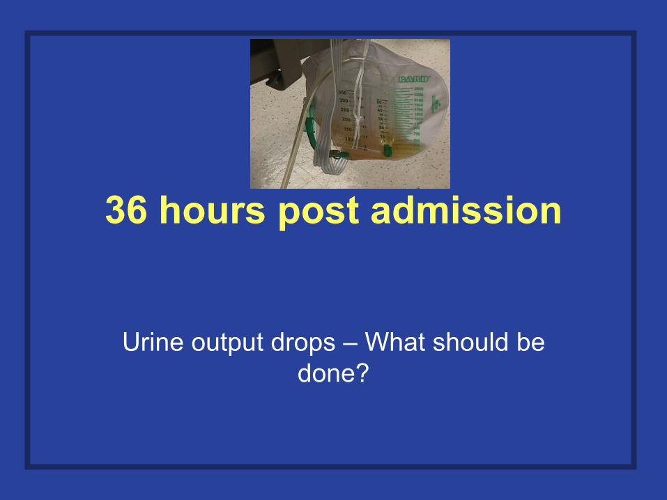
36 hours post admission
Urine output drops – What should be done?

48 hours post admission
Pulse oximeter drops and becomes difficult to read – what should be done?

Modified from criteria published in: Balk RA. Crit Care Clin. 2000;16:337-352. Kleinpell RM. Crit Care Nurs Clin N Am 2003;15:27-34.
CardiovascularTachycardiaHypotension
Altered CVP and PAOP
Renal OliguriaAnuria
↑ Creatinine
Hematologic↓ platelets,
↑ PT/INR/ ↑ aPTT↓ protein C↑ D-dimer
Hepatic Jaundice
↑ Liver enzymes↓ Albumin
CNSAltered consciousness
Confusion
MetabolicMetabolic acidosis↑ Lactate level
↓ Lactate clearance
Respiratory Tachypnea↓ PaO2
↓ PaO2/FiO2 ratio
IDENTIFYING ACUTE ORGAN DYSFUNCTION AS A MARKER OF SEVERE SEPSIS

Severe Sepsis-Associated Mortality Increases With the Number of
Dysfunctional Organs
Vincent JL, et al. Crit Care Med 1998;21:1793-800; Angus DC et al. Crit Care Med 2001;29:1303-10.
Number of Dysfunctional Organs
0%10%20%30%40%50%60%70%80%90%
AngusVincent
One Two Three Four or More
Seve
re S
epsi
s As
soci
ated
Mor
talit
y

17
What Would You Do?

18
No-ventilation Cardiopulmonary Resuscitation
• Airway opening, breathing, chest compressions (ABCs)
• Chest compressions, airway opening, breathing (CABs)

19
In a Cardiopulmonary Arrest, Which Type of Blood Gas Is Most Useful to Assess the
Resuscitation Effort: Arterial or Venous?
Venous blood
Arterial bloodSO2 - .98
SO2- .60
SO2 - .65
SO2- .61
SO2- .65
SO2 - .62
SO2 - .65
SO2- .60

20
Triple Lumen Oximetry
• Triple lumen oximetry expands ability to assess tissue oxygenation
• Values obtained from distal tip– Right atrium reading– Similar to pulmonary artery
values
• Used as end point in therapy
21
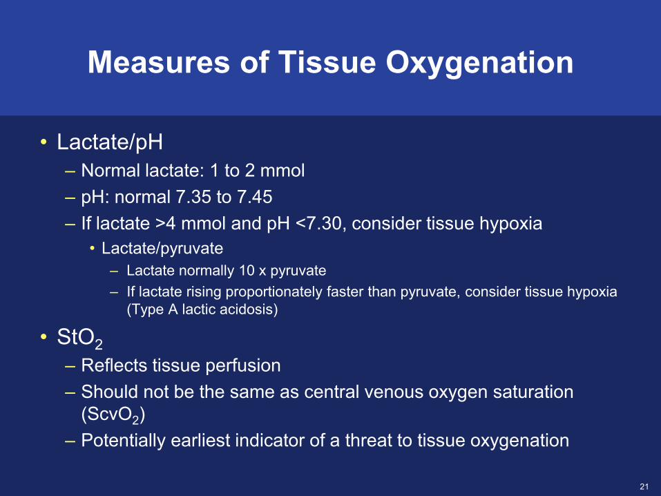
Measures of Tissue Oxygenation
• Lactate/pH– Normal lactate: 1 to 2 mmol– pH: normal 7.35 to 7.45– If lactate >4 mmol and pH <7.30, consider tissue hypoxia
• Lactate/pyruvate– Lactate normally 10 x pyruvate– If lactate rising proportionately faster than pyruvate, consider tissue hypoxia
(Type A lactic acidosis)
• StO2– Reflects tissue perfusion– Should not be the same as central venous oxygen saturation
(ScvO2)– Potentially earliest indicator of a threat to tissue oxygenation

22
Lactate as Indicator of Hypoxia
Cristae
CytosolMitochondrion
Chemical energy(high-energyelectrons)
23
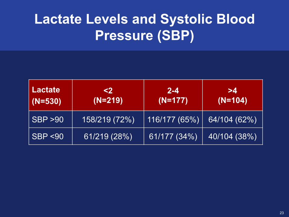
Lactate Levels and Systolic Blood Pressure (SBP)
Lactate (N=530)
<2 (N=219)
2-4 (N=177)
>4 (N=104)
SBP >90 158/219 (72%) 116/177 (65%) 64/104 (62%)
SBP <90 61/219 (28%) 61/177 (34%) 40/104 (38%)

24
Who Should Get a Lactate?
• Any patient:– With an infection and signs of systemic inflammatory
response system– Requiring rapid response– Admitted with heart failure– With overt or covert blood loss
• Others

25
Lactate as a Trigger for Hemodynamic Evaluation
Doppler Techniques
26
AORTICACCESS
PULMONARYACCESS
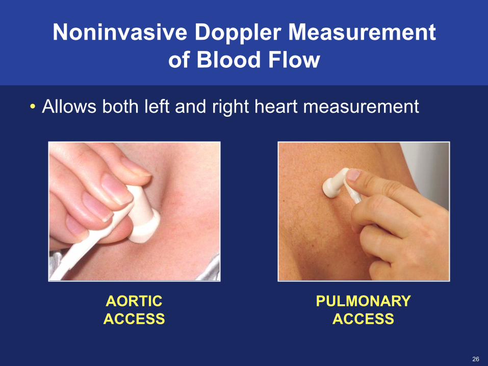
Noninvasive Doppler Measurement of Blood Flow
• Allows both left and right heart measurement

27
Any Change in Blood Flow (CO) Should Be Compared Against an
Oxygenation End Point
ScvO2 or StO2

28
Why Would a Clinician Not Get a Lactate, Especially a POCT?
• Lack of education• Other clinicians do not do it• Laboratory resistance

29
Overcoming Barriers
Implementing Lactate POCT in the Emergency Department (ED), Intensive
Care Unit, and Floors (Registered Respiratory Therapists)
30
Cardiac Assessment
Origin of Shortness of breath and Chest Pain

31
ECG and Physical Assessment
• BNP already elevated in congestive heart failure (CHF)– Activated with stretching of myocardial muscle– Helps differentiate CHF from pulmonary origin
• Promotes:– Vasodilation (reduces systemic vascular resistance, pulmonary
arterial occlusion pressure, central venous pressure)– Diuresis– Decrease in sympathetic nervous system response (reduces
norepinephrine, aldosterone)
• Normal values– <100 pg/mL

32
Shortness of Breath
• What causes SOB?• Ruling out cardiac vs pulmonary origins• Laboratory values in SOB
– BNP– Arterial blood gases (ABGs)
• Treatment for SOB

33
Causes of SOB
• SOB can be due to either cardiac or pulmonary problems. Sudden SOB is likely cardiac or pulmonary in nature
– In some cases, it could be anxiety or a neuromuscular weakness
• Cardiac causes of SOB arise from increased cardiac filling pressures, producing increased extravascular lung water. Increased lung water can:
– Flood the alveoli and interfere with gas exchange
– Also activate pulmonary stretch receptors, causing SOB
• Pulmonary causes of SOB can include (but are not limited to):
– Pneumonia, chronic lung disease, asthma, cancer, and vascular conditions
• Regardless of the cause, it is a condition that requires accurate diagnosis and rapid intervention
Pulmonarycapillary Alveolus
NORMAL VASCULAR PRESSURE
Tight periareolarconnective tissue
Loose periareolarconnective tissueLymphatic
Bronchiole
HEART FAILURE–INDUCED INCREASE TO VASCULAR PRESSUREPulmonarycapillary Alveolus
Tight periareolarconnective tissue
Loose periareolarconnective tissue
Lymphatic
Bronchiole

34
Identifying the Cause of SOB
•Clearly identifying the cause of SOB may be difficult•Patient history may provide a rapid clue as to the origin of the SOB:– A history of CHF or chronic lung disease (eg, chronic
obstructive pulmonary disease [COPD]) is more likely to have a repeat of earlier episodes
– The more objective the data, the more likely an accurate diagnosis can be made

35
Laboratory Values in SOB
• Measuring BNP levels is one of the most simple tests in identifying the cause of SOB– BNP levels are elevated (>100 pg/mL) in cardiac
causes and are normal (<100 pg/mL) in pulmonary causes
• The higher the BNP, the more likely a cardiac cause of SOB is present– Levels in excess of 400 pg/mL suggest more severe
cardiac disturbances and admission to the hospital is common with this level

36
ABG Values in SOB
• ABGs can be normal, even when a patient is short of breath
• If the PaO2 or pulse oximeter values are low (PaO2 <60 mm Hg and SpO2 <90%), then oxygen therapy may produce some relief
• If the PaCO2 and pH are low, the cause of SOB may be anxiety
• High PaCO2 values and low pHs tend to be associated with depressed breathing, but not SOB

37
Treatment of SOB
• Initially, most patients are placed on oxygen therapy, although this does little to address the cause of SOB
• If the patient has a cardiac cause of SOB, improving cardiac function is imperative
• Preload or afterload reducers or even inotropic therapy might help reduce SOB
• If the patient has a pulmonary cause of SOB that can be treated, therapies such as bronchodilators for asthma and COPD and antibiotics for pneumonia may be helpful

38
B-Type Natriuretic Peptide
• What is BNP?• How can BNP help in ruling out cardiac vs
pulmonary origins of SOB?• Clinical examples

39
How Can BNP Help in Ruling Out Cardiac vs Pulmonary Origins of SOB?
Diagnosing CHF• When a patient presents with SOB, the
patient history may provide an easy diagnosis. However, when the cause is not clear, clinicians have to differentiate between a cardiac cause and a pulmonary cause
• Sophisticated tests, such as an echocardiogram, can help diagnose the problem . However, they may not be immediately available
• If the BNP is normal, the SOB is highly likely to be noncardiac
• BNP levels >100 pg/mL suggest heart failure. The higher the level, the more likely heart failure is the cause of SOB
Normal B-TypeNatriureticPeptide
Increased B-TypeNatriureticPeptide

40
Clinical Examples
• A 66-year-old female is admitted to the ED with SOB. She had a total knee replacement last month. She has a history of coronary artery disease with a 20 pack-year history of smoking
• On examination, she has crackles throughout both lungs• Vital signs
– Blood pressure (BP): 142/84– Pulse (P): 103– Respiration rate (RR): 32– Temperature (T): 37.2– SpO2: 93 (on 2 liters per minute [lpm] nasal cannula)– BNP: 1350
• What is the cause of her SOB?– Based on the high BNP, her SOB is due to cardiac dysfunction. Other
potential problems, such as lung disease and pulmonary embolism, will not produce such a high BNP

41
Clinical Examples (cont’d)
• A 58-year-old male is admitted to the ED with SOB. He has no history of cardiac or pulmonary disease
• On examination, he has wheezing in the left lower lobe• Vital signs:
– BP: 136/86– P: 105– RR: 30– T: 37.5– SpO2: 97 (on 2 lpm nasal cannula)– BNP: 67
• What is the cause of his SOB?– Based on the normal BNP, his SOB is not due to cardiac dysfunction.
Other potential problems, such as lung disease, pneumonia, and pulmonary embolism, should be investigated to identify the cause of SOB

42
Chest Pain
• What causes chest pain (CP)?• Identifying the cause of CP• Laboratory values in CP• Treatment for CP

43
Causes of Chest Pain
• CP can be due to either cardiac or noncardiac causes • It is essential to differentiate if the cause is myocardial infarction
(MI)/ischemia vs other, often less dangerous, conditions– Acute coronary syndrome (ACS) (eg, ST elevation MI [STEMI] or non-
ST elevation MI [NSTEMI]) are life-threatening events and need immediate diagnosis and treatment
• Testing cardiac enzymes and ECG are considered essential in the diagnosis of cardiac origins of chest pain– ST-segment elevation in the ECG is highly specific for STEMI, but only
about 50% sensitive– Many patients presenting to EDs have NSTEMI
44
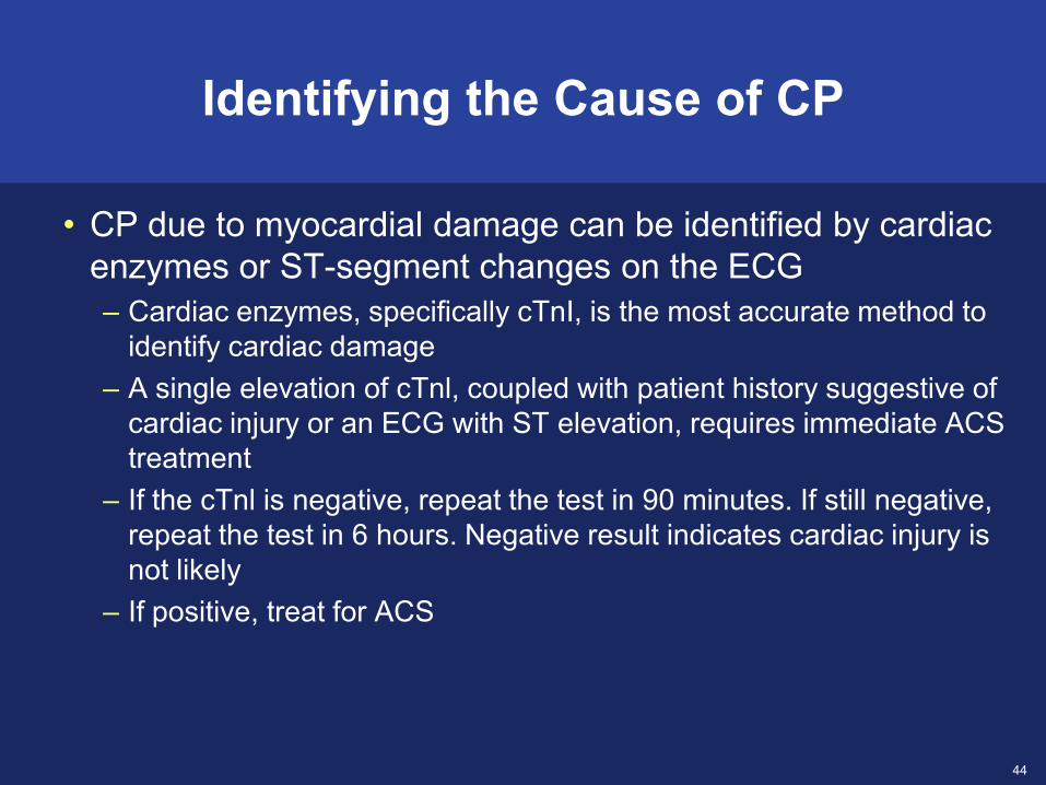
Identifying the Cause of CP
• CP due to myocardial damage can be identified by cardiac enzymes or ST-segment changes on the ECG– Cardiac enzymes, specifically cTnI, is the most accurate method to
identify cardiac damage– A single elevation of cTnl, coupled with patient history suggestive of
cardiac injury or an ECG with ST elevation, requires immediate ACS treatment
– If the cTnl is negative, repeat the test in 90 minutes. If still negative, repeat the test in 6 hours. Negative result indicates cardiac injury is not likely
– If positive, treat for ACS

45
Identifying the Cause of CP (cont’d)
• ECGs are helpful, particularly if ST elevation is present in 2 or more anatomically connected leads (eg, V1-V4 for anterior MIs; II, III, and avF for inferior MIs; and I, avL, V5 or V6 for lateral MIs)– An elevated ST segment in 2 or more leads, combined with a patient
history suggestive of cardiac origin of CP, is often enough to admit the patient to the cardiac catheterization lab

46
What Is cTnI?
• Troponin is one of a several types of proteins located in the heart.
• The cTnl is a unique form of troponin found only in myocardial tissue. Cardiac troponin has 3 components: T, C, and I
• cTnI is specific to cardiac tissue and is released into serum after myocardial necrosis

47
Use of cTnI
• cTnI is considered the most useful of the cardiac enzymes
• Use of cardiac enzymes have the best value in unclear cases of cardiac origin of chest pain (eg, patient with a left bundle branch block)

48
Interpretation of Troponin
• The American College of Cardiology and European Society of Cardiology consensus guidelines recommend using the 99th percentile of cardiac troponin values measured in a healthy reference population as the clinical decision limit
• Most healthy individuals have undetectable cTnI (<0.01 ng/mL) with a 99th percentile value of 0.4 ng/mL
• Therefore, any cTnI value >0.4 ng/mL is considered to be an elevated level indicative of myocardial injury.
• Grossly elevated cTnI values (eg, >2 ng/mL) are associated with MI
• Pattern of release in MI is BIPHASIC– Detectable in blood at 4 to 12 hours– Peaks at 12 to 38 hours– Remains elevated for 5 to 10 days
0 24 48 72 96 120 144 168Hours After Onset of MI
30
25
20
15
10
5
Trop
onin
I (n
g/m
L)

49
Limitations of Enzymes
• The most common reasons for the enzymes to be incorrect are other cardiac conditions, such as:– Blunt chest trauma– Myocarditis– CHF– Left ventricular hypertrophy

50
SummaryAcceptance of POCT Technology
• Are patients being harmed by our current practices?• What is needed to improve patient management?
– Can POCT be helpful in that management?