clinical enzymology enzymes are the protein catalysts that increase the rate of specific chemical...
TRANSCRIPT


Clinical Enzymology

• Enzymes are the protein catalysts that increase the rate of specific chemical reaction in the body.
• Enzymes are found in small amounts mainly within cells ,clotting factors and digestive enzymes function naturally after secretion: - Plasma specific – Thrombin - Secreted - Lipase, Amylase - Intracellular - transaminases, creatine kinase
• Injury or death of tissues can cause the release of tissue-specific enzymes into the bloodstream.
• Elevated enzyme levels are often indicators of tissue problems, and are used in the diagnosis of diseases.
• Enzyme activities in the body fluids are altered by pathological processes so, its measurement is used for disease investigation.
INTRODUCTION

• All known enzymes are proteins.
• They are high molecular weight compounds made up principally of chains of amino acids linked together by peptide bonds.
• Enzymes can be denatured and precipitated with salts, solvents and other reagents.
• They have molecular weights ranging from 10,000 to 2,000,000.
• Many enzymes require the presence of other compounds - cofactors - before their catalytic activity can be exerted.
• This entire active complex is referred to as the HOLOENZYME; i.e., APOENZYME (protein portion) plus the COFACTOR (coenzyme, prosthetic group or metal-ion-activator) .
Chemical Nature of Enzymes
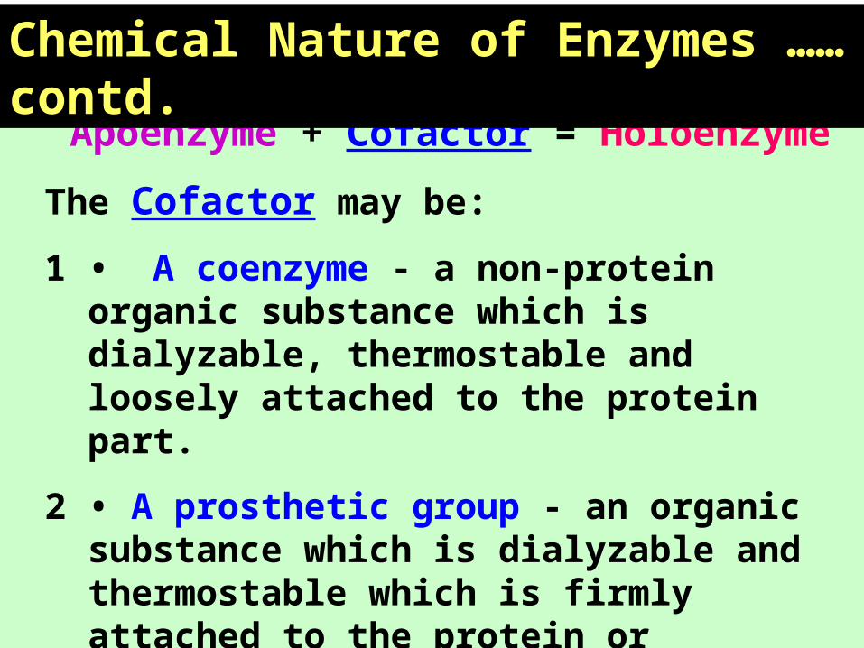
Apoenzyme + Cofactor = Holoenzyme
The Cofactor may be:
1 • A coenzyme - a non-protein organic substance which is dialyzable, thermostable and loosely attached to the protein part.
2 • A prosthetic group - an organic substance which is dialyzable and thermostable which is firmly attached to the protein or apoenzyme portion.
3 • A metal-ion-activator - these include K+, Fe++, Fe+
++, Cu++, Co++, Zn++, Mn++, Mg++, Ca++, and Mo+++.
Chemical Nature of Enzymes ……contd.
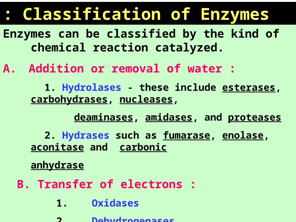
Enzymes can be classified by the kind of chemical reaction catalyzed.
A. Addition or removal of water :
1. Hydrolases - these include esterases, carbohydrases, nucleases,
deaminases, amidases, and proteases
2. Hydrases such as fumarase, enolase, aconitase and carbonic
anhydrase
B. Transfer of electrons :
1. Oxidases
2. Dehydrogenases
Classification of Enzymes:

Classification of Enzymes …… contd. C. Transfer of a radical:
1. Transglycosidases - of monosaccharides
2. Transphosphorylases and phosphomutases - of a phosphate group
3. Transaminases - of amino group
4. Transmethylases - of a methyl group
5. Transacetylases - of an acetyl group
D. Splitting or forming a C-C bond: 1. Desmolases
2. Changing geometry or structure of a molecule
3. Isomerases
E. Joining two molecules through hydrolysis of pyrophosphate bond in ATP or other tri-phosphate
1. Ligases

• One of the properties of enzymes that makes them so important as diagnostic and research tools is the specificity they exhibit relative to the reactions they catalyze.
• Greater specificity is achieved in three ways:1. Interpreting investigations in the light of clinical features
2. Test pattern recognition
3. Isoenzyme determination: AST may be due to MI or Hepatitis so, it makes
confusion in diagnosis to be confirmed by LDH levels.
- ALP in Cholestasis & bone diseases :
- Differentiated by bilirubin & transaminase levels in
Cholestasis .
- Confirmed by GGT in Cholestasis.
Specificity of Enzymes:

Specificity of Enzymes ………contd.In general, there are four distinct types of specificity:
• Absolute specificity - the enzyme will catalyze only one reaction.
• Group specificity - the enzyme will act only on molecules that have specific functional groups, such as amino, phosphate and methyl groups.
• Linkage specificity - the enzyme will act on a particular type of chemical bond regardless of the rest of the molecular structure.
• Stereochemical specificity - the enzyme will act on a particular steric or optical isomer.

Rate of entry into blood
Serum enzyme activity
Rate of removal
Inhibition
Rate of synthesis Mass of enzyme
Producing tissueTissue damage
Clearance Inactivation
Factors affecting serum enzyme activity

I- Rate of entry of enzymes into blood is affected by: a – Rate of synthesis of enzyme:
- Biliary obstruction hepatobiliary tree enzymes
- Drugs :Anticonvulsant drugs ( Phenobarbital & phenytoin)
synthesis of enzymes by the hepatocytes
b- Mass of enzyme producing cells as in:
- alkaline phosphatase: ( in active growth , Paget’s disease.
and in 3rd trimester pregnancy).
- acid phosphatase ( in cancer prostate).
c- Necrosis or Cell damage as in:
- Hepatitis transaminases
- Myocardial infarction CK
- Stored blood LDH
Factors affecting Serum enzyme activities-1

II- Enzyme inhibitors:
- Little effect on enzyme values determined in the lab.
- Organophosphorus poisoning irreversible inhibition of
cholinesterase.
III- Clearance of enzymes:
- Breakdown by Proteases and removal by the reticuloendothelial
system.
- Renal excretion of small molecular enzymes e.g. Amylase
Factors affecting Serum enzyme activities-2


Schematic diagram showing the effect of temperature on rate of nonenzyme-catalyzed and enzyme catalyzed reactions.

Enzymes have an active site- a cleft into
which substrate molecules fit
The active site contains amino acids that:
-Attract the substrate
-Assist in the chemical reactions that converts
substrate to product
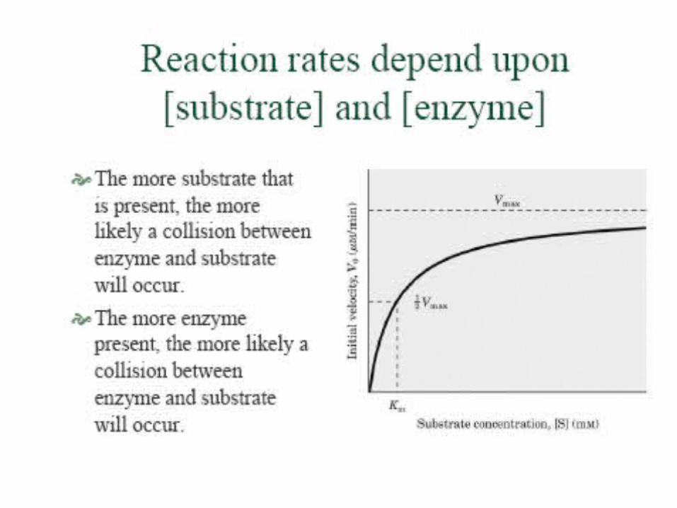
How does an enzyme work?


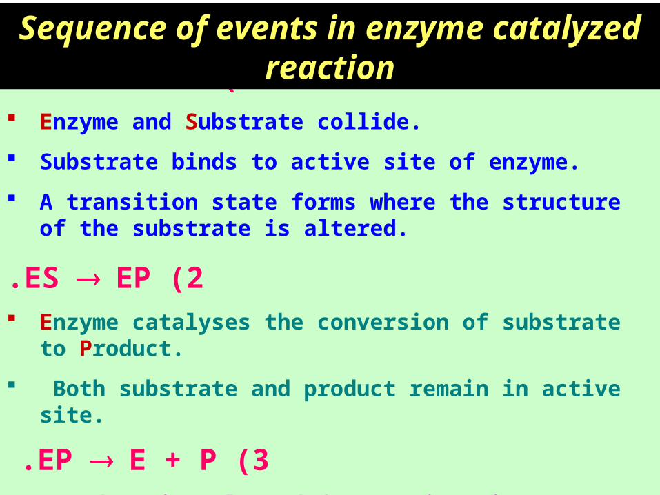
1 )E + S ES. Enzyme and Substrate collide.
Substrate binds to active site of enzyme.
A transition state forms where the structure of the substrate is altered.
2 )ES EP. Enzyme catalyses the conversion of substrate to Product.
Both substrate and product remain in active site.
3 )EP E + P. • Product is released from active site.
Sequence of events in enzyme catalyzed reaction

ISOENZYMES
• Catalyze the same reaction
• Two or more polypeptide chains
• Different polypeptide chains are products of different genes
• Differ in AA sequence and physical properties
• May be separable on the basis of charge
• Are tissue specific

Aminotransferases:• ALT • AST • GGT
ALT and AST :1. Pyridoxal dependent 2. Indicates Hepatitis, Myocardial infarction: - Elevations take > 4 hours to develop, last 4 days - ALT elevation lasts longer than AST - AST elevations are higher than ALT - Elevations occur in most definite infarctions3. Skeletal muscle damage 4. Hemolysis 5. Pancreatitis
Isoenzymes and multiple forms

GGT
• Hepatobiliary enzyme - highly inducible in 75% of the population
• Highest increases in intra or posthepatic biliary obstruction
• Higher and more persistent increases than AlP (Alkaline Phosphatase)
Isoenzymes and multiple forms
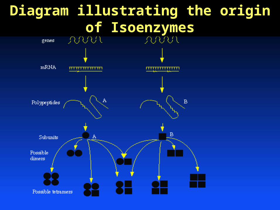
Diagram illustrating the origin of Isoenzymes

Transaminase activities in human tissues, relative to serum as unity
AST ALT
Heart 7800 450
Liver 7100 2850
Skeletal Muscle 5000 300
Kidney 4500 1200
Pancreas 1400 130
Spleen 700 80
Lung 500 45
Erythrocytes 15 7
Serum 1 1
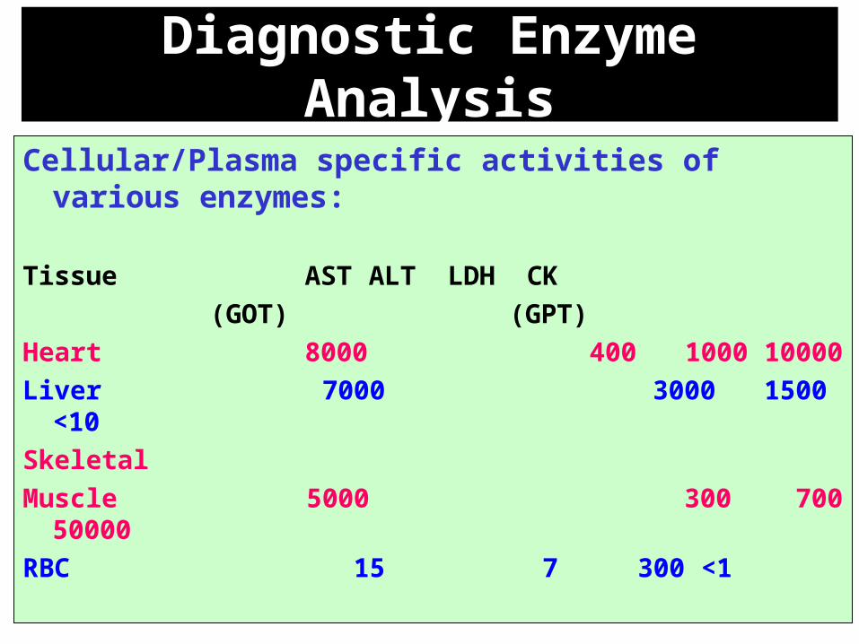
Diagnostic Enzyme Analysis
Cellular/Plasma specific activities of various enzymes:
Tissue AST ALT LDH CK
(GOT) (GPT)
Heart 8000 400 1000 10000
Liver 7000 3000 1500 <10
Skeletal
Muscle 5000 300 700 50000
RBC 15 7 300 <1


LDH-Lactate Dehydrogenase in MI• LDH is a tetramer of two non-identical subunits (LDH5(M4),
LDH4(M3H), LDH3(M2H2), LDH2(MH3), LDH1(H4)

•Acid phosphatase (ACP)
•Amylase (AMS)
•Alanine aminotransferase (ALT)
•Alkaline phosphatase (ALP)
•Aspartate aminotransferase (AST)
•Creatine kinase (CK)
•Gamma-glutamyltransferase (GGT)
•Glucose 6-phosphate dehydrogenase (G6PD)
•Lactate dehydrogenase (LDH or LD)
•Lipase (LPS)
•Plasma cholinesterase
ENZYMES OF CLINICAL INTEREST

Diagnostically Important Enzymes 1/3
Enzyme Principal Sources Principle Clinical Applications
Acid
Phosphatase (ACP)
Prostate, erythrocytes Carcinoma of prostate
Alanine aminotransferase(ALT)
Liver, Skeletal muscle, Heart
Hepatic parenchymal disease
Aldolase Skeletal muscle, heartMuscle disease
Alkaline
Phosphatase (ALP)
Liver, bone, intestinal mucosa, placenta, kidney
Bone diseases, hepatobiliary diseases
Amylase (AMS)Salivary glands, pancreas, ovaries
Pancreatic diseases
Aspartate aminotransferase(AST)
Liver, skeletal muscle, heart, kidney, erythrocytes
Myocardial infarction, hepatic parenchymal disease, muscle disease

Diagnostically Important Enzymes 2/3
Enzyme Principal Sources Principle Clinical Applications
Cholinesterase Liver Organophosphorus insecticide poisoning, suxamethonium sensitivity, hepatic parenchymal diseases
Creatine kinase (CK)Skeletal muscle, brain, heart, smooth muscle
Myocardial infarction, muscle diseases
Glutamate dehydrogenase
Liver Hepatic parenchymal disease
gamma-GT (GGT)Liver, kidney Hepatobiliary disease, alcoholism

Diagnostically Important Enzymes 3/3
Enzyme Principal Sources Principle Clinical Applications
Lactate dehydrogenase(LDH)
Heart, liver, skeletal muscle, erythrocytes, platelets, lymph nodes
Myocardial infarction, hemolysis, hepatic parenchymal disease
5’ Nucleosidase Hepatobiliary tract Hepatobiliary disease
Sorbitol dehydrogenase
Liver Parenchymal hepatic disease
Trypsin(ogen) Pancreas Pancreatic diseases
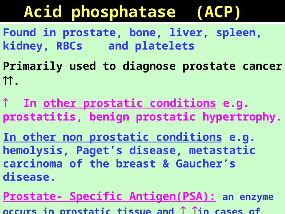
Found in prostate, bone, liver, spleen, kidney, RBCs and platelets
Primarily used to diagnose prostate cancer .
In other prostatic conditions e.g. prostatitis, benign prostatic hypertrophy.
In other non prostatic conditions e.g. hemolysis, Paget’s disease, metastatic carcinoma of the breast & Gaucher’s disease.
Prostate- Specific Antigen(PSA): an enzyme occurs in prostatic
tissue and in cases of metastatic carcinoma
Tartarate inhibits the prostatic ACP enzyme while Formaldehyde inhibits ACP from other sources
Acid phosphatase (ACP)

Alanine aminotransferase (ALT)
•Widely distributed, although the largest amounts found in the liver.
•Smaller amounts occur in the heart but usually remains normal after MI .
•Congestive cardiac failure release from the liver
•More specific for liver disease than AST.

•Widely distributed, high concentrations in intestines, liver, bone, spleen, placenta and kidney.
•The main sources of serum ALP are the hepatobiliary tree and bone disorders.
•Elevated levels during healing of fractures , active growth and during the 3rd trimester of pregnancy.
serum ALP activity in liver disease is mainly due to Cholestasis.
•Decreased levels are found in the inherited condition “ Hypophosphatasia” which is caused by defective bone calcification
Alkaline phosphatase (ALP)
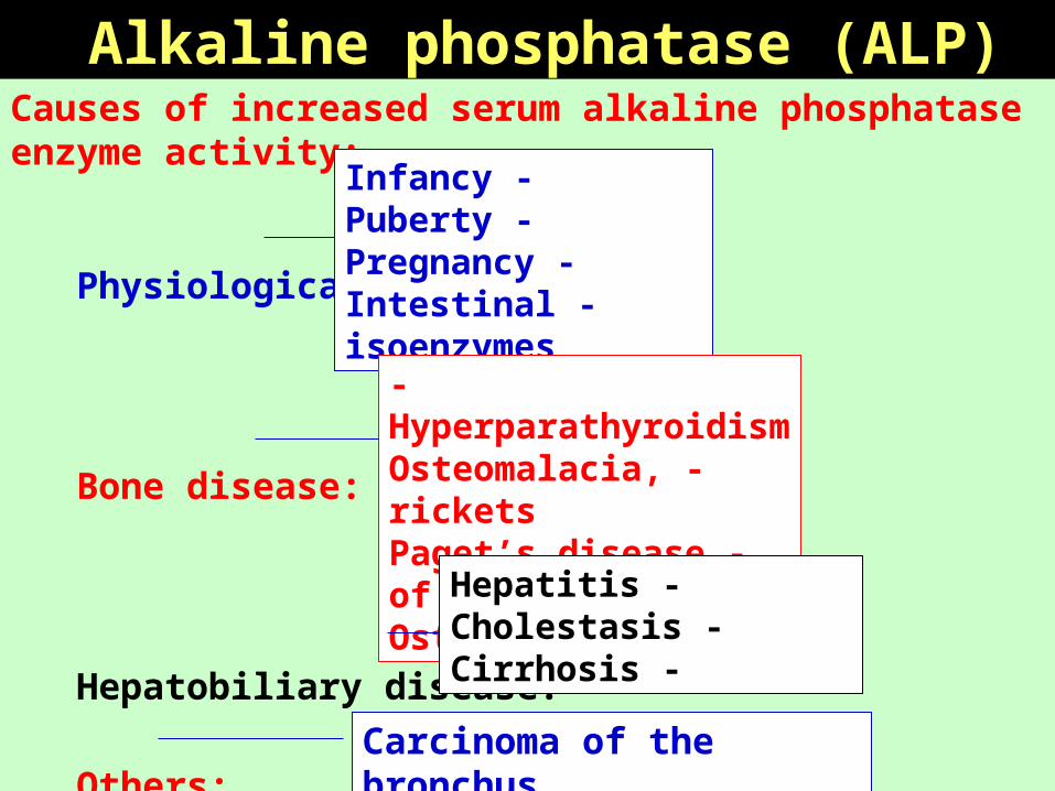
Alkaline phosphatase (ALP) Causes of increased serum alkaline phosphatase enzyme activity:
Physiological :
Bone disease:
Hepatobiliary disease:
Others:
-Infancy -Puberty
-Pregnancy -Intestinal isoenzymes
-Hyperparathyroidism -Osteomalacia, rickets
-Paget’s disease of bone -Osteomyelitis
-Hepatitis -Cholestasis
-Cirrhosis
Carcinoma of the bronchus

Alkaline Phosphatase• Bone Alkaline Phosphatase
Males
Females
4 8 12 16 20 (Years)
Alk. P
hos.un
its

• Hydrolases that split complex Polysaccharides.
- alpha-amylases (1-4 alpha links attacked at random)
- MW 40,000 - filtered by kidney
- Ca+2 requiring metalloenzyme
Sources :
1. Pancreas (p-type)
2. Salivary glands (s-type)
3. Intestinal malignancy
Amylase (AMS)

Clinical Significance : Diagnosis and monitoring of pancreatitis
1. Acute: - transient increase in activity within 2 - 12 hours
- returns to normal in 3 - 4 days
2. Extent of increase (typically 4 - 6 fold) related to probability of acute pancreatitis.
3. Serum amylase activity may be falsely normal in presence of severe dyslipidaemia - check urinary amylase.
4. Serum amylase may be raised in biliary duct obstruction.
5. Serum amylase activity may be normal in chronic pancreatitis.
6. Amylase may be raised in pleural effusions and pseudocystic fluids secondary to pancreatitis.
7. Salivary gland diseases or trauma may raise serum amylase activity.
Amylase (AMS)

Salivary Enzymes in Normal Function and Abnormal Pathology
• α-Amylase—normally present in saliva—important in digestion
______________________________________________
• Lysozyme
• Hyaluronidase
• Chondrosulfatase
• Aryl Sulfatase
• Neutral Protease
• Collagenases
Specific activities are increased in gingivitis and periodontal disease

1. Pancreatic disease (P-type):
Pancreatitis:
- Acute
- Chronic
- Complications:
• Pseudocyst
• Ascites and pleural effusion
• Abcess
Pancreatic Trauma, including investigative maneuvers
Pancreatic carcinoma
Causes of Hyperamylasemia and Hyperamylasuria

2. Disorders of non-pancreatic origin
(mechanism unknown) :
a- Renal insufficiency (mixed)
b- Neoplastic hyperamylasemia - usually bronchogenic
or ovarian (usually S-type)
c- Salivary gland lesions, e.g. mumps, calculus disease
(S-type)
d- Macroamylasemia (predominantly S-type)
Causes of Hyperamylasemia and Hyperamylasuria

• 3. Disorders of complex origin (mechanism unknown or uncertain) o Biliary tract disease o Intra-abdominal disease (other than pancreatic diseases):
o Perforated peptic ulcer (P-type) o Intestinal obstruction (P-type) o Mesenteric infarction (P-type) o Peritonitis (mixed; depends on cause) o Acute appendicitis o Ruptured ectopic pregnancy (S-type) o Aortic aneurysm with dissection
o Cerebral trauma (type depends on other organ damage) o Burns and traumatic shock o Postoperative hyperamylasemia (usually S-type) o Diabetic ketoacidosis (mixed) o Renal transplantation (S-type) o Acute alcoholism (mixed) o Drugs: - Medicinal opiates (P-type) - Heroin addiction (S-type)
Causes of Hyperamylasemia and Hyperamylasuria

• This enzyme is widely distributed in the body.
• Main sources: Heart, liver, skeletal muscle, and kidney.
• Useful in the diagnosis of MI, liver disorders and muscle damage.
•Causes of serum AST levels:
•Physiological : Neonates.
• Liver diseases: Hepatitis, hepatic necrosis , cholestasis
•Cardiac disease: Myocardial Infarction.
•Diseases of skeletal muscle: Crush injury,trauma,myopathy
•From Erythrocytes: Hemolysis
Aspartate aminotransferase (AST)

•Creatine kinase is associated with ATP regeneration in muscle and nervous tissue.
•Elevated blood levels of CK are used as indicators of MI, muscular dystrophy, and stroke.
•CK occurs as a dimer of 2 different subunits, M and B.
- CK-BB: Brain type.
- CK-MB: Hybrid type.
- CK-MM: Muscle type.
•These can be separated by electrophoresis.
•CK-MB is released from cardiac muscle cells after MI.
Creatine kinase (CK)

MM% MB% BB%
MM - skeletal muscle
<80<20< 1
MB - cardiac muscle
<60 <40 <1
BB - brain0 <3 <95
Creatine kinase (CK)A dimer - M and B protein strands which are the products of different genes - true isoenzymes.
• 5% cutoff by general agreement
• 2 of 3 - history, ECG, enzymes

• A microsomal enzyme its synthesis induced by ethanol and anticonvulsant drugs.
• Found mainly in the kidney and significant amounts in liver, brain, prostate, and pancreas.
• Used primarily for diagnosis of hepatobiliary problems.
• ALT, AST and GGT are the main liver function tests.
• Marked elevation of serum GGT level is seen in alcoholic liver disease.
serum GGT activity sometimes following MI or congestive cardiac failure.
Gamma-glutamyltransferase (GGT)

• First (and control) enzyme for pentose phosphate pathway (P.P.P.).
• Important in production of NADPH + H+, especially in RBC.
• NADPH + H+ keeps glutathione reduced.
• Antimalarial drugs are oxidants, and adversely affect this system in RBCs.
• Some populations, especially African-Americans, have a high frequency of G6PD deficiency.
• If given antimalarial drugs, or fava beans, they develop hemolytic anemia.
Glucose 6-phosphate dehydrogenase (G6PD)

• Converts pyruvate to lactate (and vice versa) during and after anaerobic metabolism.
• LDH occurs as a tetramer of 2 different subunits:
LD-1 (HHHH) from the heart:
Elevated after MI.
LD-2 (HHHM) from the kidney:
Elevated after renal infarction.
LD-3 (HHMM) from the lung, spleen and pancreas: Elevated in pulmonary embolism.
LD-4 (HMMM) and LD-5 (MMMM), both from the liver and skeletal muscle:
Elevated in injury to liver or skeletal muscle.
Lactate dehydrogenase (LDH or LD)

Control
LDH Isoenzymes1 2 3 4 5

•Breaks down fat into monoacylglycerol and free fatty acids.
•Primarily from the pancreas.
•Used to diagnose acute pancreatitis.
•Pancreatic lipases : - A group of enzymes that hydrolyze glycerol esters of long
chain fatty acids.
- Some substrate specificity e.g. LPL (Lipoprotein Lipase).
- Bile salts are necessary for activity.
-Almost exclusively used clinically in the investigation of pancreatitis.
- Increase within 2 - 12 hours of acute attack.
- May remain elevated for many days .
- More specific to acute pancreatitis than amylase.
- Less sensitive to acute exacerbations in chronic pancreatitis.
Lipase (LPS)

• Similar to cholinesterase in nervous system, degrades
acetylcholine (neurotransmitter and hormone).
• Elevated in hepatitis and cirrhosis.
• Also elevated in organophosphate (pesticide) poisoning.
• Degrades succinylcholine, a muscle relaxant given
during general anesthesia in surgery.
• Some people are deficient in plasma cholinesterase, so
the normal dose of succinylcholine would kill them
Therefore, a determination of plasma cholinesterase is
made prior to major surgery.
Plasma cholinesterase

Low Plasma Cholinesterase
Category of causeExamples
Physiological reasonsInfancy, 3rd Trimester of pregnancy
Inherited abnormalityScoline sensitivity
(ChE variants)
Acquired abnormality:
A) Liver diseaseImpaired protein synthesis
B) Industrial poisoningOrganophosphorus insecticides
C) Drug effects
Oral contraceptives, MAO inhibitors, Cytotoxic drugs

A Serine Protease (hydrolyses peptide bonds formed by the carboxyl groups of lysine/arginine).
• Inactive Zymogens secreted (Type 1 and 2) under the influence of vagus nerve .
• alpha1-antitrypsin and alpha2-macroglobulin protect serum proteins (consider alpha-1 AT deficiency).
• Little clinical application in modern practice.
Pancreatic Trypsin

Serum Enzymes
in Disease

Myocardial Infarction

Myocardial Infarction ( MI )• Necrosis of the myocardium, but not angina pectoris release of
CK, AST and LDH (HBD) into the circulation.
• CK is the first to rise (activity within 6 h of MI ).
• Total CK reaches a peak at 24-36 h.
• In uncomplicated cases, CK returns to normal within 3 days.
• Serum AST more slowly ( maximum activity within 48 h) and returns to normal in 4-5 days.
• No significant elevation in HBD seen for the 1st 24 h (reaches maximum at about 3 days & remain for up to 8 days).
• It is important to consider the timing of sample when interpreting test results.
• CK & HBD are useful as early and late indicators of MI, and more specific than AST.

Myocardial Infarction ( MI )• CK from skeletal muscle may be following intramuscular
injection, chest compression for resuscitation or electrical defibrillator.
• CK specificity is by measuring CK-MB.
• HBD activity may be due to non cardiac factors (hemolysis).
• Cardiac enzyme measurements are very sensitive indicators of MI because it is in over 95% of cases.
• They are of particular value in the following conditions:
1. Atypical clinical presentation (absence of chest pain)
2. If the patient presents some time after a suspected event.
3. Difficulty in interpreting ECG (Arrhythmia or previous MI).
4. If further MI is suspected few days of a previous one.

CK-2 & CK-3 in normal subject and in patient
24 hrs after Myocardial InfarctionCreatine Kinase isoenzymes in blood

Plasma levels following myocardial infarction
CPK---Creatine Kinase
LDH---Lactate Dehydrogenase
HBDH—α-Hydroxybutyrate dehydrogenase


Myocardial Infarction : Plasma Enzymes Changes
EnzymeAbnormal
activity detectable(h)
Peak value of abnormality(h)
Duration of abnormality
(days)
CK-MB isoenzyme3 - 1012 - 241.5 - 3
Total CK5 - 1218 - 302 - 5
AST6 - 1220 - 302 - 6
Heart-specific’ LD8 - 1630 - 485 - 14
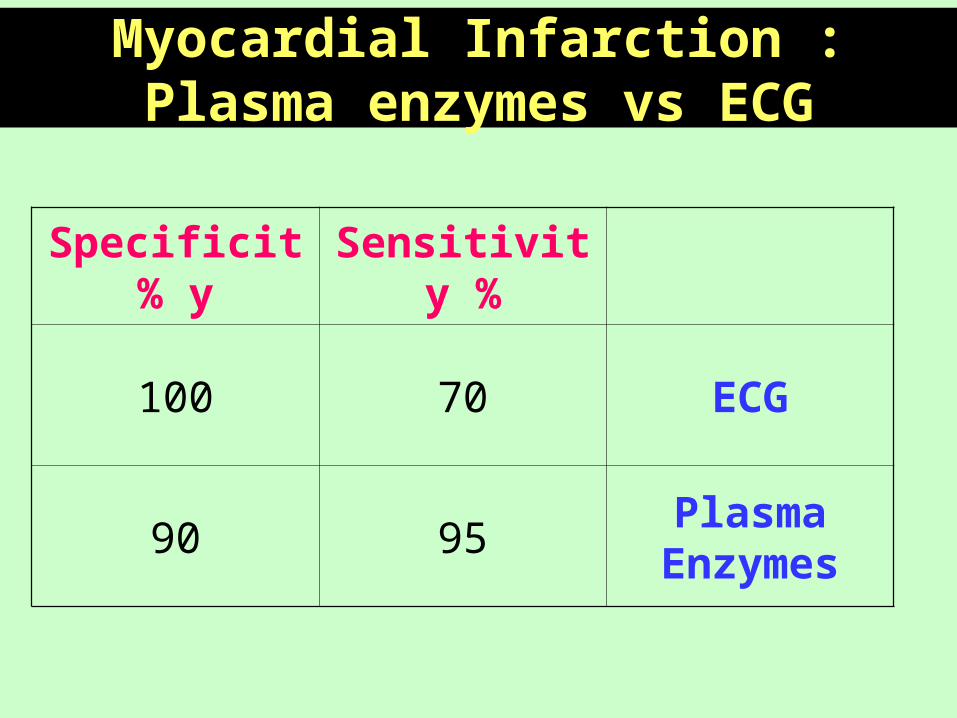
Myocardial Infarction : Plasma enzymes vs ECG
Sensitivity %Specificity%
ECG70100
Plasma Enzymes
9590
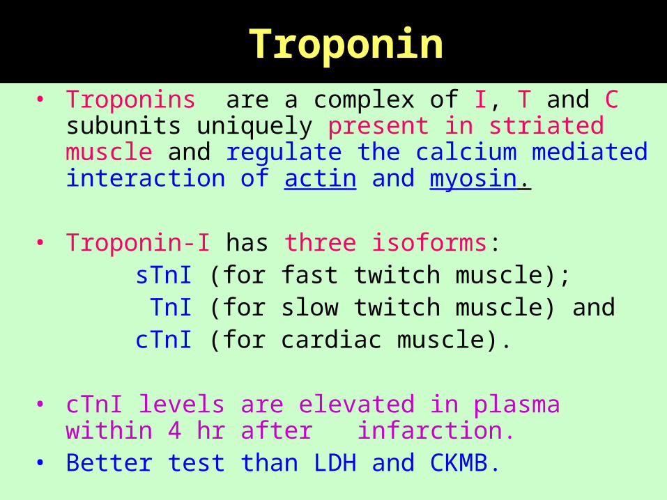
Troponin • Troponins are a complex of I, T and C subunits
uniquely present in striated muscle and regulate the calcium mediated interaction of actin and myosin.
• Troponin-I has three isoforms: sTnI (for fast twitch muscle); TnI (for slow twitch muscle) and cTnI (for cardiac muscle).
• cTnI levels are elevated in plasma within 4 hr after infarction.
• Better test than LDH and CKMB.

Muscle DiseasesMuscular Dystrophy
Toxic Myopathy
Malignant Hyperpyrexia
Traumatic Myopathy

Muscular Dystrophy• Genetically determined degenerative disorders.
• Duchenne muscular dystrophy is an X-linked recessive disorder caused by an abnormal dystrophin gene (progressive weakness of muscles).
• CK activities before the onset of clinical symptoms (values <10 times the normal upper level).
• Serum CK is in 75 % of female carriers.
• Becker’s muscular dystrophy is a benign form of Duchenne MD.
• CK elevated pattern similar to that of Duchenne MD.

Toxic Myopathy• Causes:
- Drugs & chemicals (Alcohol, D-penicillamine, ..etc) generalized myopathy
- IM injections ( Trauma & Chemical irritation)
• CK activity by narcotic analgesics given in MI.
• Rapid in body temp, shock& convulsions.(in general Anesthesia
Serum CK activity during attacks .
• Pre-operative CK should be measured in patients with a family history of malignant hyperpyrexia.
Malignant Hyperpyrexia

Traumatic Myopathies
• Muscle trauma (surgery, I.M. injection, etc..) release of enzymes
• High serum CK values occurs post-operatively .
• If MI suspected, CK-MB should be measured.
• Serum CK usually return to normal within 48 h of a single intramuscular injection.
• Vigorous exercise of short duration and prolonged moderate exercise serum CK

Liver Diseases Hepatic Necrosis
Hepatitis
Cholestasis
Jaundice Hepatocellular Damage

Measurement of serum enzyme activities for :
a - Differential Diagnosis of Jaundice.
b - Monitoring of drug toxicity.
• ALT is more specific than AST.
• Hepatocellular disease has only modest effect on ALP & GGT (up to 3 times the upper limit of normal)
• In Cholestasis, Higher values of ALP & GGT due to synthesis ( the values are 5-10 times the upper normal level) .
Liver Enzymes ( ALT, AST, GGT, ALP, LDH)

Bone Diseases - Osteoporosis
- Osteomalacia
- Tumors
- Paget’s Disease

• ALP enzyme is usually normal in Osteoporosis as osteoblastic activity is not increased
• Modest of ALP in Osteomalacia and Rickets
• Healing fractures Transient of ALP
• 1ry & 2ry Hyperparathyroidism of ALP
• In Paget’s disease of bone of ALP (10 times)
• 1ry & 2ry bone tumors of ALP (5 times normal)
Bone Enzymes - ( Alkaline Phosphatase) ALP
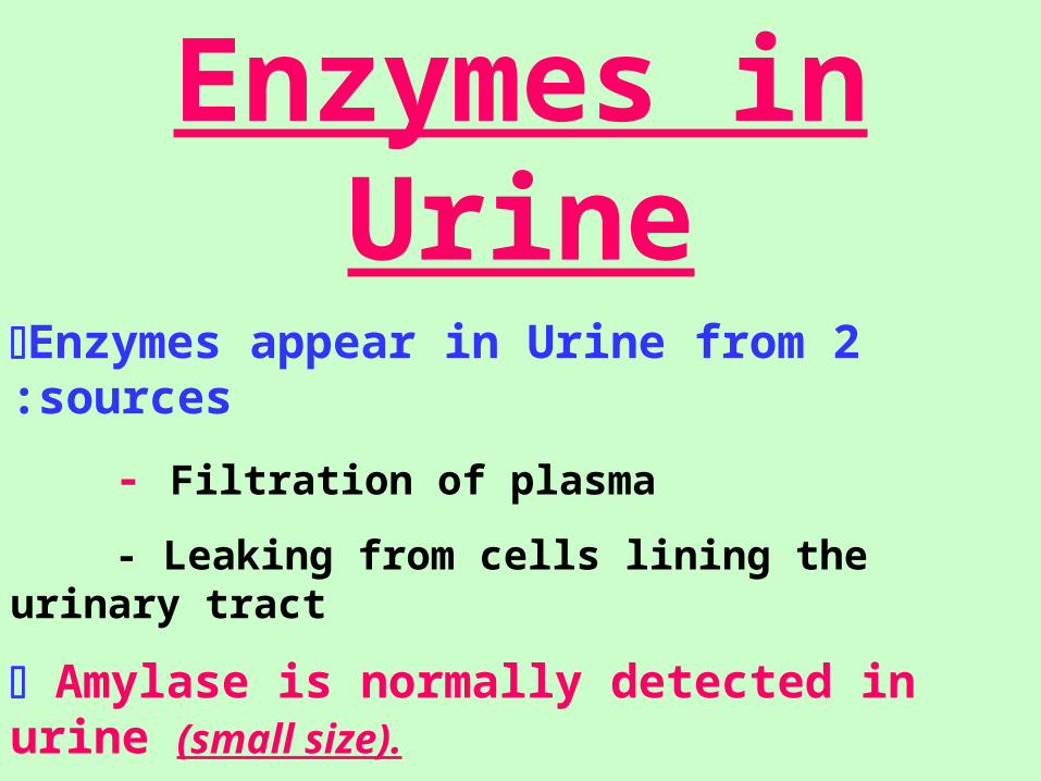
Enzymes in UrineEnzymes appear in Urine from 2 sources:
- Filtration of plasma
- Leaking from cells lining the urinary tract
Amylase is normally detected in urine (small size).
Indicators of tubular damage:
- Alkaline phosphatase
- N-acetyl--glucosaminindase (NAG)

Enzymes in Hematological Disorders
Inherited or acquired diseases
- Hemolytic diseases
- Spherocytosis
- Methemoglobinemia
G-6-PD deficiency Hemolysis on exposure to oxidant drugs as antimalarial drugs (Primaquine) or ingestion of fava beans
Pyruvate kinase , Glutathione synthetase, Hexokinase Defects Hemolysis


Thank you


EnzymesProteins.
Increase reaction rates by lowering activation
energy.
Increase rates by 106-1012.
Allow reactions to occur under much milder
conditions (low temperature, atmospheric
pressure, around neutral pH).
Enzymes do not affect the thermodynamic
properties of a reaction- they do not alter G.


Some other Enzymes of Diagnostic Value
Plasma AST in:• Liver Disease• Acute Renal Disease• Acute Pancreatitis
Alkaline Phosphatase
• Liver isoenzyme in:– Liver cancer and fatty liver
• Bone isoenzyme in:– Osteoblastic bone tumors– Maternal plasma AP up in the third trimester of
pregnancy


1. Rate of entry
- tissue damage enzyme synthesis
- extent of tissue damage
- concentration gradient
- rate of production
2. Rate of clearance
- breakdown by proteases
- removal by reticuloendothelial system
- renal excretion only for amylase (small molecular weight)
3. Induction
4. Proliferation
Factors affecting serum enzyme concentrations


Functions of Released Enzymes• Components of bone—Collagen, Hyaluronic acid,
Sulfated glycoproteins, Hydroxyapatite• Hyaluronic Acid NAG + Glucuronic Acid
HU• Sulfated Glycoproteins NAG sulfate + Glucuronic
AS, CS Acid
Acids produced breakdown the hydroxyapatite crystal lattice

Bone Resorption
Bone
Collagen type I Abnormal Hydroxyapatite Crystal
lattice
Collagenase type I
¾ and ¼ Chains
Neutral Proteinase
Amino acids and peptides