clinical group global journal of infectious diseases … · global journal of infectious diseases...
TRANSCRIPT
Global Journal of Infectious Diseases and Clinical Research
ISSN: 2455-5363 DOI CC By
028
Citation: Joy AT, Chris OI, Godwin NC (2017) Toxocariasis and Public Health: An Epidemiological Review. Glob J Infect Dis Clin Res 3(1): 028-039. DOI: http://doi.org/10.17352/2455-5363.000016
Clinical Group
AbstractAn epidemiological review of toxocariasis aimed to underpin its prevalence, proclivity and prognosis
was undertaken. Human toxocariasis constitutes one of the most common parasitic infections worldwide, which is more prevalent in developing and tropical countries. Human infection is caused by ingesting the eggs which were shed in the faeces of the defi nite dog or cat host. There is a range of clinical manifestations of toxocariasis in human, but the two classical clinical syndromes often described are visceral and ocular larva migrans. The clinical signs and complications which result from infection with this parasite are mostly dependent on the number and migration locations of Toxocara larvae. Visual identifi cation of larvae in tissues and organs is the gold standard for toxocariasis diagnosis in human, while an enzyme linked immunosorbent assay detecting Imunoglobulin-G antibodies against Toxocara excretory-secretory antigen is the reference test for immunodiagnosis. In human, loss of vision, hypereosinophilia, encephalitis and problems involving the liver, lung and the central nervous system are the most important complications. Poor hygiene, poverty and lack of education can exacerbate the exposure to Toxocara infection. Albendazole is the treatment of choice for toxocariasis. Conclusively, the present review recommends that regular stool examination and frequent chemotherapy of pets can be effective in reducing the egg number deposited in soil; reducing the number of pet animals or limiting contacts of small children with them and good hygiene practices will limit transmission of toxocariasis.
Review Article
Toxocariasis and Public Health: An Epidemiological Review
Anunobi Toochukwu Joy1, Okoye Ikem Chris2 and Nwosu Chigozie Godwin2*1Science Laboratory Department, Federal Polytechnic, Idah, Kogi State, Nigeria2Parasitology and Biomedical Diseases Research Unit; Zoology and Environmental Biology Department, University of Nigeria Nsukka, Enugu State, Nigeria
Dates: Received: 11 October, 2017; Accepted: 20 November, 2017; Published: 21 November, 2017
*Corresponding author: Nwosu Chigozie Godwin, Parasitology and Biomedical Diseases Research Unit; Zoology and Environmental Biology Department, University of Nigeria Nsukka, Enugu State, Nigeria, E-mail:
Keywords: Toxocariasis; Prevalence; Diagnosis; Larva migrans; Toxocara infection
https://www.peertechz.com
Introduction
Toxocariasis is the clinical term applied to infection in the human host with either Toxocara canis (Werner, 1782) or Toxocara cati (Schrank, 1788). Both of these are ascarid nematodes in the order Ascaridida, superfamily Ascaridiodea, and family Toxocaridae. The defi nitive hosts of T. canis and T. cati are the domestic dog and cat respectively. They can be found in different parts of the body, including the liver, heart, lungs, brain, muscle or eye [1]. The clinical signs and complications which result from infection with this parasite are mostly dependent on the number and migration locations of Toxocara larvae [2]. In human, loss of vision, hypereosophilia, encephalitis and problems involving the liver, lung and the central nervous system are most important complications caused by this parasite [2]. Other names applied to toxocariasis include Weingarten’s disease, Frimodt-Møller’s syndrome, and eosinophilic pseudoleukemia [3], and also nematode ophthalmitis, toxocaral disease, toxocarose, and covert toxocariasis [4]. T. canis and T. cati are distributed worldwide [5], which is more prevalent in tropical, developing countries and poor communities [6]. Human quest for surrounding ourselves with various domestic animals, particularly cats and dogs, has ensured a worldwide distribution for toxocariasis [7,8]. Poor hygiene, poverty and lack of education can exacerbate the exposure to Toxocara spp (Quattrocchi et al., 2012).
Etiologic Agents, Hosts And Distribution
Aetiologic Agents
Zoonotic Toxocara species include Toxocara canis, T. cati, and possibly T. vitulorum and T. pteropodis. These nematode parasites all belong to the family Toxocaridae. T. canis is generally thought to be more important than T. cati in human disease. T. cati has been implicated particularly in ocular toxocariasis [9,10]. T. vitulorum infection is thought to be a low level zoonosis mainly affecting children in the tropics. T. pteropodis, a nematode of fruit bats, was implicated in an outbreak of hepatitis associated with feces-contaminated fruit in Palm Island, Australia [11]. This association has been questioned by some authors.
Two new species have recently been identifi ed: T. malayasiensis [12], in the domestic cat and T. lyncus in caracals [13]. The zoonotic potential of these two organisms is unresolved [11,14,15]. Toxocara canis (Werner, 1782) and Toxcara cati (Schrank, 1788) are common intestinal roundworms of canids and felids, respectively that are often implicated in human toxocariasis.
Historical background: Werner described a parasitic nema-tode in dogs in 1782 which he named Ascaris canis. Johnston
029
Citation: Joy AT, Chris OI, Godwin NC (2017) Toxocariasis and Public Health: An Epidemiological Review. Glob J Infect Dis Clin Res 3(1): 028-039. DOI: http://doi.org/10.17352/2455-5363.000016
however identifi ed what Werner had described as a member of the genus Toxocara and established by Stiles in 1905. Fülle-born speculated that T canis larvae might cause granulomatous nodules in humans. In 1947, Perlingiero and Gyorgy described the fi rst case of what was probably toxocariasis. Their patient was a 2-year-old boy from Florida who had classical symptoms and esoinophilic necrotizing granulomas [3]. In 1950, Camp-bell-Wilder was the fi rst to describe toxocariasis in humans; she published a paper describing ocular granulomas in patients with endophthalmitis, Coat’s disease, or pseudoglioma. Two years later, Beaver et al published the presence of Toxocara larvae in granulomas removed from patients with symptoms similar to those in Wilder’s patients [16,7,9].
Habi tat: The eggs of T. canis are ex creted in the feces of an in fected canid host. The em bry onated eggs can live in the feces for up to three weeks. The feces are often de posited in soil or sandy areas. A host must in gest the eggs for the life cycle to con tinue. If in gested, the new habi tat be comes the in ter nal or-gans of the host. The gut is the fi rst area T. canis lar vae re side. If the host has not been pre vi ously in fected, hatched ju ve niles go throught the cir cu la tion to the lungs, then back to the gut. If in a canid host, they take up res i dence in the in tes tine and de-velop into adults. If hosts have been pre vi ously “im mu nized” jun ve niles go to the body tis sues and be come dor mant as if they were in a paratenic host. Often the in fec tious lar vae stay in the mam mary glands until a preg nancy where they are passed on to a nurs ing pup. If in a human or other non-canid host the lar vae will won der through out the or gans. These wan der-ing lar vae are called vis ceral larva mi grans. They may travel to the eyes, lungs, brain, heart, mus cles, liver, and other or gans. Here they do not de velop fur ther but can cause se vere local re-ac tions [17,18].
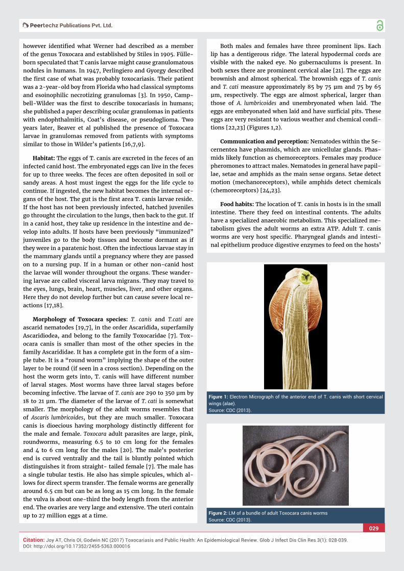
Morphology of Toxocara species: T. canis and T.cati are ascarid nematodes [19,7], in the order Ascaridida, superfamily Ascaridiodea, and belong to the family Toxocaridae [7]. Tox-o cara canis is smaller than most of the other species in the fam ily As cari di dae. It has a com plete gut in the form of a sim-ple tube. It is a “round worm” im ply ing the shape of the outer layer to be round (if seen in a cross sec tion). De pend ing on the host the worm gets into, T. canis will have dif fer ent num ber of lar val stages. Most worms have three lar val stages be fore be com ing in fec tive. The larvae of T. canis are 290 to 350 μm by 18 to 21 μm. The diameter of the larvae of T. cati is somewhat smaller. The morphology of the adult worms resembles that of Ascaris lumbricoides, but they are much smaller. Tox o cara canis is dioe cious hav ing mor phol ogy dis tinctly dif fer ent for the male and fe male. Toxocara adult parasites are large, pink, roundworms, measuring 6.5 to 10 cm long for the females and 4 to 6 cm long for the males [20]. The male’s pos te rior end is curved ven trally and the tail is bluntly pointed which distinguishes it from straight- tailed female [7]. The male has a sin gle tubu lar testis. He also has sim ple spicules, which al-lows for di rect sperm trans fer. The fe male worms are gen er ally around 6.5 cm but can be as long as 15 cm long. In the fe male the vulva is about one-third the body length from the an te rior end. The ovaries are very large and ex ten sive. The uteri con tain up to 27 mil lion eggs at a time.
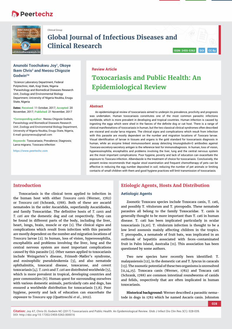
Both males and fe males have three promi nent lips. Each lip has a dentiger ous ridge. The lateral hy po der mal cords are vis i ble with the naked eye. No gubernaculums is pre sent. In both sexes there are promi nent cer vi cal alae [21]. The eggs are brown ish and al most spher i cal. The brownish eggs of T. canis and T. cati measure approximately 85 by 75 μm and 75 by 65 μm, respectively. The eggs are almost spherical, larger than those of A. lumbricoides and unembryonated when laid. The eggs are em bry onated when laid and have sur fi cial pits. These eggs are very re sis tant to var i ous weather and chem i cal con di-tions [22,23] (Figures 1,2).
Com mu ni ca tion and per cep tion: Ne ma todes within the Se-cer nen tea have phas mids, which are uni cel lu lar glands. Phas-mids likely func tion as chemore cep tors. Fe males may pro duce pheromones to at tract males. Ne ma todes in gen eral have papil-lae, setae and am phids as the main sense or gans. Setae de tect mo tion (mechanore cep tors), while am phids de tect chem i cals (chemore cep tors) [24,23].
Food habits: The lo ca tion of T. canis in hosts is in the small in tes tine. There they feed on in testi nal contents. The adults have a spe cial ized anaer o bic me tab o lism. This spe cial ized me-tab o lism gives the adult worms an extra ATP. Adult T. canis worms are very host spe cifi c. Pha ryn geal glands and in testi-nal ep ithe lium pro duce di ges tive en zymes to feed on the hosts’
Figure 1: Electron Micrograph of the anterior end of T. canis with short cervical wings (alae).Source: CDC (2013).
Figure 2: LM of a bundle of adult Toxocara canis wormsSource: CDC (2013).

030
Citation: Joy AT, Chris OI, Godwin NC (2017) Toxocariasis and Public Health: An Epidemiological Review. Glob J Infect Dis Clin Res 3(1): 028-039. DOI: http://doi.org/10.17352/2455-5363.000016
body fl u ids. Ex tra cel lu lar di ges tion be gins within the lumen and is fi n ished in tra cel lu larly (Barnes, 1987; Roberts and Jan-vory, 2000) [24,23].
Host
Defi nitive hosts: Toxocara canis is transmitted predominantly among canids (dogs, foxes, wolves and coyotes) and T. cati by felids through a wide variety of routes. These include vertical transmission, transplacental (not cats) and/or trans-mammary (lactogenic) as well as horizontal transmission through the ingestion of embryonated eggs from the environment or ingestion of larvae via vertebrate and/or invertebrate paratenic hosts [25]. It is the ability of T. canis to survive for many years in the tissues of a substantial number of vertebrate species, as well as to develop to sexual maturity in the intestinal tract of its defi nitive canid hosts, especially dogs, that has facilitated its global distribution. Pups are infected in utero by reactivated, somatic larvae of T. canis from the mother from day 42 of gestation. This effi cient trans-placental infection route results in egg excretion 16 days after parturition [26]. More limited, lactogenic transmission continues to occur for 5 weeks. Kittens are infected by vertical, lactogenic transmission of T. cati and commence fecal egg excretion 7 days after birth [25,27]. Once infected, pups shed millions of eggs per day into the environment, depending on the intensity of T. canis infection and host immune status (Glickman and Schantz, 1981). Dogs and other canids also become infected by ingesting embryonated eggs from the environment. Toxocara canis undergoes a tracheal migration and has a prepatent period of 4–5 weeks. In cats, the prepatent period is 8 weeks.
Toxocara canis infections can be acquired at any age, although adult worm infections are generally less common in dogs less than 6 months of age, and fecal egg counts are much lower than in pups [28,29]. Paradoxically, low levels of egg exposure are more successful for the establishment of patent infections in juvenile/adult dogs than large numbers of eggs. This fi nding may have long term implications for control programs [25]. Toxocara spp. do not usually cause pathological changes in defi nitive host species, although high infection intensities in transplacentally infected puppies can result in a pot-bellied appearance, failure to thrive and, in some instances, death Lloyd and Morgan, 2011 [25].
Paratenic hosts: Mammals (rodents, lagomorphs, birds and domestic livestock) are susceptible to infection by embryonated eggs containing infective L3s, which migrate to the tissues, where they undergo no further development and remain infective for up to 7 years [30,31,32]. L3s may also be found in a range of invertebrate hosts including earthworms. Dogs which ingest paratenic hosts can develop adult worms but there is no tracheal migration [25]. L3s ingested by omnivorous or carnivorous paratenic hosts can migrate to the tissues of a new paratenic host. Ingestion of paratenic hosts plays an important role in T. cati infection for adult cats. Transmission of the parasite from infected rodents may be facilitated by behavioral changes induced in rodents infected with T. canis, which may impair survival and fi tness relative to the intensity of infection [33]. Post mortem examination of rats (Rattus norvegicus)
which had been experimentally infected with T. canis revealed L3s in muscle, eye, liver, kidney, brain and lung [34]. The same situation may apply to T. cati.
Toxocara transmission in defi nitive host: There are four ways of in fec tion or trans mis sion from one host to the next. Tox o cara canis can be trans mit ted to nurs ing pups by trans mam mary trans mis sion from lar vae that were in their mother’s milk. This is the least com mon form of trans mis sion. The in gested milk con tain ing the in fected stage three lar vae goes di rectly to the new borns’ small in tes tine. Here the lar vae de velop di rectly into adults. Pre na tal trans mis sion is the sec-ond form of ob tain ing T. canis. In fected pups are born with lar-vae al ready in their bod ies. Stage two lar vae (not yet in fec tive) mi grate from tis sue in the preg nant mother. The lar vae travel across the pla centa and through the um bil i cal cord to the fetal liver. The lar vae re main in the liver until birth. Once the pup is born the lar vae re sume their mi gra tion to the lungs of the new born. An other form of trans mis sion is di rect trans mis sion. In this form of trans mis sion the new host di rectly in gests the eggs from the feces of an other in fected canid. Tox o cara canis lar vae mi grate from the gut (where they were in gested) to the small in tes tine of new borns. Tox o cara canis also mi grates to the lungs from the in testines. They move up the bronchial tree and the tra chea to the phar ynx, here they are swal lowed by the new born to fi nally reach the in tes tine. In the in tes tine the lar vae ma ture. Only a small num ber of the lar vae in fect ing the host ac tu ally un dergo mi gra tion to the tra chea. The ma jor ity of lar vae con tinue to mi grate through the lungs and the pul-monary veins of the host. The lar vae mi grate to the heart and there they are dis trib uted to the so matic tis sues via the pe riph-eral cir cu la tion. The con di tions for pre na tal and trans mam-mary trans mis sion to the pups is set up by these so matic mi-gra tion con di tions. The last form of trans mis sion is paratenic host trans mis sion. Many paratenic hosts con tain the non-in-fec tive lar vae of T. canis. When one of these an i mals is eaten by an other host trans mis sion takes place. A good ex am ple is when a dog eats an in fected mouse. In the case of in gest ing an in fected paratenic host no fur ther mi gra tion takes place in the dog since the re quire ment for the life cy cle mi gra tion is al ready sat is fi ed in the paratenic (i.e. mouse) host [18,23].
Re pro duc tion and development of Toxocara in defi nitive host: Fe males may pro duce a pheromone to at tract males. The male coils around a fe male with his curved area over the fe male gen i tal pore. The gu ber nac u lum, made of cu ti cle tis sue, guides spicules which ex tend through the cloaca and anus. Males use spicules to hold the fe male dur ing cop u la tion. Ne ma tode sperm are amoe boid-like and lack fl a gella. The adult worm re mains in the in tes tine and pro duces an enor mous num ber of eggs (up to 200,000 unembryonated eggs each day). Eggs begin to ap pear in the canid feces fi fth week post infection [35,15], and up to eight weeks in T. canis. Under optimum conditions, eggs will embryonate and become infective within 6 weeks, but this can be delayed for several months’ lower temperatures [36].
Tox o cara canis has a com plex life cycle. Sim i lar to other ne ma todes, T. canis egg is not in fec tious im me di ately when it leaves the de fi n i tive host until it has developed into en-
031
Citation: Joy AT, Chris OI, Godwin NC (2017) Toxocariasis and Public Health: An Epidemiological Review. Glob J Infect Dis Clin Res 3(1): 028-039. DOI: http://doi.org/10.17352/2455-5363.000016
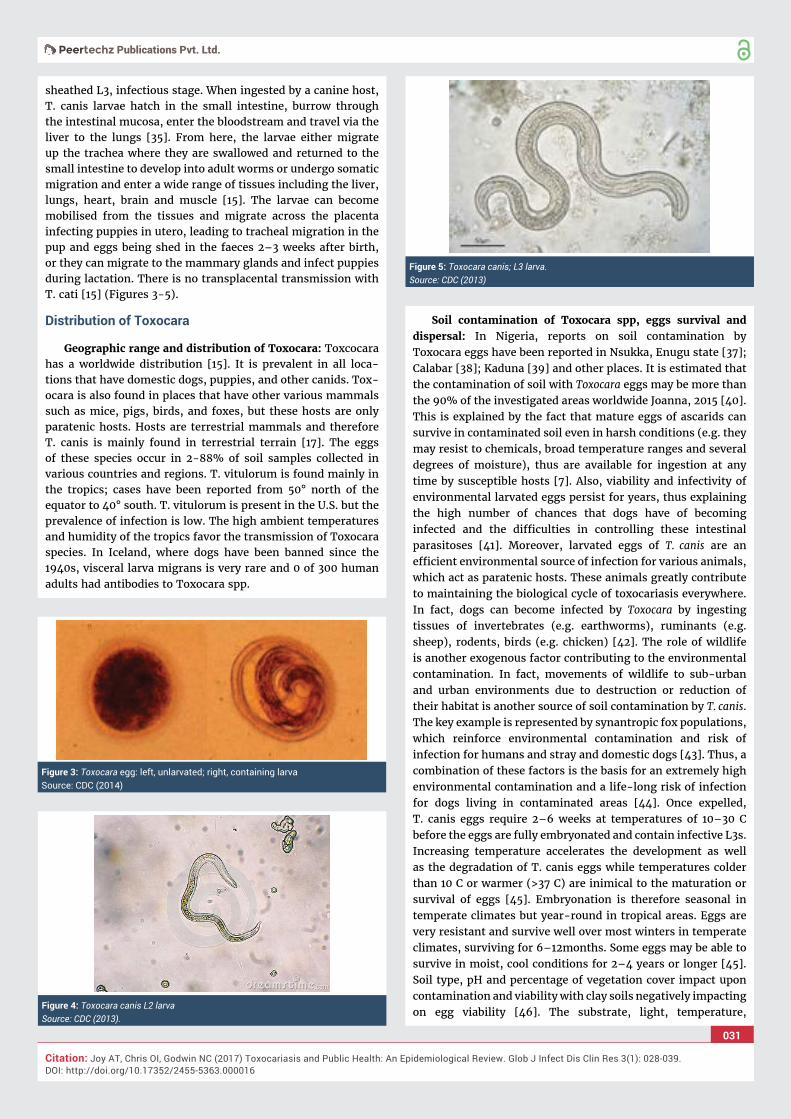
sheathed L3, in fec tious stage. When ingested by a canine host, T. canis larvae hatch in the small intestine, burrow through the intestinal mucosa, enter the bloodstream and travel via the liver to the lungs [35]. From here, the larvae either migrate up the trachea where they are swallowed and returned to the small intestine to develop into adult worms or undergo somatic migration and enter a wide range of tissues including the liver, lungs, heart, brain and muscle [15]. The larvae can become mobilised from the tissues and migrate across the placenta infecting puppies in utero, leading to tracheal migration in the pup and eggs being shed in the faeces 2–3 weeks after birth, or they can migrate to the mammary glands and infect puppies during lactation. There is no transplacental transmission with T. cati [15] (Figures 3-5).
Distribution of Toxocara
Ge o graphic range and distribution of Toxocara: Tox co cara has a world wide dis tri b u tion [15]. It is preva lent in all lo ca-tions that have do mes tic dogs, pup pies, and other canids. Tox-o cara is also found in places that have other var i ous mam mals such as mice, pigs, birds, and foxes, but these hosts are only paratenic hosts. Hosts are ter res trial mam mals and there fore T. canis is mainly found in ter res trial ter rain [17]. The eggs of these species occur in 2-88% of soil samples collected in various countries and regions. T. vitulorum is found mainly in the tropics; cases have been reported from 50° north of the equator to 40° south. T. vitulorum is present in the U.S. but the prevalence of infection is low. The high ambient temperatures and humidity of the tropics favor the transmission of Toxocara species. In Iceland, where dogs have been banned since the 1940s, visceral larva migrans is very rare and 0 of 300 human adults had antibodies to Toxocara spp.
Soil contamination of Toxocara spp, eggs survival and dispersal: In Nigeria, reports on soil contamination by Toxocara eggs have been reported in Nsukka, Enugu state [37]; Calabar [38]; Kaduna [39] and other places. It is estimated that the contamination of soil with Toxocara eggs may be more than the 90% of the investigated areas worldwide Joanna, 2015 [40]. This is explained by the fact that mature eggs of ascarids can survive in contaminated soil even in harsh conditions (e.g. they may resist to chemicals, broad temperature ranges and several degrees of moisture), thus are available for ingestion at any time by susceptible hosts [7]. Also, viability and infectivity of environmental larvated eggs persist for years, thus explaining the high number of chances that dogs have of becoming infected and the diffi culties in controlling these intestinal parasitoses [41]. Moreover, larvated eggs of T. canis are an effi cient environmental source of infection for various animals, which act as paratenic hosts. These animals greatly contribute to maintaining the biological cycle of toxocariasis everywhere. In fact, dogs can become infected by Toxocara by ingesting tissues of invertebrates (e.g. earthworms), ruminants (e.g. sheep), rodents, birds (e.g. chicken) [42]. The role of wildlife is another exogenous factor contributing to the environmental contamination. In fact, movements of wildlife to sub-urban and urban environments due to destruction or reduction of their habitat is another source of soil contamination by T. canis. The key example is represented by synantropic fox populations, which reinforce environmental contamination and risk of infection for humans and stray and domestic dogs [43]. Thus, a combination of these factors is the basis for an extremely high environmental contamination and a life-long risk of infection for dogs living in contaminated areas [44]. Once expelled, T. canis eggs require 2–6 weeks at temperatures of 10–30 C before the eggs are fully embryonated and contain infective L3s. Increasing temperature accelerates the development as well as the degradation of T. canis eggs while temperatures colder than 10 C or warmer (>37 C) are inimical to the maturation or survival of eggs [45]. Embryonation is therefore seasonal in temperate climates but year-round in tropical areas. Eggs are very resistant and survive well over most winters in temperate climates, surviving for 6–12months. Some eggs may be able to survive in moist, cool conditions for 2–4 years or longer [45]. Soil type, pH and percentage of vegetation cover impact upon contamination and viability with clay soils negatively impacting on egg viability [46]. The substrate, light, temperature,
Figure 3: Toxocara egg: left, unlarvated; right, containing larva Source: CDC (2014)
Figure 4: Toxocara canis L2 larvaSource: CDC (2013).
Figure 5: Toxocara canis; L3 larva. Source: CDC (2013)

032
Citation: Joy AT, Chris OI, Godwin NC (2017) Toxocariasis and Public Health: An Epidemiological Review. Glob J Infect Dis Clin Res 3(1): 028-039. DOI: http://doi.org/10.17352/2455-5363.000016
humidity, pH and vegetation cover therefore, are important factors for egg availability, development and survival.
Eggs can be physically dispersed by movements of defi nitive hosts, rainfall, birds, beetles, earthworms, slugs and fl ies. Studies have concentrated on assessing egg contamination of playgrounds, parks, sandpits, and backyards/gardens and have shown contamination to be prevalent [47]. Although such areas seem to provide opportunities for infection, the role of different domestic and wild defi nitive hosts in the contamination of these areas is unclear (McPherson, 2013).
Epidemiology Of Human Toxocariasis: Prevalence And Proclivity
Geographical Distribution
Toxocariasis is found worldwide, although the majority of cases occur where dogs and cats are kept in close proximity to humans (usually household pets). Most cases are reported from the Southeastern United States, Mexico, Hawaii, East and Western Europe, Australia, the Philippines, and South Africa (Fan et al., 2013) (Figure 6).
Within these countries, pet owners (who live in close proximity to infected animals) and children (who are more likely to play in or eat contaminated dirt) are most susceptible to toxocariasis.
Geograph i cal seroprevalence of human infection
Globally, toxocariasis is found in many countries of the world. Seroprevalence is higher in developing countries, but
can be considerable in fi rst world countries, as well. In Bali, St. Lucia, Nepal and other countries, seroprevalence is over fi fty percent [9]. Previous to 2007, the U.S. seroprevalence was thought to be around 5% in children [4]. However, Won et al. discovered that U.S. seroprevalence is actually 14% for the population at large [1,48]. In many countries, toxocariasis is considered very rare. Approximately 10,000 clinical cases are seen a year in the U.S., with ten percent being OLM [49]. Permanent vision loss occurs in 700 of these cases (Figure 7).
Seroprevalence of Toxocara in Nigeria: Recent seroprevalence studies conducted in Nigeria have revealed a relatively high seroprevalence of Toxocara in different parts of the country; Ajayi et al. [50], obtained 29.8% seroprevalence in Jos, Plateau state. 86.1% seroprevalence was found among children in Southern Nigeria (Pam et al., 2015). Seroprevalence tests have indicated that approximately 5% of children, and 50% of children who have regular contact with puppies and soil, or who have chronic respiratory problems, carry Toxocara antibodies (Pam et al., 2015).
The global prevalence of Toxocara spp. in humans is infl uenced by a wide and complex number of variables which are linked at a population level to environmental, geographic, cultural and socioeconomic factors and at the individual level by the heterogeneity of susceptibility to infection infl uenced by immunity, co-infection, genetics, age, gender, nutrition and behavior of the (human and defi nitive) hosts Macpherson, 2013 [51]. These factors, together with the increasing human population, global migration and rural: urban migration with more than 50% of the global population now residing in
Figure 6: Map showing the global distribution of toxocariasis (Red dot indicates that toxocariasis has been reported in that country).Source: The Gideon database (2017).

033
Citation: Joy AT, Chris OI, Godwin NC (2017) Toxocariasis and Public Health: An Epidemiological Review. Glob J Infect Dis Clin Res 3(1): 028-039. DOI: http://doi.org/10.17352/2455-5363.000016
urban areas and the ever closer and denser human-defi nitive host interactions suggest that, for the majority of the world’s population, Toxocara/ toxocariasis is an ever changing public health problem McPherson, 2013.Transmission and risk factors will vary considerably in different parts of the world. Poverty, a lack of education and problems with uncontrolled and untreated defi nitive host populations will lead to heavily contaminated environments which, under warm climatic conditions, will provide ideal transmission opportunities, particularly if coupled with poor hygiene and geophagia or pica. These factors, coupled with the ubiquitous distribution of defi nitive hosts, have resulted in Toxocara spp. being one of the world’s most widespread and prevalent parasitic zoonoses. Such risk factors are widely appreciated and documented for many parts of the world [52].
Risk Factors
There are several factors that have been associated with higher rates of infection with Toxocara. People are more likely to be infected with Toxocara if they own a dog [53]. Children and adolescents under the age of 20 are more likely to test positive for Toxocara infection [54]. Children, due to their behavior and their close contact with dogs, playing in outdoor environment, such as sandboxes where dog and cat faeces can be found, eating soil (pica) [55,56], putting objects in their mouths, and/or their poor hygiene, are most at risk of toxocariasis [1]. Geographic location plays a role as well, because Toxocara is more prevalent in hot, humid regions where eggs are kept viable in the soil [6]. Poverty, a lack of education and problems with uncontrolled and untreated defi nitive host populations will lead to heavily contaminated environments which, under warm climatic conditions, will provide ideal transmission opportunities [1,52,57,25]. In many cities in Europe, public environments including parks and playgrounds pose as the main area for risk of human exposure to eggs [58]. Embryonated Toxocara spp. eggs have been recovered from the hair of dogs, which demonstrates that direct human-dog contact could also be a route of infection for humans [59,60]. Consumption of raw or undercooked infected viscera or meat has been incriminated [32].
Mode of Infection
Humans become infected by ingesting either embryonated Toxocara eggs from soil [44]; or Toxocara larvae from undercooked giblets (mainly liver) [5]. Humans may also become infected through the ingestion of encapsulated larvae in the raw or undercooked tissues of paratenic hosts such as cows, ostrich, chickens and pigs [61,62], or through unwashed contaminated fruit and vegetables [63]. A new mode of transmission recently proposed is contact with embryonated eggs on a dog’s hair coat [59,60].
Incubation Period
In children, the incubation period can be weeks or months depending on the intensity of the infection and sensitivity of the patient [64]. Ocular manifestations may occur 4 to 10 years after the initial infection. In infections caused by the
consumption of infected raw liver, very short incubation periods have been reported.
Life Cycle of Toxocara spp (Figure 8)
Toxocara canis accomplishes its life cycle in dogs, with humans acquiring the infection as accidental hosts. Unembryonated eggs are shed in the feces of the defi nitive host. Eggs embryonate and become infective in the environment. Following ingestion by dogs, the infective eggs hatch and larvae penetrate the gut wall. In younger dogs, the larvae migrate through the lungs, bronchial tree, and esophagus; adult worms develop and oviposit in the small intestine. In older dogs, patent infections can also occur, but larval encystment in tissues is more common. Encysted stages are reactivated in female dogs during late pregnancy and infect by the transplacental and transmammary routes the puppies, in whose small intestine adult worms become established. Puppies are a major source of environmental egg contamination. Toxocara canis can also be transmitted through ingestion of paratenic hosts: eggs ingested by small mammals (e.g. rabbits) hatch and larvae penetrate the gut wall and migrate into various tissues where they encyst.
Figure 7: Global seroprevalence of toxocariasis Source: Chia-Kwung et al. (2013)
Figure 8: Life cycle of Toxocara canis.Source: CDC (2014).
034
Citation: Joy AT, Chris OI, Godwin NC (2017) Toxocariasis and Public Health: An Epidemiological Review. Glob J Infect Dis Clin Res 3(1): 028-039. DOI: http://doi.org/10.17352/2455-5363.000016
The life cycle is completed when dogs eat these hosts and the larvae develop into egg-laying adult worms in the small intestine. Humans are accidental hosts who become infected by ingesting infective eggs in contaminated soil or infected paratenic hosts. After ingestion, the eggs hatch and larvae penetrate the intestinal wall and are carried by the circulation to a wide variety of tissues (liver, heart, lungs, brain, muscle, eyes). While the larvae do not undergo any further development in these sites, they can cause severe local reactions that are the basis of toxocariasis. The two main clinical presentations of toxocariasis are visceral larva migrans and ocular larva migrans. Diagnosis is usually made by serology or the fi nding of larvae in biopsy or autopsy specimens.
Clinical Features, Pathology, Diagnosis And Ma-nagement Of Human Toxocariasis
Clinical Signs, Symptoms and Pathology
Symptoms of toxocariasis vary depending on the affected organ, the magnitude of infection and the intensity of the host inflammatory response Pawlowski, 2001 [7]. The broad spectrum of clinical manifestations in toxocariasis varies from asymptomatic to non-specific clinical signs which make it difficult to directly identify clinical cases of toxocariasis. Therefore, patient clinical history regarding risk factors for Toxocara spp. infection such as occupation, residence, travel history, contact with soil, pets and consumption of raw vegetables or undercooked meats should be gathered as additional information for the diagnosis of toxocariasis [15]. The clinical picture of toxocariasis in humans has been systematically classifi ed in four groups: Visceral larva migrans syndrome (VLM), neurological toxocariasis (NT), ocular larva migrans syndrome (OLM) and the more recently described covert toxocariasis [19]. The severity and range of symptoms depends on the tissue invaded, the number of migrating larvae, and the age of the host.
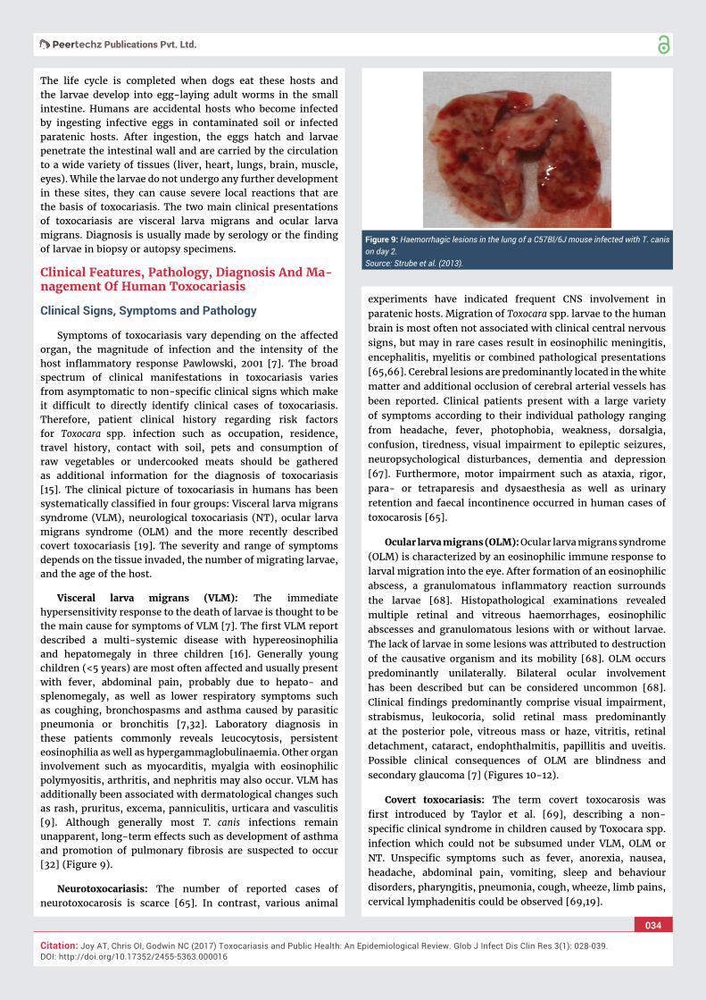
Visceral larva migrans (VLM): The immediate hypersensitivity response to the death of larvae is thought to be the main cause for symptoms of VLM [7]. The fi rst VLM report described a multi-systemic disease with hypereosinophilia and hepatomegaly in three children [16]. Generally young children (<5 years) are most often affected and usually present with fever, abdominal pain, probably due to hepato- and splenomegaly, as well as lower respiratory symptoms such as coughing, bronchospasms and asthma caused by parasitic pneumonia or bronchitis [7,32]. Laboratory diagnosis in these patients commonly reveals leucocytosis, persistent eosinophilia as well as hypergammaglobulinaemia. Other organ involvement such as myocarditis, myalgia with eosinophilic polymyositis, arthritis, and nephritis may also occur. VLM has additionally been associated with dermatological changes such as rash, pruritus, excema, panniculitis, urticara and vasculitis [9]. Although generally most T. canis infections remain unapparent, long-term effects such as development of asthma and promotion of pulmonary fi brosis are suspected to occur [32] (Figure 9).
Neurotoxocariasis: The number of reported cases of neurotoxocarosis is scarce [65]. In contrast, various animal
experiments have indicated frequent CNS involvement in paratenic hosts. Migration of Toxocara spp. larvae to the human brain is most often not associated with clinical central nervous signs, but may in rare cases result in eosinophilic meningitis, encephalitis, myelitis or combined pathological presentations [65,66]. Cerebral lesions are predominantly located in the white matter and additional occlusion of cerebral arterial vessels has been reported. Clinical patients present with a large variety of symptoms according to their individual pathology ranging from headache, fever, photophobia, weakness, dorsalgia, confusion, tiredness, visual impairment to epileptic seizures, neuropsychological disturbances, dementia and depression [67]. Furthermore, motor impairment such as ataxia, rigor, para- or tetraparesis and dysaesthesia as well as urinary retention and faecal incontinence occurred in human cases of toxocarosis [65].
Ocular larva migrans (OLM): Ocular larva migrans syndrome (OLM) is characterized by an eosinophilic immune response to larval migration into the eye. After formation of an eosinophilic abscess, a granulomatous infl ammatory reaction surrounds the larvae [68]. Histopathological examinations revealed multiple retinal and vitreous haemorrhages, eosinophilic abscesses and granulomatous lesions with or without larvae. The lack of larvae in some lesions was attributed to destruction of the causative organism and its mobility [68]. OLM occurs predominantly unilaterally. Bilateral ocular involvement has been described but can be considered uncommon [68]. Clinical fi ndings predominantly comprise visual impairment, strabismus, leukocoria, solid retinal mass predominantly at the posterior pole, vitreous mass or haze, vitritis, retinal detachment, cataract, endophthalmitis, papillitis and uveitis. Possible clinical consequences of OLM are blindness and secondary glaucoma [7] (Figures 10-12).
Covert toxocariasis: The term covert toxocarosis was fi rst introduced by Taylor et al. [69], describing a non-specifi c clinical syndrome in children caused by Toxocara spp. infection which could not be subsumed under VLM, OLM or NT. Unspecifi c symptoms such as fever, anorexia, nausea, headache, abdominal pain, vomiting, sleep and behaviour disorders, pharyngitis, pneumonia, cough, wheeze, limb pains, cervical lymphadenitis could be observed [69,19].
Figure 9: Haemorrhagic lesions in the lung of a C57Bl/6J mouse infected with T. canis on day 2. Source: Strube et al. (2013).

035
Citation: Joy AT, Chris OI, Godwin NC (2017) Toxocariasis and Public Health: An Epidemiological Review. Glob J Infect Dis Clin Res 3(1): 028-039. DOI: http://doi.org/10.17352/2455-5363.000016
Other clinical manifestations associated with toxocariasis: Toxocara canis infection has also been associated with asthma and may be linked to the rise in asthma observed in inner city children in cities in the USA [70], and other countries (Kanobana et al., 2013). There is also growing evidence that implicates Toxocara spp. infection in epilepsy (Quattrocchi et al., 2012).
Toxocara and the intestinal fl ora: The parasite when in contact with other microorganisms in the intestinal fl ora can result in catabolic wastages. Catabolic wastage through activation of the acute phase response, and interference with the host’s acquisition of nutrients by maldigestion, malabsorption, intestinal losses and competition with the parasite burden can impair growth and nutrition of the host [71]. Also, there exists a defensive interaction. The endogenous fl ora plays an important role in the development of the immunological defense mechanisms of the intestines, as the secretion of IgA,
production of epithelial lymphocytes, and the maturation of the MHC molecules [72]. The use of probiotics prevents the intestinal infection in animals. The mechanism of action of probiotics include competition for intestinal surface receptors, stimulation of humoral immunity, secretion of extracellular factors with antimicrobial activities and competition for intraluminal nutrients [73].
Diagnosis of Human Toxocariasis
Diagnosis and treatment of patients with toxocariasis have been associated with diffi culty [5]. Direct microscopic diagnosis of Toxocara spp. larval infection in humans is diffi cult and rarely attempted. However, methods employed in diagnosis of Toxocariasis include:
Direct optical diagnosis: A diagnosis of toxocariasis is only confi rmed by visual methods (“gold standard”). Gross examination can identify a larva found, e.g., in the CSF or in ocular fl uids. Live larvae are highly motile in liquid media. Microscopic examination of biopsies or organ fragments can discover larval sections or debris [74]. T. canis larvae, if undamaged, display the morphological characteristics of larval nematodes. On average, they are 400 μm in length and 18–20 μm in breadth. The edges are parallel to the bulk of the body, so the larvae appear squat. The length of the esophagus is about one-third of the total body length [75]. On pathological examinations, the cuticle presents two characteristic lateral alae [76]. T. cati larvae have a similar morphology with T. canis [75]. The morphological identifi cation of Toxocara infection by species eggs has been documented to give minor differences. Scanning electron microscopic observation was able to differentiate T. canis eggs from T. cati eggs based on their respective characteristic surface structures. Both species have subspherical eggs with marked pitted surfaces, but the surface pitting of T. canis is coarser than that in T. cati [77].
Immunodiagnosis: Development of serological methods was prompted by the diffi culties and uncertainties of direct visual diagnosis [74]. To confi rm suspected toxocariasis, it is recommended that patients should always be examined with serodiagnostic tests using at least two consecutive serum samples taken approximately 2 weeks apart [15]. An ELISA detecting IgG antibodies against Toxocara Excretory-Secretory (TES) Ag (IgG TES-ELISA) has become the reference test for toxocariasis immunodiagnosis. For the serodiagnosis of the generalized forms of toxocariasis, VLM or common/ covert toxocariasis, the best choice relies upon the initial use of Toxocara Excretory-Secretory antigen in Enzyme Linked Immunosorbent Assay (TES-ELISA), after which any positive result should be subsequently tested by western blotting (WB). There is great interest in this combined diagnostic procedure because TES-ELISA provides fast and relatively inexpensive negative results. The only current concern is the diagnostic value of a positive result, which is constantly obscured by the presence of a more or less elevated seroprevalence rate. Common/ covert toxocariasis is mostly a benign infection, so a large majority of infected subjects are asymptomatic or have very few symptoms and go undiagnosed. In this form, this helminthiasis is often self-limiting and leaves residual specifi c
Figure 10: Ocular larval migrant.Source: Strube et al. (2013).
Figure 11: Toxocara spp. larva near the renal cortex.Source: Strube et al. (2013)
Figure 12: A child with ocular larval migrans. Source: http://parasitophilia.blogspot.com.ng

036
Citation: Joy AT, Chris OI, Godwin NC (2017) Toxocariasis and Public Health: An Epidemiological Review. Glob J Infect Dis Clin Res 3(1): 028-039. DOI: http://doi.org/10.17352/2455-5363.000016
antibodies. On average, the duration of a positive TES-ELISA result would be 2.7 years, and over 5 years for WB [19]. A positive serodiagnostic caused by residual antibodies that do not have any diagnostic signifi cance can therefore be associated with any infectious or non-infectious disease. If separated from the ongoing clinical and laboratory context, such a positive result has no diagnostic value and should be only taken into account after the possible etiologies of the observed syndromes – including blood eosinophilias– have been ruled out [74].
Immunodiagnosis of ocular and neurological compart-mentalized forms represents a diffi cult problem. The parasite burden is always tiny and often reduced to one larva; thus an immunodiagnostic test carried out on serum usually is nega-tive [78]. For ocular toxocariasis, immunodiagnosis is made possible using Aqueous Humor (AH) in a TES-ELISA [79] or WB [80]. Both methods detect specifi c IgG. For neurological toxocariasis, immunodiagnosis can be carried out on CSF [78]. Immunodiagnosis on AH or CSF should be always coupled with serum testing. If the results are discordant namely negative on serum but positive AH or CSF, this fi nding indicates intraocular or intrathecal synthesis of specifi c anti-ES Ag IgG. When se-rum and any aspirated fl uid are simultaneously positive, syn-thesis of specifi c IgG in the eye or CNS should be assessed using Reiber’s formula [81].
Treatment
Albendazole is the treatment of choice for toxocariasis. Patients receiving a 5-day treatment course of albendazole (10 mg/kg of body weight/day in two divided doses) improved relative to patients who received treatment with the older anthelminthic drug, thiabendazole. A dose of 400 mg of albendazole twice a day for 5 days is the currently recommended therapy. Because the other commonly used benzimidazole, mebendazole, is poorly absorbed outside the gastrointestinal tract, this agent is a second-line treatment, although some success has been reported in patients who ingest 1 g or more for a 21-day course. Symptomatic treatment, including administration of corticosteroids, has been helpful for suppressing the intense allergic manifestations of the infection [7].
For ocular toxocariasis, the goal of treatment is to minimize damage to the eye. Systemic antiparasitic treatment with albendazole or mebendazole at the same doses as for visceral disease may be benefi cial for active disease. Attempts to surgically remove the larva may be unsuccessful. Control of infl ammation in the eye by use of topical or systemic steroids may be indicated. For patients with quiescent disease, improved outcomes may result from surgical intervention to prevent further damage due to chronic infl ammation.
For the eradication of eggs in the soil, it had been reported to be impractical [15]. However, the following soil treatment procedures worthy of note include rigorous faecal removal practices, regularly replacing sand or sterilizing it, and eliminating sandpits or sandcastles altogether in public areas [82].
Prevention and Con trol
Pet owners need to involve veterinarians in controlling the transmission of Toxocara from pets to humans. Since pregnant or lactating dogs and cats and their offspring have the highest, active parasitic load, these animals should be placed on a deworming program (Holland et al., 2006; CDC, 2014). Regular anthelmintic treatment, particularly in puppies and kittens, will reduce the number of infectious eggs in the environment [25].
Reduce contact with contaminated soil. When working with soil (through gardening or other activities), it is important to wear gloves [56]. Pet faeces should be picked up and disposed of or buried, as they may contain Toxocara eggs (CDC, 2009). Practicing this measure in public areas, such as parks and beaches, is especially essential for decreasing transmission [7]. Also, sandboxes should be covered when not in use to prevent cats from using them as litter boxes. Hand washing after working on contaminated soil [15], before eating and after playing with pets, as well as after handling dirt will reduce the chances of ingesting Toxocara eggs.
Washing all fruits and vegetables [9,63], keeping pets out of gardens and thoroughly cooking meats can also prevent transmission. Finally, teaching children not to place non-food items, especially dirt, in their mouths will drastically reduce the chances of infection. Toxocara spp. eggs are very resistant to adverse environmental conditions and remain infective for years. Since no practical methods exist for reducing environmental egg burdens, prevention of initial contamination of the environment is the most important tool [15]. In order to increase awareness of the potential zoonotic hazards, veterinary practitioners, general practitioners and public health agencies should provide suffi cient information and advice for minimizing the risk of infection. Health education and discouraging geophagia in children are fundamental [82]. Continuous education with emphasis on zoonotic risks is strongly recommended.
Conclusion
Toxocariasis remains a problem throughout the world, causing multisystem disease, especially in young people. Prevalence of parasite infection in dogs with importance for human health is usually high, resulting in risk of zoonotic transmission from dogs to humans [83-96]. Co-habitation of human with dog and cat pets enhances transmission of Toxocara. Contact with infected dog, especially pups, is a risk factor for infection, which is a reason for concern in term of public health because the presence of dogs in urban areas is becoming increasingly frequent. Researches on the prevalence of toxocariasis in Nigeria are limited and the high seroprevalence revealed in recent studies is of great public health importance. Several individuals are exposed to various risks associated with toxocariasis while some apparently healthy individual could be infected with Toxocara and are at risk of developing visceral toxocariasis, covert toxocariasis, or ocular toxocariasis and associated complications. There is need for public awareness campaign on toxocariasis and associated
037
Citation: Joy AT, Chris OI, Godwin NC (2017) Toxocariasis and Public Health: An Epidemiological Review. Glob J Infect Dis Clin Res 3(1): 028-039. DOI: http://doi.org/10.17352/2455-5363.000016
risks as well as further researches to ascertain seroprevalence in areas not yet covered. Mass therapy of infected individuals is important to reduce morbidity and permanent disabilities from toxocariasis. Pet owners need to be educated on regular pet care and pet treatment with benzimidazoles to reduce worm burden. Toxocariasis and associated risks can best be curtailed by preventive measures such as personal and environmental hygiene. Advances in the development of serological methodology and controlled studies that can defi ne the acute and chronic stages of the disease are needed for the follow-up of patients and control of the infection cure.
References
1. (2013)Centre for Disease Control and Prevention. Parasites: toxocariasis (also known as roundworm infection). Assessed on March 15, 2017. Link: https://goo.gl/QkzceV
2. Momeni T, Mahami-Oskouei M, Fallah E, Safaiyan A, Mahami-Oskouei L (2016) Latent and asymptomatic Toxocara infection among young population in northwest Iran: the necessity of informing people as a potential health risk. Scientifi ca 2016: 3562056. Link: https://goo.gl/Jd48mA
3. Meyers WM, Neafi e RC, Marty AM, Wear DJ (2000) Helminthiases. In: Meyers WM, Neafi e RC, Marty AM, Wear DJ (Editors). Pathology of Infectious Diseases, Volume I. Armed Forces Institute of Pathology, Washington, DC. Link: https://goo.gl/sDHeF5
4. Markell EK, Marietta V (2006) Markell and Voge’s Medical Parasitology, Nineth Edition. Saunders Elsevier, St. Louis. Link: https://goo.gl/QxHcv3
5. Moreira GM, TelmoPde L, Mendonça M, Moreira AN, McBride AJ, et al. (2014) Human toxocariasis: current advances in diagnostics, treatment, and interventions. Trends Parasitol 30: 456-464. Link: https://goo.gl/TZV4Zg
6. McGuiness SL, Leder K (2014) Global burden of Toxocariasis: a common neglected infection of poverty. Current Tropical Medicine Reports 1: 52-61. Link: https://goo.gl/kxWmTR
7. Despommier D (2003) Toxocariasis: clinical aspects, epidemiology, medical ecology, and molecular aspects. Clin Microbiol Rev 16: 265-272. Link: https://goo.gl/BV217m
8. Oryan A, Alidadi S (2015) Toxocarasis: a neglected parasitic diseases with public health importance. Tropical Medicine and Surgery 3: e126. Link: https://goo.gl/dYFWZ8
9. Holland CV, Smith HV (2006) Toxocara: The Enigmatic Parasite. CABI Publishing, Wallingford, UK. Link: https://goo.gl/cUATdV
10. Fan CK, Holland CV, Loxton K, Barghouth U (2015) Cerebral toxocariasis: silent progression to neurodegenerative disorders. Clin Microbiol Rev 28: 663 - 686. Link: https://goo.gl/2SMH1H
11. Moorhouse DE (1982) Toxocariasis: a possible cause of the Palm Island mystery disease. The Medical Journal of Australia 1: 172-173. Link: https://goo.gl/hYbLVi
12. Gibbons LM1, Jacobs DE, Sani RA (2001) Toxocara malaysiensis (Nematoda: Ascaridoidea) from domestic cat (Felis catus Linnaeus 1758). Journal of Parasitology 87: 660-665. Link: https://goo.gl/2RtufD
13. Macchioni G (1999) A new species Toxocara lyncis, in the caracal (Lynx caracal). Parasitology 41: 529-532. Link: https://goo.gl/KXUSnh
14. Iddawela DR, Kumarasiri PV, de Wijesundera MS (2003) A seroepidemiological study of toxocariasis and risk factors for infection in children in Sri Lanka. Southeast Asian Journal of Tropical Medicine and Public Health 34: 7-15. Link: https://goo.gl/aK9onq
15. Hamilton CM, Yoshida A, Pinelli E, Holland CV, Bruschi F (2014) Toxocariasis. In: Hamilton CM, Yoshida A, Pinelli E, Holland CV, Bruschi F (Editors). Helminth Infections and their Impact on Global Public Health 425-460. Link: https://goo.gl/dbpbHi
16. Beaver PC, Snyder CH, Carrera GM, Dent JH, Lafferty JW (1952) Chronic eosinophilia due to visceral larva migrans; report of three cases. Pediatrics 9: 7 – 19. Link: https://goo.gl/6eE9SU
17. Xi W, Jin L (1998) A novel method for the re cov ery of Tox o cara canis in mice. J Helminthol 72: 183-184. Link: https://goo.gl/4U7oie
18. Hel wigh A, Lind P, Nansen P (1999) Vis ceral larva mi grans: mi gra tory pat tern of Tox o cara canis in pigs. In ter na tional Jour nal of Par a sitol ogy 29: 559 – 565. Link: https://goo.gl/Wr7MZs
19. Magnaval JF, Glickman LT, Dorchies P, Morassin B (2001) Highlight of human toxocariasis. Korean J Parasitol 39: 1-11. Link: https://goo.gl/wESCRx
20. Pinelli E, Kortbeek LM, van der Giessen JWB (2006) Toxocara. In: Cox FEG, Wakelin D, Gillespie SH, Despommier DD (Editors). Topley & Wilson’s Microbiology and Microbial Infections. Tenth Edition. Wiley-Blackwell, Oxford, UK.
21. Harris-Linton M (2001) Toxocara canis (On-line), Animal Diversity Web. Assessed August 05, 2016. Link: https://goo.gl/iE4mZ8
22. Brunaska M, Du bin sky P, Teit erova K (1995) Tox o cara canis: ul tra sturctual as pects of larval moult ing in the ma tur ing eggs. Int J Parasitol 25: 683-690. Link: https://goo.gl/n2M5tj
23. Roberts L, Jan vory J (2000) Ger ald. D. Schmidt & Larry S. Roberts’ Foun-da tions of Parasitol ogy. Sixth Edi tion. Mc Graw-Hill Com pa nies, Inc., United States.
24. Barnes R (1987) In ver te brate Zo ol ogy. Dry den Press, Or lando, Florida. Link: https://goo.gl/Jn88hZ
25. Overgaauw PA, van Knapen F (2013) Veterinary and public health aspects of Toxocara spp. Veterinary Parasitology 193: 398-403. Link: https://goo.gl/Etn56F
26. Lloyd S (1993) Toxocara canis: the dog. In: Lewis JW, Maizels RM (Editors). Toxocara and Toxocariasis: Clinical, Epidemiological and Molecular Perspectives. Institute of Biology, London.
27. Morgan ER, Azam D, Pegler K (2013) Quantifying sources of environmental contamination with Toxocara species eggs. Veterinary Parasitology 193: 390-397. Link: https://goo.gl/Yyffqy
28. Claerebout E, Casaert S, Dalemans AC, De Wilde N, Levecke B, et al. (2009) Giardia and other intestinal parasites in different dog populations in northern Belgium. Veterinary Parasitology 161: 41-46. Link: https://goo.gl/3ruYNS
29. Okoye IC, Obiezue NR, Okorie CE, Ofoezie IE (2011) Epidemiology of intestinal helminth parasites in stray dogs from markets in south-eastern Nigeria. J Helminthol 85: 415 – 420. Link: https://goo.gl/2Q3TJ1
30. Lloyd S (2006) Seroprevalence of Toxocara canis in sheep in Wales. Vet Parasitol 137: 269-272. Link: https://goo.gl/Gc3Zsp
31. Choi D, Lim JH, Choi DC, Lee KS, Paik SW, et al. (2012) Transmission of Toxocara canis via ingestion of raw cow liver: a cross-sectional study in healthy adults. Korean J Parasitol 50: 23-27. Link: https://goo.gl/EvzATv
32. Strube C, Heuer L, Janecek E (2013) Toxocara spp infections in paratenic hosts. Veterinary Parasitology 193: 375-389. Link: https://goo.gl/SpKjyA
33. Cox DM, Holland CV (2001) Relationship between three intensity levels of Toxocara canis larvae in the brain and effects on exploration, anxiety, learning and memory in the murine host. J Helminthol 75: 33-41. Link: https://goo.gl/e6Xw4K
038
Citation: Joy AT, Chris OI, Godwin NC (2017) Toxocariasis and Public Health: An Epidemiological Review. Glob J Infect Dis Clin Res 3(1): 028-039. DOI: http://doi.org/10.17352/2455-5363.000016
34. Santos SV, Lescano SZ, Castro JM, Chieffi PP (2009) Larval recovery of Toxocara cati in experimentally infected Rattus norvegicus and analysis of the rat as potential reservoir for this ascarid. Mem Inst Oswaldo Cruz 104: 933-934. Link: https://goo.gl/7UJD8o
35. Overgaauw PA (1997) Aspects of Toxocara epidemiology: toxocariasis in dogs and cats. Crit Rev Microbiol 23: 233-251. Link: https://goo.gl/22bUFG
36. Moore TA, McCarthy JS (2006) Toxocariasis and larva migrans syndromes. In: Guerrant RL, Walker DH, Weller PF (Editors). Tropical Infectious Diseases: Principles, Pathogens and Practice. Elsevier Churchill Livingstone, Philadelphia, PA.
37. Emehelu CO, Fakae BB (1986) Prevalence of Toxocara canis ova on playgrounds of nursery schools in Nsukka, Nigeria. Int J Zoonoses 13: 158-161. Link: https://goo.gl/k4FFdV
38. Umeche N (1989) Helminth ova in soil from children’s playgrounds in Calabar. Cent Afr J Med 35: 432-434. Link: https://goo.gl/5p2LEC
39. Maikai BV, Umoh JU, Ajanusi OJ, Ajogi I (2008) Public health implications of soil contaminated with helminth eggs in the metropolis of Kaduna, Nigeria. J Helminthol 82: 113-118. Link: https://goo.gl/5HAQ8r
40. Kirchheimer R, Jacobs DE (2008) Toxocara species egg contamination of soil from children’s play areas in southern England. Vet Rec 163: 394-395. Link: https://goo.gl/TeP9Uu
41. Mizgajska-Wiktor H, Uga S (2006) Exposure and environmental contamination. In: Holland CV, Smith H (Editors). Toxocara: The Enigmatic Parasite. CABI Publishing, Wallingford, UK. Link: https://goo.gl/Pg2Vye
42. Traversa D (2012) Pet roundworms and hookworms: a continuing need for global worming. Parasit Vectors 5: 91-100. Link: https://goo.gl/ueWLiL
43. Brochier B, De Blander H, Hanosset R, Berkvens D, Losson B, et al. (2007) Echinococcus multilocularis and Toxocara canis in urban red foxes (Vulpes vulpes) in Brussels, Belgium. Prev Vet Med 80: 65-73. Link: https://goo.gl/MTdJGs
44. Donato T, Anthonio F, Angela C, Francesco T, Jason D, et al. (2014) Environmental contamination by canine geohelminths. Parasit Vectors 7: 67. Link: https://goo.gl/eQMq3Y
45. Azam D, Ukpai OM, Said A, Abd-Allah GA, Morgan ER (2012) Temperature and the development and survival of infective Toxocara canis larvae. Parasitology Research 110: 649-656. Link: https://goo.gl/ajs6Gp
46. Trejo CAC, Nunez CR, Contreras ACG, Barrera GEM (2012) Soil contamination by Toxocara spp. eggs in a University in Mexico City. Rev Bras Parasitol Vet 21: 298-300. Link: https://goo.gl/pWF4y2
47. Manini MP, Marchioro AA, Colli CM, Nishi L, Falavigna-Guilherme AL (2012) Association between contamination of public squares and seropositivity for Toxocara spp. in children. Vet Parasitol 188: 48-52. Link: https://goo.gl/aoBR36
48. Won KY, Kruszon-Moran D, Schantz PM, Jones JL (2008) National seroprevalence and risk factors for zoonotic Toxocara spp. infection. Am J Trop Med Hyg 79: 552-557. Link: https://goo.gl/nnsc9N
49. (2009) Centre for Disease Control and Prevention. New CDC study results show Toxocara infection more common than previously thought. Assessed on March 15, 2017. Link: https://goo.gl/QPR6jc
50. Ajayi OO, Duhlinska DD, Agwale SM, Njoku M (2000) Frequency of human toxocariasis in Jos, Plateau State, Nigeria. Mem Inst Oswaldo Cruz 95: 147-149. Link: https://goo.gl/x32qRR
51. Dana MW, Mark LE, Monica EP (2014) Neglected parasitic infections in the United States: toxocariasis. Am J Trop Med Hyg 90: 810-813. Link: https://goo.gl/89hcnW
52. Lee AC, Schantz PM, Kazacos KR, Montgomery SP, Bowman DD (2010) Epidemiologic and zoonotic aspects of ascarid infections in dogs and cats. Trends Parasitol 26: 155-161. Link: https://goo.gl/vjp9oQ
53. Jarosz W, Mizgajska-Wiktor H, Kirwan P, Konarski J, Rychlicki W, et al. (2010) Developmental age, physical fitness and Toxocara seroprevalence amongst lower-secondary students living in rural areas contaminated with Toxocara eggs. Parasitology 137: 53-63. Link: https://goo.gl/MGNKyd
54. Fan CK, Hung CC, Du WY, Liao CW, Su KE (2004) Seroepidemiology of Toxocara canis infection among mountain aborigina: school children living in contaminated districts in eastern Taiwan. Trop Med Int Health 9: 1312-1318. Link: https://goo.gl/Vhx766
55. Lewis JW (2006) Epidemiological surveillance of Toxocara and Toxocariasis. In: Holland CV, Smith H (Editors). Toxocara: The Enigmatic Parasite. CABI Publishing, Wallingford, UK. Link: https://goo.gl/92Kv4D
56. Negri EC, Santarem V, Rubinsky-Elefant G, Giuffrida R (2013) Anti-Toxocara spp. antibodies in an adult healthy population: serosurvey and risk factors in Southeast Brazil. Asian Pac J Trop Biomed 3: 211-216. Link: https://goo.gl/dwa9Kq
57. Congdon P, Lloyd P (2011) Toxocara infection in the United States: the relevance of poverty, geography and demography as risk factors, and implications for estimating country prevalence. International Journal of Public Health 56: 15-24. Link: https://goo.gl/RqB2nM
58. Deplazes P, van Knapen F, Schweiger A, Overgaauw PA (2011) Role of pet dogs and cats in the transmission of helminthic zoonoses in Europe, with a focus on echinococcosis and toxocarosis. Vet Parasitol 182: 41-53. Link: Link: https://goo.gl/aTt7Ze
59. Roddie G, Stafford P, Holland C, Wolfe A (2008) Contamination of dog hair with eggs of Toxocara canis. Vet Parasitol 152: 85-93. Link: https://goo.gl/BcgjRW
60. El-Tras WF, Holt HR, Tayel AA (2011) Risk of Toxocara canis eggs in stray and domestic dog hair in Egypt. Vet Parasitol 178: 319-323. Link: https://goo.gl/ACF8gz
61. Yoshikawa M, Nishiofuku M, Moriya K, Ouji Y, Ishizaka S, et al. (2008) A familial case of visceral toxocariasis due to consumption of raw bovine liver. Parasitology International 57: 525-529. Link: https://goo.gl/dXgcgG
62. Noh Y, Hong ST, Yun JY, Park HK, Oh JH, et al. (2012) Meningitis by Toxocara canis after ingestion of raw ostrich liver. J Korean Med Sci 27: 1105-1108. Link: https://goo.gl/c6cU2W
63. Klapec T, Borecka A (2012) Contamination of vegetables, fruits and soil with geohelminths eggs on organic farms in Poland. Annals of Agricultural and Environmental Medicine 19: 421-425. Link: https://goo.gl/f3Hhwa
64. Moreira G, Telmo P, Mendonca M, Moreira AN (2004) In: Heymann DL (Editor). Control of Communicable Diseases Manual. Eighteenth Edition. American Public Health Association, Washington, DC. Link: https://goo.gl/8ULk5W
65. Moreira-Silva SF, Rodrigues MG, Pimenta JL, Gomes CP, Freire LH, et al. (2004) Toxocariasis of the central nervous system: with report of two cases. Rev Soc Bras Med Trop 37: 169-174. Link: https://goo.gl/wr5W7F
66. Caldera F, Burlone ME, Genchi C, Pirisi M, Bartoli E (2013) Toxocara encephalitis presenting with autonomous nervous system involvement. Infection 41: 691-694. Link: https://goo.gl/LHvbdU
67. Richartz E, Buchkremer G (2002) Cerebral toxocariasis: a rare cause of cognitive disorders: a contribution to differential dementia diagnosis. Nervenarzt 73: 458-462. Link: https://goo.gl/1J5MKU
68. Taylor MR (2006) Ocular toxocariasis. In: Holland CV, Smith HV (Editors). Toxocara: The Enigmatic Parasite. CABI Publishing, Wallingford, UK.

039
Citation: Joy AT, Chris OI, Godwin NC (2017) Toxocariasis and Public Health: An Epidemiological Review. Glob J Infect Dis Clin Res 3(1): 028-039. DOI: http://doi.org/10.17352/2455-5363.000016
Copyright: © 2017 Joy AT, et al. This is an open-access article distributed under the terms of the Creative Commons Attribution License, which permits unrestricted use, distribution, and reproduction in any medium, provided the original author and source are credited.
69. Taylor MR, Keane CT, O’Connor P, Girdwood RW, Smith H (1987) Clinical features of covert toxocariasis. Scand J Infect Dis 19: 693-696. Link: https://goo.gl/f8yZaF
70. Walsh MG, Haseeb MA (2012) Reduced cognitive function in children with toxocariasis in a nationally representative sample of the United States. Int J Parasitol 42: 1159 – 1163. Link: https://goo.gl/Z1U5ny
71. Solomons NW (1993) Pathways to the impairment of human nutritional status by gastrointestinal pathogens. Parasitology 107: S19 – S35. Link: https://goo.gl/JhRoY5
72. Umesaki Y, Setoyama H (2000) Structure of the intestinal fl ora responsible for development of the gut immune system in a rodent model. Microbes Infect 2: 1343-1351. Link: https://goo.gl/pQWGxq
73. Rolfe RD (2000) The role of probiotic cultures in the control of gastrointestinal health. J Nutr 130(2S Suppl): 396S-402S. Link: https://goo.gl/ZXCin5
74. Fillaux J, Magnaval F (2013) Laboratory diagnosis of human toxocariasis. Vet Parasitol 193: 327-336. Link: https://goo.gl/Ywgtwc
75. Nichols RL (1956) The etiology of visceral larva migrans I: diagnostic morphology of infective second-stage Toxocara larvae. J Parasitol 42: 349-362. Link: https://goo.gl/szCy5p
76. Marty AM (2000) Toxocariasis. In: Meyers WM, Neafi e RC, Marty AM, Wear DJ (Editors). Pathology of Infectious Diseases, Volume I. Armed Forces Institute of Pathology, Washington, DC.
77. Uga S, Matsuo J, Kimura D, Rai SK, Koshino Y, et al. (2000) Differentiation of Toxocara canis and Toxocara cati eggs by light and scanning electron microscopy. Vet Parasitol 92: 287-294. Link: https://goo.gl/CiRNTR
78. Vidal JE, Sztajnbok J, Seguro AC (2003) Eosinophilic meningoencephalitis due to Toxocara canis: case report and review of the literature. Am J Trop Med Hyg 69: 341-343. Link: https://goo.gl/n36MKD
79. de Visser L, Rothova A, de Boer JH, van Loon AM, Kerkhoff FT, et al. (2008) Diagnosis of ocular toxocariasis by establishing intraocular antibody production. Am J Ophthalmol 145: 369-374. Link: https://goo.gl/scuSJj
80. Magnaval JF, Malard L, Morassin B, Fabre R (2002) Immunodiagnosis of ocular toxocariasis using Western-blot for the detection of specifi c anti-Toxocara IgG and CAP for the measurement of specifi c anti-Toxocara IgE. J Helminthol 76: 335-339. Link: https://goo.gl/eSkVze
81. Reiber H, Felgenhauer K (1987) Protein transfer at the blood cerebrospinal fl uid barrier and the quantitation of the humoral immune response within the central nervous system. Clin Chim Acta 163: 319-328. Link: https://goo.gl/6N4szu
82. Otero D, Alho AM, Nijsse R, Roelfsema J, Overgaauw P, Madeira de Carvalho L (2017) Environmental contamination with Toxocara spp. eggs in public parks
and playground sandpits of Greater Liston, Portugal. J Infect Public Health. Link: https://goo.gl/LvPahq
83. Ugbomoiko US, Ariza L, Heukelbach J (2008) Parasites of importance for human health in Nigerian dogs: high prevalence and limited knowledge of pet owners. BMC Vet Res 4: 49. Link: https://goo.gl/Ap27bs
84. Błaszkowska J, Góralska K, Wójcik A, Kurnatowski P, Szwabe K (2015) . Presence of Toxocara spp. eggs in children’s recreation areas. Ann Agric Environ Med 22: 23-37. Link: https://goo.gl/VRDXPw
85. (2014) Centre for Disease Control and Prevention. Parasites: toxocariasis (also known as roundworm infection). Assessed on March 15, 2017. Link: https://goo.gl/2Dv8q1
86. Chia-Kwung F, Chien-Wei L, Yu-Chieh C (2013) Factors affecting disease manifestation of toxocarosis in humans: genetics and environment. Vet Parasitol 193: 342-352. Link: https://goo.gl/L9zRH1
87. Duggan IC, Collier KJ, Lambert PW (2002) Evaluation of invertebrate biometrics and the infl uence of subsample size using data from some Westland, New Zealand, lowland streams. New Zealand Journal of Marine and Freshwater Research 36: 117-128.
88. (2017) GIDEON Database. Assessed on March 15, 2017.
89. Gyang PV, Akinwale OP, Lee YL, Chuang TW, Orok AB, et al. (2015) Seroprevalence, disease awareness, and risk factors for Toxocara canis infection among primary school children in Makoko, an urban slum community in Nigeria. Acta Tropica 146: 135-140. Link: https://goo.gl/p8FhFQ
90. Hasby K, Senyonjo L, Gupta S, Ladburg G, Suvari M, et al. (2016) Epidemiology of toxocariasis in England and Wales. Zoonoses and Public Health 63: 529-533. Link: https://goo.gl/ZSiDV7
91. Jean faivre T, Cimon B, Tol stu chow N, de Gen tile L, Chabasse D (1996) Pleural ef fu sion and tox o cari a sis. Tho rax 51: 106-107. Link: https://goo.gl/tTbJPj
92. Kagan IG (1957) Serum-agar double diffusion studies with Ascaris antigens. J Infect Dis 101: 11-19. Link: https://goo.gl/egdn1V
93. Kagan IG (1958) Hemagglutination tests with Ascaris antigens. J Immunol 80: 396-399. Link: https://goo.gl/CN1Eah
94. Magnaval JF, Glickman LT (2006) Management and treatment options for human toxocariasis. In: Holland CV, Smith HV (Editors). Toxocara: The Enigmatic Parasite. CABI Publishing, Wallingford, UK. Link: https://goo.gl/WAbDWd
95. Takayanagi T, Akao N, Suzuki A, To moda M, Tsuki date S (1999) New an i mal model for human oc u lar tox o cari a sis: oph thal mo scopic ob ser va tion. Br J Ophthalmol 83: 967-972. Link: https://goo.gl/RrnWsB
96. Wilder HC (1950) Nematode endophthalmitis. Trans Am Acad Ophthalmol Otolaryngol 55: 99-109. Link: https://goo.gl/VFWQGm