clinical investigational plan“a clinical evaluation of the diagnostic utility of mri scans in...
TRANSCRIPT
Study Document No: SJM-CIP-10130 Ver. D Study Name: SJM MRI Diagnostic Imaging Registry
Clinical Investigational Plan Reference: SJM-CIP-10130
SJM MRI Diagnostic Imaging Registry
“A Clinical Evaluation of the Diagnostic Utility of MRI scans in patients implanted with St. Jude Medical pacemakers, ICDs, and CRTs”
Clinical Investigation Plan (CIP)
Sponsor: St. Jude Medical, Inc.
15900 Valley View Court Sylmar, CA 91342
USA TEL: (818) 362-6822 FAX: (818) 364-5814
Template: 86357 Rev. A Page 1 of 71
This confidential document is the property of Abbott and shall not be reproduced, distributed, disclosed or used without the express written consent of Abbott
Study Document No: SJM-CIP-10130 Ver. D Study Name: SJM MRI Diagnostic Imaging Registry
Clinical Investigational Plan
PRINCIPAL INVESTIGATOR SIGNATURE PAGE
SJM MRI Diagnostic Imaging Registry
“A Clinical Evaluation of the Diagnostic Utility of MRI scans in patients implanted with St. Jude Medical pacemakers, ICDs, and CRTs”
Version D
Date: June 14, 2017
Reference #: SJM-CIP-10130
I have read and agree to adhere to the clinical investigational plan and all regulatory requirements applicable in conducting this clinical study. Principal Investigator Printed name: _______________________________________ Signature: _______________________________________ Date: ____________________
Template: 86357 Rev. A Page 2 of 71
This confidential document is the property of Abbott and shall not be reproduced, distributed, disclosed or used without the express written consent of Abbott
Study Document No: SJM-CIP-10130 Ver. D Study Name: SJM MRI Diagnostic Imaging Registry
Clinical Investigational Plan Table of Contents 1.0 SYNOPSIS ................................................................................................................................................... 7
1.1 STUDY FLOW CHART .............................................................................................................................. 10 1.2 STUDY CONTACTS .................................................................................................................................. 10
2.0 BACKGROUND AND JUSTIFICATION FOR STUDY ...................................................................... 11
3.0 RISKS AND BENEFITS OF THE CLINICAL STUDY, INCLUDING ANALYSIS OF RISKS ..... 11 3.1 DESCRIPTION OF SUBJECT POPULATION................................................................................................... 11 3.2 ANTICIPATED CLINICAL BENEFITS ........................................................................................................... 11 3.3 ANTICIPATED ADVERSE EVENTS AND ADVERSE DEVICE EFFECTS ............................................................ 12 3.4 RESIDUAL RISKS ASSOCIATED WITH THE DEVICE UNDER INVESTIGATION ............................................... 14 3.5 RISKS ASSOCIATED WITH PARTICIPATION IN THE CLINICAL STUDY.......................................................... 14 3.6 POSSIBLE INTERACTIONS WITH CONCOMITANT MEDICAL TREATMENTS AND/OR CONCURRENT MEDICAL INTERVENTIONS ................................................................................................................................................ 14 3.7 STEPS THAT WILL BE TAKEN TO CONTROL OR MITIGATE THE RISKS ......................................................... 15 3.8 RISK-TO-BENEFIT RATIONALE ................................................................................................................. 16
4.0 STUDY DESIGN ....................................................................................................................................... 16 4.1 PURPOSE ................................................................................................................................................. 16 4.2 STUDY DESIGN AND SCOPE ..................................................................................................................... 16
4.2.1 Number of Subjects Required to be Included in the Study ............................................................ 16 4.2.2 Estimated Time Needed to Enroll Subject Population .................................................................. 17
4.3 OBJECTIVES ............................................................................................................................................ 17 4.3.1 Primary Objective ......................................................................................................................... 17
4.4 ENDPOINTS ............................................................................................................................................. 17 4.4.1 Primary Endpoints ........................................................................................................................ 17
4.4.1.1 PRIMARY ENDPOINT #1: ................................................................................................................ 17 4.4.1.2 PRIMARY ENDPOINT #2: ................................................................................................................ 17
4.4.2 Additional Data ............................................................................................................................ 17 4.5 SUBJECT SELECTION ............................................................................................................................... 18
4.5.1 Inclusion Criteria ......................................................................................................................... 18 4.5.2 Exclusion Criteria ......................................................................................................................... 18
4.6 SUBJECT POPULATION............................................................................................................................. 19 4.6.1 Patient Screening .......................................................................................................................... 19 4.6.2 Point of Enrollment ...................................................................................................................... 19 4.6.3 Enrollment of Medicare Beneficiaries .......................................................................................... 19 4.6.4 Vulnerable Population .................................................................................................................. 19
4.7 INFORMED CONSENT PROCESS ................................................................................................................ 20 4.7.1 General Process ........................................................................................................................... 20
5.0 DEVICES UNDER INVESTIGATION .................................................................................................. 20 5.1 DEVICE DESCRIPTIONS............................................................................................................................ 20 5.2 DEVICE HANDLING & STORAGE ............................................................................................................. 21
6.0 PROCEDURES ......................................................................................................................................... 22 6.1 STUDY FLOW CHART .............................................................................................................................. 22 6.2 PROCEDURES .......................................................................................................................................... 23 6.3 PRE-MRI SCAN PROCEDURES ................................................................................................................. 24
6.3.1 Informed Consent & Inclusion/Exclusion Evaluation .................................................................. 24 6.3.1.1 SITUATIONS WHERE SUBJECT DOES NOT MEET ELIGIBILITY CRITERIA ............................................ 24
Template: 86357 Rev. A Page 3 of 71
This confidential document is the property of Abbott and shall not be reproduced, distributed, disclosed or used without the express written consent of Abbott
Study Document No: SJM-CIP-10130 Ver. D Study Name: SJM MRI Diagnostic Imaging Registry
Clinical Investigational Plan
6.3.2 Baseline Data Collection .............................................................................................................. 25 6.3.3 Clearing the Subject for the MRI Scan ......................................................................................... 25 6.3.4 Pre-MRI Device Assessment ......................................................................................................... 25 6.3.5 Pre-MRI Device Programming ..................................................................................................... 26 6.3.6 Setting up ECG and/or Pulse Oximetry ........................................................................................ 27
6.4 DURING MRI SCAN PROCEDURES ........................................................................................................... 27 6.4.1 MRI Scan Parameters ................................................................................................................... 27 6.4.2 Cardiac Monitoring ...................................................................................................................... 27 6.4.3 Life–threatening Ventricular Arrhythmia and Asystole Assessment ............................................. 28 6.4.4 Handling of Subjects Unable to Tolerate an MR Scan ................................................................. 28
6.5 POST-MRI SCAN PROCEDURES ............................................................................................................... 28 6.5.1 Post-MRI Device Programming and Assessment ......................................................................... 28 6.5.2 Reporting of MRI Scan-Related Adverse Device Effects .............................................................. 29 6.5.3 Clinical Evaluation of MRI Scan .................................................................................................. 30 6.5.4 Data Submission ........................................................................................................................... 30
6.6 FOLLOW-UP VISIT PROCEDURES............................................................................................................. 30 6.6.1 Significant Parameter Changes: ................................................................................................... 31 6.6.2 Subjects with significant parameter changes requiring multiple follow-up visits ........................ 31 6.6.3 Subjects requiring only a single follow-up visit ............................................................................ 31 6.6.4 Unscheduled visits ........................................................................................................................ 31 6.6.5 Device Assessment during Follow-Ups ........................................................................................ 31 6.6.6 Data Submission ........................................................................................................................... 32
6.7 SYSTEM REVISIONS ................................................................................................................................. 32 6.8 SUBJECT STUDY COMPLETION ................................................................................................................. 32 6.9 CRITERIA AND PROCEDURES FOR SUBJECT WITHDRAWAL OR SCREEN FAILURE .................................... 33 6.10 DESCRIPTION OF ACTIVITIES PERFORMED BY SPONSOR REPRESENTATIVES ....................................... 34
7.0 COMPLIANCE TO THE CIP ................................................................................................................. 34 7.1 ADHERENCE TO THE CLINICAL INVESTIGATION PLAN (PROTOCOL DEVIATIONS) ................................... 34 7.2 REPEATED AND SERIOUS NON-COMPLIANCE .......................................................................................... 35
8.0 ADVERSE DEVICE EFFECT ................................................................................................................ 36 8.1 DEFINITIONS ........................................................................................................................................... 36
8.1.1 Medical Device ............................................................................................................................. 36 8.1.2 Adverse Device Effect (ADE) ........................................................................................................ 36 8.1.3 Serious Adverse Device Effect (SADE) ......................................................................................... 37 8.1.4 Unanticipated Adverse Device Effect (UADE) ............................................................................. 37
8.2 REPORTING OF MRI SCAN-RELATED ADVERSE DEVICE EFFECTS........................................................... 37 8.2.1 Complication ................................................................................................................................ 38 8.2.2 MRI scan related complication ..................................................................................................... 38
8.3 PROCEDURE FOR ASSESSING, RECORDING AND REPORTING ADES, SADES, AND UADES: ..................... 38 8.3.1 Criteria and guidelines for non-reportable events ....................................................................... 38 8.3.2 Criteria and guidelines for reportable events ............................................................................... 38
8.4 SUBJECT DEATH ...................................................................................................................................... 39 8.4.1 Procedure for recording and reporting subject death .................................................................. 39
8.5 COMPLAINT ............................................................................................................................................ 40
9.0 DATA MANAGEMENT .......................................................................................................................... 41 9.1 DATA MANAGEMENT PLAN .................................................................................................................... 41 9.2 DOCUMENT AND DATA CONTROL ........................................................................................................... 42
9.2.1 Traceability of documents and data .............................................................................................. 42 9.2.2 Recording Data............................................................................................................................. 42
Template: 86357 Rev. A Page 4 of 71
This confidential document is the property of Abbott and shall not be reproduced, distributed, disclosed or used without the express written consent of Abbott
Study Document No: SJM-CIP-10130 Ver. D Study Name: SJM MRI Diagnostic Imaging Registry
Clinical Investigational Plan 10.0 MONITORING .................................................................................................................................... 42
11.0 REGULATORY INSPECTIONS ....................................................................................................... 42
12.0 STATISTICAL CONSIDERATIONS ................................................................................................ 43 12.1 STATISTICAL DESIGN, HYPOTHESES, METHOD AND ANALYTICAL PROCEDURES .................................. 43
12.1.1 Primary endpoint#1: ................................................................................................................................ 43 12.1.2 Primary endpoint#2: ................................................................................................................................ 43 12.1.3 Additional Data: ...................................................................................................................................... 44
12.2 SAMPLE SIZE ...................................................................................................................................... 44 12.3 PASS/FAIL CRITERIA TO BE APPLIED TO THE RESULTS OF THE CLINICAL STUDY .................................. 44 12.4 THE PROVISION FOR AN INTERIM ANALYSIS ....................................................................................... 44 12.5 CRITERIA FOR THE TERMINATION OF THE CLINICAL STUDY ON STATISTICAL GROUNDS ...................... 44 12.6 THE SPECIFICATION OF SUBGROUPS FOR ANALYSIS ............................................................................ 44 12.7 PROCEDURES THAT TAKE INTO ACCOUNT ALL THE DATA ................................................................... 45 12.8 THE TREATMENT OF MISSING, UNUSED OR SPURIOUS DATA, INCLUDING DROP-OUTS AND WITHDRAWALS ................................................................................................................................................. 45 12.9 THE EXCLUSION OF PARTICULAR INFORMATION FOR THE TESTING OF THE HYPOTHESIS, IF RELEVANT 45 12.10 IN MULTI-CENTER STUDIES, THE MINIMUM AND MAXIMUM NUMBER OF SUBJECTS TO BE INCLUDED FOR EACH CENTER ............................................................................................................................................ 45
13.0 DOCUMENT RETENTION ............................................................................................................... 45
14.0 STUDY COMMITTEES ..................................................................................................................... 45 14.1 CLINICAL EVENTS COMMITTEE (CEC) ............................................................................................... 45 14.2 STEERING COMMITTEE (SC) .............................................................................................................. 46
15.0 INVESTIGATION SUSPENSION OR TERMINATION ................................................................ 46 15.1 PREMATURE TERMINATION OF THE WHOLE CLINICAL STUDY OR OF THE CLINICAL STUDY IN ONE OR MORE INVESTIGATIONAL SITES. ........................................................................................................................ 46 15.2 RESUMING THE STUDY AFTER TEMPORARY SUSPENSION .................................................................... 47 15.3 STUDY CONCLUSION .......................................................................................................................... 48
16.0 PUBLICATION POLICY ................................................................................................................... 48 17.0 BIBLIOGRAPHY ................................................................................................................................ 48
18.0 APPENDIX A: ABBREVIATIONS .................................................................................................... 49
19.0 APPENDIX B: CIP REVISION HISTORY ...................................................................................... 50
20.0 APPENDIX C: MRI HAZARD CHECKLIST .................................................................................. 54
21.0 APPENDIX D: LIST OF CLINICAL INVESTIGATION SITES AND IRB ................................. 56
22.0 APPENDIX E: SAMPLE INFORMED CONSENT ......................................................................... 57 23.0 APPENDIX F: PRODUCT LABELING ............................................................................................ 69
24.0 APPENDIX G: CASE REPORT FORMS ......................................................................................... 70
Template: 86357 Rev. A Page 5 of 71
This confidential document is the property of Abbott and shall not be reproduced, distributed, disclosed or used without the express written consent of Abbott

Study Document No: SJM-CIP-10130 Ver. D Study Name: SJM MRI Diagnostic Imaging Registry
Clinical Investigational Plan
Table of Tables
Table 1: Anticipated Events and Anticipated Adverse Device Effects .................................. 12 Table 2: Unavoidable events related to the MRI Scan ........................................................... 14 Table 3. List of Eligible Devices by Type and Family .......................................................... 20 Table 4: Study Procedures and Data Collection ..................................................................... 23 Table 5. Recommended Device Settings for the MRI Environment ..................................... 26 Table 6. MRI Scan Parameters .............................................................................................. 27 Table 7. List of Significant Parameter Changes Requiring Multiple Follow-Ups ................ 31 Table of Figures
Figure 1: Study Flow Chart..................................................................................................... 22
Template: 86357 Rev. A Page 6 of 71
This confidential document is the property of Abbott and shall not be reproduced, distributed, disclosed or used without the express written consent of Abbott

Study Document No: SJM-CIP-10130 Ver. D Study Name: SJM MRI Diagnostic Imaging Registry
Clinical Investigational Plan 1.0 Synopsis
Details
Title: A Clinical Evaluation of the Diagnostic Utility of MRI scans in patients implanted with St. Jude Medical pacemakers, ICDs, or CRTs.
Acronym: SJM MRI Diagnostic Imaging Registry
Purpose: Assess the diagnostic utility of MRI scans in patients who are implanted with a St. Jude Medical (SJM) pacemaker, ICD, or CRT device.
Objectives: Primary Objective • Characterize the image quality, clinical impact, and diagnostic utility of MRI in
patients undergoing clinically indicated, non-thoracic MRI scans who are implanted with a St. Jude Medical pacemaker, ICD, or CRT device.
Endpoints: Primary Endpoint #1:
• The proportion of MRI scans from pacemakers or CRT-Ps providing sufficient image quality to allow for a diagnostic interpretation.
Primary Endpoint #2: • The proportion of MRI scans from ICDs or CRT-Ds providing sufficient image
quality to allow for a diagnostic interpretation.
Design: This study is a prospective, non-randomized, multi-center study of subjects implanted with an SJM pacemaker, ICD, or CRT device and who are clinically indicated for a non-thoracic MRI scan. A prospective, multi-center study design was chosen for generalizability of clinical results by enrolling subjects across multiple geographies and sites. The total duration of the study is expected to be 2 to 3 years depending on the rate of enrollment. The study will be conducted in up to 100 centers in the United States. Up to 300 subjects will be enrolled in this study from 2 main device groups (150 subjects with a pacemaker/CRT-P and 150 subjects with an ICD/CRT-D). For each of the 2 main device groups, a minimum of 25 head scans, 25 extremity scans, and 25 lumbar scans will be collected with the remainder of scans to be from any of these 3 scan regions.
Devices used:
This study includes any of the following market released St. Jude Medical pacemaker, ICD, or CRT current generation devices* and any market-released pacing or
Template: 86357 Rev. A Page 7 of 71
This confidential document is the property of Abbott and shall not be reproduced, distributed, disclosed or used without the express written consent of Abbott
Study Document No: SJM-CIP-10130 Ver. D Study Name: SJM MRI Diagnostic Imaging Registry
Clinical Investigational Plan
Details
defibrillation lead:
• Pacemakers: Assurity™, Assurity MRI™, Endurity™ and Endurity MRI™ • ICDs: Ellipse™ and Fortify Assura™ • CRTs: Allure™/Allure Quadra™/Allure Quadra MP™ CRT-P and Quadra
Assura™/Quadra Assura MP™ CRT-D
NOTE: *These devices may be part of a system that is FDA approved for MRI scanning.
Study Population All patients who meet the inclusion/exclusion criteria, sign an IRB approved informed
consent form, and have their device programmed to enter the MRI environment will be considered enrolled as a subject in this study.
The study population includes males and females implanted with a St. Jude Medical pacemaker, ICD, or CRT device system and who are clinically indicated for a non-thoracic MRI scan. Vulnerable subjects, such as minors or those unable to provide consent, are excluded from participating.
Inclusion/ Exclusion Criteria
Inclusion Criteria
Eligible patients will meet all of the following: • Patient is implanted with a market-released St. Jude Medical pacemaker, ICD,
or CRT current generation device listed in the study protocol and any market-released pacing or defibrillation lead.
• Patient’s device and all leads must be implanted for at least 6 weeks prior to the scheduled date of the MRI.
• Patient has a clinical indication for a non-thoracic MRI scan, where MRI is the imaging modality of choice that will give adequate results to manage the patient.
• Patient is scheduled for a non-thoracic MRI scan up to 1.5T. • Patient has a pacemaker, ICD, or CRT device implanted pectorally. • Patient has the ability to provide informed consent for study participation and
be willing and able to comply with the study procedures. • Patient is 18 years or above, or of legal age to give informed consent specific to
state and national law.
Exclusion Criteria
Patients will be excluded if they meet any of the following: • Patient has an ICD/CRT-D and is pacemaker dependent • Capture threshold is greater than 2.5 volts at 0.5 ms for RA and RV leads.
Template: 86357 Rev. A Page 8 of 71
This confidential document is the property of Abbott and shall not be reproduced, distributed, disclosed or used without the express written consent of Abbott
Study Document No: SJM-CIP-10130 Ver. D Study Name: SJM MRI Diagnostic Imaging Registry
Clinical Investigational Plan
Details
• Pacing lead impedance is NOT within normal range (i.e. ≥ 200 and ≤ 2000 ohms)
• High voltage lead impedance (HVLI) is NOT within normal range (i.e. ≥ 20 and ≤ 200 ohms)
• Patient has a device generator battery voltage at elective replacement interval (ERI)
• Patient has another existing active implanted medical device (e.g. neurostimulator, infusion pump, etc.) that has MR labeling that will not allow the MRI scans to be completed.
• Patient has other non-MRI compatible device or material implanted NOTE: • MRI compatible knee replacements, hip replacements, stents, etc. may
be included as long as the labeling of these devices allow for the clinically indicated MRI scans
• MRI compatible mechanical, prosthetic, and bioprosthetic heart valves may be included as long as the labeling of these devices allow for the clinically indicated MRI scans
• Non-removable dental implants may be included • Patient has a lead extender, adaptor, or capped/abandoned lead • Patient is pregnant
Data Collection MRI Scan Visit:
Pre-MRI Scan • Obtain informed consent & verify subject eligibility • Obtain medical and surgical history • Obtain demographic information • Obtain implanted SJM pacemaker, ICD, or CRT device system information • Determine the subject’s underlying rhythm • Obtain in-clinic device measurements: remaining battery capacity, capture
threshold, sense, pacing and HVLI impedances, as applicable During MRI Scan
• Monitor subject with ECG along with the site’s routine monitoring. In cases where ECG monitoring is not readily available, monitoring via pulse oximetry alone is considered acceptable
• Assess subject for adverse events
Template: 86357 Rev. A Page 9 of 71
This confidential document is the property of Abbott and shall not be reproduced, distributed, disclosed or used without the express written consent of Abbott

Study Document No: SJM-CIP-10130 Ver. D Study Name: SJM MRI Diagnostic Imaging Registry
Clinical Investigational Plan
Details
Post MRI Scan (Same day as MRI Scan) • Obtain in-clinic device measurements: remaining battery capacity, capture
threshold, sense, pacing and HVLI impedances • Determine the subject’s underlying rhythm • Evaluate subject for Adverse Device Effect (ADE), Serious Adverse Device
Effect (SADE), Unanticipated Adverse Device Effect (UADE) events and submit an AE CRF (as applicable)
• Obtain MRI scan information and clinical evaluation of MRI images • Report deviations, death, and withdrawal, as applicable
Post MRI Follow-up Visit (Scheduled Visit: 7 days and/or 1-month-Refer to flow chart below)
• Obtain in-clinic device measurements: remaining battery capacity, capture threshold, sense, pacing and HVLI impedances
• Evaluate subject for Adverse Device Effect (ADE), Serious Adverse Device Effect (SADE), Unanticipated Adverse Device Effect (UADE) events and submit an AE CRF (as applicable)
• Report deviations, death, and withdrawal, as applicable Unscheduled Visit (if applicable)
• Obtain in-clinic device measurements: remaining battery capacity, capture threshold, sense, pacing and HVLI impedances
• Evaluate subject for Adverse Device Effect (ADE), Serious Adverse Device Effect (SADE), Unanticipated Adverse Device Effect (UADE) events and submit an AE CRF (as applicable)
• Report deviations, death, and withdrawal, as applicable
1.1 Study Flow Chart Study Flow charts can be found in Section 6.1.
1.2 Study Contacts
Template: 86357 Rev. A Page 10 of 71
This confidential document is the property of Abbott and shall not be reproduced, distributed, disclosed or used without the express written consent of Abbott
Study Document No: SJM-CIP-10130 Ver. D
Study Name: SJM MRI Diagnostic Imaging Registry
Clinical Investigational Plan 2.0 Background and Justification for Study Magnetic resonance is the modality of choice for the diagnosis of many musculoskeletal, central nervous system, and cardiovascular diseases. 1, 2 MRI does not use radiation, has few side effects and is very useful to view soft tissue. In 2007, an estimated 27.5 million MRI procedures were performed in the U.S. in 7,195 hospital and non-hospital sites.3
According to the 2005 World Survey of cardiac pacing and cardioverter defibrillators, 223,425 new pacemakers were implanted in the United States in 2005. When compared to a similar survey conducted in 2001, the 2005 survey showed an increase in the number of pacemakers and defibrillators implanted throughout the world, a trend that is likely to continue into the future.4,5 It is estimated that 50-75% of patients with implantable cardiac devices will develop an indication for an MRI scan during the lifetime of their device.6 Over the past 10 years, there have been numerous patients with implanted devices who successfully underwent magnetic resonance imaging.7,8,9,10,11 In this prospective, non-randomized, multi-center study, St. Jude Medical plans to develop a registry of patients with SJM pacemakers, ICDs and CRTs who undergo a clinically indicated, non-thoracic MRI scan in order to characterize the image quality, clinical impact, and diagnostic utility of these MRI scans. Adverse events and changes in device measurements related to the MRI scan will also be reported. This study design was chosen to generalize the study results by enrolling subjects across multiple geographies and sites. 3.0 Risks and Benefits of the Clinical Study, including Analysis of Risks Since subjects enrolling in this study will have an MRI scan performed based on clinical indication at the discretion of the ordering physician, participation in this study will not add additional risk to the subject. The information gathered in this study will add to the understanding of the clinical utility of MRI scans in patients implanted with a pacemaker, ICD, or CRT device system.
3.1 Description of subject population This study intends to enroll subjects implanted with St. Jude Medical market-released pacemakers, ICDs, and CRTs with a clinical indication for a non-thoracic MRI scan. This population includes males and females 18 years of age or older.
3.2 Anticipated clinical benefits There are no direct clinical benefits to the subject as a result of their participation in this study since the decision to have an MRI is based on a clinical indication and not a result of this study. All subjects may be more closely monitored by their physician.
Template 86357 Page 11 of 71
This confidential document is the property of Abbott and shall not be reproduced, distributed, disclosed or used without the express written consent of Abbott.
Study Document No: SJM-CIP-10130 Ver. D
Study Name: SJM MRI Diagnostic Imaging Registry
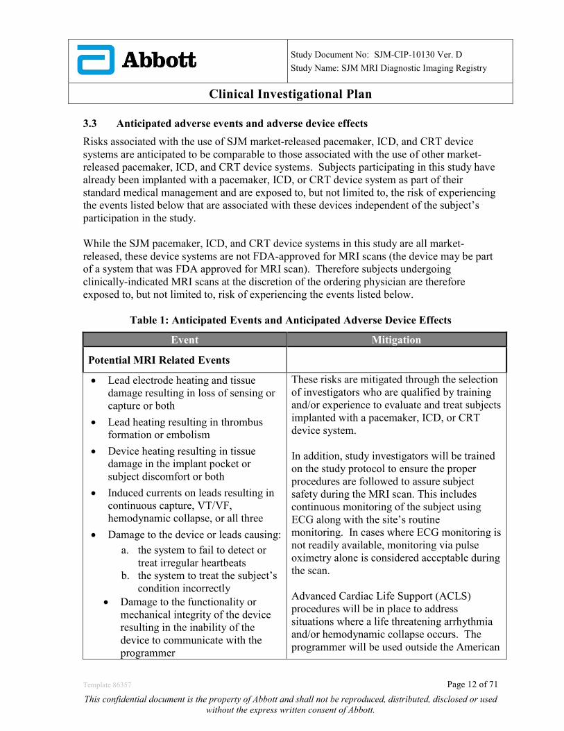
Clinical Investigational Plan 3.3 Anticipated adverse events and adverse device effects Risks associated with the use of SJM market-released pacemaker, ICD, and CRT device systems are anticipated to be comparable to those associated with the use of other market-released pacemaker, ICD, and CRT device systems. Subjects participating in this study have already been implanted with a pacemaker, ICD, or CRT device system as part of their standard medical management and are exposed to, but not limited to, the risk of experiencing the events listed below that are associated with these devices independent of the subject’s participation in the study. While the SJM pacemaker, ICD, and CRT device systems in this study are all market-released, these device systems are not FDA-approved for MRI scans (the device may be part of a system that was FDA approved for MRI scan). Therefore subjects undergoing clinically-indicated MRI scans at the discretion of the ordering physician are therefore exposed to, but not limited to, risk of experiencing the events listed below.
Table 1: Anticipated Events and Anticipated Adverse Device Effects
Event Mitigation
Potential MRI Related Events
• Lead electrode heating and tissue damage resulting in loss of sensing or capture or both
• Lead heating resulting in thrombus formation or embolism
• Device heating resulting in tissue damage in the implant pocket or subject discomfort or both
• Induced currents on leads resulting in continuous capture, VT/VF, hemodynamic collapse, or all three
• Damage to the device or leads causing: a. the system to fail to detect or
treat irregular heartbeats b. the system to treat the subject’s
condition incorrectly • Damage to the functionality or
mechanical integrity of the device resulting in the inability of the device to communicate with the programmer
These risks are mitigated through the selection of investigators who are qualified by training and/or experience to evaluate and treat subjects implanted with a pacemaker, ICD, or CRT device system. In addition, study investigators will be trained on the study protocol to ensure the proper procedures are followed to assure subject safety during the MRI scan. This includes continuous monitoring of the subject using ECG along with the site’s routine monitoring. In cases where ECG monitoring is not readily available, monitoring via pulse oximetry alone is considered acceptable during the scan. Advanced Cardiac Life Support (ACLS) procedures will be in place to address situations where a life threatening arrhythmia and/or hemodynamic collapse occurs. The programmer will be used outside the American
Template 86357 Page 12 of 71
This confidential document is the property of Abbott and shall not be reproduced, distributed, disclosed or used without the express written consent of Abbott.
Study Document No: SJM-CIP-10130 Ver. D
Study Name: SJM MRI Diagnostic Imaging Registry
Clinical Investigational Plan
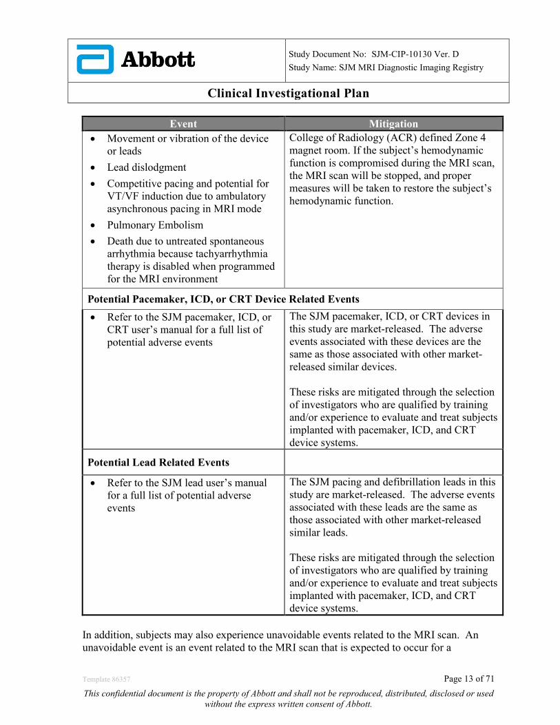
Event Mitigation • Movement or vibration of the device
or leads • Lead dislodgment • Competitive pacing and potential for
VT/VF induction due to ambulatory asynchronous pacing in MRI mode
• Pulmonary Embolism • Death due to untreated spontaneous
arrhythmia because tachyarrhythmia therapy is disabled when programmed for the MRI environment
College of Radiology (ACR) defined Zone 4 magnet room. If the subject’s hemodynamic function is compromised during the MRI scan, the MRI scan will be stopped, and proper measures will be taken to restore the subject’s hemodynamic function.
Potential Pacemaker, ICD, or CRT Device Related Events
• Refer to the SJM pacemaker, ICD, or CRT user’s manual for a full list of potential adverse events
The SJM pacemaker, ICD, or CRT devices in this study are market-released. The adverse events associated with these devices are the same as those associated with other market-released similar devices. These risks are mitigated through the selection of investigators who are qualified by training and/or experience to evaluate and treat subjects implanted with pacemaker, ICD, and CRT device systems.
Potential Lead Related Events
• Refer to the SJM lead user’s manual for a full list of potential adverse events
The SJM pacing and defibrillation leads in this study are market-released. The adverse events associated with these leads are the same as those associated with other market-released similar leads. These risks are mitigated through the selection of investigators who are qualified by training and/or experience to evaluate and treat subjects implanted with pacemaker, ICD, and CRT device systems.
In addition, subjects may also experience unavoidable events related to the MRI scan. An unavoidable event is an event related to the MRI scan that is expected to occur for a
Template 86357 Page 13 of 71
This confidential document is the property of Abbott and shall not be reproduced, distributed, disclosed or used without the express written consent of Abbott.

Study Document No: SJM-CIP-10130 Ver. D
Study Name: SJM MRI Diagnostic Imaging Registry
Clinical Investigational Plan projected duration in all subjects. Unavoidable events are not reportable unless the condition worsens or continues beyond the time frame listed below. Unavoidable events do not need to be reported on an adverse event form if they are resolved within the time frame specified. These events are expected to occur with any MRI scan.
Table 2: Unavoidable events related to the MRI Scan Event Time Frame post – MRI scan
• Claustrophobia • During MRI scan • Mild diaphoresis • During and < 1 hour post MRI scan • Sensation of bodily warmth • During and < 1 hour post MRI scan • Sensation of warmth at device pocket not
arising to the level of discomfort • During and < 1 hour post MRI scan
• Hearing impairment • < 24 hours • Body stiffness related to immobility • < 48 hours
3.4 Residual risks associated with the device under investigation While steps have been taken to identify risks associated with these devices undergoing an MRI scan (Refer to Table 1), there may be risks that are unknown at this time. 3.5 Risks associated with participation in the clinical study Potential risks associated with the MRI scan are the same as or comparable to those associated with MRI scans of an implanted medical device powered by a battery or other electrical source of power including, but not limited to, those listed in the Adverse Events and Adverse Device Effects section of the protocol. 3.6 Possible interactions with concomitant medical treatments and/or concurrent
medical interventions Since subjects are clinically indicated for an MRI scan, there are no treatments that the subject would not otherwise receive as part of the subject’s medical management related to having an implanted pacemaker, ICD, or CRT device system. The MRI scanner, methods used to scan the subject (scan sequences), and monitoring procedures in and of themselves are not investigational. As such, there are no anticipated interactions with concomitant medical treatments or concurrent medical interventions associated with the MRI scan. The device checks in this study involve testing of the device and lead that are normally done at a routine device check. As such, there are no anticipated interactions with concomitant medical treatments or concurrent medical interventions associated with this study.
Template 86357 Page 14 of 71
This confidential document is the property of Abbott and shall not be reproduced, distributed, disclosed or used without the express written consent of Abbott.

Study Document No: SJM-CIP-10130 Ver. D
Study Name: SJM MRI Diagnostic Imaging Registry
Clinical Investigational Plan 3.7 Steps that will be taken to control or mitigate the risks The risks associated with MRI scanning of subjects implanted with implantable active medical devices have been identified through clinical evaluation, including an exhaustive literature search. Risks normally associated with pacemakers, ICDs, CRTs, and transvenous leads will be minimized in the study by selecting investigators who are experienced in treating subjects implanted with these devices and are trained in the this study protocol. Subjects will be actively monitored during the MRI scan using an ECG along with the site’s routine monitoring. In cases where ECG monitoring is not readily available, monitoring via pulse oximetry alone is considered acceptable. The investigator and/or other ACLS-certified personnel will be present during the MRI scan to address cases of asystole or hemodynamic collapse that may occur during the MRI scan. Risks will also be minimized by careful assessment of each subject prior to enrollment. After enrollment, subjects in the study will be assessed as specified in this CIP to monitor the condition of the implanted system after the subject has undergone the MRI scan. In order to limit the risk of the MRI scan for a subject with an implanted pacemaker, ICD, or CRT device system, the MRI scans will be limited to non-thoracic examinations at up to 1.5T. In addition, the following precautions will be taken based on the AHA Guidelines on Safety of Magnetic Resonance Imaging in Patients with Cardiovascular Devices12:
o Advanced cardiovascular life support (ACLS) trained personnel and a “crash cart”, including defibrillator, will be available throughout the procedure to address an adverse event.
o Pre-MRI scan steps outside the MR environment
o Pre-test cardiac device functions
o For pacemaker dependent subjects, reprogram to asynchronous mode
o For ICD/CRT-D, disable bradycardia and tachycardia therapy and detection and exclude pacemaker dependent patients
o The patient’s heart rhythm and vital signs should be monitored throughout the MR procedure.
o Maintain visual and voice contact with the patient throughout the procedure.
o Instruct the patient to alert the MR system operator to any unusual sensations or problems.
After the MRI scan the pacemaker, ICD, or CRT device will be interrogated to verify appropriate device function, to evaluate pacing and sensing characteristics, and to assess any adverse events.
Template 86357 Page 15 of 71
This confidential document is the property of Abbott and shall not be reproduced, distributed, disclosed or used without the express written consent of Abbott.

Study Document No: SJM-CIP-10130 Ver. D
Study Name: SJM MRI Diagnostic Imaging Registry
Clinical Investigational Plan Overall, the clinical study design, subject selection process, and procedures developed for monitoring of the subject during the MRI scan have all been designed to minimize risks to the subject. While steps have been taken to identify and reduce or minimize risks associated with the MRI scan and participation in the study, there may be risks that are unknown at this time. 3.8 Risk-to-benefit rationale There may be no direct clinical benefit to the subject for participating in this study; no direct therapy is being provided as part of the study. However, the information gathered in this study will add to the understanding of the clinical utility of MRI scans in patients implanted with a pacemaker, ICD, or CRT device system.
4.0 Study Design 4.1 Purpose The purpose of this study is to assess the clinical utility of MRI scans in patients who are implanted with a St. Jude Medical pacemaker, ICD, or CRT device. The patient population under study includes male and female patients 18 years or older that are clinically indicated for a non-thoracic MRI scan. 4.2 Study Design and Scope This study will be performed as part of a regulated, prospective, non-randomized, multi-center clinical study. This multi-center study design was chosen for generalizability of study results by enrolling subjects across multiple geographies and varying types of sites. The total duration of this study is expected to be 2 to 3 years dependent on the rate of enrollment. The study will be conducted in up to 100 centers in the United States. Subjects will be enrolled, undergo a clinically-indicated MRI scan, and have an assessment of adverse events, device measurements, and clinical utility of the MRI scan images.
4.2.1 Number of Subjects Required to be Included in the Study The maximum number of subjects in the study is 300 from 2 main device groups (150 subjects with a pacemaker/CRT-P and 150 subjects with an ICD/CRT-D. A maximum of 45 subject enrollments will be allowed per center (15% of total subjects enrolled). For each of the 2 main device groups, a minimum of 25 head scans, 25 extremity scans, and 25 lumbar scans will be collected with the remainder of scans to be from any of these 3 scan regions.
Template 86357 Page 16 of 71
This confidential document is the property of Abbott and shall not be reproduced, distributed, disclosed or used without the express written consent of Abbott.
Study Document No: SJM-CIP-10130 Ver. D
Study Name: SJM MRI Diagnostic Imaging Registry
Clinical Investigational Plan
A patient will be considered enrolled in the study after he/she meets the inclusion/exclusion criteria, signs an IRB-approved informed consent form, and the subject’s device is programmed to enter the MRI environment.
4.2.2 Estimated Time Needed to Enroll Subject Population Enrollment in this study is expected to take approximately 2 to 3 years, depending on the rate of site activation and enrollments.
4.3 Objectives
4.3.1 Primary Objective The primary objective of this study is to characterize the image quality, clinical impact, and diagnostic utility of MRI in patients undergoing clinically indicated, non-thoracic MRI scans who are implanted with a St. Jude Medical pacemaker, ICD, or CRT device.
4.4 Endpoints
4.4.1 Primary Endpoints 4.4.1.1 Primary Endpoint #1: The proportion of MRI scans from pacemakers or CRT-Ps providing sufficient image quality to allow for a diagnostic interpretation.
4.4.1.2 Primary Endpoint #2: The proportion of MRI scans from ICDs or CRT-Ds providing sufficient image quality to allow for a diagnostic interpretation.
4.4.2 Additional Data
• Demographics: gender, age, ethnicity, race, cardiac disease history, arrhythmia history, etc.
• A summary of scan location (i.e. head, extremity, and lumbar). • A summary of clinical findings based on review of MRI scans • A summary of device electrical measurements at the MRI Scan Visit (before and
immediately after the MRI scan) and at Follow-up Visit(s). • A summary of adverse events (ADE, SADE, UADE) • A summary of MRI-related complications • Mortality • Estimate of the incidence rate of device patients undergoing a clinically indicated
MRI. The above data will be summarized by each main device group (pacemaker/CRT-P and ICD/CRT-D).
Template 86357 Page 17 of 71
This confidential document is the property of Abbott and shall not be reproduced, distributed, disclosed or used without the express written consent of Abbott.
Study Document No: SJM-CIP-10130 Ver. D
Study Name: SJM MRI Diagnostic Imaging Registry
Clinical Investigational Plan 4.5 Subject Selection A patient, who meets all of the inclusion criteria, and none of the exclusion criteria, is eligible to participate in this study.
4.5.1 Inclusion Criteria Eligible patients will meet all of the following: 1. Patient is implanted with a market-released St. Jude Medical pacemaker, ICD, or
CRT current generation device listed in the study protocol and any market-released pacing or defibrillation lead.
2. Patient’s device and all leads must be implanted for at least 6 weeks prior to the scheduled date of the MRI.
3. Patient has a clinical indication for a non-thoracic MRI scan, where MRI is the imaging modality of choice that will give adequate results to manage the patient.
4. Patient is scheduled for a non-thoracic MRI scan up to 1.5T. 5. Patient has a pacemaker, ICD, or CRT device implanted pectorally. 6. Patient has the ability to provide informed consent for study participation and be
willing and able to comply with the study procedures. 7. Patient is 18 years or above, or of legal age to give informed consent specific to
state and national law.
4.5.2 Exclusion Criteria Subjects are not eligible for clinical study participation if they meet any of the following exclusion criteria:
1. Patient has an ICD/CRT-D and is pacemaker dependent 2. Capture threshold is greater than 2.5 volts at 0.5 ms for RA and RV leads 3. Pacing lead impedance is NOT within range (i.e. ≥ 200 and ≤ 2000 ohms) 4. High voltage lead impedance (HVLI) is NOT within range (i.e. ≥ 20 and ≤ 200
ohms) 5. Patient has a device generator battery voltage at elective replacement interval
(ERI) 6. Patient has another existing active implanted medical device (e.g.
neurostimulator, infusion pump, etc.) that has MR labeling that will not allow the MRI scans to be completed.
7. Patient has other non-MRI compatible device or material implanted NOTE: • MRI compatible knee replacements, hip replacements, stents, etc. may be included
as long as the labeling of these devices allow MRI scans conducted per this protocol
Template 86357 Page 18 of 71
This confidential document is the property of Abbott and shall not be reproduced, distributed, disclosed or used without the express written consent of Abbott.
Study Document No: SJM-CIP-10130 Ver. D
Study Name: SJM MRI Diagnostic Imaging Registry
Clinical Investigational Plan
• MRI compatible mechanical, prosthetic, and bioprosthetic heart valves may be included as long as the labeling of these devices allow for MRI scans conducted per this protocol
• Non-removable dental implants may be included 8. Patient has a lead extender, adaptor, or capped/abandoned lead 9. Patient is pregnant
4.6 Subject Population
4.6.1 Patient Screening All patients presenting at the investigational site may be screened by a member of the investigational team who has been trained on the CIP and delegated to do so. Patients who do not meet the inclusion/exclusion criteria will not be eligible to participate in this study. Patients meeting the inclusion/exclusion criteria will be fully informed about the study and asked to review and sign informed consent. If the subject agrees, a duly signed and dated Patient Informed Consent will be obtained.
4.6.2 Point of Enrollment Subjects are considered enrolled after the informed consent form has been signed (Refer to section 4.7 for the Informed Consent Process), it has been verified that the subject meets all of the inclusion and none of the exclusion criteria, and the subject’s device is programmed to enter the MRI environment.
4.6.3 Enrollment of Medicare Beneficiaries This clinical study will enroll Medicare beneficiaries and therefore conforms to all standards of Medicare coverage requirements. Section 3.0 describes how all enrolled subjects, including Medicare beneficiaries, may be affected by the device under investigation. Subjects enrolled in the clinical study are expected to be consistent with the Medicare population based on age and as such the study results are expected to be generalizable to the Medicare population.
4.6.4 Vulnerable Population This clinical study will be conducted in a vulnerable population only when the study cannot be carried out in non-vulnerable populations. At the current time, this study does not allow, and will not include vulnerable patients for enrollment into the study.
Template 86357 Page 19 of 71
This confidential document is the property of Abbott and shall not be reproduced, distributed, disclosed or used without the express written consent of Abbott.

Study Document No: SJM-CIP-10130 Ver. D
Study Name: SJM MRI Diagnostic Imaging Registry
Clinical Investigational Plan 4.7 Informed Consent Process
4.7.1 General Process Prior to enrolling in the clinical study and conducting study-specific procedures, all subjects will be consented, as required by applicable regulations and the center’s IRB. Informed consent must be obtained from each subject prior to any study related procedures. Each consent form must be signed and dated by the subject and by the person obtaining the consent.
The principal investigator or his/her authorized designee will conduct the Informed Consent Process. This process will include a verbal discussion with the subject on all aspects of the clinical study that are relevant to the subject’s decision to participate in the clinical study. The subject shall be provided with the informed consent form that is written in a language that is understandable to the subject and has been approved by the center’s IRB. Failure to obtain informed consent from a subject prior to study enrollment should be reported to St. Jude Medical within 5 working days and to the reviewing center’s IRB/ consistent with the center’s IRB reporting requirements.
5.0 Devices Under Investigation 5.1 Device Descriptions This study includes any of the following market-released St. Jude Medical pacemaker, ICD, or CRT current generation devices and any market-released pacing or defibrillation lead that were previously implanted in patients with a clinical indication for the device system prior to participation in this study. Subjects with devices in the following families are eligible to enroll (See Table 3):
Table 3. List of Eligible Devices by Type and Family Device Type Device Family Pacemakers Assurity™ and Assurity MRI™
Endurity™ and Endurity MRI™ ICDs Ellipse™
Fortify Assura™ CRTs Allure™/Allure Quadra™ /Allure
Quadra MP™ CRT-P Quadra Assura™/ Quadra Assura MP™ CRT-D
The SJM pacemaker, ICD, and CRT devices are supported by the St. Jude Medical Merlin Patient Care System (Merlin PCS) programmer.
Template 86357 Page 20 of 71
This confidential document is the property of Abbott and shall not be reproduced, distributed, disclosed or used without the express written consent of Abbott.
Study Document No: SJM-CIP-10130 Ver. D
Study Name: SJM MRI Diagnostic Imaging Registry
Clinical Investigational Plan 5.2 Device Handling & Storage Instructions for use, storage and handling instructions, preparation for use and any precautions can be found in the User’s Manuals for each the market-released devices and leads.
Template 86357 Page 21 of 71
This confidential document is the property of Abbott and shall not be reproduced, distributed, disclosed or used without the express written consent of Abbott.
Study Document No: SJM-CIP-10130 Ver. D
Study Name: SJM MRI Diagnostic Imaging Registry
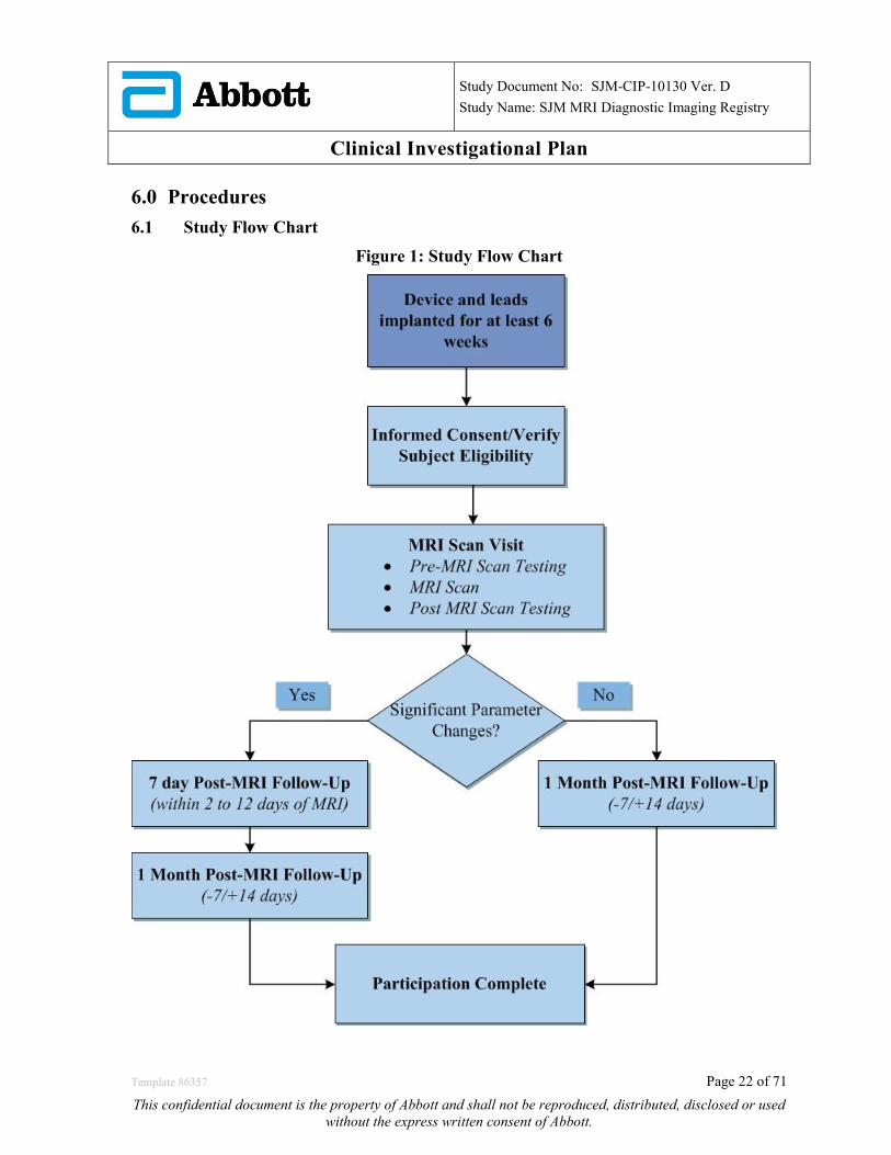
Clinical Investigational Plan 6.0 Procedures 6.1 Study Flow Chart
Figure 1: Study Flow Chart
Template 86357 Page 22 of 71
This confidential document is the property of Abbott and shall not be reproduced, distributed, disclosed or used without the express written consent of Abbott.

Study Document No: SJM-CIP-10130 Ver. D
Study Name: SJM MRI Diagnostic Imaging Registry
Clinical Investigational Plan 6.2 Procedures The study will be conducted in accordance with the CIP. All parties participating in the conduct of the study will be qualified by education, training, or experience to perform their tasks and this training will be documented appropriately. The study will not commence until St. Jude Medical receives written approval from the IRB and relevant regulatory authorities and all required documents have been collected from the site(s). All required study procedures at each specified interval are outlined in the section below. Refer to Table 4.
Table 4: Study Procedures and Data Collection
Procedure/Evaluation
Study Schedule
MRI Scan Visit Follow-Up Visit(s)
2-12 days and/or 1 Month (-7/+ 14 days) Post MRI
Scan*
Pre-MRI
During MRI
Post-MRI (Same day as
MRI Scan) Informed Consent & Inclusion/Exclusion Evaluation √
Medical and Surgical History √ Demographic Information √ Implanted SJM pacemaker, ICD, or CRT device system info. √
Complete the SJM provided MRI Hazard Checklist or a site-specific checklist, per site’s standard of care
√
Obtain device measurements (remaining battery capacity, capture threshold, sense, pacing and HVLI impedances, as applicable) at permanently programmed settings
√ √ √
Program the appropriate device settings before MRI √
Monitor subject with ECG along with the site’s routine monitoring** √
Assess subject for adverse events √ √ √ Program device to original settings after MRI scan √ Clinical Evaluation of MRI scan √ Report deviations and withdrawal √ √ √ √ *Refer to the study flow chart for follow-up schedule ** In cases where ECG monitoring is not readily available, monitoring via pulse oximetry alone is considered acceptable
Template 86357 Page 23 of 71
This confidential document is the property of Abbott and shall not be reproduced, distributed, disclosed or used without the express written consent of Abbott.
Study Document No: SJM-CIP-10130 Ver. D
Study Name: SJM MRI Diagnostic Imaging Registry
Clinical Investigational Plan 6.3 Pre-MRI Scan Procedures
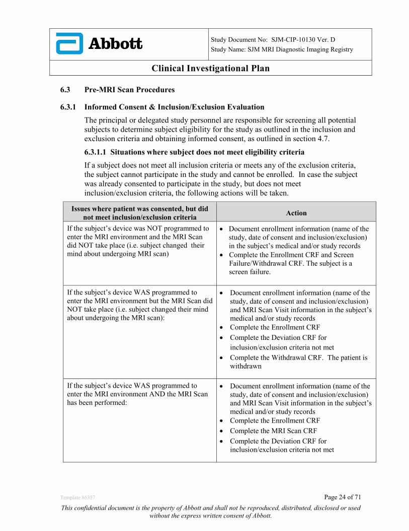
6.3.1 Informed Consent & Inclusion/Exclusion Evaluation The principal or delegated study personnel are responsible for screening all potential subjects to determine subject eligibility for the study as outlined in the inclusion and exclusion criteria and obtaining informed consent, as outlined in section 4.7.
6.3.1.1 Situations where subject does not meet eligibility criteria If a subject does not meet all inclusion criteria or meets any of the exclusion criteria, the subject cannot participate in the study and cannot be enrolled. In case the subject was already consented to participate in the study, but does not meet inclusion/exclusion criteria, the following actions will be taken.
Issues where patient was consented, but did not meet inclusion/exclusion criteria Action
If the subject’s device was NOT programmed to enter the MRI environment and the MRI Scan did NOT take place (i.e. subject changed their mind about undergoing MRI scan)
• Document enrollment information (name of the study, date of consent and inclusion/exclusion) in the subject’s medical and/or study records
• Complete the Enrollment CRF and Screen Failure/Withdrawal CRF. The subject is a screen failure.
If the subject’s device WAS programmed to enter the MRI environment but the MRI Scan did NOT take place (i.e. subject changed their mind about undergoing the MRI scan):
• Document enrollment information (name of the study, date of consent and inclusion/exclusion) and MRI Scan Visit information in the subject’s medical and/or study records
• Complete the Enrollment CRF • Complete the Deviation CRF for
inclusion/exclusion criteria not met • Complete the Withdrawal CRF. The patient is
withdrawn
If the subject’s device WAS programmed to enter the MRI environment AND the MRI Scan has been performed:
• Document enrollment information (name of the study, date of consent and inclusion/exclusion) and MRI Scan Visit information in the subject’s medical and/or study records
• Complete the Enrollment CRF • Complete the MRI Scan CRF • Complete the Deviation CRF for
inclusion/exclusion criteria not met
Template 86357 Page 24 of 71
This confidential document is the property of Abbott and shall not be reproduced, distributed, disclosed or used without the express written consent of Abbott.
Study Document No: SJM-CIP-10130 Ver. D
Study Name: SJM MRI Diagnostic Imaging Registry
Clinical Investigational Plan 6.3.2 Baseline Data Collection
Collect data on the subject including demographics, medical/surgical history, and St. Jude Medical device system information
6.3.3 Clearing the Subject for the MRI Scan The radiologist or designated radiological staff member must determine the subject’s eligibility for an MRI scan prior to the MRI scan (per standard of practice). The study MRI Hazard Checklist may be used to document a radiologist or designated member of the radiology department has cleared the subject for an MRI scan. Alternatively, the radiology department may use its own hazard checklist in lieu of the study MRI Hazard Checklist. This documentation should be maintained with the subject’s medical and/or study records.
In order to safely perform an MRI scan on a subject with a St. Jude Medical device system, the physician/clinician should follow the recommended guidelines below:
• Review the potential adverse events in an MRI environment in Table 1. • Generate a report of the patient’s permanently programmed parameters • Perform device assessment • Program recommended settings for the MRI environment • Subject receives the MRI Scan • Reprogram device to patient’s permanently programmed parameters
6.3.4 Pre-MRI Device Assessment 1) Interrogate the subject’s device using a Merlin programmer.
2) Verify the following conditions: • Capture thresholds are stable at ≤ 2.5V@ 0.5 ms for RA and RV leads • Pacing lead impedance is within range, i.e. ≥ 200 and ≤ 2000 ohms • HVLI is within range, i.e. ≥ 20 and ≤ 200 ohms • Device generator battery voltage is not at elective replacement interval (ERI) • No additional hardware (adaptors, extenders, or abandoned leads)
NOTE: If any of the above conditions are not met, the subject is a screen failure.
3) Determine the subject’s underlying rhythm • If there is no intrinsic rhythm when the device is programmed to pace at 40
bpm, the subject is considered pacemaker dependent.
4) Obtain the following measurements for all leads, as applicable, with the permanently programmed parameters. • Remaining battery capacity • Capture threshold for all leads
Template 86357 Page 25 of 71
This confidential document is the property of Abbott and shall not be reproduced, distributed, disclosed or used without the express written consent of Abbott.
Study Document No: SJM-CIP-10130 Ver. D
Study Name: SJM MRI Diagnostic Imaging Registry
Clinical Investigational Plan
• Sensing amplitude for RA and RV leads • Pacing lead impedance for all leads • HVLI impedance NOTE: RV capture thresholds are not required to be obtained if a high ventricular rate is present (e.g. 110bpm). If available, sites should use the automatically obtained pacing capture threshold from the most recent archival data as a substitute for the in-clinic capture threshold. RV sensing measurements are not required if the subject’s intrinsic rate has been established to be ≤40 beats per minute.
5) Perform capacitor maintenance (For ICD or CRT-D devices only) NOTE: This is to avoid potential damage to the ICD or CRT-D device should an automatic capacitor maintenance check be scheduled to occur while the patient is in the MRI environment. The manual capacitor maintenance will prevent the scheduled check from occurring.
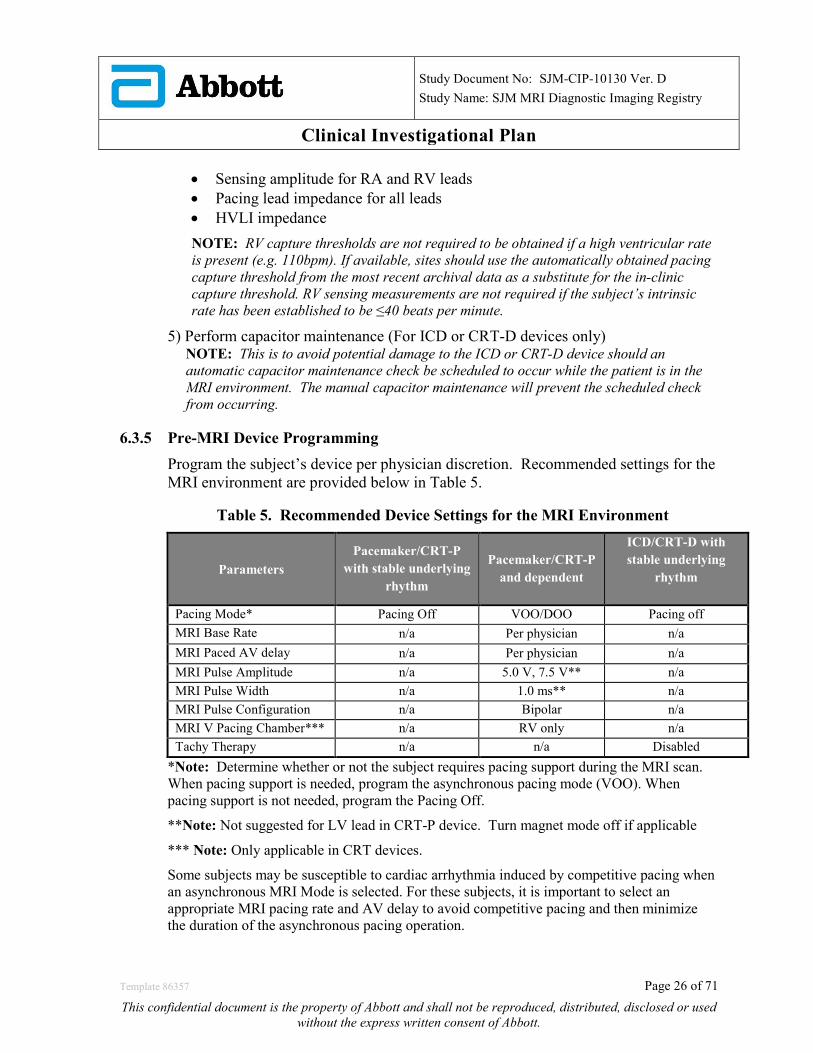
6.3.5 Pre-MRI Device Programming Program the subject’s device per physician discretion. Recommended settings for the MRI environment are provided below in Table 5.
Table 5. Recommended Device Settings for the MRI Environment
Parameters Pacemaker/CRT-P
with stable underlying rhythm
Pacemaker/CRT-P and dependent
ICD/CRT-D with stable underlying
rhythm
Pacing Mode* Pacing Off VOO/DOO Pacing off MRI Base Rate n/a Per physician n/a MRI Paced AV delay n/a Per physician n/a MRI Pulse Amplitude n/a 5.0 V, 7.5 V** n/a MRI Pulse Width n/a 1.0 ms** n/a MRI Pulse Configuration n/a Bipolar n/a MRI V Pacing Chamber*** n/a RV only n/a Tachy Therapy n/a n/a Disabled
*Note: Determine whether or not the subject requires pacing support during the MRI scan. When pacing support is needed, program the asynchronous pacing mode (VOO). When pacing support is not needed, program the Pacing Off.
**Note: Not suggested for LV lead in CRT-P device. Turn magnet mode off if applicable
*** Note: Only applicable in CRT devices.
Some subjects may be susceptible to cardiac arrhythmia induced by competitive pacing when an asynchronous MRI Mode is selected. For these subjects, it is important to select an appropriate MRI pacing rate and AV delay to avoid competitive pacing and then minimize the duration of the asynchronous pacing operation.
Template 86357 Page 26 of 71
This confidential document is the property of Abbott and shall not be reproduced, distributed, disclosed or used without the express written consent of Abbott.
Study Document No: SJM-CIP-10130 Ver. D
Study Name: SJM MRI Diagnostic Imaging Registry
Clinical Investigational Plan 6.3.6 Setting up ECG and/or Pulse Oximetry
Set up the ECG and/or pulse oximetry to monitor heart rate. Place the oximetry clip on the subject’s finger or any other appendage that results in valid pulse oximetry readings. Position MRI compatible surface electrodes on the subject to ensure the subject’s heart rate can be continuously monitored during the scan. During the MRI scan, periodically record heart rate, and blood oxygen saturation levels. Visually examine the ECG during the MRI scan. Note any abnormalities observed in the cardiac rhythm (Refer to Life–threatening Ventricular Arrhythmia and Asystole Assessment below). After the MRI scan, remove the subject from the MRI field.
Note: Monitor subject with ECG along with the site’s routine monitoring. In cases where ECG monitoring is not readily available, monitoring via pulse oximetry alone is considered acceptable.
6.4 During MRI Scan Procedures After confirmation by the electrophysiologist or device specialist that all pre-MRI system checks (mentioned above) have been met, subjects will have the clinically indicated, non-thoracic MRI scan completed by a radiology staff member.
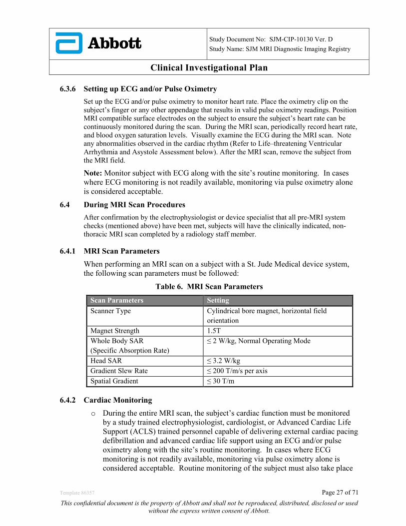
6.4.1 MRI Scan Parameters When performing an MRI scan on a subject with a St. Jude Medical device system, the following scan parameters must be followed:
Table 6. MRI Scan Parameters
Scan Parameters Setting Scanner Type
Cylindrical bore magnet, horizontal field orientation
Magnet Strength 1.5T Whole Body SAR (Specific Absorption Rate)
≤ 2 W/kg, Normal Operating Mode
Head SAR ≤ 3.2 W/kg Gradient Slew Rate ≤ 200 T/m/s per axis Spatial Gradient ≤ 30 T/m
6.4.2 Cardiac Monitoring o During the entire MRI scan, the subject’s cardiac function must be monitored
by a study trained electrophysiologist, cardiologist, or Advanced Cardiac Life Support (ACLS) trained personnel capable of delivering external cardiac pacing defibrillation and advanced cardiac life support using an ECG and/or pulse oximetry along with the site’s routine monitoring. In cases where ECG monitoring is not readily available, monitoring via pulse oximetry alone is considered acceptable. Routine monitoring of the subject must also take place
Template 86357 Page 27 of 71
This confidential document is the property of Abbott and shall not be reproduced, distributed, disclosed or used without the express written consent of Abbott.
Study Document No: SJM-CIP-10130 Ver. D
Study Name: SJM MRI Diagnostic Imaging Registry
Clinical Investigational Plan
to assess and/or confirm any clinically significant changes noted in the subject’s oxygen saturation or heart rate, as well as any clinically significant complaints not obvious with pulse oximetry. Record these changes and complaints during the MRI scan.
o ACLS procedures must be in place to address situations where a life threatening arrhythmia and/or hemodynamic collapse occurs. The programmer must be used outside the American College of Radiology (ACR) defined Zone 4 magnet room. If the subject’s hemodynamic function is compromised during the MRI scan, discontinue the MRI procedure and take proper measures to restore the subject’s hemodynamic function.
6.4.3 Life–threatening Ventricular Arrhythmia and Asystole Assessment o Monitoring of spontaneous ventricular arrhythmias and asystole must be
conducted via an ECG and/or pulse oximetry during the MRI scan. Any sustained ventricular arrhythmias or asystole (see definition below) must be documented on an Adverse Event form. Non-sustained ventricular tachycardias (NSVT) or premature ventricular contractions (PVCs) do not need to be reported as an adverse event. However, if an arrhythmia reproducibly occurs (occurring more than once during the MRI scan) while the subject is actively being scanned, report the event on an Adverse Event form.
Definitions: • Sustained Ventricular Arrhythmia: Heart Rate >150bpm for > 30 seconds with
depolarization originating in the ventricles • Asystole: A standstill > 6 seconds in electrical activity of the heart (i.e., no heart
rate for 6 seconds or more)
6.4.4 Handling of Subjects Unable to Tolerate an MR Scan In cases where the scan cannot be tolerated by the subject, remove the subject from the scanner. Assess the subject for possible adverse events, and treat the subject’s reported symptoms according to your institution’s standard of practice. Document the reason for the intolerance. At a minimum, information related to the sequence used to perform the scan, the length of time the subject was scanned, and the whole body SAR level reached should be collected and submitted to St. Jude Medical. A repeat scan is not required to be completed.
6.5 Post-MRI Scan Procedures Following the MRI Scan, remove the subject from the MRI bore.
6.5.1 Post-MRI Device Programming and Assessment 1) Interrogate the subject’s device using a Merlin programmer.
2) Reprogram device to subject’s permanently programmed parameters.
Template 86357 Page 28 of 71
This confidential document is the property of Abbott and shall not be reproduced, distributed, disclosed or used without the express written consent of Abbott.

Study Document No: SJM-CIP-10130 Ver. D
Study Name: SJM MRI Diagnostic Imaging Registry
Clinical Investigational Plan
3) Determine the subject’s underlying rhythm • If there is no intrinsic rhythm when the device is programmed to pace at 40
bpm, the subject is considered pacemaker dependent.
4) Obtain the following measurements for all leads, as applicable, with the permanently programmed parameters. • Remaining battery capacity • Capture threshold for all leads • Sensing amplitude for RA and RV leads • Pacing lead impedance for all leads • HVLI impedance
NOTE: RV capture thresholds are not required to be obtained if a high ventricular rate is present (e.g. 110bpm). If available, sites should use the automatically obtained pacing capture threshold from the most recent archival data as a substitute for the in-clinic capture threshold. RV sensing measurements are not required if the subject’s intrinsic rate has been established to be ≤40 beats per minute.
6.5.2 Reporting of MRI Scan-Related Adverse Device Effects An ADE or SADE related to the following should be reported as soon as possible, but no later than 10 working days, to St. Jude Medical: clotting, pulmonary embolism, or heating of the device pocket during the MRI scan. These events are likely to be associated with symptoms occurring during or immediately following the MRI scan and may manifest as chest pain, shortness of breath, or changes in vital signs during or immediately following the MRI scan.
To ensure all ADEs or SADEs related to or caused by the MRI scan are appropriately captured, before starting the scan, verbally instruct the subject to report symptoms of chest pain, shortness of breath or pocket discomfort that he/she experiences while being scanned or immediately after exiting the scanner. Note changes in vital signs such as changes in heart rate, room air blood oxygen saturation, and/or respiration rate that occur during the MRI scan that may suggest an ADE or SADE has occurred due to clotting, pulmonary embolus or related to lead tip or device pocket heating.
If symptoms during or immediately after the MRI scan suggest that an ADE or SADE has occurred due to clotting, pulmonary embolus or related to lead tip or device pocket heating, test to assess possible causes. Diagnostic testing may be performed in any order deemed appropriate by the investigator; if any test was not performed, provide medical justification for not performing that test:
(1) A 12-lead EKG
(2) A 2-view chest X-ray (PA and Lateral).
(3) Room air blood oxygen saturation
(4) A transthoracic echocardiogram.
Template 86357 Page 29 of 71
This confidential document is the property of Abbott and shall not be reproduced, distributed, disclosed or used without the express written consent of Abbott.

Study Document No: SJM-CIP-10130 Ver. D
Study Name: SJM MRI Diagnostic Imaging Registry
Clinical Investigational Plan
If the subject reports pocket discomfort, ask the subject for additional descriptive information and determine if the pocket is discolored or warm to the touch. EKG, chest x-ray, room air blood oxygen saturation, or transthoracic echocardiogram testing are not required to be performed for symptoms related to device pocket heating.
Sites should report an ADE or SADE if the subject experiences a significant rise in pacing threshold (1.25V @ 0.5ms or greater) from pre-MRI scan to post-MRI scan.
6.5.3 Clinical Evaluation of MRI Scan The radiologist will review the clinically-indicated MRI scan for diagnostic interpretation. The following information will be collected regarding the MRI scan, and are not limited to:
• Primary reason for MRI scan • Body region scanned (non-thoracic) (e.g. brain, cervical spine, lumbar spine, knee,
shoulder, hip, hand/wrist) • MRI System used (i.e. brand, model, software version, etc.) • Duration of scan (min) • Whole body SAR • Image quality (i.e. sufficient or insufficient to interpret the MRI image) • Clinical Diagnosis (e.g. joint abnormality, vascular abnormality, no abnormalities,
etc.)
6.5.4 Data Submission Once all the required testing has been performed at this visit, complete and submit the Enrollment and MRI Scan Case Report Forms to St. Jude Medical. If an adverse event or death occurred, submit an Adverse Event, Death Case Report Form, and Product Out of Service Case Report Form, as applicable. Report any deviations or withdrawals by submitting a Deviation or Withdrawal Case Report Form. Export the MRI scan onto a CD, or other form of electronic media in DICOM format, and send to St. Jude Medical. Upload pre and post MRI scan device session records through the EDC study portal or Merlin.net. It is recommended that the following device printouts and measurements be maintained at the site.
• FastPath Summary • Test Results with Freezes, Include Battery & Leads • Wrap-up Overview with full parameters • Upload device session record
6.6 Follow-Up Visit Procedures The subject’s scheduled follow-up visits will be based upon the change in the device parameters from the MRI Scan Visit (pre-MRI scan to post-MRI scan measurements).
Template 86357 Page 30 of 71
This confidential document is the property of Abbott and shall not be reproduced, distributed, disclosed or used without the express written consent of Abbott.

Study Document No: SJM-CIP-10130 Ver. D
Study Name: SJM MRI Diagnostic Imaging Registry
Clinical Investigational Plan If the subject had a follow-up visit at another institution, then the follow-up data, including device measurements and pertinent medical records may be obtained per the medical record release authorization statement in the informed consent.
6.6.1 Significant Parameter Changes: Table 7. List of Significant Parameter Changes Requiring Multiple Follow-Ups
A change in remaining battery capacity of ≥ 2% A change in pacing lead impedance > 50 ohms A change in high-voltage lead impedance (HVLI) ≥ 7 ohms Decrease in P wave sense amplitude ≥ 50% Decrease in R wave sense amplitude ≥ 25% Increase in capture threshold ≥ 0.5V at the pre-programmed pulse width
6.6.2 Subjects with significant parameter changes requiring multiple follow-up visits Subjects with any of the significant parameters changes shown in Table 7 above will require multiple follow-up visits to ensure patient safety and appropriate device function. A total of 2 follow-up visits will be scheduled:
1) Within 2 to 12 days after the MRI scan 2) 1 Month (-7/+ 14 days) after the MRI scan
6.6.3 Subjects requiring only a single follow-up visit Subjects that did not have any of the significant parameters changes shown in Table 7 will be required to return for a single follow-up visit at 1 month (-7/+14 days) after the MRI scan.
6.6.4 Unscheduled visits An unscheduled visit is defined as a visit that occurs after the MRI Scan Visit where the subject is seen in clinic due to an ADE, SADE, or UADE.
6.6.5 Device Assessment during Follow-Ups For any scheduled or unscheduled follow-up visit:
1) Determine the subject’s underlying rhythm • If there is no intrinsic rhythm when the device is programmed to pace at 40
bpm, the subject is considered pacemaker dependent.
2) Obtain the following measurements for all leads, as applicable, with the permanently programmed parameters.
• Remaining battery capacity • Capture threshold for all leads
Template 86357 Page 31 of 71
This confidential document is the property of Abbott and shall not be reproduced, distributed, disclosed or used without the express written consent of Abbott.
Study Document No: SJM-CIP-10130 Ver. D
Study Name: SJM MRI Diagnostic Imaging Registry
Clinical Investigational Plan
• Sensing amplitude for RA and RV leads • Pacing lead impedance for all leads • HVLI impedance NOTE: RV capture thresholds are not required to be obtained if a high ventricular rate is present (e.g. 110bpm). If available, sites should use the automatically obtained pacing capture threshold from the most recent archival data as a substitute for the in-clinic capture threshold. RV sensing measurements are not required if the subject’s intrinsic rate has been established to be ≤ 40 beats per minute.
3) Evaluate the subject for adverse events (ADEs, SADEs, UADEs).
6.6.6 Data Submission Once all the required assessments have been performed at the follow-up visit, complete and submit the Follow-Up Case Report Form to St. Jude Medical. If an adverse event, revision procedure, or death occurred, submit an Adverse Event, Death, System Revision, and/or Product Out of Service Case Report Forms, as applicable. Report any deviations or withdrawals by submitting a Deviation or Withdrawal Case Report Form.
6.7 System Revisions A System Revision CRF should be completed for all types of revisions such as pulse generator replacement, lead replacement and lead repositioning. The subject’s follow up schedule is not affected in any way. If the subject has the SJM pacemaker, ICD, or CRT device removed at any time during the study, and the subject will not receive a replacement SJM device, follow the subject for 30 days, and withdraw the subject from the study. Complete a System Revision CRF and Product Out of Service CRF, withdrawal CRF and submit through EDC. In cases where only the device pocket is revised (leads are not repositioned, leads have not been disconnected from the device), a System Revision CRF is not required to be submitted. 6.8 Subject study completion The subject’s participation in this study is complete after the last scheduled follow-up per sections 6.6.2 or 6.6.3, as applicable. When the subject’s participation in the clinical study has been completed the subject will return to standard medical care as per physician’s recommendation.
Template 86357 Page 32 of 71
This confidential document is the property of Abbott and shall not be reproduced, distributed, disclosed or used without the express written consent of Abbott.

Study Document No: SJM-CIP-10130 Ver. D
Study Name: SJM MRI Diagnostic Imaging Registry
Clinical Investigational Plan 6.9 Criteria and Procedures for Subject Withdrawal or Screen Failure Subjects must be informed about their right to withdraw from the study at any time and for any reason without sanction, penalty or loss of benefits to which the subject is otherwise entitled and withdrawal from the study will not jeopardize their future medical care or relationship with the investigator. Subjects will be asked to specify the reason for the termination, but have the right not to answer. The investigator may decide to withdraw a subject from the study at any time with reasonable rationale. The subject’s future care will not be influenced by a decision, voluntary or otherwise, to withdraw from the study. All reasonable efforts should be made to retain the subject in the clinical study until completion of the study. Reasons for subject’s withdrawal include, but are not limited to:
• Subject refuses to continue participating in the study • Subject is deceased (cause must be documented) • Subject’s non-compliance • Subject’s participation is terminated by the PI or investigator, although the
subject consented, since participation is no longer medically appropriate • Subject receives a non-SJM replacement device during a system revision
(subject will be followed for 30 days) • Subject is ‘lost to follow up’: Subject does not adhere to the scheduled follow up
visits but has not explicitly requested to be withdrawn from the clinical study. (This does not apply to missed visits). Site personnel should at all times make all reasonable efforts to locate and communicate with the subject in order to achieve subject compliance to the scheduled follow up visits:
1) A subject will be considered ‘Lost to Follow Up’ after a minimum of 2 phone calls of a physician or delegate at the investigational site to the subject or contact. These 2 phone calls need to be documented in the subject’s hospital records.
2) If these attempts are unsuccessful, a certified letter should be sent to the subject’s last known address or general practitioner (GP) and a copy of this letter should be maintained in the subject’s hospital records.
If a subject withdraws from the clinical study, the site will record the subject’s reasons for withdrawal, on a Withdrawal CRF. When subject withdrawal from the clinical study is due to an adverse event the subject will be followed until resolution of that adverse event or determination that the subject’s condition is stable. The status of the subject’s condition should be documented at the time of withdrawal.
Template 86357 Page 33 of 71
This confidential document is the property of Abbott and shall not be reproduced, distributed, disclosed or used without the express written consent of Abbott.
Study Document No: SJM-CIP-10130 Ver. D
Study Name: SJM MRI Diagnostic Imaging Registry
Clinical Investigational Plan Screening Failure: A subject may also become a screen failure based if the subject’s participation ended prior to enrollment in the study. Refer to section 6.3.1.1. Complete a Withdrawal CRF for screen failure subjects and note that the subject is a screen failure. 6.10 Description of Activities performed by Sponsor Representatives Trained sponsor personnel may perform certain activities to ensure compliance to the clinical investigational plan and may provide technical expertise. Sponsor representatives may perform the following activities:
• Verifying MRI scan parameters on the programmer at the MRI Scan Visit. • Programming of the device per protocol, and/or as directed by the
investigator/designee. • Interrogation of and testing the device at any study visit (scheduled or
unscheduled): remaining battery capacity, capture, sense, pacing and HVLI measurements
While sponsor representatives may perform these activities, the principal investigator remains responsible for ensuring all study data is collected as required per protocol. Deviations resulting from failure to comply with protocol requirements will be reported through completion of a Deviation CRF. 7.0 Compliance to the CIP 7.1 Adherence to the Clinical Investigation Plan (Protocol Deviations) A deviation is defined as an event where the clinical investigator, site personnel, sponsor or sponsor representative did not conduct the clinical study according to the Clinical Investigational Plan, IRB requirements or the Investigator Agreement. The investigator is not allowed to deviate from the CIP, except as specified under emergency circumstances. In some cases, failure to comply with the CIP may be considered failure to protect the rights, safety and well-being of subjects, since the non-compliance exposes subjects to unreasonable risks. For example, failure to adhere to the inclusion/exclusion criteria: these criteria are specifically defined by the Sponsor to exclude subjects for whom the device is not beneficial and the use involves unreasonable risks. This may be considered failure to protect the rights, safety and well-being of the enrolled subject. Similarly, failure to perform safety assessments intended to detect adverse events may be considered failure to protect the rights, safety and well-being of the enrolled subject. Investigators should seek minimization of such risks by adhering to the CIP. Simultaneously, in the event that adhering to the CIP exposes the subject to unreasonable risks, the investigator is also required to protect the rights, safety and well-being of the subject by intentionally deviating from the requirements of the CIP, so that subjects are not exposed to unreasonable risks.
Template 86357 Page 34 of 71
This confidential document is the property of Abbott and shall not be reproduced, distributed, disclosed or used without the express written consent of Abbott.

Study Document No: SJM-CIP-10130 Ver. D
Study Name: SJM MRI Diagnostic Imaging Registry
Clinical Investigational Plan It is the responsibility of the investigator to provide adequate medical care to a subject enrolled in a study. Regulations require that the PI maintain accurate, complete, and current records, including documents showing the date of and reason for every deviation from the Clinical Investigational Plan. Relevant information for each deviation will be documented on a Deviation Case Report Form. The site will submit the CRF to St. Jude Medical. Regulations require Investigators obtain approval from St. Jude Medical and the IRB [as required] before initiating changes in or deviations from the protocol, except when necessary to protect the life or physical well-being of a subject in an emergency. Under emergency circumstances, deviations from the CIP to protect the rights, safety and well-being of human subjects may proceed without prior approval of the sponsor and the EC. Such deviations shall be documented and reported to the sponsor and the EC as soon as possible, but no later than 5 working days. Prior approval must be requested when the PI anticipates, contemplates, or makes a conscious decision to depart from the CIP, except when unforeseen circumstances are beyond the investigator’s control (e.g. a subject who fails to attend a scheduled follow-up visit, a subject is too ill to perform a CIP-required test, etc.). All deviations, including those beyond the investigator’s control, must be reported on a CRF. To obtain approval, the Principal Investigator may call or email and discuss the potential deviation with St. Jude Medical or designee prior to initiating any changes. All deviations must be reported to appropriate regulatory authorities in specified timelines (if appropriate). Investigator will notify St. Jude Medical and the reviewing IRB within 5 working days of:
• Any deviation to protect the life or physical well-being of a subject in an emergency • Any failure to obtain informed consent
Investigators or the designee must notify St. Jude Medical, Inc. as soon as possible and complete the Deviation CRF. The Investigator is required to adhere to local regulatory requirements for reporting deviations to IRB. 7.2 Repeated and Serious Non-compliance In the event of repeated non-compliance or a one-time serious non-compliance, as determined by the Sponsor, a Clinical Research Associate or clinical representative will attempt to secure compliance by one or more of the following actions:
Template 86357 Page 35 of 71
This confidential document is the property of Abbott and shall not be reproduced, distributed, disclosed or used without the express written consent of Abbott.

Study Document No: SJM-CIP-10130 Ver. D
Study Name: SJM MRI Diagnostic Imaging Registry
Clinical Investigational Plan
• Visiting the investigator • Contacting the investigator by telephone • Contacting the investigator in writing • Retraining of the investigator
If an investigator is found to be repeatedly non-compliant with the signed agreement, the CIP or any other conditions of the clinical study, the Sponsor will either secure compliance or, at its sole discretion, terminate the investigator’s participation in the clinical study. 8.0 Adverse Device Effect 8.1 Definitions
8.1.1 Medical Device Any instrument, apparatus, implement, machine, appliance, implant, software, material or other similar or related article (a) Intended by the manufacturer to be used, alone or in combination, for human beings for one or more of the specific purpose(s) of o Diagnosis, prevention, monitoring, treatments or alleviation of disease, o Diagnosis, monitoring, treatment, alleviation of, or compensation for, an injury, o Investigation, replacement, modification, or support of the anatomy or of a
physiological process, o Supporting or sustaining life, o Control of conception, o Disinfection of medical devices and
(b) Which does not achieve its primary intended action in or on the human body by pharmacological, immunological or metabolic means, but which may be assisted in its intended function by such means.
8.1.2 Adverse Device Effect (ADE) An adverse device effect is an adverse event related to the use of an investigational medical device. This definition includes adverse events resulting from insufficient or inadequate instructions for use, deployment, implantation, installation, or operation, or any malfunction of the investigational medical device. This definition includes any event resulting from the use, error, or from intentional misuse of the investigational medical device.
Template 86357 Page 36 of 71
This confidential document is the property of Abbott and shall not be reproduced, distributed, disclosed or used without the express written consent of Abbott.

Study Document No: SJM-CIP-10130 Ver. D
Study Name: SJM MRI Diagnostic Imaging Registry
Clinical Investigational Plan 8.1.3 Serious Adverse Device Effect (SADE)
Adverse device effect that has resulted in any of the consequences characteristic of a serious adverse event: • Death • A serious deterioration in the health of the subject, that either resulted in:
o A life-threatening illness or injury OR o A permanent impairment to a body structure or a body function OR o An in-patient or prolonged hospitalization OR o A medical or surgical intervention to prevent life-threatening illness or
injury or permanent impairment to a body structure or a body o Fetal distress, fetal death or a congenital abnormality or birth defect
A planned hospitalization for a pre-existing condition, or a procedure required by the CIP is not considered a serious adverse event.
8.1.4 Unanticipated Adverse Device Effect (UADE) As defined in 21 CFR §812.3, unanticipated adverse device effects (UADE) are defined as any serious adverse effect on health or safety or any life-threatening problem or death caused by, or associated with, a device, if that effect, problem, or death was not previously identified in nature, severity, or degree of incidence in the clinical investigational plan or application (including a supplementary plan or application), or any other unanticipated serious problem associated with a device that relates to the rights, safety, or welfare of subjects.
If an unanticipated adverse device effect occurs, the investigator must notify St. Jude Medical and the IRB/MEC immediately, but no later than 10 working days of the investigator’s knowledge of the event, as required by 21 CFR §812.150. St. Jude Medical will take any steps necessary to investigate the event, and will be responsible for notifying FDA and all other participating IRBs/MECs and investigators.
8.2 Reporting of MRI Scan-Related Adverse Device Effects An ADE or SADE related to the following should be reported as soon as possible, but no later than 10 working days, to St. Jude Medical: clotting, pulmonary embolism, or heating of the device pocket during the MRI scan. These events are likely to be associated with symptoms occurring during or immediately following the MRI scan and may manifest as chest pain, shortness of breath, or changes in vital signs during or immediately following the MRI scan. To ensure all ADEs or SADEs related to or caused by the MRI scan are appropriately captured, follow the instructions in section 6.5.2 above.
Template 86357 Page 37 of 71
This confidential document is the property of Abbott and shall not be reproduced, distributed, disclosed or used without the express written consent of Abbott.

Study Document No: SJM-CIP-10130 Ver. D
Study Name: SJM MRI Diagnostic Imaging Registry
Clinical Investigational Plan 8.2.1 Complication
A complication is defined as an SADE that requires an invasive intervention or leads to death.
8.2.2 MRI scan related complication A complication is deemed to be MRI related if it is caused by or related to the interaction between the leads, SJM device (pacemaker, ICD, or CRT) and the MRI system that occurs during the MRI scan and includes the time the subject is within the 5 Gauss line of the MRI system or up through the completion of the MRI Scan visit. The MRI scan related complications will be based on the CEC adjudication data.
8.3 Procedure for assessing, recording and reporting ADEs, SADEs, and UADEs: Safety surveillance within this study and the safety reporting, both performed by the investigator, starts as soon as the subject is enrolled in the study. Enrollment occurs after the subject signs the main study consent, meets all of the inclusion and none of the exclusion criteria, and the subject’s device has been programmed for the MRI environment. The safety surveillance and the safety reporting will continue until the completion of the Post MRI follow-up visit(s), the subject is deceased, the subject/investigator concludes his participation into the study or the subject/investigator withdraws the subject from the study, except as otherwise specified in the CIP. Once the subject’s device has been programmed for the MRI environment, according to the protocol, all adverse device effects (whether serious or not-serious) and potential UADEs will be collected and reported to the Sponsor on an Adverse Event case report form through the EDC system.
8.3.1 Criteria and guidelines for non-reportable events Except as otherwise noted in the CIP, the following events are not reportable to the sponsor: • Unavoidable events, as listed in Table 2 that resolve within the timeframes
specified
8.3.2 Criteria and guidelines for reportable events Records relating to the subject’s subsequent medical course must be maintained and submitted (as applicable) to the Sponsor until the event has subsided or, in case of permanent impairment, until the event stabilizes and the overall clinical outcome has been ascertained. Adverse events will be monitored until they are adequately resolved. The status of the subject’s condition should be documented at each visit.
The investigator will report the event to the IRB per their reporting requirements.
Template 86357 Page 38 of 71
This confidential document is the property of Abbott and shall not be reproduced, distributed, disclosed or used without the express written consent of Abbott.
Study Document No: SJM-CIP-10130 Ver. D
Study Name: SJM MRI Diagnostic Imaging Registry
Clinical Investigational Plan
Reportable events to sponsor are considered: • ADEs, SADEs, and UADEs caused by or associated with the MRI scan (except
unavoidable events related to the MRI scan) • Unavoidable ADEs whose conditions worsen or continue beyond the time frame
listed in the Unavoidable events tables. • All ADEs and SADEs that are not associated with the MRI scan
All above events will be reported to the Sponsor, as soon as possible, but no later than 10 days of first learning of the event. A list of foreseeable adverse events and anticipated adverse device effects include, but are not limited to, those listed in Table 1. Refer to section 3.3 for full details. The Sponsor will ensure that all adverse events are reported to the relevant authorities as per regulations. The description of the adverse event, date of the adverse event, treatment and resolution of the reportable adverse events will be reported, as applicable, to the relevant authorities per regulations. Additional information may be requested, when required, by the Sponsor in order to support the reporting of AE CRFs to regulatory authorities. The investigator must notify the IRB, if appropriate, in accordance with national and local laws and regulations, of the AE CRFs reported to the Sponsor.
8.4 Subject Death
8.4.1 Procedure for recording and reporting subject death All subject deaths are to be documented and reported to the sponsor within 10 working days after becoming aware of the event. The Clinical Events Committee (CEC) (see section 14.1) reviewing adverse device effects noted in sections 8.1.2-8.3.1 for the study will also review and classify all potential subject deaths. All subject deaths that occur during this investigation must be reported to St. Jude Medical as soon as possible. Notification of death should include a detailed statement of the pertinent events and be signed by the investigator in addition to the appropriate case report forms (Death CRF, Withdrawal CRF, and Product Out of Service CRF). It is the investigator’s responsibility to notify the IRB per the IRB policy. Details of death and the following information, if available, should be provided in a letter to St. Jude Medical by the investigator summarizing the subject’s course since enrollment in the study:
• Date and time of death • Place death occurred (e.g. hospital, nursing home, subject’s home) • If death was witnessed • Identification of the rhythm at the time of death, if known (include any available
documentation)
Template 86357 Page 39 of 71
This confidential document is the property of Abbott and shall not be reproduced, distributed, disclosed or used without the express written consent of Abbott.

Study Document No: SJM-CIP-10130 Ver. D
Study Name: SJM MRI Diagnostic Imaging Registry
Clinical Investigational Plan
• Cause of death • Any other circumstances surrounding the death • Approximate time interval to death from the initiating event. • Autopsy report (if performed) • Whether it was device and/or procedure related • Whether it was related to the study • Device configuration at the time of death
If any of the above information is not available, provide an explanation in the death narrative of what attempts (and how many) were made to obtain the information, and the outcome of those attempts. At a minimum, two (2) phone calls should be placed, followed by a certified letter, to the subject’s next of kin. Provide clinical notes and witness statements. If possible, interrogate the ICD. Retrieve and print all episode diagnostics, IEGMs, and programmed parameters. Every attempt should be made to explant the device and/or leads intact. Any explanted SJM devices or leads should be returned to St. Jude Medical for analysis promptly. In the event that the device is not explanted, the above procedure must be followed to retrieve the data. The reason the device and/or lead(s) are not being returned to St. Jude Medical must be stated clearly on the case report form.
8.5 Complaint A complaint is defined as an inadequacy of a medical device with respect to its identity, quality, durability, reliability, safety or performance. Complaints may be submitted from the time of consent through the end of the study. If a complaint involves an adverse device effect category or death as described in the protocol (section 8.1-8.4), then the investigator shall notify the Sponsor by completing the adverse event or death case report form as applicable and must provide the Sponsor with all necessary documentation needed. If the complaint does not involve a reportable adverse event per protocol the investigator should notify the SJM Product Surveillance Department through one of the methods listed below as soon as possible after becoming aware of a complaint.
Complaints will be collected and reported by St. Jude Medical according to our product reporting process.
Template 86357 Page 40 of 71
This confidential document is the property of Abbott and shall not be reproduced, distributed, disclosed or used without the express written consent of Abbott.
Study Document No: SJM-CIP-10130 Ver. D
Study Name: SJM MRI Diagnostic Imaging Registry
Clinical Investigational Plan 9.0 Data Management Overall, the Sponsor will be responsible for the data handling. The sponsor and/or its affiliates will be responsible for compiling and submitting all required reports to governmental agencies. Data will be analyzed by the Sponsor and may be transferred to the Sponsor’s locations outside of the US and/or any other worldwide regulatory authority in support of a market-approval application. St. Jude Medical respects and protects personally identifiable information that we collect or maintain. The privacy of each subject and confidentiality of his/her information will be preserved in reports and when publishing any data. Confidentiality of data will be observed by all parties involved at all times throughout the clinical study. All data will be secured against unauthorized access. An electronic data capture (EDC) system will be used to collect data and store data specified in the CIP. This data collection includes all data points found on the electronic case report forms (eCRFs) for the study. The EDC system may also be used to upload source documents related, but not limited to AE CRFs and deaths.
The Principal Investigator or institution will provide direct access to source data during and after the clinical study for monitoring, audits, IRB review and regulatory authority inspections. As required, the Principal Investigator or institution will obtain permission for direct access to source documents from the subject, hospital administration and national regulatory authorities before starting the clinical study.
9.1 Data Management Plan A detailed Data Management Plan (DMP) will be established to ensure consistency of data collection. This document will include procedures used for data review, database cleaning, and issuing and resolving data queries. If appropriate, the DMP may be updated throughout the study duration. All revisions will be tracked and document controlled. CRF data will be captured in a validated electronic database management Oracle Clinical system hosted by St. Jude Medical. Only authorized site personnel will be permitted to enter the data through the electronic data capture (EDC) system deployed by St. Jude Medical. An electronic audit trail will be used to track any subsequent changes of the entered data.
Template 86357 Page 41 of 71
This confidential document is the property of Abbott and shall not be reproduced, distributed, disclosed or used without the express written consent of Abbott.
Study Document No: SJM-CIP-10130 Ver. D
Study Name: SJM MRI Diagnostic Imaging Registry
Clinical Investigational Plan 9.2 Document and Data Control
9.2.1 Traceability of documents and data The investigator will ensure accuracy, completeness, legibility and timeliness of the data reported to the sponsor on the CRFs and in all required reports.
9.2.2 Recording Data Source documents will be created and maintained by the investigational site team throughout the clinical study. The data reported on the CRFs will be derived from, and be consistent with, these source documents, and any discrepancies will be explained in writing.
All data reported for the study must be substantiated with source documentation in the subject’s medical records, clinic records or other forms of documentation provided by or approved by Sponsor. The CRFs will be validated (eCRF) by the authorized site personnel.
10.0 Monitoring It is the responsibility of St. Jude Medical as the sponsor of the study to ensure the study is conducted, recorded, and reported according to the approved protocol, subsequent amendment(s), applicable regulations, and guidance documents. Monitoring will be conducted according to the St. Jude Medical Clinical Monitoring standard operating procedure. Prior to beginning the study, St. Jude Medical will contact the investigator or designee to discuss the study and data requirements. A St. Jude Medical monitor will periodically review the subject records and associated source documents. The investigator shall make subject and study records available to the clinical monitor for monitoring. 11.0 Regulatory Inspections The investigator and/or delegate should contact St. Jude Medical immediately upon notification of a governmental agency inspection at the site. A clinical monitor or designee will assist the investigator and/or delegate in preparing for the audit. An investigator who has authority to grant access will permit authorized governmental agency employees, at reasonable times and in reasonable manner, to enter and inspect any
Template 86357 Page 42 of 71
This confidential document is the property of Abbott and shall not be reproduced, distributed, disclosed or used without the express written consent of Abbott.
Study Document No: SJM-CIP-10130 Ver. D
Study Name: SJM MRI Diagnostic Imaging Registry
Clinical Investigational Plan establishment where devices are held (including any establishment where devices are used or where records or results are kept). An investigator, or any person acting on behalf of such a person with respect to the study, will permit authorized governmental agency employees, at reasonable times and in reasonable manner, to inspect and copy all records relating to the study. An investigator will permit authorized governmental agency employees to inspect and copy records that identify subjects, upon notice that governmental agency has reason to suspect that adequate informed consent was not obtained, or that reports required to be submitted by the investigator, to the Sponsor or IRB have not been submitted or are incomplete, inaccurate, false or misleading. 12.0 Statistical considerations The following section presents statistical considerations for the study. The primary objective of this study is to characterize the image quality, clinical impact, and diagnostic utility of MRI in patients undergoing clinically indicated, non-thoracic 1.5T MRI scans who are implanted with a St. Jude Medical pacemaker, ICD, or CRT device.
12.1 Statistical design, hypotheses, method and analytical procedures
12.1.1 Primary endpoint#1: The proportion of MRI scans from pacemakers or CRT-Ps providing sufficient quality to allow for a diagnostic interpretation. The primary endpoint will be reported as the number of MRI scans providing sufficient quality to allow for a diagnostic interpretation divided by total number of MRI scans along with the 95% lower confidence bound (LCB) calculated using Clopper-Pearson exact method.
12.1.2 Primary endpoint#2: The proportion of MRI scans from ICDs or CRT-Ds providing sufficient quality to allow for a diagnostic interpretation. The primary endpoint will be reported as the number of MRI scans providing sufficient quality to allow for a diagnostic interpretation divided by total number of MRI scans along with the 95% lower confidence bound (LCB) calculated using Clopper-Pearson exact method.
Template 86357 Page 43 of 71
This confidential document is the property of Abbott and shall not be reproduced, distributed, disclosed or used without the express written consent of Abbott.
Study Document No: SJM-CIP-10130 Ver. D
Study Name: SJM MRI Diagnostic Imaging Registry
Clinical Investigational Plan 12.1.3 Additional Data:
• Demographics: gender, age, ethnicity, race, cardiac disease history, arrhythmia history, etc.
• A summary of scan location (i.e. head, extremity, lumbar) • A summary of clinical findings based on review of MRI scans • A summary of device electrical measurements at the MRI Scan Visit (before
and immediately after the MRI scan) and at subsequent Follow-up Visit(s). • A summary of adverse events (ADE, SADE, UADE) • A summary of MRI-related complications • Mortality • Estimate of the incidence rate of device patients undergoing a clinically
indicated MRI scan.
The above data will be summarized by each main device group (pacemaker/CRT-P and ICD/CRT-D).
12.2 Sample size Assuming 95% of the MRI scans are of sufficient quality to allow for a diagnostic interpretation, a sample size of 150 MRI scans for each of the two main device groups (Pacemaker/CRT-P and ICD/CRT-D) provides a 95% confidence interval half-width of 4.0%. For each of the 2 main device groups, a minimum of 25 head scans, 25 extremity scans, and 25 lumbar scans will be collected, with the remainder of scans to be from any of these 3 scan regions in order to ensure an adequate distribution of these different scan regions. 12.3 Pass/fail criteria to be applied to the results of the clinical study No pass/fail criterion is applied to the results of the clinical study. 12.4 The provision for an interim analysis There are no planned interim analyses for the study 12.5 Criteria for the termination of the clinical study on statistical grounds There is no planned termination for the study based on statistical grounds. 12.6 The specification of subgroups for analysis There are no planned subgroup analyses for the study
Template 86357 Page 44 of 71
This confidential document is the property of Abbott and shall not be reproduced, distributed, disclosed or used without the express written consent of Abbott.
Study Document No: SJM-CIP-10130 Ver. D
Study Name: SJM MRI Diagnostic Imaging Registry
Clinical Investigational Plan 12.7 Procedures that take into account all the data Analyses of primary endpoint will be performed in subjects who complete the MRI scan. Subject accountability for enrolled subjects will be performed prior to analyses of the primary endpoint. 12.8 The treatment of missing, unused or spurious data, including drop-outs and
withdrawals There are no plans to perform imputations for missing data, subject dropouts or withdrawals. If spurious data are discovered, these data will be excluded from analyses. Reasons for exclusion of any data from analyses will be summarized. 12.9 The exclusion of particular information for the testing of the hypothesis, if
relevant There is no hypothesis for this study. 12.10 In multi-center studies, the minimum and maximum number of subjects to be
included for each center A minimum of 1 subject and a maximum of 45 subjects may be included at each center enrolling in the study. 13.0 Document Retention St. Jude Medical and the Principal Investigators will maintain the clinical study documents as required by St. Jude Medical, Inc. and applicable regulatory requirements. They will take measures to prevent accidental or premature destruction of these documents. The Principal Investigator or St. Jude Medical may transfer custody of records to another person/party and document the transfer at the investigational site or at St. Jude Medical’s facility. These documents must be retained by the investigational site for a period of 2 years after clinical study conclusion and made available for monitoring or auditing by St. Jude Medical’s representative or representatives of the FDA and other applicable regulatory agencies. The Principal Investigator must ensure the availability of source documents from which the information on the case report forms was derived. 14.0 Study Committees 14.1 Clinical Events Committee (CEC) The CEC is responsible for providing an independent review and adjudication of CIP pre-defined clinical events. The CEC will consist of at least 3 members. The CEC will be comprised of independent and unblinded physicians that are not investigators or steering committee members in the trial. St. Jude Medical will appoint members of the CEC and the
Template 86357 Page 45 of 71
This confidential document is the property of Abbott and shall not be reproduced, distributed, disclosed or used without the express written consent of Abbott.

Study Document No: SJM-CIP-10130 Ver. D
Study Name: SJM MRI Diagnostic Imaging Registry
Clinical Investigational Plan chairperson. St. Jude Medical may facilitate the CEC meeting, but will not be voting members. Reportable events to be collected under this protocol will be adverse device effects outlined in section 8. The CEC will base their final adjudication on the information provided on the case report forms, medical records, and their clinical knowledge and experience. The CEC will also review and classify all subject deaths under this protocol. The primary function, responsibilities and membership of the CEC will be described in detail in a CEC charter. 14.2 Steering Committee (SC) The Steering Committee advises the sponsor during a clinical study, such as in the development of the study CIP, during the conduct of the study, during data analysis and/or presentation/publication of the study results. For this study, the Steering Committee will also monitor the overall safety through ongoing monitoring of adverse event and death rates. The primary function, responsibilities and membership of the SC will be described in detail in a Steering Committee charter.
15.0 Investigation Suspension or Termination 15.1 Premature termination of the whole clinical study or of the clinical study in one
or more investigational sites. The Sponsor reserves the right to stop the study at any stage, with appropriate written notice to the investigator. Possible reasons for early termination of the study by the sponsor, either at local, national or international level, may include, but are not limited to:
• The device/therapy fails to perform as intended • Occurrence of UADE which cannot be prevented in future cases • Sponsor’s decision • Request from Regulatory bodies • Request of Ethics Committee(s) • Concern for subject safety and welfare • Failure to secure subject Informed Consent prior to any investigational activity • Failure to report unanticipated adverse device effects within 72 hours to St. Jude
Medical and the IRB • Repeated non-compliance with this CIP or the Clinical Trial Agreement • Inability to successfully implement this CIP • Violation of applicable national or local laws and regulations • Falsification of data, or any other breach of ethics or scientific principles
The study will be terminated according to applicable regulations.
Template 86357 Page 46 of 71
This confidential document is the property of Abbott and shall not be reproduced, distributed, disclosed or used without the express written consent of Abbott.
Study Document No: SJM-CIP-10130 Ver. D
Study Name: SJM MRI Diagnostic Imaging Registry
Clinical Investigational Plan The investigator may also discontinue participation in the clinical study with appropriate written notice to the Sponsor. Should either of these events occur, the investigator will return all documents to the sponsor; provide a written statement as to why the premature termination has taken place and notify the IRB. Follow-up for all enrolled subjects will be as per CIP requirements. A Principal Investigator, IRB or regulatory authority may suspend or prematurely terminate participation in a clinical study at the investigational sites for which they are responsible. If suspicion of an unacceptable risk to subjects arises during the clinical study or when so instructed by the IRB or regulatory authority, St. Jude Medical may suspend the clinical study as appropriate while the risk is assessed. St. Jude Medical will terminate the clinical study if an unacceptable risk is confirmed. St. Jude Medical will consider terminating or suspending the participation of a particular investigational site or investigator in the clinical study if monitoring or auditing identifies serious or repeated deviations on the part of an investigator. If suspension or premature termination occurs, the terminating party will justify its decision in writing and promptly inform the other parties with whom they are in direct communication. The Principal Investigator and St. Jude Medical will keep each other informed of any communication received from IRB or regulatory authority. If for any reason St. Jude Medical suspends or prematurely terminates the study at an individual investigational site, St. Jude Medical will inform the responsible regulatory authority, as appropriate, and ensure that the IRB are notified, either by the Principal Investigator or by St. Jude Medical. If the suspension or premature termination was in the interest of safety, St. Jude Medical will inform all other Principal Investigators. If suspension or premature termination occurs, St. Jude Medical will remain responsible for providing resources to fulfill the obligations from the CIP and existing agreements for following up the subjects enrolled in the clinical study, and the Principal Investigator or authorized designee will promptly inform the enrolled subjects at his/her investigational site, if appropriate. 15.2 Resuming the study after temporary suspension When St. Jude Medical concludes an analysis of the reasons for the suspension, implements the necessary corrective actions, and decides to lift the temporary suspension, St. Jude Medical will inform the Principal Investigators, IRB, or regulatory authority, where appropriate, of the rationale, providing them with the relevant data supporting this decision. Concurrence will be obtained before the clinical study resumes from the IRB or regulatory authority where appropriate.
Template 86357 Page 47 of 71
This confidential document is the property of Abbott and shall not be reproduced, distributed, disclosed or used without the express written consent of Abbott.

Study Document No: SJM-CIP-10130 Ver. D
Study Name: SJM MRI Diagnostic Imaging Registry
Clinical Investigational Plan If subjects have been informed of the suspension, the Principal Investigator or authorized designee will inform them of the reasons for resumption.
15.3 Study Conclusion The study will be concluded when:
• All sites are closed AND • The Final report generated by St. Jude Medical has been provided to sites or St.
Jude Medical has provided formal documentation of study closure
16.0 Publication Policy The results of the clinical study will be submitted, whether positive or negative for publication. A ‘Publication Agreement’ will be signed between the Principal Investigator and the Sponsor either as a separate Publication Agreement or within the Clinical Trial Agreement. For more information on publication guidelines, please refer to the International Committee of Medical Journal Editors (ICMJE) on www.icmje.org. Upon receiving IDE approval from the FDA, this clinical study will be registered on ClinicalTrials.gov. A full report of the pre-specified outcomes, including any negative outcomes, will be made public through the ClinicalTrials.gov website according to the requirements of Section 801 of the FDA Amendments Act. If this clinical study is terminated early, the Sponsor will make every effort to accelerate the release of the pre-specified outcomes through the ClinicalTrials.gov website. 17.0 Bibliography A literature search was conducted to obtain information relevant to implanted active medical devices, including pacemakers, ICDs, CRTs, and MRI scanning to characterize and understand the effects of MRI scanning on ICDs. A list of the bibliographic references pertaining to this study can be found in the References section of this document.
Template 86357 Page 48 of 71
This confidential document is the property of Abbott and shall not be reproduced, distributed, disclosed or used without the express written consent of Abbott.
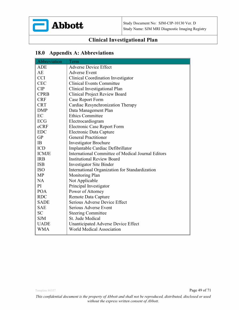
Study Document No: SJM-CIP-10130 Ver. D
Study Name: SJM MRI Diagnostic Imaging Registry
Clinical Investigational Plan 18.0 Appendix A: Abbreviations Abbreviation Term ADE AE CCI CEC CIP CPRB CRF CRT DMP EC ECG eCRF EDC GP IB ICD ICMJE IRB ISB ISO MP NA PI POA RDC SADE SAE SC SJM UADE WMA
Adverse Device Effect Adverse Event Clinical Coordination Investigator Clinical Events Committee Clinical Investigational Plan Clinical Project Review Board Case Report Form Cardiac Resynchronization Therapy Data Management Plan Ethics Committee Electrocardiogram Electronic Case Report Form Electronic Data Capture General Practitioner Investigator Brochure Implantable Cardiac Defibrillator International Committee of Medical Journal Editors Institutional Review Board Investigator Site Binder International Organization for Standardization Monitoring Plan Not Applicable Principal Investigator Power of Attorney Remote Data Capture Serious Adverse Device Effect Serious Adverse Event Steering Committee St. Jude Medical Unanticipated Adverse Device Effect World Medical Association
Template 86357 Page 49 of 71
This confidential document is the property of Abbott and shall not be reproduced, distributed, disclosed or used without the express written consent of Abbott.
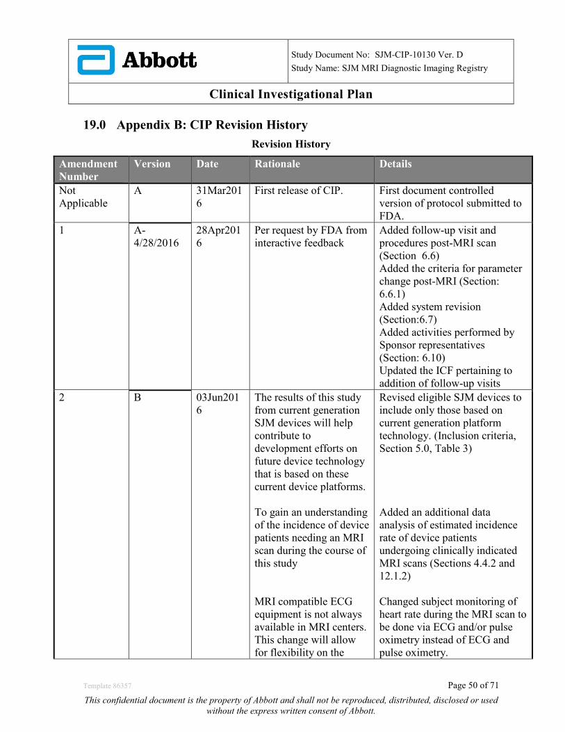
Study Document No: SJM-CIP-10130 Ver. D
Study Name: SJM MRI Diagnostic Imaging Registry
Clinical Investigational Plan 19.0 Appendix B: CIP Revision History
Revision History
Amendment Number
Version Date Rationale Details
Not Applicable
A 31Mar2016
First release of CIP. First document controlled version of protocol submitted to FDA.
1 A- 4/28/2016
28Apr2016
Per request by FDA from interactive feedback
Added follow-up visit and procedures post-MRI scan (Section 6.6) Added the criteria for parameter change post-MRI (Section: 6.6.1) Added system revision (Section:6.7) Added activities performed by Sponsor representatives (Section: 6.10) Updated the ICF pertaining to addition of follow-up visits
2 B 03Jun2016
The results of this study from current generation SJM devices will help contribute to development efforts on future device technology that is based on these current device platforms. To gain an understanding of the incidence of device patients needing an MRI scan during the course of this study MRI compatible ECG equipment is not always available in MRI centers. This change will allow for flexibility on the
Revised eligible SJM devices to include only those based on current generation platform technology. (Inclusion criteria, Section 5.0, Table 3) Added an additional data analysis of estimated incidence rate of device patients undergoing clinically indicated MRI scans (Sections 4.4.2 and 12.1.2) Changed subject monitoring of heart rate during the MRI scan to be done via ECG and/or pulse oximetry instead of ECG and pulse oximetry.
Template 86357 Page 50 of 71
This confidential document is the property of Abbott and shall not be reproduced, distributed, disclosed or used without the express written consent of Abbott.
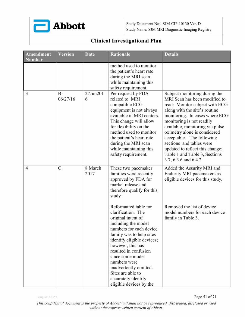
Study Document No: SJM-CIP-10130 Ver. D
Study Name: SJM MRI Diagnostic Imaging Registry
Clinical Investigational Plan
Amendment Number
Version Date Rationale Details
method used to monitor the patient’s heart rate during the MRI scan while maintaining this safety requirement.
3 B- 06/27/16
27Jun2016
Per request by FDA related to: MRI compatible ECG equipment is not always available in MRI centers. This change will allow for flexibility on the method used to monitor the patient’s heart rate during the MRI scan while maintaining this safety requirement.
Subject monitoring during the MRI Scan has been modified to read: Monitor subject with ECG along with the site’s routine monitoring. In cases where ECG monitoring is not readily available, monitoring via pulse oximetry alone is considered acceptable. The following sections and tables were updated to reflect this change: Table 1 and Table 3, Sections 3.7, 6.3.6 and 6.4.2
4 C 8 March 2017
These two pacemaker families were recently approved by FDA for market release and therefore qualify for this study Reformatted table for clarification. The original intent of including the model numbers for each device family was to help sites identify eligible devices; however, this has resulted in confusion since some model numbers were inadvertently omitted. Sites are able to accurately identify eligible devices by the
Added the Assurity MRI and Endurity MRI pacemakers as eligible devices for this study. Removed the list of device model numbers for each device family in Table 3.
Template 86357 Page 51 of 71
This confidential document is the property of Abbott and shall not be reproduced, distributed, disclosed or used without the express written consent of Abbott.

Study Document No: SJM-CIP-10130 Ver. D
Study Name: SJM MRI Diagnostic Imaging Registry
Clinical Investigational Plan
Amendment Number
Version Date Rationale Details
family trademark name listed in labelling. Minor typos corrected for clarification
Misspelling was corrected in the Study Flow Chart in Section 1.1 and in the Informed Consent Form Template.
5 D 15 June 2017
The sponsor is interested in evaluating the effect of MRI on image quality/artifacts separately for Pacemakers/CRT-Ps and ICDs/CRT-Ds since there is a significant difference in the components in devices with defibrillation capabilities (e.g. high voltage capacitor, high voltage transformer and larger battery) which may have an impact on image quality/artifacts. Minor change for clarification/consistency
Minor change for clarification/consistency Minor change for clarification/consistency
Modified the primary endpoint to be evaluated separately for PM/CRT-Ps and ICD/CRT-Ds Increased the total enrollment from 150 to 300 subjects which will come from 150 PM/CRT-Ps and 150 ICD/CRT-Ds. Updated enrollment cap per site from 22 to 45 to maintain 15% of total enrollments (300). Updated the study duration from 1.5 to 2 years to 2 to 3 years in Sections 4.2 and 4.2.2. Section 6.10. Replaced reference to Sponsor field clinical engineers with Sponsor representatives. Section 8.3.2 and 8.4.1 replaced 72 hour adverse event and death reporting period to 10 days to be consistent with prior sections 8.2 and 8.1.4. Sections 4.2.1 and 12.2 now clarify that once the minimum
Template 86357 Page 52 of 71
This confidential document is the property of Abbott and shall not be reproduced, distributed, disclosed or used without the express written consent of Abbott.
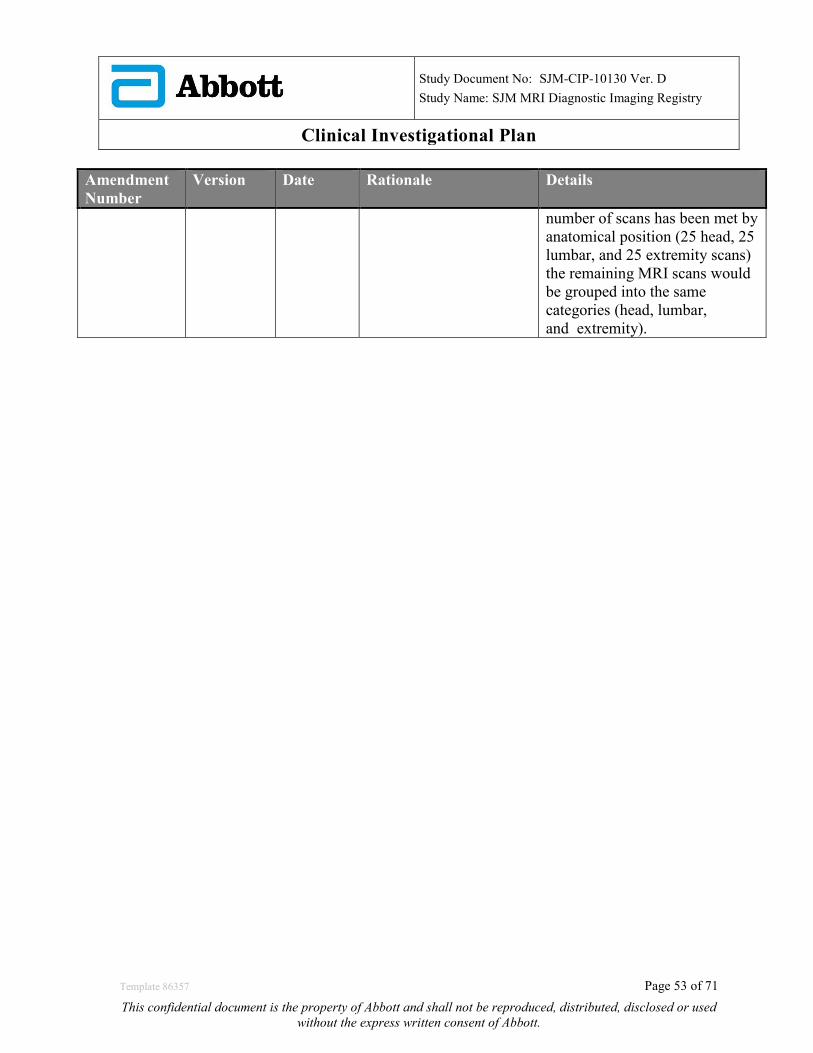
Study Document No: SJM-CIP-10130 Ver. D
Study Name: SJM MRI Diagnostic Imaging Registry
Clinical Investigational Plan
Amendment Number
Version Date Rationale Details
number of scans has been met by anatomical position (25 head, 25 lumbar, and 25 extremity scans) the remaining MRI scans would be grouped into the same categories (head, lumbar, and extremity).
Template 86357 Page 53 of 71
This confidential document is the property of Abbott and shall not be reproduced, distributed, disclosed or used without the express written consent of Abbott.
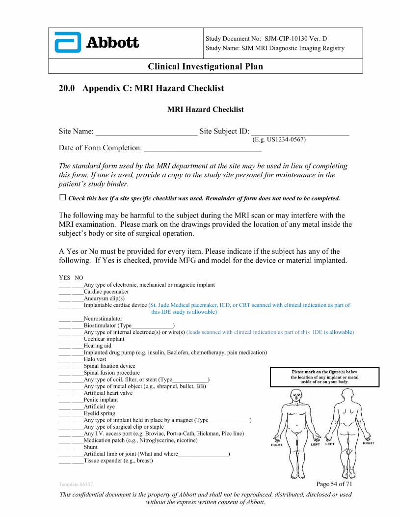
Study Document No: SJM-CIP-10130 Ver. D
Study Name: SJM MRI Diagnostic Imaging Registry
Clinical Investigational Plan 20.0 Appendix C: MRI Hazard Checklist
MRI Hazard Checklist Site Name: __________________________ Site Subject ID: _________________________ (E.g. US1234-0567) Date of Form Completion: ______________________________ The standard form used by the MRI department at the site may be used in lieu of completing this form. If one is used, provide a copy to the study site personel for maintenance in the patient’s study binder.
□ Check this box if a site specific checklist was used. Remainder of form does not need to be completed. The following may be harmful to the subject during the MRI scan or may interfere with the MRI examination. Please mark on the drawings provided the location of any metal inside the subject’s body or site of surgical operation. A Yes or No must be provided for every item. Please indicate if the subject has any of the following. If Yes is checked, provide MFG and model for the device or material implanted. YES NO ____ ____Any type of electronic, mechanical or magnetic implant ____ ____Cardiac pacemaker ____ ____Aneurysm clip(s) ____ ____Implantable cardiac device (St. Jude Medical pacemaker, ICD, or CRT scanned with clinical indication as part of this IDE study is allowable) ____ ____Neurostimulator ____ ____Biostimulator (Type______________) ____ ____Any type of internal electrode(s) or wire(s) (leads scanned with clinical indication as part of this IDE is allowable) ____ ____Cochlear implant ____ ____Hearing aid ____ ____Implanted drug pump (e.g. insulin, Baclofen, chemotherapy, pain medication) ____ ____Halo vest ____ ____Spinal fixation device ____ ____Spinal fusion procedure ____ ____Any type of coil, filter, or stent (Type____________) ____ ____Any type of metal object (e.g., shrapnel, bullet, BB) ____ ____Artificial heart valve ____ ____Penile implant ____ ____Artificial eye ____ ____Eyelid spring ____ ____Any type of implant held in place by a magnet (Type______________) ____ ____Any type of surgical clip or staple ____ ____Any I.V. access port (e.g. Broviac, Port-a-Cath, Hickman, Picc line) ____ ____Medication patch (e.g., Nitroglycerine, nicotine) ____ ____Shunt ____ ____Artificial limb or joint (What and where_________________) ____ ____Tissue expander (e.g., breast)
Template 86357 Page 54 of 71
This confidential document is the property of Abbott and shall not be reproduced, distributed, disclosed or used without the express written consent of Abbott.

Study Document No: SJM-CIP-10130 Ver. D
Study Name: SJM MRI Diagnostic Imaging Registry
Clinical Investigational Plan ____ ____Removable dentures, false teeth or partial plate ____ ____Diaphragm, IUD, Pessary (Type___________________) ____ ____Surgical mesh (Location____________________) ____ ____Body piercing (Location____________________) ____ ____Wig, hair implants ____ ____Tattoos or tattooed eyeliner ____ ____Radiation seeds (e.g., cancer treatment) ____ ____Any implanted items (e.g., pins, rods, screws, nails, plates, wires) ____ ____Any hair accessories (e.g., bobby pins, barrettes, clips) ____ ____Jewelry ____ ____Any other type of implanted item (Type____________________) Instructions for patients 1. You are urged to use the ear plugs or headphones that we supply for use during your MRI examination since some
patients may find the noise levels unacceptable and the noise levels may affect your hearing. 2. Remove all jewelry (e.g., necklaces, pins, rings). 3. Remove all hair pins, bobby pins, rings). 4. Remove all dentures, false teeth, partial dental plates. 5. Remove all hearing aids. 6. Remove eyeglasses. 7. Remove your watch, pager, cell phone, credit and bank cards, and all other cards with a magnetic strip. 8. Remove body piercing objects. 9. Use gown, if provided, or remove all clothing with metal fasteners, zippers. Subject attests that the above information is correct to the best of subject’s knowledge. Subject has read or has had contents read to subject, understands the entire contents of this form, and had the opportunity to ask questions regarding the information on this form. Subject signature______________________________________________________________________________
Hazard Checklist for Radiology Personnel The following items are not permitted to be used during the MRI scan.
• Endotracheal tube • Foley catheter with temperature sensor and/or metal clip • Swan-Ganz catheter • Rectal probe • Extraventricular device • Esophageal probe • Arterial line transducer • Tracheotomy tube
• Guidewires
Radiology Personnel Completing Checklist
• Print Name:________________________________ Signature___________________________ Date:__________
Template 86357 Page 55 of 71
This confidential document is the property of Abbott and shall not be reproduced, distributed, disclosed or used without the express written consent of Abbott.
Study Document No: SJM-CIP-10130 Ver. D
Study Name: SJM MRI Diagnostic Imaging Registry
Clinical Investigational Plan 21.0 Appendix D: List of Clinical Investigation Sites and IRB A list of Clinical Investigational sites and IRB is kept under a separate cover and is available upon request.
Template 86357 Page 56 of 71
This confidential document is the property of Abbott and shall not be reproduced, distributed, disclosed or used without the express written consent of Abbott.
Study Document No: SJM-CIP-10130 Ver. D
Study Name: SJM MRI Diagnostic Imaging Registry
Clinical Investigational Plan 22.0 Appendix E: Sample Informed Consent
STUDY TITLE AND NUMBER A Clinical Evaluation of the Diagnostic Utility of MRI
scans in patients implanted with St. Jude Medical Pacemakers, ICDs, and CRTs
SPONSOR
St. Jude Medical, Inc. (SJM)
PRINCIPAL INVESTIGATOR
SITE NAME
Introduction You are being asked to take part in this research study because you are currently implanted with a St. Jude Medical implantable cardiac device (i.e. pacemaker, implanted cardioverter-defibrillator (ICD), or cardiac resynchronization therapy (CRT)) and your doctor has recommended that you have a magnetic resonance imaging (MRI) scan for clinical purposes. This form explains why this research is being done and what your role will be if you decide to participate. This form also talks about the possible risks that may happen if you take part in this study. This study is sponsored by St. Jude Medical. This company manufactures medical devices intended to treat various medical conditions. Please read this form, and ask your study doctor any questions you may have about the study so that your questions may be answered before you decide if you want to take part in the study. Please take your time and talk about this information with your family, friends, or family doctor. This consent form may contain some words that you do not understand. It is important that you understand what is in this form. It will explain the different activities you will be asked to do or participate if you take part in the research study and what the risks might be; whether or not you do take part is entirely your choice. Please ask the study doctor or the study staff to explain any words or information that you do not understand. If you decide you want to take part in the research project, you will be asked to sign the consent section before any study-related activities are performed. By signing it you are telling us that you:
Template 86357 Page 57 of 71
This confidential document is the property of Abbott and shall not be reproduced, distributed, disclosed or used without the express written consent of Abbott.
Study Document No: SJM-CIP-10130 Ver. D
Study Name: SJM MRI Diagnostic Imaging Registry
Clinical Investigational Plan
• Understand what you have read • Consent to take part in the research project • Consent to have the tests and treatments that are described • Consent to the use of your personal and health information as described.
Taking part in this study is entirely voluntary. If you don’t wish to take part, you don’t have to. You will receive the best possible care whether or not you take part in the study. Refusing participation will not involve any penalty or loss of benefit. If you decide to take part in this study, you must sign your name at the end of this form. No research activity can be performed until you sign this form. What is the purpose of this study? The purpose of this study is to characterize the image quality and clinical usefulness of MRI scans in patients with St. Jude Medical cardiac devices. What is the device being tested? The devices being tested are St. Jude Medical pacemakers, ICDs, and CRTs that have been approved for use by the Food and Drug Administration (FDA) and are commercially available. However, these devices have not yet been FDA-approved to undergo an MRI scan. This study is testing the quality of the images and clinical usefulness of MRI scans in patients with these implantable cardiac devices that have already been recommended by their doctor to have an MRI scan for clinical reasons. What will be requested from you if you take part in this study? This research study includes a screening/qualification phase to determine if you are a good candidate for the study. Your doctor or other study personnel will ask you medically related questions, such as whether you have been implanted with a St. Jude Medical pacemaker, ICD, or CRT device for at least 6 weeks. Other questions you may be asked include:
• Age • Gender • Past medical history • Past surgical history • Whether you have any type of permanent implants (for example, staples, wires,
pins, rods, screws, plates, cardiac stents, insulin pumps, meshes, dentures, braces, etc.) that are metallic
• Whether you’ve had any accidents or been injured by metallic objects
Template 86357 Page 58 of 71
This confidential document is the property of Abbott and shall not be reproduced, distributed, disclosed or used without the express written consent of Abbott.
Study Document No: SJM-CIP-10130 Ver. D
Study Name: SJM MRI Diagnostic Imaging Registry
Clinical Investigational Plan In order to participate in this study, you must meet all the inclusion criteria and none of the exclusion criteria to safely undergo an MRI scan for this study. Based on your answers and your medical history, your doctor will decide if you qualify to take part in the SJM MRI Diagnostic Imaging Registry. If you do not qualify for the study, your participation will end. If your doctor determines that you qualify, and you decide to take part in this study, the figure below describes your involvement in this trial. You will be having an MRI scan as part of your routine clinical care as recommended by your doctor. The data collection for this study is experimental and would occur only if you choose to participate in this trial.
Template 86357 Page 59 of 71
This confidential document is the property of Abbott and shall not be reproduced, distributed, disclosed or used without the express written consent of Abbott.
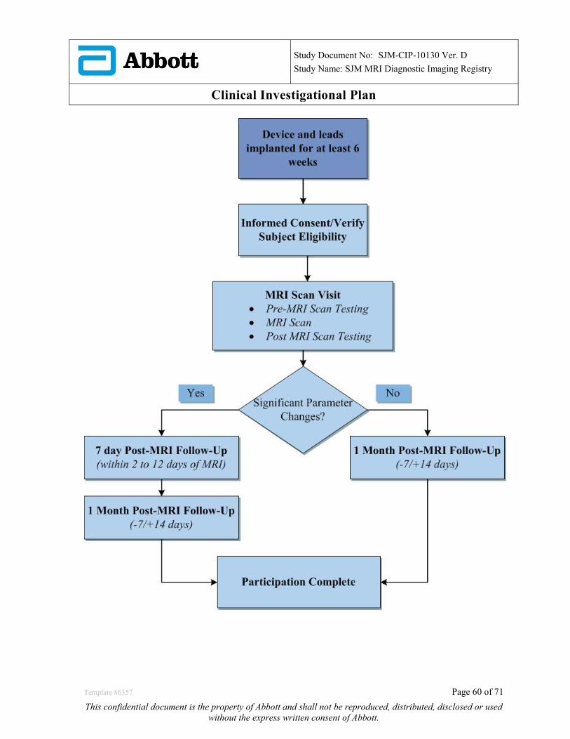
Study Document No: SJM-CIP-10130 Ver. D
Study Name: SJM MRI Diagnostic Imaging Registry
Clinical Investigational Plan
Template 86357 Page 60 of 71
This confidential document is the property of Abbott and shall not be reproduced, distributed, disclosed or used without the express written consent of Abbott.
Study Document No: SJM-CIP-10130 Ver. D
Study Name: SJM MRI Diagnostic Imaging Registry
Clinical Investigational Plan There may be a representative from the sponsor at your study visit and the representative may carry out some of the study procedures. The study doctor may direct a representative from the sponsor to collect information from your implanted device. At the study doctor’s direction, the sponsor representative may also program your device or run tests to see if your device is working as expected. The sponsor’s representative will work under the direction of your study doctor or other care provider. You will be required to have your implanted lead(s) and device tested in the same way as a regular device check-up at the MRI visit (before and after the MRI scan).The following information will be collected.
a. Capture threshold (how much energy it takes to make your heart beat) b. Sensing threshold (how well your device can sense your heart beat) c. Pacing impedance of your implanted lead d. High voltage lead impedance of your implanted lead e. Remaining battery capacity
The section below describes the procedures that will occur in detail 1. MRI Scan Visit
Before the MRI Scan:
• A pregnancy test may be given as part of your routine care (for women of child bearing age). If you are pregnant, you will be withdrawn from the study.
• Before the actual MRI scan, your implanted device will be tested using a programmer in the same way as at a regular clinical check-up. You should not feel any discomfort during this procedure.
• Your device settings may be changed to prepare for the MRI scan • A member of your study doctor’s staff or the radiological team will ask you a
series of questions (MRI Hazard Checklist) to make sure you meet the safety requirement to receive the MRI scan.
• You may be asked to undress, and put on a gown for the MRI scan.
During the MRI Scan:
• Once you are cleared to have an MRI scan, you will be placed on a padded table and positioned for your exam. Electrodes (sticky pads) may be placed on your chest, and the electrodes will be connected to an electrocardiogram machine (ECG). The ECG allows the MRI staff to monitor your heart rhythm during the exam.
• A pulse oximeter may be placed on your finger tip. The pulse oximeter is a painless finger clip used to measure the amount of oxygen in your body. This
Template 86357 Page 61 of 71
This confidential document is the property of Abbott and shall not be reproduced, distributed, disclosed or used without the express written consent of Abbott.
Study Document No: SJM-CIP-10130 Ver. D
Study Name: SJM MRI Diagnostic Imaging Registry
Clinical Investigational Plan
allows the doctor to see how much oxygen content is in your blood during the MRI.
• A soft padded coil may be placed at the area where the pictures will be taken. Another coil may be placed on or behind your head.
• The table on which you are lying will be moved to the center of the MRI magnet, which looks like a short narrow tube. Even though the tube is open, some people feel confined in small places. If this bothers you, please notify the MRI staff.
• During the exam, you will be monitored. The MRI staff is able to see and hear you. You will be able to hear the MRI staff, too.
• When MRI pictures are taken, radio signals and magnetic fields are used to generate an image. When this happens, it is normal for the MRI machine to make loud, banging, and clicking noises. You may be asked to wear earplugs or headphones for your comfort during the exam.
• You may feel warm during the scan. This is expected. If you feel discomfort or have pain in the area where your device is implanted, please let your study doctor or other study staff know.
• When the MRI exam is finished, you will be taken out of the MRI magnet and the pulse oximeter and/or ECG electrodes will be removed.
After the MRI Scan:
• After you have your scan, your device will be tested using a programmer and reset to your regular settings. Your device settings may be adjusted if changes occurred during the MRI. This device testing is done the same way as at a regular clinical check-up. You should not feel any discomfort while your device is being tested.
Follow-Up Visits The number of scheduled follow-up visits will be based upon the change, if any, in your device measurements from the MRI Scan Visit (pre-MRI scan to post-MRI scan measurements); these will range from 1 to 2 visits.
• Patients that did not have any of the significant change in device measurements will be required to return for one follow-up visit 1 month (-7/+ 14 days) after the MRI scan.
• Patients with significant change in device measurements in device will be required to return for 2 follow-up visits:
1. Within 2 to 12 days after the MRI scan 2. 1 Month (-7/+14 days) after the MRI scan
Template 86357 Page 62 of 71
This confidential document is the property of Abbott and shall not be reproduced, distributed, disclosed or used without the express written consent of Abbott.

Study Document No: SJM-CIP-10130 Ver. D
Study Name: SJM MRI Diagnostic Imaging Registry
Clinical Investigational Plan During the follow-up visit(s), your device will be tested using a programmer in the same way as at a regular clinical check-up. You should not feel any discomfort during this procedure. You will also be asked about any changes in your health status since your previous visit. Your study participation will end following the completion of the follow-up visit(s). How long will the study last? If you agree to take part in the study, your involvement will last from the time you consent to take part in this study through the end of your follow-up visit (s). Please ask your doctor how long your MRI scan visit is expected to take. The duration of your MRI scan visit depends on the body part(s) you need scanned and the complexity of the scan your physician has requested. Up to 300 people will take part in this research at approximately 100 sites in the United States. What are the possible risks and discomforts? There are risks, discomforts, and inconveniences associated with any research study to you (or to an embryo, unborn child or nursing infant if you become pregnant). These deserve careful thought. While in the study, you will receive an MRI scan. The risks and adverse effects of the MRI scan are listed below but they will vary from person to person. Your participation in this study will not add any additional risks to those you will have already been exposed to by having the MRI scan for clinical purposes. You should talk with the study doctor if you have any questions. Risks associated with MRI scans of patients with Cardiac Devices: a) Potential MRI Scan Adverse Events:
• Claustrophobia (fear of enclosed spaces) • Mild diaphoresis (sweating) • Hearing impairment (difficulty hearing) • Sensation of bodily warmth • Sensation of warmth at device pocket not arising to the level of discomfort • Body stiffness related to immobility
There will be emergency personnel and equipment on hand for your safety. Please tell the study doctor, nurse or technician if you experience any unusual sensations or discomfort during the scan.
Template 86357 Page 63 of 71
This confidential document is the property of Abbott and shall not be reproduced, distributed, disclosed or used without the express written consent of Abbott.

Study Document No: SJM-CIP-10130 Ver. D
Study Name: SJM MRI Diagnostic Imaging Registry
Clinical Investigational Plan b) Potential Pacemaker, ICD, or CRT System Adverse Events:
• Lead electrode heating and tissue (heart muscle) damage, resulting in loss of sensing or capture or both: A portion of the lead may heat up causing damage to the heart muscle resulting in the loss of some of the device’s electrical functions.
• Lead heating resulting in a blood clot or blockage of a blood vessel • Device heating resulting in tissue damage in the implant pocket or patient
discomfort or both • Induced currents on leads resulting in continuous capture (continuous pacing of
the heart), VT/VF (abnormal beating of the lower heart chambers), hemodynamic collapse (blood circulation failure), or all three
• Damage to the device or leads causing: o the system to fail to detect or treat irregular heartbeats o the system to treat the patient’s condition incorrectly
• Damage to the functionality or mechanical integrity of the device resulting in the inability of the device to communicate with the programmer
• Movement or vibration of the device or leads • Lead dislodgment (lead comes loose from the heart) • When the device is programmed to be in the MRI environment, it paces
independent of the natural heart rhythm. This pacing may happen at the same time of a natural heart beat and may cause VT/VF (abnormal beating of the lower heart chambers).
• Syncope (fainting) due to loss of pacing if no pacing support is programmed with MRI settings
• Pulmonary embolism (blockage of the lung’s blood vessel) • Death due to untreated spontaneous arrhythmia because tachyarrhythmia therapy
is disabled when the device is programmed to be in an MRI environment
These risks would also apply if you received an MRI scan with your device regardless of your participation in this study. There may be other risks or discomforts to you (or to an embryo, unborn child or nursing infant if you become pregnant) that are not known at this time. If important information is learned during the course of this research study, your doctor will be notified by St. Jude Medical. Your doctor will discuss with you important new information that is learned during the course of this study that may affect your condition or willingness to continue to take part in this research study. What are the risks for women of childbearing age? If you are a woman who is able to become pregnant, it is expected that you will use an effective method of birth control to prevent exposing a fetus to a potentially dangerous
Template 86357 Page 64 of 71
This confidential document is the property of Abbott and shall not be reproduced, distributed, disclosed or used without the express written consent of Abbott.

Study Document No: SJM-CIP-10130 Ver. D
Study Name: SJM MRI Diagnostic Imaging Registry
Clinical Investigational Plan environment with unknown risk. If you are pregnant or currently breast feeding, you may not participate in this study because you or your child may be exposed to an unknown risk. If you plan to become pregnant in the next 6 months, you should discuss your participation with your study doctor. Patients who become pregnant while taking part in the study should contact the study doctor right away. What are the possible benefits to you or others? There are no direct benefits to you by participating in this research study. The information gathered in this study will add to the understanding of treatment options for patients with implanted cardiac devices who need to have an MRI scan performed. If you do not want to take part in this study, what other options are available to you? Your study doctor will discuss other options available to you, such as your choice not to take part in this study. If you choose to take part in this study, what are the costs? For all patients enrolled in the study, the cost of the MRI scan performed at the MRI visit, and the cost of the device checks performed during the study will be covered by the study sponsor. Will you receive payment for taking part in this study?
You will receive a payment of $25 for each of the post-MRI scan follow-up visit(s) to help cover the costs of travel and parking related to your participation in the research study What if the device needs to be removed? In the event your St. Jude Medical device(s) has to be removed, as a result of the MRI it will be returned to St. Jude Medical for analysis. Should you withdraw from this study and choose to have your St. Jude Medical device(s) removed, the cost will be your responsibility. In the event of your death, your implanted St. Jude Medical device(s) may be removed and returned to St. Jude Medical for analysis. The study doctor will get your family’s approval prior to removing the device(s). What if you are injured because of this study? If you suffer any injuries, illnesses, or complications as a direct result of participation in this study, the Sponsor will pay for all reasonable and necessary treatment of injuries, undesirable side effects, or adverse reactions directly resulting from participation in this study, so long as
Template 86357 Page 65 of 71
This confidential document is the property of Abbott and shall not be reproduced, distributed, disclosed or used without the express written consent of Abbott.
Study Document No: SJM-CIP-10130 Ver. D
Study Name: SJM MRI Diagnostic Imaging Registry
Clinical Investigational Plan you followed all study instructions. The Sponsor will not cover the cost of injuries resulting from participation in the study to the extent that they are caused by your failure, or that of the hospital or study doctor, to follow study instructions or other negligence, the natural progression of an underlying condition (whether diagnosed or not) or pre-existing condition, or events that would have been expected using currently approved therapies for your condition. However, signing this consent form in no way limits your legal rights against the Sponsor, investigators, or anyone else, and you do not release the study doctors or participating institutions from their legal and professional responsibilities. During the study, if you experience any injuries, illnesses, or complications from taking part in this study, please contact Dr. ______________ at ___-___-_____. What are your rights if you decide to take part in this study? Your signature on this consent form means that you have received information about this research study and that you agree to be a part of the study. You may stop taking part in the study at any time without penalty or loss of benefits to which you are otherwise entitled. If you wish to stop taking part in this research study for any reason, you should contact Dr. ______________ at ___-___-_____. If you do withdraw your consent during the study, the study doctor and relevant study staff will not collect additional personal information from you, although personal information already collected will be retained to ensure that the results of the research project can be measured properly and to comply with the law. You should be aware that data collected by the sponsor up to the time you withdraw will be part of the study results. Your study doctor or designee will discuss with you what follow-up is required if you decide to withdraw, or are withdrawn from the study before the study is finished. Your doctor or the sponsor of the study (St. Jude Medical, Inc.) may also stop your participation in the study at any time, without your consent, for any reason.
How will your information be kept confidential? If you decide to take part in this study, your medical records and personal information will be kept confidential to the extent allowed by federal, state, and local law. A special code (number combination) will be used to identify your personal health information. However, information from the study may be exported to countries where different data protection laws apply. The data protection laws in other countries may be less strict than those of your country.
Template 86357 Page 66 of 71
This confidential document is the property of Abbott and shall not be reproduced, distributed, disclosed or used without the express written consent of Abbott.

Study Document No: SJM-CIP-10130 Ver. D
Study Name: SJM MRI Diagnostic Imaging Registry
Clinical Investigational Plan If you decide to participate in the study, the study sponsor and others who work with the study, such as the study staff and Institutional Review Board (IRB) will see health information about you. The IRB is a group of people who perform independent review of research as required by laws governing this type of research. U.S. Food and Drug Administration (FDA), other regulatory agencies, IRB, and sponsor’s representative may inspect and copy your medical records. The information collected about you may be used in several ways. Information about you and your health that might identify you may be given to others to carry out the research study. Your study doctor may use some of the information in making decisions about your care. The sponsor may use the information in any of the following ways:
• To analyze and make conclusions about the results of the study, • In documents sent to the government agencies throughout the world including the U.S.
Food and Drug Administration (FDA) regarding this study, • For reporting undesirable events to the FDA and other government health agencies, • To provide overall study results to other study doctors, including in publications, • To conduct new medical research, to reanalyze the study results in the future or to
combine your information with information from other studies, • To develop new medical products and procedures, and other product-development
related activities. Your name will not be provided for publications in medical journals. While using the information in these ways, the sponsor may give study data to its affiliated companies in the U.S. or other countries. The sponsor may also share the information with its business partners or companies it hires to provide study-related services. Information received during the study will not be used for any mailing lists or sold to anyone for marketing purposes. Your permission for the use, retention, and sharing of your identifiable health information will expire upon completion of the study. At that time the research information not already in your medical record will not be given to the sponsor. A description of this clinical study will be available on http://www.ClinicalTrials.gov, as required by U.S. Law. This Web site will not include information that can identify you. At most, the Web site will include a summary of the results. You can search the Web site at any time. Please note information may not be available on ClinicalTrials.gov until the study is completed and the device is marketed. If you receive medical care from a doctor other than your study doctor while taking part in this study, you agree that your medical records will be made available for the collection of data related to this study.
Template 86357 Page 67 of 71
This confidential document is the property of Abbott and shall not be reproduced, distributed, disclosed or used without the express written consent of Abbott.

Study Document No: SJM-CIP-10130 Ver. D
Study Name: SJM MRI Diagnostic Imaging Registry
Clinical Investigational Plan Who can you contact for study information? If you have any questions about the study or taking part in this study, please contact Dr. ______________ at ___-___-_____. In addition, if you have any concerns, complaints or questions about your rights as a research patient or an injury that you believe is a research-related, please contact:
Name of person at IRB: Title of person at IRB: IRB phone number: IRB email, if known:
Consent and authorization for participation in this study Taking part in this study is entirely voluntary. You are making a decision on whether or not to take part in the research study. Your signature indicates that you have read the information in this form and have decided to take part in the research study. You will be given a signed copy of this form to keep. I have read all of the above information in this consent and authorization form. I have had the opportunity to ask questions and have received answers concerning areas I did not understand.
Name of Participant (please print)
Signature Date Time: __________
Name of Person Obtaining
Consent (please print)
Signature Date
Template 86357 Page 68 of 71
This confidential document is the property of Abbott and shall not be reproduced, distributed, disclosed or used without the express written consent of Abbott.

Study Document No: SJM-CIP-10130 Ver. D
Study Name: SJM MRI Diagnostic Imaging Registry
Clinical Investigational Plan 23.0 Appendix F: Product Labeling The product labeling for the devices, instructions for use and packaging are all commercially available and will be provided to FDA upon request.
Template 86357 Page 69 of 71
This confidential document is the property of Abbott and shall not be reproduced, distributed, disclosed or used without the express written consent of Abbott.
Study Document No: SJM-CIP-10130 Ver. D
Study Name: SJM MRI Diagnostic Imaging Registry
Clinical Investigational Plan 24.0 Appendix G: Case Report Forms The case report forms for the study are kept under a separate cover, and are available upon request.
Template 86357 Page 70 of 71
This confidential document is the property of Abbott and shall not be reproduced, distributed, disclosed or used without the express written consent of Abbott.
Study Document No: SJM-CIP-10130 Ver. D
Study Name: SJM MRI Diagnostic Imaging Registry
Clinical Investigational Plan References
1 Quint DJ. Indications for emergent MRI of the central nervous system. JAMA. 2000; 283:853–855. 2 Pennell DJ, Sechtem UP, Higgins CB, et al. Clinical indications for cardiovascular magnetic resonance (CMR): Consensus Panel report. Eur Heart J. 2004; 25:1940 –1965.. 3 imv Medical Information Division. 2007 MRI Market Summary Report. Prepared for St. Jude Medical June 2008. 4 Mond H G, Irwin M, Ector H, and Proclemer A. The world survey of cardiac pacing and cardioverter- defibrillators: calendar year 2005 an International Cardiac Pacing and Electrophysiology Society (ICPES) Project. PACE 2008 Sep;31(9):1202-12. 5 Mond H G, Irwin M, Morillo C, Ector H. The world survey of cardiac pacing and cardioverter- defibrillators: Calendar year 2001. PACE 2004 Feb;27:955-64 6 Kalin R and Stanton R. Current Clinical Issues for MRI Scanning of Pacemaker and Defibrillator Patients. PACE 2005;28(4):326-328. 7 Gimbel J R, Johnson D, Levine P A, Wilkoff B L. Safe performance of magnetic resonance imaging on five patients with permanent cardiac pacemakers. PACE 1996;19:913-919. 8 Valhaus C, Sommer T, Lewalter T. et al. Interference with cardiac pacemakers by magnetic resonance imaging : Are there irreversible changes at 0.5 Tesla? PACE 2001; 24: 489-495. 9 Luechinger R, Duru F, Scheidegger, M B, et al. Force and torque effects of a 1.5Tesla MRI scanner on cardiac pacemakers and ICDs. PACE 2001; 24:199-205. 10 Loewy J, Loewy A, and Kendall EJ. Reconsiderations of pacemakers and MR imaging. RadioGraphics 2004; 24:1257-1268. 11 Roguin A, Zviman M M, Meininger G T, et al. Modern pacemaker and implantable cardioverter defibrillator system scan be magnet safe : In vitro and in vivo assessment of safety and function at 1.5T. Circulation 2004; 110:475-482. 12 Levine GN, Gomes AS, Arai AE, et. al. Safety of magnetic resonance imaging in patients with cardiovascular devices: an American Heart Association scientific statement from the Committee on Diagnostic and Interventional Cardiac Catheterization, Council on Clinical Cardiology, and the Council on Cardiovascular Radiology and Intervention: Circulation. 2007;116(24):2878-2891.
Template 86357 Page 71 of 71
This confidential document is the property of Abbott and shall not be reproduced, distributed, disclosed or used without the express written consent of Abbott.