clinical management of root canal perforations: is the ... · general practitioner to refer the...
TRANSCRIPT
Glendale Microendodontics • 1138 n. Brand Boulevard, suite B • Glendale, ca 91202telephone: (818) 552-endo • www.GlendaleMicroendodontics.coM
Endodontic treatment (aka RCT) can be, both, a very re-warding and sometimes chal-lenging dental procedure for the practitioner. While gener-ally endodontic treatment may be straightforward once the root canals have been located and negotiated to length, some-times iatrogenic perforation of the pulp chamber floor or the root becomes a stressful reality for the clinician, and perplex-ing, if not upsetting, for the patient. Is such a tooth doomed in those circumstances? In this month’s Newsletter, I will ad-dress the different factors (loca-tion, size, length of time since perforation, repair material of choice, use of magnification, and the experience of the clini-cian dealing with perforation) that may determine the suc-cess of perforation repair, and the long term retention of such teeth. I will end by presenting some clinical cases from our practice. Procedural accidents pres-ent a source of frustration to the dental clinician. One such accident is the perforation of the tooth during endodontic treatment. However, contrary
to the belief that once a tooth has been perforated, that its prognosis becomes poor to hopeless. Perfora-tion repair can be a very success-ful and predictable procedure, a procedure that is routinely per-formed in our clinic. The factors that determine the success of teeth that have had a perforation include: lo-cation (sub-ossous, coronal, furcal, mid-root, or apical); size (small, medium, or large); length of time since the perfo-ration (recent, or long stand-ing); repair material (MTA, amalgam, Dycal, composite, or IRM); use of magnification (none, loupes, endoscope, or microscope); and the experi-ence of the operator (none, low, medium or extensive). Perforations that are of small size, are sub-osseous in the coronal aspect of the root, are repaired immediately with MTA (due to its sealing ability and its biocompatibility) using a surgi-cal operating microscope (SOM) by an experienced clinician has the best prognosis for long term success. However, perforations of different sizes (provided they are below the crest of the bone) and at different levels of the
tooth will often have good long term success rates if it is re-paired with MTA under proper isolation and moisture control, delivered by specialized carri-ers, using the SOM. The critical keys to successful management include an experienced opera-tor using proper protocol and material under the SOM. Suc-cessful recalls of teeth repaired with MTA date back close to 20 years. It becomes important for a general practitioner to refer the patient who has experienced the unfortunate event of a per-foration as soon as reasonably possible. It behooves the dentist as well as the patient to be seen by an endodontist with exten-sive experience in dealing with procedural accidents, and one who makes full use of a SOM. Both patient and referring doc-tor will often be pleasantly sur-prised as to the long term suc-cess and predictability of such procedure; thereby averting the loss of the tooth and maintain-ing the patient’s natural tooth for a long period of time.
(See photos on reverse side.)
Clinical Management of Root CanalPerforations: Is the Tooth Doomed?
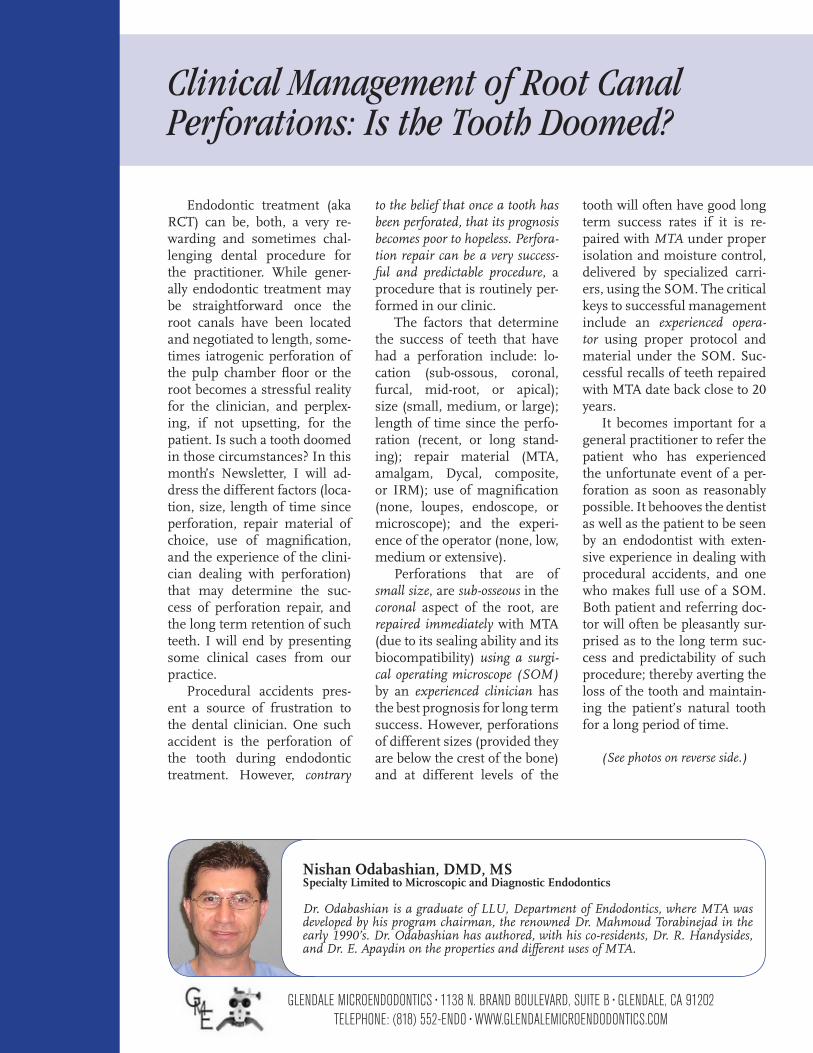
Nishan Odabashian, DMD, MSSpecialty Limited to Microscopic and Diagnostic Endodontics
Dr. Odabashian is a graduate of LLU, Department of Endodontics, where MTA was developed by his program chairman, the renowned Dr. Mahmoud Torabinejad in the early 1990’s. Dr. Odabashian has authored, with his co-residents, Dr. R. Handysides, and Dr. E. Apaydin on the properties and different uses of MTA.
Glendale Microendodontics • 1138 n. Brand Boulevard, suite B • Glendale, ca 91202telephone: (818) 552-endo • www.GlendaleMicroendodontics.coM
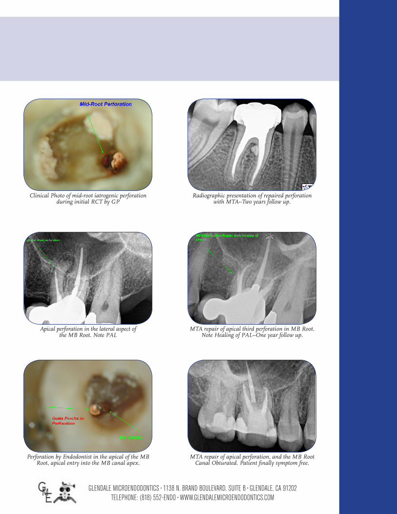
Apical perforation in the lateral aspect of the MB Root. Note PAL
Radiographic presentation of repaired perforation with MTA--Two years follow up.
MTA repair of apical third perforation in MB Root. Note Healing of PAL--One year follow up.
Clinical Photo of mid-root iatrogenic perforationduring initial RCT by GP
Perforation by Endodontist in the apical of the MB Root, apical entry into the MB canal apex.
MTA repair of apical perforation, and the MB Root Canal Obturated. Patient finally symptom free.