clinical neurological examination - cardionotes · pdf fileclinical neurological examination...
TRANSCRIPT
Clinical Neurological Examination
Dr. Satish Kumar, MD
This module will instruct medical students and post graduate trainees on how to perform a
thorough neurological examination. It stresses examination technique, so that the student may perform
the exam in a real clinical setting with authority and confidence. Each examining maneuver is
photographed clearly, with a concise, relevant discussion. An organizational overview of the
examination is provided early in the module to facilitate memorization and overall comprehension.
Furthermore, the neurological terminology pertaining to the examination is fully explained, allowing
one to communicate the results of the exam to fellow medical personnel or to the medicolegal record.
Enjoy the module, and please contact me at [email protected] with any suggestions or
comments.
Introduction- a few basics
The objective of a neurological exam is fourfold:
1. To identify an abnormality in the nervous system.
2. To differentiated peripheral from central nervous system lesions.
3. To try to localize & identify the nature of lesion.
4. To establish internal consistency, i.e. does the patient cooperate fully and are the findings in a
specific patient only a variant of normality?
The neurological exam
The neurological exam can be divided into nine areas. The outline below should be memorized.
Having immediate recall of this outline allows the examiner to quickly proceed through the exam
without omitting any sections. The exam should be performed in an organized, step-wise manner.
I. Preparation for the Neurological Examination
II. General Appearance, including posture, motor activity, vital signs and perhaps meningeal
signs if indicated.
III. Mini Mental Status Exam, including speech observation.
IV. Cranial Nerves, I through XII.
V. Motor System, including muscle atrophy, tone and power.
VI. Sensory System, including vibration, position, pin prick, temperature, light touch and
higher sensory functions.
VII. Reflexes, including deep tendon reflexes, clonus, Hoffman's response and plantar reflex.
VIII. Coordination, gait and Rhomberg's Test
IX. Examining the comatose patient
Throughout these lessons you will see blue text (like this for example). This blue color indicates a
clinical tip or medical pearl.

Preparing for the neurological examination
The patient should be awake and alert, sitting on the examining table facing the examiner. The room
should be quiet and adequately illuminated. It is imperative that the patient is naked, save the patient's
underwear and hospital gown, to perform a full, initial neurological screening of the patient. Future or
serial examinations may be more directed and may not require removal of clothing. It is also important
that the patient is cooperative, non-intoxicated and is able to follow commands during the examination. If
this is not the case, then errors may occur. Eyeglasses, if required, should be worn by the patient and are
a requisite to perform a full ophthalmologic examination.
It is important for the physician to be prepared as well. When the physician is well versed with the
organization of the neurological examination, is equipped with the correct tools, and is constantly
anticipating the next part of the examination, the exam will run smoothly and rapidly with minimal
patient discomfort. The physician should have a basic expectation, derived from the history taken
previously, of what neurological findings may be present in a patient before an examination. These
expectations must be tested and confirmed during the exam to establish a working diagnosis. One must
remember to fully examine a patient and be very objective when results are documented, because initial
expectations are often incorrect. In summary, it is important to fully examine a new patient and keep an
open mind.
The tools required to perform a neurological exam.
There are six basic tools required.-
1. Tendon Hammer
2. Tuning Fork
3. Ophthalmoscope
4. Visual Acuity Card
5. Cotton Wisp
6. Soap
The reflex hammer and accessories.
The reflex hammer is used to illicit deep tendon reflexes
throughout the body. This important item may come with
detachable pin and brush accessories that are used to test
for the sensory modalities of pin prick and light touch,
respectively. If your reflex hammer does not have these
items, then a common safety pin will be an adequate
replacement.
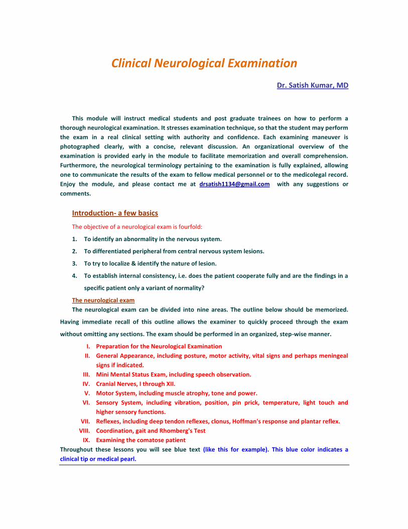
The 256 hertz tuning fork.
This item has multiple uses during the exam. It is used to
test vibration sense throughout the body, to evaluate
conductive versus neurological hearing loss, and may be
placed under either warm or cold water (remember to dry
it off before use) and then utilized for temperature
sensation evaluation. Although one may purchase 126 and
512 hertz tuning forks (the 512 is better for auditory
evaluation and the 126 is optimal for vibratory
examination) the 256 hertz fork is adequate for an initial
examination of both modalities. To cause the fork to
vibrate, wrap it sharply on the palm of your hand before
each time you touch its base to the patient's skin.
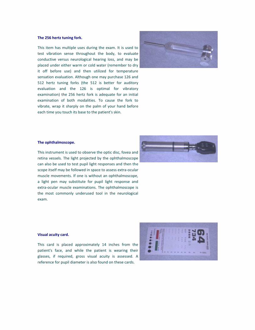
The ophthalmoscope.
This instrument is used to observe the optic disc, fovea and
retina vessels. The light projected by the ophthalmoscope
can also be used to test pupil light responses and then the
scope itself may be followed in space to assess extra-ocular
muscle movements. If one is without an ophthalmoscope,
a light pen may substitute for pupil light response and
extra-ocular muscle examinations. The ophthalmoscope is
the most commonly underused tool in the neurological
exam.
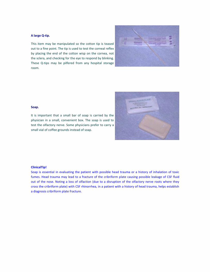
Visual acuity card.
This card is placed approximately 14 inches from the
patient's face, and while the patient is wearing their
glasses, if required, gross visual acuity is assessed. A
reference for pupil diameter is also found on these cards.
A large Q-tip.
This item may be manipulated so the cotton tip is teased
out to a fine point. The tip is used to test the corneal reflex
by placing the end of the cotton wisp on the cornea, not
the sclera, and checking for the eye to respond by blinking.
These Q-tips may be pilfered from any hospital storage
room.
Soap.
It is important that a small bar of soap is carried by the
physician in a small, convenient box. The soap is used to
test the olfactory nerve. Some physicians prefer to carry a
small vial of coffee grounds instead of soap.
ClinicalTip!
Soap is essential in evaluating the patient with possible head trauma or a history of inhalation of toxic
fumes. Head trauma may lead to a fracture of the cribriform plate causing possible leakage of CSF fluid
out of the nose. Noting a loss of olfaction (due to a disruption of the olfactory nerve roots where they
cross the cribriform plate) with CSF rhinorrhea, in a patient with a history of head trauma, helps establish
a diagnosis cribriform plate fracture.
General Appearance
Have the patient sit facing you on the examining table. Take a few seconds to actively observe the patient,
and continue to actively observe the patient during the exam.
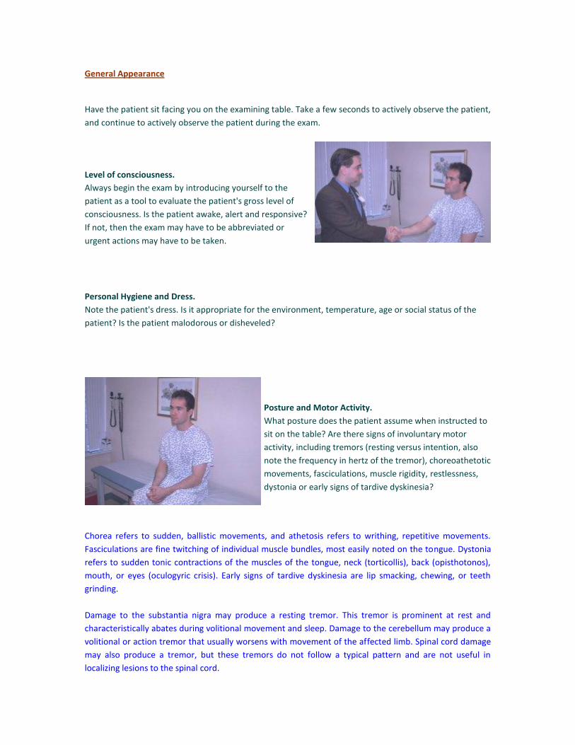
Level of consciousness.
Always begin the exam by introducing yourself to the
patient as a tool to evaluate the patient's gross level of
consciousness. Is the patient awake, alert and responsive?
If not, then the exam may have to be abbreviated or
urgent actions may have to be taken.
Personal Hygiene and Dress.
Note the patient's dress. Is it appropriate for the environment, temperature, age or social status of the
patient? Is the patient malodorous or disheveled?
Posture and Motor Activity.
What posture does the patient assume when instructed to
sit on the table? Are there signs of involuntary motor
activity, including tremors (resting versus intention, also
note the frequency in hertz of the tremor), choreoathetotic
movements, fasciculations, muscle rigidity, restlessness,
dystonia or early signs of tardive dyskinesia?
Chorea refers to sudden, ballistic movements, and athetosis refers to writhing, repetitive movements.
Fasciculations are fine twitching of individual muscle bundles, most easily noted on the tongue. Dystonia
refers to sudden tonic contractions of the muscles of the tongue, neck (torticollis), back (opisthotonos),
mouth, or eyes (oculogyric crisis). Early signs of tardive dyskinesia are lip smacking, chewing, or teeth
grinding.
Damage to the substantia nigra may produce a resting tremor. This tremor is prominent at rest and
characteristically abates during volitional movement and sleep. Damage to the cerebellum may produce a
volitional or action tremor that usually worsens with movement of the affected limb. Spinal cord damage
may also produce a tremor, but these tremors do not follow a typical pattern and are not useful in
localizing lesions to the spinal cord.
Height, Build and Weight.
Is the patient obese or cachectic? If cachectic, note any wasting of the temporalis muscles. Note the
general body proportions and look for any gross deformities. Also check for dysmorphic features,
including low set ears, wide set eyes, small mandible, mongoloid facies, etc.
Vital Signs.
These include temperature, pulse, respiratory rate and blood pressure. It is essential that the vitals always
be taken as an initial assessment of a patient. Emergency measures may have to be taken for drastically
abnormal vital signs.
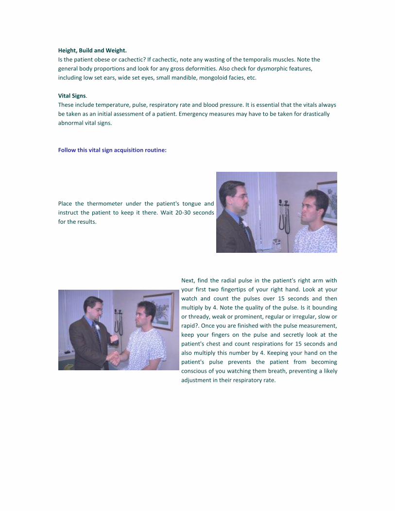
Follow this vital sign acquisition routine:
Place the thermometer under the patient's tongue and
instruct the patient to keep it there. Wait 20-30 seconds
for the results.
Next, find the radial pulse in the patient's right arm with
your first two fingertips of your right hand. Look at your
watch and count the pulses over 15 seconds and then
multiply by 4. Note the quality of the pulse. Is it bounding
or thready, weak or prominent, regular or irregular, slow or
rapid?. Once you are finished with the pulse measurement,
keep your fingers on the pulse and secretly look at the
patient's chest and count respirations for 15 seconds and
also multiply this number by 4. Keeping your hand on the
patient's pulse prevents the patient from becoming
conscious of you watching them breath, preventing a likely
adjustment in their respiratory rate.
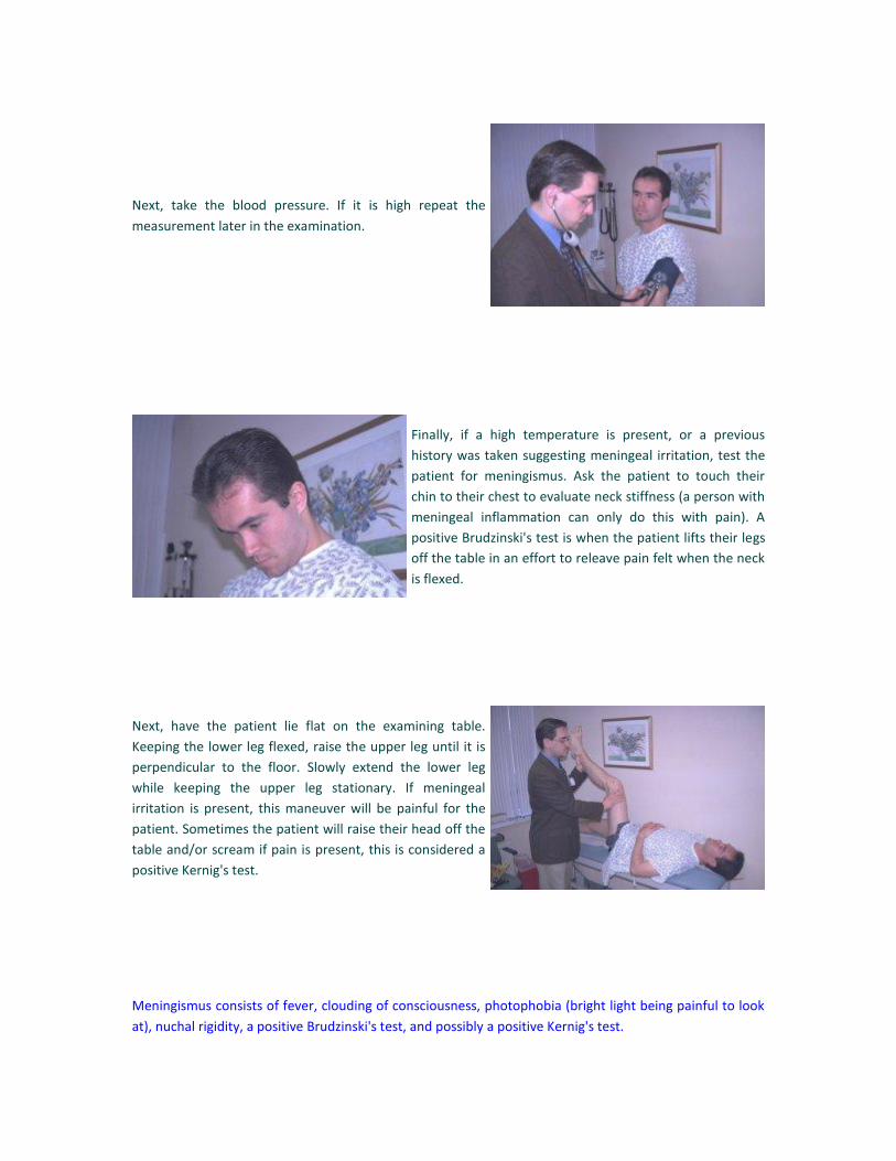
Next, take the blood pressure. If it is high repeat the
measurement later in the examination.
Finally, if a high temperature is present, or a previous
history was taken suggesting meningeal irritation, test the
patient for meningismus. Ask the patient to touch their
chin to their chest to evaluate neck stiffness (a person with
meningeal inflammation can only do this with pain). A
positive Brudzinski's test is when the patient lifts their legs
off the table in an effort to releave pain felt when the neck
is flexed.
Next, have the patient lie flat on the examining table.
Keeping the lower leg flexed, raise the upper leg until it is
perpendicular to the floor. Slowly extend the lower leg
while keeping the upper leg stationary. If meningeal
irritation is present, this maneuver will be painful for the
patient. Sometimes the patient will raise their head off the
table and/or scream if pain is present, this is considered a
positive Kernig's test.
Meningismus consists of fever, clouding of consciousness, photophobia (bright light being painful to look
at), nuchal rigidity, a positive Brudzinski's test, and possibly a positive Kernig's test.

Special Topic: Classic Cerebrospinal Fluid Characteristics
Idiopathic Seizures Clear CSF with normal protein, normal glucose, no WBC's, no RBC's,
normal opening pressure and normal % Gamma globulin.
Bacterial Meningitis: Milky CSF with increased protein, decreased glucose, high WBC's
(PMN predominate), few RBC's, mildly increased opening pressure
and normal % Gamma globulin.
Guillain-Barre Syndrome: Yellow CSF with very high protein (up to a gram), normal glucose, no
WBC's, no RBC's, normal opening pressure and normal % Gamma
globulin.
Subarachnoid Hemorrhage: Yellow CSF with increased protein, normal glucose, few WBC's,
inumerable RBC's, mildly increased opening pressure and normal %
Gamma globulin.
Herpes Simplex Encephalitis: Cloudy CSF with increased protein, normal glucose, increased WBC's
(lymphocyte predominate), few RBC's, increase in opening pressure
and normal % Gamma globulin.
Viral Meningitis: Cloudy CSF with increased protein, normal glucose, increased WBC's
(lymphocyte predominate), no RBC's, normal opening pressure and
normal % Gamma globulin.
Multiple Sclerosis: Clear CSF with mild increase in protein, normal glucose, few WBC's
(lymphocytic predominate), no RBC's, normal opening pressure,
increased % Gamma globulin.
Benign Intracranial Hypertension: Clear CSF with normal protein, normal glucose, no WBC's, no RBC's,
increased opening pressure and normal % Gamma globulin.
COGNITION
The mini mental status exam is an important diagnostic tool used to evaluate a patient's orientation,
concentration and memory (i.e.,cognition). Although it is not imperative to perform a mini mental exam
each time you evaluate a patient neurologically, a baseline score should be established when a patient is
first examined. Serial mini mentals may be performed if deficits in cognition are noted at a later date or
discovered upon the initial examination. The mini mental exam should be photocopied from any standard
psychiatry text and carried with the student until it is administered frequently enough to be committed to
memory. Once the student becomes proficient at instructing the patient to perform this battery, it will
become a quick and efficient part of the neurological exam. More indepth neurocognitive tests may be
necessary if deficits are discovered. Furthermore, it is important to assess a patient's ability to follow
commands to perform a comprehensive, meaningful neurological exam.
The mini mental exam is scored out of a total of 30 points. A score of 24 or higher is considered within
normal range, although specific deficits may be noted and investigated further. A score of below 24 is
indicative of dementia.
PERFORMANCE OF THE MINI MENTAL EXAMINATION.
Orientation.
Date: Ask the patient to state the date. The patient achieves one point for each correct answer of
the following: year, month, day, season and numerical date (a total of five points). If the patient
does not volunteer all of these, ask specifically for the parts omitted.
Location: Ask the patient to state where they are. The patient achieves one point for state,
country, town, hospital and floor (a total of five points). Once again, if parts are omitted, ask for
them specifically.
Registration.
Ask the patient if you can test their memory. State the name of three unrelated items (dog,
pencil, ball) and then ask the patient to repeat the three items. The patient gets one point for
each item repeated, i.e. registered (a total of three points). Ask the patient to remember these
items, because you will ask for the patient to repeat them again later in the examination. In
order to evaluate recent memory later in the exam, make certain all three objects have been
registered. You may have to repeat them 5-6 times.
Attention and Calculation.
Ask the patient to begin with 100 and count backwards by subtracting 7's. The patient receives 1
point for each correct answer with a maximum of five points. If the patient is unable to subtract,
have them spell the word WORLD in reverse, getting 1 point for each correct letter.
Recall.
Ask the patient to repeat the three words that they were asked to remember. Score 0-3.
Language.
Naming: Show the patient a wrist watch and then ask them to name the object shown to them.
Repeat this question showing the patient a pen or pencil. Score 0-2. Repetition: Ask the patient
to repeat the following sentence: "No ifs, ands, or buts." Score 0-1.
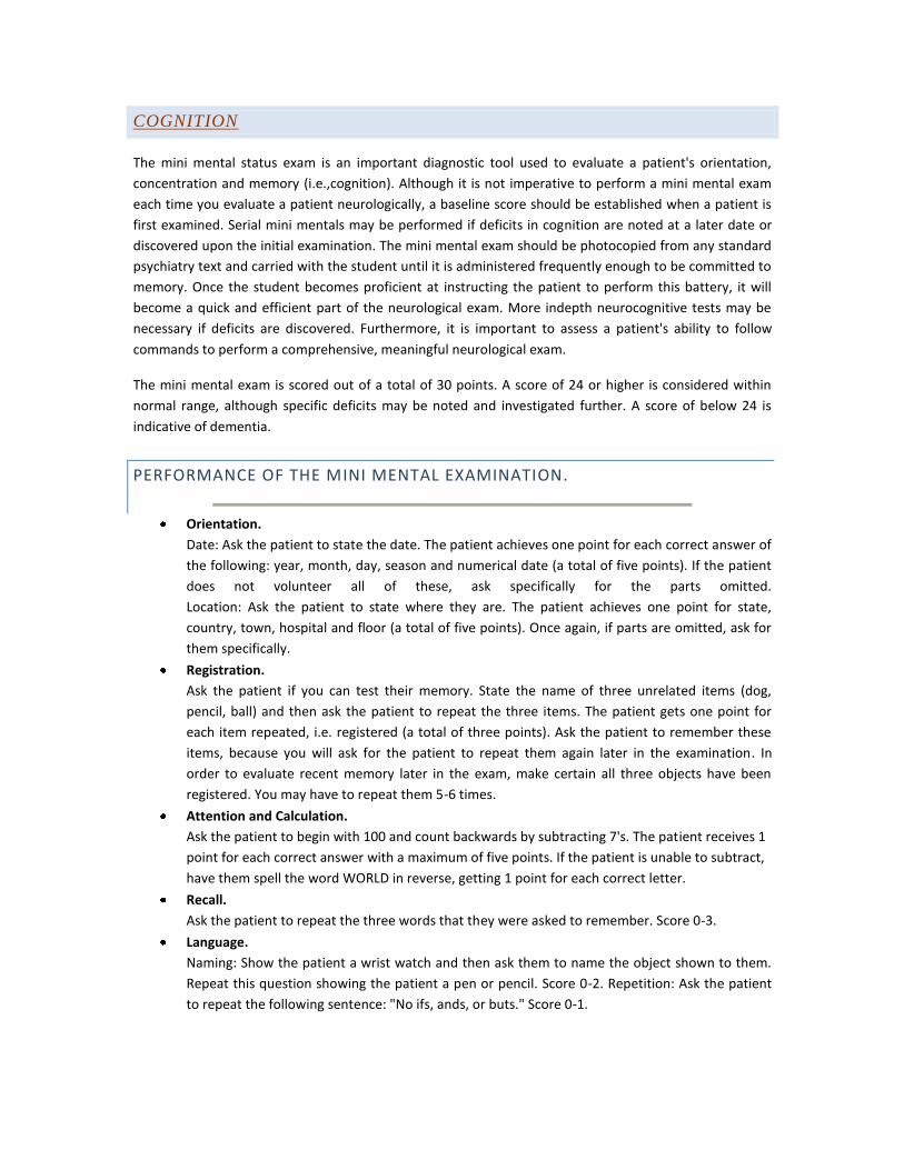
Three step command: Hand the patient a piece of paper
and state the following command: "hold this piece of
paper, fold the paper in half, and place the paper on
the floor". One point for each correct movement,
maximum of 3.
Reading: On a blank sheet of paper write clearly the following: CLOSE YOUR EYES. Show this paper to the
patient and ask them to read it to themselves and do what it says. Score 0-1.
Writing: Give the patient a piece of paper and pen and ask the patient to write any sentence they would
like. The sentence must contain a noun and a verb, yet correct punctuation is not required. Score 0-1.
Copying: On a sheet of paper draw two intersecting pentagons and then ask the patient to copy these
objects. Score 0-1. All ten angles must be present with appropriate intersection points. Ignore tremor.
Consciousness.
Estimate the patient's level of consciousness: alert, lethargic, obtunded, stuporous, or comatose.
An alert patient is vigilantly attentive and keen. A lethargic patient is dull, sluggish and appears
half asleep. An obtunded patient opens their eyes, responds slowly to questions, is somewhat
confused, and has a decreased interest in their environment. A stuporous patient is near
unconscious with apparent mental inactivity and reduced ability to respond to stimulation.
Comatose patients are unconscious and unresponsive.
Add the up the total score and note the level of consciousness. Some clinicians defer the mini mental
status examination until the end of the neurological examination for patients who are likely to have a
normal mini mental status examination. The rationale for this change is that many patient's find some
questions offensive and pointless and often become irritated, thus jeopardizing cooperation during the
rest of the neurological examination.
EXAMINATION OF THE CRANIAL NERVES
When testing the cranial nerves one must be cognizant of asymmetry. The following is a summary of the
cranial nerves and their respective functioning.
I - Smell
II - Visual acuity, visual fields and ocular fundi
II,III - Pupillary reactions
III,IV,VI - Extra-ocular movements, including opening of the eyes
V - Facial sensation, movements of the jaw, and corneal reflexes
VII - Facial movements and gustation
VIII - Hearing and balance
IX,X - Swallowing, elevation of the palate, gag reflex and gustation
V,VII,X,XII - Voice and speech
XI - Shrugging the shoulders and turning the head
XII - Movement and protrusion of tongue
Lesions of the nervous system above the spinal cord are often classified as peripheral or central in
location. Peripheral lesions are lesions of the cranial nerve nuclei, the cranial nerves or the neuromuscular
junctions. Central lesions are lesions in the brainstem (not involving a cranial nerve nucleus), cerebrum or
cerebellum. If there is a lesion in the brainstem involving a cranial nerve nucleus along with other areas of
the brain stem, then the lesion is considered both central and peripheral.
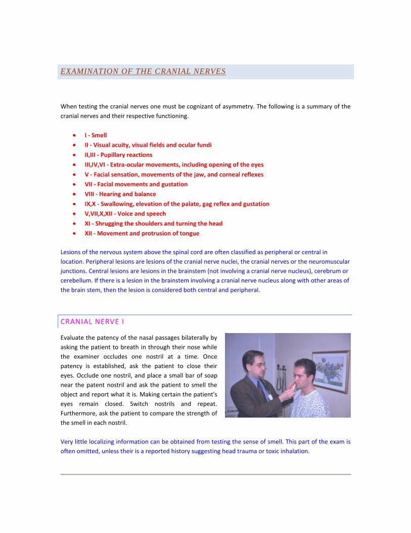
CRANIAL NERVE I
Evaluate the patency of the nasal passages bilaterally by
asking the patient to breath in through their nose while
the examiner occludes one nostril at a time. Once
patency is established, ask the patient to close their
eyes. Occlude one nostril, and place a small bar of soap
near the patent nostril and ask the patient to smell the
object and report what it is. Making certain the patient's
eyes remain closed. Switch nostrils and repeat.
Furthermore, ask the patient to compare the strength of
the smell in each nostril.
Very little localizing information can be obtained from testing the sense of smell. This part of the exam is
often omitted, unless their is a reported history suggesting head trauma or toxic inhalation.

CRANIAL NERVE II
First test visual acuity by using a pocket visual acuity
chart. Perform this part of the examination in a well lit
room and make certain that if the patient wears glasses,
they are wearing them during the exam. Hold the chart
14 inches from the patient's face, and ask the patient to
cover one of their eyes completely with their hand and
read the lowest line on the chart possible. Have them
repeat the test covering the opposite eye. If the patient
has difficulty reading a selected line, ask them to read
the one above. Note the visual acuity for each eye.
Next evaluate the visual fields via confrontation. Face the
patient one foot away, at eye level. Tell the patient to
cover their right eye with their right hand and look the
examiner in the eyes. Instruct the patient to remain
looking you in the eyes and say "now" when the
examiner's fingers enter from out of sight, into their
peripheral vision. Once this is understood, cover your left
eye with your left hand (the opposite eye of the patient)
and extend your arm and first 2 fingers out to the side as
far as possible. Beginning with your hand and arm fully extended, slowly bring your outstretched fingers
centrally, and notice when your fingers enter your field of vision. The patient should say now at the same
time you see your own fingers. Repeat this maneuver a total of eight times per eye, once for every 45
degrees out of the 360 degrees of peripheral vision. Repeat the same maneuver with the other eye.
Using an ophthalmoscope, observe the optic disc,
physiological cup, retinal vessels and fovea. Note the
pulsations of the optic vessels, check for a blurring of the
optic disc margin and a change in the optic disc's color
form its normal yellowish orange
The initial change in the ophthalmoscopic examination in
a patient with increased intracranial pressure is the loss
of pulsations of the retinal vessels. This is followed by
blurring of the optic disc margin and possibly retinal
hemorrhages.
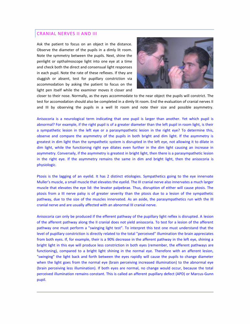
CRANIAL NERVES II AND III
Ask the patient to focus on an object in the distance.
Observe the diameter of the pupils in a dimly lit room.
Note the symmetry between the pupils. Next, shine the
penlight or opthalmoscope light into one eye at a time
and check both the direct and consensual light responses
in each pupil. Note the rate of these reflexes. If they are
sluggish or absent, test for pupillary constriction via
accommodation by asking the patient to focus on the
light pen itself while the examiner moves it closer and
closer to their nose. Normally, as the eyes accommodate to the near object the pupils will constrict. The
test for accomodation should also be completed in a dimly lit room. End the evaluation of cranial nerves II
and III by observing the pupils in a well lit room and note their size and possible asymmetry.
Anisocoria is a neurological term indicating that one pupil is larger than another. Yet which pupil is
abnormal? For example, if the right pupil is of a greater diameter than the left pupil in room light, is their
a sympathetic lesion in the left eye or a parasympathetic lesion in the right eye? To determine this,
observe and compare the asymmetry of the pupils in both bright and dim light. If the asymmetry is
greatest in dim light than the sympathetic system is disrupted in the left eye, not allowing it to dilate in
dim light, while the functioning right eye dilates even further in the dim light causing an increase in
asymmetry. Conversely, if the asymmetry is greatest in bright light, then there is a parasympathetic lesion
in the right eye. If the asymmetry remains the same in dim and bright light, then the anisocoria is
physiologic.
Ptosis is the lagging of an eyelid. It has 2 distinct etiologies. Sympathetics going to the eye innervate
Muller's muscle, a small muscle that elevates the eyelid. The III cranial nerve also innervates a much larger
muscle that elevates the eye lid: the levator palpebrae. Thus, disruption of either will cause ptosis. The
ptosis from a III nerve palsy is of greater severity than the ptosis due to a lesion of the sympathetic
pathway, due to the size of the muscles innervated. As an aside, the parasympathetics run with the III
cranial nerve and are usually affected with an abnormal III cranial nerve.
Anisocoria can only be produced if the efferent pathway of the pupillary light reflex is disrupted. A lesion
of the afferent pathway along the II cranial does not yield anisocoria. To test for a lesion of the afferent
pathway one must perform a "swinging light test". To interpret this test one must understand that the
level of pupillary constriction is directly related to the total "perceived" illumination the brain appreciates
from both eyes. If, for example, their is a 90% decrease in the afferent pathway in the left eye, shining a
bright light in this eye will produce less constriction in both eyes (remember, the efferent pathways are
functioning), compared to a bright light shining in the normal eye. Therefore with an afferent lesion,
"swinging" the light back and forth between the eyes rapidly will cause the pupils to change diameter
when the light goes from the normal eye (brain perceiving increased illumination) to the abnormal eye
(brain perceiving less illumination). If both eyes are normal, no change would occur, because the total
perceived illumination remains constant. This is called an afferent pupillary defect (APD) or Marcus-Gunn
pupil.
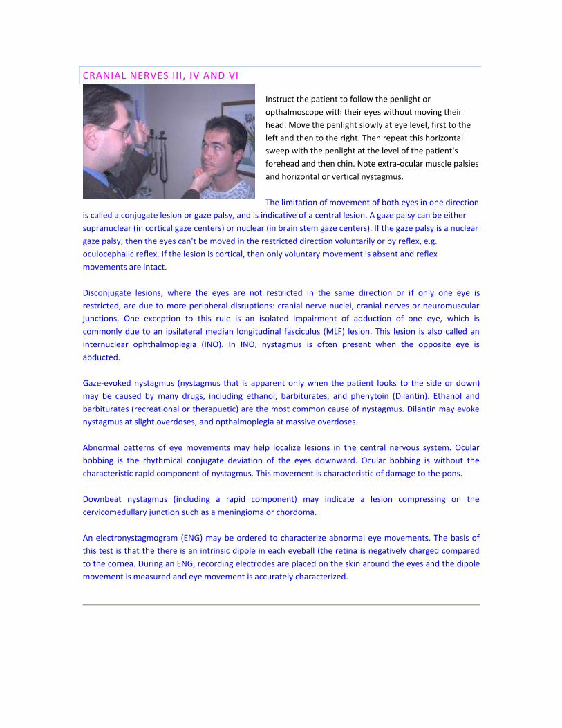
CRANIAL NERVES III, IV AND VI
Instruct the patient to follow the penlight or
opthalmoscope with their eyes without moving their
head. Move the penlight slowly at eye level, first to the
left and then to the right. Then repeat this horizontal
sweep with the penlight at the level of the patient's
forehead and then chin. Note extra-ocular muscle palsies
and horizontal or vertical nystagmus.
The limitation of movement of both eyes in one direction
is called a conjugate lesion or gaze palsy, and is indicative of a central lesion. A gaze palsy can be either
supranuclear (in cortical gaze centers) or nuclear (in brain stem gaze centers). If the gaze palsy is a nuclear
gaze palsy, then the eyes can't be moved in the restricted direction voluntarily or by reflex, e.g.
oculocephalic reflex. If the lesion is cortical, then only voluntary movement is absent and reflex
movements are intact.
Disconjugate lesions, where the eyes are not restricted in the same direction or if only one eye is
restricted, are due to more peripheral disruptions: cranial nerve nuclei, cranial nerves or neuromuscular
junctions. One exception to this rule is an isolated impairment of adduction of one eye, which is
commonly due to an ipsilateral median longitudinal fasciculus (MLF) lesion. This lesion is also called an
internuclear ophthalmoplegia (INO). In INO, nystagmus is often present when the opposite eye is
abducted.
Gaze-evoked nystagmus (nystagmus that is apparent only when the patient looks to the side or down)
may be caused by many drugs, including ethanol, barbiturates, and phenytoin (Dilantin). Ethanol and
barbiturates (recreational or therapuetic) are the most common cause of nystagmus. Dilantin may evoke
nystagmus at slight overdoses, and opthalmoplegia at massive overdoses.
Abnormal patterns of eye movements may help localize lesions in the central nervous system. Ocular
bobbing is the rhythmical conjugate deviation of the eyes downward. Ocular bobbing is without the
characteristic rapid component of nystagmus. This movement is characteristic of damage to the pons.
Downbeat nystagmus (including a rapid component) may indicate a lesion compressing on the
cervicomedullary junction such as a meningioma or chordoma.
An electronystagmogram (ENG) may be ordered to characterize abnormal eye movements. The basis of
this test is that the there is an intrinsic dipole in each eyeball (the retina is negatively charged compared
to the cornea. During an ENG, recording electrodes are placed on the skin around the eyes and the dipole
movement is measured and eye movement is accurately characterized.
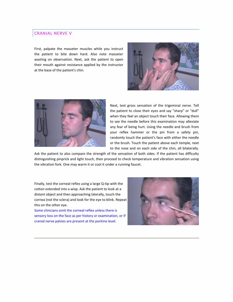
CRANIAL NERVE V
First, palpate the masseter muscles while you instruct
the patient to bite down hard. Also note masseter
wasting on observation. Next, ask the patient to open
their mouth against resistance applied by the instructor
at the base of the patient's chin.
Next, test gross sensation of the trigeminal nerve. Tell
the patient to close their eyes and say "sharp" or "dull"
when they feel an object touch their face. Allowing them
to see the needle before this examination may alleviate
any fear of being hurt. Using the needle and brush from
your reflex hammer or the pin from a safety pin,
randomly touch the patient's face with either the needle
or the brush. Touch the patient above each temple, next
to the nose and on each side of the chin, all bilaterally.
Ask the patient to also compare the strength of the sensation of both sides. If the patient has difficulty
distinguishing pinprick and light touch, then proceed to check temperature and vibration sensation using
the vibration fork. One may warm it or cool it under a running faucet.
Finally, test the corneal reflex using a large Q-tip with the
cotton extended into a wisp. Ask the patient to look at a
distant object and then approaching laterally, touch the
cornea (not the sclera) and look for the eye to blink. Repeat
this on the other eye.
Some clinicians omit the corneal reflex unless there is
sensory loss on the face as per history or examination, or if
cranial nerve palsies are present at the pontine level.
CRANIAL NERVE VII
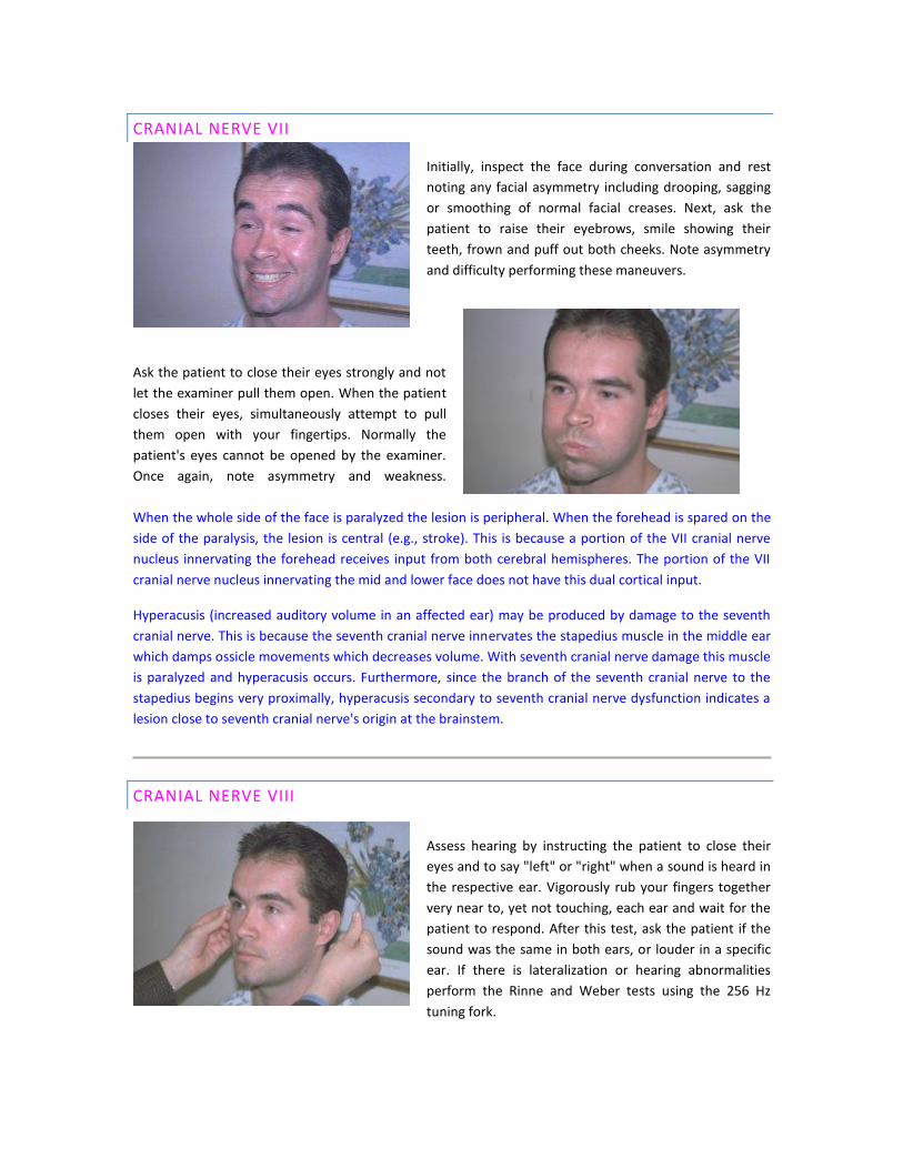
Initially, inspect the face during conversation and rest
noting any facial asymmetry including drooping, sagging
or smoothing of normal facial creases. Next, ask the
patient to raise their eyebrows, smile showing their
teeth, frown and puff out both cheeks. Note asymmetry
and difficulty performing these maneuvers.
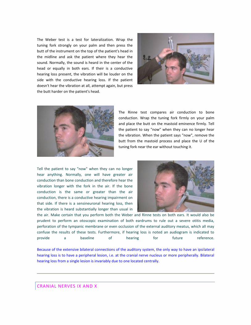
Ask the patient to close their eyes strongly and not
let the examiner pull them open. When the patient
closes their eyes, simultaneously attempt to pull
them open with your fingertips. Normally the
patient's eyes cannot be opened by the examiner.
Once again, note asymmetry and weakness.
When the whole side of the face is paralyzed the lesion is peripheral. When the forehead is spared on the
side of the paralysis, the lesion is central (e.g., stroke). This is because a portion of the VII cranial nerve
nucleus innervating the forehead receives input from both cerebral hemispheres. The portion of the VII
cranial nerve nucleus innervating the mid and lower face does not have this dual cortical input.
Hyperacusis (increased auditory volume in an affected ear) may be produced by damage to the seventh
cranial nerve. This is because the seventh cranial nerve innervates the stapedius muscle in the middle ear
which damps ossicle movements which decreases volume. With seventh cranial nerve damage this muscle
is paralyzed and hyperacusis occurs. Furthermore, since the branch of the seventh cranial nerve to the
stapedius begins very proximally, hyperacusis secondary to seventh cranial nerve dysfunction indicates a
lesion close to seventh cranial nerve's origin at the brainstem.
CRANIAL NERVE VIII
Assess hearing by instructing the patient to close their
eyes and to say "left" or "right" when a sound is heard in
the respective ear. Vigorously rub your fingers together
very near to, yet not touching, each ear and wait for the
patient to respond. After this test, ask the patient if the
sound was the same in both ears, or louder in a specific
ear. If there is lateralization or hearing abnormalities
perform the Rinne and Weber tests using the 256 Hz
tuning fork.
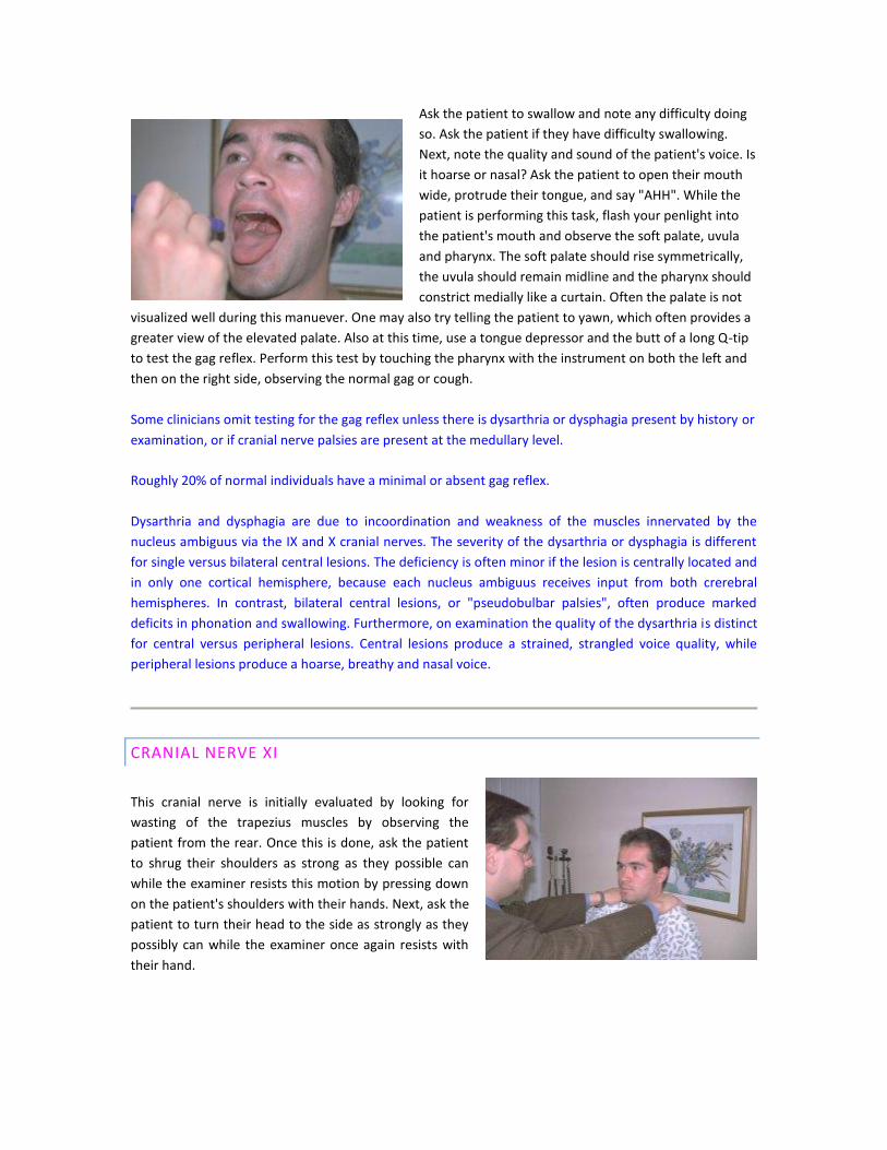
The Weber test is a test for lateralization. Wrap the
tuning fork strongly on your palm and then press the
butt of the instrument on the top of the patient's head in
the midline and ask the patient where they hear the
sound. Normally, the sound is heard in the center of the
head or equally in both ears. If their is a conductive
hearing loss present, the vibration will be louder on the
side with the conductive hearing loss. If the patient
doesn't hear the vibration at all, attempt again, but press
the butt harder on the patient's head.
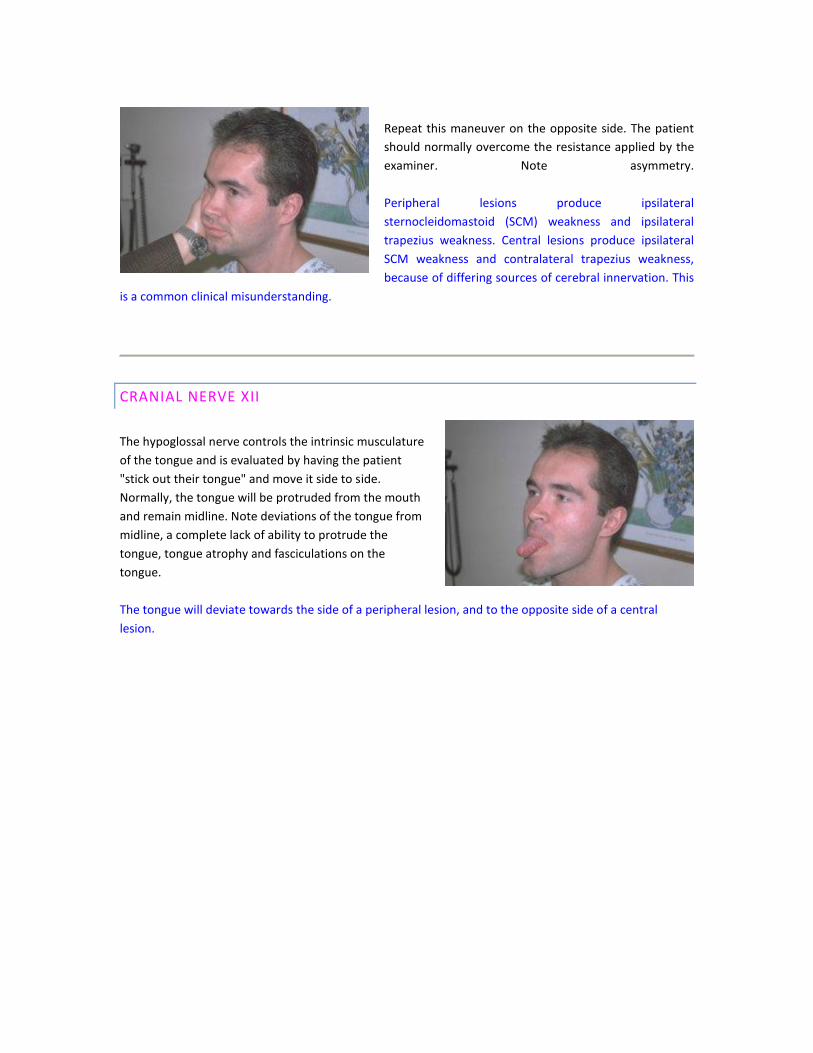
The Rinne test compares air conduction to bone
conduction. Wrap the tuning fork firmly on your palm
and place the butt on the mastoid eminence firmly. Tell
the patient to say "now" when they can no longer hear
the vibration. When the patient says "now", remove the
butt from the mastoid process and place the U of the
tuning fork near the ear without touching it.
Tell the patient to say "now" when they can no longer
hear anything. Normally, one will have greater air
conduction than bone conduction and therefore hear the
vibration longer with the fork in the air. If the bone
conduction is the same or greater than the air
conduction, there is a conductive hearing impairment on
that side. If there is a sensineuronal hearing loss, then
the vibration is heard substantially longer than usual in
the air. Make certain that you perform both the Weber and Rinne tests on both ears. It would also be
prudent to perform an otoscopic examination of both eardrums to rule out a severe otitis media,
perforation of the tympanic membrane or even occlusion of the external auditory meatus, which all may
confuse the results of these tests. Furthermore, if hearing loss is noted an audiogram is indicated to
provide a baseline of hearing for future reference.
Because of the extensive bilateral connections of the auditory system, the only way to have an ipsilateral
hearing loss is to have a peripheral lesion, i.e. at the cranial nerve nucleus or more peripherally. Bilateral
hearing loss from a single lesion is invariably due to one located centrally.
CRANIAL NERVES IX AND X
Ask the patient to swallow and note any difficulty doing
so. Ask the patient if they have difficulty swallowing.
Next, note the quality and sound of the patient's voice. Is
it hoarse or nasal? Ask the patient to open their mouth
wide, protrude their tongue, and say "AHH". While the
patient is performing this task, flash your penlight into
the patient's mouth and observe the soft palate, uvula
and pharynx. The soft palate should rise symmetrically,
the uvula should remain midline and the pharynx should
constrict medially like a curtain. Often the palate is not
visualized well during this manuever. One may also try telling the patient to yawn, which often provides a
greater view of the elevated palate. Also at this time, use a tongue depressor and the butt of a long Q-tip
to test the gag reflex. Perform this test by touching the pharynx with the instrument on both the left and
then on the right side, observing the normal gag or cough.
Some clinicians omit testing for the gag reflex unless there is dysarthria or dysphagia present by history or
examination, or if cranial nerve palsies are present at the medullary level.
Roughly 20% of normal individuals have a minimal or absent gag reflex.
Dysarthria and dysphagia are due to incoordination and weakness of the muscles innervated by the
nucleus ambiguus via the IX and X cranial nerves. The severity of the dysarthria or dysphagia is different
for single versus bilateral central lesions. The deficiency is often minor if the lesion is centrally located and
in only one cortical hemisphere, because each nucleus ambiguus receives input from both crerebral
hemispheres. In contrast, bilateral central lesions, or "pseudobulbar palsies", often produce marked
deficits in phonation and swallowing. Furthermore, on examination the quality of the dysarthria is distinct
for central versus peripheral lesions. Central lesions produce a strained, strangled voice quality, while
peripheral lesions produce a hoarse, breathy and nasal voice.
CRANIAL NERVE XI
This cranial nerve is initially evaluated by looking for
wasting of the trapezius muscles by observing the
patient from the rear. Once this is done, ask the patient
to shrug their shoulders as strong as they possible can
while the examiner resists this motion by pressing down
on the patient's shoulders with their hands. Next, ask the
patient to turn their head to the side as strongly as they
possibly can while the examiner once again resists with
their hand.
Repeat this maneuver on the opposite side. The patient
should normally overcome the resistance applied by the
examiner. Note asymmetry.
Peripheral lesions produce ipsilateral
sternocleidomastoid (SCM) weakness and ipsilateral
trapezius weakness. Central lesions produce ipsilateral
SCM weakness and contralateral trapezius weakness,
because of differing sources of cerebral innervation. This
is a common clinical misunderstanding.
CRANIAL NERVE XII
The hypoglossal nerve controls the intrinsic musculature
of the tongue and is evaluated by having the patient
"stick out their tongue" and move it side to side.
Normally, the tongue will be protruded from the mouth
and remain midline. Note deviations of the tongue from
midline, a complete lack of ability to protrude the
tongue, tongue atrophy and fasciculations on the
tongue.
The tongue will deviate towards the side of a peripheral lesion, and to the opposite side of a central
lesion.
Special Topic: Pathology found on Opthalmologic Examination
Papilledema. Note swelling of the disc,
hemorrhages, and exudates, with preservation of
the physiologic cup.
Optic Atrophy. Note the chalky white disc with
discrete margins. Optic atrophy is a late finding
with increased intracranial pressure.
Central Retinal Artery Occlusion. Note the diffusely
pale retina and prominent central fovea which is
usually blended in with the normal, pink retina.
Central Retinal Vein Occlusion. The disc is
massively swollen with diffuse hemorrhages and
cotton-wool spots.
Proliferative Diabetic Retinopathy. Note the multiple
hemorrhages, exudates and neovascularization
throughout the retina. Chorioretinal striae extend
towards the area of fibrovascular proliferation in the
lower portion of the photograph.
Cytomegalovirus Retinitis. Note the area of retinal
necrosis and hemorrhage along the lower portion
of the photograph. Common in patinets with
immunodeficiency, especially AIDS.
THE MOTOR SYSTEM EXAMINATION
The motor system evaluation is divided into the following: body positioning, involuntary movements,
muscle tone and muscle strength.
Upper motor neuron lesions are characterized by weakness, spasticity, hyperreflexia, primitive reflexes
and the Babinski sign. Primitive reflexes include the grasp, suck and snout reflexes. Lower motor neuron
lesions are characterized by weakness, hypotonia, hyporeflexia, atrophy and fasciculations.
Fasciculations are fine movements of the muscle under the skin and are indicative of lower motor neuron
disease. They are caused by denervation of whole motor units leading to acetylcholine hypersensitivity at
the denervated muscle. Atrophy of the affected muscle is usually concurrent with fasciculations.
Fibrillations are spontaneous contractions of individual muscle fibers and are therefore not observed with
the naked eye.
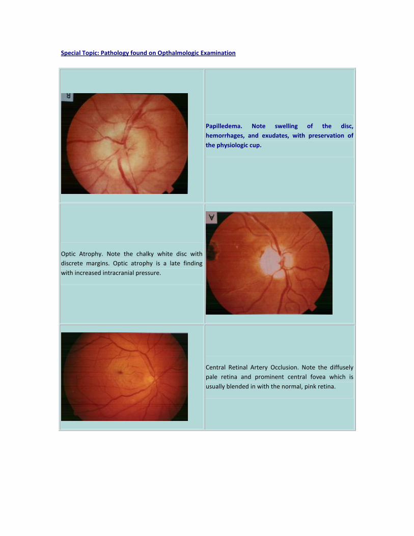
Note the position of the body that the patient assumes
when sitting on the examination table.
Paralysis or weakness may become evident when a
patient assumes an abnormal body position. A central
lesion usually produces greater weakness in the
extensors than in the flexors of the upper extremities,
while the opposite is true in the lower extremities: a
greater weakness in the flexors than in the extensors.
Next, examine the patient for tics, tremors and fasciculations. Note their location and quality. Also note if
they are related to any specific body position or emotional state.
Systematically examine all of the major muscle groups of the body.
For each muscle group:
1. Note the appearance or muscularity of the muscle (wasted, highly developed, normal).
2. Feel the tone of the muscle (flaccid, clonic, normal).
3. Test the strength of the muscle group.
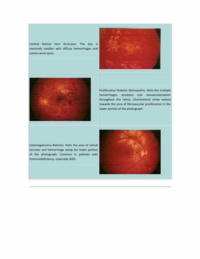
Muscle Power Ratiting Scale*:
0 No muscle contraction is detected
1 A trace contraction is noted in the muscle by palpating the muscle while the
patient attempts to contract it.
2 The patient is able to actively move the muscle when gravity is eliminated.
3 The patient may move the muscle against gravity but not against resistance
from the examiner.
4 The patient may move the muscle group against some resistance from the
examiner.
5 The patient moves the muscle group and overcomes the resistance of the
examiner. This is normal muscle strength.
*Since this rating scale is skewed towards weakness, many clinicians further subclassify their finding
by adding a + or -, e.g., 5- or 3+.
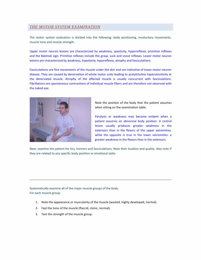
Starting with the deltoids, ask the patient to raise both
their arms in front of them simultaneously as strongly as
then can while the examiner provides resistance to this
movement. Compare the strength of each arm.
The deltoid muscle is innervated by the C5 nerve root via
the axillary nerve.
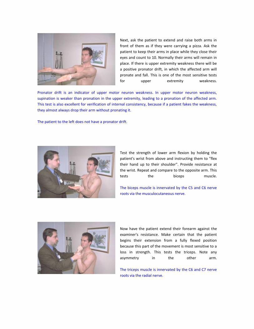
Next, ask the patient to extend and raise both arms in
front of them as if they were carrying a pizza. Ask the
patient to keep their arms in place while they close their
eyes and count to 10. Normally their arms will remain in
place. If there is upper extremity weakness there will be
a positive pronator drift, in which the affected arm will
pronate and fall. This is one of the most sensitive tests
for upper extremity weakness.
Pronator drift is an indicator of upper motor neuron weakness. In upper motor neuron weakness,
supination is weaker than pronation in the upper extremity, leading to a pronation of the affected arm.
This test is also excellent for verification of internal consistency, because if a patient fakes the weakness,
they almost always drop their arm without pronating it.
The patient to the left does not have a pronator drift.
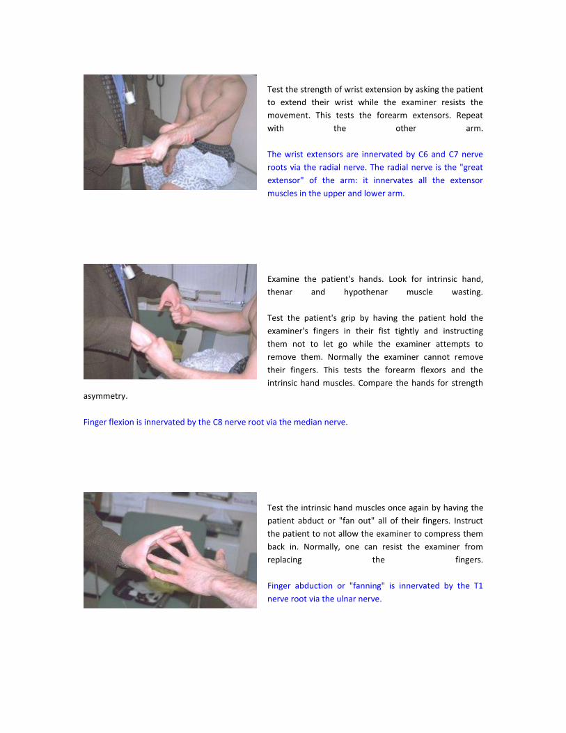
Test the strength of lower arm flexion by holding the
patient's wrist from above and instructing them to "flex
their hand up to their shoulder". Provide resistance at
the wrist. Repeat and compare to the opposite arm. This
tests the biceps muscle.
The biceps muscle is innervated by the C5 and C6 nerve
roots via the musculocutaneous nerve.
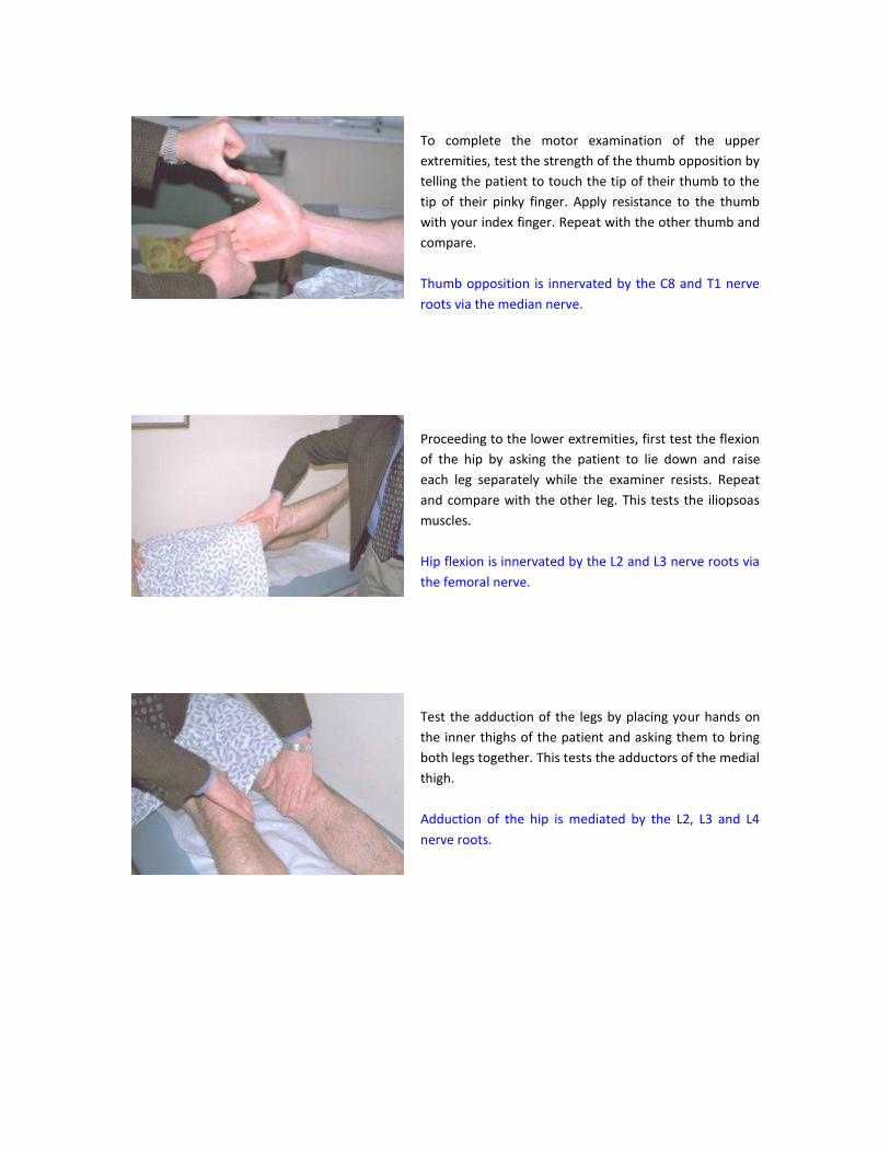
Now have the patient extend their forearm against the
examiner's resistance. Make certain that the patient
begins their extension from a fully flexed position
because this part of the movement is most sensitive to a
loss in strength. This tests the triceps. Note any
asymmetry in the other arm.
The triceps muscle is innervated by the C6 and C7 nerve
roots via the radial nerve.
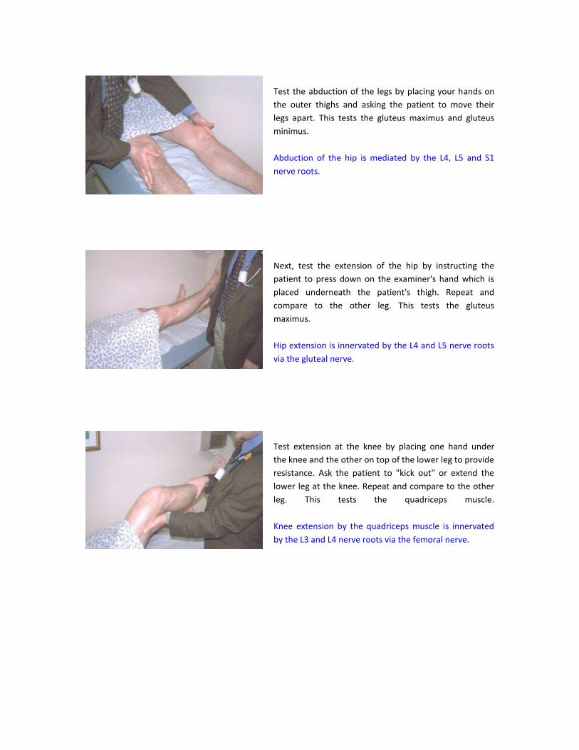
Test the strength of wrist extension by asking the patient
to extend their wrist while the examiner resists the
movement. This tests the forearm extensors. Repeat
with the other arm.
The wrist extensors are innervated by C6 and C7 nerve
roots via the radial nerve. The radial nerve is the "great
extensor" of the arm: it innervates all the extensor
muscles in the upper and lower arm.
Examine the patient's hands. Look for intrinsic hand,
thenar and hypothenar muscle wasting.
Test the patient's grip by having the patient hold the
examiner's fingers in their fist tightly and instructing
them not to let go while the examiner attempts to
remove them. Normally the examiner cannot remove
their fingers. This tests the forearm flexors and the
intrinsic hand muscles. Compare the hands for strength
asymmetry.
Finger flexion is innervated by the C8 nerve root via the median nerve.
Test the intrinsic hand muscles once again by having the
patient abduct or "fan out" all of their fingers. Instruct
the patient to not allow the examiner to compress them
back in. Normally, one can resist the examiner from
replacing the fingers.
Finger abduction or "fanning" is innervated by the T1
nerve root via the ulnar nerve.
To complete the motor examination of the upper
extremities, test the strength of the thumb opposition by
telling the patient to touch the tip of their thumb to the
tip of their pinky finger. Apply resistance to the thumb
with your index finger. Repeat with the other thumb and
compare.
Thumb opposition is innervated by the C8 and T1 nerve
roots via the median nerve.
Proceeding to the lower extremities, first test the flexion
of the hip by asking the patient to lie down and raise
each leg separately while the examiner resists. Repeat
and compare with the other leg. This tests the iliopsoas
muscles.
Hip flexion is innervated by the L2 and L3 nerve roots via
the femoral nerve.
Test the adduction of the legs by placing your hands on
the inner thighs of the patient and asking them to bring
both legs together. This tests the adductors of the medial
thigh.
Adduction of the hip is mediated by the L2, L3 and L4
nerve roots.
Test the abduction of the legs by placing your hands on
the outer thighs and asking the patient to move their
legs apart. This tests the gluteus maximus and gluteus
minimus.
Abduction of the hip is mediated by the L4, L5 and S1
nerve roots.
Next, test the extension of the hip by instructing the
patient to press down on the examiner's hand which is
placed underneath the patient's thigh. Repeat and
compare to the other leg. This tests the gluteus
maximus.
Hip extension is innervated by the L4 and L5 nerve roots
via the gluteal nerve.
Test extension at the knee by placing one hand under
the knee and the other on top of the lower leg to provide
resistance. Ask the patient to "kick out" or extend the
lower leg at the knee. Repeat and compare to the other
leg. This tests the quadriceps muscle.
Knee extension by the quadriceps muscle is innervated
by the L3 and L4 nerve roots via the femoral nerve.
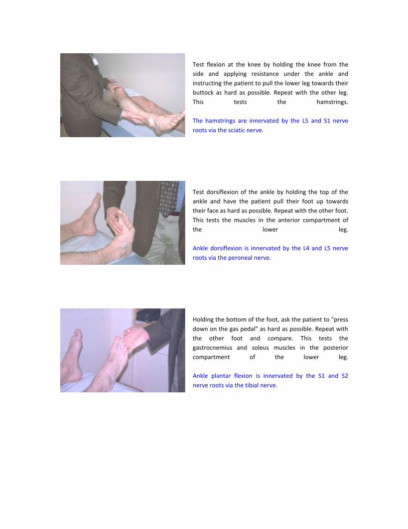
Test flexion at the knee by holding the knee from the
side and applying resistance under the ankle and
instructing the patient to pull the lower leg towards their
buttock as hard as possible. Repeat with the other leg.
This tests the hamstrings.
The hamstrings are innervated by the L5 and S1 nerve
roots via the sciatic nerve.
Test dorsiflexion of the ankle by holding the top of the
ankle and have the patient pull their foot up towards
their face as hard as possible. Repeat with the other foot.
This tests the muscles in the anterior compartment of
the lower leg.
Ankle dorsiflexion is innervated by the L4 and L5 nerve
roots via the peroneal nerve.
Holding the bottom of the foot, ask the patient to "press
down on the gas pedal" as hard as possible. Repeat with
the other foot and compare. This tests the
gastrocnemius and soleus muscles in the posterior
compartment of the lower leg.
Ankle plantar flexion is innervated by the S1 and S2
nerve roots via the tibial nerve.
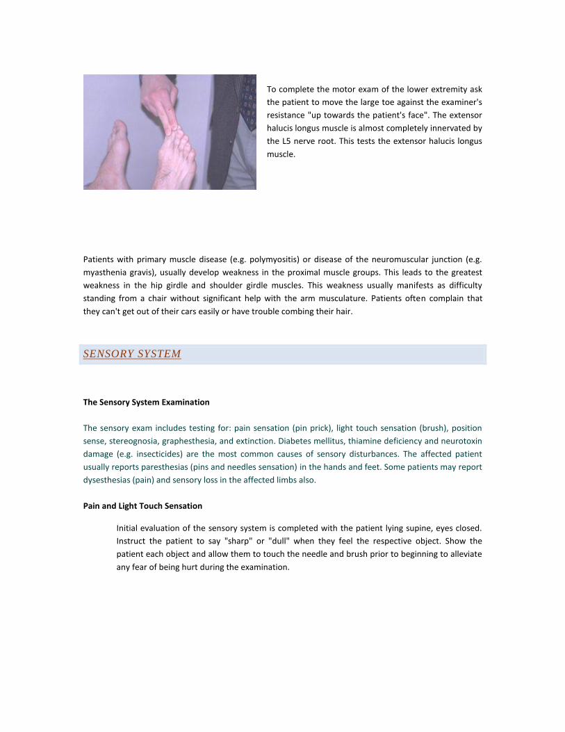
To complete the motor exam of the lower extremity ask
the patient to move the large toe against the examiner's
resistance "up towards the patient's face". The extensor
halucis longus muscle is almost completely innervated by
the L5 nerve root. This tests the extensor halucis longus
muscle.
Patients with primary muscle disease (e.g. polymyositis) or disease of the neuromuscular junction (e.g.
myasthenia gravis), usually develop weakness in the proximal muscle groups. This leads to the greatest
weakness in the hip girdle and shoulder girdle muscles. This weakness usually manifests as difficulty
standing from a chair without significant help with the arm musculature. Patients often complain that
they can't get out of their cars easily or have trouble combing their hair.
SENSORY SYSTEM
The Sensory System Examination
The sensory exam includes testing for: pain sensation (pin prick), light touch sensation (brush), position
sense, stereognosia, graphesthesia, and extinction. Diabetes mellitus, thiamine deficiency and neurotoxin
damage (e.g. insecticides) are the most common causes of sensory disturbances. The affected patient
usually reports paresthesias (pins and needles sensation) in the hands and feet. Some patients may report
dysesthesias (pain) and sensory loss in the affected limbs also.
Pain and Light Touch Sensation
Initial evaluation of the sensory system is completed with the patient lying supine, eyes closed.
Instruct the patient to say "sharp" or "dull" when they feel the respective object. Show the
patient each object and allow them to touch the needle and brush prior to beginning to alleviate
any fear of being hurt during the examination.
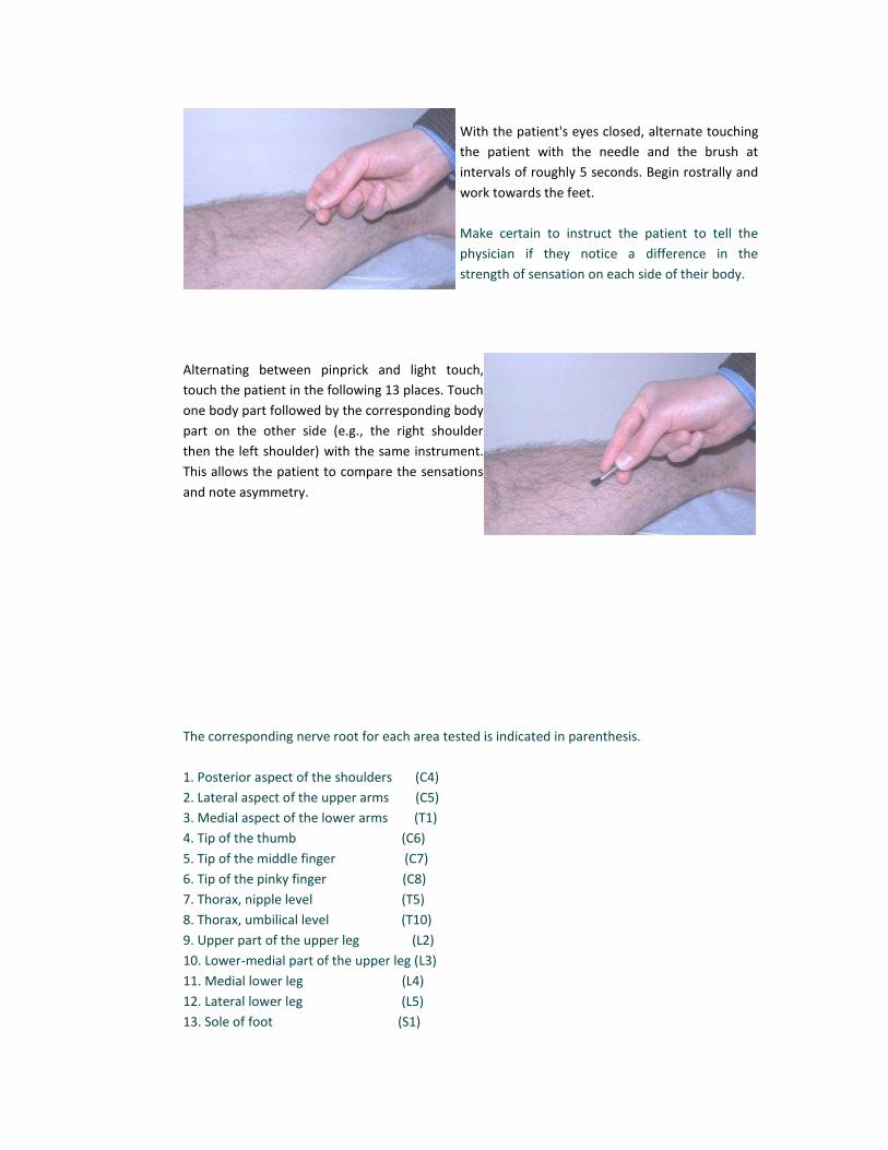
With the patient's eyes closed, alternate touching
the patient with the needle and the brush at
intervals of roughly 5 seconds. Begin rostrally and
work towards the feet.
Make certain to instruct the patient to tell the
physician if they notice a difference in the
strength of sensation on each side of their body.
Alternating between pinprick and light touch,
touch the patient in the following 13 places. Touch
one body part followed by the corresponding body
part on the other side (e.g., the right shoulder
then the left shoulder) with the same instrument.
This allows the patient to compare the sensations
and note asymmetry.
The corresponding nerve root for each area tested is indicated in parenthesis.
1. Posterior aspect of the shoulders (C4)
2. Lateral aspect of the upper arms (C5)
3. Medial aspect of the lower arms (T1)
4. Tip of the thumb (C6)
5. Tip of the middle finger (C7)
6. Tip of the pinky finger (C8)
7. Thorax, nipple level (T5)
8. Thorax, umbilical level (T10)
9. Upper part of the upper leg (L2)
10. Lower-medial part of the upper leg (L3)
11. Medial lower leg (L4)
12. Lateral lower leg (L5)
13. Sole of foot (S1)
If there is a sensory loss present, test vibration sensation and temperature sensation with the
tuning fork. Also concentrate the sensory exam in the area of deficiency.
Position Sense
Test position sense by having the patient, eyes closed, report if their large toe is "up" or "down"
when the examiner manually moves the patient's toe in the respective direction. Repeat on the
opposite foot and compare. Make certain to hold the toe on its sides, because holding the top or
bottom provides the patient with pressure cues which make this test invalid.
Fine touch, position sense (proprioception) and vibration sense are conducted together in the
dorsal column system. Rough touch, temperature and pain sensation are conducted via the
spinothalamic tract. Loss of one modality in a conduction system is often associated with the loss
of the other modalities conducted by the same tract in the affected area.
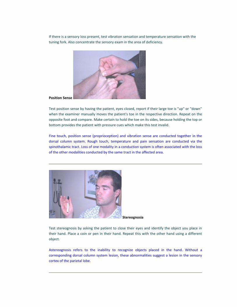
Stereognosia
Test stereognosis by asking the patient to close their eyes and identify the object you place in
their hand. Place a coin or pen in their hand. Repeat this with the other hand using a different
object.
Astereognosis refers to the inability to recognize objects placed in the hand. Without a
corresponding dorsal column system lesion, these abnormalities suggest a lesion in the sensory
cortex of the parietal lobe.
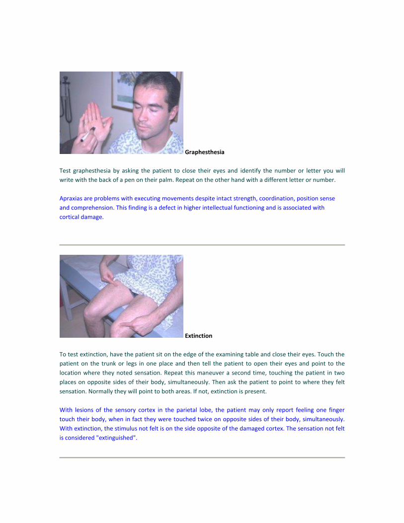
Graphesthesia
Test graphesthesia by asking the patient to close their eyes and identify the number or letter you will
write with the back of a pen on their palm. Repeat on the other hand with a different letter or number.
Apraxias are problems with executing movements despite intact strength, coordination, position sense
and comprehension. This finding is a defect in higher intellectual functioning and is associated with
cortical damage.
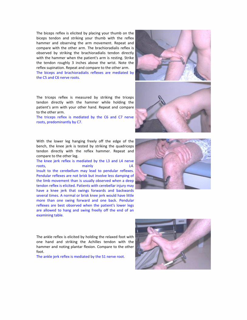
Extinction
To test extinction, have the patient sit on the edge of the examining table and close their eyes. Touch the
patient on the trunk or legs in one place and then tell the patient to open their eyes and point to the
location where they noted sensation. Repeat this maneuver a second time, touching the patient in two
places on opposite sides of their body, simultaneously. Then ask the patient to point to where they felt
sensation. Normally they will point to both areas. If not, extinction is present.
With lesions of the sensory cortex in the parietal lobe, the patient may only report feeling one finger
touch their body, when in fact they were touched twice on opposite sides of their body, simultaneously.
With extinction, the stimulus not felt is on the side opposite of the damaged cortex. The sensation not felt
is considered "extinguished".

DEEP TENDON REFLEXES
Using a reflex hammer, deep tendon reflexes are elicited in all 4 extremities. Note the extent or power of
the reflex, both visually and by palpation of the tendon or muscle in question.
Rate the reflex with the following scale:
5+ Sustained clonus
4+ Very brisk, hyperreflexive, with clonus
3+ Brisker or more reflexive than normally.
2+ Normal
1+ Low normal, diminished
0.5+ A reflex that is only elicited with reinforcement
0 No response
Reinforcement is accomplished by asking the patient to clench their teeth, or if testing lower extremity
reflexes, have the patient hook together their flexed fingers and pull apart. This is known as the Jendrassik
maneuver.
It is key to compare the strength of reflexes elicited with each other. A finding of 3+, brisk reflexes
throughout all extremities is a much less significant finding than that of a person with all 2+, normal
reflexes, and a 1+, diminished left ankle reflex suggesting a distinct lesion.
Have the patient sit up on the edge of the examination bench with one hand on top of the other, arms
and legs relaxed. Instruct the patient to remain relaxed.
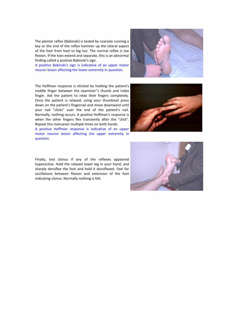
The biceps reflex is elicited by placing your thumb on the biceps tendon and striking your thumb with the reflex hammer and observing the arm movement. Repeat and compare with the other arm. The brachioradialis reflex is observed by striking the brachioradialis tendon directly with the hammer when the patient's arm is resting. Strike the tendon roughly 3 inches above the wrist. Note the reflex supination. Repeat and compare to the other arm. The biceps and brachioradialis reflexes are mediated by the C5 and C6 nerve roots.
The triceps reflex is measured by striking the triceps tendon directly with the hammer while holding the patient's arm with your other hand. Repeat and compare to the other arm. The triceps reflex is mediated by the C6 and C7 nerve roots, predominantly by C7.
With the lower leg hanging freely off the edge of the bench, the knee jerk is tested by striking the quadriceps tendon directly with the reflex hammer. Repeat and compare to the other leg. The knee jerk reflex is mediated by the L3 and L4 nerve roots, mainly L4. Insult to the cerebellum may lead to pendular reflexes. Pendular reflexes are not brisk but involve less damping of the limb movement than is usually observed when a deep tendon reflex is elicited. Patients with cerebellar injury may have a knee jerk that swings forwards and backwards several times. A normal or brisk knee jerk would have little more than one swing forward and one back. Pendular reflexes are best observed when the patient's lower legs are allowed to hang and swing freelly off the end of an examining table.
The ankle reflex is elicited by holding the relaxed foot with one hand and striking the Achilles tendon with the hammer and noting plantar flexion. Compare to the other foot. The ankle jerk reflex is mediated by the S1 nerve root.
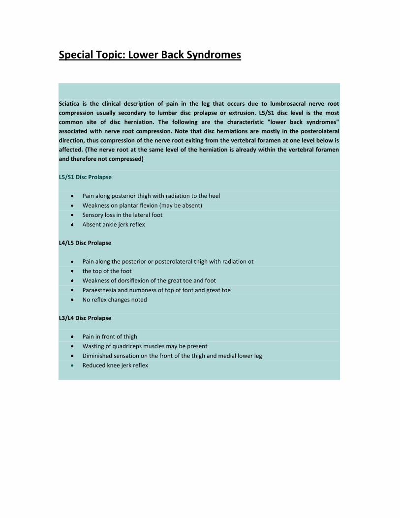
The plantar reflex (Babinski) is tested by coarsely running a key or the end of the reflex hammer up the lateral aspect of the foot from heel to big toe. The normal reflex is toe flexion. If the toes extend and separate, this is an abnormal finding called a positive Babinski's sign. A positive Babinski's sign is indicative of an upper motor neuron lesion affecting the lower extremity in question.
The Hoffman response is elicited by holding the patient's middle finger between the examiner''s thumb and index finger. Ask the patient to relax their fingers completely. Once the patient is relaxed, using your thumbnail press down on the patient's fingernail and move downward until your nail "clicks" over the end of the patient's nail. Normally, nothing occurs. A positive Hoffman's response is when the other fingers flex transiently after the "click". Repeat this manuever multiple times on both hands. A positive Hoffman response is indicative of an upper motor neuron lesion affecting the upper extremity in question.
Finally, test clonus if any of the reflexes appeared hyperactive. Hold the relaxed lower leg in your hand, and sharply dorsiflex the foot and hold it dorsiflexed. Feel for oscillations between flexion and extension of the foot indicating clonus. Normally nothing is felt.
Special Topic: Lower Back Syndromes
Sciatica is the clinical description of pain in the leg that occurs due to lumbrosacral nerve root
compression usually secondary to lumbar disc prolapse or extrusion. L5/S1 disc level is the most
common site of disc herniation. The following are the characteristic "lower back syndromes"
associated with nerve root compression. Note that disc herniations are mostly in the posterolateral
direction, thus compression of the nerve root exiting from the vertebral foramen at one level below is
affected. (The nerve root at the same level of the herniation is already within the vertebral foramen
and therefore not compressed)
L5/S1 Disc Prolapse
Pain along posterior thigh with radiation to the heel
Weakness on plantar flexion (may be absent)
Sensory loss in the lateral foot
Absent ankle jerk reflex
L4/L5 Disc Prolapse
Pain along the posterior or posterolateral thigh with radiation ot
the top of the foot
Weakness of dorsiflexion of the great toe and foot
Paraesthesia and numbness of top of foot and great toe
No reflex changes noted
L3/L4 Disc Prolapse
Pain in front of thigh
Wasting of quadriceps muscles may be present
Diminished sensation on the front of the thigh and medial lower leg
Reduced knee jerk reflex
COORDINATION, GAIT AND RHOMBERG TEST
Coordination
Coordination is evaluated by testing the patient's ability to perform rapidly alternating and point-to-point
movements correctly.
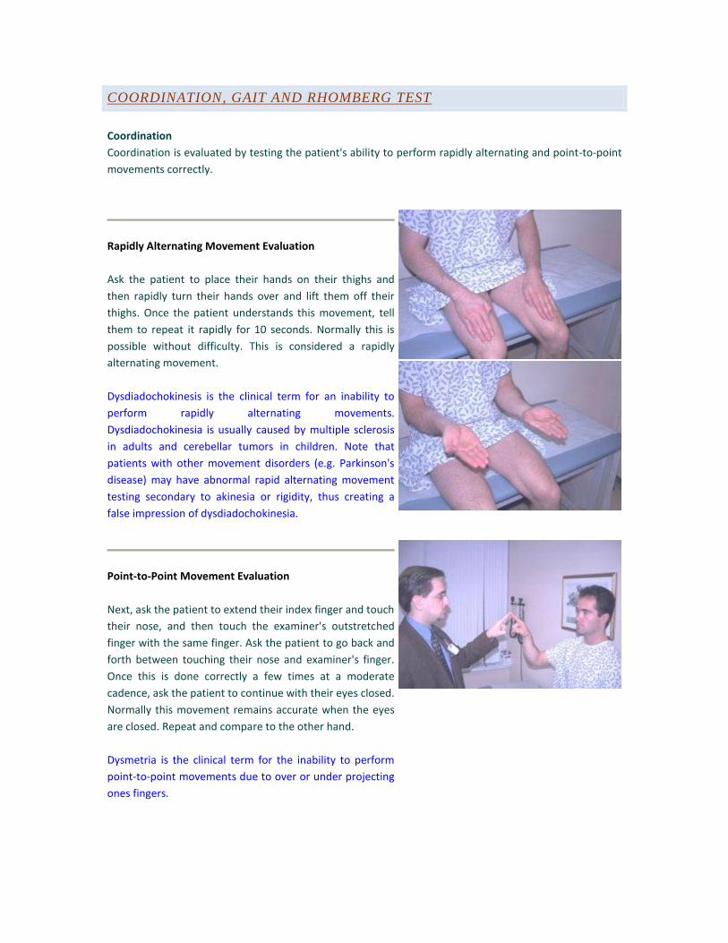
Rapidly Alternating Movement Evaluation
Ask the patient to place their hands on their thighs and
then rapidly turn their hands over and lift them off their
thighs. Once the patient understands this movement, tell
them to repeat it rapidly for 10 seconds. Normally this is
possible without difficulty. This is considered a rapidly
alternating movement.
Dysdiadochokinesis is the clinical term for an inability to
perform rapidly alternating movements.
Dysdiadochokinesia is usually caused by multiple sclerosis
in adults and cerebellar tumors in children. Note that
patients with other movement disorders (e.g. Parkinson's
disease) may have abnormal rapid alternating movement
testing secondary to akinesia or rigidity, thus creating a
false impression of dysdiadochokinesia.
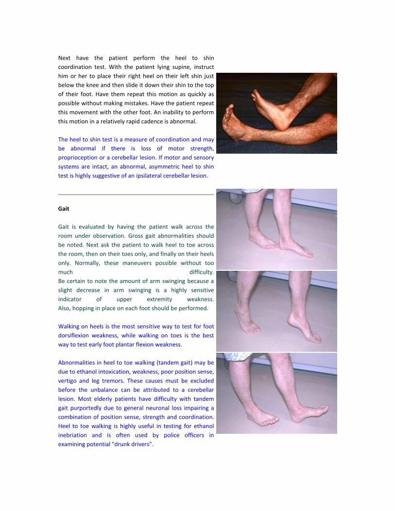
Point-to-Point Movement Evaluation
Next, ask the patient to extend their index finger and touch
their nose, and then touch the examiner's outstretched
finger with the same finger. Ask the patient to go back and
forth between touching their nose and examiner's finger.
Once this is done correctly a few times at a moderate
cadence, ask the patient to continue with their eyes closed.
Normally this movement remains accurate when the eyes
are closed. Repeat and compare to the other hand.
Dysmetria is the clinical term for the inability to perform
point-to-point movements due to over or under projecting
ones fingers.
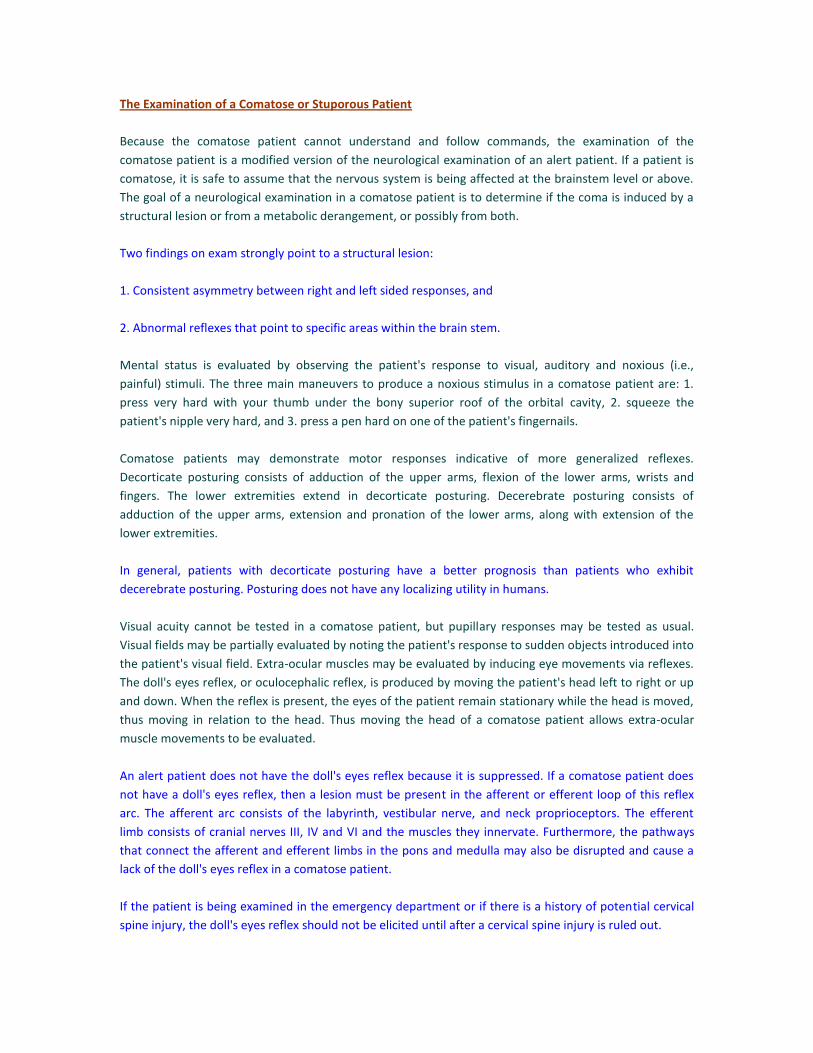
Next have the patient perform the heel to shin
coordination test. With the patient lying supine, instruct
him or her to place their right heel on their left shin just
below the knee and then slide it down their shin to the top
of their foot. Have them repeat this motion as quickly as
possible without making mistakes. Have the patient repeat
this movement with the other foot. An inability to perform
this motion in a relatively rapid cadence is abnormal.
The heel to shin test is a measure of coordination and may
be abnormal if there is loss of motor strength,
proprioception or a cerebellar lesion. If motor and sensory
systems are intact, an abnormal, asymmetric heel to shin
test is highly suggestive of an ipsilateral cerebellar lesion.
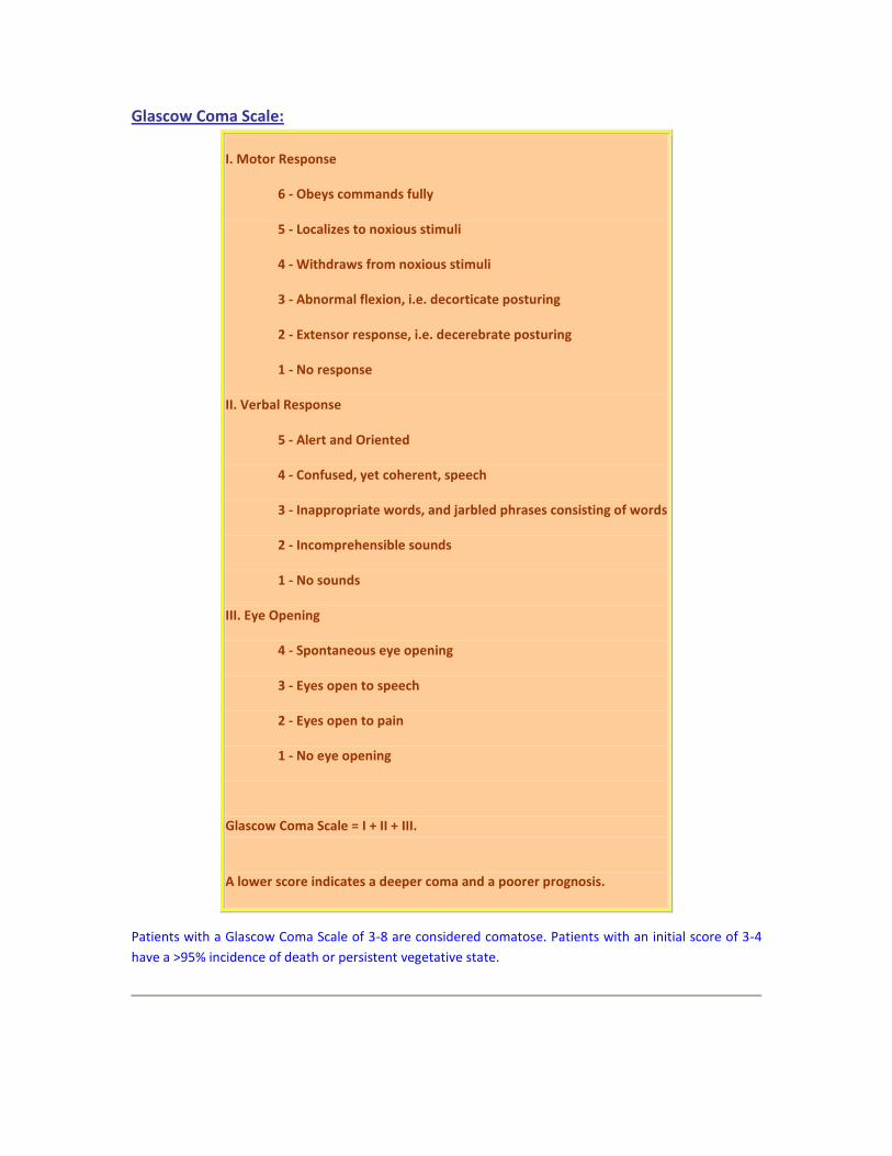
Gait
Gait is evaluated by having the patient walk across the
room under observation. Gross gait abnormalities should
be noted. Next ask the patient to walk heel to toe across
the room, then on their toes only, and finally on their heels
only. Normally, these maneuvers possible without too
much difficulty.
Be certain to note the amount of arm swinging because a
slight decrease in arm swinging is a highly sensitive
indicator of upper extremity weakness.
Also, hopping in place on each foot should be performed.
Walking on heels is the most sensitive way to test for foot
dorsiflexion weakness, while walking on toes is the best
way to test early foot plantar flexion weakness.
Abnormalities in heel to toe walking (tandem gait) may be
due to ethanol intoxication, weakness, poor position sense,
vertigo and leg tremors. These causes must be excluded
before the unbalance can be attributed to a cerebellar
lesion. Most elderly patients have difficulty with tandem
gait purportedly due to general neuronal loss impairing a
combination of position sense, strength and coordination.
Heel to toe walking is highly useful in testing for ethanol
inebriation and is often used by police officers in
examining potential "drunk drivers".

Rhomberg Test
Next, perform the Romberg test by having the patient
stand still with their heels together. Ask the patient to
remain still and close their eyes. If the patient loses their
balance, the test is positive.
To achieve balance, a person requires 2 out of the
following 3 inputs to the cortex: 1. visual confirmation of
position, 2. non-visual confirmation of position (including
proprioceptive and vestibular input), and 3. a normally
functioning cerebellum. Therefore, if a patient loses their
balance after standing still with their eyes closed, and is
able to maintain balance with their eyes open, then there
is likely to be lesion in the cerebellum. This is a positive
Rhomberg.
To conclude the gait exam, observe the patient rising from
the sitting position. Note gross abnormalities.
The Examination of a Comatose or Stuporous Patient
Because the comatose patient cannot understand and follow commands, the examination of the
comatose patient is a modified version of the neurological examination of an alert patient. If a patient is
comatose, it is safe to assume that the nervous system is being affected at the brainstem level or above.
The goal of a neurological examination in a comatose patient is to determine if the coma is induced by a
structural lesion or from a metabolic derangement, or possibly from both.
Two findings on exam strongly point to a structural lesion:
1. Consistent asymmetry between right and left sided responses, and
2. Abnormal reflexes that point to specific areas within the brain stem.
Mental status is evaluated by observing the patient's response to visual, auditory and noxious (i.e.,
painful) stimuli. The three main maneuvers to produce a noxious stimulus in a comatose patient are: 1.
press very hard with your thumb under the bony superior roof of the orbital cavity, 2. squeeze the
patient's nipple very hard, and 3. press a pen hard on one of the patient's fingernails.
Comatose patients may demonstrate motor responses indicative of more generalized reflexes.
Decorticate posturing consists of adduction of the upper arms, flexion of the lower arms, wrists and
fingers. The lower extremities extend in decorticate posturing. Decerebrate posturing consists of
adduction of the upper arms, extension and pronation of the lower arms, along with extension of the
lower extremities.
In general, patients with decorticate posturing have a better prognosis than patients who exhibit
decerebrate posturing. Posturing does not have any localizing utility in humans.
Visual acuity cannot be tested in a comatose patient, but pupillary responses may be tested as usual.
Visual fields may be partially evaluated by noting the patient's response to sudden objects introduced into
the patient's visual field. Extra-ocular muscles may be evaluated by inducing eye movements via reflexes.
The doll's eyes reflex, or oculocephalic reflex, is produced by moving the patient's head left to right or up
and down. When the reflex is present, the eyes of the patient remain stationary while the head is moved,
thus moving in relation to the head. Thus moving the head of a comatose patient allows extra-ocular
muscle movements to be evaluated.
An alert patient does not have the doll's eyes reflex because it is suppressed. If a comatose patient does
not have a doll's eyes reflex, then a lesion must be present in the afferent or efferent loop of this reflex
arc. The afferent arc consists of the labyrinth, vestibular nerve, and neck proprioceptors. The efferent
limb consists of cranial nerves III, IV and VI and the muscles they innervate. Furthermore, the pathways
that connect the afferent and efferent limbs in the pons and medulla may also be disrupted and cause a
lack of the doll's eyes reflex in a comatose patient.
If the patient is being examined in the emergency department or if there is a history of potential cervical
spine injury, the doll's eyes reflex should not be elicited until after a cervical spine injury is ruled out.

The oculovestibular reflex, or cold calorics, is produced by placing the patient's upper body and head at 30
degrees off horizontal, and injecting 50-100cc of cold water into an ear. The water has the same effect on
the semicircular canal as if the patient's head was turned to the opposite side of the injection. Therefore,
the patient's eyes will look towards the ear of injection. This eye deviation lasts for a sustained period of
time. This is an excellent manuever to assess extra-ocular muscles in the comatose patient with possible
cervical spine injury.
If the oculovestibular reflex is absent, a lesion of the pons, medulla, or less commonly the III, IV, IV or VIII
nerves is present. Unlike the oculocephalic reflex, the oculovestibular reflex is present in awake patients.
In alert patients, this reflex not only induces eye deviation, it also produces nystagmus in the direction of
the non-injected ear. The slow phase is towards the injected ear and the fast phase is away.
Cranial nerve V may be tested in the comatose patient with the corneal reflex test. Cranial nerve VII may
be examined by observing facial grimicing in response to a noxious stimulus. Cranial nerves IX an X may be
evaluated with the gag reflex.
The motor system is assessed by testing deep tendon reflexes, feeling the resistance of the patient's limbs
to passive movements, and testing the strength of posturing and local withdrawl movements. Local
withdrawl movements may be elicited by pressing a pen hard on the patient's fingernail and observing if
the patient withdrawls the respective limb from the noxious stimulus.
Upper motor neuron lesions are characterized by spasticity. Spasticity is increased muscle tone leading to
resistance of the limbs to passive manipulation. This spasticity classically results in the clasp-knife
response. The clasp-knife response is when the spastic limb is passively moved with great resistance,
when suddenly the limb "gives", becoming very easy to move. The clasp knife response is most prominent
in the muscle groups least affected by the upper motor lesion, e.g., flexors in the upper extremities or
extensors in the lower extremities.
The sensory system can only be evaluated by observing the patient's response, or lack of response, to
noxious stimuli in different parts of the body.
In addition to withdrawing from noxious stimuli, patient's may localize towards noxious stimuli.
Localization indicates a shallower coma compared to the patient that withdraws.
A common prognostic assessment, called the Glascow Coma Scale, is often used to measure the depth of
coma. The Glascow Coma Scale is often used serially as a means to follow a comatose patient clinically. It
has 3 sections: I. best motor response, II. best verbal response, and III. eye opening.
Glascow Coma Scale:
I. Motor Response
6 - Obeys commands fully
5 - Localizes to noxious stimuli
4 - Withdraws from noxious stimuli
3 - Abnormal flexion, i.e. decorticate posturing
2 - Extensor response, i.e. decerebrate posturing
1 - No response
II. Verbal Response
5 - Alert and Oriented
4 - Confused, yet coherent, speech
3 - Inappropriate words, and jarbled phrases consisting of words
2 - Incomprehensible sounds
1 - No sounds
III. Eye Opening
4 - Spontaneous eye opening
3 - Eyes open to speech
2 - Eyes open to pain
1 - No eye opening
Glascow Coma Scale = I + II + III.
A lower score indicates a deeper coma and a poorer prognosis.
Patients with a Glascow Coma Scale of 3-8 are considered comatose. Patients with an initial score of 3-4
have a >95% incidence of death or persistent vegetative state.