clinical nuclear cardiology - booksca.ca
TRANSCRIPT


Clinical NuclearCardiology
State of the Artand Future Directions

Clinical NuclearCardiology
State of the Artand Future Directions
FOURTH EDITION
Barry L. Zaret, MDRobert W. Berliner Professor of Medicine
Professor of Diagnostic RadiologySection of Cardiovascular MedicineYale University School of Medicine
New Haven, Connecticut
George A. Beller, MDRuth C. Heede Professor of Cardiology
Professor of MedicineCardiovascular DivisionDepartment of Medicine
University of Virginia Health SystemCharlottesville, Virginia

1600 John F. Kennedy Blvd.Ste 1800Philadelphia, PA 19103-2899
Clinical Nuclear Cardiologty ISBN: 978-0-323-05796-7
Copyright # 2010, 2005, 1999, 1993 by Mosby, Inc., an affiliate of Elsevier Inc.
No part of this publication may be reproduced or transmitted in any form or by any means, elec-tronic or mechanical, including photocopying, recording, or any information storage and retrievalsystem, without permission in writing from the publisher. Permissions may be sought directlyfrom Elsevier’s Rights Department: phone: (+1) 215 239 3804 (US) or (+44) 1865 843830 (UK);fax: (+44) 1865 853333; e-mail: [email protected]. You may also complete yourrequest on-line via the Elsevier website at http://www.elsevier.com/permissions.
Notice
Cardiology is an ever-changing field. Standard safety precautions must be followed, but as newresearch and clinical experience broaden our knowledge, changes in practice, treatment, anddrug therapy may become necessary or appropriate. Readers are advised to check the mostcurrent information provided by the manufacturer of each product to be administered toverify the recommended dose or formula, the method and duration of administration, andcontraindications. It is the responsibility of the licensed practitioner, relying on their ownexperience and knowledge of the patient, to determine dosages and the best treatment foreach individual patient. To the fullest extent of the law, neither the Publisher nor the Editorsassume any liability for any injury and/or damage to persons or property arising out of orrelated to any use of the material contained in this book.
The publisher
Library of Congress Cataloging-in-Publication Data
Clinical nuclear cardiology: state of the art and future directions/[edited by] Barry L. Zaret, GeorgeA. Beller. — 4th ed.
p. ; cm.Includes bibliographical references and index.ISBN 978-0-323-05796-7 (alk. paper)1. Heart—Radionuclide imaging. I. Zaret, Barry L. II. Beller, George.[DNLM: 1. Heart—radionuclide imaging. 2. Myocardial Ischemia—radionuclide imaging.3. Radionuclide Imaging—instrumentation. 4. Radionuclide Imaging—methods. WG 141.5.R3 C6415 2010]
RC683.5.R33N833 2010616.10207575–dc22
2009022837
Executive Publisher: Natasha Andjelkovic
Developmental Editor: Julie Goolsby
Project Manager: Nayagi Athmanathan
Design Direction: Ellen Zanolle
Printed in China
Last digit is the print number: 9 8 7 6 5 4 3 2 1

To Myrna Zaret, my wife of almost 50 years, my muse, my companion, my love.
Barry Zaret
To my wonderful and supportive wife, Katherine Brooks, and my six delightful grandchildren,Max, Pietro, Giacomo, Emily, Colin and Grace.
George Beller


CONTRIBUTORS
NIKOLAOS ALEXOPOULOS, MDResearch Scholar, Division of Cardiology, Department ofMedicine, Emory University, Atlanta, Georgia;Researcher, 1st Cardiology, University of Athens MedicalSchool, Athens, Greece
Coronary Artery Computed Tomography Angiography
TIMOTHY M. BATEMAN, MDProfessor, Department of Medicine, University ofMissouri School of Medicine; Co-Director,Cardiovascular Radiologic Imaging, Department ofCardiovascular Disease, Mid America Heart Institute ofSaint Luke’s Hospital, Kansas City, Missouri.
Radiation Considerations for Cardiac Nuclear and ComputedTomography Imaging
JEROEN J. BAX, MD, PhDCardiology, Leiden University Medical Center, Leiden,The Netherlands
Stress Myocardial Perfusion Imaging in Patients withDiabetes Mellitus
Myocardial Viability: Comparison with Other Techniques
ROB S. BEANLANDS, MD, FRCPCProfessor, Medicine (Cardiology)/Radiology, Departmentof Medicine, University of Ottawa, Chief,Cardiac Imaging; Director, National Cardiac PET Centre,Cardiology Department, University of Ottawa HeartInstitute, Ottawa, Ontario, Canada
Diagnosis and Prognosis in Cardiac Disease Using CardiacPET Perfusion Imaging
GEORGE A. BELLER, MDRuth C. Heede Professor of Cardiology and Professor ofMedicine, Cardiovascular Division, Department ofMedicine, University of Virginia Health System,Charlottesville, Virginia
Comparison of Noninvasive Techniques for MyocardialPerfusion Imaging
FRANK M. BENGEL, MDAssociate Professor of Radiology and Medicine, Directorof Cardiovascular Nuclear Imaging, Division of NuclearMedicine, Department of Radiology, Johns HopkinsUniversity School of Medicine, Baltimore, Maryland
Cardiac Neurotransmission Imaging: Positron EmissionTomography
DANIEL S. BERMAN, MDProfessor of Medicine, David Geffen School of Medicineat University of California, Los Angeles, Chief,Department of Cardiac Imaging, Cedars-Sinai MedicalCenter, Los Angeles, California
Digital/Fast SPECT: Systems and Software
Regional and Global Ventricular Function and Volumes fromSPECT Perfusion Imaging
Prognostic Implications of MPI Stress SPECT
ROBERT O. BONOW, MDGoldberg Distinguished Professor of Cardiology,Division of Cardiology, Northwestern University FeinbergSchool of Medicine; Chief, Division of Cardiology,Co-Director, BluhmCardiovascular Institute,NorthwesternMemorial Hospital, Chicago, Illinois
Assessment of Myocardial Viability with Thallium-201 andTechnetium-Based Agents
KENNETH A. BROWN, MD, FACC, FAHA,
FASNCDirector, Nuclear Cardiology and Cardiac StressLaboratory, Department of Cardiology, University ofVermont College of Medicine, Burlington, Vermont
Nuclear Imaging in Revascularized Patients with CoronaryArtery Disease
MATTHEW M. BURG, PhDAssociate Clinical Professor of Medicine, Yale UniversitySchool of Medicine, New Haven; VA-CT HealthcareSystem, West Haven, Connecticut
Mechanistic and Methodological Considerations for theImaging of Mental Stress Ischemia
DENNIS A. CALNON, MD, FACC, FASE, FASNCDirector, Nuclear Cardiology, McConnell Heart Hospitalat Riverside Methodist Hospital, Columbus, Ohio
Atlas of Cases
JOHN M. CANTY, JR, MDAlbert and Elizabeth Rekate Professor of Medicine,Chief, Division of Cardiovascular Medicine, Departmentof Medicine, University at Buffalo; Staff Cardiologist,VAWestern New York Healthcare System at Buffalo,Buffalo, New York
Pathophysiologic Basis of Hibernating Myocardium
vii

IGNASI CARRIO, MDChair and Professor, Nuclear Medicine, UniversitatAutonoma de Barcelona; Director, Department ofNuclear Medicine, Hospital De La Santa Creu I Sant Pau,Barcelona, Spain
Cardiac Neurotransmission Imaging: Single-Photon EmissionComputed Tomography
JAMES A. CASE, PhDAssociate Professor, Director of Physics, University ofMissouri, Kansas City, Missouri
Attenuation Correction and Scatter Correction of MyocardialPerfusion SPECT Images
JI CHEN, PhDAssistant Professor, Department of Radiology, EmoryUniversity, Atlanta, Georgia
SPECT Processing, Quantification, and Display
S. JAMES CULLOM, PhDDirector, Research and Development, CardiovascularImaging Technologies, LLC, Kansas City; AdjunctProfessor, Department of Nuclear Engineering andSciences, University of Missouri, Columbia; TechnicalDirector, ASPIRE Foundation, Kansas City, Missouri
Radiation Considerations for Cardiac Nuclear and ComputedTomography Imaging
SETH T. DAHLBERG, MDAssociate Professor, Medicine and Radiology, University ofMassachusetts Medical School; Director of NuclearCardiology, Divisions of Nuclear Medicine and Cardiology,UMassMemorialMedicalCenter,Worcester,Massachusetts
Imaging for Preoperative Risk Stratification
ROBERT A. DEKEMP, PhDAssociate Professor, Medicine and Engineering, Universityof Ottawa; Head Imaging Physicist, National Cardiac PETCentre, University of Ottawa Heart Institute, Ottawa,Ontario, Canada
Diagnosis and Prognosis in Cardiac Disease Using CardiacPET Perfusion Imaging
E. GORDON DEPUEY, MDProfessor, Department of Radiology, ColumbiaUniversity College of Physicians and Surgeons; Directorof Nuclear Medicine, Radiology, St. Luke’s-RooseveltHospital, New York, New York
Single-Photon Emission Computed Tomography Artifacts
MARCELO F. DI CARLI, MDAssociate Professor of Radiology andMedicine, HarvardMedical School; Director of Noninvasive CardiovascularImaging Program,Chief ofNuclearMedicine andMolecularImaging,Departments of Radiology andMedicine, BrighamandWomen’s Hospital, Boston, Massachusetts
PET/CT and SPECT/CT Hybrid Imaging
Assessment of Myocardial Viability with Positron EmissionTomography
TRACY L. FABER, PhDProfessor, Department of Radiology, Emory University,Atlanta, Georgia
SPECT Processing, Quantification, and Display
JAMES A. FALLAVOLLITA, MDProfessor, Department of Medicine, Universityat Buffalo; Staff Cardiologist, VAWesternNew York Healthcare System at Buffalo, Buffalo,New York
Pathophysiologic Basis of Hibernating Myocardium
ANTONIO B. FERNANDEZ, MDCardiovascular Imaging Fellow, Yale University Schoolof Medicine, VA-CT Healthcare System, West Haven,Connecticut
Mechanistic and Methodological Considerations for theImaging of Mental Stress Ischemia
ALBERT FLOTATS, MDAssociate Professor, Department of Nuclear Medicine,Universitat Autonoma de Barcelona; Consultant,Nuclear Medicine, Hospital De La Santa Creu I Sant Pau,Barcelona, Spain
Cardiac Neurotransmission Imaging: Single-Photon EmissionComputed Tomography
OLIVER GAEMPERLI, MDDepartment of Cardiac Imaging, University HospitalZurich, Zurich, Switzerland
Hybrid Cardiac Imaging
SANJIV SAM GAMBHIR, MD, PhDProfessor, Department of Radiology and Bioengineering;Director, Molecular Imaging Program at Stanford(MIPS); Chief, Division of Nuclear Medicine, StanfordHospital and Clinics, Stanford University, Stanford,California
Molecular Imaging of Gene Expression and Cell Therapy
ERNEST V. GARCIA, PhDProfessor of Radiology, Emory University, Atlanta,Georgia
SPECT Processing, Quantification, and Display
GUIDO GERMANO, PhD, MBAProfessor, Department of Medicine, University ofCalifornia School of Medicine, Los Angeles;Director, Artificial Intelligence Program, Department ofMedicine, Cedars-Sinai Medical Center, Los Angeles,California
Digital/Fast SPECT: Systems and Software
Regional and Global Ventricular Function and Volumes fromSPECT Perfusion Imaging
RAYMOND J. GIBBONS, MDProfessor of Medicine, Department of CardiovascularDiseases, Mayo Clinic, Rochester, Minnesota
Appropriate Use of Nuclear Cardiology
viii Contributors

DAVID K. GLOVER, ME, PhDAssociate Professor, Department of Medicine andCardiovascular Division, University of Virginia,Charlottesville, Virginia
Overview of Tracer Kinetics and Cellular Mechanisms ofUptake
State-of-the-Art Instrumentation for PET and SPECT Imagingin Small Animals
DENNIS A. GOODMAN, MD, FACP, FACC, FCCPClinical Associate Professor, Medicine, University ofCalifornia, San Diego, La Jolla, California; Past Chiefof Cardiology, Scripps Memorial Hospital, La Jolla,California; Senior Cardiologist, Tisch/BellvueHospital, New York, New York; Clinical AssociateProfessor, Medicine, New York University, New York,New York
Coronary Artery Calcification: Pathogenesis, Imaging, andRisk Stratification
ROBERT J. GROPLER, MDProfessor of Radiology, Medicine, and BiomedicalEngineering; Lab Chief, Cardiovascular ImagingLaboratory, Mallinckrodt Institute of Radiology,Washington University School of Medicine; AttendingPhysician, Radiology and Medicine, Barnes-JewishHospital, St. Louis, Missouri
Imaging of Myocardial Metabolism
RORY HACHAMOVITCH, MD, MSCDepartment of Cardiovascular Medicine ClevelandClinic Cleveland, Ohio Prognostic Implications ofMPI Stress SPECT
Prognostic Implications of MPI Stress SPECT
GARY V. HELLER, MD, PhDProfessor of Medicine, University of Connecticut Schoolof Medicine, Farmington; Director, Nuclear Cardiology,Associate Director, Cardiac Division, Hartford Hospital,Hartford, Connecticut
Imaging in Women
THOMAS A. HOLLY, MDAssociate Professor of Medicine and Radiology,Northwestern University Feinberg School of Medicine;Medical Director, Nuclear Cardiology, NorthwesternMemorial Hospital, Chicago, Illinois
Assessment of Myocardial Viability with Thallium-201 andTechnetium-Based Agents
AMI E. ISKANDRIAN, MD, MACC, FAHA,
FASNCDistinguished Professor of Medicine and Radiology,Department of Medicine, University of Alabama atBirmingham, Birmingham, Alabama
Coronary Artery Disease Detection: Pharmacologic StressSPECT
DIWAKAR JAIN, MD, FACC, FRCP, FASNCProfessor of Medicine, Department of Cardiology;Director of Nuclear Cardiology Laboratory, DrexelUniversity College of Medicine; Attending Physician,Cardiology, Hahnemann University Hospital,Philadelphia, Pennsylvania.
Atlas of Cases
PHILIPP A. KAUFMANN, MDProfessor and Director of Cardiac Imaging, ZurichCenter for Integrative Human Physiology, University ofZurich, Zurich, Switzerland
Hybrid Cardiac Imaging
SANJIV KAUL, MDProfessor of Medicine, Division Chief, Division ofCardiovascular Medicine, Oregon Health ScienceUniversity, Portland, Oregon
Myocardial Perfusion Imaging with ContrastEchocardiography
JANUSZ K. KIKUT, MDAssistant Professor of Radiology and Nuclear Medicine,University of Vermont; Director of Nuclear Medicineand PET/CT, Department of Radiology, Fletcher AllenHealth Care, Burlington, Vermont
Nuclear Imaging in Revascularized Patients with CoronaryArtery Disease
MICHAEL A. KING, PhDProfessor, Department of Radiology, University ofMassachusetts Medical School, Worcester, Massachusetts
Attenuation/Scatter/Resolution Correction: Physics Aspects
JUHANI KNUUTI, MD, PhDProfessor, Turku PET Centre, University of Turku, Turku,Finland
Assessment of Myocardial Viability with Positron EmissionTomography
MICHAEL C. KONTOS, MDAssociate Professor, Internal Medicine (Division ofCardiology, Pauley Heart Center), Radiology andEmergency Medicine; Associate Director, Acute CardiacCare; Co-Director, Stress Laboratory; Associate Professor,Internal Medicine, Department of Cardiology,Radiology, and Emergency Medicine, VirginiaCommonwealth University, Richmond, Virginia
Imaging Patients with Chest Pain in the EmergencyDepartment
CHRISTOPHER M. KRAMER, MDProfessor, Departments of Medicine and Radiology;Director, Cardiovascular Imaging Center, University ofVirginia Health System, Charlottesville, Virginia
Myocardial Perfusion: Magnetic Resonance Imaging
ixContributors
BIJOY KUNDU, PhDAssistant Professor, Department of Radiology, Universityof Virginia, Charlottesville, Virginia
State-of-the-Art Instrumentation for PET and SPECT Imagingin Small Animals
AVIJIT LAHIRI, MBBS, MSC, MRCP, FACC, FESCConsultant Cardiologist and Director, Clinical Imagingand Research Centre (CIRC), The Wellington Hospital,St John’s Wood, London; Medical Director, BritishCardiac Research Trust; Honorary Professor, MiddlesexUniversity; Honorary Senior Lecturer, Imperial College,London, United Kingdom
Coronary Artery Calcification: Pathogenesis, Imaging,and Risk Stratification
JEFFREY A. LEPPO, MDProfessor, Department of Medicine and Radiology,University of Massachusetts, Worcester; Chief ofCardiology, Department of Medicine, Berkshire MedicalCenter, Pittsfield, Massachusetts
Imaging for Preoperative Risk Stratification
JONATHAN R. LINDNER, MDProfessor of Medicine, Associate Chief for Education,Division of Cardiovascular Medicine, Oregon HealthScience University, Portland, Oregon
Myocardial Perfusion Imaging with ContrastEchocardiography
JOHN J. MAHMARIAN, MDProfessor of Medicine, Department of Cardiology, WeillCornell Medical College, New York, New York; Director,Nuclear Cardiology and CT Services, Methodist DeBakeyHeart and Vascular Center, The Methodist Hospital,Houston, Texas
Risk Stratification for Acute ST-Segment Elevation andNon-ST-Segment Elevation Myocardial Infarction
DALTON S. McLEAN, MDCardiology Fellow, Division of Cardiology, Departmentof Medicine, Emory University, Atlanta, Georgia
Coronary Artery Computed Tomography Angiography
TODD D. MILLER, MDProfessor of Medicine, Department of CardiovascularDiseases, Mayo Clinic, Rochester, Minnesota
Appropriate Use of Nuclear Cardiology
ALAN R. MORRISON, MD, PhDCardiovascular Medicine Fellow, Department of InternalMedicine, Yale University School of Medicine, NewHaven, Connecticut
Molecular Imaging Approaches for Evaluation of MyocardialPathophysiology: Angiogenesis, Ventricular Remodeling,Inflammation, and Cell Death
LAURA FORD-MUKKAMALA, DO, FACCClinical Cardiologist, Department of Cardiology, BillingsClinic, Billings, Montana
Imaging in Women
JAGAT NARULA, MD, PhDProfessor of Medicine and Chief of Cardiology; MedicalDirector, Memorial Heart and Vascular Institute; MedicalDirector, Edwards Life Sciences Center for AdvancedCardiovascular Technology, University of California,Irvine, Irvine, California
Radionuclide Imaging of Inflammation in Atheroma
TINSU PAN, PhDAssociate Professor, Department of Imaging Physics, TheUniversity of Texas, MD Anderson Cancer Center,Houston, Texas
Attenuation/Scatter/Resolution Correction: Physics Aspects
AMIT R. PATEL, MDDirector of Cardiac Magnetic Resonance, AssistantProfessor of Medicine, Department of Medicine,University of Chicago, Chicago, Illinois
Myocardial Perfusion: Magnetic Resonance Imaging
JAMES A. PATTON, PhDProfessor, Department of Radiology and RadiologicalSciences, Vanderbilt University Medical Center,Nashville, Tennessee
Digital/Fast SPECT: Systems and Software
LINDA R. PETERSON, MDAssociate Professor of Medicine and Radiology,Cardiovascular Division and Division of Geriatrics andNutritional Sciences, Department of Medicine,Washington University School of Medicine, St. Louis,Missouri
Imaging of Myocardial Metabolism
DON POLDERMANS, MD, PhD, FESCProfessor, Department of Medicine, Erasmus MedicalCentre, Department of Vascular Surgery, Rotterdam,The Netherlands
Myocardial Viability: Comparison with Other Techniques
DONNA M. POLK, MD, MPHDirector, Preventive Cardiology, Department ofCardiology, Hartford Hospital, Hartford, Connecticut
Imaging in Women
P. HENDRIK PRETORIUS, PhDAssociate Professor, Department of Radiology, Universityof Massachusetts Medical School, Worcester,Massachusetts
Attenuation/Scatter/Resolution Correction: Physics Aspects
x Contributors

PAOLO RAGGI, MDAssociate Professor of Medicine, Division of Cardiology,Department of Medicine, Emory University, Atlanta,Georgia
Coronary Artery Computed Tomography Angiography
RAYMOND R. RUSSELL, MD, PhDAssociate Professor, Departments of Medicine andDiagnostic Radiology, Yale-New Haven Hospital,New Haven, Connecticut
Principles of Myocardial Metabolism as They Relate toImaging
Coronary Artery Disease Detection: Exercise Stress SPECT
ANTTI SARASTE, MD, PhDResearch Fellow, Nuklearmedizinische Klinik undPoliklinik, Klinikum rechts der Isar der TechnischeUniversitat Munchen, Munich, Germany
Cardiac Neurotransmission Imaging: Positron EmissionTomography
HEINRICH R. SCHELBERT, MD, PhDThe George V. Taplin Professor, Department ofMolecular and Medical Pharmacology, University ofCalifornia, Los Angeles, California
State-of-the-Art Instrumentation for PET and SPECT Imagingin Small Animals
Myocardial Blood Flow Measurement: Evaluating CoronaryPathophysiology and Monitoring Therapy
THOMAS HELLMUT SCHINDLER, MD, PhDAssistant Professor, Department of Medicine andCardiology, Cardiovascular Center, School of Medicineat the University Hospitals of Geneva, Geneva,Switzerland
Myocardial Blood Flow Measurement: Evaluating CoronaryPathophysiology and Monitoring Therapy
AREND F.L. SCHINKEL, MD, PhDCardiology, Thoraxcenter, Erasmus Medical Center,Rotterdam, The Netherlands
Myocardial Viability: Comparison with Other Techniques
MARKUS SCHWAIGER, MD, FACC, FAProfessor, Nuclear Medicine, Technische UniversitatMunchen; Director, Nuclear Medicine, Klinikum rechtsder Isar, Munich, Germany
Cardiac Neurotransmission Imaging: Positron EmissionTomography
LESLEE J. SHAW, PhDProfessor of Medicine, Department of Medicine, EmoryUniversity, Atlanta, Georgia
Cost Effectiveness of Myocardial Perfusion Single-PhotonEmission Computed Tomography
HOSSAM M. SHERIF, MDPost-Doctoral Nuclear Cardiology Research Fellow,Nuclear Medicine, Technical University of Munich,Munich, Germany; Assistant Professor, CardiovascularCritical Care Medicine, Cairo University Hospitals,Cairo, Egypt
Cardiac Neurotransmission Imaging: Positron EmissionTomography
ALBERT J. SINUSAS, MDProfessor, Department of Internal Medicine andDiagnostic Radiology, Yale University School ofMedicine; Director, Department of CardiovascularNuclear Imaging and Stress Laboratory, Yale-New HavenHospital, New Haven, Connecticut
Role of Intact Biological Models for Evaluation of Radiotracers
Molecular Imaging Approaches for Evaluation of MyocardialPathophysiology: Angiogenesis, Ventricular Remodeling,Inflammation, and Cell Death
PIOTR SLOMKA, PhDProfessor, Department of Medicine, University ofCalifornia, Los Angeles; Research Scientist, Departmentof Medicine and Imaging, Cedars-Sinai Medical Center,Los Angeles, California
Digital/Fast SPECT: Systems and Software
PREM SOMAN, MD, PhD, FRCP (UK), FACCAssistant Professor of Medicine, Department ofMedicine, University of Pittsburgh Medical Center,Pittsburgh, Pennsylvania
Radionuclide Imaging in Heart Failure
ROBERT SOUFER, MDProfessor of Medicine, Chief of Cardiology, VA-CTHealthcare System, Yale University School of Medicine,West Haven, Connecticut
Mechanistic and Methodological Considerations for theImaging of Mental Stress Ischemia
H. WILLIAM STRAUSS, MDProfessor of Radiology (Nuclear Medicine), Weill CornellMedical College; Attending Physician, NuclearMedicine, Memorial Sloan Kettering Cancer Center,New York, New York
Radionuclide Imaging of Inflammation in Atheroma
RAGHUNANDAN DUDDA SUBRAMANYA,
MBBS, MDFellow in Cardiology, Drexel University College ofMedicine, Philadelphia; Fellow in Cardiology,Hahnemann University Hospital, Philadelphia; Fellowin Cardiology, Abington Memorial Hospital, Abington,Pennsylvania
Atlas of Cases
xiContributors

INES VALENTA, MDResearch Fellow, Department of Medicine andCardiology, Cardiovascular Center, School of Medicineat the University Hospitals of Geneva, Geneva,Switzerland
Myocardial Blood Flow Measurement: Evaluating CoronaryPathophysiology and Monitoring Therapy
SERGE D. VAN KRIEKINGE, PhDAssistant Professor, Department of Medicine, Universityof California, Los Angeles, School of Medicine; ResearchScientist, Artificial Intelligence Program, Department ofMedicine, Cedars-Sinai Medical Center, Los Angeles,California
Regional and Global Ventricular Function and Volumes fromSPECT Perfusion Imaging
SHREENIDHI VENURAJU, MBBS, MRCPClinical Research Fellow in Cardiology, Cardiac Imaging,Clinical Imaging and Research Centre, WellingtonHospital, London, United Kingdom
Coronary Artery Calcification: Pathogenesis, Imaging, andRisk Stratification
JOHAN W.H. VERJANS, MDCardiology, University Medical Center Utrecht, Utrecht,Netherlands; Cardiology, University of California,Irvine, Irvine, California
Radionuclide Imaging of Inflammation in Atheroma
FRANS J.TH. WACKERS, MD, PhDProfessor Emeritus of Diagnostic Radiology andMedicine, Senior Research Scientist, Departments ofDiagnostic Radiology and Internal Medicine, YaleUniversity School of Medicine, New Haven, Connecticut
Coronary Artery Disease Detection: Exercise Stress SPECT
Stress Myocardial Perfusion Imaging in Patients withDiabetes Mellitus
ERNST E. VAN DER WALL, MD, PhDDepartment of Cardiology, Leiden University MedicalCenter, Leiden, The Netherlands
Myocardial Viability: Comparison with Other Techniques
DENNY D. WATSON, PhDProfessor of Radiology, Director of Nuclear Cardiology,University of Virginia Health System, Charlottesville,Virginia
Overview of Tracer Kinetics and Cellular Mechanisms ofUptake
JOSEPH C. WU, MD, PhD, FACCAssistant Professor, Medicine (Cardiology) andRadiology, Stanford University School of Medicine,Stanford, California
Molecular Imaging of Gene Expression and Cell Therapy
AJAY KUMAR YERRAMASU, MBBS, MRCPClinical Research Fellow in Cardiology, Cardiac Imaging,Clinical Imaging and Research Centre, WellingtonHospital, London, United Kingdom
Coronary Artery Calcification: Pathogenesis, Imaging, andRisk Stratification
KEIICHIRO YOSHINAGA, MD, PhDAssociate Professor, Molecular Imaging, HokkaidoUniversity Graduate School of Medicine, Sapporo,Hokkaido, Japan
Diagnosis and Prognosis in Cardiac Disease Using CardiacPET Perfusion Imaging
BARRY L. ZARET, MDRobert W. Berliner Professor of Medicine and Professorof Diagnostic Radiology, Department of InternalMedicine, Section of Cardiovascular Medicine, YaleUniversity School of Medicine; Attending, InternalMedicine/Cardiovascular Medicine, Yale-New HavenHospital, New Haven, Connecticut
Cardiac Performance
Imaging in Patients Receiving Cardiotoxic Chemotherapy
Radionuclide Imaging of Inflammation in Atheroma
MARIA CECILIA ZIADI, MDClinical Research Fellow in Molecular Function andImaging, Nuclear Cardiology, University of Ottawa HeartInstitute, Ottawa, Ontario, Canada
Diagnosis and Prognosis in Cardiac Disease Using CardiacPET Perfusion Imaging
GILBERT J. ZOGHBI, MD, FACC, FSCAIAssistant Professor of Medicine, Department ofMedicine, University of Alabama at Birmingham;Birmingham Veterans Association Medical Center,Birmingham, Alabama
Coronary Artery Disease Detection: Pharmacologic StressSPECT
xii Contributors


PREFACE
INTRODUCTION: INTEGRATEDCARDIOVASCULAR IMAGING—THE FUTURE
This book first appeared in 1993; the preceding thirdedition was published in 2005. Each new edition hasbeen associated with significant expansion, revision,and updating, as well as significant changes in orienta-tion that reflect new advances in the field. At the timeof this fourth edition, nuclear cardiology is firmly estab-lished as a key noninvasive modality for the clinicalevaluation of patients with cardiovascular disease. Con-comitantly, there have been further advances in instru-mentation, radiopharmaceutical development, and newclinical research, leading to additional understandingof clinical utility, cost-effectiveness, appropriateness,and relationship of imaging findings to patient out-comes. Nuclear cardiology has been incorporated intomajor large multicenter clinical trials. In addition, asother modes of cardiovascular imaging have approachedmaturity, there has been movement toward integratingthe various imaging modalities under the broadumbrella of cardiovascular multimodality imaging. Thecardiovascular imager of the future will likely be trainedin more than one modality and will be housed in dedi-cated imaging centers that offer a variety of imagingapproaches. It will be the imager’s job to determine thestudy most appropriate for answering the posed clinicalquestion. In recognition of this trend, new chapters areincluded in this edition that provide additional focuson non-nuclear cardiovascular imaging modalities suchas computed tomography, magnetic resonance imaging,and contrast echocardiography.
The fourth edition continues to focus on nuclear car-diology and represents a major effort to incorporate newadvances in clinical nuclear cardiology, therebyproviding a road map for up-to-date clinical use. In addi-tion, we seek to point out the new directions in whichnuclear cardiology as well as integrated cardiovascularimaging are headed. Our goals in the fourth edition con-tinue to be twofold: first, to present the most up-to-dateand comprehensive clinically applicable data availablein the field, thereby offering both the practitioner andstudent/trainee the current clinical state of the art; andsecond, to present the newest andmost exciting directionsin the field that reflect both technologic and biological
advances. To meet these combined goals, the book hasonce again expanded—now to a total of 45 chapters—and also includes a totally new and expanded atlas ofcase presentations to provide concrete examples of theclinical relevance of nuclear cardiology. Once again,the book is grouped into nine specific sections. Twentyof the 45 chapters, as well as the atlas, are totally new.An almost equal number of chapters have been elimi-nated, and, all remaining chapters have been revised,updated, and, in certain instances, consolidated.
Section 1 addresses issues related to radiopharmace-uticals and tracer kinetics. The three chapters in thissection provide information concerning tracer kineticsand cellular mechanisms of uptake, principles of myocar-dial metabolism as they relate to imaging, and the roleof intact biological models in evaluating radiotracers.
Section 2 deals with instrumentation. The eight chap-ters in this section address issues relating to processing,quantification, and display of single-photon emissiontomography (SPECT) data, SPECT artifacts, attenuation/scatter/resolution and correction from both physicsand clinical standpoints, hybrid imaging, digital/fastSPECT imaging, radiation considerations of imagingtechnologies, and small-animal imaging.
Section 3, as in the previous edition, contains twochapters dealing with cardiac function and performance,as evaluated by blood-pool imaging and gated SPECTimaging.
Section 4 addresses major issues that relate to perfu-sion imaging and detection of coronary disease. The 12chapters in this section address the issues of coronaryartery disease detection by exercise and pharmacologicstress, their prognostic implications, assessment of myo-cardial perfusion imaging by magnetic resonance imag-ing, echocardiography, and positron emissiontomography (PET), and hybrid imaging. Specific chap-ters deal with computed tomography angiography anduse of computed tomography to assess coronary arterycalcification. Chapters also address cost-effectiveness aswell as the appropriate use criteria for nuclear cardiol-ogy. A chapter also compares the various noninvasiveapproaches for assessment of myocardial perfusion.
Section 5 focuses on disease- and gender-specificissues. Specific chapters focus on imaging in women,imaging for preoperative risk assessment, revascularizedpatients, patients with diabetes mellitus, imaging in
xvii

the heart failure population, imaging of patients receiv-ing cardiotoxic chemotherapy, mental stress imaging,and the use of PET measurements of myocardial bloodflow to evaluate cardiovascular pathophysiology andtherapeutic efficacy.
Section 6 addresses acute coronary syndromes. Thetwo chapters in this section deal with imaging in theemergency department and risk stratification of patientswith acute myocardial infarction, based on new datafrom a multicenter randomized trial.
Section 7 contains four chapters focusing on myocar-dial viability. Viability assessment with SPECT studies,PET, and other techniques is addressed in three specificchapters, while an additional chapter focuses on thepathophysiologic basis of hibernating myocardium.
Section 8 contains three chapters on tracer-specificimaging techniques. These three chapters deal with
imaging of myocardial metabolism and cardiac neuro-transmission imaging with either SPECT or PET.
Section 9 deals with new molecular approaches andcontains three chapters dealing with molecular imagingof angiogenesis matrix metalloproteases and cell death,vascular abnormalities, and imaging of gene expressionand cell therapy. Such techniques are primarily beingevaluated in preclinical experimental models but havealready shown promise in early clinical studies.
The Atlas of Cases is the final section of the book andis designed to provide complementary information tothe numerous clinical issues discussed in the text. Itexemplifies the substantial clinical utility of nuclear car-diology and shows a variety of images set in their clini-cal context. This atlas is significantly expanded fromthe one included in the third edition.
Barry L. Zaret and George A. Beller
xviii Preface


Chapter 1
Overview of Tracer Kineticsand Cellular Mechanisms
of Uptake
DENNY D. WATSON AND DAVID K. GLOVER
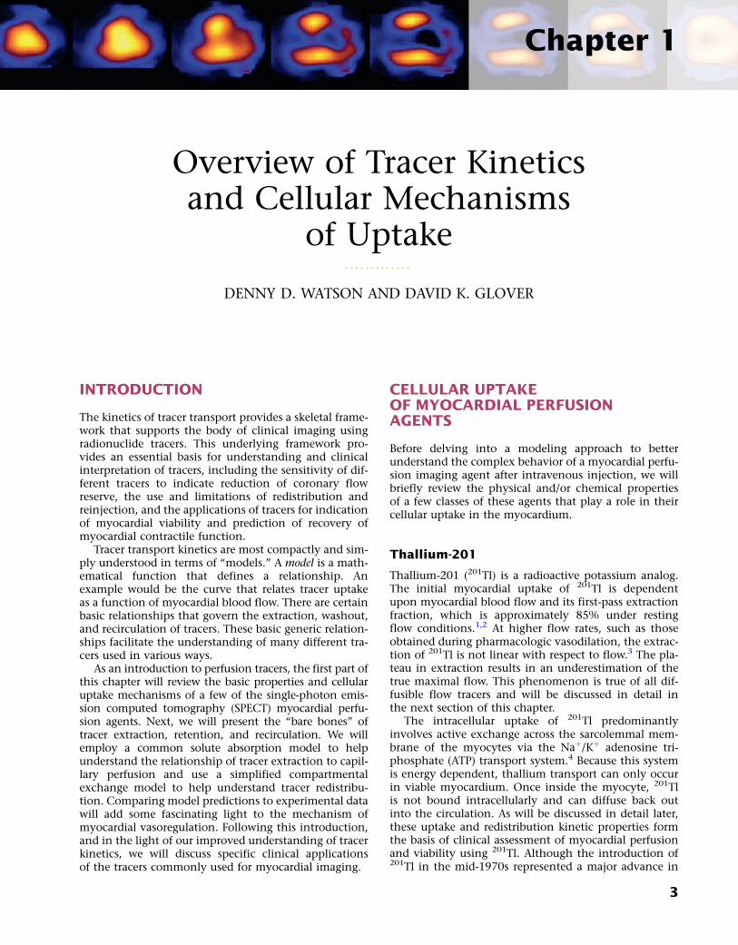
INTRODUCTION
The kinetics of tracer transport provides a skeletal frame-work that supports the body of clinical imaging usingradionuclide tracers. This underlying framework pro-vides an essential basis for understanding and clinicalinterpretation of tracers, including the sensitivity of dif-ferent tracers to indicate reduction of coronary flowreserve, the use and limitations of redistribution andreinjection, and the applications of tracers for indicationof myocardial viability and prediction of recovery ofmyocardial contractile function.
Tracer transport kinetics are most compactly and sim-ply understood in terms of “models.” A model is a math-ematical function that defines a relationship. Anexample would be the curve that relates tracer uptakeas a function of myocardial blood flow. There are certainbasic relationships that govern the extraction, washout,and recirculation of tracers. These basic generic relation-ships facilitate the understanding of many different tra-cers used in various ways.
As an introduction to perfusion tracers, the first part ofthis chapter will review the basic properties and cellularuptake mechanisms of a few of the single-photon emis-sion computed tomography (SPECT) myocardial perfu-sion agents. Next, we will present the “bare bones” oftracer extraction, retention, and recirculation. We willemploy a common solute absorption model to helpunderstand the relationship of tracer extraction to capil-lary perfusion and use a simplified compartmentalexchange model to help understand tracer redistribu-tion. Comparing model predictions to experimental datawill add some fascinating light to the mechanism ofmyocardial vasoregulation. Following this introduction,and in the light of our improved understanding of tracerkinetics, we will discuss specific clinical applicationsof the tracers commonly used for myocardial imaging.
CELLULAR UPTAKEOF MYOCARDIAL PERFUSIONAGENTS
Before delving into a modeling approach to betterunderstand the complex behavior of a myocardial perfu-sion imaging agent after intravenous injection, we willbriefly review the physical and/or chemical propertiesof a few classes of these agents that play a role in theircellular uptake in the myocardium.
Thallium-201
Thallium-201 (201Tl) is a radioactive potassium analog.The initial myocardial uptake of 201Tl is dependentupon myocardial blood flow and its first-pass extractionfraction, which is approximately 85% under restingflow conditions.1,2 At higher flow rates, such as thoseobtained during pharmacologic vasodilation, the extrac-tion of 201Tl is not linear with respect to flow.3 The pla-teau in extraction results in an underestimation of thetrue maximal flow. This phenomenon is true of all dif-fusible flow tracers and will be discussed in detail inthe next section of this chapter.
The intracellular uptake of 201Tl predominantlyinvolves active exchange across the sarcolemmal mem-brane of the myocytes via the Naþ/Kþ adenosine tri-phosphate (ATP) transport system.4 Because this systemis energy dependent, thallium transport can only occurin viable myocardium. Once inside the myocyte, 201Tlis not bound intracellularly and can diffuse back outinto the circulation. As will be discussed in detail later,these uptake and redistribution kinetic properties formthe basis of clinical assessment of myocardial perfusionand viability using 201Tl. Although the introduction of201Tl in the mid-1970s represented a major advance in
3

nuclear cardiology, its physical properties are not idealfor gamma camera imaging. The low-energy 69- to 80-keV x-ray photopeak can result in attenuation artifactsand the relatively long 73-hour half-life limits the maxi-mal dose that can be safely administered.
Monovalent CationicTechnetium-99m-Labeled Tracers
Technetium-99m (99mTc) is a generator-produced isotopethat is readily available and has a number of advantagesover 201Tl for gamma camera imaging. The higher-energy140-keV principle photopeak is ideal for detection usingstandard collimated gamma cameras with less attenua-tion, and its short 6-hour half-life allows for a higheradministered dose yielding improved count statistics.
Over the years, there have been a number of 99mTc-labeled myocardial perfusion imaging agents that havebeen investigated as replacements for 201Tl. The mostsuccessful ones to date are the lipophilic monovalentcationic agents, 99mTc-sestamibi (sestamibi, Cardiolite)and 99mTc-tetrofosmin (tetrofosmin, Myoview), that arenow widely used for clinical studies. Following an intra-venous injection, the first-pass extraction fractions ofsestamibi and tetrofosmin are approximately 65% and54%, respectively, under basal resting flow conditions.5,6
Because of their lower extraction fractions comparedwith 201Tl, the plateau in tracer uptake observed duringhyperemia occurs at lower flow rates. The effect of this“roll-off” in extraction at lower flow rates is to diminishthe relative difference in tracer activities between high-flow regions and those myocardial regions subtendedby a coronary stenosis, making it more difficult to detectmilder stenoses.
Although these agents are members of two distinctchemical classes of compounds, isonitriles and dipho-sphines, respectively, they share several common prop-erties. Unlike 201Tl, which utilizes a specific membrane-active transporter, these tracers are passively drawnacross the sarcolemmal and mitochondrial membranesalong a large electronegative transmembrane potentialgradient, owing to their lipophilicity and positivecharge.7 Once inside the mitochondria, these cationictracers are tightly bound by the potential gradient suchthat there is a very slow net efflux resulting in prolongedmyocardial retention times. Although ATP is not directlyrequired for the intracellular sequestration of cationictracers, as it is for 201Tl, the influx and retention of thesetracers are energy dependent because the presence of anormal electronegative transmembrane gradient isrequired. With irreversible injury, the mitochondrialand sarcolemmal membranes are depolarized, and theuptake of these cationic tracers is impaired.8 Accord-ingly, like 201Tl, the cationic 99mTc-labeled agents canbe used to assess myocardial viability.
In addition to the lower plateau in extraction men-tioned, another disadvantage to both sestamibi andtetrofosmin is the problem of photon scatter from theadjacent liver that can interfere with the interpretationof myocardial perfusion defects, particularly in the infe-rior left ventricular wall. Accordingly, there has beenrenewed interest in recent years to design improved
cationic 99mTc-labeled tracers that exhibit more rapidliver clearance. 99mTc-(N)(PNP5)(DBODC5)þ (DBODC5)is a lipophilic nitride that is rapidly taken up andretained by the myocardium in a manner that is mecha-nistically similar to sestamibi and tetrofosmin. However,studies in both rats and dogs demonstrated thatDBODC5 cleared more rapidly from the liver than eitherof these other cationic tracers, with virtually no liveractivity observed after only 1 hour.9,10 The first-passextraction fraction of DBODC5 is intermediate to thatof sestamibi and tetrofosmin.10 Although there is noimprovement in the ability of DBODC5 to track myocar-dial blood flow at hyperemic flow rates, its morefavorable biodistribution properties offer a potentialadvantage that warrants further investigation.
Another new lipophilic cationic tracer with improvedbiodistribution and very rapid liver clearance is 99mTc-[N(MPO)(PNP5)]þ (MPO). The myocardial uptake ofMPO in Sprague Dawley rats was reported to be betweenthat of sestamibi and DBODC5 over 2 hours.11 Interest-ingly, the heart-liver ratio of MPO at 30 minutes afterinjection was more than twice that of DBODC5 andapproximately 4 times higher than that of sestamibi.11
With such rapid liver clearance, clinically useful imagesmight be obtainable as early as 15 minutes post injec-tion. At the present time, the first-pass extraction frac-tion studies have not been conducted using MPO.
Neutral Lipophilic Tracers
99mTc-teboroxime (teboroxime) is a member of a class ofneutral lipophilic molecules known as BATOs (Boronicacid Adducts of Technetium diOxime). After intravenousinjection, the initial instantaneous uptake of teborox-ime is high, with a first-pass extraction fraction ofapproximately 90%—higher than even 201Tl.12,13 How-ever, unlike the cationic 99mTc-labeled myocardial perfu-sion tracers discussed earlier that are retained in themyocardium, teboroxime exhibits rapid flow-dependentmyocardial clearance in under 10 minutes. Thus,although the myocardial extraction fraction that isobserved immediately after injection is very high, therapid clearance of this tracer results in a loss of defectcontrast within the first 5 minutes post injection.14
Additionally, because the myocardial clearance rate ofteboroxime is flow dependent, with slower clearancefrom ischemic versus normally perfused zones, the dif-ferential clearance rates give the scintigraphic equivalentof “redistribution,” with an apparent filling-in of the ini-tial perfusion defects over time, as is observed with201Tl.15 The mechanism for such rapid clearance is thatteboroxime is believed not to cross the sarcolemmalmembrane into the intracellular space of the myocyte,remaining instead within the intravascular space inassociation with the endothelial layer.16 Furthermore,its myocardial uptake is passive, not dependent oneither active transport or other energy-dependent pro-cesses. Thus, teboroxime is considered to be a pure per-fusion tracer.
Although teboroxime was approved for clinical imag-ing at the same time as sestamibi, its rapid dynamicmyocardial clearance kinetics proved difficult to image
4 SECTION 1 Radiopharmaceuticals/Tracer Kinetics

using the relatively slow, single-head gamma camerasthat were standard in the early 1990s. With the excitingnew generation of fast cardiac SPECT instrumentationthat has recently become available on the market, theremay be renewed interest in this tracer in the future.
Another neutral lipophilic perfusion tracer that hasundergone Phase III clinical testing is 99mTc-N-NOET(NOET). Like teboroxime, NOET exhibits a first-passextraction fraction that is higher than either sestamibior tetrofosmin, with flow-dependent differential clear-ance of the tracer from the myocardium.17,18 Becauseof the differential clearance from ischemic versus nor-mal zones, NOET has been shown to undergo apparentredistribution like teboroxime, albeit at a slowerrate.18,19 Another similarity between NOET and teborox-ime involves their mechanism of localization in themyocardium. NOET is also believed to remain withinthe intravascular space in association with the endothe-lial layer.20 Because of its accessibility, NOET clearancecan be affected by a host of intravascular factors. Experi-mental studies demonstrated that the myocardial clear-ance rate of NOET could be accelerated not only byincreasing the flow rate but also by elevating the bloodlipid concentration.16,21 Like teboroxime, the uptakeand retention of NOET does not involve active orenergy-dependent processes, and thus it would also beconsidered a pure perfusion tracer.
In summary, the advent of the 99mTc-labeled myocar-dial perfusion imaging agents, particularly the lipophiliccationic tracers, sestamibi and tetrofosmin, representeda major advance by virtue of their superior imagingproperties compared with 201Tl. Some aspects of thesetracers may not be ideal, but in general they have shownexcellent diagnostic accuracy and have fueled thegrowth of the field of nuclear cardiology for nearly 20years. New SPECT perfusion tracers that exhibit bothimproved myocardial first-pass extraction fraction andmore favorable biodistribution properties are clearlywarranted.
MODELING TRACER EXTRACTION
If a tracer is injected intravenously, the number of traceratoms passing through a capillary bed will be propor-tional to the fraction of total cardiac output passingthrough the capillary bed. If all the tracer atoms wereextracted in a single pass through the capillary bed, thenumber of tracer atoms per unit volume of tissue wouldthen be proportional to the fraction of cardiac outputperfusing the unit volume of tissue. The only tracersthat approximate this ideal are microspheres.
The tracers used for clinical imaging of myocardialblood flow are not completely extracted. For these tra-cers, the fraction of tracer extracted on passing througha capillary bed depends on the blood flow through thecapillary bed. A model based on the work of Gosselinand Stibitz 22 provides insight into this process. Themodel is that of a diffusible tracer traveling througha cylindrical capillary. The tracer can diffuse outwardfrom the blood across the capillary endothelium, butit can also diffuse back into the blood from outside
the capillary endothelium. The outward and back-diffusion coefficients can be different. The extractioncoefficient reflects the net loss in tracer concentrationbetween the arterial and venous ends of the capillary.This leads to a tracer “extraction fraction” of the form:
1� e�PSb ð1Þ
where PS is a product of capillary permeability and surfacearea, and b is the capillary blood flow. The relationshipbetween blood flow and tracer extraction predicted bythis model is shown graphically in Figure 1-1. The topcurve with PS ¼ 2 would represent a tracer with highfirst-pass extraction, such as 201Tl. The lower curve withPS ¼ 1 would represent a tracer with lower first-passextraction, similar to sestamibi and tetrofosmin. The termfirst-pass extraction is often used to characterize radionu-clide tracers, but it is not often carefully defined. Sincethe extracted fraction of tracer is flow dependent, thefirst-pass extraction indicates the fraction of extractedtracer measured at baseline resting blood flow. In Fig-ure 1-1, the first-pass extraction of the two tracers shownwould be about 86% for the upper line and about 64% forthe lower line.
The amount of tracer taken up by the myocardiumshortly after bolus injection is the product of extractionfraction and myocardial blood flow per unit volume,denoted by the letter b. This product is:
Myocardial Extraction / b ð1� e�PSb Þ ð2Þ
Although the equation was derived for soluteexchange in a single capillary, it can be shown that thefunctional form remains unchanged for a generalizeddistribution of capillaries if the parameters are taken torepresent the averages over the entire capillary distribu-tion. The curve with the functional form shown hasbeen ubiquitous in representing myocardial uptake as afunction of myocardial blood flow. Figure 1-2 shows
0 1 2 3 4
1.0
0.90.8
0.70.6
0.50.4
0.3
0.2
0.1
0.0
Ext
ract
ion
frac
tion
Capillary blood flow relative to normal
Initial Myocardial Extraction
PS � 2PS � 1
Figure 1-1 Tracer extraction fraction as predicted by theGosselin and Stibitz model. Curves are shown for PS ¼ 1,representing a tracer with first-pass extraction similar to themolecular Tc-99 m tracers, and PS ¼ 2, representing a tracerwith first-pass extraction of Tl-201.
51 Overview of Tracer Kinetics and Cellular Mechanisms of Uptake

some experimental data of sestamibi extraction versusblood flow. The solid line of Figure 1-2 has the func-tional form of Equation 2. It fits the experimental dataquite well if the PS coefficient is chosen empirically tobest fit the data. However, if we substitute the PS coeffi-cient that best agrees with the first-pass extraction data,it results in the dashed line of Figure 1-2 and produces apoor fit for the flow-versus-extraction curve. The dashedline predicts a more extreme reduction of tracer extrac-tion with increasing myocardial blood than experimen-tally observed.
The same PS product should predict both themeasured first-pass extraction coefficient and the flow-versus-extraction curve. The fact that it does notindicates that something is wrong with the model.A possible problem with the simple Gosselin and Stibitzmodel is that it does not account for myocardial flowregulation by opening and closing of capillary channels.Selective opening and closing of parallel capillary chan-nels is thought to be an important mechanism to regu-late capillary resistance and myocardial blood flow.This has been experimentally demonstrated.23,24 Furtherevidence for the role of capillary closure has been morerecently found in the context of contrast echocardio-graphy25 and for sestamibi perfusion measurements inthe dog model.26
To account for the effect of variable capillaryvolumes, we wish to extend the basic model as follows:The first factor in Eq. 2 is replaced by F, which representsflow per unit myocardial volume. The term b in theexponential represents flow per unit of open capillaryvolume. We now introduce a new relationship:
F
b¼ 1� e�F ð3Þ
Equation 3 allows for flow in the open capillaries tobe different from flow per unit myocardial volume deter-mined by the arterial supply vessels. Equation 3 furtherintroduces the assumption that capillary blood volume
decreases with decreasing flow due to capillary closure,and it increases to some maximum value when allthe capillary channels are fully utilized at high flow.Figure 1-3 shows the relative capillary volume assumedby Eq. 3. This is in qualitative accord with the observa-tions of Wu et al.23 The exact way that capillary volumechanges in the course of vasoregulation is unknown.Our purpose here is limited to that of showing whateffect variable capillary volume would have on tracerextraction.
The effect of capillary closure can be seen inFigure 1-4. The curves of first-pass extraction becomeless blood-flow dependent. The first-pass extraction frac-tion at low flow is less than would be predicted by thebasic model of Gosselin and Stibitz,22 and the decreaseof extracted fraction with increasing blood flow is lesssevere. The curves of Figure 1-4 are plotted for PS ¼ 1.6and 3.1, which represent the values that fit the experi-mentally measured extraction fractions of 0.64 and0.86 for sestamibi and 201Tl, respectively. These values,obtained from first-pass extraction data, were used tocompute the myocardial uptake-versus-flow curves, and
0 1 2 3 4 5 6 7
3
2
1
0
Ext
ract
ion
� n
orm
al
Flow � normal
Extraction vs Flow During Adenosine
Figure 1-2 The solid curve shows the basic Gosselin andStibitz model using the value of PS that produces the best fitfor the extraction-versus-flow data. The dashed curve usesthe value of PS that produces the best prediction of first-passextraction. The model cannot simultaneously predict bothsets of data using the same PS value. This indicates a flaw inthe model.
0 1 2 3 4
1.0
0.90.8
0.7
0.60.50.4
0.3
0.2
0.1
0.0
Rel
ativ
e ca
pilla
ry v
olum
e
Flow per unit myocardial volume
Figure 1-3 Relationship between the fraction of opencapillary volume and myocardial blood flow per unit of myo-cardial volume.
0 1 2 3 4
0.2
0.4
0.6
0.8
1.0
0.0
Flow per unit myocardial volume
Ext
ract
ion
frac
tion
Initial Myocardial ExtractionCorrected for partial capillary closure
PS � 3.1PS � 1.6
Figure 1-4 These curves show the changes in first-passextraction caused by the introduction of variable capillaryvolume as assumed in Figure 1-2. Curves are for values ofPS ¼ 1.6 and PS ¼ 3.1, which predict first-pass extractions of0.64 and 0.86, respectively, for Tc-sestamibi and Tl-201.
6 SECTION 1 Radiopharmaceuticals/Tracer Kinetics