clinical, pathologic, and radiologic features of orbital
TRANSCRIPT
Review began 10/07/2021 Review ended 11/14/2021 Published 11/17/2021
© Copyright 2021Williams et al. This is an open accessarticle distributed under the terms of theCreative Commons Attribution License CC-BY 4.0., which permits unrestricted use,distribution, and reproduction in anymedium, provided the original author andsource are credited.
Clinical, Pathologic, and Radiologic Features ofOrbital Solitary Fibrous Tumors andMeningiomasMichael Williams , Talal Ahmad , Lawrence S. Chin , Timothy E. Richardson , Rajiv Mangla , Sultan M.Zain , Kavya Mirchia
1. Pathology, State University of New York Upstate Medical University, Syracuse, USA 2. Neurosurgery, State Universityof New York Upstate Medical University, Syracuse, USA 3. Radiology, State University of New York Upstate MedicalUniversity, Syracuse, USA
Corresponding author: Sultan M. Zain, [email protected]
AbstractA wide variety of benign and malignant tumors can arise from different structures in the orbital and peri-orbital area, affecting the eye and the optic nerve. This spectrum of tumors includes primary and metastaticcarcinomas, lymphomas, melanomas, soft tissue tumors, and primary tumors of the retina, optic disc, andoptic nerve. These also extend to relatively rare entities such as solitary fibrous tumor and meningioma ofthe orbit and optic nerve, which can present with very similar clinical and radiologic features, although thetumor grades, treatment plans, and outcomes can vary widely. In this report, we present two clinical casesof solitary fibrous tumor [central nervous system (CNS) World Health Organization (WHO) grade 2 and 3)and compare their clinical presentation, radiologic and histologic features, treatment, and clinical outcomesto a group of three orbital meningiomas (CNS WHO grade 1 and 2). In the context of these five cases oforbital lesions, we review the current clinical, pathologic, and radiologic literature on orbital tumors,focusing primarily on solitary fibrous tumors and meningiomas, along with an expanded discussion on thediagnostic criteria of both entities, as well as the treatment and prognosis of these lesions.
Categories: Pathology, Radiology, NeurosurgeryKeywords: optic nerve sheath meningioma, magnetic resonance imaging, solitary fibrous tumor, meningioma,hemangiopericytoma, orbit
IntroductionNonmetastatic neoplastic processes affecting the meninges can be broadly categorized as meningeal ormesenchymal. Meningiomas were first characterized in modern literature in 1922 [1]. They are the mostfrequent tumor of the central nervous system (CNS), comprising approximately 37.1% of all CNS neoplasms[2]. In the mesenchymal category, solitary fibrous tumor (SFT) was first described in 1942 [3] and is relativelyrare in the CNS, arising primarily from the dura and constituting <1% of all CNS neoplasms [4]. Within theorbit itself, these tumors are both relatively rare. Meningiomas account for approximately 2% of tumors inthe orbit, with a slight female predilection [5,6], whereas the frequency of SFTs is unknown due to its rarity,although it has likely been historically underdiagnosed [7], with a slight male predilection [8].Microscopically, SFTs typically have spindle cell morphology, in a “patternless pattern” distribution, with orwithout areas of intervening collagenous stroma, and staghorn-like vessels [4]. Lesions with higher orbitalincidences include vascular lesions, hematolymphoid tumors, metastatic tumors, inflammatory lesions, andlacrimal gland tumors [6]. Due to the variety of tumors that can affect the orbit, the differential diagnosis isoften wide at the time of clinical examination, and radiologic features distinguishing these lesions may bedifficult to parse out.
Here, we evaluated two patients with CNS World Health Organization (WHO) grade 2 and 3 solitary fibroustumors of the orbit and compared their initial presentation, radiologic features, histologic findings, andclinical outcomes to three patients diagnosed with CNS WHO grade 1 and 2 meningiomas of the orbit. Inaddition, we review the literature to discuss the radiologic features associated with the orbital presentationof these two tumor entities.
Case PresentationMethodologyHistologic Preparations
Hematoxylin and eosin (H&E)-stained slides for all cases were prepared from 4 μm thick sections offormalin-fixed, paraffin-embedded (FFPE) tissue using standard protocols. Immunohistochemistry wasperformed on 4 μm paraffin sections following heat-induced epitope retrieval using CC1 (Ventana, Tucson,AZ, USA), followed by staining with signal transducer and activator of transcription 6 (STAT6) (Cell Marque,
1 1 2 1 3
3 3
Open Access CaseReport DOI: 10.7759/cureus.19678
How to cite this articleWilliams M, Ahmad T, Chin L S, et al. (November 17, 2021) Clinical, Pathologic, and Radiologic Features of Orbital Solitary Fibrous Tumors andMeningiomas. Cureus 13(11): e19678. DOI 10.7759/cureus.19678
Rocklin, CA, USA), cluster of differentiation (CD) 34 (Ventana, Tucson, AZ, USA) progesterone receptor (PR)(Ventana, Tucson, AZ, USA), epithelial membrane antigen (EMA) (Ventana, Tucson, AZ, USA), somatostatinreceptor 2 (SSTR2) (Abcam, Cambridge, MA, USA; performed at Mayo Laboratories, Rochester, MN, USA),and Ki-67 (Dako, Carpinteria, CA, USA) on either a Ventana Benchmark XT or Ventana Benchmark Ultraautomated stainer, using Ventana UltraView Universal DAB Detection kits.
Next-Generation Sequencing
Targeted genome sequencing was performed on DNA isolated from FFPE tissue using next-generationsequencing (NGS) panels to evaluate 324 genes and gene rearrangements, microsatellite instability (MSI),and overall tumor mutation burden (TMB) in case 2 (Foundation Medicine, Cambridge, MA).
Case reportsCase 1
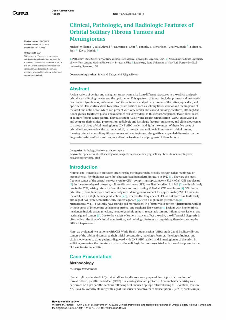
A 53-year-old man presented with a six-month history of progressive blurry vision, right-sided proptosis,and restricted upward gaze. Magnetic resonance imaging (MRI) revealed a 3.3 × 3.2 × 1.8 cm T1/T2isointense, diffuse, space-occupying mass with homogenous enhancement on T1 with gadolinium contrastin the superior right orbit with mild depression of the right eye (Figures 1A-1C). He underwent a rightcranio-orbitotomy and en-bloc excision of the lesion. The microscopic evaluation demonstrated a denselyhypercellular spindle cell neoplasm with branching “staghorn” vessels (Figure 2A), focal necrosis, andfrequent mitotic figures (Figure 2B), up to 10/10 high-power fields (HPF). Immunohistochemical stains wereperformed with the tumor cells showing strong STAT6 staining (Figure 2C) with weak, patchy CD34 staining(Figure 2D). The Ki-67 proliferation index was elevated with a quantitative count from 10% to 20% (Figure2E). The tumor cells were negative for glial fibrillary acidic protein (GFAP), EMA, S-100, SOX10, E-Cadherin,and D2-40. The final diagnosis as per the 2016 WHO Classification of Tumors was anaplastichemangiopericytoma (HPC), CNS WHO grade 3. With the 2021 WHO edition retiring the hybrid classificationof solitary fibrous tumor/hemangiopericytoma (SFT/HPC), the revised diagnosis would be a solitary fibroustumor, CNS WHO grade 3. As of this manuscript’s composition, seven months after surgery, the patient isalive and has been referred for radiation therapy and clinical follow-up.
FIGURE 1: Radiologic images of case 1.(A) Coronal T1-FS imaging demonstrates a supraorbital lobular isointense mass in the right superior extraconalportion of the right orbit (red arrow). (B) Coronal T2 imaging shows an isointense mass (red arrow) with intactextraocular muscles and optic nerves bilaterally. (C) Coronal T1-FS post-gadolinium imaging shows diffuseenhancement with mild inferior depression of the right orbital globe (red arrow).
FS: fat saturation
2021 Williams et al. Cureus 13(11): e19678. DOI 10.7759/cureus.19678 2 of 14

FIGURE 2: Histologic images of case 1.Microscopic images demonstrating (A) a spindle cell neoplasm with a “patternless pattern” and “staghornvessels,” (B) multiple mitotic figures, (C) STAT6 nuclear positivity, (D) weak CD34 positivity, (E) and elevated Ki-67proliferation index. Panels A and C are captured at a total magnification of 100×, scale bars = 500 µm; panel B iscaptured at a total magnification of 400×, scale bar = 100 µm; panels D and E are captured at a totalmagnification of 50×, scale bars = 500 µm.
STAT6: signal transducer and activator of transcription 6; CD34: cluster of differentiation 34
Case 2
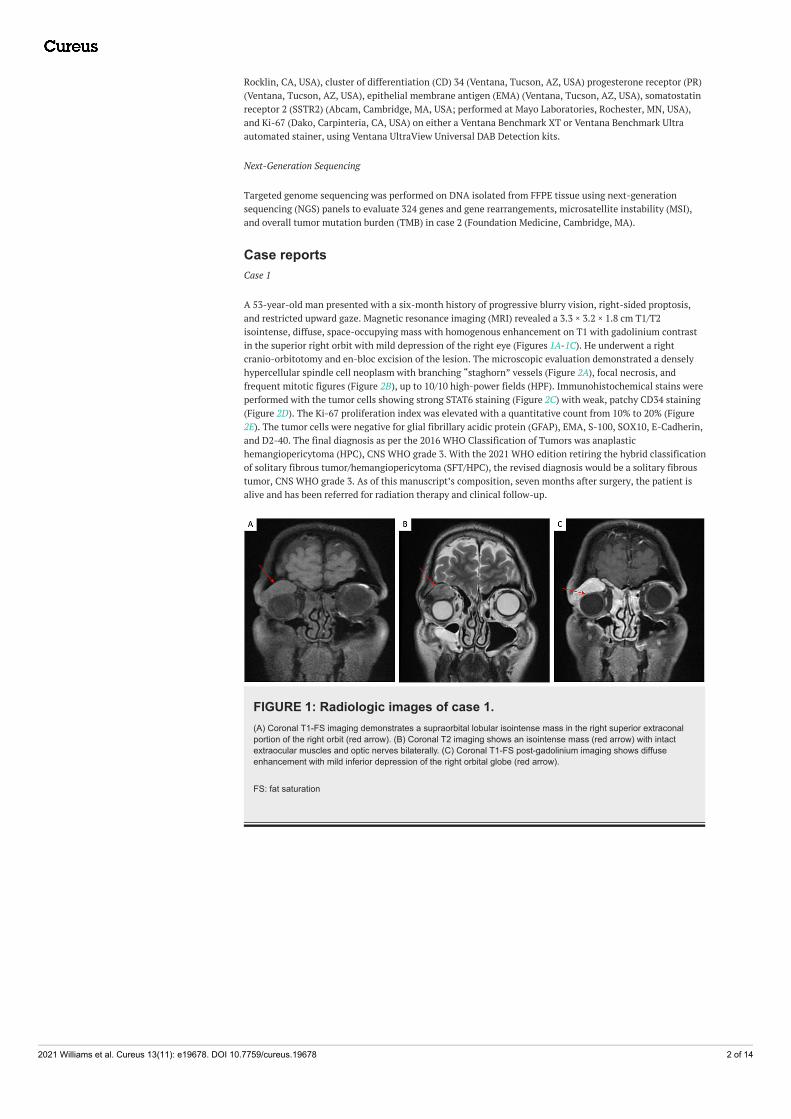
A 37-year-old man presented with a two-year history of gradual visual loss in his right eye and mild rightexophthalmos. Imaging revealing a 4.7 × 4.3 × 3.6 cm lobulated, homogenously enhancing mass along thefloor of the anterior middle cranial fossa involving the right sphenoid bone, sphenoid sinus, and projectinginto the cavernous sinus and posterior orbit with involvement of the optic canal and internal carotid artery(Figures 3A-3C). The patient had undergone a preoperative partial embolization of the feeder arteries foundon angiogram and a right-sided pterional craniotomy and endoscopic nasal subtotal resection of the lesion.The microscopic evaluation demonstrated marked hypercellularity with spindle-shaped cells arrangedhaphazardly with an overall “patternless pattern” and staghorn-like vessels. The nuclei were mildlypleomorphic with a rich reticulin network. The tumor cells were positive for vimentin with variable CD99positivity. CD34, EMA, and B-cell lymphoma 2 (BCL-2) were focally positive, with GFAP, PR, S-100, Desmin,and Neurofilament being negative. The Ki-67 proliferation index was up to 4%. An NGS panel (FoundationMedicine, Cambridge, MA, USA) demonstrated NAB2-STAT6 fusion.
2021 Williams et al. Cureus 13(11): e19678. DOI 10.7759/cureus.19678 3 of 14
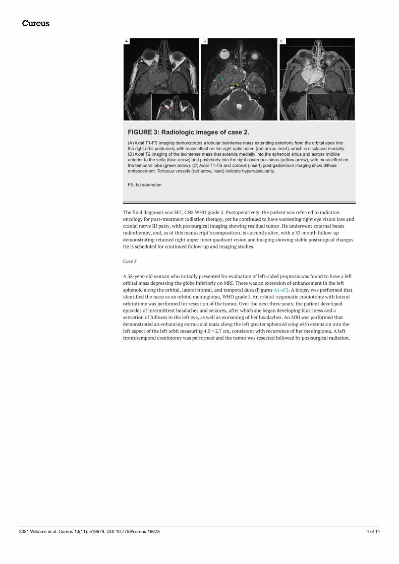
FIGURE 3: Radiologic images of case 2.(A) Axial T1-FS imaging demonstrates a lobular isointense mass extending anteriorly from the orbital apex intothe right orbit posteriorly with mass effect on the right optic nerve (red arrow, inset), which is displaced medially.(B) Axial T2 imaging of the isointense mass that extends medially into the sphenoid sinus and across midlineanterior to the sella (blue arrow) and posteriorly into the right cavernous sinus (yellow arrow), with mass effect onthe temporal lobe (green arrow). (C) Axial T1-FS and coronal (insert) post-gadolinium imaging show diffuseenhancement. Tortuous vessels (red arrow, inset) indicate hypervascularity.
FS: fat saturation
The final diagnosis was SFT, CNS WHO grade 2. Postoperatively, the patient was referred to radiationoncology for post-treatment radiation therapy, yet he continued to have worsening right eye vision loss andcranial nerve III palsy, with postsurgical imaging showing residual tumor. He underwent external beamradiotherapy, and, as of this manuscript’s composition, is currently alive, with a 33-month follow-updemonstrating retained right upper inner quadrant vision and imaging showing stable postsurgical changes.He is scheduled for continued follow-up and imaging studies.
Case 3
A 38-year-old woman who initially presented for evaluation of left-sided proptosis was found to have a leftorbital mass depressing the globe inferiorly on MRI. There was an extension of enhancement in the leftsphenoid along the orbital, lateral frontal, and temporal dura (Figures 4A-4C). A biopsy was performed thatidentified the mass as an orbital meningioma, WHO grade I. An orbital-zygomatic craniotomy with lateralorbitotomy was performed for resection of the tumor. Over the next three years, the patient developedepisodes of intermittent headaches and seizures, after which she began developing blurriness and asensation of fullness in the left eye, as well as worsening of her headaches. An MRI was performed thatdemonstrated an enhancing extra-axial mass along the left greater sphenoid wing with extension into theleft aspect of the left orbit measuring 4.0 × 2.7 cm, consistent with recurrence of her meningioma. A leftfrontotemporal craniotomy was performed and the tumor was resected followed by postsurgical radiation.
2021 Williams et al. Cureus 13(11): e19678. DOI 10.7759/cureus.19678 4 of 14
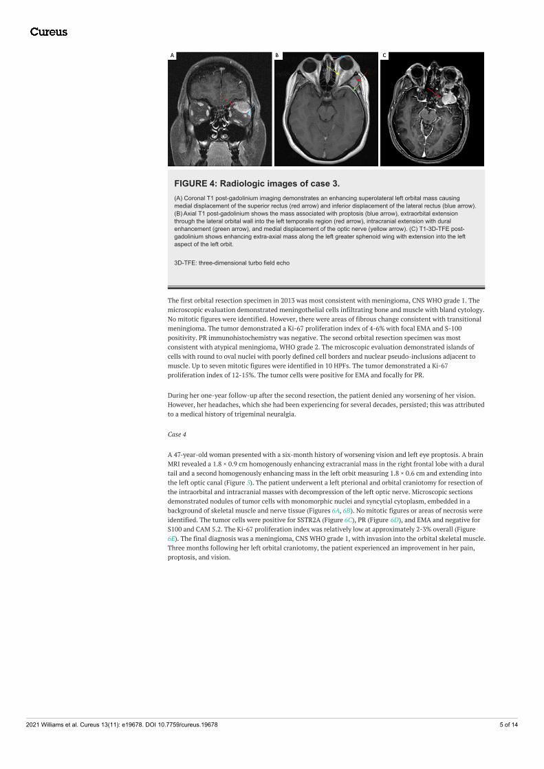
FIGURE 4: Radiologic images of case 3.(A) Coronal T1 post-gadolinium imaging demonstrates an enhancing superolateral left orbital mass causingmedial displacement of the superior rectus (red arrow) and inferior displacement of the lateral rectus (blue arrow).(B) Axial T1 post-gadolinium shows the mass associated with proptosis (blue arrow), extraorbital extensionthrough the lateral orbital wall into the left temporalis region (red arrow), intracranial extension with duralenhancement (green arrow), and medial displacement of the optic nerve (yellow arrow). (C) T1-3D-TFE post-gadolinium shows enhancing extra-axial mass along the left greater sphenoid wing with extension into the leftaspect of the left orbit.
3D-TFE: three-dimensional turbo field echo
The first orbital resection specimen in 2013 was most consistent with meningioma, CNS WHO grade 1. Themicroscopic evaluation demonstrated meningothelial cells infiltrating bone and muscle with bland cytology.No mitotic figures were identified. However, there were areas of fibrous change consistent with transitionalmeningioma. The tumor demonstrated a Ki-67 proliferation index of 4-6% with focal EMA and S-100positivity. PR immunohistochemistry was negative. The second orbital resection specimen was mostconsistent with atypical meningioma, WHO grade 2. The microscopic evaluation demonstrated islands ofcells with round to oval nuclei with poorly defined cell borders and nuclear pseudo-inclusions adjacent tomuscle. Up to seven mitotic figures were identified in 10 HPFs. The tumor demonstrated a Ki-67proliferation index of 12-15%. The tumor cells were positive for EMA and focally for PR.
During her one-year follow-up after the second resection, the patient denied any worsening of her vision.However, her headaches, which she had been experiencing for several decades, persisted; this was attributedto a medical history of trigeminal neuralgia.
Case 4
A 47-year-old woman presented with a six-month history of worsening vision and left eye proptosis. A brainMRI revealed a 1.8 × 0.9 cm homogenously enhancing extracranial mass in the right frontal lobe with a duraltail and a second homogenously enhancing mass in the left orbit measuring 1.8 × 0.6 cm and extending intothe left optic canal (Figure 5). The patient underwent a left pterional and orbital craniotomy for resection ofthe intraorbital and intracranial masses with decompression of the left optic nerve. Microscopic sectionsdemonstrated nodules of tumor cells with monomorphic nuclei and syncytial cytoplasm, embedded in abackground of skeletal muscle and nerve tissue (Figures 6A, 6B). No mitotic figures or areas of necrosis wereidentified. The tumor cells were positive for SSTR2A (Figure 6C), PR (Figure 6D), and EMA and negative forS100 and CAM 5.2. The Ki-67 proliferation index was relatively low at approximately 2-3% overall (Figure6E). The final diagnosis was a meningioma, CNS WHO grade 1, with invasion into the orbital skeletal muscle.Three months following her left orbital craniotomy, the patient experienced an improvement in her pain,proptosis, and vision.
2021 Williams et al. Cureus 13(11): e19678. DOI 10.7759/cureus.19678 5 of 14

FIGURE 5: Radiologic images of case 4.Axial T1-FS post-gadolinium imaging shows a homogenously enhancing mass in the left orbit, adjacent to thelateral wall and extending into the left optic canal (green arrow), with mass effect on the optic nerve (red arrow).The mass extends to the anterior surface of the left temporal lobe and abuts the left cavernous sinus (blue arrow).Axial 3D-SPGR post-gadolinium imaging demonstrates a homogeneously enhancing extra-axial mass overlyingthe right frontal lobe, compatible with a second meningioma (inset).
FS: fat saturation; 3D-SGPR: three-dimensional spoiled gradient echo
2021 Williams et al. Cureus 13(11): e19678. DOI 10.7759/cureus.19678 6 of 14

FIGURE 6: Histologic images of case 4.Microscopic images demonstrate (A, B) islands of meningioma cells with adjacent muscle, (C) SSTR2A positivity,(D) PR positivity, (E) and mildly elevated Ki-67 proliferation index. Panels A, B, C, and D are captured at a totalmagnification of 200×, scale bars = 200 µm; panel E is captured at a total magnification of 100×, scale bar = 500µm.
SSTR2A: somatostatin receptor type 2A; PR: progesterone receptor
Case 5
An eight-year-old female initially presented to the emergency department due to a four-month history ofleft eye proptosis, strabismus, and progressive blindness. A computed tomography (CT) scan of the headdemonstrated a 2.5 × 1.7 × 2.0 cm intraconal mass surrounding the optic nerve sheath and extending intothe left medial rectus muscle. Posteriorly, the mass extended all the way to the apex of the left orbit,involving the left superior orbital fissure (Figure 7). A left frontotemporal craniotomy was performed toremove the intraorbital mass.
2021 Williams et al. Cureus 13(11): e19678. DOI 10.7759/cureus.19678 7 of 14

FIGURE 7: Radiologic images of case 5.Axial T1-FS post-gadolinium imaging demonstrates a diffusely enhancing mass surrounding the optic nervesheath complex showing a “tram-track” appearance (red arrow). Coronal T1-FS post-gadolinium imaging: cuff ofenhancing tumor (blue arrow) around a central nonenhancing dot (optic nerve) (green arrow) with a doughnut sign(inset). The mass posteriorly extends to the apex of the left orbit involving the left superior orbital fissure.
FS: fat saturation
The resected specimen was most consistent with meningioma, primary meningothelial pattern, CNS WHOgrade 1. Microscopic sections demonstrated uniform tumor cells with oval nuclei and delicate chromatin ina lobular and whorling pattern with focal involvement of the adjacent soft tissue and skeletal muscle.Psammoma bodies were present. No mitotic figures or areas of necrosis were identified. Tumor cells stainedpositive for EMA and PR. S-100 and GFAP were focally positive. During her six-month follow-up, the patientwas feeling well aside from some mild proptosis. There were no complaints of worsening vision or any otherneurological impairment.
DiscussionThis report presents an institutional case review of two cases of orbital SFT and three cases of orbital/opticnerve sheath meningioma (ONSM), detailing the multidisciplinary aspects of clinical presentation,radiological workup, and histopathology of these neoplasms. Due to their location, the number of adjacentstructures, and the wide differential diagnosis, these neoplasms present a relatively unique set of radiologicand surgical challenges.
Meningeal SFT is a mesenchymal fibroblastic tumor first reported in the pleura [9], soon followed by otheranatomical sites. Its histological spectrum ranges from a “patternless” pattern of ovoid or spindle cells witha variable amount of collagenous acellular stroma between tumor cells (SFT phenotype) to high cellularitywith reticular fibers and prominent staghorn-shaped, thin-walled vessels (HPC phenotype) [10,11]. In the
2021 Williams et al. Cureus 13(11): e19678. DOI 10.7759/cureus.19678 8 of 14
CNS, SFT is a relatively rare tumor, constituting <1% of all CNS neoplasms [4], although its incidence in theorbit is currently unknown [7].
The 2016 WHO Classification of Tumours of the Central Nervous System revised 4th edition combined SFTand HPC into a single entity based on their shared high frequency of NAB2-STAT6 fusion proteins [10,12].Both located at 12q13.3, NAB2 (NGFI-A-binding protein 2) encodes a transcriptional repressor, while STAT6is a transcription factor that modulates signaling in the immune system [13]. Whole-genome sequencingstudies [13,14] have revealed multiple fusion types, including the most frequent NAB2ex4-STAT6ex2/3,corresponding to the classic pleural/pulmonary SFT, and the second most frequent NAB2ex6-STAT6ex16/17,commonly found in younger patients [14]. Nuclear STAT6 immunostaining has shown high specificity andsensitivity as a molecular surrogate for the fusion protein, allowing for the rapid diagnosis ofSFT [15]. SFT cases frequently demonstrate immunohistochemical reactivity to vimentin, CD34, BCL-2,CD99, and occasional staining with both EMA and SMA [11,15,16].
The proposed grading criteria somewhat vary between the updated CNS classification system (withcontinued studies attempting to improve the CNS grading system [17]), the WHO Classification of Tumoursof Soft Tissue and Bone, and the WHO Classification of Tumours of the Eye [7], although it is worth notingthat in any of these grading schemes, case 1 would be designated as grade 3 and case 2 would be designatedas grade 2. In the 2021 WHO Classification of Tumours of the Central Nervous System revised 5th edition,the term HPC has been retired, with the combined designation of SFT/HPC, as described in the 2016 4thedition, no longer being used. This was done due to an increasing emphasis on molecular markers and aneed to fully conform with soft tissue pathology nomenclature. Furthermore, neoplasms are now gradedwithin types using Arabic numerals rather than across types using Roman numerals, which is moreconsistent with the grading of non-CNS tumors [18]. SFTs, in particular, can be graded as 1, 2, or 3. Otherhistologic variants of SFT have also been described in the literature at various sites, including the fat-forming variant that has previously been identified in the orbit [11], a papillary variant [19], and a giant cell-rich variant [20]. The differential diagnosis includes fibrous meningioma, schwannomas, and poorlydifferentiated synovial sarcoma [11,15,16].
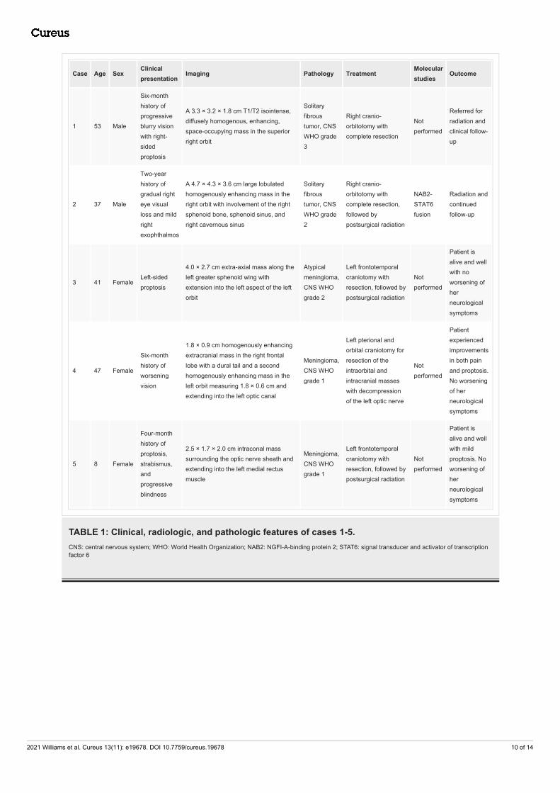
The clinical behavior of SFT depends on the extent of surgical intervention and the presence of high-riskfeatures in the orbit. WHO grade 1 SFTs reported within the CNS have a good prognosis, while those withWHO grade 2/3 [4] features or high-risk features in the orbit [7] require long-term follow-up with thepossibility of adjuvant radiotherapy due to a high rate of recurrence and possibility of extracranialmetastasis. Both patients in this study were grade 2 or 3 and had input from radiation oncology as part of amultidisciplinary approach to their treatment. Case 2 underwent radiotherapy due to partial resection, andboth are currently being closely followed due to their high-grade disease. Table 1 presents the clinical,radiologic, and pathologic features of all the cases presented in this report. Table 2 presents the features ofSFT/HPCs according to the WHO classifications.
2021 Williams et al. Cureus 13(11): e19678. DOI 10.7759/cureus.19678 9 of 14
Case Age SexClinicalpresentation
Imaging Pathology TreatmentMolecularstudies
Outcome
1 53 Male
Six-monthhistory ofprogressiveblurry visionwith right-sidedproptosis
A 3.3 × 3.2 × 1.8 cm T1/T2 isointense,diffusely homogenous, enhancing,space-occupying mass in the superiorright orbit
Solitaryfibroustumor, CNSWHO grade3
Right cranio-orbitotomy withcomplete resection
Notperformed
Referred forradiation andclinical follow-up
2 37 Male
Two-yearhistory ofgradual righteye visualloss and mildrightexophthalmos
A 4.7 × 4.3 × 3.6 cm large lobulatedhomogenously enhancing mass in theright orbit with involvement of the rightsphenoid bone, sphenoid sinus, andright cavernous sinus
Solitaryfibroustumor, CNSWHO grade2
Right cranio-orbitotomy withcomplete resection,followed bypostsurgical radiation
NAB2-STAT6fusion
Radiation andcontinuedfollow-up
3 41 FemaleLeft-sidedproptosis
4.0 × 2.7 cm extra-axial mass along theleft greater sphenoid wing withextension into the left aspect of the leftorbit
Atypicalmeningioma,CNS WHOgrade 2
Left frontotemporalcraniotomy withresection, followed bypostsurgical radiation
Notperformed
Patient isalive and wellwith noworsening ofherneurologicalsymptoms
4 47 Female
Six-monthhistory ofworseningvision
1.8 × 0.9 cm homogenously enhancingextracranial mass in the right frontallobe with a dural tail and a secondhomogenously enhancing mass in theleft orbit measuring 1.8 × 0.6 cm andextending into the left optic canal
Meningioma,CNS WHOgrade 1
Left pterional andorbital craniotomy forresection of theintraorbital andintracranial masseswith decompressionof the left optic nerve
Notperformed
Patientexperiencedimprovementsin both painand proptosis.No worseningof herneurologicalsymptoms
5 8 Female
Four-monthhistory ofproptosis,strabismus,andprogressiveblindness
2.5 × 1.7 × 2.0 cm intraconal masssurrounding the optic nerve sheath andextending into the left medial rectusmuscle
Meningioma,CNS WHOgrade 1
Left frontotemporalcraniotomy withresection, followed bypostsurgical radiation
Notperformed
Patient isalive and wellwith mildproptosis. Noworsening ofherneurologicalsymptoms
TABLE 1: Clinical, radiologic, and pathologic features of cases 1-5.CNS: central nervous system; WHO: World Health Organization; NAB2: NGFI-A-binding protein 2; STAT6: signal transducer and activator of transcriptionfactor 6
2021 Williams et al. Cureus 13(11): e19678. DOI 10.7759/cureus.19678 10 of 14
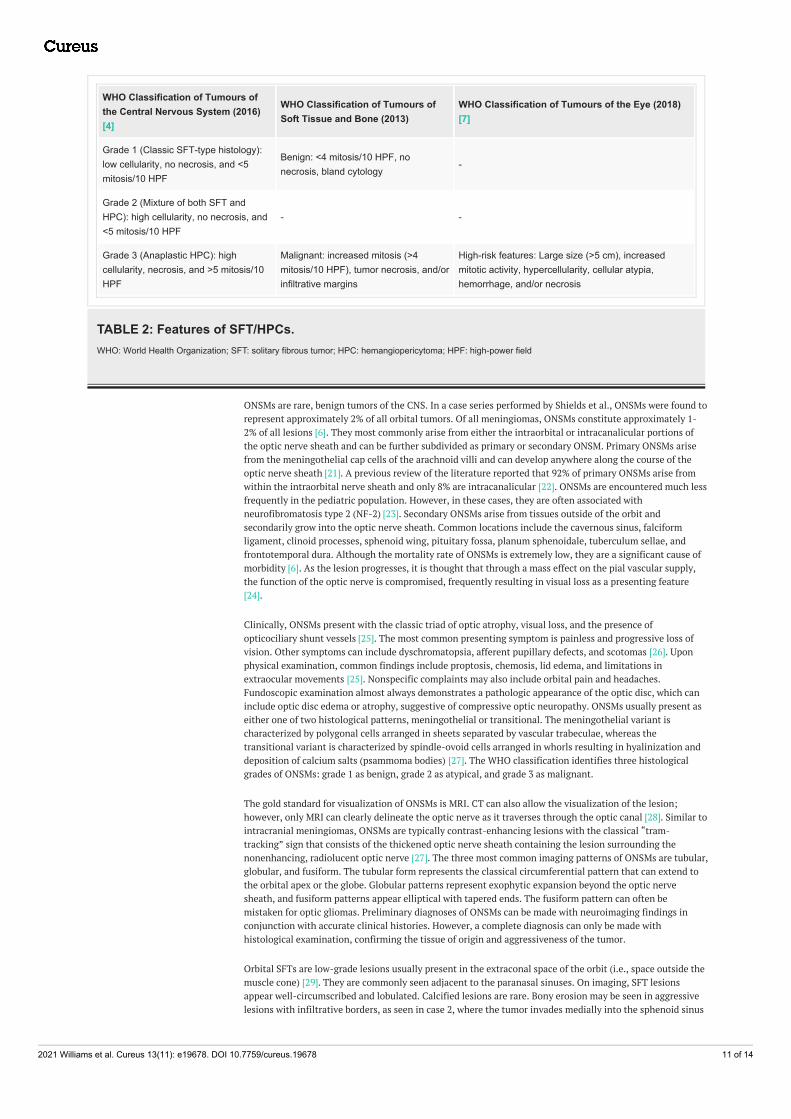
WHO Classification of Tumours ofthe Central Nervous System (2016)[4]
WHO Classification of Tumours ofSoft Tissue and Bone (2013)
WHO Classification of Tumours of the Eye (2018)[7]
Grade 1 (Classic SFT-type histology):low cellularity, no necrosis, and <5mitosis/10 HPF
Benign: <4 mitosis/10 HPF, nonecrosis, bland cytology
-
Grade 2 (Mixture of both SFT andHPC): high cellularity, no necrosis, and<5 mitosis/10 HPF
- -
Grade 3 (Anaplastic HPC): highcellularity, necrosis, and >5 mitosis/10HPF
Malignant: increased mitosis (>4mitosis/10 HPF), tumor necrosis, and/orinfiltrative margins
High-risk features: Large size (>5 cm), increasedmitotic activity, hypercellularity, cellular atypia,hemorrhage, and/or necrosis
TABLE 2: Features of SFT/HPCs.WHO: World Health Organization; SFT: solitary fibrous tumor; HPC: hemangiopericytoma; HPF: high-power field
ONSMs are rare, benign tumors of the CNS. In a case series performed by Shields et al., ONSMs were found torepresent approximately 2% of all orbital tumors. Of all meningiomas, ONSMs constitute approximately 1-2% of all lesions [6]. They most commonly arise from either the intraorbital or intracanalicular portions ofthe optic nerve sheath and can be further subdivided as primary or secondary ONSM. Primary ONSMs arisefrom the meningothelial cap cells of the arachnoid villi and can develop anywhere along the course of theoptic nerve sheath [21]. A previous review of the literature reported that 92% of primary ONSMs arise fromwithin the intraorbital nerve sheath and only 8% are intracanalicular [22]. ONSMs are encountered much lessfrequently in the pediatric population. However, in these cases, they are often associated withneurofibromatosis type 2 (NF-2) [23]. Secondary ONSMs arise from tissues outside of the orbit andsecondarily grow into the optic nerve sheath. Common locations include the cavernous sinus, falciformligament, clinoid processes, sphenoid wing, pituitary fossa, planum sphenoidale, tuberculum sellae, andfrontotemporal dura. Although the mortality rate of ONSMs is extremely low, they are a significant cause ofmorbidity [6]. As the lesion progresses, it is thought that through a mass effect on the pial vascular supply,the function of the optic nerve is compromised, frequently resulting in visual loss as a presenting feature[24].
Clinically, ONSMs present with the classic triad of optic atrophy, visual loss, and the presence ofopticociliary shunt vessels [25]. The most common presenting symptom is painless and progressive loss ofvision. Other symptoms can include dyschromatopsia, afferent pupillary defects, and scotomas [26]. Uponphysical examination, common findings include proptosis, chemosis, lid edema, and limitations inextraocular movements [25]. Nonspecific complaints may also include orbital pain and headaches.Fundoscopic examination almost always demonstrates a pathologic appearance of the optic disc, which caninclude optic disc edema or atrophy, suggestive of compressive optic neuropathy. ONSMs usually present aseither one of two histological patterns, meningothelial or transitional. The meningothelial variant ischaracterized by polygonal cells arranged in sheets separated by vascular trabeculae, whereas thetransitional variant is characterized by spindle-ovoid cells arranged in whorls resulting in hyalinization anddeposition of calcium salts (psammoma bodies) [27]. The WHO classification identifies three histologicalgrades of ONSMs: grade 1 as benign, grade 2 as atypical, and grade 3 as malignant.
The gold standard for visualization of ONSMs is MRI. CT can also allow the visualization of the lesion;however, only MRI can clearly delineate the optic nerve as it traverses through the optic canal [28]. Similar tointracranial meningiomas, ONSMs are typically contrast-enhancing lesions with the classical “tram-tracking” sign that consists of the thickened optic nerve sheath containing the lesion surrounding thenonenhancing, radiolucent optic nerve [27]. The three most common imaging patterns of ONSMs are tubular,globular, and fusiform. The tubular form represents the classical circumferential pattern that can extend tothe orbital apex or the globe. Globular patterns represent exophytic expansion beyond the optic nervesheath, and fusiform patterns appear elliptical with tapered ends. The fusiform pattern can often bemistaken for optic gliomas. Preliminary diagnoses of ONSMs can be made with neuroimaging findings inconjunction with accurate clinical histories. However, a complete diagnosis can only be made withhistological examination, confirming the tissue of origin and aggressiveness of the tumor.
Orbital SFTs are low-grade lesions usually present in the extraconal space of the orbit (i.e., space outside themuscle cone) [29]. They are commonly seen adjacent to the paranasal sinuses. On imaging, SFT lesionsappear well-circumscribed and lobulated. Calcified lesions are rare. Bony erosion may be seen in aggressivelesions with infiltrative borders, as seen in case 2, where the tumor invades medially into the sphenoid sinus
2021 Williams et al. Cureus 13(11): e19678. DOI 10.7759/cureus.19678 11 of 14

and across the midline anterior to the sella and posteriorly into the right cavernous sinus. A characteristicimaging feature is isointensity on T1-weighted and T2-weighted images with avid enhancement on post-gadolinium images, as seen in our case. T1 and T2 isointensity can help differentiate SFTs from cavernousmalformations. SFTs are vascular tumors [30,31], as seen in case 2, with tortuous vessels indicatinghypervascularity; they are also usually encapsulated tumors.
Differential diagnoses for vasculogenic lesions of the orbit include cavernous malformations, which are themost common benign lesions of the orbit and are typically well-circumscribed. However, they occur at thelateral aspect of the intraconal space (i.e., space within the muscle cone). Rarely though, conal andextraconal cavernous malformations may occur. Cavernous malformations [32] do not cause erosion of thebone or direct invasion of the surrounding structures such as the extraconal muscles or optic nerve. UnlikeSFTs, they are seen in infants. They usually displace surrounding structures. As opposed to SFTs, cavernoushemangiomas [33] appear hyperintense in T2-weighted images and do not show avid enhancement on post-contrast images.
Another differential of orbital lesions is lymphoma (primary or systemic), which often cannot be reliablydifferentiated on images. Usually, lymphoma is seen in the extraconal [34,35] compartment of the orbit, andapproximately 40% of the cases involve the lacrimal gland [36]. Lymphomas can be seen as well-circumscribed or ill-defined lesions. Bony erosion is rare. On MRI, lesions appear isointense on T1 andhyperintense on T2 with enhancement on post-contrast images.
Orbital or ONSMs occur in the intraconal compartment and show a classic tram-tracking appearance [37], asseen on axial images of case 3, which depicts thickened nonenhancing optic nerve encased by the enhancingtumor. Classic tram-tracking and a doughnut sign (usually seen on coronal images), as depicted in case 5,are also characteristic of orbital meningioma. These signs appear due to the sparing of the substance of thenerve. This helps in differentiating ONSM from optic nerve gliomas. Optic nerve gliomas, which are alsointraconal in nature, are usually seen in patients with NF-2 [38] and show the invasion of the optic nerve bythe tumor with the nerve sometimes appearing tortuous or kinked on imaging. Optic nerve glioma withoutNF-2 characteristically causes fusiform dilatation of the optic nerve on imaging.
Differentials for extraconal lesions on imaging in addition to SFTs include peripheral sheath tumors such asschwannoma and neurofibromas. Schwannomas appear hyperintense on T2-weighted images such ascavernous malformations; however, a differentiating feature of schwannomas is their heterogeneity on T2due to mixed solid and cystic components [39]. On post-contrast imaging, schwannomas usually showheterogeneous enhancement. Neurofibromas show a “bag-of-worm” appearance on imaging and typicallyappear hyperintense on T2 and heterogeneously hyperintense on T1-weighted images. The most commonprimary malignancy of the orbit is choroidal melanoma [40-42]. Melanin causes T1 and T2-shorteningeffects, meaning it appears hyperintense on T1 and hypointense on T2-weighted images. Metastasis canalso result in orbital lesions, with breast cancer being the most common cancer to cause orbital metastasis[43].
When managing patients with ONSM and orbital SFT, the primary goal of treatment is the preservation ofvision and, if possible, prevention of tumor progression. When left untreated, these lesions almost alwaysresult in visual deterioration. Historically, management of ONSM has involved careful observation in caseswhere there is negligible visual decline. However, inevitably, up to 85% of patients experience visual decline,warranting medical intervention [11]. Unfortunately, complete surgical resection almost always leads tocompromised visual function. In cases where the visual function is significantly compromised or intracranialextension has occurred, surgical intervention may be the only option [44]. In attempts to preserve visualfunction, subtotal resection with postoperative radiotherapy has also become a popular therapy. This isapparent, especially with the advent of stereotactic fractionated radiosurgery, which allows treatment with asufficient dose of radiation in a more focused manner to minimize radiation-induced complications [45].
ConclusionsOrbital SFTs, given the wide differential diagnosis for an orbital tumor, can make for a difficult diagnosis.The location and proximity to several structures also pose a treatment challenge. In this case series, we havehighlighted radiographic and histological features of both SFTs and ONSMs diagnosed at our institution,outlined their clinical outcomes, and reviewed the literature on clinical, radiologic, and pathologic featuresof SFTs, as well as other possible orbital tumors. The clinical outlook for SFTs is variable and depends notonly on tumor grading but also on the presence of high risk, which if present usually suggests a need foradjuvant radiotherapy to decrease the risk of recurrence and extracranial metastasis. Our patients diagnosedwith SFT were either CNS WHO grade 2 or 3 and thus had radiation oncology on board, with both currentlyremaining clinically stable. In contrast, ONSMs, also on the differential for an orbital tumor, are benign butproblematic given their impact on visual function. Generally, the primary goal of treatment for SFTs andONSMs is the same, that is, to preserve visual function. While surgery alone is often utilized for themanagement of ONSM, adjuvant radiotherapy helps avoid total resection, which is associated with a higherrisk of visual compromise.
2021 Williams et al. Cureus 13(11): e19678. DOI 10.7759/cureus.19678 12 of 14

Additional InformationDisclosuresHuman subjects: Consent was obtained or waived by all participants in this study. Conflicts of interest: Incompliance with the ICMJE uniform disclosure form, all authors declare the following: Payment/servicesinfo: All authors have declared that no financial support was received from any organization for thesubmitted work. Financial relationships: All authors have declared that they have no financialrelationships at present or within the previous three years with any organizations that might have aninterest in the submitted work. Other relationships: All authors have declared that there are no otherrelationships or activities that could appear to have influenced the submitted work.
References1. Cushing H: The meningiomas (dural endotheliomas): their source and favoured seats of origin . Brain. 1922,
45:282-316.2. Ostrom QT, Gittleman H, Truitt G, Boscia A, Kruchko C, Barnholtz-Sloan JS: CBTRUS Statistical Report:
primary brain and other central nervous system tumors diagnosed in the United States in 2011-2015. NeuroOncol. 2018, 20:iv1-86. 10.1093/neuonc/noy131
3. Stout AP, Murray MR: Hemangiopericytoma: a vascular tumor featuring Zimmermann's pericytes . Ann Surg.1942, 116:26-33. 10.1097/00000658-194207000-00004
4. Giannini C, Rushing EJ, Hainfellner JA, et al.: Solitary fibrous tumour/haemangiopericytoma. WHOClassification of Tumours of the Central Nervous System. Louis DN, Ohgaki H, Wiestler OD (ed): IARC,Lyon, France; 2016. 249-54.
5. Rodriguez F, Cummings T, Specht CS, Rushing EJ: Meningioma of the optic nerve . WHO Classification ofTumours of the Eye. Grossniklaus HE, Eberhart CG, Kivelä TT (ed): IARC, Lyon, France; 2018. 139-40.
6. Shields JA, Shields CL, Scartozzi R: Survey of 1264 patients with orbital tumors and simulating lesions: the2002 Montgomery Lecture, part 1. Ophthalmology. 2004, 111:997-1008. 10.1016/j.ophtha.2003.01.002
7. Mudhar HS, Rushing EJ, Rodriguez F, Vemuganti G: Solitary fibrous tumour/haemangiopericytoma. WHOClassification of Tumours of the Eye. Grossniklaus HE, Eberhart CG, Kivelä TT (ed): IARC, Lyon, France;2018. 141-42.
8. Le CP, Jones S, Valenzuela AA: Orbital solitary fibrous tumor: a case series with review of the literature .Orbit. 2014, 33:145-51. 10.3109/01676830.2013.853806
9. Klemperer P, Coleman BR: Primary neoplasms of the pleura. A report of five cases . Am J Ind Med. 1992,22:1-31. 10.1002/ajim.4700220103
10. Louis DN, Perry A, Reifenberger G, et al.: The 2016 World Health Organization Classification of Tumors ofthe Central Nervous System: a summary. Acta Neuropathol. 2016, 131:803-20. 10.1007/s00401-016-1545-1
11. Thway K, Ng W, Noujaim J, Jones RL, Fisher C: The current status of solitary fibrous tumor: diagnosticfeatures, variants, and genetics. Int J Surg Pathol. 2016, 24:281-92. 10.1177/1066896915627485
12. Fritchie KJ, Jin L, Rubin BP, et al.: NAB2-STAT6 gene fusion in meningeal hemangiopericytoma and solitaryfibrous tumor. J Neuropathol Exp Neurol. 2016, 75:263-71. 10.1093/jnen/nlv026
13. Chmielecki J, Crago AM, Rosenberg M, et al.: Whole-exome sequencing identifies a recurrent NAB2-STAT6fusion in solitary fibrous tumors. Nat Genet. 2013, 45:131-2. 10.1038/ng.2522
14. Barthelmeß S, Geddert H, Boltze C, et al.: Solitary fibrous tumors/hemangiopericytomas with differentvariants of the NAB2-STAT6 gene fusion are characterized by specific histomorphology and distinctclinicopathological features. Am J Pathol. 2014, 184:1209-18. 10.1016/j.ajpath.2013.12.016
15. Farooq Z, Badar Z, Zaccarini D, Tavernier FB, Mohamed A, Mangla R: Recurrent solitary fibrous tumor oflumbar spine with vertebral body involvement: imaging features and differential diagnosis with report of acase. Radiol Case Rep. 2016, 11:450-5. 10.1016/j.radcr.2016.08.012
16. Chen H, Xiao CW, Wang T, et al.: Orbital solitary fibrous tumor: a clinicopathologic study of ten cases withlong-term follow-up. Acta Neurochir (Wien). 2012, 154:249-55; discussion 255. 10.1007/s00701-011-1254-4
17. Macagno N, Vogels R, Appay R, et al.: Grading of meningeal solitary fibrous tumors/hemangiopericytomas:analysis of the prognostic value of the Marseille Grading System in a cohort of 132 patients. Brain Pathol.2019, 29:18-27. 10.1111/bpa.12613
18. Louis DN, Perry A, Wesseling P, et al.: The 2021 WHO Classification of Tumors of the Central NervousSystem: a summary. Neuro Oncol. 2021, 23:1231-51. 10.1093/neuonc/noab106
19. Yao ZG, Wu HB, Hao YH, et al.: Papillary solitary fibrous tumor/hemangiopericytoma: an uncommonmorphological form with NAB2-STAT6 gene fusion. J Neuropathol Exp Neurol. 2019, 78:685-93.10.1093/jnen/nlz053
20. Furusato E, Valenzuela IA, Fanburg-Smith JC, Auerbach A, Furusato B, Cameron JD, Rushing EJ: Orbitalsolitary fibrous tumor: encompassing terminology for hemangiopericytoma, giant cell angiofibroma, andfibrous histiocytoma of the orbit: reappraisal of 41 cases. Hum Pathol. 2011, 42:120-8.10.1016/j.humpath.2010.05.021
21. Saeed P, Rootman J, Nugent RA, White VA, Mackenzie IR, Koornneef L: Optic nerve sheath meningiomas.Ophthalmology. 2003, 110:2019-30. 10.1016/S0161-6420(03)00787-5
22. Dutton JJ: Optic nerve sheath meningiomas. Surv Ophthalmol. 1992, 37:167-83. 10.1016/0039-6257(92)90135-g
23. Berman D, Miller NR: New concepts in the management of optic nerve sheath meningiomas . Ann Acad MedSingap. 2006, 35:168-74.
24. Kim JW, Rizzo JF, Lessell S: Controversies in the management of optic nerve sheath meningiomas . IntOphthalmol Clin. 2005, 45:15-23. 10.1097/01.iio.0000176367.16758.f4
25. Sibony PA, Krauss HR, Kennerdell JS, Maroon JC, Slamovits TL: Optic nerve sheath meningiomas. Clinicalmanifestations. Ophthalmology. 1984, 91:1313-26. 10.1016/s0161-6420(84)34148-3
26. Carrasco JR, Penne RB: Optic nerve sheath meningiomas and advanced treatment options . Curr Opin
2021 Williams et al. Cureus 13(11): e19678. DOI 10.7759/cureus.19678 13 of 14

Ophthalmol. 2004, 15:406-10. 10.1097/01.icu.0000138617.53435.d927. Turbin RE, Pokorny K: Diagnosis and treatment of orbital optic nerve sheath meningioma . Cancer Control.
2004, 11:334-41. 10.1177/10732748040110050828. Lindblom B, Truwit CL, Hoyt WF: Optic nerve sheath meningioma. Definition of intraorbital,
intracanalicular, and intracranial components with magnetic resonance imaging. Ophthalmology. 1992,99:560-6. 10.1016/s0161-6420(92)31932-3
29. Tailor TD, Gupta D, Dalley RW, Keene CD, Anzai Y: Orbital neoplasms in adults: clinical, radiologic, andpathologic review. Radiographics. 2013, 33:1739-58. 10.1148/rg.336135502
30. Bilaniuk LT: Orbital vascular lesions. Role of imaging . Radiol Clin North Am. 1999, 37:169-83, xi.10.1016/s0033-8389(05)70085-3
31. Smoker WR, Gentry LR, Yee NK, Reede DL, Nerad JA: Vascular lesions of the orbit: more than meets the eye .Radiographics. 2008, 28:185-204; quiz 325. 10.1148/rg.281075040
32. Ansari SA, Mafee MF: Orbital cavernous hemangioma: role of imaging . Neuroimaging Clin N Am. 2005,15:137-58. 10.1016/j.nic.2005.02.009
33. Khan SN, Sepahdari AR: Orbital masses: CT and MRI of common vascular lesions, benign tumors, andmalignancies. Saudi J Ophthalmol. 2012, 26:373-83. 10.1016/j.sjopt.2012.08.001
34. Cytryn AS, Putterman AM, Schneck GL, Beckman E, Valvassori GE: Predictability of magnetic resonanceimaging in differentiation of orbital lymphoma from orbital inflammatory syndrome. Ophthalmic PlastReconstr Surg. 1997, 13:129-34. 10.1097/00002341-199706000-00007
35. Flanders AE, Espinosa GA, Markiewicz DA, Howell DD: Orbital lymphoma. Role of CT and MRI . Radiol ClinNorth Am. 1987, 25:601-13.
36. Kousoubris PD, Rosman DA: Radiologic evaluation of lacrimal and orbital disease . Otolaryngol Clin NorthAm. 2006, 39:865-93, vi. 10.1016/j.otc.2006.07.005
37. Mafee MF, Goodwin J, Dorodi S: Optic nerve sheath meningiomas. Role of MR imaging . Radiol Clin NorthAm. 1999, 37:37-58, ix. 10.1016/s0033-8389(05)70077-4
38. Kornreich L, Blaser S, Schwarz M, et al.: Optic pathway glioma: correlation of imaging findings with thepresence of neurofibromatosis. AJNR Am J Neuroradiol. 2001, 22:1963-9.
39. Wang Y, Xiao LH: Orbital schwannomas: findings from magnetic resonance imaging in 62 cases . Eye (Lond).2008, 22:1034-9. 10.1038/sj.eye.6702832
40. Mafee MF, Putterman A, Valvassori GE, Campos M, Capek V: Orbital space-occupying lesions: role ofcomputed tomography and magnetic resonance imaging. An analysis of 145 cases. Radiol Clin North Am.1987, 25:529-59.
41. Lemke AJ, Kazi I, Felix R: Magnetic resonance imaging of orbital tumors . Eur Radiol. 2006, 16:2207-19.10.1007/s00330-006-0227-0
42. Bilaniuk LT, Rapoport RJ: Magnetic resonance imaging of the orbit . Top Magn Reson Imaging. 1994, 6:167-81.
43. Peyster RG, Shapiro MD, Haik BG: Orbital metastasis: role of magnetic resonance imaging and computedtomography. Radiol Clin North Am. 1987, 25:647-62.
44. Kennerdell JS, Maroon JC, Malton M, Warren FA: The management of optic nerve sheath meningiomas . AmJ Ophthalmol. 1988, 106:450-7. 10.1016/0002-9394(88)90882-3
45. Andrews DW, Faroozan R, Yang BP, et al.: Fractionated stereotactic radiotherapy for the treatment of opticnerve sheath meningiomas: preliminary observations of 33 optic nerves in 30 patients with historicalcomparison to observation with or without prior surgery. Neurosurgery. 2002, 51:890-902; discussion 903-4.10.1097/00006123-200210000-00007
2021 Williams et al. Cureus 13(11): e19678. DOI 10.7759/cureus.19678 14 of 14