clinical study prognosis of multifocal papillary thyroid...
TRANSCRIPT

Hindawi Publishing CorporationInternational Journal of EndocrinologyVolume 2013, Article ID 809382, 6 pageshttp://dx.doi.org/10.1155/2013/809382
Clinical StudyPrognosis of Multifocal Papillary Thyroid Carcinoma
Sheng-Fong Kuo,1 Shu-Fu Lin,1 Tzu-Chieh Chao,2 Chuen Hsueh,3
Kun-Ju Lin,4 and Jen-Der Lin1
1 Division of Endocrinology and Metabolism, Department of Internal Medicine, Chang Gung Memorial Hospital,Chang Gung University, Taoyuan 333, Taiwan
2Department of General Surgery, Chang Gung Memorial Hospital, Chang Gung University, Taoyuan 333, Taiwan3Department of Pathology, Chang Gung Memorial Hospital, Chang Gung University, Taoyuan 333, Taiwan4Department of Nuclear Medicine; Chang Gung Memorial Hospital, Chang Gung University, Taoyuan 333, Taiwan
Correspondence should be addressed to Jen-Der Lin; [email protected]
Received 11 September 2013; Revised 9 December 2013; Accepted 11 December 2013
Academic Editor: Mario Maggi
Copyright © 2013 Sheng-Fong Kuo et al. This is an open access article distributed under the Creative Commons AttributionLicense, which permits unrestricted use, distribution, and reproduction in any medium, provided the original work is properlycited.
This studywas to investigate the clinical features and therapeutic outcomes ofmultifocal papillary thyroidmicrocarcinoma (PTMC).A total of 2,418 papillary thyroid carcinoma (PTC) patients had undergone thyroidectomy in one medical center between 1977 and2010. There were 483 (20.0%) diagnosed with multifocal PTC. The percentage of multifocal PTC was higher in PTMC patients(22.0%) than in non-PTMC patients (19.5%). Demographic and clinical characteristics of PTMC and multifocal PTC in PTCpatients were traced. Multifocal PTC patients presented with smaller tumors at an older age, and a higher percentage underwenttotal or complete thyroidectomy. These patients also showed a higher incidence of postoperative disease progression than didunifocal PTC patients. Comparison of 483 patients with multifocal PTMC and non-PTMC tumors showed a higher incidence ofpostoperative disease progression in patients with non-PTMC; otherwise, there was no statistical difference in disease-specific andtotal mortality between these two groups. In conclusion, the incidence of multifocal PTMCwas not lower than that of non-PTMC,and postoperative therapies were necessary for both multifocal PTMC and non-PTMC patients.
1. Introduction
Over the past 2 decades, numerous clinical reports have notedan increased incidence of both papillary thyroid carcinoma(PTC) and papillary thyroid microcarcinoma (PTMC) [1, 2].Although most PTC patients show a good prognosis oninitial followup, the mortality rate for PTC after long-termfollowup is 5–10%. Multifocal PTC has higher recurrencethan that of unifocal PTC [3, 4] and mainly results fromradiation exposure, genetic mutation, and/or intrathyroidspread [5]. Differences in therapeutic intervention and long-term followup between patients with multifocal PTC andPTMC have generated considerable controversy; however,these alternative approaches are necessary in order to provideappropriate treatment. The purpose of the present study isto investigate the clinical features, therapeutic outcomes, andpercentage of multifocal PTC in both PTMC and non-PTMCtumors in areas unexposed to radiation. Additionally, the
percentages of PTMC and multifocal PTC found duringrecent decades were retrospectively analyzed.
2. Subjects and Methods
For our retrospective analysis, we collected the records of2,418 PTC patients who had undergone thyroidectomy atthe Chang Gung Medical Center (CGMC) (Linkou, Taiwan)between 1977 and 2010. All of the patients were followedupuntil the end of 2011 and were staged in accordance with thetumor-node-metastasis (TNM) staging criteria proposed bythe Union for International Cancer Control (6th edition) [6].Patients who were not followedup for at least 1 year, as well asthose who underwent an initial thyroid surgery at a differenthospital, were excluded from the study.
Preoperative thyroid ultrasonography and fine needleaspiration cytology (FNAC) examinations were performed

2 International Journal of Endocrinology
for 1,908 of 2,418 patients with thyroid nodules [7]. Patientswith cytologically proven malignancy or suspected malig-nancy were advised to undergo thyroidectomy. Total thy-roidectomywas performed for 1,999 of 2,418 (82.7%) patients.Out of all 2,418 cases, 483 displayed multifocal PTC. Ofthese multifocal PTC patients, 454 (94.0%) underwent totalthyroidectomy. Following total thyroidectomy, 1,809 patientsreceived postoperative thyroid remnant ablation, radioactiveiodine (131I) therapy, and long-term followup at CGMC, withthe exception of the low-risk T1a without metastasis group.Ablation of thyroid remnant was performed 4 to 6 weeksafter surgery, with an 131I ablation dose of 1.1–3.7 GBq (30–100mCi). Cases in which foci of 131I uptake, cytologicalfindings, or histological findings indicated extension beyondthe thyroid bed were classified as postoperative progressivedisease. In these cases, patients were given higher 131I thera-peutic doses (3.7–7.4GBq (100–200mCi)) that were repeatedat 6- to 12-month intervals. Permission was obtained fromthe Institutional Review Board (IRB) and ethics committeeof CGMC for a retrospective review of the medical records ofstudy subjects.The IRB waived the requirement for obtaininginformed consent. Confidentiality of the research subjectswas maintained in accordance with the requirements of theIRB of CGMC.
Pathological classification was performed for all patientsaccording to World Health Organization guidelines [8].The largest tumor size during the first thyroid surgery wasrecorded. Patients were categorized as PTMC if the largesttumor diameter was ≤1 cm; otherwise, patients were catego-rized as non-PTMC. As per our previous study, multifocalPTMC was defined as two or more tumor sites with adiameter ≤1 cm [9]. Noninvasive radiologic and nuclearmedical studies were selected based on clinical indicationsfor each patient and included the following: chest radiog-raphy, computed tomography, magnetic resonance imaging,bone scan, thallium-201 scan, and fluoro-18-deoxyglucosepositron-emission tomography. Postoperative persistent dis-ease status was defined as a persistent local regional tumoror distant metastases as detected by noninvasive methodsduring the first postoperative year. At the end of 2011,patients were categorized as progression free (PF) on thebasis of negative results on 131I whole-body scan (WBS),absence of visible tumor outside the neck area, and absenceof local or distant metastases in noninvasive examinations.Clinical postoperative progression of PTC was defined asthe presence of cytologically or pathologically confirmedlesions or detectable stimulated thyroglobulin (Tg) levels(>1.2 ng/mL).
Admission records were reviewed for the following data:age, gender, primary tumor size, ultrasonographic findings,FNAC results, thyroid function before surgery, surgicalmeth-ods, histopathological findings, TNM stage, serum Tg levels4 to 6 weeks after surgery, Tg antibody titers, therapeutic 131Iscanning results, 131I accumulated dose, postoperative chestradiography findings, clinical status for the analysis of distantmetastases vianoninvasive radiologic and nuclear medicalstudies, treatment outcomes, cause of death, and survivalstatus.
Figure 1: Number of papillary thyroid carcinoma (PTC) patientsand percentage of papillary thyroid microcarcinoma and multifocalPTC during different periods.
Data are expressed as the mean ± standard error ofthe mean. Univariate statistical analysis was performed todetermine the significance of various factors according to theKaplan-Meier method and the log-rank test [10]. A 𝑃 value<0.05 was considered statistically significant. Survival rateswere calculated according to the Kaplan-Meier method andcompared with the Breslow and Mantel-Cox tests.
3. Results
For all 2,418 PTC patients, the number of total PTC, PTMC,and multifocal PTC cases in different periods is illustratedin Figure 1. The percentage of multifocal PTC and PTMCincreased 6.1-fold during the period before 1990 and 1.9-fold during the period between 2006 and 2010. Among the483 multifocal PTC patients, 108 presented with multifocalPTMC (22.4%) (Table 1). A significantly higher percentageof non-PTMC patients underwent total or complete thy-roidectomy (96.8% non-PTMC versus 84.3% PTMC). Therewere no differences in the gender or mean age of patientsbetween groups.The incidence of TNM stage I was higher forPTMC tumors than for non-PTMC tumors; in addition, thenon-PTMC group had a higher rate of postoperative diseaseprogression (Table 1). Otherwise, there were no statisticaldifferences with regard to thyroid cancer disease-specific andtotal mortality between the 2 groups after a follow-up periodof 6.1 years.
To identify risk factors for postoperative progression,clinical and demographic information for multifocal PTCcases was grouped by postoperative progression status andanalyzed (Table 2). The results indicated that male gender,larger tumor size, advanced TNM stage, and high postop-erative Tg level were significant factors for postoperativeprogression.
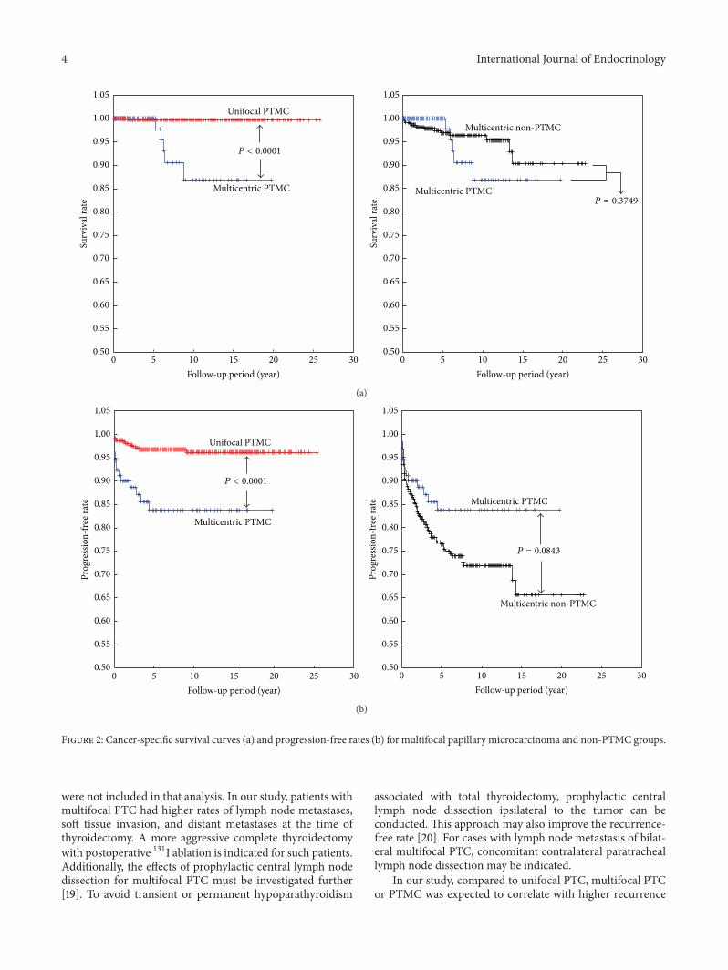
Cancer-specific survival and postoperative progressionanalysis were performed to compare multifocal PTMC withother types of PTC. The thyroid cancer-specific survivalrates for unifocal PTMC and multifocal PTMC were 99.7%
International Journal of Endocrinology 3
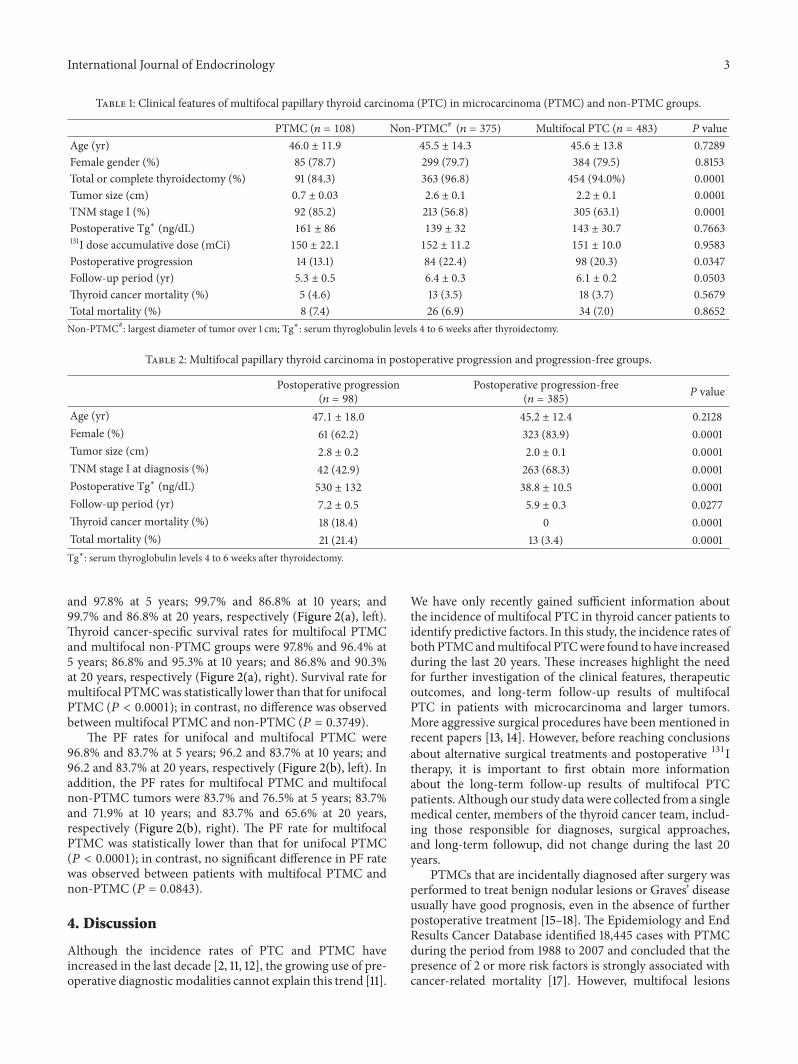
Table 1: Clinical features of multifocal papillary thyroid carcinoma (PTC) in microcarcinoma (PTMC) and non-PTMC groups.
PTMC (𝑛 = 108) Non-PTMC#(𝑛 = 375) Multifocal PTC (𝑛 = 483) 𝑃 value
Age (yr) 46.0 ± 11.9 45.5 ± 14.3 45.6 ± 13.8 0.7289Female gender (%) 85 (78.7) 299 (79.7) 384 (79.5) 0.8153Total or complete thyroidectomy (%) 91 (84.3) 363 (96.8) 454 (94.0%) 0.0001Tumor size (cm) 0.7 ± 0.03 2.6 ± 0.1 2.2 ± 0.1 0.0001TNM stage I (%) 92 (85.2) 213 (56.8) 305 (63.1) 0.0001Postoperative Tg∗ (ng/dL) 161 ± 86 139 ± 32 143 ± 30.7 0.7663131I dose accumulative dose (mCi) 150 ± 22.1 152 ± 11.2 151 ± 10.0 0.9583Postoperative progression 14 (13.1) 84 (22.4) 98 (20.3) 0.0347Follow-up period (yr) 5.3 ± 0.5 6.4 ± 0.3 6.1 ± 0.2 0.0503Thyroid cancer mortality (%) 5 (4.6) 13 (3.5) 18 (3.7) 0.5679Total mortality (%) 8 (7.4) 26 (6.9) 34 (7.0) 0.8652Non-PTMC#: largest diameter of tumor over 1 cm; Tg∗: serum thyroglobulin levels 4 to 6 weeks after thyroidectomy.
Table 2: Multifocal papillary thyroid carcinoma in postoperative progression and progression-free groups.
Postoperative progression(𝑛 = 98)
Postoperative progression-free(𝑛 = 385)
𝑃 value
Age (yr) 47.1 ± 18.0 45.2 ± 12.4 0.2128Female (%) 61 (62.2) 323 (83.9) 0.0001Tumor size (cm) 2.8 ± 0.2 2.0 ± 0.1 0.0001TNM stage I at diagnosis (%) 42 (42.9) 263 (68.3) 0.0001Postoperative Tg∗ (ng/dL) 530 ± 132 38.8 ± 10.5 0.0001Follow-up period (yr) 7.2 ± 0.5 5.9 ± 0.3 0.0277Thyroid cancer mortality (%) 18 (18.4) 0 0.0001Total mortality (%) 21 (21.4) 13 (3.4) 0.0001Tg∗: serum thyroglobulin levels 4 to 6 weeks after thyroidectomy.
and 97.8% at 5 years; 99.7% and 86.8% at 10 years; and99.7% and 86.8% at 20 years, respectively (Figure 2(a), left).Thyroid cancer-specific survival rates for multifocal PTMCand multifocal non-PTMC groups were 97.8% and 96.4% at5 years; 86.8% and 95.3% at 10 years; and 86.8% and 90.3%at 20 years, respectively (Figure 2(a), right). Survival rate formultifocal PTMCwas statistically lower than that for unifocalPTMC (𝑃 < 0.0001); in contrast, no difference was observedbetween multifocal PTMC and non-PTMC (𝑃 = 0.3749).
The PF rates for unifocal and multifocal PTMC were96.8% and 83.7% at 5 years; 96.2 and 83.7% at 10 years; and96.2 and 83.7% at 20 years, respectively (Figure 2(b), left). Inaddition, the PF rates for multifocal PTMC and multifocalnon-PTMC tumors were 83.7% and 76.5% at 5 years; 83.7%and 71.9% at 10 years; and 83.7% and 65.6% at 20 years,respectively (Figure 2(b), right). The PF rate for multifocalPTMC was statistically lower than that for unifocal PTMC(𝑃 < 0.0001); in contrast, no significant difference in PF ratewas observed between patients with multifocal PTMC andnon-PTMC (𝑃 = 0.0843).
4. Discussion
Although the incidence rates of PTC and PTMC haveincreased in the last decade [2, 11, 12], the growing use of pre-operative diagnosticmodalities cannot explain this trend [11].
We have only recently gained sufficient information aboutthe incidence of multifocal PTC in thyroid cancer patients toidentify predictive factors. In this study, the incidence rates ofboth PTMCandmultifocal PTCwere found to have increasedduring the last 20 years. These increases highlight the needfor further investigation of the clinical features, therapeuticoutcomes, and long-term follow-up results of multifocalPTC in patients with microcarcinoma and larger tumors.More aggressive surgical procedures have been mentioned inrecent papers [13, 14]. However, before reaching conclusionsabout alternative surgical treatments and postoperative 131Itherapy, it is important to first obtain more informationabout the long-term follow-up results of multifocal PTCpatients. Although our study datawere collected from a singlemedical center, members of the thyroid cancer team, includ-ing those responsible for diagnoses, surgical approaches,and long-term followup, did not change during the last 20years.
PTMCs that are incidentally diagnosed after surgery wasperformed to treat benign nodular lesions or Graves’ diseaseusually have good prognosis, even in the absence of furtherpostoperative treatment [15–18]. The Epidemiology and EndResults Cancer Database identified 18,445 cases with PTMCduring the period from 1988 to 2007 and concluded that thepresence of 2 or more risk factors is strongly associated withcancer-related mortality [17]. However, multifocal lesions
4 International Journal of Endocrinology
302520151050302520151050
1.05
1.00
0.95
0.90
0.85
0.80
0.75
0.70
0.65
0.60
0.55
0.50
1.05
1.00
0.95
0.90
0.85
0.80
0.75
0.70
0.65
0.60
0.55
0.50
Surv
ival
rate
Surv
ival
rate
Unifocal PTMC
Multicentric PTMC Multicentric PTMC
P < 0.0001
Multicentric non-PTMC
Follow-up period (year) Follow-up period (year)
P = 0.3749
(a)
302520151050302520151050
Unifocal PTMC
Multicentric PTMC
Multicentric PTMC
P < 0.0001
Multicentric non-PTMC
Follow-up period (year) Follow-up period (year)
P = 0.0843
Prog
ress
ion-
free r
ate
Prog
ress
ion-
free r
ate
1.05
1.00
0.95
0.90
0.85
0.80
0.75
0.70
0.65
0.60
0.55
0.50
1.05
1.00
0.95
0.90
0.85
0.80
0.75
0.70
0.65
0.60
0.55
0.50
(b)
Figure 2: Cancer-specific survival curves (a) and progression-free rates (b) for multifocal papillary microcarcinoma and non-PTMC groups.
were not included in that analysis. In our study, patients withmultifocal PTC had higher rates of lymph node metastases,soft tissue invasion, and distant metastases at the time ofthyroidectomy. A more aggressive complete thyroidectomywith postoperative 131I ablation is indicated for such patients.Additionally, the effects of prophylactic central lymph nodedissection for multifocal PTC must be investigated further[19]. To avoid transient or permanent hypoparathyroidism
associated with total thyroidectomy, prophylactic centrallymph node dissection ipsilateral to the tumor can beconducted. This approach may also improve the recurrence-free rate [20]. For cases with lymph node metastasis of bilat-eral multifocal PTC, concomitant contralateral paratracheallymph node dissection may be indicated.
In our study, compared to unifocal PTC, multifocal PTCor PTMC was expected to correlate with higher recurrence

International Journal of Endocrinology 5
rates or poorer prognosis. Among 483 multifocal PTCpatients, there were 23 (4.8%) presented as distant metas-tases at the time of thyroidectomy. In addition, 23 of 98(23.5%) postoperative progressive patients were presentedwith distant metastases. Additionally, total and disease-specific mortality rates were not increased in patients withmultifocal PTC. More data and a longer follow-up periodare needed to draw firm conclusions. Diverse mechanismssuch as multiple independent tumors or intrathyroid spreadoriginating from a single tumor mass were suggested foroccurrences of multifocal PTC [5, 21]. In our study, theincidence of multifocal PTMC was not lower than thatof multifocal non-PTMC. This finding indicates that thepattern of multifocal PTC manifests in early-stage thyroidcancer.
There is some controversy regarding the use of 131Iablation after total thyroidectomy to prevent recurrence oflow- and intermediate-risk PTMC [22–24]. In our multivari-ate statistical analysis, we identified extrathyroid invasion,solid pattern, tumor multifocality, and absence of a tumorcapsule as significant and independent risk factors for PTMCrecurrence [25], although less information was availableabout postoperative 131I therapy for multifocal PTC. In ourstudy, the 131I doses used to treat multifocal and unifocalPTC patients were not statistically different, and a higherrate of postoperative progression was noted in the multifocalgroup. Additional prospectively designed studies are requiredto determine the effect of higher 131I doses on preventingrecurrence in patients with multifocal PTC.
Along with the study limitations described above, 17.3%of our patients did not undergo total thyroidectomy, and thethyroid remnant might have contained incidental microcar-cinomas. Differences between PTMC and non-PTMCgroupstreated with total thyroidectomy may also represent a bias.Additionally, some of the patients did not receive 131I forremnant ablation.
In conclusion, multifocal PTMC occurred more fre-quently than non-PTMC. Additionally, postoperative dis-ease progression and cancer mortality rates were higher inmultifocal PTC than in unifocal PTC in both the PTMCand larger tumor groups. Furthermore, total thyroidectomysuccessfully reduced the postoperative disease progressionrate in multifocal PTC patients with larger tumors.
Conflict of Interests
The authors declare that there are no conflicting financialinterests.
Acknowledgments
This work was supported by grants to Jen-Der Lin from theNational Science Council in Taiwan (NMRPD1B0311) andChang GungMemorial Hospital Grants CMRPG3B1942.Thefunding source had no role in study design, data collectionand analysis, decision to publish, or paper preparation.
References
[1] J. D. Cramer, P. Fu, K. C. Harth, S. Margevicius, and S. M.Wilhelm, “Analysis of the rising incidence of thyroid cancerusing the Surveillance, Epidemiology and End Results nationalcancer data registry,” Surgery, vol. 148, no. 6, pp. 1147–1152, 2010.
[2] S. C. Londero, A. Krogdahl, L. Bastholt et al., “Papillary thyroidcarcinoma in Denmark 1996–2008: an investigation of changesin incidence,”Cancer Epidemiology, vol. 37, no. 1, pp. e1–e6, 2013.
[3] S.-F. Kuo, T.-C. Chao, H.-Y. Chang, C. Hsueh, C.-H. Yang, andJ.-D. Lin, “Prognostic evaluation of patients with multicentricpapillary thyroid microcarcinoma,” Journal of the FormosanMedical Association, vol. 110, no. 8, pp. 511–517, 2011.
[4] R. Ivanova, P. Soares, P. Castro, and M. Sobrinho-Simoes,“Diffuse (or multinodular) follicular variant of papillary thy-roid carcinoma: a clinicopathologic and immunohistochemicalanalysis of ten cases of an aggressive form of differentiatedthyroid carcinoma,” Virchows Archiv, vol. 440, no. 4, pp. 418–424, 2002.
[5] R. Giannini, C. Ugolini, C. Lupi et al., “The heterogeneousdistribution of BRAFmutation supports the independent clonalorigin of distinct tumor foci in multifocal papillary thyroid car-cinoma,”The Journal of Clinical Endocrinology and Metabolism,vol. 92, no. 9, pp. 3511–3516, 2007.
[6] TNM Classification of Malignant Tumours, John Wiley & Sons,New York, NY, USA, 6th edition, 2002, edited by L. H. Sobinand C. Wittwkind.
[7] J.-D. Lin, T.-C. Chao, B.-Y. Huang, S.-T. Chen, H.-Y. Chang, andC. Hsueh, “Thyroid cancer in the thyroid nodules evaluated byultrasonography and fine-needle aspiration cytology,” Thyroid,vol. 15, no. 7, pp. 708–717, 2005.
[8] R. A. Delellis, R. V. Lloyd RV, and P. U. Heitx, “Pathologyand genetics of tumors of endocrine organs,” in World HealthOrganization of Tumours, pp. 73–76, IARC, Lyon, France, 2004.
[9] J.-D. Lin, T.-C. Chao, C. Hsueh, and S.-F. Kuo, “High recurrentrate of multicentric papillary thyroid carcinoma,”The Annals ofSurgical Oncology, vol. 16, no. 9, pp. 2609–2616, 2009.
[10] D. D. Zhang, X.-H. Zhou, D. H. Freeman Jr., and J. L. Freeman,“A non-parametric method for the comparison of partial areasunder ROC curves and its application to large health care datasets,” Statistics in Medicine, vol. 21, no. 5, pp. 701–715, 2002.
[11] S. C. Londero, A. Krogdahl, L. Bastholt et al., “Papillary thyroidmicrocarcinoma in Denmark 1996–2008: a national study ofepidemiology and clinical significance,” Thyroid, vol. 23, no. 9,pp. 1159–1164, 2013.
[12] J.-D. Lin, “Increased incidence of papillary thyroid microcar-cinoma with decreased tumor size of thyroid cancer,” MedicalOncology, vol. 27, no. 2, pp. 510–518, 2010.
[13] H. Mazeh, Y. Samet, D. Hochstein et al., “Multifocality in well-differentiated thyroid carcinomas calls for total thyroidectomy,”The American Journal of Surgery, vol. 201, no. 6, pp. 770–775,2011.
[14] J. A. Ricci and A. E. Alfonso, “Multifocal micropapillarythyroid cancer: a new indication for total thyroidectomy?”TheAmerican Surgeon, vol. 78, no. 11, pp. 1211–1214, 2012.
[15] C.-Y.Wang andT.-C. Chang, “Toxic nodular goiter with thyroidpapillary microcarcinoma,” ANZ Journal of Surgery, vol. 80, no.1-2, p. 117, 2010.
[16] T.-C. Chao, J.-D. Lin, L.-B. Jeng, and M.-F. Chen, “Thyroidcancer with concurrent hyperthyroidism,” Archives of Surgery,vol. 134, no. 2, pp. 130–134, 1999.
6 International Journal of Endocrinology
[17] N. Neuhold, A. Schultheis, M. Hermann, G. Krotla, O. Koperek,and P. Birner, “Incidental papillary microcarcinoma of thethyroid—further evidence of a very low malignant potential: aretrospective clinicopathological study with up to 30 years offollow-up,”Annals of Surgical Oncology, vol. 18, no. 12, pp. 3430–3436, 2011.
[18] X.-M. Yu, Y. Wan, R. S. Sippel, and H. Chen, “Should allpapillary thyroid microcarcinomas be aggressively treated?: ananalysis of 18,445 cases,” Annals of Surgery, vol. 254, no. 4, pp.653–660, 2011.
[19] M. Barczynski, A. Konturek, M. Stopa, and W. Nowak, “Pro-phylactic central neck dissection for papillary thyroid cancer,”British Journal of Surgery, vol. 100, no. 3, pp. 410–418, 2013.
[20] D. Giordano, R. Valcavi, G. B. Thompson et al., “Complicationsof central neck dissection in patients with papillary thyroidcarcinoma: results of a study on 1087 patients and review of theliterature,”Thyroid, vol. 22, no. 9, pp. 911–917, 2012.
[21] E. Kuhn, L. Teller, S. Piana, J. Rosai, andM. J.Merino, “Differentclonal origin of bilateral papillary thyroid carcinoma, with areview of the literature,” Endocrine Pathology, vol. 23, no. 2, pp.101–107, 2012.
[22] H. J. Kim, N. K. Kim, J. H. Choi et al., “Radioactive iodineablation does not prevent recurrences in patients with papillarythyroid microcarcinoma,” Clinical Endocrinology, vol. 78, no. 4,pp. 614–620, 2013.
[23] C. Buffet, J. L. Golmard, C. Hoang et al., “Scoring systemfor predicting recurrences in patients with papillary thyroidmicrocarcinoma,” European Journal of Endocrinology, vol. 167,no. 2, pp. 267–275, 2012.
[24] K. M. Creach, B. A. Siegel, B. Nussenbaum, and P. W. Grigsby,“Radioactive iodine therapy decreases recurrence in thyroidpapillarymicrocarcinoma,” ISRNEndocrinology, vol. 2012, Arti-cle ID 816386, 6 pages, 2012.
[25] G. Ardito, L. Revelli, R. Giustozzi et al., “Aggressive papillarythyroid microcarcinoma: prognostic factors and therapeuticstrategy,” Clinical Nuclear Medicine, vol. 38, no. 1, pp. 25–28,2013.

Submit your manuscripts athttp://www.hindawi.com
Stem CellsInternational
Hindawi Publishing Corporationhttp://www.hindawi.com Volume 2014
Hindawi Publishing Corporationhttp://www.hindawi.com Volume 2014
MEDIATORSINFLAMMATION
of
Hindawi Publishing Corporationhttp://www.hindawi.com Volume 2014
Behavioural Neurology
EndocrinologyInternational Journal of
Hindawi Publishing Corporationhttp://www.hindawi.com Volume 2014
Hindawi Publishing Corporationhttp://www.hindawi.com Volume 2014
Disease Markers
Hindawi Publishing Corporationhttp://www.hindawi.com Volume 2014
BioMed Research International
OncologyJournal of
Hindawi Publishing Corporationhttp://www.hindawi.com Volume 2014
Hindawi Publishing Corporationhttp://www.hindawi.com Volume 2014
Oxidative Medicine and Cellular Longevity
Hindawi Publishing Corporationhttp://www.hindawi.com Volume 2014
PPAR Research
The Scientific World JournalHindawi Publishing Corporation http://www.hindawi.com Volume 2014
Immunology ResearchHindawi Publishing Corporationhttp://www.hindawi.com Volume 2014
Journal of
ObesityJournal of
Hindawi Publishing Corporationhttp://www.hindawi.com Volume 2014
Hindawi Publishing Corporationhttp://www.hindawi.com Volume 2014
Computational and Mathematical Methods in Medicine
OphthalmologyJournal of
Hindawi Publishing Corporationhttp://www.hindawi.com Volume 2014
Diabetes ResearchJournal of
Hindawi Publishing Corporationhttp://www.hindawi.com Volume 2014
Hindawi Publishing Corporationhttp://www.hindawi.com Volume 2014
Research and TreatmentAIDS
Hindawi Publishing Corporationhttp://www.hindawi.com Volume 2014
Gastroenterology Research and Practice
Hindawi Publishing Corporationhttp://www.hindawi.com Volume 2014
Parkinson’s Disease
Evidence-Based Complementary and Alternative Medicine
Volume 2014Hindawi Publishing Corporationhttp://www.hindawi.com