clitocybe nebularis bba - general subjectsweb.bf.uni-lj.si/bi/biokemija/separati/pohbba08.pdf ·...
TRANSCRIPT

�������� ����� ��
Purification, characterization and cloning of a ricin B-like lectin frommushroom clitocybe nebularis with antiproliferative activity against humanleukemic t cells
Jure Pohleven, Natasa Obermajer, Jerica Sabotic, Sabina Anzlovar, KristinaSepcic, Janko Kos, Bogdan Kralj, Borut Strukelj, Joze Brzin
PII: S0304-4165(08)00262-6DOI: doi:10.1016/j.bbagen.2008.11.006Reference: BBAGEN 26610
To appear in: BBA - General Subjects
Received date: 30 July 2008Revised date: 19 November 2008Accepted date: 22 November 2008
Please cite this article as: Jure Pohleven, Natasa Obermajer, Jerica Sabotic, SabinaAnzlovar, Kristina Sepcic, Janko Kos, Bogdan Kralj, Borut Strukelj, Joze Brzin, Pu-rification, characterization and cloning of a ricin B-like lectin from mushroom clitocybenebularis with antiproliferative activity against human leukemic t cells, BBA - GeneralSubjects (2008), doi:10.1016/j.bbagen.2008.11.006
This is a PDF file of an unedited manuscript that has been accepted for publication.As a service to our customers we are providing this early version of the manuscript.The manuscript will undergo copyediting, typesetting, and review of the resulting proofbefore it is published in its final form. Please note that during the production processerrors may be discovered which could affect the content, and all legal disclaimers thatapply to the journal pertain.

ACC
EPTE
D M
ANU
SCR
IPT
ACCEPTED MANUSCRIPTClitocybe nebularis ricin B-like lectin1
PURIFICATION, CHARACTERIZATION AND CLONING OF A RICIN B-LIKE
LECTIN FROM MUSHROOM Clitocybe nebularis WITH ANTIPROLIFERATIVE
ACTIVITY AGAINST HUMAN LEUKEMIC T CELLS
Jure Pohlevena,*, Nataša Obermajerb, Jerica Sabotiča, Sabina Anžlovara,c, Kristina
Sepčićc, Janko Kosa,b, Bogdan Kraljd, Borut Štrukelja,b and Jože Brzina
a Department of Biotechnology, Jožef Stefan Institute, Jamova 39, SI-1000 Ljubljana, Slovenia
b Faculty of Pharmacy, University of Ljubljana, Aškerčeva 7, SI-1000 Ljubljana, Slovenia
c Department of Biology, Biotechnical Faculty, University of Ljubljana, Večna pot 111, SI-1000
Ljubljana, Slovenia
d Department of Environmental Sciences, Jožef Stefan Institute, Jamova 39, SI-1000
Ljubljana, Slovenia
* Corresponding author. Tel.: +386 1477 3306, Fax: +386 1477 3984.
E-mail address: [email protected]
Keywords: Mushroom; Clitocybe nebularis; Ricin B-like lectin; Antiproliferative effect;
Leukemic T cells
The nucleotide sequence reported in this paper has been submitted to the GenBank™/EMBL
Data Bank with accession number EU682006.

ACC
EPTE
D M
ANU
SCR
IPT
ACCEPTED MANUSCRIPTClitocybe nebularis ricin B-like lectin2
Abstract
Background: Lectins are a diverse group of carbohydrate-binding proteins exhibiting
numerous biological activities and functions.
Methods: Two-step serial carbohydrate affinity chromatography was used to isolate a
lectin from the edible mushroom clouded agaric (Clitocybe nebularis). It was characterized
biochemically, its gene and cDNA cloned and the deduced amino acid sequence analyzed. Its
activity was tested by hemagglutination assay and carbohydrate-binding specificity determined
by glycan microarray analysis. Its effect on proliferation of several human cell lines was
determined by MTS assay.
Results: A homodimeric lectin with 15.9-kDa subunits agglutinates human group A,
followed by B, O, and bovine erythrocytes. Hemagglutination was inhibited by glycoprotein
asialofetuin and lactose. Glycan microarray analysis revealed that the lectin recognizes human
blood group A determinant GalNAcα1-3(Fucα1-2)Galβ-containing carbohydrates, and
GalNAcβ1-4GlcNAc (N,N’-diacetyllactosediamine). The lectin exerts antiproliferative activity
specific to human leukemic T cells.
Conclusions: The protein belongs to the ricin B-like lectin superfamily, and has been
designated as Clitocybe nebularis lectin (CNL). Its antiproliferative effect appears to be elicited
by binding to carbohydrate receptors on human leukemic T cells.
General Significance: CNL is one of the few mushroom ricin B-like lectins that have
been identified and the only one so far shown to possess immunomodulatory properties.

ACC
EPTE
D M
ANU
SCR
IPT
ACCEPTED MANUSCRIPTClitocybe nebularis ricin B-like lectin3
1. Introduction
Fungi are well-known for their nutritional and medicinal value due to their content of a
variety of bioactive substances with pharmacological properties [1]. For this reason, mushrooms
and their derivatives are increasingly used worldwide in traditional medicine and as dietary
supplements [2]. Numerous mushroom-derived compounds with antitumor,
immunomodulatory, antiviral, antimicrobial and other activities have been isolated and
identified, including polysaccharides, polysaccharide-protein complexes, triterpenes, phenols,
peptides, and proteins such as lectins [3].
Lectins are biologically active proteins that have been isolated from humans, animals,
plants and microorganisms, and also from fungi. They constitute a diverse group of proteins that
specifically bind different types of carbohydrates. The widespread occurrence of lectins
suggests their role in basic biological functions. Most lectins contain more than one
carbohydrate-binding site and can therefore agglutinate cells or cross-link cell surface
carbohydrates [4]. Their biological activity can be exerted through binding and clustering
glycosylated receptors on cell surfaces, leading to signal transduction pathway activation, and
by binding to extracellular matrix glycoconjugates, cytoplasmic and nuclear glycoproteins. In
this manner, lectins can be involved in various cellular processes, including cell adhesion,
migration, differentiation, apoptosis and proliferation, and thus possess immunomodulatory
properties and have potential roles in cancer [5].
Many lectins have been isolated and characterized from higher fungi [6,7], and six
different folds have been identified for mushroom lectins [8,9]. One of them is a β-trefoil fold
typical of the ricin B domain in the crystal structures of Laetiporus sulphureus lectin [10] and
Marasmius oreades agglutinin [11]. In addition, other fungal lectins that contain ricin B-like
lectin domains have been isolated from Rhizoctonia solani [12] and Polyporus squamosus [13].
A lectin from a basidiomycete, clouded agaric (Clitocybe nebularis), has been reported [14] but
has not been characterized at the molecular level. In previous work, our group isolated a novel

ACC
EPTE
D M
ANU
SCR
IPT
ACCEPTED MANUSCRIPTClitocybe nebularis ricin B-like lectin4
inhibitor of cysteine proteinases, clitocypin, from this mushroom [15] and also several distinct
lectins with specificities for different carbohydrates which have been examined for biological
activities. In this study, a ricin B-like lectin designated Clitocybe nebularis lectin (CNL) has
been isolated, characterized biochemically and its gene, cDNA and deduced amino acid
sequences determined and analyzed. Moreover, the effect on proliferation of several human cell
lines was assayed in order to evaluate the antitumor and immunomodulatory potential of CNL.
2. Materials and methods
2.1. Fungal material
Basidiocarps of the mycorrhizal basidiomycete Clitocybe nebularis were collected in
their natural habitat in Kras forest, Slovenia in October 2004 and frozen at –30ºC until use. A
specimen is deposited at the Department of Biotechnology, Jožef Stefan Institute, Ljubljana,
Slovenia. The cultured mycelium isolated from the same specimen was confirmed by ribosomal
DNA spacer sequencing to belong to the C. nebularis species [16]. It is kept in the collection of
fungi, lichens and higher plants at the Slovenian Forestry Institute, Ljubljana, Slovenia.
2.2. Purification of CNL
After defrosting, C. nebularis fruiting bodies (1000 g fresh weight) were homogenized
and extracted in 1000 ml of 20 mM Tris/HCl buffer, pH 7.5, containing 0.4 M NaCl (Buffer A).
The homogenate was centrifuged for 15 min at 11,000 × g and 4°C. The resulting supernatant
was filtered and subjected to two-step serial carbohydrate affinity chromatography, using
lactosyl- and glucosyl-Sepharose 4B columns (Pharmacia Fine Chemicals, Uppsala, Sweden).
The columns were prepared as described [17], and equilibrated with Buffer A. The extract was

ACC
EPTE
D M
ANU
SCR
IPT
ACCEPTED MANUSCRIPTClitocybe nebularis ricin B-like lectin5
loaded on a lactosyl-Sepharose column, which was then washed with Buffer A to remove
unbound material. Adsorbed proteins were eluted with either 0.2 M lactose or 0.01 M NaOH in
the same buffer. In the latter case, fractions were immediately neutralized with 2 M Tris/HCl
buffer, pH 6.5. The eluted lactose-binding proteins were then applied to a glucosyl-Sepharose
column and the unbound fractions (containing CNL) were collected, pooled and concentrated
using an Amicon UM-10 ultrafiltration membrane (Amicon Corp., Lexington, MA). The purity
of isolated CNL was assessed by a reversed-phase high-performance liquid chromatography
(RP-HPLC; Hewlett-Packard Series 1100 system, Germany) on a Chromsep HPLC column
(ChromSpher C8, 100 × 3 mm; Chrompack international, The Netherlands). The initial solvent
was water containing 0.1% trifluoroacetic acid (TFA) as ion-pairing agent. Proteins were eluted
with a linear gradient of acetonitrile/water (90:10, v/v) solution containing 0.1% TFA. The
eluting solution was increased linearly from 0 to 45% over 5 min, 45-65% over 30 min, and 65-
100% over 5 min. The lectin was then dried in a SpeedVac concentrator (Savant Instruments,
inc., Hicksville, NY).
2.3. Polyacrylamide gel electrophoresis and isoelectric focusing
The purity and molecular mass of CNL were estimated by sodium dodecyl sulfate-
polyacrylamide gel electrophoresis (SDS-PAGE) using homogeneous 15% (w/v) acrylamide gel
on a mini-Protean II apparatus from Bio-Rad Laboratories (Hercules, CA). Samples were
dissolved in electrophoresis buffer, pH 6.8, containing 5% SDS and heated at 100C in either
the presence or absence of 5% (v/v) 2-mercaptoethanol for 10 min. Gels were stained with 0.1%
(w/v) Coomassie Brilliant Blue R-250. Molecular mass was estimated using low molecular
weight standard proteins of 14.4-97 kDa (LMW Calibration Kit for SDS Electrophoresis,
Amersham Pharmacia Biotech). Non-denatured CNL was subjected to native PAGE on
PhastGel Gradient 8-25 with PhastGel Native Buffer Strips, using the PhastSystem (Pharmacia
ACC
EPTE
D M
ANU
SCR
IPT
ACCEPTED MANUSCRIPTClitocybe nebularis ricin B-like lectin6
LKB Biotechnology), according to the instructions provided. Isoelectric focusing was carried
out with a Pharmacia PhastSystem, using a polyacrylamide gel with a pH gradient of 3-9
(PhastGel™ IEF 3-9, Pharmacia) following the manufacturer’s protocol. The isoelectric point
was estimated using a Pharmacia Broad pI Calibration Kit for a pH range of 3-10.
2.4. Size exclusion chromatography
The approximate molecular mass of native CNL was estimated by analytical size
exclusion fast protein liquid chromatography (FPLC; ÄKTA FPLC System, Amersham
Pharmacia Biotech, Sweden). The column (Superdex™ 75 HR/10/30, Pharmacia Biotech Inc.)
was equilibrated with 50 mM Tris/HCl buffer, pH 7.5, containing 0.3 M NaCl, with 0.2 M
lactose to prevent interactions between the lectin and column matrix. The column was calibrated
under the same conditions with Dalton standards MS II (Serva, Heidelberg).
2.5. Electrospray ionization-mass spectrometry
The molecular mass of the HPLC-purified CNL was determined by electrospray
ionization (ESI) mass spectrometry. The protein was dissolved in water/methanol (60:40, v/v)
solution containing 0.1% formic acid, and analyzed on an AutoSpecQ (Micromass, Manchester,
UK), and on a Q-Tof Premier (Micromass MS Technologies/Waters, Manchester, UK) mass
spectrometer. The protein sample was introduced into the ion source (needle voltage of 4 kV) at
a flow rate of 5 µl/min using a syringe pump. The mass spectrum, with multiply charged ion
series, was deconvoluted to obtain the molecular mass of the CNL.
2.6. Amino acid sequencing

ACC
EPTE
D M
ANU
SCR
IPT
ACCEPTED MANUSCRIPTClitocybe nebularis ricin B-like lectin7
N-terminal and peptide sequences were determined by automated Edman degradation
using a Procise Protein Sequencing System 492 (PE Applied Biosystems, Foster City, CA).
HPLC-purified CNL was denatured by heating at 100°C for 30 min and digested with trypsin or
α-chymotrypsin (enzyme/substrate ratio of 1:100 (w/w)) in 0.1 M N-methyl morpholine acetate
buffer, pH 8.1, for 4 h at 37°C. Cyanogen bromide cleavage was carried out in 80% formic acid
at room temperature in the dark for 36 h. Resulting peptide fragments were separated by RP-
HPLC and sequenced.
2.7. Molecular cloning of the gene and cDNA encoding CNL
High molecular weight genomic DNA was isolated from frozen powdered C. nebularis
fruiting bodies as described [18]. Total RNA was extracted using RNeasy Plant Mini Kit
(Qiagen, Hilden, Germany) according to the manufacturer’s protocol for isolation from plant
tissues and filamentous fungi. First strand complementary DNA (cDNA) was synthesized from
the total RNA by reverse transcription-polymerase chain reaction (PCR) using a GeneAmp
RNA PCR Core Kit (Applied Biosystems, Foster city, CA) with anchored oligo(dT)-adapter
primer dT(17)3’RACE (Table S1, Supplementary data). For PCR amplification of the partial cnl
gene and cDNA sequences, genomic DNA and reverse transcript were used as templates.
Forward (CNL-D-1f) and reverse (CNL-D-1r) degenerate primers (Table S1) were designed
based on the partial amino acid sequence in regions with the lowest degeneracy. On the basis of
the resulting partial nucleic acid sequences, specific primers were designed to amplify the
complete gene and cDNA sequences.
3’ rapid amplification of cDNA ends (3’ RACE) was carried out using the 3’RACE
adapter primer paired with a forward primer CNL-RACE-1f (Table S1) specific for the cDNA
fragment. cDNA synthesized from total RNA was used as a template. The resulting PCR
product (100 times diluted) served as a template for secondary PCR using a specific nested
primer CNL-RACE-2f (Table S1).
ACC
EPTE
D M
ANU
SCR
IPT
ACCEPTED MANUSCRIPTClitocybe nebularis ricin B-like lectin8
Genome walking PCR was used to amplify the complete cnl gene with its promoter and
terminator regions. Genome walking libraries were constructed using GenomeWalker™
Universal Kit (BD Biosciences Clontech, Palo Alto, CA) according to the manufacturer’s
instructions. High molecular weight genomic DNA (2.5 μg) was digested separately with four
restriction enzymes (DraI, EcoRV, PvuII, StuI) at 37°C overnight. After purification by ethanol
precipitation, GenomeWalker™ Adaptors were ligated to the digested DNA at 16°C overnight.
The resulting ligation mixes were used as templates in genome walking PCR amplifications,
using adapter primer AP1 and a nested primer AP2 provided by the manufacturer, paired with
nested forward gene-specific primers (CNL-term-1 and CNL-term-2; Table S1) for downstream
amplification or nested reverse gene-specific primers (CNL-prom-1 and CNL-prom-2; Table
S1) for upstream amplification. PCR amplification conditions were used as suggested by the
manufacturer.
2.8. Sequence analysis
All PCR products were cloned into pGEM-T Easy Vector System I (Promega, Madison,
WI) and sequenced using the Automated DNA Sequencing Service at MWG Biotech
(Ebersberg, Germany). Overlapping fragments were assembled using the BioEdit program
(http://www.mbio.ncsu.edu/BioEdit/bioedit.html) complete putative cnl gene and cDNA
sequences were obtained. Both were confirmed by amplifying the full-length sequences using
forward (CNL-1f) and reverse (CNL-1r) primers (Table S1).
Gene promoter analysis was performed using TESS (transcription element search
software at http://www.cbil.upenn.edu/cgi-bin/tess/tess). The cDNA-deduced amino acid
sequence of CNL (Swiss-Prot accession no. B2ZRS9) was analyzed using online proteomics
tools at the ExPASy server (http://www.expasy.org/tools/). Similarity searches were performed
using BLASTn and BLASTp algorithms at the NCBI server

ACC
EPTE
D M
ANU
SCR
IPT
ACCEPTED MANUSCRIPTClitocybe nebularis ricin B-like lectin9
(http://www.ncbi.nlm.nih.gov/blast/Blast.cgi), as well as FASTA3 and PSI-BLAST [19]
programs at the EBI server (http://www.ebi.ac.uk/Tools/similarity.html). In addition, the CNL
amino acid sequence was analyzed for protein domain and family identification using SMART
(http://smart.embl-heidelberg.de/), and the SUPERFAMILY program [20]
(http://supfam.cs.bris.ac.uk/SUPERFAMILY/). Using SMART, a multiple alignment of the
CNL amino acid sequence with ricin B domain sequences was performed and manually adjusted
in the BioEdit program.
2.9. Molecular modeling of CNL
The secondary structure of CNL was predicted using PSIPRED V2.6
(http://bioinf.cs.ucl.ac.uk/psipred/psiform.html). Structurally conserved regions of the lectin
were identified by hydrophobic cluster analysis using the online Drawhca program
(http://mobyle.rpbs.univ-paris-diderot.fr/cgi-bin/portal.py?form=HCA). Fully automated fold
recognition was carried out using the LOOPP program
(http://cbsuapps.tc.cornell.edu/loopp.aspx) to predict the three-dimensional structure of CNL.
The predicted model was visualized using the PyMOL Molecular Graphics System (DeLano
Scientific, San Carlos, CA).
2.10. Hemagglutination assay
The specificity of CNL for different types of erythrocytes was determined by
hemagglutination using human groups A, AB, B, O, and bovine erythrocytes. Red blood cells
were washed three times with 0.9% saline and twice with 20 mM Tris/HCl buffer, pH 7.5,
containing 130 mM NaCl (erythrocyte buffer). Erythrocyte suspension (2%), corresponding to a
final concentration of 4.3 × 10⁸ erythrocytes/ml, was pipetted into a microtiter plate (Nunclon
Delta SI, Nunc, Denmark), 100 µl per well, and 1-20 µl of CNL were added to give final

ACC
EPTE
D M
ANU
SCR
IPT
ACCEPTED MANUSCRIPTClitocybe nebularis ricin B-like lectin10
concentrations of 1-325 µg/ml. Agglutination was examined visually after 1 hour incubation at
room temperature.
A preliminary study of carbohydrate-binding specificity of CNL was performed by
hemagglutination inhibition assay using mono- and disaccharides (D-glucose (Kemika, Zagreb,
Croatia), D-galactose, D-sucrose, D-lactose (Fluka, Buchs, Switzerland), β-D-fructose, D-
mannose (Sigma, St. Louis, Missouri)), and a glycoprotein asialofetuin (Sigma, St. Louis,
Missouri). Prior to the test, microtiter plate wells were blocked with 0.1% (w/v) bovine serum
albumin. CNL, at a final agglutinating concentration of 18.6 µg/ml, was incubated with serial
dilutions of carbohydrate inhibitors in a microtiter plate for 1 h. Human group A erythrocytes
suspended in erythrocyte buffer, pH 7.5, were then added.
The thermal stability of CNL was assessed by incubation at different concentrations for
30 min at 4 to 100°C, and storing overnight at 4°C. Samples were assayed by hemagglutination
using human group A erythrocytes in erythrocyte buffer, pH 7.5. To study the activity of CNL
in the pH range of 5-9, the lectin was diluted to different concentrations with 20 mM
Mes/NaOH buffer, pH 5, 5.5, 6 or 6.5, containing 130 mM NaCl, or with erythrocyte buffer, pH
7, 7.5, 8, 8.5 or 9. Human group A erythrocytes were suspended in the same buffers and
hemagglutinating activity assayed. Activity of the lectin at lower and higher pH values could
not be tested due to the spontaneous lysis of erythrocytes.
2.11. Glycan microarray analysis
To determine fine carbohydrate-binding specificity of CNL, glycan microarray analysis
was performed by The Consortium for Functional Glycomics (Core H;
http://www.functionalglycomics.org/static/consortium/resources/resourcecoreh.shtml).
Microarrays (Mammalian Printed Array version 3.2) containing ~400 different glycan structures
in replicates of 6 were printed as described [21]. The lectin was biotinylated using No-Weight™
NHS-PEO4-Biotin (Pierce, Rockford, IL) according to the manufacturer’s instructions.

ACC
EPTE
D M
ANU
SCR
IPT
ACCEPTED MANUSCRIPTClitocybe nebularis ricin B-like lectin11
Streptavidin-conjugated horseradish peroxidase was used to confirm biotinylation of CNL
transferred to a PVDF membrane (Immobilon, Millipore, Bedford, MA) and the activity of the
biotinylated lectin was examined by hemagglutination assay. In a binding assay, microarray
slides were incubated with various concentrations of biotinylated CNL (200, 1 and 0.1 µg/ml).
Slides were washed and bound lectin detected using fluorescently labeled streptavidin.
Microarray slides were subjected to imaging, fluorescence was measured and images analyzed
[21].
2.12. Cell proliferation assay
U-937 human pro-monocytic lymphoma cell line (CRL-1593.2; ATCC, Manassas, VA),
and Mo-T and Jurkat human leukemic T cell lines (CRL-8066 and TIB-152; ATCC, Manassas,
VA) were used and maintained according to the supplier’s instructions. In addition, MCF-10A
neoT cell line originating from the MCF-10 human fibrocystic breast epithelial cell line
(provided by Prof. Bonnie F. Sloane from Wayne State University, Detroit, MI) was used and
cultured as described previously [22]. U-937, Mo-T, Jurkat and MCF-10A neoT cells were
resuspended in the appropriate growth media to final concentrations of 4 × 10⁵ cells/ml, and
samples (100 µl/well) added to a 96-well microtiter polystyrene plate (Costar, Schiphol-Rijk,
The Netherlands). Differentiated U-937 cells with macrophage-like properties were obtained by
treating U-937 cells with phorbol 12-myristate 13-acetate (PMA; Sigma, St. Louis, MO) for 24
h at 37°C under 5% CO2. Non-adherent cells were washed away with warm (37°C) medium.
CNL dissolved in phosphate buffer saline (PBS), pH 7.4, was added to the growth medium to
different final concentrations. The appropriate volume of PBS was added to control samples of
cells devoid of the lectin and incubated for 4 days at 37°C. Cells were then washed and their
viability assessed using the CellTiter 96® AQueous One Solution Cell Proliferation Assay
(MTS colorimetric assay; Promega, Madison, WI) according to the instructions provided by the
manufacturer. Percentages of cell proliferation were determined as (Atest cells/Acontrol cells) × 100,

ACC
EPTE
D M
ANU
SCR
IPT
ACCEPTED MANUSCRIPTClitocybe nebularis ricin B-like lectin12
where Atest cells and Acontrol cells are the absorbances of formazan measured for cells treated with
different concentrations of CNL, and for non-treated control cells, respectively. In order to see if
the effect was elicited due to carbohydrate-binding properties of CNL another control
experiment was performed, where the lectin was preincubated with 0.1 M lactose diluted in PBS
and added to cells. Two independent experiments were performed in triplicate for each cell type.
A comparative analysis of proliferation of non-treated versus treated cells was performed with
two-sample t-tests, using the Origin software (Origin 7.5 SR4, OriginLab Corporation,
Northampton, MA).
3. Results
3.1. Purification of CNL
Purified CNL (~8 mg) was isolated from Clitocybe nebularis fruiting bodies (1000 g)
by two-step serial carbohydrate affinity chromatography. In the first step, a crude extract was
loaded onto a lactosyl-Sepharose column. Lactose-binding proteins were obtained by elution
with 0.2 M lactose or 0.01 M NaOH, with respective yields of ~7 mg and ~18 mg. In the second
step, CNL was separated from other lactose-binding proteins by application to a glucosyl-
Sepharose column. The unbound CNL was collected and concentrated, and its purity assessed
using RP-HPLC, revealing a single major peak (not shown).
3.2. CNL molecular mass and isoelectric point
After lactosyl-Sepharose affinity chromatography and elution with either lactose or
NaOH, three bands were obtained by SDS-PAGE analysis, corresponding to apparent masses

ACC
EPTE
D M
ANU
SCR
IPT
ACCEPTED MANUSCRIPTClitocybe nebularis ricin B-like lectin13
of 19 kDa, 17.5 kDa, and 15.5 kDa (Fig. S1A, lane 2, Supplementary data). The glucosyl-
Sepharose unbound CNL migrated as a single band and corresponded to a ~19-kDa protein (Fig.
S1A, lane 3), irrespective of prior treatment with reducing agent (not shown). Native CNL was
analyzed by size exclusion FPLC. It eluted as two peaks with approximate molecular masses of
33 kDa and 16 kDa, corresponding to about 97% and 3% of total protein and indicating a dimer
and a monomer in slow equilibrium (Fig. 1). These results were also confirmed by native PAGE
(data not shown). ESI-mass spectrometry analysis of HPLC-purified CNL revealed one major
peak with a molecular mass of 15,903.5 Da. There was also a minor peak observed which is
most probably an ionization artifact, since its 22 Da higher molecular mass is consistent with
the sodium adduct (M+Na)+ of CNL (not shown).
Isoelectric focusing of CNL revealed one major and two minor bands which indicate
heterogeneity at the primary structure level. The isoelectric point of CNL was determined to be
approximately 4.3 (Fig. S1B, lane 2, Supplementary data).
3.3. Sequence analysis of purified CNL
The fact that CNL was blocked to Edman degradation at the amino terminus suggested
its modification. Efforts to deblock the lectin using TFA in methanol (1:1, v/v) at 47°C for 48 h
[23] were unsuccessful. However, a small fraction of the total protein enabled its sequence to be
determined. Treatment with proteases yielded peptide fragments only when the lectin was
previously denatured by heating at 100°C for 30 min, indicating that native CNL is resistant to
proteolytic cleavage. The peptides were sequenced and around 85% of the complete sequence
was determined (Fig. S2, Supplementary data). Two unidentified amino acid residues were
detected, namely Trp50 which cannot be identified by Edman degradation and Tyr61 which was
probably modified (Fig. S2).
3.4. Characterization of a cnl gene and CNL-encoding cDNA sequence

ACC
EPTE
D M
ANU
SCR
IPT
ACCEPTED MANUSCRIPTClitocybe nebularis ricin B-like lectin14
PCR amplification using degenerate primers with genomic DNA and reverse transcript
as templates yielded fragments of 386 and 275 bp, respectively. Specific nested primers were
designed and the complete cnl gene with its promoter and terminator regions, as well as the
downstream region of the cDNA sequence, were obtained by genome walking and 3’ RACE
methods, respectively. The overlapping segments were assembled and the complete cnl gene
and cDNA sequences were amplified to confirm the determined nucleotide sequences
containing the complete open reading frame encoding the full-length CNL. Alignment of the cnl
gene and cDNA sequences showed no differences (Fig. S2). The 669-bp gene is composed of
five exons (147, 96, 74, 61, 72 bp) and four introns (63, 48, 52, 56 bp) (Fig. S2). The intron-
exon boundaries closely match the consensus splice sites predicted for eukaryotic genes, i.e.
GTRNGY for the 5’-splice site and YNYAG for the 3’-splice site [24]. The 264-bp promoter
sequence contains a consensus TATA box sequence (TATAAAA) at position -76, and a
predicted transcription initiation site (TCACCCTC) at position -39 (Fig. S2). A putative TATA
box, located 37 nt upstream from the putative transcription initiation site, is consistent with the
locations found in several other fungal genes, which are generally 30-60 nt upstream from the
transcriptional start site [24]. The 620-bp terminator sequence of cnl does not contain the
consensus eukaryotic polyadenylation signal sequence AATAAA, however a putative variant
signal, AATCAA, was identified 21 nt upstream of the poly(A) tail. The same polyadenylation
signal sequence was reported for the Onchocerca volvulus galectin (Ov-gbp-2) gene (32 nt
upstream) [25], the lignin peroxidase (LG2) gene from basidiomycete Phanerochaete
chrysosporium (24 nt upstream) [26], the calpain-like protease (palBory) gene from Aspergillus
oryzae [27] and the Gossypium hirsutum Δ-12 fatty acid desaturase (FAD2-3) gene (34 nt
upstream) [28].
BLASTn and FASTA3 similarity searches against public sequence databases using the
complete cnl gene, its coding region, or cDNA sequence as queries revealed no significant
similarities (cut-off e < 10).

ACC
EPTE
D M
ANU
SCR
IPT
ACCEPTED MANUSCRIPTClitocybe nebularis ricin B-like lectin15
3.5. Analysis of the cDNA-deduced CNL amino acid sequence
The amino acid sequence was translated from the open reading frame and aligned with
the cnl gene and cDNA sequences in Fig. S2. The deduced amino acid sequence indicates that
the unprocessed polypeptide is composed of 149 amino acids that match the residues
determined by amino acid sequencing of the purified lectin. However, two alternative amino
acid residues are observed, Thr31 and Leu32 instead of Val31 and Ile32 (Fig. S2).
Using online proteomics tools, the N-terminus of the primary translation product was
predicted with 85% likelihood to be post-translationally processed by N-terminal methionine
excision and further modified by N-acetylation of the following serine residue. The predicted N-
acetyl group is consistent with the blocked N-terminus detected by N-terminal amino acid
sequencing of purified CNL. This was also confirmed by the calculated molecular mass of the
149 amino acid-long unprocessed protein (15,992.8 Da), and the molecular mass calculated for
the predicted mature CNL lacking the initiating methionine (15,861.6 Da) but acetylated
(+42.0372 Da), which totals 15,903.6 Da. This calculated molecular mass of the predicted
mature CNL corresponds to the 15,903.5-Da molecular mass of the purified protein determined
by mass spectrometry, indicating that the protein is modified post-translationally. Mature CNL
contains no cysteine residues and only one methionine. The calculated isoelectric point of the
protein is 4.87, that differs slightly from the isoelectric point determined experimentally (4.3;
Fig. S1B, lane 2).
3.6. CNL is a member of the ricin B-like lectin superfamily
The CNL amino acid sequence similarity searches against protein databases, using
BLASTp and FASTA3, and PSI-BLAST which is more sensitive to weak but biologically
relevant sequence similarities [19], returned numerous fungal and bacterial proteins, including

ACC
EPTE
D M
ANU
SCR
IPT
ACCEPTED MANUSCRIPTClitocybe nebularis ricin B-like lectin16
proteases and proteins involved in carbohydrate metabolism. These proteins all contain ricin B-
type lectin domains that generally exhibit low sequence similarity to CNL. However, some
proteins containing ricin B-like lectin domains with around 20% amino acid sequence identity
to CNL were identified. These include the zinc metalloendopeptidase flavastacin precursor from
Flavobacterium meningosepticum (Swiss-Prot Q47899), predicted proteins from mushrooms
Coprinopsis cinerea (A8PCJ2) and Laccaria bicolor (B0DSD3), ricin B-related lectin from
fungus Polyporus squamosus (Q75WT8), mushroom Marasmius oreades agglutinin (Q8X123)
and Aspergillus oryzae predicted protein (Q2U1N6). A profile hidden Markov model-based
searches for detecting distant family members [29], using SMART (with included Pfam
domains), revealed that CNL consists of a ricin B lectin domain ranging from residues 6 to 141.
It lacks a signal peptide, suggesting that the molecule is not secreted via the endoplasmic
reticulum–Golgi apparatus pathway. Additionally, a SUPERFAMILY sequence search
predicted that CNL is a member of the all-β class of proteins with a β-trefoil fold and a member
of the ricin B-like lectins superfamily.
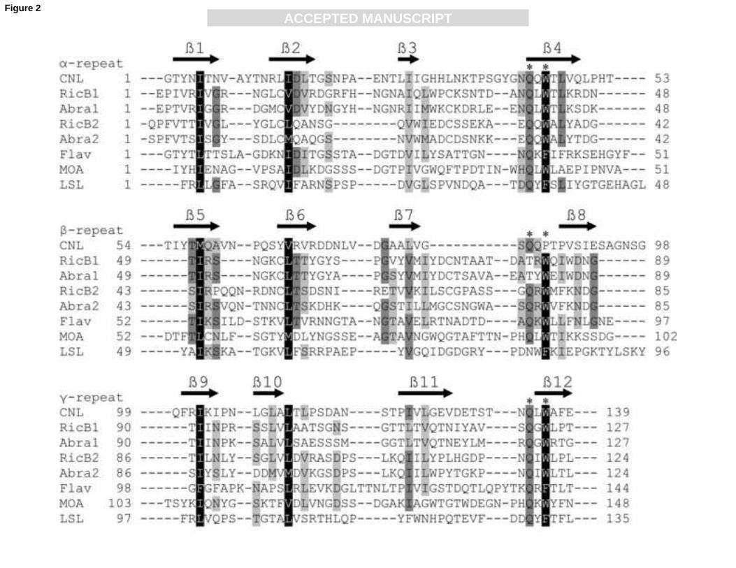
The ricin B-like lectin domain of CNL was aligned with the representative ricin B-type
lectin domains of proteins with known structures [10,11,30,31] (Fig. 2). Closer examination of
the multiple amino acid sequence alignment revealed extensive divergence between the ricin B-
type domains, which was also noted by Hazes [32]. Despite low sequence identity, the domains
share common conserved residues and internal homology of amino acid sequences. The three
distinct homologous subdomains, designated as α-, β- and γ-repeats [33], are also apparent in
CNL and contain glutamine – any residue – tryptophan, QxW, motifs typical of ricin B-like
lectin (QxW)3 domains [32] (Fig. 2). The three CNL repeats display divergence that is most
evident in the less conserved β-repeat that does not contain a genuine QxW motif. The β-repeats
are also most divergent in other (QxW)3 domains, including RicB1, Abra1 and LSL (Fig. 2).
Besides more or less conserved QxW motifs, CNL contains conserved hydrophobic residues
important for the hydrophobic core of the fold (Fig. 2). CNL presumably contains putative
carbohydrate-binding sites in the α- and γ-repeats, though the residues involved in sugar binding

ACC
EPTE
D M
ANU
SCR
IPT
ACCEPTED MANUSCRIPTClitocybe nebularis ricin B-like lectin17
are hard to predict on the basis of variant carbohydrate-binding site residues determined from
the crystallographic analysis of ricin B-type lectin-sugar complex structures [10,11,30,31,34].
3.7. Molecular modeling of CNL predicts a β-trefoil fold typical of ricin B-like lectins
The predicted secondary structure of CNL indicates that the protein contains twelve β-
strands (four per repeat), which is consistent with the all-β class of proteins. The number and
positions of CNL β-strands (Fig. 2) are consistent with secondary structures of other mushroom
ricin B-like lectins [10,11]. To further ascertain a possible structural relationship between CNL
and ricin B-type lectins, hydrophobic cluster analysis and three-dimensional structure prediction
were performed. Structural similarities were readily recognized on comparing the HCA plots of
CNL and ricin B-type lectins (not shown), suggesting that these proteins have similar three-
dimensional structures. Finally, fully automated fold recognition was carried out using the
LOOPP program. The predicted three-dimensional structure of CNL revealed that it adopts a β-
trefoil fold (Fig. 3) typical of ricin B-type lectins [10,11,30,31]. It consists of three distinct
repetitive homologous subdomains (α-, β-, and γ-repeats), which form a pseudo-threefold
symmetry around the hydrophobic core of the fold.
3.8. CNL specificity for different types of erythrocytes and for carbohydrates
Hemagglutination assay showed that the lectin agglutinated all types of red blood cells
tested. The lowest minimum concentrations of CNL exhibiting agglutination were observed for
human groups A, AB and B (8.9, 12.8, and 14.1 µg/ml, respectively), followed by group O
(47.6 µg/ml), and bovine (148.1 µg/ml ) erythrocytes.
The preliminary study of carbohydrate specificity by hemagglutination inhibition assay
showed that, of all the carbohydrate inhibitors tested, CNL agglutination was inhibited by the
glycoprotein asialofetuin and lactose. The minimum inhibitory concentrations were 0.955

ACC
EPTE
D M
ANU
SCR
IPT
ACCEPTED MANUSCRIPTClitocybe nebularis ricin B-like lectin18
mg/ml for the glycoprotein and 1.676 mg/ml (4.65 mM) for lactose. Furthermore, the fine
carbohydrate-binding specificity of CNL was studied by glycan microarray analysis which
revealed that the lectin recognizes N,N’-diacetyllactosediamine (GalNAcβ1–4GlcNAc) and
human blood group A determinant-containing sugar epitopes. These include human blood group
A-specific tetrasaccharide A tetra type II (GalNAcα1-3(Fucα1-2)Galβ1-4GlcNAc), followed by
A tetra Lac (GalNAcα1-3(Fucα1-2)Galβ1-4Glc), A tetra type I (GalNAcα1-3(Fucα1-2)Galβ1-
3GlcNAc), and human blood group A-specific trisaccharide A tri (GalNAcα1–3(Fucα1-2)Galβ).
The list of glycans with the highest affinities for CNL is presented in Table 1. The glycan
microarray results indicate CNL recognition of terminal non-reducing N-acetylgalactosamine
(GalNAcα/β-) and recognition of N-acetylglucosamine (-GlcNAc) or N-acetyllactosamine (-
Galβ1-4GlcNAc) at the reducing end.
3.9. Thermal and pH stability of CNL
CNL exhibited unchanged hemagglutinating activity after 30-min incubation at
temperatures up to 50°C, while at higher temperatures it decreased. Under the conditions used
14% of the initial activity was retained even after heating at 100°C. CNL retained more than
70% of its maximum agglutinating activity over the tested pH range (5-9), with the optimum at
6-6.5.
3.10. Antiproliferative effect of CNL on human leukemic T cell lines
The effect of CNL on proliferation of several human cell lines was assessed by the MTS
assay. No effect was observed on pro-monocytic lymphoma and fibrocystic breast epithelial
cells. However, a dose-dependent antiproliferative effect of CNL was observed on both
leukemic T cell lines, i.e. Mo-T (Fig. 4) and Jurkat. At concentrations of 25 μg/ml and above,
the number of viable leukemic cells was significantly reduced (p < 0.05) with a ~60% decrease

ACC
EPTE
D M
ANU
SCR
IPT
ACCEPTED MANUSCRIPTClitocybe nebularis ricin B-like lectin19
at the highest concentration (100 μg/ml) of CNL. In a control experiment, in which CNL was
preincubated with 0.1 M lactose, the antiproliferative effect was abolished (Fig. 4), the
carbohydrate-binding sites of CNL being blocked by the sugar.
4. Discussion
A novel ricin B-like lectin, designated Clitocybe nebularis lectin (CNL), was purified
from mushroom C. nebularis. The lectin consists of a single ricin B domain, has a molecular
mass of 15.9 kDa and isoelectric point around 4.3. Under non-denaturing conditions, CNL
forms a homodimeric structure by noncovalent self-association.
Two-step serial carbohydrate affinity chromatography proved to be the simplest and
most rapid method for separating CNL from other lectins and impurities. Besides commonly
used carbohydrates (lactose in our case) for lectin elution from the affinity column, NaOH
solution was also used and proved to be more efficient. In addition, subsequent removal of the
lectin-bound lactose after elution with the sugar was also avoided by NaOH elution.
The carbohydrate-binding specificity of CNL was determined by glycan microarray
analysis. It revealed specificity for N,N’-diacetyllactosediamine and human blood group A
determinant-containing carbohydrates, which is in agreement with the agglutination of human
group A erythrocytes. These results indicate CNL recognition of terminal non-reducing
GalNAcα/β- and recognition of -GlcNAc or -Galβ1-4GlcNAc at the reducing end. However, the
method of purification using lactose and hemagglutination inhibition assay suggest some
recognition of terminal non-reducing β-galactose, which was also shown by the microarray
analysis.
The complete cnl gene, consisting of five exons and four introns, and CNL-encoding
cDNA were obtained by molecular cloning. A putative TATA box was found in the promoter
region at a position that suggests transcriptional activity of the gene. Moreover, the deduced

ACC
EPTE
D M
ANU
SCR
IPT
ACCEPTED MANUSCRIPTClitocybe nebularis ricin B-like lectin20
amino acid sequence matched almost exactly the residues determined for purified CNL,
indicating that the obtained gene encodes a very similar isoform of the lectin. Heterogeneity of
purified CNL was also observed by isoelectric focusing (Fig. S1B, lane 2). The two variant
amino acid residues observed in CNL (Fig. S2) are probably not important for the structure or
significantly involved in carbohydrate binding, as concluded from the multiple sequence
alignment of ricin B-type lectin domains. Heterogeneity is common for mushroom lectins
including ricin B-like lectins [13,35,36] and could be the result of gene polymorphism, or the
presence of highly conserved multiple gene families encoding the isolectins [37].
The in silico prediction analyses of the deduced amino acid sequence suggest that the
primary translation product undergoes post-translational modification by removal of the
initiating methionine and acetylation of the following serine. This was consistent with the
blocked N-terminus detected by amino acid sequencing of purified CNL and confirmed by
comparing the calculated molecular mass of the putative mature lectin and mass spectrometry
analysis of purified CNL, revealing masses of 15,903.6 Da and 15,903.5 Da, respectively. The
molecular mass analysis, together with the fact that the lectin lacks signal peptide and thus is
synthesized on free ribosomes, indicate that CNL is not glycosylated.
Similarity searches for homologous proteins in databases revealed numerous fungal and
bacterial proteins containing ricin B-like lectin domains that show low sequence identity to
CNL. Ricin B-like lectins constitute a family of carbohydrate-binding proteins with structures
known from the lectin domains of plant toxins ricin and abrin [30,31]. Although a high degree
of divergence between members of the ricin B-like lectin family is its characteristic feature [32],
further investigation of the uncertain relationship between CNL and ricin B-like lectins was
conducted. A profile hidden Markov model-based searches, that provide greater sensitivity
when searching for evolutionarily distant proteins [29], identified a ricin B lectin domain in
CNL. In addition, the lectin was classified as a member of the ricin B-like lectin superfamily,
which groups together proteins with structural, functional and sequence evidence for a common
evolutionary ancestor [20]. Multiple sequence alignment of CNL with representative ricin B-

ACC
EPTE
D M
ANU
SCR
IPT
ACCEPTED MANUSCRIPTClitocybe nebularis ricin B-like lectin21
type lectin domains revealed that, despite low sequence identity, CNL contains the internal
homology displayed in the three repeats with QxW motifs and conserved hydrophobic residues
structurally important for the hydrophobic core of the fold, all typical of ricin B-type lectins
[32]. The secondary structure prediction, hydrophobic clustering analysis and predicted three-
dimensional structure together lead to the conclusion that CNL adopts the β-trefoil fold typical
of ricin B-type lectins. Besides amino acid sequence analysis, there is biochemical evidence
supporting CNL inclusion in the ricin B-like lectin superfamily. The agglutinating and
carbohydrate-binding activities of CNL, recognizing asialofetuin and lactose, corresponds to the
properties of ricin B chain [38] and mushroom ricin B-like lectins [13,36,39]. However, fine
carbohydrate-binding specificities of mushroom ricin B-like lectins show no particular
similarities, except for higher or lower affinities for lactose and N-acetyllactosamine-containing
carbohydrates.
CNL could be distantly related to a ricin B-like lectin with known structure, designated
as Marasmius oreades agglutinin (MOA; Swiss-Prot Q8X123). This agglutinin was isolated
from the mushroom M. oreades which is related to C. nebularis, both belonging to the
basidiomycete order Agaricales and to the same family of Tricholomataceae. Of the proteins
with known folds, MOA displays the highest similarity score with CNL. Further, both proteins
are acidic and acetylated at their N-terminus [35]. MOA apparently lacks sequence homology to
ricin B-type lectin domains but still adopts the highly conserved β-trefoil fold (PDB code 2iho)
[11], similar to that in the predicted three-dimensional structure of CNL (Fig. 3). We therefore
conclude that, despite low amino acid sequence identity, there are strong grounds for CNL
being structurally related to ricin B-type lectins, sharing similar β-trefoil folds and being
classified as a member of the ricin B-like lectin superfamily.
Finally, CNL was shown to possess antiproliferative activity against leukemic T
lymphocytes. The effect is comparable to those described for other mushroom lectins [40,41]
but, to our knowledge, no antiproliferative activity has been reported for ricin B-like lectins that
do not contain a toxic domain. The inhibition of proliferation of leukemic T cells by CNL was

ACC
EPTE
D M
ANU
SCR
IPT
ACCEPTED MANUSCRIPTClitocybe nebularis ricin B-like lectin22
abolished by lactose, which binds to and blocks the carbohydrate-binding sites of CNL.
Furthermore, other lectins isolated from C. nebularis with sugar specificities different from
those of CNL did not inhibit the proliferation of leukemic T cells (results not shown). Therefore,
the antiproliferative effect appears to be at least partially elicited by CNL recognizing and
binding to cell surface glycoconjugates specific for this particular lectin. The fact that the effect
was not exhibited on pro-monocytic lymphoma and fibrocystic breast epithelial cell lines
suggests that the antiproliferative effect of CNL is specific to leukemic T lymphocytes which
presumably possess appropriate receptors. Similarly, the ricin B chain of the plant toxin ricin
has been shown to bind cell surface glycoconjugates and thus facilitate internalization of the
protein by endocytosis, which is the first step in mediating cytotoxicity by the toxic domain
[42]. Moreover, studies on human galectin-1 have shown that the galectin induces apoptosis of
T cells by cross-linking the specific T cell surface glycoprotein receptors [43].
In conclusion, CNL is a mushroom-derived, biologically active carbohydrate-binding
protein. It is one of the few mushroom ricin B-like lectins that have been identified and the only
one so far shown to possess immunomodulatory properties. It exhibits antiproliferative effects
specific to human leukemic T cells. Therefore, the lectin has potential therapeutic applications
in treating T cell mediated autoimmune and inflammatory disorders and hematopoietic
malignancies, and could be used for targeting leukemic T cells.
Acknowledgements
The authors thank Prof. Dr. Roger Pain for critical review of the manuscript and Adrijana
Leonardi for amino acid sequencing analysis. The authors would also like to acknowledge The
Consortium for Functional Glycomics and the Protein-Carbohydrate Interaction Core H at
Emory University School of Medicine, Atlanta, GA, supported by NIGMS Grant GM62116, for
ACC
EPTE
D M
ANU
SCR
IPT
ACCEPTED MANUSCRIPTClitocybe nebularis ricin B-like lectin23
glycan array analysis. The work was supported by the Ministry of Higher Education, Science
and Technology of the Republic of Slovenia under Grants No. J4-9425-0106-06 and P4-0127.
List of abbreviations
CNL, Clitocybe nebularis lectin; RP-HPLC, reversed-phase high-performance liquid
chromatography; TFA, trifluoroacetic acid; SDS-PAGE, sodium dodecyl sulfate-
polyacrylamide gel electrophoresis; FPLC, fast protein liquid chromatography; PCR,
polymerase chain reaction; ESI, electrospray ionization; 3’ RACE, 3’ rapid amplification of
cDNA ends; RicB1, ricin B chain lectin domain 1; Abra1, abrin-a B chain lectin domain 1;
RicB2, ricin B chain lectin domain 2; Abra2, abrin-a B chain lectin domain 2; Flav, flavastacin;
MOA, Marasmius oreades agglutinin; LSL, Laetiporus sulphureus lectin.
References
[1] R. Chang, Functional properties of edible mushrooms, Nutr. Rev. 54 (1996) S91-S93.
[2] S.P. Wasser, D. Sokolov, S.V. Reshetnikov, M. Timor-Tismenetsky, Dietary
supplements from medicinal mushrooms: Diversity of types and variety of regulations,
Int. J. Med. Mushr. 2 (2000) 1-19.
[3] U. Lindequist, T.H.J. Niedermeyer, W.-D. Julich, The pharmacological potential of
mushrooms, Evid. Based Complement. Alternat. Med. 2 (2005) 285-299.
[4] A. Varki, R. Cummings, J. Esko, H. Freeze, G. Hart, J. Marth, Essentials of
glycobiology, Cold Spring Harbor Laboratory Press, New York, 1999.
[5] N.L. Perillo, M.E. Marcus, L.G. Baum, Galectins: versatile modulators of cell adhesion,
cell proliferation, and cell death, J. Mol. Med. 76 (1998) 402-412.

ACC
EPTE
D M
ANU
SCR
IPT
ACCEPTED MANUSCRIPTClitocybe nebularis ricin B-like lectin24
[6] J. Guillot, G. Konska, Lectins in higher fungi, Biochem. Syst. Ecol. 25 (1997) 203-230.
[7] H. Wang, T.B. Ng, V.E.C. Ooi, Lectins from mushrooms, Mycol. Res. 102 (1998) 897-
906.
[8] A. Imberty, E.P. Mitchell, M. Wimmerová, Structural basis of high-affinity glycan
recognition by bacterial and fungal lectins, Curr. Opin. Struct. Biol. 15 (2005) 525-534.
[9] G. Cioci, E.P. Mitchell, V. Chazalet et al., β-propeller crystal structure of Psathyrella
velutina lectin: An integrin-like fungal protein interacting with monosaccharides and
calcium, J. Mol. Biol. 357 (2006) 1575-1591.
[10] J.M. Mancheño, H. Tateno, I.J. Goldstein, M. Martínez-Ripoll, J.A. Hermoso,
Structural analysis of the Laetiporus sulphureus hemolytic pore-forming lectin in
complex with sugars, J. Biol. Chem. 280 (2005) 17251-17259.
[11] E. Grahn, G. Askarieh, Ǻ. Holmner et al., Crystal structure of the Marasmius oreades
mushroom lectin in complex with a xenotransplantation epitope, J. Mol. Biol. 369
(2007) 710-721.
[12] L. Candy, W.J. Peumans, L. Menu-Bouaouiche et al., The Gal/GalNAc-specific lectin
from the plant pathogenic basidiomycete Rhizoctonia solani is a member of the ricin-B
family, Biochem. Biophys. Res. Commun. 282 (2001) 655-661.
[13] H. Tateno, Harry C. Winter, Irwin J. Goldstein, Cloning, expression in Escherichia coli
and characterization of the recombinant Neu5Acα2,6Galβ1,4GlcNAc-specific high-
affinity lectin and its mutants from the mushroom Polyporus squamosus, Biochem. J.
382 (2004) 667-675.
[14] V. Horejsí, J. Kocourek, Studies on lectins. XXXVI. Properties of some lectins prepared
by affinity chromatography on O-glycosyl polyacrylamide gels, Biochim. Biophys.
Acta 538 (1978) 299-315.
[15] J. Brzin, B. Rogelj, T. Popovič, B. Štrukelj, A. Ritonja, Clitocypin, a new type of
cysteine proteinase inhibitor from fruit bodies of mushroom Clitocybe nebularis, J.
Biol. Chem. 275 (2000) 20104-20109.

ACC
EPTE
D M
ANU
SCR
IPT
ACCEPTED MANUSCRIPTClitocybe nebularis ricin B-like lectin25
[16] J. Sabotič, D. Gaser, B. Rogelj, K. Gruden, B. Štrukelj, J. Brzin, Heterogeneity in the
cysteine protease inhibitor clitocypin gene family, Biol. Chem. 387 (2006) 1559-1566.
[17] G. Levi, V.I. Teichberg, Isolation and physicochemical characterization of electrolectin,
a beta-D-galactoside binding lectin from the electric organ of Electrophorus electricus,
J. Biol. Chem. 256 (1981) 5735-5740.
[18] E.M. Möller, G. Bahnweg, H. Sandermann, H.H. Geiger, A simple and efficient
protocol for isolation of high molecular weight DNA from filamentous fungi, fruit
bodies, and infected plant tissues, Nucleic Acids Res. 20 (1992) 6115-6116.
[19] S.F. Altschul, T.L. Madden, A.A. Schäffer et al., Gapped BLAST and PSI-BLAST: A
new generation of protein database search programs, Nucleic Acids Res. 25 (1997)
3389-3402.
[20] J. Gough, C. Chothia, SUPERFAMILY: HMMs representing all proteins of known
structure. SCOP sequence searches, alignments and genome assignments, Nucleic Acids
Res. 30 (2002) 268-272.
[21] O. Blixt, S. Head, T. Mondala et al., Printed covalent glycan array for ligand profiling
of diverse glycan binding proteins, Proc. Natl. Acad. Sci. U.S.A. 101 (2004) 17033-
17038.
[22] N. Obermajer, A. Premzl, T. Zavašnik-Bergant, B. Turk, J. Kos, Carboxypeptidase
cathepsin X mediates β2-integrin-dependent adhesion of differentiated U-937 cells, Exp.
Cell Res. 312 (2006) 2515-2527.
[23] M.T. Gheorghe, H. Jörnvall, T. Bergman, Optimized alcoholytic deacetylation of N-
acetyl-blocked polypeptides for subsequent Edman degradation, Anal. Biochem. 254
(1997) 119-125.
[24] S.J. Gurr, S.E. Unkles, J.R. Kinghorn, The structure and organization of nuclear genes
of filamentous fungi, IRL Press, Oxford, 1987.
[25] G.T. Joseph, T. Huima, A. Klion, S. Lustigman, A novel developmentally regulated
galectin of Onchocerca volvulus, Mol. Biochem. Parasitol. 106 (2000) 187-195.

ACC
EPTE
D M
ANU
SCR
IPT
ACCEPTED MANUSCRIPTClitocybe nebularis ricin B-like lectin26
[26] M.H. Gold, M. Alic, Molecular biology of the lignin-degrading basidiomycete
Phanerochaete chrysosporium, Microbiol. Rev. 57 (1993) 605-622.
[27] E. Futai, H. Sorimachi, S.-Y. Jeong, K. Kitamoto, S. Ishiura, K. Suzuki, Aspergillus
oryzae palBory encodes a calpain-like protease: Homology to Emericella nidulans PalB
and conservation of functional regions, J. Biosci. Bioeng. 88 (1999) 438-440.
[28] I.L. Pirtle, W. Kongcharoensuntorn, M. Nampaisansuk, J.E. Knesek, K.D. Chapman,
R.M. Pirtle, Molecular cloning and functional expression of the gene for a cotton Δ-12
fatty acid desaturase (FAD2), Biochim. Biophys. Acta 1522 (2001) 122-129.
[29] S.R. Eddy, Profile hidden Markov models, Bioinformatics 14 (1998) 755-763.
[30] W. Montfort, J.E. Villafranca, A.F. Monzingo et al., The three-dimensional structure of
ricin at 2.8 Ǻ, J. Biol. Chem. 262 (1987) 5398-5403.
[31] T.H. Tahirov, T.-H. Lu, Y.-C. Liaw, Y.-L. Chen, J.-Y. Lin, Crystal structure of abrin-a
at 2.14 Ǻ, J. Mol. Biol. 250 (1995) 354-367.
[32] B. Hazes, The (QxW)(3) domain: A flexible lectin scaffold, Protein Sci. 5 (1996) 1490-
1501.
[33] E. Rutenber, M. Ready, J.D. Robertus, Structure and evolution of ricin B chain, Nature
326 (1987) 624-626.
[34] H. Tateno, I.J. Goldstein, Partial identification of carbohydrate-binding sites of a
Galα1,3Galβ1,4GlcNAc-specific lectin from the mushroom Marasmius oreades by site-
directed mutagenesis, Arch. Biochem. Biophys. 427 (2004) 101-109.
[35] R.P. Kruger, H.C. Winter, N. Simonson-Leff, J.A. Stuckey, I.J. Goldstein, J.E. Dixon,
Cloning, expression, and characterization of the Galα1,3Gal high affinity lectin from
the mushroom Marasmius oreades, J. Biol. Chem. 277 (2002) 15002-15005.
[36] H. Tateno, I.J. Goldstein, Molecular cloning, expression, and characterization of novel
hemolytic lectins from the mushroom Laetiporus sulphureus, which show homology to
bacterial toxins, J. Biol. Chem. 278 (2003) 40455-40463.

ACC
EPTE
D M
ANU
SCR
IPT
ACCEPTED MANUSCRIPTClitocybe nebularis ricin B-like lectin27
[37] E.J.M. Van Damme, W.J. Peumans, A. Pusztai, S. Bardocz, Handbook of plant lectins:
Properties and biomedical applications, John Wiley and Sons, New York, 1998.
[38] J.H. Wu, T. Singh, A. Herp, A.M. Wu, Carbohydrate recognition factors of the lectin
domains present in the Ricinus communis toxic protein (ricin), Biochimie 88 (2006)
201-217.
[39] H.C. Winter, K. Mostafapour, I.J. Goldstein, The mushroom Marasmius oreades lectin
is a blood group type B agglutinin that recognizes the Galα1,3Gal and
Galα1,3Galβ1,4GlcNAc porcine xenotransplantation epitopes with high affinity, J. Biol.
Chem. 277 (2002) 14996-15001.
[40] L. Yu, D.G. Fernig, J.A. Smith, J.D. Milton, J.M. Rhodes, Reversible inhibition of
proliferation of epithelial cell lines by Agaricus bisporus (edible mushroom) lectin,
Cancer Res. 53 (1993) 4627-4632.
[41] C. Zhao, H. Sun, X. Tong, Y. Qi, An antitumour lectin from the edible mushroom
Agrocybe aegerita, Biochem. J. 374 (2003) 321-327.
[42] K. Sandvig, B. van Deurs, Endocytosis and intracellular transport of ricin: recent
discoveries, FEBS Lett. 452 (1999) 67-70.
[43] K.E. Pace, C. Lee, P.L. Stewart, L.G. Baum, Restricted receptor segregation into
membrane microdomains occurs on human T cells during apoptosis induced by
galectin-1, J. Immunol. 163 (1999) 3801-3811.

ACC
EPTE
D M
ANU
SCR
IPT
ACCEPTED MANUSCRIPTClitocybe nebularis ricin B-like lectin28
Figure captions
Fig. 1. Size exclusion chromatography elution profile of CNL using FPLC. Inset, SDS-PAGE
analysis of peaks a, b, and c. Approximate molecular masses of eluted CNL are indicated at the
top of the chromatogram.
Fig. 2. Sequence alignment of the CNL ricin B-like lectin domain with representative ricin B-
type lectin domains of proteins with known structures. RicB1, ricin B chain lectin domain 1,
and RicB2, ricin B chain lectin domain 2 (Swiss-Prot P02879); Abra1, abrin-a B chain lectin
domain 1, and Abra2, abrin-a B chain lectin domain 2 (P11140); Flav, flavastacin (Q47899);
MOA, Marasmius oreades agglutinin (Q8X123); LSL, Laetiporus sulphureus lectin (Q7Z8V1).
Identical amino acid residues are shaded dark gray, conserved hydrophobic residues forming a
hydrophobic core are shaded black and other similar residues are shaded light gray. The ricin B-
type domains consist of three homologous repeats (α, β, and γ; denoted above sequence names),
each containing the QxW motif indicated by two asterisks above the sequence (* *). The
predicted β-strands of CNL secondary structure are indicated by black arrows above the amino
acid sequence.
Fig. 3. Ribbon diagram of the predicted three-dimensional structure of CNL. The lectin adopts a
β-trefoil fold typical of ricin B-type lectins, consisting of three subdomains depicted in the
following colors: α-repeat, light gray; β-repeat, black; γ-repeat, dark gray. N and C mark the N-
and C-terminal ends of the domain.
Fig. 4. Antiproliferative effect of CNL on leukemic Mo-T cells. Bars represent means ± SD of
percentage of control cell proliferation, each assessed in three independent experiments. The
gray bar represents the control experiment, in which CNL was preincubated with 0.1 M lactose.

ACC
EPTE
D M
ANU
SCR
IPT
ACCEPTED MANUSCRIPTClitocybe nebularis ricin B-like lectin29
Asterisks above bars indicate statistically significant differences (p < 0.05) between the
proliferation of non-treated and treated cells.
Supplementary data
Fig. S1. SDS-PAGE (A) and isoelectric focusing (B) analysis of CNL. (A) Lane 1, Pharmacia
low molecular weight standard proteins (14.4 to 97 kDa); lane 2, lactose-binding proteins
isolated by lactosyl-Sepharose affinity chromatography with elution by 0.01 M NaOH solution;
lane 3, purified CNL. (B) Lane 1, Pharmacia Broad pI Calibration Kit for a pH range of 3-10;
lane 2, RP-HPLC-purified CNL.
Fig. S2. Alignment of the complete cnl gene and CNL-encoding cDNA sequences with deduced
amino acid sequence. The genomic sequence is denoted by gDNA and introns are marked with
hyphens in the cDNA sequence. The amino acid sequence translated from the open reading
frame is given below the nucleotide sequences. ‡, amino acid residues determined by amino
acid sequencing; two variant residues are shaded gray and unidentified residues are denoted by
X. Nucleotides and amino acids are numbered, starting with the start codon (ATG) printed in
boldface. The stop codon (TGA) is indicated by an asterisk. The predicted TATA box is shaded
black, the predicted transcription initiation site is shaded gray, and the putative polyadenylation
signal in the 3’-UTR is underlined.
Table S1 – see Tables

ACC
EPTE
D M
ANU
SCR
IPT
ACCEPTED MANUSCRIPT
Table 1
Carbohydrate-binding specificity of CNL determined by glycan microarray analysis
Glycans with the highest affinities for CNL are listed. Human blood group A-specific tetrasaccharide A
tetra type II is shaded black, A tetra Lac dark gray, A tetra type I light gray, trisaccharide A tri printed
bold, and N,N’-diacetyllactosediamine is underlined. Microarray slides were incubated with a lectin
concentration of 1 µg/ml.
No.a Glycan structure-spacera RFUb %CVc
330 GalNAcα1-3(Fucα1-2)Galβ1-4GlcNAcβ1-3Galβ1-4GlcNAcβ1-3Galβ1-
4GlcNAcβ-Sp0
29275 7
91 GalNAcβ1-4GlcNAcβ-Sp0 25032 3
329 GalNAcα1-3(Fucα1-2)Galβ1-4GlcNAcβ1-3Galβ1-4GlcNAcβ-Sp0 24756 3
365 GalNAcα1-3(Fucα1-2)Galβ1-4GlcNAcβ1-2Manα1-3(GalNAcα1-3(Fucα1-
2)Galβ1-4GlcNAcβ1-2Manα1-6)Manβ1-4GlcNAcβ1-4GlcNAcβ-Sp20
24391 3
80 GalNAcα1-3(Fucα1-2)Galβ1-4GlcNAcβ-Sp0 20390 5
140 Galβ1-4GalNAcα1-3(Fucα1-2)Galβ1-4GlcNAcβ-Sp8 19569 6
82 GalNAcα1-3(Fucα1-2)Galβ1-4Glcβ-Sp0 17513 17
81 GalNAcα1-3(Fucα1-2)Galβ1-4GlcNAcβ-Sp8 17009 3
92 GalNAcβ1-4GlcNAcβ-Sp8 15791 13
376 Galβ1-3GalNAcα1-3(Fucα1-2)Galβ1-4GlcNAc-Sp14 11685 3
390 GalNAcα1-3(Fucα1-2)Galβ1-3GalNAcα1-3(Fucα1-2)Galβ1-4GlcNAcβ-Sp0 11469 3
368 GalNAcα1-3(Fucα1-2)Galβ1-3GlcNAcβ1-2Manα1-3(GalNAcα1-3(Fucα1-
2)Galβ1-3GlcNAcβ1-2Manα1-6)Manβ1-4GlcNAcβ1-4GlcNAcβ-Sp20
10290 4
78 GalNAcα1-3(Fucα1-2)Galβ1-3GlcNAcβ-Sp0 9242 8
29 [3OSO3]Galβ1-3(Fucα1-4)GlcNAcβ-Sp8 7500 8
83 GalNAcα1-3(Fucα1-2)Galβ-Sp8 6367 6
297 GalNAcα1-3(Fucα1-2)Galβ-Sp18 6106 2
a Mammalian Printed Array Version 3.2 consisting of 406 glycans in replicates of six was used. A list of
glycans and designation of spacer arms are available at
http://www.functionalglycomics.org/static/consortium/resources/resourcecoreh8.shtml.

ACC
EPTE
D M
ANU
SCR
IPT
ACCEPTED MANUSCRIPT
b Relative fluorescence units represent the degree of binding of fluorescently labeled streptavidin to
biotin-labeled CNL, which indicates the specificity of CNL for individual glycans. The value represents
the mean of four replicates (the highest and lowest point from six replicates has been removed).
c Percentage of coefficient of variation determined as standard deviation/mean × 100.

ACC
EPTE
D M
ANU
SCR
IPT
ACCEPTED MANUSCRIPTFigure 1
ACC
EPTE
D M
ANU
SCR
IPT
ACCEPTED MANUSCRIPTFigure 2

ACC
EPTE
D M
ANU
SCR
IPT
ACCEPTED MANUSCRIPTFigure 3

ACC
EPTE
D M
ANU
SCR
IPT
ACCEPTED MANUSCRIPTFigure 4