cloning and characterization of the iron-sulfur subunit ... · sdh- mutants have been scarce in ......
TRANSCRIPT
THE JOURNAL OF BIOLOGICAL CHEMISTRY 0 1990 by The American Society for Biochemistry and Molecular Biology, Inc.
Vol. 265, No. 18, Issue of June 25, pp. 10419-10423,199O Printed in U.S. A.
Cloning and Characterization of the Iron-Sulfur Subunit Gene of Succinate Dehydrogenase from Succharomyces cereuisiae*
(Received for publication, October 20, 1989)
Angela Lombardo, Kathleen Carine, and Immo E. Scheffler From the DeDartment of Biologv and the Center for Molecular Genetics, University of California, San Diego, La Jolla, California 92043 I”
We describe the cloning and characterization of the complete gene for the iron-sulfur protein subunit of succinate dehydrogenase (EC 1.3.99.1) from Saccha- romyces cerevisiae. The promoter and coding sequence have been cloned into an Escherichia coli-yeast shuttle vector. The cloned gene complements the defect in a succinate dehydrogenase-deficient yeast mutant iso- lated by us, and gene expression is fully responsive to induction by glucose deprivation, indicating that the promoter is intact.
Succinate dehydrogenase (EC 1.3.99.1) is an essential en- zyme for respiration. It is the only enzyme of the tricarboxylic acid cycle which is associated with the inner mitochondrial membrane. Succinate dehydrogenase itself consists of two subunits of 27 and 70 kDa, which are referred to as the iron protein (IP) and flavor protein (FP) subunits, respectively. Both contain non-heme iron-sulfur clusters, and the FP sub- unit also has a covalently linked flavin. It is believed that the succinate dehydrogenase complex is bound via the IP subunit to two small integral membrane proteins of 13.5 and 15.5 kDa (in mammals), and together these proteins constitute the succinate:ubiquinone oxidoreductase (complex II) of the elec- tron transport chain (l-3).
There are numerous unanswered questions relating to the biogenesis of mitochondria; among these are questions dealing with the post-translational modifications and the mechanism of assembly of complexes on the inner mitochondrial mem- brane. Being composed of only four peptides, complex II appears to be the simplest of all the complexes in the electron transport chain, making it a good model system in which to study these problems.
The techniques of molecular genetics have proven to be very powerful in such analyses. However, two requirements first have to be met: one needs the cloned genes, and it is desirable to have mutants. Sdh- mutants have been scarce in eukaryotic systems and have thus received relatively little attention. Two preliminary reports describing yeast Sdh- mutants have appeared some time ago (4, 5), and there is a single report of a Neurosporu mutant with severely reduced succinate dehydrogenase activity (6). The precise lesion in these mutants has not been elucidated. We have described a
* The research described here was supported by Grant GM33752 from the United States Public Health Service (to I. E. S.) and in part by a grant from the American Cancer Society. The costs of publication of this article were defrayed in part by the payment of page charges. This article must therefore be hereby marked “aduertisement” in accordance with 18 U.S.C. Section 1734 solely to indicate this fact.
The nucleotide sequence(s) reported in this paper has been submitted to the GenBankTM/EMBL Data Bank with accession number(s) JO5487.
mammalian succinate dehydrogenase-deficient mutant (7,8), and it appears to be defective in the IP subunit.’
Recently we have described an application of the polymer- ase chain reaction to clone partial cDNAs and partial genomic DNAs of the IP gene from a wide variety of species, including mammals and Saccharornyces cereuisiae (9). This has permit- ted us to construct an IP-deficient yeast mutant by targeted gene disruption (10).
The partial genomic clone was used to clone the complete gene from S. cereuisiae. This paper describes the isolation and characterization of this gene.
EXPERIMENTAL PROCEDURES
Vectors and Probes-A genomic yeast library (11) in the vector YEP13 (12) as well as a plasmid containing the yeast URA3 gene (13) were generously supplied by Dr. M. Yaffe’s laboratory (Univer- sity of California San Diego).
A partial genomic fragment of the S. cereuisiae SDH IP gene was cloned as described (9), and it was used in screening the library.
The yeast, E. coli shuttle vector pRS315 (14), was kindly provided by Dr. C. Wills (University of California San Diego).
Partially overlapping restriction fragments from the genomic clone BglII-BglII (1 kb)’ and PstI-PstI (1.4 kb) were subcloned into the vector pGEM-3Zf(-) from Promega Biotec. From these two clones several other subclones were made for the purpose of sequencing, using the same vector.
Specific oligonucleotides were synthesized by Operon Technologies (Alameda, CA).
Yeast Strains-The parental yeast strain DLl (15), obtained from Dr. Yaffe’s laboratory, had the genotype SDH leu2 ura.3 h&3. We have described the selection of an succinate dehydrogenase-deficient mutant after targeted gene disruption (10). Its genotype is sdh leu2 his3. The following media were employed: YPD (1% Bacto-yeast extract, 2% Bacto-peptone, 2% dextrose); YPG (1% Bacto-yeast extract, 2% Bacto-peptone, 3% glycerol).
Yeast Transformation-Yeast transformations were performed as described by Ito et al. (16). The transformants were selected in SD plates (synthetic minimal medium with 0.67% Bacto-yeast nitrogen base without amino acids, 2% dextrose, 2% Bacto-agar), supple- mented with 20 fig/ml uracil, 20 fig/ml L-histidine, and 30 fig/ml L- leucine for the parental strain DLl, with histidine and leucine for the sdh mutant, and with histidine for the transformant which must have the Sdh’ His- phenotype. Single colonies were passed once on the same SD medium with only 0.1% dextrose.
Northern Analyses-Northern analyses were carried out by stand- ard protocols (17) and as described by us (10).
Primer Extension-Total RNA (30-50 pg) and 3 x lo6 cpm of end- labeled primer (20 nucleotides, positions 122-142; see Fig. 2) were heated in reverse transcriptase buffer (50 mM Tris-Hcl, pH 8.3, 8 mM MgCl,, 25 mM KCl, and 20 mM dithiothreitol) to 70 “C for 5 min, allowed to anneal at 42 “C for 10 min, and extended in the presence of 0.25 mM dNTPs with 50 units of Moloney murine leukemia virus reverse transcriptase for 30 min at 42 “C. The reaction mixture was placed at 65 “C for 10 min and chilled on ice for 1 min. A second extension reaction was performed by the addition of 0.25 mM dNTPs
’ R. Boggs and I. E. Scheffler, unpublished data. ’ The abbreviation used is: kb, kilobase(
10419
by guest on June 8, 2018http://w
ww
.jbc.org/D
ownloaded from

10420 S. cerevkiue Succinate Dehydrogenase Iron-Sulfur Gene
and 40 units of avian myeloblastosis virus reverse transcriptase at 42 “C for 90 min. Following alkaline hydrolysis and neutralization, the cDNA was extracted twice with phenol-chloroform, ethanol pre- cipitated, and fractionated on a 6% polyacrylamide, 7 M urea gel along with appropriate sequencing reactions carried out with a-?+ dATP.
Ribonuclease Protection Assay-The protection assay was per- formed with total RNA as described in (17). A BglII-PstI fragment (815 base pairs) (see Fig. 2) was subcloned into the BarnHI-PstI site of pGEM-BZf(-). The plasmid was linearized by AuaI digestion upstream of the BglII site, and antisense RNA was synthesized using SP6 RNA polymerase. After gel purification of the RNA probe, the hybridization was performed at 45 “C overnight. The protected prod- uct was analyzed on a 6% polyacrylamide, 7 M urea sequencing gel along with molecular weight standards.
Other Methods-Yeast cells were grown as described by Hase et al. (18), and mitochondria were prepared by the procedure of Wills et al. (19). The assay for succinate dehydrogenase has been described (10).
The polymerase chain reaction was carried out as described (9) except that a temperature cycler from ERICOMP Inc. was used for the incubations.
DNA sequencing was performed with the Sequenase kit from United States Biochemical Corp. using either universal primers for the pGEM vector or specific oligonucleotides synthesized by Operon Technologies (Alameda, CA).
Southern analyses were performed by standard protocols (17). The DNA from yeast was isolated by the procedure of Cryer et al. (20).
Radioactive probes were made by the random primer DNA labeling system from Bethesda Research Laboratories, and end labeling of primers was performed as described (17).
The S. cereuisiae Chromo-Di-Hybridizer was purchased from Clon- tech Laboratories, Inc.
Restriction Enzymes, Isotopes, and Other Reagents-Restriction enzymes (HindIII, BanHI), SP6 RNA polymerase, and Moloney murine leukemia virus reverse transcriptase were purchased from Bethesda Research Laboratories or from New England BioLabs (BglII, PstI, SacI, XbaI). Avian myeloblastosis virus reverse transcrip- tase was from United States Biochemical Corp. They were used according to the instructions provided by the suppliers.
[a-“P]dCTP (specific activity, 3000 Ci/mmol), [a-““P]CTP (spe- cific activity > 400 Ci/mmol), [T-~‘P]ATP (specific activity > 5000 Ci/mmol), and ?S-dATP (specific activity > 1000 Ci/mmol) were purchased from Amersham Corp.
All other chemicals were of the highest grade available.
RESULTS
Selection of a Genomic Clone with the SDH IP Gene-A polymerase chain reaction was carried out with 10 aliquots of a yeast genomic library in the vector YEPl3 (see “Experi- mental Procedures”), each containing a subpopulation of ap- proximately 500 clones. One aliquot gave a very strong signal by ethidium bromide staining of the polymerase chain reac- tion products on minigels, and it was confirmed to contain succinate dehydrogenase IP sequences by Southern analysis. Cells from this aliquot were plated out for low density screens (21), in which the partial yeast succinate dehydrogenase IP genomic DNA (9) was used as a probe. Several positive clones were isolated for plasmid preparation and Southern analysis.
To verify that the genomic clones contained the complete gene for the IP subunit, plasmid DNA from individual clones was transfected into an succinate dehydrogenase-deficient yeast mutant (leu2 his3) in which the IP gene had been inactivated by targeted gene disruption (10). Transformants were first isolated by selection for complementation of the leu2 mutation. Total RNA from wild type, the succinate dehydrogenase-deficient mutant, and the transformants was subjected to Northern analysis. The disruption of the endog- enous gene by a vector sequence led to the transcription of a larger mRNA from the upstream portion of the IP gene and the vector (10). In the transformed cells, a wild-type mRNA is expressed in addition to the abnormal mRNA of the mutant, suggesting that the promoter and coding sequence of the IP gene are present in the genomic clones (results not shown).
A BgZII-BglII fragment of - 1 kb and a partially overlapping P&I-P&I fragment of - 1.4 kb were identified, respectively, to represent the upstream and downstream portions of the gene by hybridization with the polymerase chain reaction clone (9). Identical fragments could also be detected in a Southern analysis of total genomic DNA from wild-type cells (not shown). The combined BglII-PstI fragment (-2.2 kb) could be expected to contain the promoter and complete coding sequence for the SDH IP gene (Fig. 1).
Subcloning-The whole gene was subcloned as a BglII-PstI (-2.2 kb) fragment into pGEM-3Zf(-). The complete insert from this derivative was excised by the flanking restriction sites Hind111 and Sac1 from the original polylinker, and this HindIII-Sac1 fragment was cloned into the polylinker of the shuttle vector pRS315 of Sikorski and Hieter (14), yielding the vector pRS IP7. This vector could be transferred directly into the sdh leu2 his3 mutant, where it can exist as a stable replicating plasmid by virtue of its CEN and ARS sequences 04).
Expression from the Subcloned Gene-The LEU2 gene in the vector pRS IP7 was used in the selection of leucine prototrophs. Six such transformed clones were subsequently analyzed for the presence of mRNA containing succinate dehydrogenase IP coding sequence. In contrast to the sdh leu2 his3 mutant, all six Leu+ clones now also expressed the normal l.l-kb succinate dehydrogenase IP mRNA in addition to the abnormal chimeric mRNA from the endogenous dis- rupted IP gene. Thus, the pRS IP7 vector contains a complete SDH IP gene including the promoter. As another functional test of the promoter, we examined the induction of elevated levels of this mRNA in cells grown under glucose deprivation. Both the abnormal and the normal succinate dehydrogenase IP mRNA levels are increased significantly (Fig. 2). tines A and B show the induction in wild-type cells, lanes C and D show the corresponding induction in cells transformed with pRS IP7, and lane E shows the untransformed mutant cells in glucose-rich medium. Mutant cells transformed with the vector pRS315 alone yielded a band similar to that shown in lane E. It might be pointed out that the transformed cells grew well in glucose-deficient medium, indicating that they had regained the competence for respiration.
As final test of the expression of the cloned IP gene on the pRS IP7 vector, assays of succinate dehydrogenase activity in isolated mitochondria were carried out (see “Experimental Procedures”). Although the mutant had no activity, clearly such an activity was restored to the transformed cells. When grown under identical conditions, the succinate dehydrogen- ase activity in transformed cells was comparable or slightly higher than that in the wild type. Specific activities were 142
Cl 1 2 kb I I I
= q
B i $
z
8 2
.,’ 1
=..._ 4
,,I’ . . ._.
,,.’ q ,’
g ., I , ’ 2 $j
FIG. 1. Restriction map of the genomic DNA fragment from S. cerevisiae containing the SDH IP subunit gene. The -2.2- kb BglII-PstI fragment was subcloned into the shuttle vector pRS315 (14) vieldine a vector DRS IP7 from which the IP gene could be .,< y expressed. The BglII-DheI segment shown below is used to indicate the complete coding region (black), i.e. the open reading frame for the IP subunit and the promoter region (cross-hatched).
by guest on June 8, 2018http://w
ww
.jbc.org/D
ownloaded from

S. cerevisiae Succinate Dehydrogenase Iron-Sulfur Gene 10421
ABCD E
+ - + - +
kb
237 - 0
1.35 -
a
0.24 -
FIG. 2. Northern analysis of total RNA from wild-type (lanes A and B) and transformed cells (lanes C and D) grown in glucose-rich (YPD) (+) and in glucose-poor (YPG) (-) me- dium. The mutant cells (lane E) were grown in YPD medium. A URA3 probe was used to monitor equal loading of the gels (not shown).
(k9.6) nmol of succinate oxidized/min/mg of mitochondrial protein for wild type and 173 (&ll) nmol/min/mg for the transformed cells3 The similarity of these activity levels parallels the comparable amounts of succinate dehydrogenase IP mRNA in these cells.
Additional subcloning experiments have been aimed at deducing the minimal upstream region that is fully functional as a promoter, and it could be shown that 465 nucleotides could be deleted from the upstream BglII site to the NdeI site (Fig. 1) without any effect on the expression of this gene and its further induction at low glucose concentrations (results not shown).
Sequencing-The nucleotide sequence was established by first sequencing subcloned restriction fragments in pUC or pGEM vectors with universal primers and subsequently by extension of specific oligonucleotide primers from the vicinity of the ends of partial sequences. Both strands were sequenced. The clone derived from the polymerase chain reaction had also been sequenced independently to establish the amino acid sequence for comparison with that of other species (9). The sequencing strategy is presented in Fig. 3A.
The sequence and other relevant information are presented in Fig. 3B. The sequence starts at the BglII site almost 700 nucleotides upstream of the open reading frame (Fig. 1) and continues about 120 nucleotides beyond the stop codon. A continuous open reading frame starts at position 1 and ter- minates with the stop codon at position 798. A typical eukar- yotic polyadenylation signal, TAATAAA, is found down- stream of the stop codon at positions 861-868. The promoter region includes a TATAA box (positions -117 to -123) and a potential CCAAT box (positions -148 to -151).
A comparison with the amino acid sequence of the corre- sponding bovine subunit (22) is shown in Fig. 4. The extensive homology is not unexpected and has already been emphasized (9), but the proteins from the two species differ significantly at their N termini and C termini. Assuming that the size of the two subunits is very similar, one can place the N-terminal
’ In our previous paper (10) the specific activity reported should have been 0.135 rmol/min/mg of mitochondrial protein.
fragment from Fig. 1. The box represents the open reading frame. The segment overlapping the BglII and X&I sites in the open reading frame was cloned by polymerase chain reaction (PCR) (9). Solid lines below show subcloned fragments. Solid arrows show sequencing from universal primers in the vectors used for subcloning; stippled arrows show sequencing with specific primers. bp, base pairs. B, nucleotide and amino acid sequences for the S. cereuisiue SDH IP gene. The first 11 lines represent the promoter region. The open reading frame starts at position 1 and extends to position 798.
ATATTAAATHTPRLKTFKVYRWNPDEPSAKPHMSYQVDLNFR AQT WA I K AI 0 KTGD “TEI ON I N I
RSCREGICGSCAMNIGGRNTLAClCKIDaNESKQLKIYPLPHMFIVKDLVPDLTNFYQQY NG TRR T L VS Y"1 s A
KSIQPYLQRSSFPKUGTEVLQSIEDRKKLDGLYECILCACCSTSCPSYWWNQEQYLGPA" E KKKDESQG KQY E DGDK
LMQAYRWLIDSRDQATKTRKAMLNNSMSLYRCHTIMNCTRTCPKGLNPGIAEIKK M DF EE L KLQDPF E K
SLAFA MMATYKEKQASA
FIG. 4. Comparison of the amino acid sequences of the yeast and bovine succinate dehydrogenase IP mature subunits. The sequence for yeast is given at the top. Positions at which the bovine protein differs are listed below. The precise position of cleavage of the signal peptide from the mature subunit has not yet been deter- mined. The overlined segments are cysteine-rich regions involved in the formation of the iron sulfur clusters, which are highly conserved in all species examined so far (9, 22).
of the mature yeast IP subunit near the alanine/threonine cluster, leaving a signal sequence of about 20 amino acids.
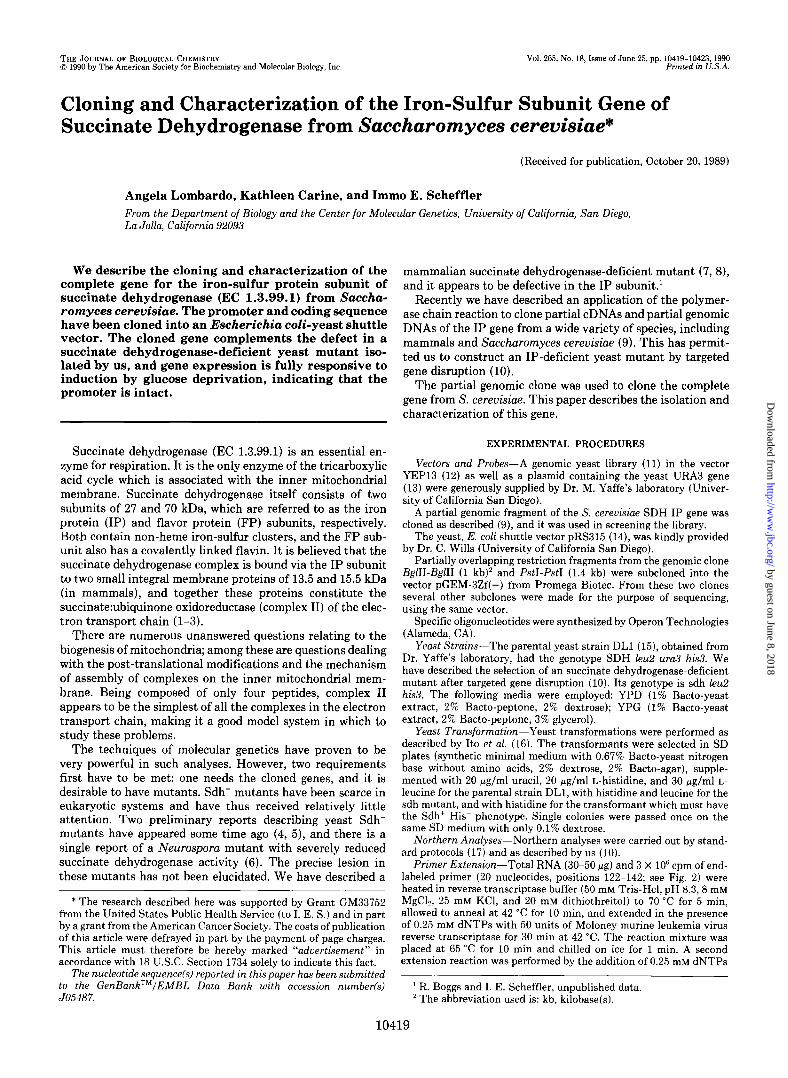
Mapping the Transcription Start Site-Both nuclease pro- tection and primer extension assays were performed to estab- lish the precise start of transcription from the IP gene. The results from a nuclease protection experiment are shown in Fig. 5A. Three major bands of 0.143,0.147, and 0.158 kb were protected, placing transcription initiation at approximately -42, -46, and -57 nucleotides from the start codon. These
by guest on June 8, 2018http://w
ww
.jbc.org/D
ownloaded from
10422 S. cerevisiae Succinate Dehydrogenase Iron-Sulfur Gene
A B
Kb AGCT
.142 -
FIG. 5. Mapping of the 5’ end of the succinate dehydrogen- ase IP mRNA. Panel A, ribonuclease protection assay as described under “Experimental Procedures.” An end-labeled 1-kb DNA ladder was used as the standard. Panel B, primer extension assay as de- scribed under “Experimental Procedures.” The right side of the panel shows the sequencing reaction carried out on the BglII-BglII fragment (Fig. 2) using the same primer.
ORIGIN
CRRONOSOMR
IV, VII
xv
XVI, XIII
II, XIV, x
xl
v, vm h- -1,
IX
III VI I
FIG. 6. Mapping of the SDH IP gene on chromosome VII by hybridization to yeast chromosomes separated by pulsed field electrophoresis. The left lane was probed with an succinate dehy- drogenase IP fragment and the right lane with a URA3 fragment.
bands were absent from the tRNA controls. Fig. 5B shows the results of a primer extension experiment, which allowed the precise mapping of the transcription start to the single nu- cleotide level. Three major start sites were confirmed at positions -44 (G), -50 (A), and -59 (G) relative to the start codon, or 73, 67, and 58 nucleotides downstream from the TATAA box.
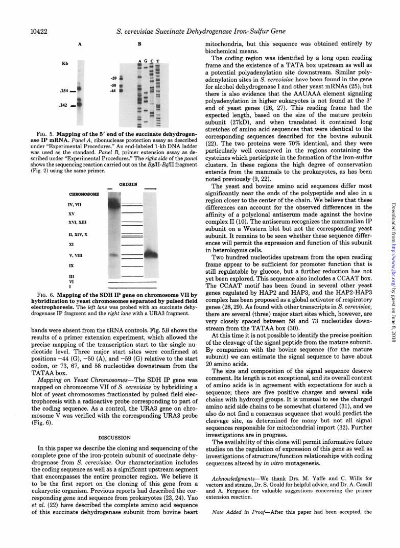
Mapping on Yeast Chromosomes-The SDH IP gene was mapped on chromosome VII of S. cereuisiae by hybridizing a blot of yeast chromosomes fractionated by pulsed field elec- trophoresis with a radioactive probe corresponding to part of the coding sequence. As a control, the URA3 gene on chro- mosome V was verified with the corresponding URA3 probe (Fig. 6).
DISCUSSION
In this paper we describe the cloning and sequencing of the complete gene of the iron-protein subunit of succinate dehy- drogenase from S. cereuisiae. Our characterization includes the coding sequence as well as a significant upstream segment that encompasses the entire promoter region. We believe it to be the first report on the cloning of this gene from a eukaryotic organism. Previous reports had described the cor- responding gene and sequence from prokaryotes (23,24). Yao et al. (22) have described the complete amino acid sequence of this succinate dehydrogenase subunit from bovine heart
mitochondria, but this sequence was obtained entirely by biochemical means.
The coding region was identified by a long open reading frame and the existence of a TATA box upstream as well as a potential polyadenylation site downstream. Similar poly- adenylation sites in S. cerevisiae have been found in the gene for alcohol dehydrogenase I and other yeast mRNAs (25), but there is also evidence that the AAUAAA element signaling polyadenylation in higher eukaryotes is not found at the 3’ end of yeast genes (26, 27). This reading frame had the expected length, based on the size of the mature protein subunit (27kD), and when translated it contained long stretches of amino acid sequences that were identical to the corresponding sequences described for the bovine subunit (22). The two proteins were 70% identical, and they were particularly well conserved in the regions containing the cysteines which participate in the formation of the iron-sulfur clusters. In these regions the high degree of conservation extends from the mammals to the prokaryotes, as has been noted previously (9, 22).
The yeast and bovine amino acid sequences differ most significantly near the ends of the polypeptide and also in a region closer to the center of the chain. We believe that these differences can account for the observed differences in the affinity of a polyclonal antiserum made against the bovine complex II (10). The antiserum recognizes the mammalian IP subunit on a Western blot but not the corresponding yeast subunit. It remains to be seen whether these sequence differ- ences will permit the expression and function of this subunit in heterologous cells.
Two hundred nucleotides upstream from the open reading frame appear to be sufficient for promoter function that is still regulatable by glucose, but a further reduction has not yet been explored. This sequence also includes a CCAAT box. The CCAAT motif has been found in several other yeast genes regulated by HAP2 and HAP3, and the HAPP-HAP3 complex has been proposed as a global activator of respiratory genes (26,29). As found with other transcripts in S. cerevisiae, there are several (three) major start sites which, however, are very closely spaced between 56 and 73 nucleotides down- stream from the TATAA box (30).
At this time it is not possible to identify the precise position of the cleavage of the signal peptide from the mature subunit. By comparison with the bovine sequence (for the mature subunit) we can estimate the signal sequence to have about 20 amino acids.
The size and composition of the signal sequence deserve comment. Its length is not exceptional, and its overall content of amino acids is in agreement with expectations for such a sequence; there are five positive charges and several side chains with hydroxyl groups. It is unusual to see the charged amino acid side chains to be somewhat clustered (31), and we also do not find a consensus sequence that would predict the cleavage site, as determined for many but not all signal sequences responsible for mitochondrial import (32). Further investigations are in progress.
The availability of this clone will permit informative future studies on the regulation of expression of this gene as well as investigations of structure/function relationships with coding sequences altered by in vitro mutagenesis.
Acknowledgments-We thank Drs. M. Yaffe and C. Wills for vectors and strains, Dr. S. Gould for helpful advice, and Dr. A. Cassill and A. Ferguson for valuable suggestions concerning the primer extension reaction.
Note Added in Proof-After this paper had been accepted, the
by guest on June 8, 2018http://w
ww
.jbc.org/D
ownloaded from

S. cerevisiae Succinate Dehydrogenase Iron-Sulfur Gene 10423
cloning and sequencing of the human IP subunit was described by 16. Ito, H., Fukuda, Y., Murata, K., and Kimura, A. (1983) J. Bac- Kita, K., Oya, H., Gennis, R. B., Ackrell, B. A. C., and Kasahara, M. teriol. 153, 163-168 (1990) Biochem. Biophvs. Res. Commun. 166, 101-108. 17. Ausubel, F. M., Brent, R., Kingston, R. E., Moore, D. D., Seidman,
1.
2. 3.
4.
5. 6.
7.
8.
9.
10.
11.
12.
13.
14. 15.
REFERENCES
Hate& Y., Galante, Y. M., Stiggal, D. L., and Ragan, C. I. (1979) Methods Enzymal. 56,577~602.
Hatefi. Y. (1985) Annu. Reu. Biochem. 54.1015-1069 Singer; T. P., and Johnson, M. K. (1985) PEBS Z&t. 190, 189-
198 De Kok, J., Muller, J. L. M., and Slater, E. C. (1975) Biochim.
Biophys. Acta 387,441-450 Ciriacy, M. (1977) Mol. Gen. Genet. 154,213-220 Edwards, D. L., Belsole, D. M., Guzik, H. J., and Unger, B. W.
(1979) J. Bacterial. 137,900-904 Soderberg, K., Ditta, G. S., and Scheffler, I. E. (1977) Cell 10,
697-706 Mascarello, J. T., Soderberg, K., and Scheffler, I. E. (1980)
Cytogenet. Cell Genet. 28, 121-135 Gould, S. J., Subramani, S., and Scheffler, I. E. (1989) Proc. Natl.
Acad. Sci. U. S. A. 86,1934-1938 Lombardo, A., and Scheffler. I. E. (1989) J. Biol. Chem. 264.
18873-18877 Jensen, R., Sprague, G. F., Jr., and Herskowitz, I. (1983) Proc.
Natl. Acad. Sci. U. S. A. 80.3035-3039 Broach, J. R., Strathern, J. W., and Hicks, J. B. (1979) Gene
(Am&.) 8, 121-133 Chevalier, M-R., Bloch, J-C., and Lacroute, F. (1980) Gene
(Am&.) 11, 11-19 Sikorski, R. S., and Hieter, P. (1989) Genetics 122, 19-27 van Loon, A. P. G. M., van Eijk, E., and Grivell, L. A. (1983)
EMBO J. 2,1765-1770
J. G., Smith. J. A.,.and Struhl, K. (1988) Current Protocols in Molecular Biolagv. Wilev Interscience. New York
18. Hase, T., Muller, U., Riezman, H., and Schatz, G. (1984) EMBO J. 3,3157-3164
19. Wills, C., Martin, T., and Melham, T. (1986) Arch. Biochem. Biophys. 246,306-320
20. Crver. D. R.. Eccleshal. R.. and Marmur. J. (1975) Methods Cell hi. 12,39-44
21. Benton. W. D.. and Davis. R. W. (1977) Science 196.180 22. Yao, Y, Wakabayashi, S.; Matsuda, S:, Matsubara,‘H., Yu, L.,
and Yu, C.-A. (1987) in Iron-Sulfur Protein Research (Matsu- bara, H., Katsube, Y., and Wada, K., eds) pp. 240-244, Sprin- ger-Verlag, New York
23. Cole, S. T., Grundstrom, T., Jaurin, B., Robinson, J. J., and Weiner, J. H. (1982) Eur. J. Biochem. 126, 211-216
24. Darlison, M. G., and Guest, J. R. (1984) Biochem. J. 223, 507- 517
25. Bennetzen, J. L., and Hall, B. D. (1981) J. Bial. Chem. 257, 3018-3025
26. Henikoff, S., and Cohen, E. H. (1984) Mol. Cell. Biol. 4, 1515- 1520
27. Zaret, K. S., and Sherman, F. (1984) J. Mol. Biol. 177, 107-135 28. Keng, T., and Guarente, L. (1987) Proc. Natl. Acad. Sci. U. S. A.
84,9114-9117 29. Chodosh, L. A., Olesen, J., Hahn, S., Baldwin, A. S., Guarente,
L., and Sharp, P. A. (1988) Cell 53, 25-35 30. Struhl, K. (1987) Cell 49,295-297 31. Roise, D., and Schatz, G. (1988) J. Biol. Chem. 263,4509-4511 32. Hendrick, J. P., Hodges, P. E., and Rosenberg, L. E. (1989) Proc.
Natl. Acad. Sci. U. S. A. 86,4056-4060
by guest on June 8, 2018http://w
ww
.jbc.org/D
ownloaded from

A Lombardo, K Carine and I E Schefflerdehydrogenase from Saccharomyces cerevisiae.
Cloning and characterization of the iron-sulfur subunit gene of succinate
1990, 265:10419-10423.J. Biol. Chem.
http://www.jbc.org/content/265/18/10419Access the most updated version of this article at
Alerts:
When a correction for this article is posted•
When this article is cited•
to choose from all of JBC's e-mail alertsClick here
http://www.jbc.org/content/265/18/10419.full.html#ref-list-1
This article cites 0 references, 0 of which can be accessed free at
by guest on June 8, 2018http://w
ww
.jbc.org/D
ownloaded from