combined pet/mri: global warming—summary report of … · introduction the 6th annual positron...
TRANSCRIPT

Mol Imaging Biol (2018) 20:4Y20DOI: 10.1007/s11307-017-1123-5* The Author(s), 2017. This article is an open access publicationPublished Online: 2 October 2017
REVIEW ARTICLE
Combined PET/MRI: Global Warming—SummaryReport of the 6th International Workshopon PET/MRI, March 27–29, 2017, Tübingen,GermanyD. L. Bailey,1 B. J. Pichler,2 B. Gückel,3 G. Antoch,4 H. Barthel,5 Z. M. Bhujwalla,6 S. Biskup,7
S. Biswal,8 M. Bitzer,9 R. Boellaard,10 R. F. Braren,11 C. Brendle,12 K. Brindle,13,14 A. Chiti,15,16
C. la Fougère,17 R. Gillies,18 V. Goh,19,20 M. Goyen,21 M. Hacker,22 L. Heukamp,23
G. M. Knudsen,24 A. M. Krackhardt,25 I. Law,26 J. C. Morris,27 K. Nikolaou,3 J. Nuyts,28
A. A. Ordonez,29 K. Pantel,30 H. H. Quick,31,32 K. Riklund,33 O. Sabri,5 B. Sattler,5
E. G. C. Troost,34,35,36,37 M. Zaiss,38 L. Zender,39 Thomas Beyer40
1Department of Nuclear Medicine, Royal North Shore Hospital, and Faculty of Health Sciences, University of Sydney, Sydney, Australia2Werner Siemens Imaging Center, Department of Preclinical Imaging and Radiopharmacy, Eberhard-Karls-Universität, Tübingen,Germany
AbstractThe 6th annual meeting to address key issues in positron emission tomography (PET)/magneticresonance imaging (MRI) was held again in Tübingen, Germany, from March 27 to 29, 2017.Over three days of invited plenary lectures, round table discussions and dialogue boarddeliberations, participants critically assessed the current state of PET/MRI, both clinically and asa research tool, and attempted to chart future directions. The meeting addressed the use of PET/MRI and workflows in oncology, neurosciences, infection, inflammation and chronic painsyndromes, as well as deeper discussions about how best to characterise the tumourmicroenvironment, optimise the complementary information available from PET and MRI, andhow advanced data mining and bioinformatics, as well as information from liquid biomarkers(circulating tumour cells and nucleic acids) and pathology, can be integrated to give a morecomplete characterisation of disease phenotype. Some issues that have dominated previousmeetings, such as the accuracy of MR-based attenuation correction (AC) of the PET scan, werefinally put to rest as having been adequately addressed for the majority of clinical situations.Likewise, the ability to standardise PET systems for use in multicentre trials was confirmed, thusremoving a perceived barrier to larger clinical imaging trials. The meeting openly questionedwhether PET/MRI should, in all cases, be used as a whole-body imaging modality or whether inmany circumstances it would best be employed to give an in-depth study of previously identifieddisease in a single organ or region. The meeting concluded that there is still much work to bedone in the integration of data from different fields and in developing a common language for allstakeholders involved. In addition, the participants advocated joint training and education forindividuals who engage in routine PET/MRI. It was agreed that PET/MRI can enhance ourunderstanding of normal and disrupted biology, and we are in a position to describe the in vivo
Correspondence to: Thomas Beyer; e-mail: [email protected]

nature of disease processes, metabolism, evolution of cancer and the monitoring of response topharmacological interventions and therapies. As such, PET/MRI is a key to advancing medicineand patient care.
Key words: PET/MRI, MR-PET, Hybrid imaging, Molecular imaging, PET/CT, PET, MRI,Quantification, Infection, Inflammation, Oncology, Neurology, Multi-parametric imaging
3Department of Diagnostic and Interventional Radiology, University of Tübingen, Tübingen, Germany4Department of Diagnostic and Interventional Radiology, Medical Faculty, University Dusseldorf, 40225, Dusseldorf, Germany5Department of Nuclear Medicine, University Hospital Leipzig, Leipzig, Germany6Division of Cancer Imaging Research, Department of Radiology, The Johns Hopkins University School of Medicine, Baltimore, MD,21205, USA7Praxis für Humangenetik Tübingen, Paul-Ehrlich-Str. 23, 72076, Tübingen, Germany8Molecular Imaging Program at Stanford (MIPS) and Bio-X, Department of Radiology, Stanford University School of Medicine, Stanford,CA, USA9Department of Internal Medicine I, Eberhard-Karls University, Tübingen, Germany10Department of Nuclear Medicine and Molecular Imaging, University of Groningen, University Medical Center Groningen, Groningen,The Netherlands11Institute of Diagnostic and Interventional Radiology, Klinikum rechts der Isar, Technische Universität München, Munich, Germany12Diagnostic and Interventional Neuroradiology, Department of Radiology, Eberhard Karls University, Hoppe-Seyler-Straße 3, 72076,Tübingen, Germany13Cancer Research UK Cambridge Institute, Li Ka Shing Centre, Robinson Way, Cambridge, CB2 0RE, UK14Department of Biochemistry, University of Cambridge, Tennis Court Road, Cambridge, CB2 1GA, UK15Department of Biomedical Sciences, Humanitas University, Milan, Italy16Department of Nuclear Medicine, Humanitas Research Hospital, Milan, Italy17Department of Radiology, Nuclear Medicine and Clinical Molecular Imaging, Eberhard-Karls-Universität, Tübingen, Germany18Department of Cancer Imaging and Metabolism, H. Lee Moffitt Cancer Center and Research Institute, Tampa, FL, 33621, USA19Cancer Imaging, School of Biomedical Engineering & Imaging Sciences, King’s College London, London, UK20Department of Radiology, Guy’s & St Thomas’ Hospitals London, London, UK21GE Healthcare GmbH, Beethovenstrasse 239, Solingen, Germany22Division of Nuclear Medicine, Department of Biomedical Imaging and Image-Guided Therapy, Medical University of Vienna, Vienna,Austria23New Oncology GmbH, Köln, Germany24Neurobiology Research Unit, Rigshospitalet and Faculty of Health and Medical Sciences, University of Copenhagen, Copenhagen,Denmark25III. Medical Department, Klinikum rechts der Isar, Technische Universität München, Munich, Germany26Department of Clinical Physiology, Nuclear Medicine and PET, Rigshospitalet, University of Copenhagen, Copenhagen, Denmark27Knight Alzheimer Disease Research Center, Washington University School of Medicine, St Louis, MO, USA28Nuclear Medicine & Molecular Imaging, KU Leuven, Leuven, Belgium29Department of Pediatrics, Center for Infection and Inflammation Imaging Research, Johns Hopkins University School of Medicine,Baltimore, MD, USA30Institute of Tumor Biology, University Medical Center Hamburg-Eppendorf, Hamburg, Germany31High Field and Hybrid MR Imaging, University Hospital Essen, Essen, Germany32Erwin L. Hahn Institute for MR Imaging, University of Duisburg-Essen, Essen, Germany33Department of Radiation Sciences, Umea University, Umea, Sweden34OncoRay—National Center for Radiation Research in Oncology, Dresden, Germany35Institute of Radiooncology—OncoRay, Helmholtz-Zentrum Dresden-Rossendorf, Dresden, Germany36Department of Radiotherapy, University Hospital Carl Gustav Carus and Medical Faculty of Technische Universität Dresden, Dresden,Germany37German Cancer Consortium (DKTK), Partner Site Dresden, Dresden, Germany38High Field Magnetic Resonance, Max Planck Institute for Biological Cybernetics, Tübingen, Germany39Department of Internal Medicine VIII, University Hospital Tübingen, Tübingen, Germany40QIMP Group, Center for Medical Physics and Biomedical Engineering General Hospital Vienna, Medical University Vienna, 4L,Waehringer Guertel 18-20, 1090, Vienna, Austria
Bailey D.L. et al.: Combined PET/MRI: Global Warming 5

IntroductionThe 6th annual positron emission tomography (PET)/magnetic resonance imaging (MRI) workshop in theuniversity town of Tübingen, Germany, was held overMarch 27–29, 2017. The initial Tübingen workshop in2012 was the first of its kind to be specifically devoted toaddressing methodological, clinical and research aspects ofhybrid imaging using PET/MRI [1]. Attendees at the currentworkshop came from virtually all continents with themajority originating from Europe and only one in fivehaving attended past workshops.
In the time since the previous workshop of 2016, the useof PET/MRI has continued to expand at a similar rate as ithas done since its introduction [1–5]. To say this in adifferent way would be that the installation rate of new PET/MRI systems has not been as remarkable as that previouslywitnessed with the initial introduction of PET/X-ray com-puted tomography (CT). Instead, barriers to installing PET/MRI remain significant: the capital cost of the equipment,high recurrent operating costs, lack of appropriately trainedstaff to operate and interpret the PET/MRI studies and thelack of an evidence base demonstrating a proven role for thisform of hybrid imaging in clinical use. The past 12 monthshave, however, seen accelerating expansion in Asia inparticular, and hence, the use of the term global warmingfor this year’s summary descriptor to capture the mood ofthe workshop as PET/MRI is now truly a global tool withincreasing use, especially clinically, thus reflective of ageneral Bwarming^ of the imaging community to the valueof the technology. Another factor aiding the continuingincrease in acceptance of PET/MRI is that the design of thesystems, which originally came in a variety of configura-tions, appears now to have settled on one in which the twomodalities are fully integrated in a single gantry thuspermitting simultaneous acquisitions with both modalities.
At times, debate at previous Tübingen workshops wasdominated by issues that were considered to be technicallydeficient or compromised in the early systems, such as howto use MRI-based image sequences to produce appropriatecorrection maps to compensate for photon attenuation in thePET images [2, 3]. As long as issues such as these remainedunanswered, it was difficult to focus on developing futureconcepts for maximally exploiting PET/MRI. It was agreedat this workshop that this particular issue was finally solvedto a level of accuracy sufficient for the majority of clinicalapplications and/or to a level comparable to that seen inPET/CT, i.e. overall uncertainties seen with PET/MRI are noworse than those seen in PET/CT and were considered to beclinically acceptable. One participant during the Physics andInstrumentation dialogue board even called the topic ofMRI-based attenuation correction a Bcase closed^, if only forapplications of PET/MRI of the adult brain in the intactskull. Yet, it needs to be acknowledged that as with PET/CT,a careful inspection of PET/MRI image quality andquantification remains warranted. Hence, it was noticeable
at this year’s meeting that more discussion was devoted tofuture applications of PET/MRI than previously, with adesire to begin Bgoing deeper^ into the interrogation of theinformation contained in the images.
As in the previous meetings’ reports, we will attempt toprovide succinct summaries of highlight lectures, dialogueboards and major outcomes of the discussion boards.Likewise, we will highlight progress achieved, or lackthereof, in specific areas. Finally, we will adhere again tothe general conventions of previous reports to indicateprogress (↑), steady state (↔) and regression (↓) in keyaspects of PET/MRI. The key to the summary tables ofchanges in PET/MRI with respect to the status of theprevious year is shown in Table 1.
Finally, as the style of the lectures and discussions wasslightly changed for this year’s workshop in an attempt toventure into detailed discussions of selected applications ofPET/MRI, we will not follow the previous convention ofdetailing new evidence, which has emerged and futurechallenges in each area, but rather we will attempt to capturethe recurring themes that emerged during the discussions.
Highlight LecturesHighlight Lecture 1: PET/MRI Workflow
The 2017 meeting started with an invited presentation on theconsiderations and challenges when developing optimisedworkflows for PET/MRI. There are a number of keyconsiderations for operating a PET/MRI system clinically.First, the economics/logistics are much more challengingthan for PET/CT. Second, PET/MRI scans are far moredemanding on readers. Finally, the scans are more expensivethan PET/CT. The lecture suggested some niche clinicalroles for PET/MRI and compared it to stand-alone imagingwith the other modalities (Table 2).
It was suggested that the workflows that have beendeveloped for PET/MRI in neurology and cardiology arestraightforward [6]. A simple neurological PET/MRI exam-ination with 2-deoxy-2-[18F]fluoro-D-glucose ([18F]FDG)can be completed in 20 min, whilst a comprehensive multi-parametric protocol using a F-18-labelled amyloid ligand forthe assessment of dementia might require at least 45 min;however, all imaging is captured in a single session.Similarly, in cardiology, a complete examination can beachieved in around 45 min, as the imaging of a single organis ideally suited to PET/MRI. An example of the comple-
Table 1. Key to current status of PET/MRI
↑ Documented evidence of improvement in science and methodology↗ Suggestion of improvement in methodology, but requires further
investigation↔ No change, but satisfactory status since previous workshop↘ Little advancement in science and methodology despite previous
recognition of need for improvement↓ Less clear evidence than previously suggested
6 Bailey D.L. et al.: Combined PET/MRI: Global Warming
mentary nature of PET and MRI in cardiology would be touse [18F]FDG to assess myocardial viability whilst usingMRI to assess perfusion and other parameters (wall motion,etc) [3], or to use absolute PET perfusion measurements inconjunction with scar tissue evaluation from late enhance-ment MRI.
The workflows in oncology, however, are ratherchallenging and it may be that for adult patients PET/MRI imaging could be restricted to studying primary(target) lesions, a single body region (e.g., thorax,abdomen, pelvis) or organ (e.g., brain, heart, liver,pancreas) using BPET/CT guidance^. As a consequenceof lengthy MR protocols resulting in longer availabletime to acquire the PET scan, the amount of radioactivityadministered could be reduced (and hence decreaseradiation exposure to the subject), or the adoption ofcontinuous list-mode dynamic acquisitions and subse-quent modelling and parametric PET image generation.As one of the main roles for PET/CT in oncology todayis in staging the extent of disease for metastasis (M) andnodal spread (N)—and less so for primary tumour (T)staging—it may be that the role which emerges for PET/MRI reverts more to characterising the primary tumour,perhaps using non-FDG radiopharmaceuticals. For exam-ple, one could conceive of a protocol where a dynamicPET/MRI scan is acquired from injection of theradiopharmaceutical up to 30–60 min over the regioncontaining the primary tumour, to capture all of the MRIsequences of interest and the PET kinetics of uptake inthe primary lesion, and then the patient is taken for aconventional whole-body staging PET/CT scan com-mencing 60–90 min after injection to complete themetastasis and nodal evaluation. This concept has beenproposed as early as 2009 by Hicks and Lau [7].Reiterating discussions from previous Tübingen work-shops, the use of DWI-MRI in patients with [18F]FDGavid primaries and lymph nodes was considered obsolete[4]; however, MRI, including DWI, expresses a highsensitivity and specificity for small liver lesions eventhough the [18F]FDG-PET may be negative.
Highlight Lecture 2: Image-Guided Radiotherapy
The second highlight lecture was dedicated to the valueof image-guided radiation therapy. Over the past years,wide-bore PET/MR systems that can serve the demandsof the dedicated radiotherapy equipment have beendesigned and are being used. In this lecture, the valueof [18F]FDG-PET for individualised target volume
delineation, e.g. in lung cancer patients, was brieflyreviewed. Moreover, the use of non-[18F]FDG radio-tracers in radiation treatment planning (RTP) andmonitoring of response was emphasised. Clinically, theability to monitor hypoxia and re-oxygenation duringcombined radio-chemotherapy offers the radiation oncol-ogist greater insight into potential response duringtreatment, which should ultimately lead to betteroutcomes.
Prospective clinical studies personalising treatment on thebasis of hypoxia-PET readout are being designed or havestarted patient accrual. 3′-Deoxy-3′-[18F]fluorothymidine([18F]FLT) depicting another relevant tumour characteristicin radiotherapy, i.e. tumour cell proliferation, was shown tobe of predictive value in head and neck squamous cellcarcinoma patients; however, the limited availability of thetracer has hindered its wider adoption in larger clinical trialsand ultimately clinical routine. In theory, [18F]FDG-PET, thehypoxia PET-tracers [18F]fluoromisonidazole ([18F]FMISO)or 3-[18F]fluoro-2-(4-(2-nitro-1H-imidazol-1-yl)methyl)([18F]HX4), and [18F]FLT-PET may be of additive value indefining the biological target volume. Furthermore, theinternational phase II clinical BPET BOOST^ study inadvanced stage non-small cell lung cancer (ClinicalTrials.govidentifier: NCT01024829) was mentioned, in which anenhanced Bboost dose^ is delivered to either the [18F]FDG-PET avid tumour sub-volumes or to the entire gross tumourvolume whilst maintaining the dose to the surrounding organsat risk (NCT01024829). Moreover, PET/MR imaging isincreasingly being utilised as an objective measure for normaltissue toxicity, e.g. radiation-pneumonitis in lung cancerpatients [8] or neurocognitive decline in primary brain tumourpatients, or as an indirect indicator of primary tumour response,e.g. in HNSCC [9]. A final word of caution addressed thenecessity of geometrically accurate MR images in the era ofimage-guided high-precision radiotherapy.
In conclusion, PET/MRI appears to have a number ofpotential uses in radiotherapy, including the adoption ofintegrated functional imaging to monitor response andadapt during treatments when significant changes areobserved (Fig. 1). However, cross-specialty studies areneeded that specifically employ radiotracers other thanstandard [18F]FDG for both the primary tumour as wellas normal tissue.
Highlight Lecture 3: Imaging the TissueMicroenvironment
The final highlight lecture presented a tour de forcereview of fundamental discoveries related to cancer atthe cellular level which has been suggested as a futuretopic of interest for advanced PET/MRI: the assessmentof tumour microenvironment. It was proposed that PET/MRI might be able to examine similar features seen inthe tumour microenvironment scaled up to the whole
Table 2. Suggested current clinical roles for stand-alone CT, PET and MRI(courtesy of A Beer, Würzburg)
CT Robust, rapid whole-body assessmentPET BProblem solving^ whole-body toolMRI BProblem solving^ specific regional imaging tool
Bailey D.L. et al.: Combined PET/MRI: Global Warming 7

organ or organism level. Some of the parametersdiscussed could be implemented already today, sincethey are based on existing imaging biomarkers, such ashypoxia PET agents (e.g. [18F]FMISO) or the imaging ofcollagen fibres, that can act as a Bhighway^ along whichcancer cells migrate, using diffusion MRI.
Some studies of cancer cell migration have suggestedfocal adhesion kinase (FAK) as a potential therapeutic targetin solid carcinomas. FAK is an intracellular tyrosine kinaserecruited to sites of integrin clustering or focal adhesions andis a multi-functional regulator of cell signalling within thetumour microenvironment. FAK is a major mediator ofsignal transduction by cell surface receptors includingintegrins, cytokine receptors and growth factors and couldeven potentially be used to inhibit cells migrating byadhesion. Based on preclinical evidence presented duringthis lecture, a FAK imaging biomarker would be useful tostudy this effect in vivo in combination with MRI sequencesof apparent diffusion coefficient (ADC) (collagen distribu-tion), pH and hypoxia, so as to better our understanding ofthe tumour microenvironment.
The tissue macroenvironment and its relationship tocachexia in cancer patients was also reviewed. Magneticresonance spectroscopy (MRS) of lipids and cholesterol inserum has been shown to be prognostic for cachexia andbuilds on the idea of a tumour metabolic Bsecretome^ incachexia which could be used for prognostication and/ormonitoring response to treatment using a holistic imagingapproach with PET/MRI.
In light of recent advances in theranostic imaging, arole for PET/MRI was suggested looking at targetedprodrug detection and its action in tumours such as theconversion of 5-fluorocytosine (5-FC) to the active anti-cancer drug 5-fluorouracil (5-FU) in prostate cancertreatment, and the prospect of studying the effects ofphoto-immunotherapy [10]. These approaches go farbeyond the capabilities of PET/CT and demonstratepotential unique future applications for PET/MRI.
Dialogue Boards
Dialog Board 1: Physics and Instrumentation
One of the main topics which has dominated discussions inpast workshops has been the accuracy of attenuationcorrection (AC) using MR-based techniques. A number ofstrategies have been investigated, resulting in over 250publications over the past several years. Attenuation due tothe MR hardware alone can account for up to 20 % signalloss in the reconstructed PET image [11–13]. Nevertheless,there was general agreement between the participants in thisDialogue Board, and many clinical users, that this issue canbe put to rest since MR-AC has been solved to the degreerequired for clinical use based on accepted image metrics.Sequences, such as ultra-short echo time (UTE) and zeroecho time (ZTE), now provide sufficient information toidentify bone or ancillary positioning aids and coils to aid inbuilding an accurate attenuation map of the head for PET/MR neuroapplications [14]. Truncation of the body at theperiphery due to the limited size of the MRI field-of-viewcan been mitigated with the B0 homogenization usinggradient enhancement (BHUGE^) correction [15];implementations of this type or alternative solutions varybetween the manufacturers.
Of note, the variety of available algorithms for MR-based attenuation correction challenges the clinicalreaders. Unlike in PET/CT, where a single fast Bpush-button^ whole-body CT scan provides all the necessaryinformation required for AC, similar corrections in PET/MRI resemble a BLEGO construction kit^: multipleBbuilding blocks^ are needed to form a whole-body dataset for attenuation correction in PET/MR: Dixon se-quences for AC of the soft tissues, UTE and ZTEsequences to provide the skull bones, bone models toadd major bones, HUGE or MLAA data to correct fortruncation artefacts, and CT-based templates to correctfor RF coils and the patient table. It is clear that this
Fig. 1. Patient with oropharyngeal carcinoma pre- and post-RT. a [18F]FDG-PET/CT prior to RT. Oral mucosa delineated inpink, clinical target volume (CTV) in red. b [18F]FDG-PET/CT during week 4 of RT. For the analysis, the CTV was subtractedfrom mucosa. ( Courtesy of S. Zschaeck, MD, Charité Berlin)
8 Bailey D.L. et al.: Combined PET/MRI: Global Warming

rather complex approach of PET/MR attenuation correc-tion is prone to errors and requires thorough integrationto provide the Bpush-button^ ease of use, accuracy androbustness of the CT component for AC in PET/CT.
In light of the above discussions, it was agreed thatalthough numerous methods for AC in PET/MRI have beendeveloped and evaluated in various studies, not all of thesedifferent techniques have found their way as productversions into the existing PET/MRI systems. Thus, thePET/MRI community faces the situation that at single sitessome methods are available, whilst others use differentmethods, different vendors and different AC applications.This leads to a certain inhomogeneity when using differentAC methods, as not only one single version of AC exists asis the case for PET/CT. This also emphasises the furtherneed towards standardisation efforts in PET/MRI attenuationcorrection.
The meeting was reminded, however, that the reproduc-ibility of SUVmax measurements with [18F]FDG PET/CT canbe of the order of up to ± 50 % as shown in a study wheresubjects were scanned on two consecutive days [16]. Thesedifferences are due to many factors including biological andinstrumentation components, and small differences in atten-uation correction factors between methods should be viewedin this context.
The ability of PET data and MRI data to be used in theimage reconstruction of the other modality (i.e. MR-informed PET reconstruction, PET-informed MRI recon-struction) has previously been suggested and explored [17].The dialogue board concluded that using the MRI data candefinitely have an impact and improve the spatial resolutionand image quantification of PET images (Fig. 2), but thePET data have not yet provided any discernible improve-ment in the MRI reconstructions due to their poorer spatialresolution. Motion detected in the MRI scans can be used tocorrect for motion in both the MRI and PET data during thereconstruction process [14]. First implementations of thesetechniques are becoming available on commercial PET/MRIsystems. Finally, one further comment on attenuationcorrection was added in that the accuracy of the attenuationcorrection could be further improved using time-of-flightinformation from the PET data, if available, in the MLAAalgorithm [18–20]. This is now being actively pursued withthe emergence of time-of-flight (ToF) PET/MRI systems.Yet, even without MLAA, improved image quality can beachieved by the use of improved ToF in regular ToF OSEMand it was shown that ToF already mitigates MR-basedattenuation correction artefacts [21].
Table 3 summarises the progress made in the areas ofPET/MRI physics and instrumentation. Of note, no such
Fig. 2. Joint PET and MR image reconstruction. Simulated PET and undersampled (5 of 8 coils) T1-w MR data. Firstindependent reconstructions were performed, using least squares reconstruction with joint total variation (TV) prior to MR andML reconstruction with a TV prior to PET. Then, a simultaneous MR and PET reconstruction with the joint TV prior wasperformed. The resolution of the PET image improves a little, whilst the changes to the MR image appear very minor at best(courtesy of J Nuyts, Leuven/BE).
Bailey D.L. et al.: Combined PET/MRI: Global Warming 9

dedicated session was scheduled for the 4th (2015) or 5th(2016) workshops, when these topics were dealt with as partof the clinical topics. Overall, progress was made in all ofthe key areas. As discussed above, the accuracy androbustness of MR-AC methods are well understood, andresearch endeavors are now turning to the validation of newconcepts for multi-parametric imaging, such as the adoptionof an image-derived arterial blood input function [22].
Dialogue Board 2: Oncology—Status Quo
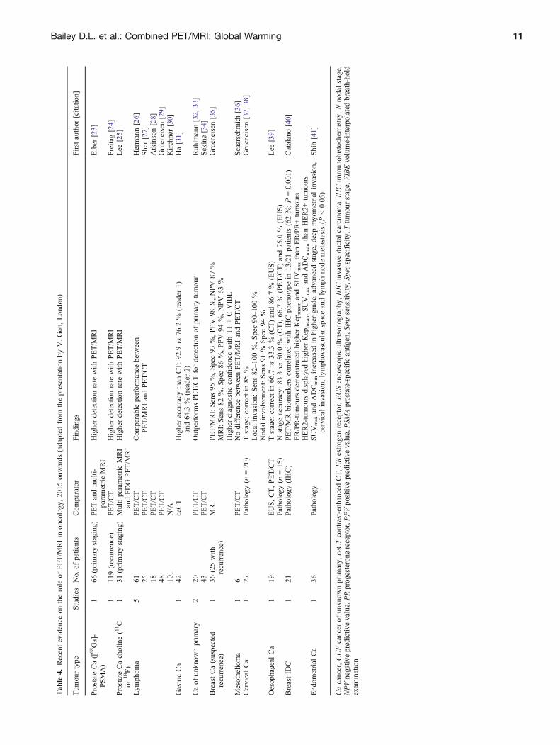
The session started with a review of the current evidence forthe role of PET/MRI in oncology (Table 4). From this, theareas where evidence exists that PET/MRI has beendemonstrated to provide better information and characteri-sation of disease are in prostate cancer [23] and, assuggested previously at these workshops, in paediatriconcology [42]. Areas where new evidence is emerginginclude gastrointestinal cancers [43, 44], breast cancer [40,41], gynaecological malignancies [37] and hepato-pancreatobiliary cancers [45]. The value of PET/MRI inthese applications was seen again to be in a comprehensiveregional evaluation and not in Bwhole-body^ applications; asstated by one of the panellists: BThe added value of PET/MRI starts locally^.
The dialogue board continued with a discussion of PET/MRI tissue characterisation and molecular diagnostics. Itwas felt that modern oncology practice is making its greatestadvances today in DNA sequencing/phenotyping to guidethe use of targeted therapies. This information can betransformed into knowledge about Btumour resistance^,which, it was suggested, is more likely attributable to clonalevolution producing heterogeneity [46]. This message wasreinforced later in the symposium in the dialogue board onemerging areas and discovery (see below). PET/MRI wasreported to be extremely valuable in avoiding samplingerrors during biopsy caused by tumour heterogeneity [47].
Rather than trying to minimise the amount of scanningthat would be performed, it was suggested that primarytumour characterisation for an individual might requiremultiple probes to characterise the individual disease; one
example given was in the evaluation of primary lesions inhepatocellular carcinoma (HCC) using both [18F]FDG and[18F]fluorocholine (FCh). Whereas previously the value of[18F]FDG in HCC has been questioned due to the differentglycolytic pathways that the tumour can access [48] leadingto a negative [18F]FDG signal, it was argued that the bestunderstanding of the tumour biology and heterogeneitywould be attained by performing both scans to betterunderstand the regional phenotype of the heterogeneouscancer cells. This is akin to the approach taken in PETimaging of neuroendocrine tumours using a somatostatinreceptor radiopharmaceutical such as [68Ga]DOTATATEcombined with [18F]FDG [49, 50].
The characterisation of HCC can be regarded as amolecular extension to the concept of radiomics, which hasbeen applied primarily to anatomical image analysis incombination with non-imaging biomarkers. By integratingmolecular imaging information with radiomics, we move toa Bradiomics+^ concept that is one step closer to Bin vivopathology^, which has the potential to become the standardin the near future for oncology patient management.
The theme of multi-parametric imaging and tumourheterogeneity continued in the presentations and discussions.In pancreatic adenocarcinoma, three distinct phenotypeswith different genetic subtypes have been identified thatexhibit different metabolic subtypes following either glyco-lytic or lipogenic pathways, or a combination of both [51]. Anovel technique (Fig. 3) for spatially aligning surgicalresection specimens with the images from PET/MRI wasdescribed using a histopathological processing algorithmbased on a 3-D print mould generated from pre-operativein vivo imaging, enabling accurate co-registration betweenthe excised tissue and the in situ imaging data. A multi-parametric analysis comparing [18F]FDG SUV versus ADCfrom MRI was used to classify heterogeneity and is apotential target area of application for ML-based clusteringof physical tissue compartments.
Table 5 summarises the status quo of PET/MRI inoncology. Generally, the use of PET/MRI for oncologyindications continues to grow and methodological progresshas been made continuously. Unfortunately, the communitystill lacks a set of standardised acquisition protocols(pending the engagement of the medical specialists’ organi-sations), and further efforts are needed to resolve residualbias from MR-AC, the presence of truncation artefacts and,more importantly, motion effects.
Dialogue Board 3: Oncology—Where to Go?
This dialogue board addressed the theme of how PET/MRIcan best complement the latest developments in oncologytherapeutics. As immunotherapy is one of the hottest topicsat present in oncology, discussion centred on whether itcould be improved by the use of functional imagingtechniques. Additionally, the discussants considered how
Table 3. Progress of physics and instrumentation developments since thefirst workshop
Feature 2012 2013 2014 2015 2016 2017
Critical evaluation of MR-ACmethods
↗ ↑ ↑ – – ↑
Validation of MR-based motioncorrection
↔ ↔ ↗ – – ↗
Agreement on acceptable lowerlimits of quantitative accuracyof PET following MR-AC
↘ ↘ ↔ – – ↗
Clinical introduction of advanced,MR-based quantitative parame-ters (e.g. image-derived inputfunctions)
↓ ↘ ↘ – – ↗
10 Bailey D.L. et al.: Combined PET/MRI: Global Warming
Tab
le4.
Recentevidence
ontherole
ofPET/M
RIin
oncology,2015
onwards
(adapted
from
thepresentatio
nby
V.Goh,London)
Tum
ourtype
Studies
No.
ofpatients
Com
parator
Findings
Firstauthor
[citatio
n]
ProstateCa([68Ga]-
PSMA)
166
(primarystaging)
PETandmulti-
parametricMRI
Higherdetectionrate
with
PET/M
RI
Eiber
[23]
1119(recurrence)
PET/CT
Higherdetectionrate
with
PET/M
RI
Freitag[24]
ProstateCacholine(11C
or18F)
131
(primarystaging)
Multi-parametricMRI
andFDG
PET/M
RI
Higherdetectionrate
with
PET/M
RI
Lee
[25]
Lym
phom
a5
61 25 18 48 101
PET/CT
PET/CT
PET/CT
PET/CT
N/A
Com
parableperformance
between
PET/M
RIandPET/CT
Hermann[26]
Sher[27]
Atkinson[28]
Grueneisen[29]
Kirchner[30]
Gastric
Ca
142
ceCT
Higheraccuracy
than
CT:92
.9vs
76.2
%(reader1)
and64.3
%(reader2)
Ha[31]
Caof
unknow
nprim
ary
220 43
PET/CT
PET/CT
OutperformsPET/CTfordetectionof
prim
arytumou
rRuh
lmann[32,
33]
Sekine[34]
BreastCa(suspected
recurrence)
136
(25with
recurrence)
MRI
PET/M
RI:Sens95
%,Spec93
%,PPV
98%,NPV
87%
MRI:Sens82
%,Spec86
%,PPV
94%,NPV
63%
Higherdiagno
stic
confidence
with
T1+CVIBE
Grueneisen[35]
Mesothelio
ma
16
PET/CT
Nodifference
betweenPET/M
RIandPET/CT
Scaarschm
idt[36]
CervicalCa
127
Patho
logy
(n=20)
Tstage;
correctin
85%
Local
invasion
:Sens82–100
%,Spec90–100
%Nod
alinvo
lvem
ent:Sens91
%Spec94
%
Grueneisen[37,
38]
Oesop
hageal
Ca
119
EUS,CT,PET/CT
Patho
logy
(n=15
)Tstage:
correctin
66.7
vs33
.3%
(CT)and86
.7%
(EUS)
Nstageaccuracy:83
.3vs
50.0
%(CT),66
.7%
(PET/CT)and75
.0%
(EUS)
Lee
[39]
BreastID
C1
21Patho
logy
(IHC)
PET/M
Rbiom
arkers
correlated
with
IHCph
enotyp
ein
13/21patients(62%;P=0.001)
ER/PR-tum
ours
demonstratedhigher
Kep
meanandSUVmax
than
ER/PR+tumours
HER2-tumours
displayedhigh
erKep
mean,SUVmax
andADCmeanthan
HER2+
tumou
rs
Catalano[40]
Endom
etrial
Ca
136
Pathology
SUVmax
andADCminincreasedin
high
ergrade,
advanced
stage,
deep
myo
metrial
invasion
,cervical
invasion,lymphovascularspaceandlymph
node
metastasis(P
G0.05
)Shih[41]
Cacancer,CUPcancer
ofun
know
nprim
ary,
ceCTcontrast-enhancedCT,ERestrog
enreceptor,EUSendoscop
icultrason
ograph
y,ID
Cinvasive
ductal
carcinom
a,IH
Cim
munohistochem
istry,
Nno
dalstage,
NPVnegativ
epredictiv
evalue,PRprogesterone
receptor,P
PVpositiv
epredictiv
evalue,PSM
Aprostate-specificantig
en,S
enssensitivity,S
pecspecificity
,Ttumourstage,VIBEvolume-interpolated
breath-hold
exam
ination
Bailey D.L. et al.: Combined PET/MRI: Global Warming 11

the latest developments in relatively non-invasive tumourphenotyping using liquid biopsy could be combined with theresults of imaging of lesion heterogeneity using PET/MRI.Liquid biopsy of circulating tumour cells and DNA ormiRNA is playing an increasing role in screening and earlydetection (especially in recurrence), the detection of micro-metastases and in the monitoring of therapies where they canbe used as an early indication of response from simple bloodsampling [52, 53]. However, it was recognised that blood-based tests present a Bglobal^ picture as changes inphenotype are often observed serially, thus requiringregional identification, indicating an example of whereliquid biopsy and imaging would perform a complementaryrole.
As this dialogue board was addressing the question ofBWhere to Go?^, the role of mouse models and patient-derived xenografts (PDXs) were discussed. Preclinicalimaging with PET/MRI will likely have a role in thesemodels along with bioluminescence techniques. Finally, theunderstanding of the role of tumour-infiltrating T cells wasconsidered and whether labelling T cells with Zr-89 or othernuclides would allow in vivo monitoring to predict response
to immunotherapy (Fig. 4). However, careful selection andmatching of targets, probes and nuclides is required.Especially, functional impairment by direct targeting of Tcells needs to be excluded.
Panel Discussion: Dialogue Boards 2 and3—Oncology
The discussion was kicked off with an upfront statementby one of the panellists who stated Bthe imaging fieldwas disconnected from oncology for years^, therebyattesting to another statement below regarding a fre-quently observed approach towards interpreting resultsfrom imaging examinations in isolation. The panel wasthen asked to respond to the question of where theythought oncological therapy response assessment will bein 10 years’ time. The initial response was that there isincreasing pressure to predict what response might belikely to occur in an individual patient, partly due to thehigh cost of the therapies. The power of predictingpatient-specific therapy response was felt to be of ever-
Fig. 3. Co-registration of imaging and histopathology data. a Work flow schematic. b Pre-operative T2w image with annotatedresection margins. c 3D print mold. f Fixated axially processed specimen with g annotated tissue blocks. h Stitching of tissueslices. i Screenshot of in-house written software for co-registration and regional analysis of imaging and histopathology data(courtesy of Rickmer Braren, Katja Steiger, Franz Irlinger and Maximilian Baust).
Table 5. Progress indicators for PET/MRI in oncology
Feature 2012 2013 2014 2015 2016 2017
Definition of key clinical applications ↔ ↔ ↗ ↗ ↗ ↗Diagnostic quality of PET in PET/MRI equivalent to PET quality in PET/CT ↔ ↔ ↗ ↗ ↗ ↑Resolving quantitative bias from MR-AC ↘ ↔ ↔ ↔ ↗ ↗Clinical data available on diagnostic accuracy of PET(/MRI) in oncology ↔ ↔ ↗ ↔ ↗ ↗PET/MRI protocol standardisation ↓ ↔ ↘ ↔ ↔ ↔Clinical evidence on the usefulness of PET/MRI in paediatric oncology ↔ ↗ ↗ ↗ ↔ ↔Reduced radiation exposure as a key driver for paediatric PET/MRI ↗ ↑ ↑ ↔ ↘ ↘
12 Bailey D.L. et al.: Combined PET/MRI: Global Warming

increasing importance given the evolution of resistanceof solid tumours to successive lines of therapies. It wasmentioned that 95 % of the oncology drugs fail duringphase 1 trials, which can be regarded as an argumenttowards Braising the bar on pre-clinical testing^, i.e.questioning whether animal models are suitable surro-gates for predicting efficacy in humans. It was felt thatimaging, and molecular imaging in particular, will be anincreasingly important tool due to the heterogeneity andevolution seen in disease progression and that ultimatelyimaging could be more important than traditional biopsyin this regard due to its ability to identify evolvingclones. A more careful and systematic validation ofmultiple imaging parameters, as provided by PET/MRI,in the clinical context was considered an important stepbusting acceptance of imaging parameters as bio- andsurrogate markers.
The panel was also asked to speculate as to theappropriate timing of imaging to assess response. Aftersome discussion, with suggestions from Bas soon as24 hours^ after commencing treatment to Bnot before 6–8 weeks^, it was agreed that if the therapy is inducingsenescence [54] then imaging could be used as soon as2 weeks after commencing therapy. One of the panellistssuggested that we still do not have all of the appropriatetracers to study cellular biology to match the advances inoncology today. He suggested that some of the probesneeded to demonstrate tumour stresses during therapy andthe stress support pathways should be able to demonstrateapoptosis, senescence and hypoxia. The forum’s generalresponse was that Bwe have the tracers and we have all thetechnology and tools, but what we do not understand is whatthe signal is telling us at present^. This returned to the themeof better developing multi-parametric analyses and the rolethat predictive analytics may play in this domain; oneattendee summarised it by saying Bwe should stop talking
about the Bimages^, but rather promote the image content asmineable data, that is, as an imaging assay^.
The discussion moved on to looking at pseudo-progression in the light of immunotherapies, which is a realclinical challenge. Thus, imaging strategies need to bedeveloped to directly track T cells within the tumourindicating response to treatment and therefore representinga valuable surrogate marker (Fig. 5). In addition, the abilityto follow-up on treatment response and tumour progressionwith the help of liquid biopsies with or without imaging wasdiscussed. It was concluded that, today, liquid biopsies alonewould not be sufficient to do the job, and that imaging couldadd valuable information especially in therapy responsemonitoring providing information about the change inphenotype of known and the location of evolving(metastatic) disease which may have implications forsubsequently adjusted therapies.
The panel discussion ended with some speculation aboutthe role that functional imaging could take to assist intriaging patients appropriately to targeted therapies. It wasfelt that some education of regulators and the industry wasstill needed to develop a paradigm that would test individualpatients prior to commencing therapy in an attempt tocontain costs and produce better outcomes.
Dialogue Board 4: Neurology—Status Quo
The dialogue board in neurology started by reasserting thatthe issue of AC for PET/MRI in neurology was solved towithin the uncertainties associated with the overall proce-dure for the given modality. The only possible exceptions atthis stage are in the cases of scanning children’s brains usingatlas-based techniques for MR-derived AC or where therehas been prior craniotomy and, therefore, disruption of theskull contour. This has now resulted in progress in analysing
Fig. 4. Selection of target structures on effector T cells as well as probes and nuclides to develop safe and efficient imagingstrategies to track T cells during immunotherapy (courtesy of Sabine Mall and Angela Krackhardt).
Bailey D.L. et al.: Combined PET/MRI: Global Warming 13

the data from brain PET/MRI, as previously researcherswere preoccupied with solving the attenuation correctionproblem.
Presentations from the panellists documented the currentclinical uses of PET/MRI in neurology as being in studies ofdementia using [18F]FDG, neurodegenerative disorders,neuro-oncology, epilepsy, a small number of cases ofcerebrovascular disease and various other diverse conditionsincluding encephalitis and sarcoidosis [56]. The PET/MRIstudies were being used in a variety of ways includingdiagnosis, characterising pathology, demonstrating the ex-tent of neuronal injury and monitoring progression, anddifferent radioligands can be used depending on the focus ofthe examination. The use of imaging biomarkers inAlzheimer’s disease (AD) with PET/MRI can be performedin a 20–30-min session including an early image afterinjection of the a fluorinated amyloid PET tracer to give ap e r f u s i o n / BFDG- l i k e^ imag e ( r e g i on a l b l o odflow/metabolism) that can be compared with the subsequentimage of the binding to cerebral amyloid. Mention was madeof further imaging biomarkers such as the α4β2 nicotinicacetylcholine receptor which can be imaged using [18F]-flubatine [57]. Another promising approach to multi-modality brain imaging, particularly in dementia, utilisesrest state-fMRI in combination with structural MRI and[18F]FDG PET to determine the functional connectivitybetween affected brain areas [58]. Finally, the use of PET/MRI in neurological imaging trials requires the judicious
choice of an AC method (based on MR sequences or othermodalities) that should stay standard for the duration of thestudy. Lastly, it was noted that fully integrated PET/MRI hasproven to be beneficial for fully quantitative brain imaging,including non-/invasive pharmacokinetic modelling.
Discussion of research in rodents using PET/MRI focusedon investigating the connectivity between metabolism (using[18F]FDG), perfusion (using [15O]H2O) and fMRI using theblood oxygen level dependent (BOLD) technique. A numberof interesting results demonstrating both expected correla-tion in metabolism and perfusion in some brain areas as wellas unexpected disconnections between the same parametersin other areas were seen [59]. PET/MRI, operating over themedium to long temporal scale compared to ultra-fasttechniques methods such as electroencephalography (EEG)and magnetoencephalography (MEG), was seen as the onlyway to inves t igate such connect ions ( termedBcometomics^—connectivity via metabolomics).
The great strength of PET/MRI in brain imaging, whetherresearch or clinical, was thought to be the Bone-stop shop^option, where all of the imaging required to characterisepatient disease could be performed in a single session. Thisis also reflected in the continuous progress made in theapplications of PET/MRI for neurology (Table 6). Ofinterest, one of the panellists made a point on theBsimultaneity of PET and MRI, that had no other advantagethan increase patient comfort and convenience^, which mayhave come as a surprise to some PET/MRI users.
Dialogue Board 5: Neurology—Where to Go?
This dialogue board posed the question of whether thepredominant use of brain PET/MRI in the future would befor routine clinical applications or in research. Ampleevidence was provided that there is an increasing roleclinically for brain PET/MRI [6]. The case was made for thesubstitution of imaging biomarkers to objectively diagnosedementia in place of the current syndromic tools whichclinicians are left to rely upon. With potentially expensivetherapies on the horizon in AD, it was felt that regulatorsand medical service providers should be encouraged to useimaging as an objective biomarker to select the appropriate
Fig. 5. Tracking of intravenously injected T-cell receptor(TCR)-transduced central memory T cells within antigen-expressing tumours by Zr-89-labelled aTCRmu-F(ab′)2 usingPET/CT. a TCR-transgenic T cells were injected intrave-nously after tumour engraftment followed by i.v. injection of[89Zr]-labelled aTCRmu-F(ab′)2 48 h after adoptive T-celltransfer. b Heterogeneity of T-cell infiltration, as seen in thezoom in, has been validated by semi-quantitative analysisusing immunohistochemistry [55] (courtesy of Sabine Malland Angela Krackhardt).
Table 6. Progress indicators for PET/MRI in neurology
Feature 2012 2013 2014 2015 2016 2017
Improved understanding of brainphysiology and functionthrough the use of combinedPET/MRI
↔ ↗ ↗ ↗ ↗ ↗
Methodological progress forimproved quantification ofPET/MRI neurological exami-nations (AC, IDIF, SUV)
↔ ↔ ↗ ↗ ↗ ↗
MR-based motion correction forroutine clinical use
↓ ↘ ↔ ↔ ↔ ↗
14 Bailey D.L. et al.: Combined PET/MRI: Global Warming
patients for these therapies as well as to monitor response.On the research side, elegant work using multi-modality
neuroimaging with pharmacological challenges was shownwhich demonstrated that pharmacological PET could beused to study drug penetration and kinetics, to identifypharmacodynamic effects, and could be used in drugoccupancy studies which could be combined with simulta-neous blood flow measurements using arterial spin labelling[60] as well as functional and structural MR imaging [61,62].
Addressing the issue of where to go with future brainPET/MRI studies, the integration of imaging with humangenetics testing was explored. Using next-generation se-quencing technologies, it is now possible to sequencehundreds and even thousands of genes in parallel. Thisdramatically increases the chance to find the cause of thedisease in many heterogeneous neurologic diseases. From adiagnostic point of view, panels of genes that are known tocause a particular disease including all differential diagnosescan be investigated simultaneously with very high coverageand, if negative, whole exome or whole genome sequencingis followed. Next-generation sequencing allows also thedetection of mosaicism in blood and other tissue samples.
Panel Discussion: Dialogue Boards 4 and5—Neurology
The panel discussion on the neuroscience subjects reflectedthe relative maturity and acceptance of PET/MRI in studyingthe brain. The panellists agreed that PET/MRI is aconvenient means to an efficient work-up of patientssuspected of Alzheimer’s disease; however, the addeddiagnostic benefit of fully integrated PET/MRI in thispatient group was considered to be small.
PET/MRI can help increase patient comfort as well asprovide doctors with the option to perform a wide range ofstructural and molecular imaging assessments for diseasecharacterisation and/or monitoring within a single investiga-tion. This is of utmost importance for trials comprising avariety of MRI/fMRI imaging sequences along with exten-sive and complex pharmacokinetic PET investigations. Thepanellists agreed that PET/MRI is capable of providingmolecular imaging-based evidence to support early diagno-sis/staging/disease and therapy monitoring of dementia andother brain disorders in clinical routine. Furthermore, it is apowerful tool in complex research settings of neurosciencecomprising molecular imaging and pharmacokinetic/pharmacodynamic analysis.
In clinical research, a large number of physiologicallyrelevant measurements can now be made simultaneouslyusing PET and MRI including metabolism, neurotransmis-sion, receptor expression, cerebral blood flow/perfusion,tissue environment and pathological conditions (e.g. amyloiddeposition). This opens for investigation of fundamentalphysiological and neurochemical aspects of, e.g. changes in
neurotransmission in relation to blood flow under physio-logical or pharmacological stimulation. Such knowledge canbe instrumental to assess drug effects in the individualpatients, and allows for a precision medicine approach. Thenext challenge seen by the panellists was how to integratethese measurements with genetic fingerprinting to delvemore deeply into describing a more comprehensive pheno-typical classification of disease.
An additional emerging topic might also be the assess-ment of active plaques in demyelinating diseases likemultiple sclerosis. In addition, the combination of PET/MRI with fast temporal scale techniques like EEG mightgive further insight in pathophysiological processes in thebrain, e.g. in epilepsy.
Dialogue Board 6: Infection and Inflammation
The topics of infection and inflammation imaging wereintroduced at last year’s workshop for the first time [5]. Atthe time, an enormous number of targets were presented forpotentially imaging infection and inflammation, but therewere relatively few imaging biomarkers specifically devel-oped to exploit the vast majority of these, and hencediscussion last year centred on adapting well-establishednuclear medicine techniques which have been around formany years using radiolabelled antibiotics, immune cells andantibodies, membrane ligands, antimicrobial peptides, ironmetabolism ([67Ga]-citrate) and metabolic tracers. However,many existing agents fall short of distinguishing infectionfrom sterile inflammation (e.g. FDG). In contrast, at themeeting this year, a number of new compounds werepresented for bacteria-specific PET imaging. Non-invasiveanatomical analysis with MRI plus pathogen-specific PETimaging could significantly improve patient outcomes byrapidly identifying a source of infection and monitoring theresponse to treatment [63].
Imaging of infection has been developed by the Centerfor Infection & Inflammation Imaging Research (Ci3R) atJohns Hopkins University over the past few years. Using asystematic screening approach, they evaluated a largenumber of potential agents for selective bacterial accumula-tion and found ten compounds that could be used asbacteria-specific imaging biomarkers, showing interestingresults with para-aminobenzoic acid (PABA), deoxy-mannitol and deoxy-sorbitol [64]. Among other biomarkers,they presented results using [18F]fluoro-deoxysorbitol([18F]FDS), which can be synthesised from radiolabelledFDG [65]. [18F]FDS is selectively accumulated by gram-negative Enterobacteriaceae such as Escherichia coli,Yersinia, Klebsiella, Enterobacter and Salmonella, includingmulti-drug-resistant organisms. Using a murine myositismodel, [18F]FDS was able to differentiate infection sitesfrom sterile infection in immunocompetent and neutropenicmice, thus suggesting that the uptake is actually by thebacteria rather than by migrating neutrophils. Potential
Bailey D.L. et al.: Combined PET/MRI: Global Warming 15

advantages of imaging active infection include a muchearlier path to diagnosis and instituting appropriate antibiotictherapy rather than waiting for blood cultures (Table 7).
The session then turned to a new area for theworkshop: the imaging of chronic pain and how itsinvestigation could benefit from the use of [18F]FDGand PET/MRI. In many Western societies, morbidity andproductivity loss due to chronic pain constitutes one ofthe largest burdens on society. Chronic pain was said toaffect more people than those suffering from cancer, heartdisease and diabetes combined. The source of chronicpain is often difficult to identify and diagnose withconventional morphological imaging. Bone scanningusing [99mTc]phosphonate has long been known to havea role in identifying the site of the cause of pain reflectedin increased osteoblastic reactivity—the concept ofBwhere it’s hot it hurts^ attributed to Schuster. Similarly,increased PET radiotracer uptake preliminarily appears tomap to areas of increased pain-relevant or pain-generatingpathology, potentially affording clinicians the ability tomake improved image-informed, objective managementdecision to minimise pain in chronic pain sufferers. TheMRI component of PET/MRI is considered essentialbecause of the high spatial resolution and high tissuecontrast which, when combined with the [18F]FDG signal,can identify the exact location of the inflammatoryresponse. As soft tissue is involved in many of thesesyndromes, PET/CT was thought to be of less value dueto the poorer tissue contrast that is usually seen with CTcompared to MRI. Additionally, because PET and MRIdata sets are acquired simultaneously, the fidelity of theco-registration of both data sets is likely more accuratethan what can be achieved with PET/CT. Spatial mis-registration between PET and CT data is a well-knownphenomenon since both data sets are acquired separatelyin time and, thus, small patient movements between thetwo scan acquisitions can result in PET measurementerrors between small adjacent structures such as a nerveroot and a neighbouring facet joint. The ability toaccurately delineate abnormal radiotracer uptake in anerve root versus the facet joint, for example, could bethe difference in a successful outcome since management
decisions are made according to the location of PETabnormality.
[18F]FDG has been used off-label in a clinical trial tostudy patients with complex regional pain syndrome, chronicsciatica and other pain syndromes. As in the discussion ofinfection and inflammation imaging, new imaging bio-markers of specific targets were being developed andcharacterised in human subjects, for example, to identifysigma-1 receptor (σ1R) ligands such as [18F]FTC-146 in thesetting of chronic pain [66, 67]. Sigma-1 receptors, a uniqueclass of intercellular chaperone proteins, have a modulatoryrole in ion channels and other neurotransmitter systems.Accordingly, σ1Rs have been found to be important in pain,inflammation, neuronal protection, neurodegeneration, can-cer, addiction and psychiatric diseases. Early results haveshowed important differences between asymptomatic volun-teers and those suffering from chronic pain.
Dialogue Board 7: Emerging Areas
The final dialogue board of the workshop addressed theissue of advances in multi-parametric imaging. The firstcontribution looked at how it is now possible to investigatemultiple aspects of tumour metabolism by combininghyperpolarised C-13-labelled cell substrates with PETbiomarkers, such as [18F]FDG [68, 69]. Dynamic nuclearpolarisation can increase the signal-to-noise ratio in solutionstate 13C NMR spectroscopy and imaging experiments by910,000× [70]. Fundamental questions such as why cancershave high rates of aerobic glycolysis (Warburg effect) couldpotentially be addressed using 1-[13C]pyruvate and[18F]FDG in animal tumour models. The combination ofmagnetic resonance spectroscopic imaging of hyperpolarised1-[13C]pyruvate, when combined with [18F]FDG to imageglycolysis, and further measures such as hypoxia bio-markers, could provide insight into cancer metabolism andtumour heterogeneity and how various pathways (e.g.HIF1α, MYC, mTOR, PTEN) are involved (Table 8).
The second contribution was on the development ofadaptive therapies and insights into how cancers Bevolve^.Evolution is driven by the genomic plasticity that is inherentto cancers, in combination with microenvironmental selec-tion. As tumours contain multiple microenvironmentalhabitats, these will result in distinct lineages of tumour cellsgenerating intratumoural heterogeneity. Addition of therapychanges the adaptive landscape and selects for cells that areresistant. Notably, these resistant clades of cells are oftencross resistant to other therapies, leading to unmanageabledisease [71]. This has been mathematically modelled toshow that emergence of resistance can be forestalled withadaptive dosing based on tumour response [72]. This ishighly relevant to PET/MRI studies as the monitoring ofresponse becomes a critical factor in adaptive dosing and inpredicting pathways that the emerging resistant cell linesmay exploit. The large number of probes that can be used
Table 7. Progress indicators for PET/MRI in infection and inflammation
2012 2013 2014 2015 2016 2017
Improved tissue characterisationby combined PET/MRI
– – – – ↗ ↗
Development of newradiopharmaceuticals for PETuse in general
– – – – ↗ ↗
Standardise imaging protocols – – – – ↔ ↔Standardise image interpretation
criteria– – – – ↔ ↔
Definition of key clinicalapplications
– – – – ↗ ↔
16 Bailey D.L. et al.: Combined PET/MRI: Global Warming
with PET and MRI should provide tools with which tomonitor the differential response expected from the differentphenotypes of cancer cells present.
The final presentation looked at a promising technique forMRI, known as chemical exchange saturation transfer(CEST) [73, 74] with glucoCEST, which uses the hydroxylgroups of the glucose molecule to study glucose uptake andmetabolism [75]. The glucoCEST imaging procedure usesmillimolar amounts of contrast and demonstrates vascularand extracellular signals and may potentially be able tofollow metabolic products of the agent through variousphases of the Krebs cycle. This could provide an interestingcomparison with [18F]FDG, which is trapped upon entry intothe cell and does not proceed through the metabolic cascade.A further agent which has already been tested in tumourpatients is 3-O-methyl-D-glucose (OMG) [76–78]. Of note,chemical shift selective CEST, e.g. amide proton transfer,works best with very high magnetic field strengths and, thus,is not applicable with current whole-body PET/MRI sys-tems. However, the glucose injection approach employingthe difference of images before and after injection againprovides glucose-related selectivity at clinical field strengthsand lower chemical shift selectivity.
This dialogue board brought out some of the liveliestdiscussions, attesting to the keen interests of the audience inreviewing new, less standard-of-care applications of PET/MRI. A strong desire towards implementing existing MR-only techniques (e.g. CEST imaging, hyperpolarisation) intoPET/MRI was expressed. Whilst progress in conceptualisa-tion of such ideas was evident, practical implementationswere lagging behind, pr imar i ly because theseimplementations are linked to higher investments in person-nel and infrastructure, and as such may be limited to a fewselected sites. In that case, it becomes ever more importantto share new insights into emerging areas in order to ensureexpedited translation into the clinic where and wheneverapplicable.
Round Table DiscussionsRound Table 1: Reading PET/MRI—Who andHow?
Round Table 1 hosted a spirited debate about the best way toreport clinical PET/MRI scans. The panellists representedthe radiological, nuclear medicine and hybrid imaging
communities. From the opening remarks of each, it wasclear that this issue is one that had been prominent in thepanellists’ minds for some time.
The majority of the panellists introduced PET/MRI intheir institutions with clinical scanning reported by dualreaders—an MRI expert and a nuclear medicine expert. Thiswas seen as a sensible approach in the initial start-up phase,but some felt that this would be financially unsustainable inthe long term. All agreed that it was imperative that thepotential of MRI should be fully exploited and not Bdumbeddown^ as some panellists suggested happened to the CTcomponent of PET/CT whenever its use is limited to Blowdose^ mode for anatomical localisation and attenuationcorrection only.
Having accepted that dual reporting was not the future forPET/MRI in the long term, the debate moved to whether adual certified radiology/nuclear medicine-trained individualis appropriate or, in fact, whether a new specialisation as aBhybrid imager^ would be more appropriate. The hybridimager would be someone particularly trained in theinterpretation of multi-parametric image data in an inte-grated, synergistic fashion, rather than as a pair ofcomplementary scans acquired in a single session. Trainingprogrammes will need to be modified to reflect any suchchanges in specialisation which, unfortunately, still happenat a national level even within the European Union. Therespective professional organisations will need to cooperateto produce the best outcome and not revert to trying tosimply Bprotect their patch^.
Eventually, the healthcare environment will need toaccept the statement made by one of the panellists: BHigh-end technology imaging requires high-end reading^; localvariations may apply.
Round Table 2: Imaging Versus Liquid Biopsy
The participants in this round table discussion emphasisedthat liquid biopsy today is able to characterise multiplemutations that may not be present in every individual lesionand, therefore, is not prone to the sampling errors associatedwith conventional tissue biopsy using fine needle aspirationor core biopsy samples. From this point of view, it wassuggested that it was an extremely valuable technology formonitoring the development of resistance in cancers. Liquidbiopsy may point to the use of different imaging biomarkers
Table 8. Progress indicators for PET/MRI for applications in emerging areas
2012 2013 2014 2015 2016 2017
Fully integrated PET/MRI exclusively offers the largest variety of multi-parametric biomarkers ↔ ↗ ↑ ↑ ↑ ↑Validation of advanced multi-parametric biomarkers in clinical research (beyond Bimage fusion^) ↘ ↔ ↗ ↗ ↔ ↔Contributions of small animal imaging to the understanding of multi-parametric biomarkers ↔ ↗ ↑ ↑ ↗ ↗Using standardised approaches for assessing the accuracy of PET/MRI and towards multi-parametric
image analysis– – – ↗ ↔ ↔
Bailey D.L. et al.: Combined PET/MRI: Global Warming 17

if multiple driver mutations are demonstrated in the sample.Panellists pointed to the massive imbalance between therelatively modest amount spent on diagnostic techniquescompared with the high cost of the therapies that are oftendelivered without adequately characterising the most appro-priate pathway to target prior to treatment. One panellistcalled for Bspending more money on tests that help usunderstand disease and tailor much more expensivetherapies.^
It was clear from the discussion that liquid biopsy andimaging are complementary rather than competing technol-ogies. Liquid biopsy is able to identify different phenotypesin cancer cells but remains a global measure, whereasimaging with PET/MRI using appropriate imaging probescan be used to identify differences between lesions due toclonal variation which may have an influence on the choiceof the most appropriate therapy, especially when using aregionally targeted approach such as with radiotherapy ororgan-specific chemotherapy or radioembolisation.
The discussion diverted multiple times onto a topic thatsurfaced repeatedly during this workshop, which is thewealth of data that we have at hand, augmented by imaging-based biomarker information, and that generally goesuntouched. The panel argued that Beven if we do notinterpret all information [aka data] today, we may need thesedata for later^, pointing to the ever-evolving field of big dataand deep learning. Such a data repository would clearlybenefit from wrapping any type of non-imaging (andimaging) data in a structured, DICOM-type manner so asmake them accessible on existing viewing and storagesystems, such as PACS.
ConclusionIn continuation of the discussions of the 5th Tübingenworkshop, this meeting has focussed much more onemerging areas and provided a balance between state-of-the-art practices and strategies (BWhere to go?^) in key areasof application (oncology and neurology). The dialogueboards were complemented with highlight presentations ofpromising applications of PET/MRI, such as RTP and theassessment of the tumour microenvironment, an area thatclearly would benefit from cross-speciality engagementmuch beyond that required for operating a PET/MRIprogramme.
Two interesting take-home messages from this year’smeeting pertinent to the clinical community were as follows:(a) PET/MRI is perhaps better used as a local tumourphenotyping methodology rather than as a competitor towhole-body PET/CT imaging (except for paediatrics), and(b) the simultaneity of PET and MRI as offered by fullyintegrated PET/MRI systems is seen as useful mainly forincreased patient comfort—for the actual use of PET andMRI information, maximisation of stand-alone performanceand quality was understood as key.
In the summary review talk, the theme given to thisyear’s workshop was Bglobal warming^. We have come along way from the 1st workshop in 2012 when the topic wasmainly Bhow does PET/MRI compare with PET/CT^. Overthe years and through the engagement of many avid usersand pioneers, the potential and capabilities of PET/MRI havebeen shaped and are becoming much more apparent.Together, the PET/MRI community has created a space forengagement of many capable individuals and groups. Aglobal team effort is of the essence in turning this potentialinto a reality with real value for the research community andfor patients. Global warming was chosen, because likeclimate change, PET/MRI is now known to many people. Ithas gone global and it has started to warm people up to itsuse.
Acknowledgements. Open access funding provided by Medical University ofVienna. We wish to thank all the participants for their active contribution atthe 6th Tübingen Workshop on PET/MRI and for the lively discussions. Inparticular, we like to acknowledge the contributions from A. Beer (Ulm,Germany), B. Bender (Tübingen, Germany), U. Ernemann (Tübingen,Germany), S. Gatidis (Tübingen, Germany), F. Jansen (GE Healthcare), A.Kalemis (Siemens Healthineers), M. Kean (Melbourne, Australia), O. Ratib(Geneva, Switzerland), M. Reimold (Tübingen, Germany), P. Schirmacher(Heidelberg, Germany), F. Seith (Tübingen, Germany), B. van Beers (Paris,France) and L. Umutlu (Essen, Germany).
Funding Information The workshop was supported by the GermanResearch Foundation (GU 511/5-1, AOBJ 633946). We would further liketo acknowledge the generous support of the workshop sponsors: BayerHealthCare, GE Healthcare, Hermes Medical Solutions, Lilly DeutschlandGmbH, MDPI AG, Mediso Medical Imaging Systems, mim Software,Mirada Medical, Π.pmod Biomedical Image Quantification, RAPIDBiomedical and Siemens Healthineers.
Compliance with Ethical Standards
Conflict of Interest
M Goyen is an employee of GE Healthcare.M Heukamp is with NeoOncology GmbH.
Open Access This article is distributed under the terms of the CreativeCommons At t r i bu t i on 4 .0 In t e rna t i ona l L i c en se (h t t p : / /creativecommons.org/licenses/by/4.0/), which permits unrestricted use,distribution, and reproduction in any medium, provided you give appropri-ate credit to the original author(s) and the source, provide a link to theCreative Commons license, and indicate if changes were made.
References
1. Bailey DL, Barthel H, Beyer T et al (2013) Summary report of thefirst international workshop on PET/MR imaging, March 19–23,2012, Tubingen, Germany. Mol Imaging Biol 15:361–371
2. Bailey DL, Barthel H, Beuthin-Baumann B et al (2014) CombinedPET/MR: where are we now? Summary report of the secondinternational workshop on PET/MR imaging April 8–12, 2013,Tubingen, Germany. Mol Imaging Biol 16:295–310
3. Bailey DL, Antoch G, Bartenstein P et al (2015) Combined PET/MR:the real work has just started. Summary report of the thirdinternational workshop on PET/MR imaging; February 17–21, 2014,Tubingen, Germany. Mol Imaging Biol 17:297–312
4. Bailey DL, Pichler BJ, Guckel B et al (2015) Combined PET/MRI:multi-modality multi-parametric imaging is here: summary report ofthe 4th international workshop on PET/MR imaging; February 23–27,2015, Tubingen, Germany. Mol Imaging Biol 17:595–608
5. Bailey DL, Pichler BJ, Guckel B et al (2016) Combined PET/MRI:from status quo to status go. Summary report of the fifth international
18 Bailey D.L. et al.: Combined PET/MRI: Global Warming
workshop on PET/MR imaging; February 15–19, 2016; Tubingen,Germany. Mol Imaging Biol 18:637–650
6. Barthel H, Schroeter ML, Hoffmann KT, Sabri O (2015) PET/MR indementia and other neurodegenerative diseases. SeminNuclMed 45:224–233
7. Hicks RJ, Lau EW (2009) PET/MRI: a different spin from under therim. Eur J Nucl Med Mol Imaging 36(Suppl 1):S10–S14
8. De Ruysscher D, Houben A, Aerts HJ et al (2009) Increased 18F-deoxyglucose uptake in the lung during the first weeks of radiotherapyis correlated with subsequent radiation-induced lung toxicity (RILT): aprospective pilot study. Radiother Oncol: J Eur Soc Ther RadiolOncol 91:415–420
9. Zschaeck S, Lock S, Leger S et al (2017) FDG uptake in normaltissues assessed by PET during treatment has prognostic value fortreatment results in head and neck squamous cell carcinomasundergoing radiochemotherapy. Radiother Oncol 122:437–444
10. Mitsunaga M, Ogawa M, Kosaka N et al (2011) Cancer cell-selectivein vivo near infrared photoimmunotherapy targeting specific mem-brane molecules. Nature Med 17:1685–1691
11. Navalpakkam BK, Braun H, Kuwert T, Quick HH (2013) Magneticresonance-based attenuation correction for PET/MR hybrid imagingusing continuous valued attenuation maps. Investig Radiol 48:323–332
12. Paulus DH, Tellmann L, Quick HH (2013) Towards improvedhardware component attenuation correction in PET/MR hybridimaging. Phy Med Biol 58:8021–8040
13. Paulus DH, Quick HH (2016) Hybrid positron emission tomography/magnetic resonance imaging: challenges, methods, and state of the art ofhardware component attenuation correction. Investig Radiol 51:624–634
14. Rausch I, Quick HH, Cal-Gonzalez J, et al. (2017) Technical andinstrumentational foundations of PET/MRI. Eur J Radiol doi.org/10.1016/j.ejrad.2017.04.004
15. Blumhagen JO, Braun H, Ladebeck R et al (2014) Field of viewextension and truncation correction for MR-based human attenuationcorrection in simultaneous MR/PET imaging. Med Phys 41:022303
16. Velasquez LM, Boellaard R, Kollia G et al (2009) Repeatability of18F-FDG PET in a multicenter phase I study of patients with advancedgastrointestinal malignancies. J Nucl Med 50:1646–1654
17. Knoll F, Holler M, Koesters T et al (2016) Joint MR-PETreconstruction using a multi-channel image regularizer. IEEE TransMed Imaging. https://doi.org/10.1109/TMI.2016.2564989
18. Nuyts J, Dupont P, Stroobants S, Benninck R, Mortelmans L, SuetensP (1999) Simultaneous maximum a posteriori reconstruction ofattenuation and activity distributions from emission sinograms. IEEETrans Med Imaging 18:393–403
19. Rezaei A, Defrise M, Bal G et al (2012) Simultaneous reconstructionof activity and attenuation in time-of-flight PET. IEEE Trans MedImaging 31:2224–2233
20. Rezaei A, Defrise M, Nuyts J (2014) ML-reconstruction for TOF-PETwith simultaneous estimation of the attenuation factors. IEEE TransMed Imaging 33:1563–1572
21. Delso G, Khalighi M, Ter Voert E et al (2017) Effect of time-of-flightinformation on PET/MR reconstruction artifacts: comparison of free-breathing versus breath-hold MR-based attenuation correction. Radi-ology 282:229–235
22. Jochimsen TH, Zeisig V, Schulz J et al (2016) Fully automatedcalculation of image-derived input function in simultaneous PET/MRIin a sheep model. Eur J Nucl Med Mol Imaging Phys 3:2
23. Eiber M, Weirich G, Holzapfel K et al (2016) Simultaneous Ga-PSMA HBED-CC PET/MRI improves the localization of primaryprostate cancer. Europ Urol 70:829–836
24. Freitag MT, Radtke JP, Hadaschik BA et al (2016) Comparison ofhybrid 68Ga-PSMA PET/MRI and 68Ga-PSMA PET/CT in theevaluation of lymph node and bone metastases of prostate cancer.Eur J Nucl Med Mol Imaging 43:70–83
25. Lee MS, Cho JY, Kim SY et al (2017) Diagnostic value of integratedPET/MRI for detection and localization of prostate cancer: compar-ative study of multiparametric MRI and PET/CT. J Magn ResonaImaging: J Magn Reson Imaging 45:597–609
26. Herrmann K, Queiroz M, Huellner MW et al (2015) Diagnosticperformance of FDG-PET/MRI and WB-DW-MRI in the evaluationof lymphoma: a prospective comparison to standard FDG-PET/CT.BMC Canc 15:1002
27. Sher AC, Seghers V, Paldino MJ et al (2016) Assessment ofsequential PET/MRI in comparison with PET/CT of pediatriclymphoma: a prospective study. Am J Roentgenol 206:623–631
28. Atkinson W, Catana C, Abramson JS et al (2016) Hybrid FDG-PET/MR compared to FDG-PET/CT in adult lymphoma patients. AbdomRadiol (NY) 41:1338–1348
29. Grueneisen J, Sawicki LM, Schaarschmidt BM et al (2016) Evaluationof a fast protocol for staging lymphoma patients with integrated PET/MRI. PLoS One 11:e0157880
30. Kirchner J, Deuschl C, Grueneisen J et al (2017) 18F-FDG PET/MRIin patients suffering from lymphoma: how much MRI information isreally needed? Eur J Nucl Med Mol Imaging 44:1005–1013
31. Ha TK, Choi YY, Song SY, Kwon SJ (2011) F18-fluorodeoxyglucose-positron emission tomography and computedtomography is not accurate in preoperative staging of gastric cancer.J Korean Surg Soc 81:104–110
32. Ruhlmann V, Poeppel TD, Brandt AS et al (2016) 18F-FDG PET/MRIevaluation of retroperitoneal fibrosis: a simultaneous multiparametricapproach for diagnosing active disease. Eur J Nucl Med Mol Imaging43:1646–1652
33. Ruhlmann V, Poeppel TD, Brandt AS et al (2016) Erratum to: 18F-FDG PET/MRI evaluation of retroperitoneal fibrosis: a simultaneousmultiparametric approach for diagnosing active disease. Eur J NuclMed Mol Imaging 43:1395
34. Sekine T, Barbosa FG, Sah BR et al (2017) PET/MR outperformsPET/CT in suspected occult tumors. Clin Nucl Med 42:e88–e95
35. Grueneisen J, Nagarajah J, Buchbender C et al (2015) Positronemission tomography/magnetic resonance imaging for local tumorstaging in patients with primary breast cancer: a comparison withpositron emission tomography/computed tomography and magneticresonance imaging. Investig Radiol 50:505–513
36. Schaarschmidt BM, Sawicki LM, Gomez B et al (2016) Malignantpleural mesothelioma: initial experience in integrated 18F-FDG PET/MR imaging. Clin Imaging 40:956–960
37. Grueneisen J, Schaarschmidt BM, Heubner M et al (2015) IntegratedPET/MRI for whole-body staging of patients with primary cervicalcancer: preliminary results. Eur J Nucl Med Mol Imaging 42:1814–1824
38. Grueneisen J, Schaarschmidt BM, Heubner M et al (2015) Implemen-tation of FAST-PET/MRI for whole-body staging of female patientswith recurrent pelvic malignancies: a comparison to PET/CT. Eur JRadiol 84:2097–2102
39. Lee G, I H, Kim SJ et al (2014) Clinical implication of PET/MRimaging in preoperative esophageal cancer staging: comparison withPET/CT, endoscopic ultrasonography, and CT. J Nucl Med 55:1242–1247
40. Catalano OA, Horn GL, Signore A et al (2017) PET/MR in invasiveductal breast cancer: correlation between imaging markers andhistological phenotype. Br J Cancer 116:893–902
41. Shih IL, Yen RF, Chen CA et al (2015) Standardized uptake value andapparent diffusion coefficient of endometrial cancer evaluated withintegrated whole-body PET/MR: correlation with pathological prog-nostic factors. J Magn Reson Imaging: J Magn Reson Imaing42:1723–1732
42. Schafer JF, Gatidis S, Schmidt H et al (2014) Simultaneous whole-body PET/MR imaging in comparison to PET/CT in pediatriconcology: initial results. Radiology 273:220–231
43. Lee DH, Kim SH, Im SA et al (2016) Multiparametric fully-integrated18FDG PET/MRI of advanced gastric cancer for prediction ofchemotherapy response: a preliminary study. Eur Radiol 26:2771–2778
44. Lee DH, Kim SH, Joo I et al (2016) Comparison between 18F-FDGPET/MRI and MDCT for the assessment of preoperative staging andresectability of gastric cancer. Eur J Radiol 85:1085–1091
45. Chen BB, Tien YW, Chang MC et al (2016) PET/MRI in pancreaticand periampullary cancer: correlating diffusion-weighted imaging,MR spectroscopy and glucose metabolic activity with clinical stageand prognosis. Eur J Nucl Med Mol Imaging 43:1753–1764
46. Vogelstein B, Papadopoulos N, Velculescu VE et al (2013) Cancergenome landscapes. Science 339:1546–1558
47. Gerlinger M, Catto JW, Orntoft TF et al (2015) Intratumourheterogeneity in urologic cancers: from molecular evidence to clinicalimplications. Eur Urol 67:729–737
48. Alvarez JV, Belka GK, Pan TC et al (2014) Oncogene pathwayactivation in mammary tumors dictates FDG-PET uptake. Cancer Res74:7583–7598
49. Chan D, Pavlakis N, Schembri G et al (2017) Dual somatostatinreceptor/FDG PET/CT imaging in metastatic neuroendocrine tumours:
Bailey D.L. et al.: Combined PET/MRI: Global Warming 19

proposal for a novel grading scheme with prognostic significance.Theranostics 7:1149–1158
50. Oh S, Prasad V, Lee DS, Baum RP (2011) Effect of peptide receptorradionuclide therapy on somatostatin receptor status and glucosemetabolism in neuroendocrine tumors: intraindividual comparison ofGa-68 DOTANOC PET/CT and F-18 FDG PET/CT. Int J MolImaging 2011:524130
51. Daemen A, Peterson D, Sahu N et al (2015) Metabolite profilingstratifies pancreatic ductal adenocarcinomas into subtypes withdistinct sensitivities to metabolic inhibitors. Proc Nat Acad Sci(USA) 112:E4410–E4417
52. Bidard FC, Peeters DJ, Fehm T et al (2014) Clinical validity ofcirculating tumour cells in patients with metastatic breast cancer: apooled analysis of individual patient data. Lancet Oncol 15:406–414
53. Wan JC, Massie C, Garcia-Corbacho J et al (2017) Liquid biopsiescome of age: towards implementation of circulating tumour DNA. NatRev Cancer 17:223–238
54. Nardella C, Clohessy JG, Alimonti A, Pandolfi PP (2011) Pro-senescence therapy for cancer treatment. Nat Rev Cancer 11:503–511
55. Mall S, Yusufi N, Wagner R et al (2016) Immuno-PET imaging ofengineered human T cells in tumors. Cancer Res 76:4113–4123
56. Heiss WD (2009) The potential of PET/MR for brain imaging. Eur JNucl Med Mol Imaging 36(Suppl 1):S105–S112
57. Becker G, Wilke S, Schoenknecht P, Sabri O (2012) PET quantifi-cation of (−)-[18F]-flubatine binding to nicotinic alpha4beta2 acetyl-choline receptors in human brains. J Cereb Blood Flow Metabol32:S172–S173
58. Tahmasian M, Shao J, Meng C et al (2016) Based on the networkdegeneration hypothesis: separating individual patients with differentneurodegenerative syndromes in a preliminary hybrid PET/MR study.J Nucl Med 57:410–415
59. Wehrl HF, Hossain M, Lankes K et al (2013) Simultaneous PET-MRIreveals brain function in activated and resting state on metabolic,hemodynamic and multiple temporal scales. Nat Med 19:1184–1189
60. Stegger L, Martirosian P, Schwenzer N et al (2012) SimultaneousPET/MR imaging of the brain: feasibility of cerebral blood flowmeasurements with FAIR-TrueFISP arterial spin labeling MRI. ActaRadiol 53:1066–1072
61. Sander CY, Hooker JM, Catana C et al (2013) Neurovascularcoupling to D2/D3 dopamine receptor occupancy using simultaneousPET/functional MRI. Proc Nat Acad Sci (USA) 110:11169–11174
62. Sander CY, Hooker JM, Catana C et al (2016) Imaging agonist-induced D2/D3 receptor desensitization and internalization in vivowith PET/fMRI. Neuropsychopharmacology 41:1427–1436
63. Jain SK (2017) The promise of molecular imaging in the study andtreatment of infectious diseases. Mol Imaging Biol 19:341–347
64. Ordonez AA, Weinstein EA, Bambarger LE et al (2017) A systematicapproach for developing bacteria-specific imaging tracers. J Nucl Med58:144–150
65. Weinstein EA, Ordonez AA, DeMarco VP et al (2014) ImagingEnterobacteriaceae infection in vivo with 18F-fluorodeoxysorbitolpositron emission tomography. Sci Transl Med 6:259ra146
66. James ML, Shen B, Nielsen CH et al (2014) Evaluation of sigma-1receptor radioligand 18F-FTC-146 in rats and squirrel monkeys usingPET. J Nucl Med 55:147–153
67. Hjornevik T, Cipriano PW, Shen B et al (2017) Biodistribution andradiation dosimetry of 18F-FTC-146 in humans. J Nucl Med. https://doi.org/10.2967/jnumed.117.192641
68. Brindle KM (2015) Imaging metabolism with hyperpolarized 13C-labeled cell substrates. J Am Chem Soc 137:6418–6427
69. Serrao EM, Brindle KM (2016) Potential clinical roles for metabolicimaging with hyperpolarized [1-13C]pyruvate. Front Oncol 6:59
70. Ardenkjaer-Larsen JH, Fridlund B, Gram A et al (2003) Increase insignal-to-noise ratio of 910,000 times in liquid-state NMR. Proc NatAcad Sci (USA) 100:10158–10163
71. Gillies RJ, Flowers CI, Drukteinis JS, Gatenby RA (2012) A unifyingtheory of carcinogenesis, and why targeted therapy doesn’t work. EurJ Radiol 81(Suppl 1):S48–S50
72. Enriquez-Navas PM, Kam Y, Das T et al (2016) Exploitingevolutionary principles to prolong tumor control in preclinical modelsof breast cancer. Sci Trans Med 8:327ra324
73. Liu G, Moake M, Har-el YE et al (2012) In vivo multicolor molecularMR imaging using diamagnetic chemical exchange saturation transferliposomes. Magn Reson Med 67:1106–1113
74. van Zijl PC, Yadav NN (2011) Chemical exchange saturation transfer(CEST): what is in a name and what isn’t? Magn ResonMed 65:927–948
75. Walker-Samuel S, Ramasawmy R, Torrealdea F et al (2013) In vivoimaging of glucose uptake and metabolism in tumors. Nat Med19:1067–1072
76. Rivlin M, Tsarfaty I, Navon G (2014) Functional molecular imagingof tumors by chemical exchange saturation transfer MRI of 3-O-methyl-D-glucose. Magn Reson Med 72:1375–1380
77. Schuenke P, Paech D, Koehler C et al (2017) Fast and quantitativeT1rho-weighted dynamic glucose enhanced MRI. Sci Rep 7:42093
78. Xu X, Yadav NN, Knutsson L et al (2015) Dynamic glucose-enhanced (DGE) MRI: translation to human scanning and first resultsin glioma patients. Tomography 1:105–114
20 Bailey D.L. et al.: Combined PET/MRI: Global Warming