companion animal practice approach to ocular examination in small
TRANSCRIPT

Companion animal praCtiCe
146 In Practice April 2011 | Volume 33 | 146–154
Approach to ocular examination in small animals
natasha mitchell
doi:10.1136/inp.d1810
Natasha Mitchell graduated from University College Dublin in 1998. She worked in small animal practice in the UK and Australia before undertaking a residency at an ophthalmology referral practice in Herefordshire in 2006. She now runs her own referral veterinary ophthalmology business in Limerick, Ireland. She holds the RCVS certificate in veterinary ophthalmology and is currently studying for the RCVS diploma.
While a thorough ocular examination is clearly important in the diagnostic work-up of dogs and cats presenting with eye conditions, an assessment of the eyes should also be included in every routine health check in small animal practice. Eye examinations require basic equipment, good clinical observation and lots of practice. It is also important for clinicians to be familiar with the normal anatomy of the eye, so that abnormalities can be recognised and the significance of the findings appreciated. This article describes a step-by-step approach to examination of the eye in dogs and cats, and highlights the key diagnostic tests that should be carried out as part of this procedure.
Preparation
Basic instrumentationRelatively basic and inexpensive equipment can be sufficient to diagnose many ocular conditions. The main requirements are a good focal light source and a method of magnification. Essential equipment (Fig 1) includes:
A focal light source such as a pen-light/Finoff trans-■■
illuminator/otoscope/direct ophthalmoscope;A means of magnification such as an otoscope/■■
direct ophthalmoscope/ 2x to 4x loupes;A condensing 20 to 30 dioptre lens (eg, Volk, ■■
Heine, Nikon);Third eyelid forceps (eg, von Graefe forceps).■■
Fig 1: Direct ophthalmoscope, Finoff transilluminator (both of which are interchangeable on the rechargeable handle), a condensing lens and forceps suitable for third eyelid examination
Fig 2: Ophthalmic disposables, including fluorescein strips, microbiology swabs, a topical anaesthetic, Schirmer tear test strips, a mydriatic and a disposable lacrimal cannula
Additional instruments (available at referral cen-tres, but also useful in practices with an interest in ophthalmology) include:
Slit-lamp biomicroscope (eg, Kowa SL-15);■■
Binocular indirect ophthalmoscope (eg, Keeler, ■■
Heine, Neitz);Monocular indirect ophthalmoscope (eg, pan- ■■
optic ophthalmoscope; Welch Allyn);Tonometer (eg, Tono-Pen or TonoVet);■■
Goniolens (eg, Koeppe or Barkan).■■
Disposables for diagnostic proceduresDisposable supplies (Fig 2) also required when carry-ing out an eye examination include:
Cotton wool;■■
Sterile saline;■■
A mydriatic (1 per cent tropicamide such as Mydria-■■
cyl [Alcon] or Minims vials [Smith & Nephew]);Fluorescein dye (1 per cent sodium fluorescein, ■■
available as Fluorets sterile strips [Chauvin] or in Minims vials [Smith & Nephew]);A topical local anaesthetic (eg, 1 per cent proxy-■■
metacaine [Minims vials; Smith & Nephew]);Schirmer Tear Test (Intervet Schering-Plough), Sno ■■
Strips (Smith & Nephew) or Tearex Schirmer Strips (Dioptrix);Swabs for bacterial culture and virus isolation;■■
Sterile disposable plastic or metal lacrimal cannulae.■■
146-154 Eye exam.indd 146 24/3/11 13:26:41
group.bmj.com on April 1, 2011 - Published by inpractice.bmj.comDownloaded from

Companion animal praCtiCe
149In Practice April 2011 | Volume 33 | 146–154
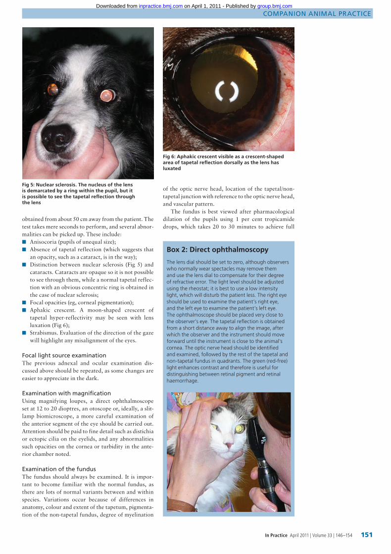
Examination roomA full ocular examination involves carrying out various tests with the lights on and in the dark (to minimise reflections). It should therefore be possible to com-pletely darken the examination room. Animals should be allowed a few minutes to acclimatise to the room. Small animals are best examined on a table, but large dogs may be examined sitting in a corner. The patient should be gently restrained, and ocular examination is usually tolerated well when approached calmly. An assistant is useful, or else owners should be shown how to gently restrain the animal with one arm over the body and the other hand resting under the chin. Some dogs may require a muzzle, but this does not hinder the ocular examination. Sedation should be avoided unless it is absolutely necessary, as it can cause enophthalmos, downward rotation of the globe, protrusion of the third eyelid, changes in pupil size or may alter the results of some tests (eg, the Schirmer tear test and intraocular pressure measurement), depending on the agent used.
Ophthalmic examination
Both eyes of any animal must be examined, even if only one has an obvious problem. The apparently normal eye may provide clues about the diagnosis as it may be at an earlier stage of the same condition. Systemic dis-ease may cause secondary ocular signs, which may aid the diagnosis of the underlying condition. Conversely, an animal presenting with an eye complaint should undergo a full clinical examination in addition to an ocular examination, in case it represents an ocular manifestation of a systemic disease.
It may be helpful to record the findings of an exam-ination on an ophthalmic examination chart, and the use of drawings will aid future monitoring. A good quality macro photograph is a useful way of recording the appearance of the eye. Almost all digital cameras have a macro function, which allows close-up focus. An eye model and drawings are really helpful for explaining the location and effect of the lesions within the eye to owners.
A step-by-step approach should be used to exam-ine all parts of the eye in a specific order. By follow-ing such a routine, it is less likely that abnormalities will be missed. However, while the protocol should be kept relatively consistent, not every diagnostic test is required for each patient, and the choice of appro-priate tests is made during the examination. Although there are many steps, with practice, a thorough eye examination can be carried out in less than 10 min-utes. The recommended order of a thorough ocular examination is discussed below.
Lights on . . .HistoryAs with any aspect of veterinary medicine, accu-rate history taking is crucial. Information should be obtained about the animal’s breed, age, general health, current medications, travel history, vision, signs of ocular pain, duration and progression of the problem, changes in appearance and abnormalities of related and in-contact animals. Hereditary ocular dis-ease is relatively common, and it is a good idea to have
access to the list of breeds currently part of the BVA/ KC/SDS Eye Scheme, which can be found on the British Veterinary Association’s website (www.bva.co.uk/canine_health_schemes/Eye_Scheme.aspx). Apart from hereditary disease, several breeds are pre-disposed to various ocular conditions. A list of these can be found in the appendices of many textbooks, and may help to diagnose certain conditions.
Distance examinationWhile the history is being obtained from owners, dogs and cats may be allowed free to explore the consulta-tion room. During this time, the attitude of the patient in an unfamiliar environment should observed to check their ability to navigate around obstacles and note any obvious abnormal gait or head position. The lighting level of the room may be temporarily reduced to see if this makes a difference to an animal’s behaviour. A maze test (Box 1) is a useful method of assessing vision in dogs (there is little point in performing the test in cats), and can be carried out at this stage or after the ocular examination.
physical examinationWith the animal placed on a table, a physical examina-tion should be performed first in case other related or unrelated signs are present.
Gross general examinationA gross general examination should be carried out to check facial symmetry along with eye symmetry, size, movement and direction of gaze. The presence and nature of any ocular discharge should be noted. The animal should be observed for obvious signs of ocular pain, such as blepharospasm and epiphora. The ‘hands-off’ approach should be used initially to avoid disturbing the conformation of the eyelids, which could alter the appearance of entropion or ectropion, and stimulating changes such as increased tear produc-tion or changes in eyelid position. If a Schirmer tear test or bacteriological swabbing are being considered, this is an appropriate time to perform these.
Close inspectionClose inspection of the eye and adnexa should be carried out using a focal light source. It is best to examine the external eye first before assessing the internal aspect. The eyelids should anatomically fit well with the globe, and they should be checked for entropion, ectropion or macropalpebral fissures. The palpebral and bulbar conjunctiva should be assessed, and the presence and location of lacrimal punctate observed. The presence and position of the third eyelid should be noted, and if a more detailed inspection of the posterior aspect is required, it may be grasped with atraumatic forceps and carefully retracted from the globe. The episclera, sclera and limbus should be examined next, and the general appearance of the ocular surface noted to make sure there is a nice healthy tear film and to check for any obvious opacities or surface irregularities of the cor-nea. The anterior chamber should be clear, and neither excessively deep or shallow. The iris should be a normal colour and have a smooth round pupil margin. The lens should be observed behind the pupil, and may appear white or cloudy if there are cataracts.
Box 1: Maze test in dogs
A maze test involves placing obstacles of different sizes and colour around a room and asking the owner to call the dog from the other side of the room. This helps to easily assess a dog’s ability to navigate around the objects in the room. The test should be performed in photopic conditions (lights on) and repeated in scotopic conditions (low lighting) with a few obstacles moved to ensure the dog has not memorised the layout.
146-154 Eye exam.indd 149 24/3/11 13:29:01
group.bmj.com on April 1, 2011 - Published by inpractice.bmj.comDownloaded from
Companion animal praCtiCe
150 In Practice April 2011 | Volume 33 | 146–154
neuro-ophthalmic examinationOphthalmic disease such as blindness may have a neurological component, and a full neurological examination may be required based on the results and interpretation of a neuro-ophthalmic examination, which should include the following tests:
Palpebral reflex. ■■ Observe the completeness of eyelid closure before and after stimulation of the medial and lateral canthus with touch, which nor-mally elicits a complete blink. It is important to establish a normal blink response before judging the results of the menace response and dazzle reflex (see below), to ensure that the animal is capable of responding as expected. A normal result confirms an intact sensory pathway (trigeminal nerve) and motor pathway (facial nerve). An abnormal result (lack of a blink) indicates poor sensation or, more commonly, facial nerve paralysis;Menace response. ■■ This is assessed by making a threatening movement towards each eye in turn, taking care not to touch the patient or create a wind current. A normal response is a blink with aversion of the head, which confirms vision and an intact facial nerve. A negative menace response usually indicates blindness, although animals with cerebel-lar lesions and normal vision also have a negative menace response;Dazzle reflex. ■■ Shine a strong light into each eye in turn. This should elicit a normal involuntary avoid-ance response, which comprises a blink or partial blink and head aversion. The pathway for this reflex is not fully understood. A negative dazzle reflex is a poor prognostic indicator for vision. A positive result suggests function of the visual pathway from the eye to the rostral colliculus, so it is precorti-cal and does not involve the visual cortex. This test is used to check for potential vision in cases with some opacity of the ocular media (eg, cataracts and hyphaema); Pupillary light reflex (PLR)■■ . Shine a bright light into the lateral aspect of each eye in turn. A nor-mal response is constriction of the pupil being stimulated (direct response) along with slightly less constriction of the unstimulated pupil (consen-sual response). Knowledge of the neuroanatomical pathway of this reflex can help to pinpoint the loca-tion of a lesion in some cases. It is important to note that this response is not an assessment of vision as
blind animals can have normal PLRs (eg, those with cataracts or occipital cortex lesions) and those with normal vision can have absent PLRs (eg, animals with iris atrophy). If the pupil does not constrict in response to light, it is possible that:
The light source is too weak;■●
The animal is very stressed, with excessive ■●
sympathetic nervous stimulation. In such cases, there will be bilateral mydriasis;
The iris is very thin leading to atrophy of the iris ■●
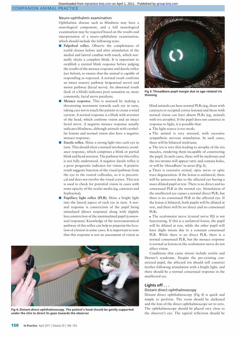
muscles, rendering them incapable of constricting the pupil. In such cases, there will be mydriasis and the iris stroma will appear tatty and contain holes, or will be ‘threadbare’ in areas (Fig 3);
There is extensive retinal, optic nerve or optic ■●
tract degeneration. If the lesion is unilateral, there will be anisocoria due to the affected eye having a more dilated pupil at rest. There is no direct and no consensual PLR in the normal eye. Stimulation of the unaffected eye causes a normal direct PLR, but there is no consensual PLR in the affected eye. If the lesion is bilateral, both pupils will be dilated at rest, and there will be no direct and no consensual PLR;
The oculomotor nerve (cranial nerve III) is not ■●
functioning. If this is a unilateral lesion, the pupil will be dilated at rest, while the other pupil will have slight miosis due to a constant consensual PLR. While there is no direct PLR, there is a normal consensual PLR, but the menace response is normal as lesions in the oculomotor nerve do not affect vision.Conditions that cause miosis include uveitis and
Horner’s syndrome. Despite the pre-existing con-stricted pupil, the affected iris should still constrict further following stimulation with a bright light, and there should be a normal consensual response in the unaffected eye.
Lights off . . . Distant direct ophthalmoscopyDistant direct ophthalmoscopy (Fig 4) is quick and simple to perform. The room should be darkened and the lens of the direct ophthalmoscope set to zero. The ophthalmoscope should be placed very close to the observer’s eye. The tapetal reflection should be
Fig 3: Threadbare pupil margin due to age-related iris thinning
Fig 4: Distant direct ophthalmoscopy. The patient’s head should be gently supported under the chin to direct its gaze towards the observer
146-154 Eye exam.indd 150 24/3/11 13:29:10
group.bmj.com on April 1, 2011 - Published by inpractice.bmj.comDownloaded from
Companion animal praCtiCe
151In Practice April 2011 | Volume 33 | 146–154
obtained from about 50 cm away from the patient. The test takes mere seconds to perform, and several abnor-malities can be picked up. These include:
Anisocoria (pupils of unequal size);■■
Absence of tapetal reflection (which suggests that ■■
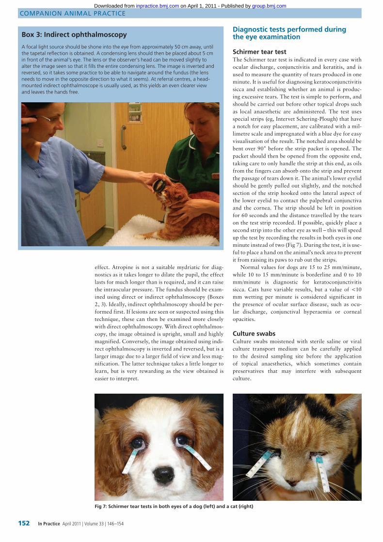
an opacity, such as a cataract, is in the way);Distinction between nuclear sclerosis (Fig 5) and ■■
cataracts. Cataracts are opaque so it is not possible to see through them, while a normal tapetal reflec-tion with an obvious concentric ring is obtained in the case of nuclear sclerosis;Focal opacities (eg, corneal pigmentation);■■
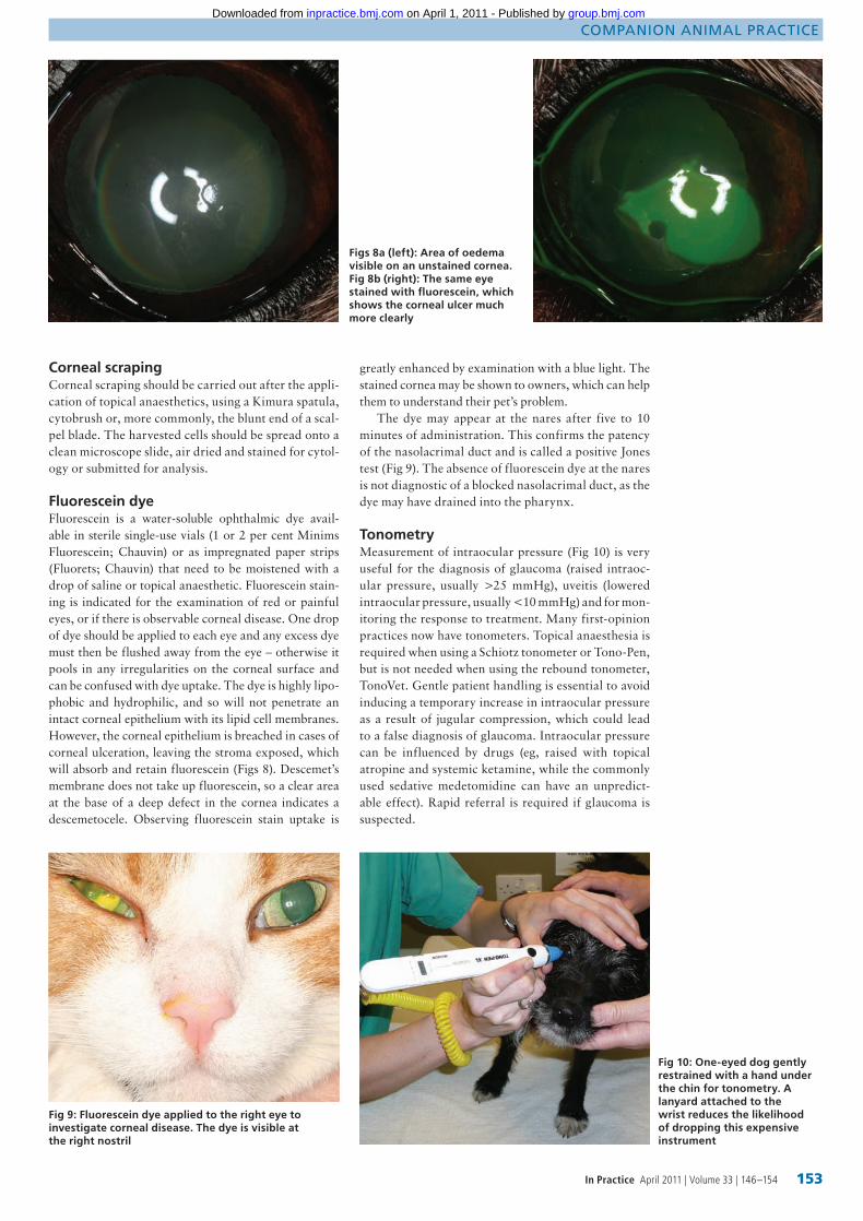
Aphakic crescent. A moon-shaped crescent of ■■
tapetal hyper-reflectivity may be seen with lens luxation (Fig 6);Strabismus. Evaluation of the direction of the gaze ■■
will highlight any misalignment of the eyes.
Focal light source examinationThe previous adnexal and ocular examination dis-cussed above should be repeated, as some changes are easier to appreciate in the dark.
examination with magnificationUsing magnifying loupes, a direct ophthalmoscope set at 12 to 20 dioptres, an otoscope or, ideally, a slit-lamp biomicroscope, a more careful examination of the anterior segment of the eye should be carried out. Attention should be paid to fine detail such as distichia or ectopic cilia on the eyelids, and any abnormalities such opacities on the cornea or turbidity in the ante-rior chamber noted.
examination of the fundusThe fundus should always be examined. It is impor-tant to become familiar with the normal fundus, as there are lots of normal variants between and within species. Variations occur because of differences in anatomy, colour and extent of the tapetum, pigmenta-tion of the non-tapetal fundus, degree of myelination
Fig 5: Nuclear sclerosis. The nucleus of the lens is demarcated by a ring within the pupil, but it is possible to see the tapetal reflection through the lens
Fig 6: Aphakic crescent visible as a crescent-shaped area of tapetal reflection dorsally as the lens has luxated
Box 2: Direct ophthalmoscopy
The lens dial should be set to zero, although observers who normally wear spectacles may remove them and use the lens dial to compensate for their degree of refractive error. The light level should be adjusted using the rheostat; it is best to use a low intensity light, which will disturb the patient less. The right eye should be used to examine the patient’s right eye, and the left eye to examine the patient’s left eye. The ophthalmoscope should be placed very close to the observer’s eye. The tapetal reflection is obtained from a short distance away to align the image, after which the observer and the instrument should move forward until the instrument is close to the animal’s cornea. The optic nerve head should be identified and examined, followed by the rest of the tapetal and non-tapetal fundus in quadrants. The green (red-free) light enhances contrast and therefore is useful for distinguishing between retinal pigment and retinal haemorrhage.
of the optic nerve head, location of the tapetal/non-tapetal junction with reference to the optic nerve head, and vascular pattern.
The fundus is best viewed after pharmacological dilation of the pupils using 1 per cent tropicamide drops, which takes 20 to 30 minutes to achieve full
146-154 Eye exam.indd 151 24/3/11 13:29:19
group.bmj.com on April 1, 2011 - Published by inpractice.bmj.comDownloaded from
Companion animal praCtiCe
152 In Practice April 2011 | Volume 33 | 146–154
effect. Atropine is not a suitable mydriatic for diag-nostics as it takes longer to dilate the pupil, the effect lasts for much longer than is required, and it can raise the intraocular pressure. The fundus should be exam-ined using direct or indirect ophthalmoscopy (Boxes 2, 3). Ideally, indirect ophthalmoscopy should be per-formed first. If lesions are seen or suspected using this technique, these can then be examined more closely with direct ophthalmoscopy. With direct ophthalmos-copy, the image obtained is upright, small and highly magnified. Conversely, the image obtained using indi-rect ophthalmoscopy is inverted and reversed, but is a larger image due to a larger field of view and less mag-nification. The latter technique takes a little longer to learn, but is very rewarding as the view obtained is easier to interpret.
Diagnostic tests performed during the eye examination
Schirmer tear testThe Schirmer tear test is indicated in every case with ocular discharge, conjunctivitis and keratitis, and is used to measure the quantity of tears produced in one minute. It is useful for diagnosing keratoconjunctivitis sicca and establishing whether an animal is produc-ing excessive tears. The test is simple to perform, and should be carried out before other topical drops such as local anaesthetic are administered. The test uses special strips (eg, Intervet Schering-Plough) that have a notch for easy placement, are calibrated with a mil-limetre scale and impregnated with a blue dye for easy visualisation of the result. The notched area should be bent over 90˚ before the strip packet is opened. The packet should then be opened from the opposite end, taking care to only handle the strip at this end, as oils from the fingers can absorb onto the strip and prevent the passage of tears down it. The animal’s lower eyelid should be gently pulled out slightly, and the notched section of the strip hooked onto the lateral aspect of the lower eyelid to contact the palpebral conjunctiva and the cornea. The strip should be left in position for 60 seconds and the distance travelled by the tears on the test strip recorded. If possible, quickly place a second strip into the other eye as well – this will speed up the test by recording the results in both eyes in one minute instead of two (Fig 7). During the test, it is use-ful to place a hand on the animal’s neck area to prevent it from raising its paws to rub out the strips.
Normal values for dogs are 15 to 25 mm/minute, while 10 to 15 mm/minute is borderline and 0 to 10 mm/minute is diagnostic for keratoconjunctivitis sicca. Cats have variable results, but a value of <10 mm wetting per minute is considered significant in the presence of ocular surface disease, such as ocu-lar discharge, conjunctival hyperaemia or corneal opacities.
Culture swabsCulture swabs moistened with sterile saline or viral culture transport medium can be carefully applied to the desired sampling site before the application of topical anaesthetics, which sometimes contain preservatives that may interfere with subsequent culture.
Box 3: Indirect ophthalmoscopy
A focal light source should be shone into the eye from approximately 50 cm away, until the tapetal reflection is obtained. A condensing lens should then be placed about 5 cm in front of the animal’s eye. The lens or the observer’s head can be moved slightly to alter the image seen so that it fills the entire condensing lens. The image is inverted and reversed, so it takes some practice to be able to navigate around the fundus (the lens needs to move in the opposite direction to what it seems). At referral centres, a head-mounted indirect ophthalmoscope is usually used, as this yields an even clearer view and leaves the hands free.
Fig 7: Schirmer tear tests in both eyes of a dog (left) and a cat (right)
146-154 Eye exam.indd 152 24/3/11 13:29:33
group.bmj.com on April 1, 2011 - Published by inpractice.bmj.comDownloaded from
Companion animal praCtiCe
153In Practice April 2011 | Volume 33 | 146–154
Corneal scrapingCorneal scraping should be carried out after the appli-cation of topical anaesthetics, using a Kimura spatula, cytobrush or, more commonly, the blunt end of a scal-pel blade. The harvested cells should be spread onto a clean microscope slide, air dried and stained for cytol-ogy or submitted for analysis.
Fluorescein dyeFluorescein is a water-soluble ophthalmic dye avail-able in sterile single-use vials (1 or 2 per cent Minims Fluorescein; Chauvin) or as impregnated paper strips (Fluorets; Chauvin) that need to be moistened with a drop of saline or topical anaesthetic. Fluorescein stain-ing is indicated for the examination of red or painful eyes, or if there is observable corneal disease. One drop of dye should be applied to each eye and any excess dye must then be flushed away from the eye – otherwise it pools in any irregularities on the corneal surface and can be confused with dye uptake. The dye is highly lipo-phobic and hydrophilic, and so will not penetrate an intact corneal epithelium with its lipid cell membranes. However, the corneal epithelium is breached in cases of corneal ulceration, leaving the stroma exposed, which will absorb and retain fluorescein (Figs 8). Descemet’s membrane does not take up fluorescein, so a clear area at the base of a deep defect in the cornea indicates a descemetocele. Observing fluorescein stain uptake is
Figs 8a (left): Area of oedema visible on an unstained cornea. Fig 8b (right): The same eye stained with fluorescein, which shows the corneal ulcer much more clearly
greatly enhanced by examination with a blue light. The stained cornea may be shown to owners, which can help them to understand their pet’s problem.
The dye may appear at the nares after five to 10 minutes of administration. This confirms the patency of the nasolacrimal duct and is called a positive Jones test (Fig 9). The absence of fluorescein dye at the nares is not diagnostic of a blocked nasolacrimal duct, as the dye may have drained into the pharynx.
TonometryMeasurement of intraocular pressure (Fig 10) is very useful for the diagnosis of glaucoma (raised intraoc-ular pressure, usually >25 mmHg), uveitis (lowered intraocular pressure, usually <10 mmHg) and for mon-itoring the response to treatment. Many first-opinion practices now have tonometers. Topical anaesthesia is required when using a Schiotz tonometer or Tono-Pen, but is not needed when using the rebound tonometer, TonoVet. Gentle patient handling is essential to avoid inducing a temporary increase in intraocular pressure as a result of jugular compression, which could lead to a false diagnosis of glaucoma. Intraocular pressure can be influenced by drugs (eg, raised with topical atropine and systemic ketamine, while the commonly used sedative medetomidine can have an unpredict-able effect). Rapid referral is required if glaucoma is suspected.
Fig 9: Fluorescein dye applied to the right eye to investigate corneal disease. The dye is visible at the right nostril
Fig 10: One-eyed dog gently restrained with a hand under the chin for tonometry. A lanyard attached to the wrist reduces the likelihood of dropping this expensive instrument
146-154 Eye exam.indd 153 24/3/11 13:29:57
group.bmj.com on April 1, 2011 - Published by inpractice.bmj.comDownloaded from
Companion animal praCtiCe
154 In Practice April 2011 | Volume 33 | 146–154
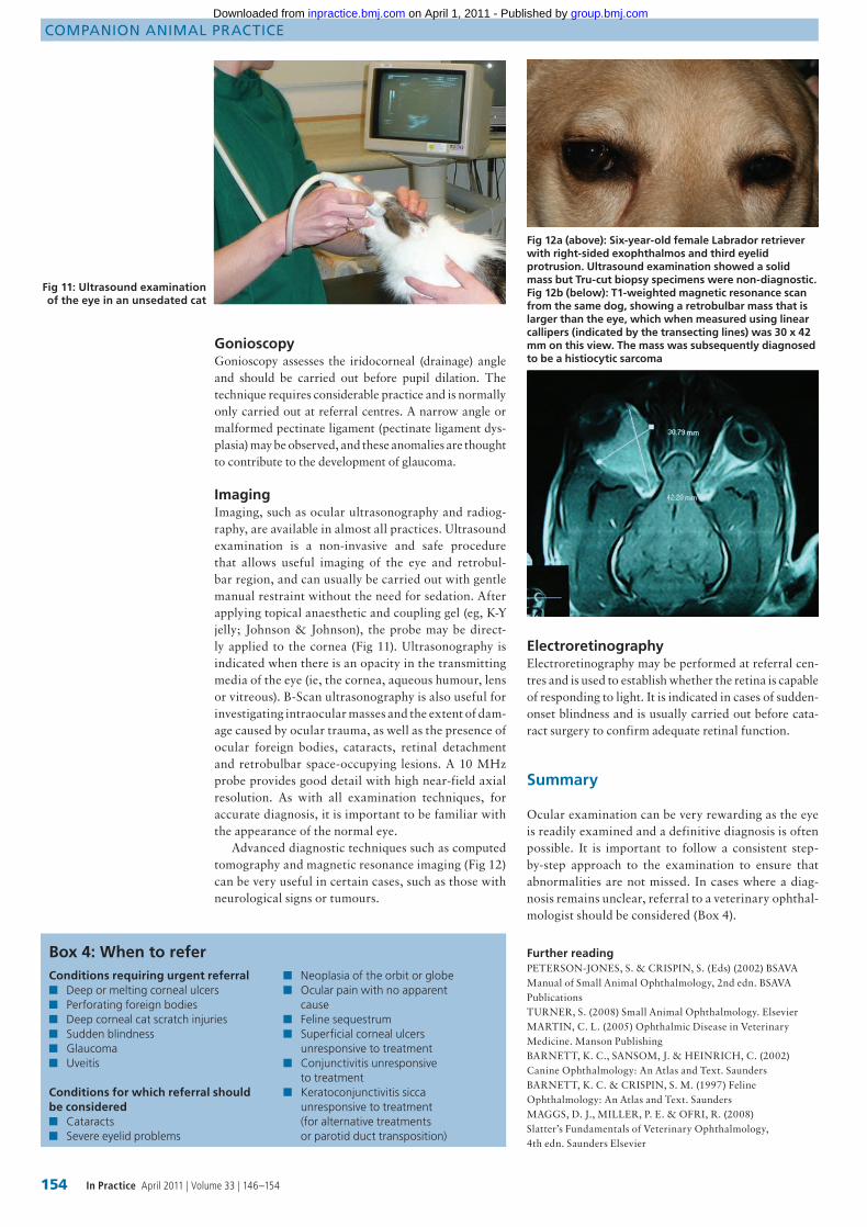
Fig 11: Ultrasound examination of the eye in an unsedated cat
Further readingPETERSON-JONES, S. & CRISPIN, S. (Eds) (2002) BSAVA Manual of Small Animal Ophthalmology, 2nd edn. BSAVA PublicationsTURNER, S. (2008) Small Animal Ophthalmology. ElsevierMARTIN, C. L. (2005) Ophthalmic Disease in Veterinary Medicine. Manson PublishingBARNETT, K. C., SANSOM, J. & HEINRICH, C. (2002) Canine Ophthalmology: An Atlas and Text. SaundersBARNETT, K. C. & CRISPIN, S. M. (1997) Feline Ophthalmology: An Atlas and Text. SaundersMAGGS, D. J., MILLER, P. E. & OFRI, R. (2008) Slatter’s Fundamentals of Veterinary Ophthalmology, 4th edn. Saunders Elsevier
GonioscopyGonioscopy assesses the iridocorneal (drainage) angle and should be carried out before pupil dilation. The technique requires considerable practice and is normally only carried out at referral centres. A narrow angle or malformed pectinate ligament (pectinate ligament dys-plasia) may be observed, and these anomalies are thought to contribute to the development of glaucoma.
ImagingImaging, such as ocular ultrasonography and radiog-raphy, are available in almost all practices. Ultrasound examination is a non-invasive and safe procedure that allows useful imaging of the eye and retrobul-bar region, and can usually be carried out with gentle manual restraint without the need for sedation. After applying topical anaesthetic and coupling gel (eg, K-Y jelly; Johnson & Johnson), the probe may be direct-ly applied to the cornea (Fig 11). Ultrasonography is indicated when there is an opacity in the transmitting media of the eye (ie, the cornea, aqueous humour, lens or vitreous). B-Scan ultrasonography is also useful for investigating intraocular masses and the extent of dam-age caused by ocular trauma, as well as the presence of ocular foreign bodies, cataracts, retinal detachment and retrobulbar space-occupying lesions. A 10 MHz probe provides good detail with high near-field axial resolution. As with all examination techniques, for accurate diagnosis, it is important to be familiar with the appearance of the normal eye.
Advanced diagnostic techniques such as computed tomography and magnetic resonance imaging (Fig 12)can be very useful in certain cases, such as those with neurological signs or tumours.
ElectroretinographyElectroretinography may be performed at referral cen-tres and is used to establish whether the retina is capable of responding to light. It is indicated in cases of sudden-onset blindness and is usually carried out before cata-ract surgery to confirm adequate retinal function.
Summary
Ocular examination can be very rewarding as the eye is readily examined and a definitive diagnosis is often possible. It is important to follow a consistent step-by-step approach to the examination to ensure that abnormalities are not missed. In cases where a diag-nosis remains unclear, referral to a veterinary ophthal-mologist should be considered (Box 4).
Fig 12a (above): Six-year-old female Labrador retriever with right-sided exophthalmos and third eyelid protrusion. Ultrasound examination showed a solid mass but Tru-cut biopsy specimens were non-diagnostic. Fig 12b (below): T1-weighted magnetic resonance scan from the same dog, showing a retrobulbar mass that is larger than the eye, which when measured using linear callipers (indicated by the transecting lines) was 30 x 42 mm on this view. The mass was subsequently diagnosed to be a histiocytic sarcoma
Box 4: When to referConditions requiring urgent referral
Deep or melting corneal ulcers■■
Perforating foreign bodies■■
Deep corneal cat scratch injuries■■
Sudden blindness■■
Glaucoma■■
Uveitis■■
Conditions for which referral should be considered
Cataracts ■■
Severe eyelid problems■■
Neoplasia of the orbit or globe■■
Ocular pain with no apparent ■■
causeFeline sequestrum■■
Superficial corneal ulcers ■■
unresponsive to treatmentConjunctivitis unresponsive ■■
to treatmentKeratoconjunctivitis sicca ■■
unresponsive to treatment (for alternative treatments or parotid duct transposition)
146-154 Eye exam.indd 154 24/3/11 13:30:12
group.bmj.com on April 1, 2011 - Published by inpractice.bmj.comDownloaded from

doi: 10.1136/inp.d1810 2011 33: 146-154In Practice
Natasha Mitchell animalsApproach to ocular examination in small
http://inpractice.bmj.com/content/33/4/146.full.htmlUpdated information and services can be found at:
These include:
serviceEmail alerting
the box at the top right corner of the online article.Receive free email alerts when new articles cite this article. Sign up in
Notes
http://group.bmj.com/group/rights-licensing/permissionsTo request permissions go to:
http://journals.bmj.com/cgi/reprintformTo order reprints go to:
http://group.bmj.com/subscribe/To subscribe to BMJ go to:
group.bmj.com on April 1, 2011 - Published by inpractice.bmj.comDownloaded from