comparative modeling of pivotal enzymes, mura and murz, of enterococcus faecalis and identification...
TRANSCRIPT

ORIGINAL RESEARCH
Comparative modeling of pivotal enzymes, MurA and MurZ,of Enterococcus faecalis and identification of potential inhibitorsby computational methods
Mansimran Khokhar • Navkiran Kaur • Vaibhav Jain • Rajat Sandhir •
Ankur Gautam • Prasad V. Bharatam • Rupinder Tewari
Received: 9 January 2013 / Accepted: 10 September 2013 / Published online: 19 September 2013
� Springer Science+Business Media New York 2013
Abstract The emergence of multi-drug-resistant bacterial
pathogens along with labor-intensive and time-consuming
drug discovery methods has put an emphasis on alternative
approaches which can overcome these drawbacks. One such
approach is in silico identification of novel bacterial inhibi-
tors using structure-based drug design methodology. In the
present study, novel inhibitors have been proposed against
two essential enzymes (MurA and MurZ) involved in the
biosynthesis of cell wall of Enterococcus faecalis, a serious
nosocomial bacterial pathogen. Homology models of MurA
and MurZ of E. faecalis were constructed using template
MurA of Enterobacter cloacae and Haemophilus influenza,
respectively. The validation of the refined models, using
Ramachandran, Errat and ProSA energy plots, showed that
constructed models were reliable and of good quality. The
active site amino acid residues, as predicted by CASTp
server, were in accordance with the residues identified from
MurA crystal structures available for other bacteria. The
present study revealed for the first time the importance of two
additional amino acid residues (Thr305 and Tyr329) in
MurA and MurZ enzymes, which might be crucial for the
binding of ligands/inhibitors with the target. Based on the
GLIDE score and interaction profile, 5-sulfonoxy-
anthranilic acid derivatives, T6361 and T6362 were found to
be the potential inhibitors of MurA and MurZ enzymes.
Keywords Enterococcus faecalis � Peptidoglycan �MurA � MurZ � Homology modeling � Molecular docking
Introduction
Enterococcus faecalis (E. faecalis) is a bacterial pathogen,
which has gained notoriety over the past few decades as the
leading cause of nosocomial infections causing urinary
tract problems, bacteremia, intra-abdominal infections and
endocarditis with a high mortality rate (Bao et al., 2009;
Sav et al., 2010). Though antibiotic therapy is available for
treatments of this infection, drug-resistant clinical isolates
of E. faecalis are being regularly isolated from laboratories
all over the world (Sood et al., 2008). This grim situation
along with the fact that limited research is being carried out
to screen novel Enterococci inhibitors’ demands for an
urgent need for new drug targets and specific inhibitors.
The availability of the gene and protein sequences of
pathogens, including E. faecalis, has brought forth exten-
sive information that can be useful for novel drug target
identification and drug development (Meinke et al., 2004).
A peptidoglycan (PG) layer is an integral component of
bacterial cell wall. The absence of PG has been shown
beyond doubt to cause cell death (Babajan et al., 2011).
The essentiality of PG layer along with its absence in
eukaryotic cells has resulted in exploiting the enzymes of
PG biosynthesis as potential drug targets. The initial steps
in PG biosynthesis are mediated by a group of Mur
enzymes (Samland et al., 2001; Klein and Bachelier,
2006). As Mur enzymes are absent in humans and have no
human homologs, they represent a promising point of
M. Khokhar � N. Kaur � A. Gautam � R. Tewari (&)
Centre for Microbial Biotechnology, Panjab University, CIL
Building, Chandigarh, India
e-mail: [email protected]
V. Jain � P. V. Bharatam
Department of Pharmacoinformatics, National Institute of
Pharmaceutical Education and Research (NIPER), Mohali, India
R. Sandhir
Department of Biochemistry, Panjab University, Chandigarh,
India
123
Med Chem Res (2014) 23:1819–1828
DOI 10.1007/s00044-013-0785-z
MEDICINALCHEMISTRYRESEARCH

attack for novel antibacterial drugs. In Gram-positive
bacteria including E. faecalis, murA gene has two copies
(murA and murZ) which encode proteins with similar
enzymatic activity. Studies have also brought forth the fact
that removal of both the genes is necessary for cell wall
disruption in E. faecalis (Du et al., 2000). Although,
structure of MurA enzymes from Gram-negative bacteria
has been deciphered and inhibitors reported (Dunsmore
et al., 2008; Eschenburg et al., 2005), but none from Gram-
positive bacteria has been reported. In this article, attempt
has been made to develop homology models for MurAef
and MurZef enzymes of E. faecalis and novel inhibitors
against these targets have been identified.
Materials and methods
Homology modeling of MurAef and MurZef
The protein sequences of MurAef and MurZef were retrieved
from UNIPROT database (http://www.uniprot.org/). To find
a suitable template for model building, NCBI BLASTp
(Altschul et al., 1990) was performed against Protein Data
Bank (PDB). ClustalW (http://www.ebi.ac.uk/Tools/msa/
clustalw2) tool was used to carry out protein multiple
sequence alignments. The three dimensional (3D) model of
the enzymes along with the ligand present in the template
was built using Modeller9v8 (http://www.salilab.org/
modeller/). Subsequently, model validation was carried out
using PROCHECK (Laskowski et al., 1993) and ProSA-web
(Sippl, 1993) programs. PROCHECK scrutinizes the ste-
reochemical quality of a protein structure and presents a
Ramachandran plot of the query structure. Steric clashes
observed in the crude models were recognized by the
ERRAT plot and the structural loops showing error greater
than 90 % were refined through loop modeling using Mod-
eller Loop class (Colovos and Yeates, 1993). PDBsum server
was used to analyze the secondary structure of the proteins
(Laskowski et al., 2009). The active site prediction of the
modeled enzymes was performed using the computed atlas
of surface topography of proteins (CASTp) server. It pro-
vides identification and measurements of surface accessible
pockets as well as interior inaccessible cavities for proteins
by analytically measuring the area and volume of each
pocket and cavity, both in solvent accessible surface and
molecular surface (Kaistha and Sinha, 2009).
Molecular docking
Retrieval of ligands and database generation
The literature sources were surveyed for small inhibitory
compounds and a total of 108 compounds were retrieved.
These compounds were chosen on their reports of inhib-
iting MurA from various microorganisms e.g., Staphylo-
coccus aureus, Mycobacterium tuberculosis, E. cloacae,
H. influenza, and Bacillus anthracis. The 3D structures of
these ligands were built using SYBYL 7.1 molecular
modeling package installed on a Silicon Graphics Fuel
Workstation (Baviskar et al., 2011). Gasteiger-Huckel
partial charges were applied to the atoms of the ligand
followed by minimization of the structures by Powell
method using Tripos force field. Finally, 3D database of
ligands was generated in SYBYL.
Ligand and enzyme structure preparation
The LigPrep module of Maestro (Maestro 9.0, Schrodinger,
LLC, New York) interface was used for the preparation of
ligand structures from the generated database. It optimizes
each ligand structure to produce low energy, tautomeric,
and reasonable ionization state conformations within a
range of pH 7.0 ± 2.0 using OPLS_2005 force field.
The refined and validated structures of MurAef and MurZef
enzymes were processed using Protein Preparation wizard
tool incorporated in Maestro interface. Hydrogen atoms were
added and the right bond orders were assigned to the enzymes.
The carboxylic acid side chains of all the Asp and Glu residues
were deprotonated and basic residues Lys and Arg side chains
were protonated so as to mimic correct ionization state at
physiological pH. In addition, correct tautomeric state of His
and the spatial arrangements of hydroxyl and amide groups of
Asn and Gln were defined using Protassign utility (Schro-
dinger Suite, 2009). To remove the steric clashes between the
atoms, enzyme structures were minimized using Impref utility
until the average root-mean-square-deviation (RMSD) of the
complete atoms came down to 0.3 A.
Receptor grid generation
Receptor grid generation module of GLIDE was used to
define the receptor grid. The ligands UNAG and UD1
(bound to MurAef and MurZef) were taken as reference to
designate the active site of MurAef and MurZef, respec-
tively. The centroid of the bound ligand was used to define
the inner box of the grid by extending the dimension to
10 A and the external box by extending up to 20 A. This
step determines the position and size of the active site of
the target, thus setting up Glide constraints (Schrodinger
Suite, 2009). These constraints were then used to fit/dock
ligands into the target site.
Molecular docking
Docking simulations were performed using Grid-based
Ligand Docking with Energetics (GLIDE) program. Glide
1820 Med Chem Res (2014) 23:1819–1828
123

explores for possible locations of the ligand in the active site
region of the enzymes using a set of hierarchal filters. It
searches for favorable interactions between one or more
conformations of the ligands and a protein (Friesner et al.,
2004). The ligand is minimized in the field of the receptor
using the OPLS-AA force field. The ChemScore algorithm
was used to perform scoring, recognizing favorable hydro-
phobic and hydrogen bond interactions (Eldridge et al., 1997).
Fig. 1 ERRAT plot of (a) MurAef and (b) MurZef enzymes
Med Chem Res (2014) 23:1819–1828 1821
123
Results and discussion
Template selection
The 430 amino acids long protein sequence of MurAef was
retrieved from UNIPROT/SwissProtKB database (primary
accession number: Q836E5). After performing BLASTP
analysis against PDB database, maximum sequence homol-
ogy was found with MurA from E. cloacae (PDB ID: 1Q3G,
Ligand: UDA (30-1-carboxy-1-phosphonooxy-ethoxy-uri-
dine-diphosphate-N-acetylglucosamine), Resolution: 2.65 A)
(Eschenburg et al., 2003). It exhibited 46 % sequence identity
and an E value of 7e-121. Correspondingly, the template
search for MurZef (primary accession number: Q831A8)
demonstrated maximum sequence homology with MurA from
H. influenza [PDB ID: 2RL1, Ligand: UD1 (uridine-diphos-
phate-N-acetylglucosamine), resolution: 2.20 A] (Yoon et al.,
2008). It exhibited 48 % identity with an E value of 3e-131.
Owing to the fact that both MurAec (1Q3G) and MurAhi
(2RL1) exhibited high sequence identity and the PDB’s had
bound ligands in their crystal structures, made them suitable
for being used as templates. This allowed us to model the
ligand within our homology models. In addition, the resolu-
tion of both of these crystal structures was high (1Q3G: 2.65 A
and 2RL1: 2.20 A), and they posses overall good B-factor,
therefore, they were used as templates for homology modeling
for MurAef and MurZef proteins, respectively.
Homology modeling
Based on the information obtained from the sequence
alignment of target (MurAef) and template (1Q3G) protein
sequence, the homology model of MurAef was generated by
Modeller9v8. 100 models were generated and the model with
lowest discrete optimized protein energy (DOPE) and Mol-
pdf scores was selected for further optimization. Steric cla-
shes were observed in the loop regions of the crude model,
therefore, these regions were further subjected to loop
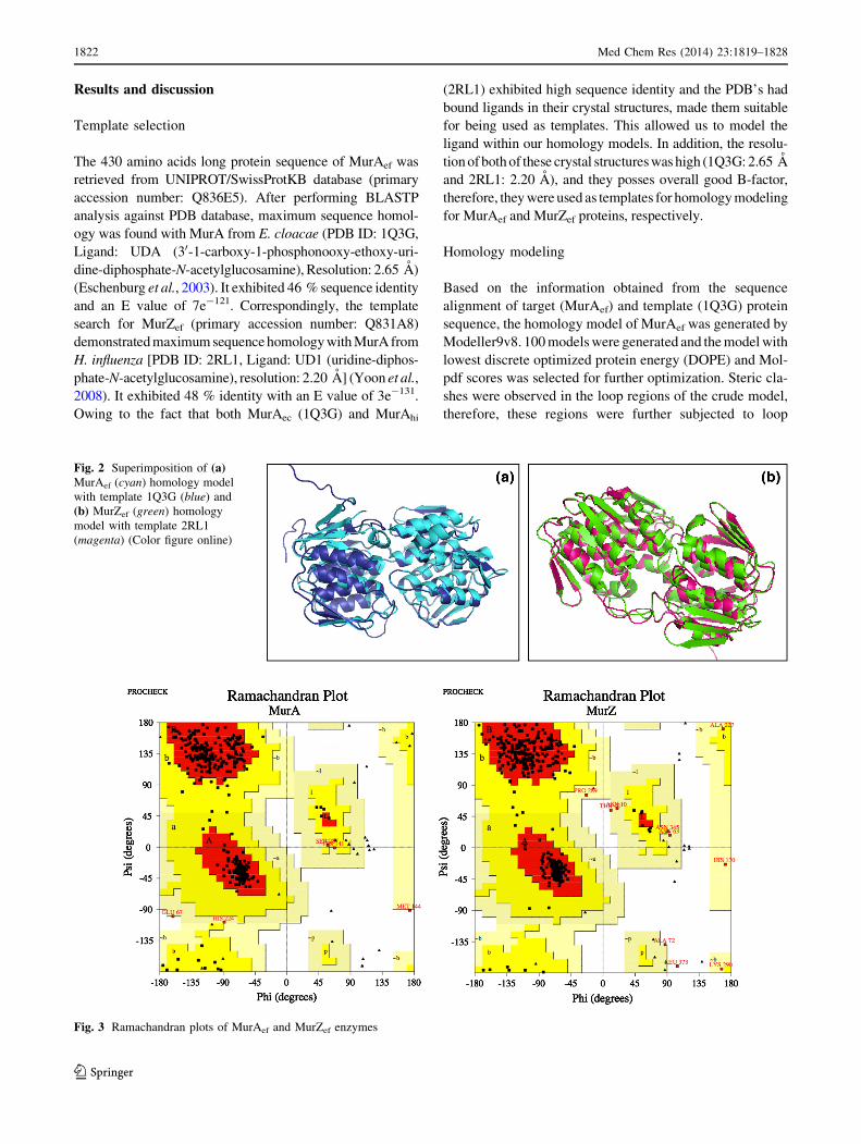
Fig. 2 Superimposition of (a)MurAef (cyan) homology model
with template 1Q3G (blue) and
(b) MurZef (green) homology
model with template 2RL1
(magenta) (Color figure online)
Fig. 3 Ramachandran plots of MurAef and MurZef enzymes
1822 Med Chem Res (2014) 23:1819–1828
123
modeling using Modeller Loop class. The model of MurAef
also contained the modeled ligand: UDP-N-acetylglucosa-
mine (UNAG) (substrate). Similarly, homology model of
MurZef was generated using 2RL1 as the template structure
and contained the modeled ligand: UDP-N-acetylglucosa-
mine, referred to as UD1 (substrate). After a few cycles of
loop refinement, the 3D models of MurA and MurZ exhibited
an overall quality factor of 94.16 and 92.64 %, respectively
(Fig. 1).
The superimposition of Ca chains of 1Q3G and MurAef
(Fig. 2a) revealed an RMSD value of 0.421 A. Also RMSD
value of 0.199 A was observed when 2RL1 and MurZef
(Fig. 2b) were superimposed. The RSMD values were in
acceptable range i.e., B0.5 A, suggesting the validity of the
models constructed.
Evaluation of homology models of MurA and MurZ
enzymes
Validation tools like PROCHECK, VERIFY3D, and
ERRAT were used for protein structure evaluation. The
MurAef Ramachandran plot, generated by PROCHECK
(Fig. 3), revealed that 89 % residues were in the most
favored region, 9.3 % in the additionally allowed region,
1.1 % in the generously allowed region and 0.5 % in the
disallowed region, thereby indicating that MurAef model
was geometrically acceptable. For MurZef, the Rama-
chandran plot demonstrated that 89.8 % residues were in
the most favored region, 7.9 % in the additionally allowed
region, 1.8 % in the generously allowed region and 0.5 %
in the disallowed region (Fig. 3). This data suggested the
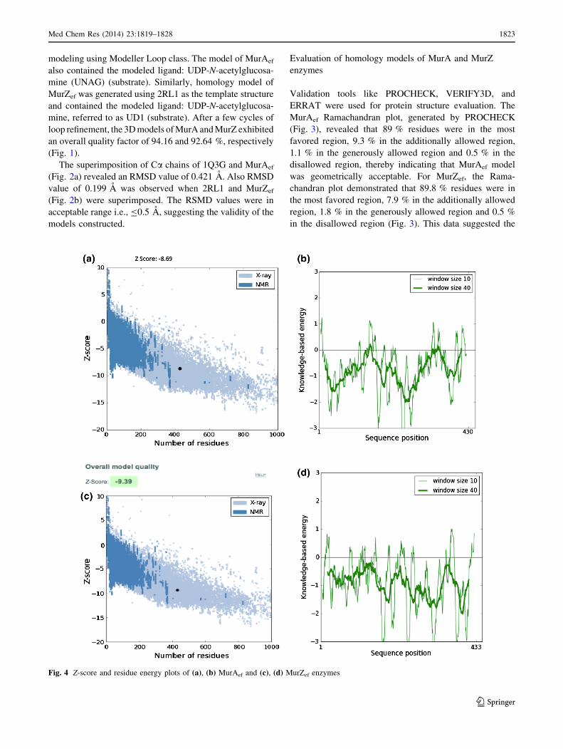
Fig. 4 Z-score and residue energy plots of (a), (b) MurAef and (c), (d) MurZef enzymes
Med Chem Res (2014) 23:1819–1828 1823
123

geometrical and stereochemical acceptability of the model.
As the residues in the disallowed regions did not show any
involvement in the active site/ligand binding, they were left
as such and the model was not further modified.
The reliability and accuracy of the developed models was
further evaluated by generating knowledge-based energy
curves for MurAef and MurZef using ProSA-web (Fig. 4)
(Wiederstein and Sippl, 2007). The z-score calculated by
ProSA indicated the overall quality of the models. The z-
score of MurAef (Fig. 4a) and MurZef (Fig. 4c) was observed
to be well within the z-score range of experimentally
determined NMR solved protein structures. The plots
(Fig. 4b, d) of residue energy exhibit the local model quality
by plotting energies as a function of amino acid sequence
position with different window size. Also, the ProSA-web
application requires only C-a backbone; therefore, it allows
us to evaluate the models early in structure determination
process. These energy plots describe the problematic parts of
the model corresponding to the positive values. In our model,
most of the amino acid sequences exhibit negative knowl-
edge-based energy, signifying that the overall quality of the
model is satisfactory.
Secondary structure analysis
The secondary structure of MurAef was analyzed using
PDBsum server, which showed that it consisted of 17
helices, 6 beta sheets, 20 strands, 5 beta hairpins, 1 beta
bulge, 2 beta–alpha–beta motifs, 31 helix–helix interac-
tions, 23 beta turns, and 4 gamma turns (Fig. 5a) (Dundas
et al., 2006). Similarly, the secondary structure of MurZef
depicted 19 helices, 6 beta sheets, 22 strands, 5 beta hair-
pins, 2 beta bulges, 2 beta-alpha–beta motifs, 29 helix–
helix interactions, and 22 beta turns (Fig. 5b) (Dundas
et al., 2006). As structure and function of the protein are
inter-correlated, this analysis provides detailed insight into
the structure of the MurA/MurZ enzymes, which could be
used to determine their function by comparing the topology
with existing 3D structures of Mur enzymes of different
organisms.
Active site prediction
Active site prediction, of constructed enzyme models,
using CASTp server suggested different binding pockets.
Based on the volume and area of the cavities and pocket,
ranking was assigned to the predictive active site. Rank 1
predicted active site cavity in MurAef and MurZef enzymes
composed of 70 and 74 amino acid residues, respectively.
These showed the presence of Lys22, Asn23, Asp49,
Arg92, Cys116, Arg121, and Arg398 in the active site of
MurAef (Fig. 6a). The amino acid residues predicted wereFig. 5 Diagrammatic representation of secondary structural elements
of (a) MurAef and (b) MurZef enzymes
1824 Med Chem Res (2014) 23:1819–1828
123
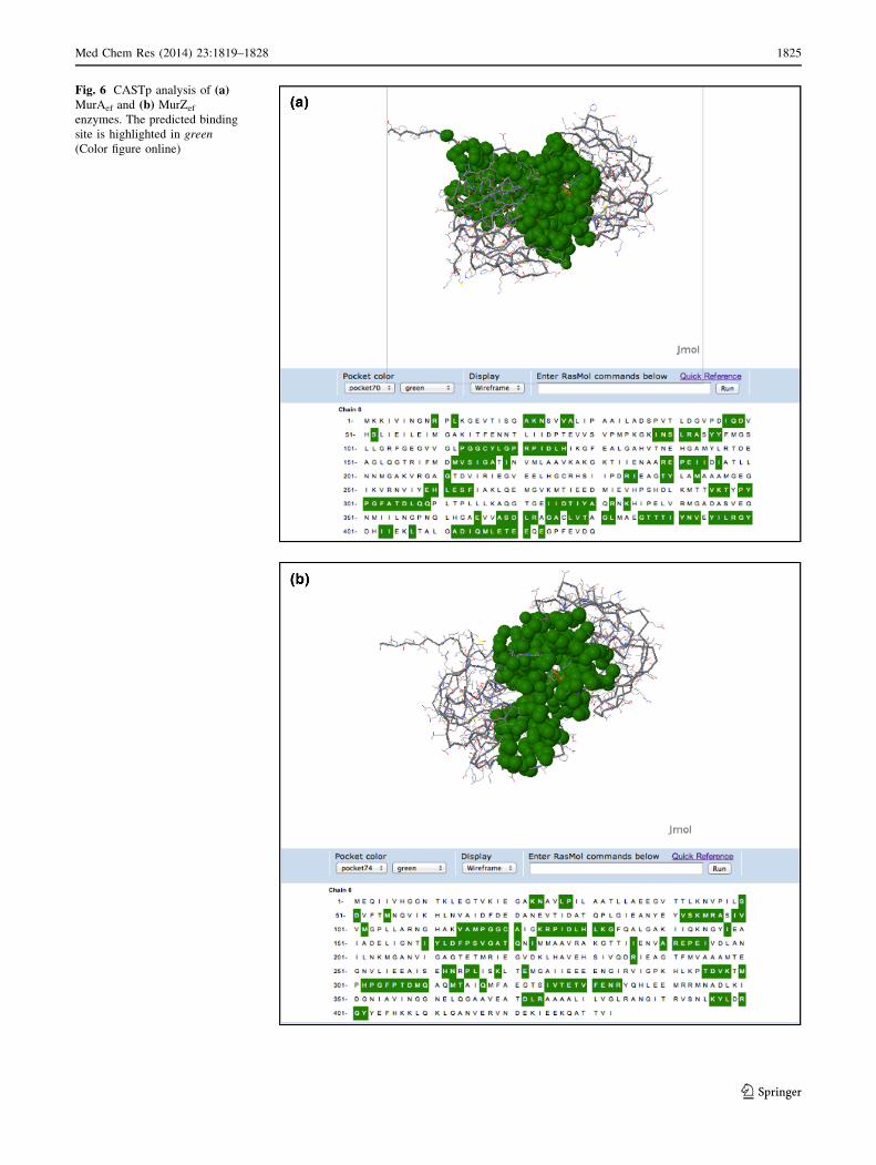
Fig. 6 CASTp analysis of (a)MurAef and (b) MurZef
enzymes. The predicted binding
site is highlighted in green
(Color figure online)
Med Chem Res (2014) 23:1819–1828 1825
123

also found to be present in the active site of the template
structure (1Q3G) and have been reported to be imperative
for other bacteria as well (Jackson et al., 2009; Kim et al.,
1996; Skarzynski et al., 1996). Similarly, Lys23, Arg96,
Cys120 Arg125, Asp308, and Arg400 were found to be
present in the active sites of MurZef, template 2RL1
(Fig. 6b) and other bacteria as well (Gautam et al., 2011;
Schonbrunn et al., 2000).
Virtual screening employs computational methods for
the identification of molecules that may be biologically
active against a target protein. We have adopted Struc-
ture-based virtual screening, for the identification of novel
inhibitors against the target protein(s) MurA and MurZ of
E. faecalis. A small library of compounds was built for
the purpose of performing molecular docking. Due to the
absence of 3D structure of target proteins, we developed
homology models of the same. These models were gen-
erated using Modeller, which were then subjected to loop
refinement and optimization. The final refined homology
models showed satisfactory Ramachandran plot statistics,
Errat plot quality factor. Moreover, the online validation
server (ProSA web) showed that the Z-score and energy
of protein folding of the models was in good agreement
with the available protein structures in PDB, which
favored the overall quality of the structures. These anal-
yses validated the homology models and prove that they
are robust as well as reliable enough to be used for virtual
screening purpose.
Table 1 Docking score and hydrogen bonding interactions of ligands with MurA and MurZ enzymes
Ligand Structure GLIDE docking score Hydrogen bonding interactions
MurA MurZ MurA MurZ
T6361 -7.466 -7.984 (Fig. 7b) Arg92, Arg121,
Pro122, His 126,
Ile165, Gly166
Lys23, Arg96, Arg125,
His130, Ser166,
Gly168, Thr307
T6362 -8.252 (Fig. 7a) -7.439 Arg121, His126,
Ile165, Gly166,Ala167, Thr305,
Tyr329
Arg96, Arg125,
Val167, Arg400
Carbidopa
NH
HO
H2N
O
OH
OH
-5.665 -6.789 Asp49, Arg121,
Arg332, Thr 305,
Tyr329
Lys23, Asn24, Arg125,
Gln171, Asp308
Residues in bold represent the amino acids reported to be essential for enzymatic activity [8, 11, 27, 28, 29, 30, 32]. These interactions were in
accordance with the ones shown by available crystal structures of MurA enzymes from different organisms
1826 Med Chem Res (2014) 23:1819–1828
123
Molecular docking analysis
In order to study the molecular level interactions and
binding affinity of the reported inhibitors (active for Mur of
other organisms) against MurAef and MurZef, they were
subjected to molecular docking into the active site of
enzyme structures using GLIDE (Table 1) program. For
selecting the ligands as probable inhibitor of MurAef and
MurZef, a limit of Glide score (\-5.00) and hydrogen
bonding interactions (C3) with the critical amino acid
residues of the active site was set. Based on the minimal
energy binding score, an indicative of good docking,
ligands T6362 and T6361 were found to be the best pos-
sible inhibitors of MurAef and MurZef respectively. Both
these inhibitors are derivatives of 5-sulfonoxy-anthranilic
acid. Previous studies reveal the action of T6361 as a
competitive inhibitor of the substrate (UNAG). It targets a
loop from Pro112 to Pro125 (which induces a conforma-
tional change in the enzyme), thus preventing the access of
UNAG to the active site. Binding mode of T6362 (Fig. 7a)
revealed hydrogen bonding with main chain atoms of Arg92,
Gly166, and Thr305 and with side chain atoms of Arg121,
His126, Ile165, Ala167, and Tyr329. It also showed p-
interactions with Tyr95 and His126. The binding mode of
T6361 (Fig. 7b) revealed hydrogen bonding with main chain
atoms of Lys23, Arg96, and Gly168 and with side chains
Arg125, His130, Ser166, and Thr307. It also showed p-
interactions with Phe331. These interactions were in
accordance with the ones shown by available crystal struc-
tures of MurA enzymes from different organisms. Carbi-
dopa which has been identified as a promiscuous inhibitor,
and reported to inhibit even MurA C115D mutant also
showed a good docking profile. These compounds have
previously been reported to inhibit MurA of different
organisms thereby advocating their broad host range (BHR)
activity (Dunsmore et al., 2008; Laskowski et al., 1993).
GLIDE evidence also suggested two residues (Thr305/307
and Tyr329), which might be noteworthy but have not been
reported previously. The inference can be drawn out of the
fact that, the interactions shown by these residues have been
repeated while studying docking with different ligands. In
the absence of any known ligands for MurAef and MurZef,
these inhibitors could act as leads and thus can be used as
starting point for designing other inhibitors as well.
Conclusions
Using in silico structure-based drug designing approach,
computational models of enzymes (MurA and MurZ) of a
serious nosocomial bacterium, E. faecalis have been
developed. Molecular docking procedure has resolved to
three molecules namely T6361, T6362, and Carbidopa,
which can act as potential anti-microbial agents. A report
of these compounds also inhibiting MurA of different
organisms advocates their BHR activity.
References
Altschul SF, Grish W, Miller W, Myers EW, Lipman DJ (1990) Basic
local alignment search tool. J Mol Biol 215:403–410
Babajan B, Chaitanya M, Rajsekhari C, Gowsia D, Madhusudhana P,
Naveen M, Chittai SK, Anuradha CM (2011) Comprehensive
structural and functional characterization of Mycobacterium
tuberculosis UDP-NAG enolpyruvyl transferase (Mtb–MurA)
and prediction of its accurate binding affinities with inhibitors.
Interdiscip Sci Comput Life Sci 3:204–216
Bao Y, Saknic T, Laverde D, Wobser D, Benachour A, Theilacker C,
Hartke A, Huebner J (2009) Role of mprF1 and mprF2 in the
pathogenicity of Enterococcus faecalis. PLoS One 7:38458
Fig. 7 Interaction profile of (a) T6362 with MurAef and of (b) T6361with MurZef (the yellow dashed lines mark the hydrogen bonding
interactions) (Color figure online)
Med Chem Res (2014) 23:1819–1828 1827
123

Baviskar AT, Madaan C, Preet R, Mohapatra P, Jain V, Agrawal A,
Guchhait SK, Kundu CN, Banerjee CN, Bharatam PV (2011)
N-Fused imidazoles as novel anticancer agents that inhibit
catalytic activity of topoisomerase IIa and induce apoptosis in
G1/S Phase. J Med Chem 54:5013–5030
Colovos C, Yeates TO (1993) Verification of protein structures:
patterns of nonbonded atomic interactions. Protein Sci
9:1511–1519
Du W, Brown JR, Sylvester DR, Huang J, Chalker AF, So CY,
Holmes DJ, Payne DJ, Wallis NJ (2000) Two active forms of
UDP-N-acetylglucosamine enolpyruvyl transferase in gram-
positive bacteria. J Bacteriol 182:4146–4152
Dundas J, Ouyang Z, Tseng J, Binkowski A, Turpaz Y, Liang J (2006)
CASTp: computed atlas of surface topography of proteins with
structural and topographical mapping of functionally annotated
residues. Nucleic Acid Res 34:W116–W118
Dunsmore CJ, Miller K, Blake KL, Patching SG, Henderson PJF,
Gaenett JA, Stubbings WJ, Phillips SVE, Palestrant DJ, Angeles
JDL, Leeds JA, Choprab I, Fishwicka CWG (2008) 2-Aminote-
tralones: novel inhibitors of MurA and MurZ. Bioorg Med Chem
Lett 18:1730–1734
Eldridge MD, Murray CW, Auton TR, Paolini GV, Mee RP (1997)
Empirical scoring functions: I. The development of a fast
empirical scoring function to estimate the binding affinity of
ligands in receptor complexes. J Comput Aided Mol Des
11:425–445
Eschenburg S, Kabsch W, Healy ML, Schoenbrunn E (2003) A new
view of the mechanism of UDP-N-acetylglucosamine eno-
lpyruvyl transferase (MurA) and 5-enolpyruvylshikimate-3-
phosphate synthase (AroA) derived from X-ray structures of
their tetrahedral reaction intermediate states. J Biol Chem
278:49215–49222
Eschenburg S, Priestman M, Schonbrunn E (2005) Evidence that the
fosfomycin target Cys115 in UDP-N-acetylglucosamine eno-
lpyruvyl transferase (MurA) is essential for product release.
J Biol Chem 280:3757–3763
Friesner RA, Banks JL, Murphy RB, Halgren TA, Klicic JJ, Mainz
DT, Repasky MP, Knoll EH, Shelley M, Perry JK, Shaw DE,
Francis P, Shenkin PS (2004) Glide: a new approach for rapid,
accurate docking and scoring. 1. Method and assessment of
docking accuracy. J Med Chem 47:1739–1749
Gautam A, Rishi P, Tewari R (2011) UDP-N-acetylglucosamine
enolpyruvyl transferase as a potential target for antibacterial
chemotherapy: recent developments. Appl Microbiol Biotechnol
92:211–225
Jackson SG, Zhang F, Chindemi P, Junop MS, Berti PJ (2009)
Evidence of kinetic control of ligand binding and staged product
release in MurA (Enolpyruvyl UDP-GlcNAc synthase)-catalyzed
reactions. Biochemistry 48:11715–11723
Kaistha SD, Sinha R (2009) Homology modeling of phosphoryl
thymidine kinase of enterohemorrhagic Escherichia coli OH:
157. Bioinformation 6:240–243
Kim DH, Lees WJ, Kempsell KE, Lane WS, Duncan K, Walsh CT
(1996) Characterization of a Cys115 to Asp substitution in the
Escherichia coli cell wall biosynthetic enzyme UDP-GlcNAce-
nolpyruvyl transferase (MurA) that confers resistance to inacti-
vation by the antibiotic fofomycin. Biochemistry 35:4923–4928
Klein CD, Bachelier A (2006) Molecular modeling and bioinformat-
ical analysis of the antibacterial target enzyme MurA from a
drug design perspective. J Comput Aided Mol Des 20:621–628
Laskowski RA, MacArthur MW, Moss DS, Thornton JM (1993)
PROCHECK: a program to check the stereochemical quality of
protein structures. J Appl Crystallogr 26:283–291
Laskowski RA, Chistyakov VV, Thornton JM (2009) PDBSUM
more: new summaries and analyses of the known 3-D structures
proteins and nucleic acids. Nucleic Acid Res 33:266–268
Meinke A, Henics T, Nagy E (2004) Bacterial genomes pave the way
to novel vaccines. Curr Opin Microbiol 7:314–320
Samland AK, Esfarjani TE, Amrhein N, Macheroux P (2001)
Asparagine 23 and Aspartate 305 are essential residues in the
active site of UDP-N-acetylglucosamine enolpyruvyl transferase
from Enterobacter cloacae. Biochemistry 40:1550–1559
Sav IG, Heikens E, Huebner J (2010) Pathogenesis and immunity in
enterococcal infections. Clin Microbiol Infect 16:533–540
Schonbrunn E, Eschenburg S, Krekel F, Luger K, Amrhein N (2000)
Role of the loop containing residue 115 in the induced-fit
mechanism of the bacterial cell wall biosynthetic enzyme MurA.
Biochemistry 39:2164–2173
Schrodinger Suite (2009) Protein preparation wizard; Epik version
2.0, Schrodinger, LLC, New York, NY, 2009; Impact version
5.5, Schrodinger, LLC, New York, NY, 2009; Prime version 2.1,
Schrodinger, LLC, New York, NY, 2009
Sippl MJ (1993) Recognition of errors in three-dimensional structures
in proteins. Proteins 17:355–362
Skarzynski T, Mistry A, Wonacott A, Hutchinson SE, Kelly VA,
Duncan K (1996) Structure of UDP-N-acetylglucosamine eno-
lpyruvyl transferase, an enzyme essential for the synthesis of
bacterial peptidoglycan, complexed with substrate UDP-N-ace-
tylglucosamine and the drug fosfomycin. Structure 4:1465–1474
Sood S, Malhotra M, Das BK, Kapil A (2008) Enterococcal infections
& antimicrobial resistance. Indian J Med Res 128:111–121
Wiederstein M, Sippl MJ (2007) ProSA-web: interactive web service
for the recognition of errors in three-dimensional structures of
proteins. Nucleic Acid Res 35:W407–W410
Yoon HJ, Lee SJ, Mikami B, Park HJ, Yoo J, Suh SW (2008) Crystal
structure of UDP-N-acetylglucosamine enolpyruvyl transferase
from Haemophilus influenza in complex with UDP-N-acetylglu-
cosamine and fosfomycin. Proteins 71:1032–1037
1828 Med Chem Res (2014) 23:1819–1828
123