comparative observations on cephaleuros parasiticus … y. b. k. & jose, g. 1979. biology of...
TRANSCRIPT

Algae 2014, 29(2): 121-126http://dx.doi.org/10.4490/algae.2014.29.2.121
Open Access
Note
Copyright © 2014 The Korean Society of Phycology 121 http://e-algae.kr pISSN: 1226-2617 eISSN: 2093-0860
Comparative observations on Cephaleuros parasiticus and C. virescens (Trentepohliaceae, Chlorophyta) from India
Yasuo Suto1, E. K. Ganesan2,a,* and John A. West3
15-11-46, Agenogi, Matsue, Shimane, Japan2Instituto Oceanográfico, Universidad de Oriente, Cumaná 6101, Venezuela3School of Botany, University of Melbourne, Parkville, VIC 3010, Australia
Cephaleuros parasiticus and C. virescens were collected from Kerala and Tamil Nadu, India. Macroscopic and micro-
scopic features were observed and their comparative features were discussed. The lesions of C. parasiticus occur on the
upper and lower leaf surfaces although zoosporangia form only on the lower surface. The thalli grow subepidermally and
intramatrically, causing necrosis of whole leaf tissue. On the other hand C. virescens thalli develop on the upper surface
and zoosporangia form on the upper surface, the thalli grow subcuticularly, and only the host epidermal and palisade
cells are necrosed. Syzygium aromaticum and Polyalthia longifolia are new host plants of C. parasiticus and C. virescens,
respectively.
Key Words: Cephaleuros parasiticus; C. virescens; comparative observations; host plants; Polyalthia; Syzygium
INTRODUCTOIN
Cephaleuros grows on living leaves, and other parts, of
woody plants mainly in the tropics and the subtropics.
Three species of Cephaleuros have been reported in India;
C. parasiticus Karsten on Camellia sinensis (L.) O. Kuntze
in tea plantations (Petch 1923, Ponmurugan et al. 2010,
Ramya et al. 2013), C. solutus Karsten on Pyrus sp. in Vara-
nasi (Chowdary and Jose 1979) and C. virescens Kunze on
various host plants from various districts (Cunningham
1897, Mann and Hutchinson 1904, Saxena 1961, Panikkar
et al. 1989, Gokhale and Shaikh 2012).
Although C. virescens has been reported as the patho-
gen of various plants, Printz (1939) indicated that it is
probably comprised of several species. It is important
to identify correctly these species for diagnose the algal
disease. We had a chance to collect C. parasiticus and C.
virescens at the same time and observed macroscopic and
microscopic morphology, and their comparative features
of them.
MATERIALS AND METHODS
Four samples were collected as follows:
Cephaleuros parasiticus, host: Syzygium aromaticum
Merr. et Perry (Clove tree); locality: Hadar Park, Munnar,
Kerala, India; collection: Sep 26, 2013, by Ganesan, E. K.
(YSH-3010).
C. virescens, host: Polyalthia longifolia (Sonn.) Thw.
(Mast tree or Cemetery tree); locality: Chennai, Tamiladu,
India; collections: Sep 5, Nov 7, and Dec 13, 2013, by Ga-
nesan, E. K. (YSH-3011, 3012, and 3013).
Specimens used in this study are deposited in the pri-
Received February 21, 2014, Accepted May 26, 2014
*Corresponding AuthorE-mail: [email protected]: +91-044-42695151, Fax: +91-044-27107101aPresent address: 3-A, Srinivas Terrace, 52, II Main Road, Gandhi Nagar, Adyar, Chennai, Tamil Nadu 600 020, India
This is an Open Access article distributed under the terms of the Creative Commons Attribution Non-Com-
mercial License (http://creativecommons.org/licenses/by-nc/3.0/) which permits unrestricted non-commercial use, distribution, and reproduction in any medium, provided the original work is properly cited.
Algae 2014, 29(2): 121-126
http://dx.doi.org/10.4490/algae.2014.29.2.121 122
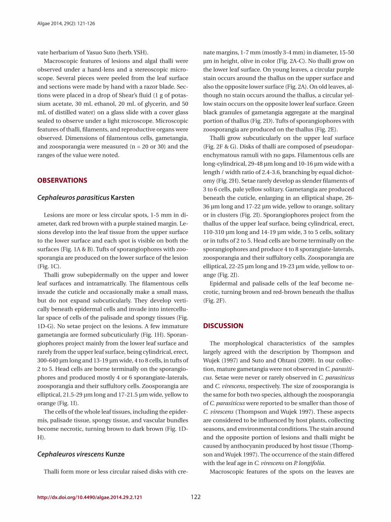
nate margins, 1-7 mm (mostly 3-4 mm) in diameter, 15-50
μm in height, olive in color (Fig. 2A-C). No thalli grow on
the lower leaf surface. On young leaves, a circular purple
stain occurs around the thallus on the upper surface and
also the opposite lower surface (Fig. 2A). On old leaves, al-
though no stain occurs around the thallus, a circular yel-
low stain occurs on the opposite lower leaf surface. Green
black granules of gametangia aggregate at the marginal
portion of thallus (Fig. 2D). Tufts of sporangiophores with
zoosporangia are produced on the thallus (Fig. 2E).
Thalli grow subcuticularly on the upper leaf surface
(Fig. 2F & G). Disks of thalli are composed of pseudopar-
enchymatous ramuli with no gaps. Filamentous cells are
long-cylindrical, 29-48 μm long and 10-16 μm wide with a
length / width ratio of 2.4-3.6, branching by equal dichot-
omy (Fig. 2H). Setae rarely develop as slender filaments of
3 to 6 cells, pale yellow solitary. Gametangia are produced
beneath the cuticle, enlarging in an elliptical shape, 26-
36 μm long and 17-22 μm wide, yellow to orange, solitary
or in clusters (Fig. 2I). Sporangiophores project from the
thallus of the upper leaf surface, being cylindrical, erect,
110-310 μm long and 14-19 μm wide, 3 to 5 cells, solitary
or in tufts of 2 to 5. Head cells are borne terminally on the
sporangiophores and produce 4 to 8 sporangiate-laterals,
zoosporangia and their suffultory cells. Zoosporangia are
elliptical, 22-25 μm long and 19-23 μm wide, yellow to or-
ange (Fig. 2J).
Epidermal and palisade cells of the leaf become ne-
crotic, turning brown and red-brown beneath the thallus
(Fig. 2F).
DISCUSSION
The morphological characteristics of the samples
largely agreed with the description by Thompson and
Wujek (1997) and Suto and Ohtani (2009). In our collec-
tion, mature gametangia were not observed in C. parasiti-
cus. Setae were never or rarely observed in C. parasiticus
and C. virescens, respectively. The size of zoosporangia is
the same for both two species, although the zoosporangia
of C. parasiticus were reported to be smaller than those of
C. virescens (Thompson and Wujek 1997). These aspects
are considered to be influenced by host plants, collecting
seasons, and environmental conditions. The stain around
and the opposite portion of lesions and thalli might be
caused by anthocyanin produced by host tissue (Thomp-
son and Wujek 1997). The occurrence of the stain differed
with the leaf age in C. virescens on P. longifolia.
Macroscopic features of the spots on the leaves are
vate herbarium of Yasuo Suto (herb. YSH).
Macroscopic features of lesions and algal thalli were
observed under a hand-lens and a stereoscopic micro-
scope. Several pieces were peeled from the leaf surface
and sections were made by hand with a razor blade. Sec-
tions were placed in a drop of Shear’s fluid (1 g of potas-
sium acetate, 30 mL ethanol, 20 mL of glycerin, and 50
mL of distilled water) on a glass slide with a cover glass
sealed to observe under a light microscope. Microscopic
features of thalli, filaments, and reproductive organs were
observed. Dimensions of filamentous cells, gametangia,
and zoosporangia were measured (n = 20 or 30) and the
ranges of the value were noted.
OBSERVATIONS
Cephaleuros parasiticus Karsten
Lesions are more or less circular spots, 1-5 mm in di-
ameter, dark red brown with a purple stained margin. Le-
sions develop into the leaf tissue from the upper surface
to the lower surface and each spot is visible on both the
surfaces (Fig. 1A & B). Tufts of sporangiophores with zoo-
sporangia are produced on the lower surface of the lesion
(Fig. 1C).
Thalli grow subepidermally on the upper and lower
leaf surfaces and intramatrically. The filamentous cells
invade the cuticle and occasionally make a small mass,
but do not expand subcuticularly. They develop verti-
cally beneath epidermal cells and invade into intercellu-
lar space of cells of the palisade and spongy tissues (Fig.
1D-G). No setae project on the lesions. A few immature
gametangia are formed subcuticularly (Fig. 1H). Sporan-
giophores project mainly from the lower leaf surface and
rarely from the upper leaf surface, being cylindrical, erect,
300-640 μm long and 13-19 μm wide, 4 to 8 cells, in tufts of
2 to 5. Head cells are borne terminally on the sporangio-
phores and produced mostly 4 or 6 sporangiate-laterals,
zoosporangia and their suffultory cells. Zoosporangia are
elliptical, 21.5-29 μm long and 17-21.5 μm wide, yellow to
orange (Fig. 1I).
The cells of the whole leaf tissues, including the epider-
mis, palisade tissue, spongy tissue, and vascular bundles
become necrotic, turning brown to dark brown (Fig. 1D-
H).
Cephaleuros virescens Kunze
Thalli form more or less circular raised disks with cre-

Suto et al. Cephaleuros in India
123 http://e-algae.kr
Fig. 1. Cephaleuros parasiticus in Syzygium aromaticum. (A) Lesions on leaf surface (left, upper surface; right, lower surface). Notice lesions on both surfaces. (B) Enlarged view of lesions with purple stain on upper surface. (C) Tufts of sporangiophores with zoosporangia forming on lower leaf surface of lesion. (D) Transverse section of lesion showing invasion of filaments (arrows) into tissue of leaf. Necrosis of epidermal cells (ep), palisade cells (ps), and spongy cells (sp). (E) Transverse section of lesion showing development of subepidermal thallus (arrows) of both leaf surfaces. Note necrosis of all leaf tissue, epidermal cells (ep), palisade cells (ps), and spongy cells (sp). (F) Vertical section of lesion showing algal filaments (arrows) expanding subepidermally. (G) Vertical section of lesion showing algal filaments (arrows) expanding among cells of spongy tissue. (H) Transverse section of lesion showing development of thallus (arrows) beneath epidermal cells and formation of immature gametangia (ga) forming beneath cuticle. Necrosis of epidermal cells (ep), palisade cells (ps), and some spongy cells (sp). (I) Sporangiophores (sph) with zoosporangia (sp). Scale bars represent: A, 1 cm; B, 1 mm; C & I, 100 μm; D, E & H, 50 μm; F & G, 10 μm.
A C
D
B
E
G
F
I
H

Algae 2014, 29(2): 121-126
http://dx.doi.org/10.4490/algae.2014.29.2.121 124
Fig. 2. Cephaleuros virescens in Polyalthia longifolia. (A) Thalli with purple stain on upper surface of young leaf. (B) Purple stain not visible on upper surface of old leaf. (C) Enlarged view of a circular disk. (D) Granules of gametangia aggregating. (E) Bush of sporangiophores with zoosporangia formed on thallus. (F) Transverse section of leaf showing development of thallus (arrow) beneath cuticle (cu) of upper leaf surface. Notice necrosis of epidermal cells (ep) and palisade cells (ps). (G) Algal filaments creeping between cuticle (cu) and epidermal cells (ep) of leaf. (H) Surface view of thallus showing pseudoparenchymatous ramuli with no gaps and crenate margin. (I) Surface view of gametangia produced from creeping filamentous cells. (J) Sporangiophore (sph), head cell (hc), suffultory cell (sc), and zoosporangium (sp). Scale bars represent: A & B, 1 cm; C, 1 mm; D & E, 100 μm; F & H-J, 50 μm; G, 10 μm.
A C
D
B
E
G
F
HI
J
Suto et al. Cephaleuros in India
125 http://e-algae.kr
REFERENCES
Chowdary, Y. B. K. & Jose, G. 1979. Biology of Cephaleuros
Kunze in nature. Phykos 18:1-9.
Cunningham, D. D. 1897. On certain diseases of fungal and
algal origin affecting economic plants in India. Sci.
Mem. Med. Off. Army India 10:95-130.
Gokhale, M. V. & Shaikh, S. S. 2012. Host range of a parasitic
alga Cephaleuros virescens Kunz. ex Fri. from Maharash-
tra state, India. Plant Sci. Feed 2:1-4.
Guiry, M. D. & Guiry, G. M. 2014. AlgaeBase. World-wide
electronic publication, National University of Ireland,
Galway. Available from: http://www.algaebase.org. Ac-
cessed Feb 22, 2014.
Krishnamurthy, V. 2000. Algae of India and neighbouring
countries. I. Chlorophycota. Oxford & IBH Publishing
Co., New Delhi, 209 pp.
Mann, H. H. & Hutchinson, C. M. 1904. Red rust; a serious
blight of the tea plant. Indian Tea Assoc. Bull. 4:1-26.
Panikkar, W. V. N., Ampili, P. & Chauhan, V. D. 1989. Observa-
tions on Cephaleuros virescens Kunze from Kerala, India.
J. Econ. Taxon. Bot. 13:67-70.
Petch, T. 1923. The diseases of the tea Bush. MacMillan & Co.,
London, 220 pp.
Ponmurugan, P., Saravanan, D. & Ramya, M. 2010. Culture
and biochemical analysis of a tea algal pathogen, Ceph-
aleuros parasiticus. J. Phycol. 46:1017-1023.
Printz, H. 1939. Vorarbeiten zu einer Monographie der Tren-
tepohliaceae. Nytt Mag. Naturvidensk. 80:137-210.
Ramya, M., Ponmurugan, P. & Saravanan, D. 2013. Manage-
ment of Cephaleuros parasiticus Karst. (Trentepholiales:
Trentepohliaceae), an algal pathogen of tea plant, Ca-
mellia sinensis (L) (O. Kuntze). Crop Protection 44: 66-
74.
Sarma, P. 1986. The freshwater Chaetophorales of New Zea-
land. Beih. Nova Hedwigia Beih. 58:1-169.
Saxena, P. N. 1961. Algae of India. 1. Chaetophorales. Bull.
Natl. Bot. Gard. 57:1-59.
Suto, Y. & Ohtani, S. 2009. Morphology and taxonomy of five
Cephaleuros species (Trentepohliaceae, Chlorophyta)
from Japan, including three new species. Phycologia
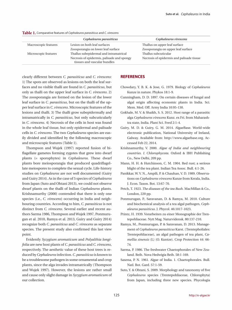
clearly different between C. parasiticus and C. virescens:
1) The spots are observed as lesions on both the leaf sur-
faces and no visible thalli are found in C. parasiticus, but
only as thalli on the upper leaf surface in C. virescens. 2)
The zoosporangia are formed on the lesion of the lower
leaf surface in C. parasiticus, but on the thalli of the up-
per leaf surface in C. virescens. Microscopic features of the
lesions and thalli. 3) The thalli grow subepidermally and
intramatrically in C. parasiticus, but only subcuticularly
in C. virescens. 4) Necrosis of the cells in host was found
in the whole leaf tissue, but only epidermal and palisade
cells in C. virescens. The two Cephaleuros species are eas-
ily divided and identified by the following macroscopic
and microscopic features (Table 1).
Thompson and Wujek (1997) reported fusion of bi-
flagellate gametes forming zygotes that grew into dwarf
plants (= sporophytes) in Cephaleuros. These dwarf
plants bore meiosporangia that produced quadriflagel-
late meiospores to complete the sexual cycle. Life-history
studies on Cephaleuros are not well documented (Guiry
and Guiry 2014). As in the case of 5 species of Cephaleuros
from Japan (Suto and Ohtani 2013), we could not observe
dwarf plants on the thalli of Indian Cephaleuros plants.
Krishnamurthy (2000) contended that there is only one
species (i.e., C. virescens) occurring in India and neigh-
bouring countries. According to him, C. parasiticus is not
distinct from C. virescens. Several earlier and recent au-
thors Sarma 1986, Thompson and Wujek 1997, Ponmuru-
gan et al. 2010, Ramya et al. 2013, Guiry and Guiry 2014)
recognize both C. parasiticus and C. virescens as separate
species. The present study also confirmed this last view
point.
Evidently Syzygium aromaticum and Polyalthia longi-
folia are new host plants of C. parasiticus and C. virescens,
respectively. The aesthetic value of these host trees is re-
duced by Cephaleuros infection. C. parasiticus is known to
be a troublesome pathogen in some ornamental and crop
plants, since the alga invades intramatrically (Thompson
and Wujek 1997). However, the lesions are rather small
and cause only slight damage in Syzygium aromaticum of
our collection.
Table 1. Comparative features of Cephaleuros parasiticus and C. virescens
Cephaleuros parasiticus Cephaleuros virescens
Macroscopic features Lesion on both leaf surfaces Thallus on upper leaf surfaceZoosporangia on lower leaf surface Zoosporangia on upper leaf surface
Microscopic features Thallus subepidermal and intramatrical Thallus subcuticularNecrosis of epidermis, palisade and spongy
tissues and vascular bundlesNecrosis of epidermis and palisade tissue

Algae 2014, 29(2): 121-126
http://dx.doi.org/10.4490/algae.2014.29.2.121 126
Thompson, R. H. & Wujek, D. E. 1997. Trentepohliales: Ceph-
aleuros, Phycopeltis, and Stomatochroon: morphology,
taxonomy, and ecology. Science Publishers Inc., Enfield,
NH, 149 pp.
48:213-236.
Suto, Y. & Ohtani, S. 2013. Seasonal development of five
Cephaleruos species (Trentepohliaceae, Chlorophyta)
on the leaves of woody plants and the behaviors of their
gametes and zoospores. Phycol. Res. 61:105-115.