comparative studies on glial markersjeb.biologists.org/content/jexbio/95/1/167.full.pdf ·...
TRANSCRIPT
J. exp. BioL (1981), 95, 167-180With 2 figuresPrinted m Great Britain
COMPARATIVE STUDIES ON GLIAL MARKERS
BY BETTY I. ROOTS
Department of Zoology and Erindale College, University of Toronto
SUMMARY
Macromolecular markers for glial cells have been sought for a variety ofreasons. One of the earliest was the need for a means of assessing the purityof cell and subcellular fractions prepared from nervous tissue. While there isstill a requirement for this kind of tool, emphasis has shifted towards seekinginformation on biochemical differentiation among cells and their functionalinteractions. A brief general review will be made of glial markers and two ofthese, 2',3'-cyclic nucleotide 3'-phosphohydrolase (CNP) and glutaminesynthetase (GS), will be considered in detail. Until recently studies ofmarkers have been concentrated on the higher vertebrates and those onlower vertebrates and invertebrates have hardly begun. However, suchcomparative studies may lead to fresh insight into old problems. Forexample, CNP has long been regarded as a marker for myelin and oligoden-drocytes but it has not been possible to attribute a functional role to it andits relation to myelination has remained obscure. The finding that it is presentin the glia of a moth Manduca sexta which lacks myelin provides a stimulusfor a fresh approach to the problem. Another example is provided bystudies on GS. This enzyme is found in astrocyte feet and preliminaryresults indicate that it is localized also in the perineurial glia of Aplytiaganglia. These results lead to a reconsideration of the perennial questionof the possible role of astrocyte feet in barrier mechanisms. Extension ofcomparative studies may not only raise, new questions but also providesome answers.
INTRODUCTION
The early motivation for the search for macromolecular markers for specific glialcell types was the need to assess the purity of the fractions separated from the nervoussystem by various means. With the explosion of interest in the bulk preparation ofcells, parts of cells, e.g. neuronal perikarya, and subcellular fractions derived fromspecific cell types, the search for markers was intensified. The development of celllines in culture, especially those derived from tumours also demanded a means ofassessing accurately the nature of the component cells. Often this proves to be acomplex problem, as is the case with the C6 rat glioma clone which manifests abewildering combination of characteristics of both oligodendrocytes and astrocytes.The need for a tool to monitor fractions and tissue cultures still exists. For example,glial fibrillary acidic protein (GFAP) which is a marker for astrocytes has been usedrecently to check the purity of a preparation of Purkinje cells from developing ratcerebellum (Woodhams et al. 1980). Another somewhat different example is the usebf GFAP and glutamine synthetase (GS), another astrocytic marker, to demonstrate
168 BETTY I. ROOTS
the presence of astrocyte feet on microvessels isolated from rat brain (White, Dutton& Norenberg, 1981). Cell type-specific markers for cultures of human neural cellshave been reviewed recently by Kennedy and his co-workers (Kennedy, Lisak &Raff, 1980). However, emphasis has shifted towards seeking information on thedifferentiation and maturation of cells and on the functional interaction between celltypes. Such studies have been hampered by the inability to identify cells at all timesusing morphological criteria alone. The development and use of biochemical markersas a complementary approach in studies of relationships between cells during develop-ment is beginning to be very productive. An example, which will be discussed in moredetail below, is the study of Rhesus monkey brain development by Levitt & Rakic(1980) using localization of GFAP.
One of the limitations in the use of markers is that cells may express the moleculeat only one stage in their development, e.g. GFAP in ependymal cells discussed below(Roessmann et al. 1980), or discontinuously according to their physiological milieu.An example of the latter is the failure of Schwann cells to synthesize galactocerebro-side in culture although they do so in vivo (Raff et al. 1978). On the other hand whereit is possible to identify cells morphologically, markers provide a tool for studying cellmaturation and for probing physiological relationships. Several of the markers, e.g.GS, and carbonic anhydrase (CA), are enzymes and knowledge of their distributionand of their appearance during ontogeny should be particularly useful in giving insightinto the differentiation of cell function and clues to the relationships between cells inthe nervous system.
Morphological characterization of glial cell types in the invertebrates is in its infancy(Roots, 1978; Lane, 1981) and would profit greatly from the application of bio-chemical markers. The definition of cells and their function in phylogeny as well asontogeny should lead to a better understanding of neurone-glia interactions. Pre-liminary results of studies on GS in invertebrate glia, discussed below, indicate thatcomparative studies may provide fresh insight into old problems.
The development of immunocytochemical techniques has led to a rapid burgeoningof studies using glial markers so that much progress has been made since the reviewsof Varon (1978) and Varon & Somjen (1979). Immunological techniques have variousproblems associated with them, for example heterogeneity of the material used toraise the anti-sera, and cross-reactivity. Although detailed discussion of these prob-lems is beyond the scope of the present review, some consideration will be givenwhere appropriate.
A general review of advances made in the field of glial markers follows with par-ticular attention given to the contributions made by comparative studies.
GLIAL FIBRILLARY ACIDIC PROTEIN (GFAP)
Astrocytes, both fibrous and protoplasmic, are characterized by the presence of finefilaments in their cytoplasm which, as the name suggests, are particularly abundantin the fibrous form of the cell. The protein which composes these filaments was firstisolated from multiple sclerosis plaques (Eng et al. 1971) which consist almost entirelyof fibrous astrocytes and thus provide an excellent starting point for the preparationof GFAP. Antibodies to GFAP were prepared and immunological studies of its
Comparative studies on glial markers 169
distribution were launched (Uyeda, Eng & Bignami, 1972). Subsequent preparationsfrom other material, normal human brain tissue, other mammalian brains and nervoustissue of lower vertebrates were contaminated to varying degrees with tubulin andneurofilament protein leading to the suggestion that the proteins may contain similarsubunits (Yen et al. 1976, see also the review by Bignami, Dahl & Rueger, 1980),which has not however been substantiated (Davison & Jones, 1981).
However, as pointed out by Eng (1979) tubulin is much less antigenic than GFAPand antisera raised against a mixture of highly antigenic GFAP and weakly antigenictubulin may still be very useful for immunocytochemical studies provided propercontrol absorptions are made. The problem of tubulin contamination has been over-come by the development of a method of preparation using immunoaffinity chroma-tography (Rueger, Dahl & Bignami, 1978).
Recently the question of the specificity of GFAP for astrocytes has been raisedagain by Jessen and Mirsky (1980) who have demonstrated that enteric glial cells inthe rat myenteric plexus are rich in GFAP and by Yen & Fields (1981) who report theoccurrence of either GFAP or a cross-reacting antigen in rat sciatic nerve. Bignami &Dahl (1977) investigated the occurrence of GFAP in human and rabbit sciatic nervesand found that non-specific fluorescence may occur in vitro with GFAP antisera.
Artefactual staining in sections has not been observed (Bignami & Dahl, 1977) andthe immunoperoxidase staining used in electron microscopy permits the preciselocalization of GFAP with, so far as is known, no redistribution. It is with the use ofthese techniques with this glial marker that recent advances in the analysis of neurone-glia relations have been made.
The main thrust of earlier work, mainly immunofluorescence studies, was theestablishment of the specificity of GFAP for mature astrocytes (Bignami & Dahl,1974 a, 1977) including those in tissue culture (Lim et al. 1978; Sensenbrenner, 1978),and for Bergmann glia, a specialized form of astrocyte found in the cerebellum(Bignami & Dahl, 19746; Schachner et al. 1977). The chief application of these andsimilar studies being the identification of these cells in normal and pathological tissueand the elucidation of the nature of cell lines in culture. Some interest in developmentwas also manifested, e.g. GFAP was shown to be present in radial glia (Antanitus,Choi & Lapham, 1976) lending credance to the view that these cells are related toastrocytes. It is this aspect of development which is receiving attention at the presenttime.
Roessman and his colleagues (Roessman et al. 1980) have followed the occurrenceof GFAP in human ependymal cells during development. Adult ependymal cells lackGFAP but between the 15th week of gestation and full term the protein is present inthe foetal cells. Thus the appearance of GFAP marks a stage in the differentiation ofependymal cells. A curious feature is that adult ependymal cells do possess filamentswhich are similar to those in astrocytes. Possible explanations are that the filamentslose their antigenicity in the adult cell, or that two distinct types of filament are pro-duced at different stages in development. This question has not been resolved. In thisstudy it was shown also that GFAP appears transiently in tanycytes* at a time in
• Tanycytes are glial cells in mammalian brain whose perikarya are in a ventricular position and whichhave long tapering processes which may reach the pia] surface.
170 BETTY I. ROOTS
development which led the authors to conclude that tanycytes represent a form ofependymal differentiation and are not related to radial glia.
In tracing cell lineages during development a major problem has been the inabilityto distinguish cells at early stages of development. Radial glial cells which are re-sponsible for guiding neurones in their migrations during development (Rakic, 1972)are very difficult to distinguish from the elongated migrating neurones. As Levitt &Rakic (1980) point out knowledge of the precise number and distribution of radial gliaduring development would lead to better understanding of the organization of thecentral nervous system and define the extent of glial participation in the guidance anddirection of neurones. The sensitivity and precision of the immunoperoxidase local-ization of GFAP at the electron microscopic level makes such quantitative studiespossible and it has been applied to the problem by Levitt & Rakic (1980) in a study ofradial glial cells and astrocytes in developing Rhesus monkey brain. Several classes ofglial cells were found to be present at considerably earlier stages of development thanpreviously recognized in primate brain. Hitherto it had been thought that only oneclass of cell was present in early development. Radial glia are the first class of astrocyticcell to be detected, appearing in the first dimester of the 165-day gestational period.In all parts of the brain the ventricular and pial surfaces are constantly connected byelongated glia. Many radial cell processes also penetrate the axonal tracts at rightangles, thus a scaffold of pathways is provided for the guidance of neurones throughincreasingly complex terrain. The pattern of the distribution of radial glia processeswas found to be unique to each area of the brain and to change systematically duringdevelopment. When their role in guidance is no longer required radial glia transforminto astrocytes, both protoplasmic and fibrous, thus transitional cells are found firstin areas where cell migration first ceases. Thus the use of the marker GFAP hasprovided new insight into neurone-glia interactions during development.
S-IOO PROTEIN
This highly soluble, highly acidic protein has had a chequered career as a putativeglial marker. Early studies appeared to indicate that it was located specifically in gliaalthough present in both astrocytes and oligodendrocytes. Then followed reports ofits presence in neurones. It was then speculated that the production of S-100 ratherthan its localization constituted the true glial marker (Varon & Somjen, 1979). Now,this year, Ghandour and his colleagues have re-examined the status of S-100 (Ghan-dour et al. 1981). The results of their experiments using light and electron microscopeimmunocytological techniques show that the protein is present only in astrocytes.They argue that previous reports of its occurrence in oligodendrocytes and neuronesare due to a number of different technical artifacts which they analyse and describe.These new findings await confirmation, meanwhile the functional significance ofS-ioo remains obscure.
GLYCEROL-3-PHOSPHATE DEHYDROGENASE (GPDH)
The soluble cytoplasmic enzyme L-a-glycerol-3-phosphate dehydrogenase (EC1.1.1.8) in rat brain is exclusively located in oligodendrocytes which appear to be
Comparative studies on glial markers 171
target cells for glucocorticoids (De Vellis et al. 1978; Leveille et al. 1980). While inrat brain GPDH and its inducibility by hydrocortisone should be viewed as oligo-dendroglial markers it is by no means clear that this is the case in mouse brain or forprimary rat brain glial cultures (see Varon & Somjen, 1979). Its low activity in multiplesclerosis plaques is consistent with a predominantly oligodendrocyte location inhuman brain (Hirsch, Blanco & Parks, 1980).
GALACTOCEREBROSIDE (GC)
Galactocerebroside, the major glycolipid component of myelin, is found also inoligodendrocytes and Schwann cells and has been investigated as a possible markerfor these cells, particularly in culture (Raff et al. 1978; Steck & Perruisseau, 1980). Itis of interest in the present context as an example of a marker whose expressiondepends upon the physiological environment of the cells. Thus Raff and his colleaguesfound that cultured Schwann cells derived from neonatal rat sciatic nerve do notreact with anti-galactocerebroside antisera. On the basis of unpublished observations,they considered it unlikely that the Schwann cells do not make detectable amounts ofGC before myelination or that GC although present is inaccessible to the antibodyand favoured the explanation that Schwann cells normally synthesize GC in vivo butstop doing so in culture.
CARBONIC ANHYDRASE (CA)
Carbonic anhydrase (EC 4.2.1.1) exists in several isozymic forms all of which aremonomeric 30000 dalton proteins. The two major isoenzymes CAI and II havedifferent amino acid sequences and exhibit different enzyme kinetics (Sciaky et al.1976) and are immunologically distinct (Wistrand & Rao, 1968). Not all studies havedistinguished between the isoenzymes but recent reports (Ghandour et al. 1980 a, b)indicate that only CAII is intrinsic to nervous tissue where it is present in bothsoluble and insoluble membrane-bound fractions.
Carbonic anhydrase was first proposed as a general glial marker by Giacobini (1961,1962). Subsequently several groups of investigators have reported its presence inprimary cultures of rat brain astrocytes (Sensenbrenner, 1978; Kimelberg et al. 1978;Church, Kimelberg & Sapirstein, 1980). However, it should be noted that suchcultures contain other cells which may be oligodendroglial in nature (see review byVan Calker & Hamprecht, 1980). The insoluble form of CA was found to be associatedwith myelin (Sapirstein, Lees & Trachtenberg, 1978) which would indicate anoligodendroglial location. Staining of both astrocytes and oligodendrocytes by animmunohistochemical reaction was reported by Roussel and his co-workers (Rousselet al. 1979). Recently, Ghandour and his colleagues (Ghandour et al. 1980 a, b, c)found CA to be located exclusively in oligodendrocytes in rat cerebellum and claim(1980 a) that the astrocyte location found by Roussel et al. is erroneous due to theincorrect identification of cells in their electron micrographs. It is difficult to resolvethis question from an examination of published micrographs, so clearly more workneeds to be done. The answer may well lie in the physiological conditions in whichglial cells of different types express their potential for CA production. (Cf. discussiontf»f GC above.)
172 BETTY I. ROOTS
The primary function of CA is thought to be the catalysis of the reversible hydrationof carbon dioxide. From this stems the potentiality of its involvement in the regulationof pH, secretory activities, photosynthesis and the movement of ions (Giacobini, 1961;Bourke et al. 1975; Bundy, 1977; Kimelberg, Biddlecome & Bourke, 1979). Incultures of astrocytes both norepinephrine and cAMP increase the activity of solubleCA and (Na+ + K+) ATPase. The effects of inhibitors indicate that the response tonorepinephrine is mediated by changes in intracellular cAMP. Since the release ofnorepinephrine is stimulated by increased extracellular concentrations of K+ ions, ithas been suggested that in the nervous system soluble CA plays a role in K+ ionhomeostasis and that its activity is modulated by the putative transmitter norepine-phrine via cAMP (Kimelberg et al. 1978; Church et al. 1980). It appears that mem-brane-associated CA is not affected and its function is enigmatic.
2',3'-CYCLIC NUCLEOTIDE 3'-PHOSPHOHYDROLASE (CNP)
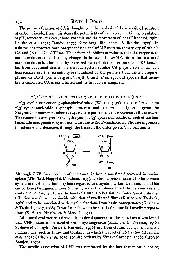
2',3'-cyclic nucleotide 3'-phosphohydrolase (EC 3.1.4.37) is also referred to as2',3'-cyclic nucleotide 3'-phosphodiesterase and has erroneously been given theEnzyme Commission number 3.1.4.16. It is perhaps the most curious of the markers.The reaction it catalyses is the hydrolysis of 2',3'-cyclic nucleotides of each of the fourbases, adenine, guanine, cytidine and undine to the 2'-nucleotides. The rate is greatestfor adenine and decreases through the bases in the order given. The reaction is
HOCHj HOCHj
- HT [ H
OH
HO O
HO—P—OIOH
Although CNP does occur in other tissues, in fact it was first discovered in bovinespleen (Whitfield, Heppel & Markham, 1955), it is found predominantly in the nervoussystem in myelin and has long been regarded as a myelin marker. Drummond and hisco-workers (Drummond, Iyer & Keith, 1962) first showed that the nervous systemcontained at least ten times the level of CNP as other tissues. Subsequently its dis-tribution was shown to coincide with that of myelinated fibres (Kurihara & Tsukada,1967) and to be associated with myelin fractions from brain homogenates (Kurihara& Tsukada, 1967, 1968). It was later shown to be enriched in purified myelin prepara-tions (Kurihara, Nussbaum & Mandel, 1971).
Additional evidence was derived from developmental studies in which it was foundthat CNP increases in parallel with myelinogenesis (Kurihara & Tsukada, 1968;Sarlieve et al. 1976; Toews & Horrocks, 1976) and from studies of myelin-deficientmutant mice, such as Jimpy and Quaking, in which the level of CNP is low (Kuriharaet al. 1971; Sarlieve et al. 1976; see also reviews by Sims & Carnegie, 1978; Varon &Somjen, 1979).
The myelin association of CNP was reinforced by the fact that it could not bej
Comparative studies on glial markers 173
letected in the nervous systems of the Dungeness crab {Cancer magister), the squid{Rossia pacifica), the octopus {Octopus dolfeini), the pink starfish {Pisaster brevisspinosus) and the Pacific prawn {Pondalus platyceros)* all of which are unmyelinated(Drummond, Eng & Mclntosh, 1971).
Like other myelin components, e.g. GC (see above) and myelin basic protein, CNPis found also in oligodendrocytes isolated from the nervous system (Raine, Poduslo &Norton, 1971; Poduslo, 1975; Szuchet & Stefansson, 1980). However, long termcultures of these cells do not show CNP activity. This could be due to their havingdedifferentiated although they still exhibit most of the morphological characteristicsof oligodendrocytes, or they could simply stop expressing this function in culture(Szuchet & Stefansson, 1980). If the latter is the case it is parallel to that of Schwanncells and the synthesis of GC (see above).
The relationship between CNP and myelin, however, is not a simple one. In anumber of elasmobranch fishes, the spiny dogfish, Squalus acanthias (Drummondet al. 1971), the lemon shark, Negaprion brevirostris, the nurse shark, Gmglymostomacirratum, the brown shark, Carcharhinus milberti, and the sting rays, Dasyatis sayi andD. sabina (Trams & Brown, 1974) the forebrain exhibits a higher CNP activity thanmore myelinated medulla or spinal cord. In the dogfish the ratio of CNP activity tocerebroside, a myelin lipid, was higher in the forebrain than in medulla or spinal cord(Drummond et al. 1971). A closer examination of the association of CNP with myelinin mammalian brain reveals a similar apparent anomaly. Although CNP activity i9indeed higher in heavily myelinated areas than in regions containing fewer myelinatedtracts, for example it is high in the spinal cord, the medulla and cerebral white matterbut low in grey matter, it is not related to the amount of myelin as measured by theamount of cerebroside. The CNP/cerebroside ratio in the spinal cord of the dog isabout one-fifth of that for both white and grey matter (Drummond et al. 1971). Fromthese observations it may be concluded that there are regional differences in myelincomposition and that CNP cannot be used as a quantitative marker for myelin.
Myelin is not homogeneous and several fractions can be isolated on sucrose gradi-ents. Compact, multilamellate myelin separates out in the lightest fractions, whereasthe heaviest fraction contains single membrane fragments which are derived frominner and outer lamellae, oligodendroglial membrane, and paranodal segments ofmyelin, and vesicles of unknown origin. The latter fraction has the highest CNPactivity and the lowest myelin basic protein content, while compact myelin has a verylow CNP activity and a high basic protein content (Shapira et al. 1978).
Waehneldt & Lane (1980) studied the distribution of CNP in membrane fractionsduring development and found that it shifted gradually to heavier fractions. Theysuggest that CNP is localized in oligodendroglial membranes, where it precedesmyelin basic protein in development, and is gradually extruded from maturing formsof myelin. Thus in fully mature compact myelin the concentration of CNP is very low.
Very recently Wells & Sprinkle (1981) have purified rat CNP and shown that it isa basic hydrophobic protein with a MW of 48000-50000. Since it shows similar
• Note that only the cerebral and visceral ganglia of the prawn were examined. If this species issimilar to others, e.g. Leander terratus, myelin-like wrappings may occur round axons in the opticpeduncles, circumoesophageal connectives and abdominal cords. However, it is not known whether this
hnyelin-like material shows CNP activity.

174 BETTY I. ROOTS
electrophoretic migration and protein band pattern as Wolfgram proteins and h a s !similar amino acid composition they consider CNP to be a, if not the, primarycomponent of the Wolfgram proteins W1 and W% of myelin. Moreover, not only doantisera to CNP recognize both Wx and W2 but some CNP activity is preserved inWolfgram proteins isolated using protective conditions. The Wolfgram proteins Wx
and Wt have been localized in the dense line of the innermost and outermost myelinlamellae and in the plasma membrane and peripheral cytoplasm of oligodendroglialcells in rat brain by the use of immunocytochemical methods at both light and electronmicroscopical levels (Roussel et al. 1977, 1978; Mandel et al. 1978). Although Rousseland his colleagues (Roussel et al. 1978) considered the limitation of staining to theinner and outermost lamellae to be due to the inaccessibility of Wolfgram proteins inthe compact central lamellae to the antibodies, in view of the finding that there is littleCNP in compact myelin the demonstrated localization probably represents accuratelythe disposition of the proteins.
Supporting evidence for the Wolfgram identify of CNP comes from studies onacclimation to environmental temperature changes in gold fish. As may be seen fromTable 1 the CNP activity of brain myelin isolated from goldfish acclimated to 5 °Cis significantly higher than of that from fish acclimated to 25 °C (D. F. Matheson,
Table 1. Effect of temperature acclimation on the CNP activity of brain myelin of goldfish
CNP activity*Acclimation temperature Mean ±8.D.
5°C 2-337 ±o-434«25 °C
• ftmoles 2'-AMP formed/min/mg myelin protein. Numbers in superscript are number of deter-minations. Value for 50 is significantly higher than 25 °C: P < o-oi (Student's / test, double-tailed).
R. Oei & B. I. Roots, unpublished observations). A 50000 MW protein in goldfishbrain myelin which shows similar electrophoretic migration in SDS gels to a ratWolfgram protein is also present in a larger amount in myelin from fish acclimated to5 °C (D. P. Selivonchick, K. Fujimoto, H. C. Agrawal & B. I. Roots, unpublishedobservations). Since the amounts of cerebroside and total myelin protein do notchange with acclimation temperature (Selivonchick & Roots, 1976), both the CNP/cerebroside and Wolfgram(?)/cerebroside ratios are greater in 50 than in 250 goldfishbrain myelin. This confirms the observations made above that myelin compositionmay vary and that CNP activity cannot be used as a quantitative marker.
It is difficult to conceive of a function for CNP activity in myelin or indeed in anyother tissue. The possibility that the substrate for CNP in vivo is something other thanthe 2',3'-cyclic nucleotides was raised by Olafson and his associates (Olafson, Drum-mond & Lee, 1969). Since there are no reports of 2'-3'-cyclic nucleotides in mam-malian tissues (Sims & Carnegie, 1978) this would seem to be highly probable, at leastfor mammals. As yet, no viable alternative substrate has been suggested. Several linesof evidence point towards a function in membrane reorganization. Firstly there is theclose association of CNP activity with myelinogenesis. Secondly, it has been foundthat CNP activity changes in the optic nerve of goldfish concommitant with theremodelling of the nerve which occurs during acclimation to environmental
Comparative studies on glial markers 175
^ i r e changes. With acclimation the axon diameter distribution spectrum changes, thusin the optic nerve of 5 °C acclimated fish there is a greater proportion of axons largerthan 0-9 /tm in diameter than in 25 °C fish nerves (cf. 71 % versus 22 %) The thick-ness and periodicity of the myelin also change (Matheson et al. 1978; D. F. Matheson,M. S. Diocee, S. T. Hussain & B. I. Roots, unpublished observations). The CNPactivity is significantly higher in the optic nerves of 50 than 25 °C acclimated fish(Matheson, Oei & Roots, 1979). Thus there is both a considerable reorganization ofmyelin and a change in CNP activity. Finally, Sims & Carnegie (1978) report severalinstances of changes in CNP activity in cases where membrane structure is known tobe altered, e.g. in viral transformation of normal tissue. However, there is nothing inany of these lines of evidence to preclude the possibility that CNP is purely a struc-tural protein whose enzyme activity is coincidental. That this may not be so isindicated by some other information. CNP activity is increased two-fold in C6TK~and C9DE clones of the Ce rat glioma by the neurotransmitter norepinephrine. Aneffect which is mediated by a /?-adrenergic receptor and an increase in intracellularcyclic AMP (McMorris, 1977). Whether norepinephrine regulates CNP activity in vivois unknown. In the tobacco horn moth, Manduca sexta, norepinephrine causes a133-fold increase in cyclic AMP levels in the glia. These cells also show CNP activityalthough at a much lower level than is found in myelin (Taylor, Dyer & Newburgh,1976). It would be interesting to know whether or not CNP activity is also stimulatedby norepinephrine in Manduca. There is increasing interest in the role of cyclic AMPas a second messenger and in its relationships with neurotransmitters (Van Calker &Hamprecht, 1980; Levitan & Benson, 1981). One might speculate that somehowCNP is involved in these interactions and their postulated roles in membrane phen-omena, and suggest that a fresh approach to the problem of the function of CNP bemade along these lines.
GLUTAMINE SYNTHETASE(GS)
Glutamine synthetase (L-glutamate: ammonia ligase; ADP-forming; EC 6.3.1.2)catalyses the biosynthetic reaction
glutamate + ammonia + ATP < > glutamine + ADP + Pt.
This reaction serves to detoxify ammonia and is involved in the metabolism of theneurotransmitters y-aminobutyric acid (GABA) and glutamic acid. Both these func-tions are associated with the smaller, fast turnover glutamate compartment of mam-malian brain (Berl & Clarke, 1969), which, it has been suggested, may be representedby the glia (Balazs, Patel & Richter, 1970). Localization of GS in glia by light micro-scopical immunohistochemical techniques (Martinez-Hernandez, Bell & Norenberg,1977) provided evidence that glia do indeed constitute the smaller glutamate compart-ment in mammalian brain. The subsequent demonstration by ultrastructural immuno-cytochemistry that GS is not only restricted to glia but confined to. astrocytes estab-lished the enzyme as an astrocyte marker (Norenberg & Martinez-Hernandez, 1979).It is present in both fibrous and protoplasmic astrocytes, in Bergmann glia, and inMiiller cells in the retina which are also astrocytic in nature (Riepe & Norenberg,^177). High GS activity is shown also by astrocytes in culture (Schousboe, Svenneby
176 BETTY I. ROOTS
and Hertz, 1977). In rat brain trace amounts were also seen in some ependymal ce l^but not in tanycytes, thus confirming the findings from studies of GFAP that epen-dymal cells and astrocytes are related, but that tanycytes represent another form ofependymal differentiation. Comparative studies in a number of vertebrates, man, dog,chick, toad and goldfish on the distribution of GS have shown that in all these animalsthe enzyme has been confined to astrocytes or ependymo-glial cells (M. D. Norenberg,personal communication). In the lower vertebrates ependymal cells predominate andthe main evolutionary development has been a reduction in their number with aprogressive increase in other cell types (see Roots, 1978). Thus it would appear thatastrocytes have assumed the ependymal cell functions associated with GS.
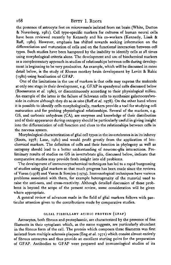
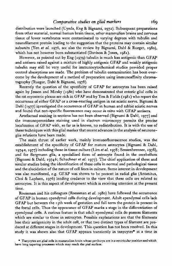
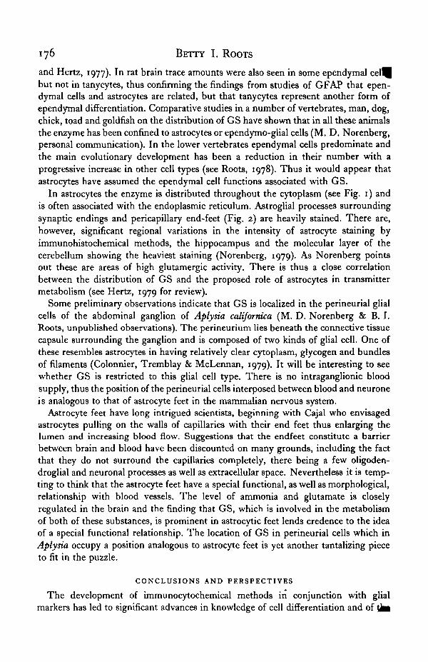
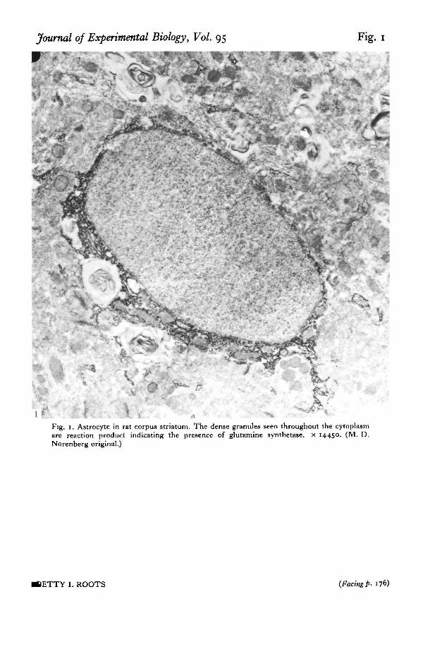
In astrocytes the enzyme is distributed throughout the cytoplasm (see Fig. 1) andis often associated with the endoplasmic reticulum. Astroglial processes surroundingsynaptic endings and pericapillary end-feet (Fig. 2) are heavily stained. There are,however, significant regional variations in the intensity of astrocyte staining byimmunohistochemical methods, the hippocampus and the molecular layer of thecerebellum showing the heaviest staining (Norenberg, 1979). As Norenberg pointsout these are areas of high glutamergic activity. There is thus a close correlationbetween the distribution of GS and the proposed role of astrocytes in transmittermetabolism (see Hertz, 1979 for review).
Some preliminary observations indicate that GS is localized in the perineurial glialcells of the abdominal ganglion of Aplysia californica (M. D. Norenberg & B. I.Roots, unpublished observations). The perineurium lies beneath the connective tissuecapsule surrounding the ganglion and is composed of two kinds of glial cell. One ofthese resembles astrocytes in having relatively clear cytoplasm, glycogen and bundlesof filaments (Colonnier, Tremblay & McLennan, 1979). It will be interesting to seewhether GS is restricted to this glial cell type. There is no intraganglionic bloodsupply, thus the position of the perineurial cells interposed between blood and neuroneis analogous to that of astrocyte feet in the mammalian nervous system.
Astrocyte feet have long intrigued scientists, beginning with Cajal who envisagedastrocytes pulling on the walls of capillaries with their end feet thus enlarging thelumen and increasing blood flow. Suggestions that the endfeet constitute a barrierbetween brain and blood have been discounted on many grounds, including the factthat they do not surround the capillaries completely, there being a few oligoden-droglial and neuronal processes as well as extracellular space. Nevertheless it is temp-ting to think that the astrocyte feet have a special functional, as well as morphological,relationship with blood vessels. The level of ammonia and glutamate is closelyregulated in the brain and the finding that GS, which is involved in the metabolismof both of these substances, is prominent in astrocytic feet lends credence to the ideaof a special functional relationship. The location of GS in perineurial cells which inAplysia occupy a position analogous to astrocyte feet is yet another tantalizing pieceto fit in the puzzle.
CONCLUSIONS AND PERSPECTIVES
The development of immunocytochemical methods in conjunction with glialmarkers has led to significant advances in knowledge of cell differentiation and of tfci
Journal of Experimental Biology, Vol. 95 Fig. 1
Fig. 1. Astrocyte in rat corpus striatum. The dense granules seen throughout the cytoplasmare reaction product indicating the presence of glutamine synthetase. x 14450. (M. D.Norenberg original.)
JETTY I. ROOTS (Facing P- 176)
Journal of Experimental Biology, Vol. 95 Fig. 2
'1Fig, a. Capillary surrounded by astrocyte-feet containing reaction product indicating gluta-
mine synthetase. x 15300. (M. D. Norenberg original.)
BETTY I. ROOTS
Comparative studies on glial markers 177
p i e of glia in the development of the nervous system. An outstanding example is thestudy of the role of radial glia during the development of Rhesus monkey brain, ft isto be expected that many more studies will be made using similar techniques.
Another fruitful line of research using markers, which has barely begun, is the studyof the effects of hormones and transmitters on glial cells. Such studies, as the exampleof the induction of CA by norepinephrine shows, may give clues as to how neuronalactivity may be linked metabolically to that of glia. The pursuit of this line of enquiryhas exciting prospects.
Finally, there is the vast potential of comparative studies yet to be realized.
I thank especially Dr Michael D. Norenberg for his generosity in providing theelectron micrographs which appear as Figs 1 and 2 and for permission to quote someof his unpublished observations. I should also like to express my appreciation to DrNancy J. Lane for her helpful comments on the manuscript. The careful typing ofthe manuscript by Mrs Margaret Clements is gratefully acknowledged.
REFERENCES
ANTANITUS, D. S., CHOI, B. H. & LAPHAM, L. W. (1976). The demonstration of glial fibrillary acidicprotein in the cerebrum of the human fetus by indirect immunofluorescence. Brain Res. 103, 613-616.
BALAZS, R., PATEL, A. J. & RICHTER, D. (1970). Metabolic compartments in the brain: their propertiesand relation to morphological structures. In Metabolic Compartmentation in the Brain (ed. R. Balazsand J. E. Cremer), pp. 167-184. New York: John Wiley.
BERL, S. & CLARKE, D. D. (1969). Compartmentation of amino acid metabolism. In Handbook ofNeuroehemistry, vol. 11 (ed. A. Lajtha), pp. 447-472. New York: Plenum Press.
BICNAMI, A. & DAHL, D. (1974a). Astrocyte-specific protein and neuroglial differentiation. An im-munofluorescence study with antibodies to the glial fibrillary acidic protein. J. comp. Neurol. 153,27-38.
BIONAMI, A. & DAHL, D. (19746). The development of Bergmann glia in mutant mice with cerebellarmalformations: reeler, staggerer and weaver. Immunofluorescence study with antibodies to the glialfibrillary acidic protein. J. comp. Neurol. 155, 219-230.
BIGNAMI, A. & DAHL, D. (1977). Specificity of the glial fibrillary acidic protein for astroglia. J. Hitto-chem. Cytochem. as, 466-469.
BIONAMI, A., DAHL, D. & RUEGER, D. C. (1980). Glial fibrillary acidic protein (GFA) in normal neuralcells and in pathological conditions. Adv. Cell. Neurobiol. 1, 285-310.
BOURKE, R. S., KIMELBERG, H. K., WEST, C. R. & BREMMER, A. M. (1975). The effect of HCO," onthe swelling and ion uptake of monkey cerebral cortex under conditions of raised extracellularpotassium. J. Neurochem. as, 323-328.
BUNDY, H. F. (1977). Carbonic anhydrase. Comp. Biochem. Physiol. 57 B, 1-7.CHURCH, G. A., KIMELBKRO, H. K. & SAPIRSTEIN, V. A. (1980). Stimulation of carbonic anhydrase
activity and phosphorylation in primary astroglial cultures by norepinephrine. J. Neurochem. 34,873-879.
COLONNIER, M., TREMBLAY, J. P. & MCLENNAN, H. (1979). Synaptic contacts on glial cells in theabdominal ganglion of Aplytia califomica. J. comp. Neurol. 188, 391—400.
DAVISON, P. F. & JONES, R. N. (1981). Filament proteins in central, cranial, and peripheral mammaliannerves. J. Cell Biol. 88, 67-72.
DE VELLIS, J., MCGINNIS, G., BREEN, G., LEVEILLB, P., BENNET, K. & MCCARTHY, K. (1978). Hormonaleffects on differentiation in neural cultures. In Cell, Tissue, and Organ Culture in Neurobiology (ed.S. FedorofT and L. Hertz), pp. 485-512. New York: Academic Press.
DRUMMOND, G. I., IYER, N. T. & KEITH, J. (1962). Hydrolysis of ribonucleoside 2',3'-cyclic phosphatesby a diesterase from brain. J. biol. Chem. 337, 3535-3539.
DRUMMOND, G. I., ENO, D. Y. & MCINTOSH, C. A. (1971). Ribonucleoside 2',3'-cyclic phosphatediesterase activity and cerebroside levels in vertebrate and invertebrate nerve. Brain Res. a8, 153-163.
ENO, L. F. (1979). Letter to the Editor. J. Histochem. Cytochem. VJ, 694-696.ENG, L. F., VANDERHAEOHEN, J. J., BICNAMI, A. & GERSTL, B. (1971). An acidic protein isolated from^fc astrocytes. Brain Res. a8, 351-354-
i 7 8 BETTY I. ROOTS
GHANDOUR, M. S., LANGLEY, O. K., VINCENDON, G., GOMBOS, F., FILIPPI, D., LIMOZIN, N., DALMASS^C. & LAURENT, G. (1980a). Immunochemical and immunohistochemical study of carbonic anhydraseII in adult rat cerebellum: a marker for oligodendrocytes. Neuroscience 5, 550-572.
GHANDOUR, M. S., VrNCENDON, G., GOMBOS, G., LIMOZIN, N., FILIPPI, D., DALMASSO, C. & LAURENT,G. (19806). Carbonic anhydrase and oligodendroglia in developing rat cerebellum: a biochemical andimmunohistological study. Devi Biol. 77, 73-83.
GHANDOUR, M. S., VINCENDON, G. & GOMBOS, G. (1980c). Astrocyte and oligodendrocyte distributionin adult rat cerebellum: an immunohistological study. J. Neurocytol. 9, 637-646.
GHANDOUR, M. S., LANGLEY, O. K., LABOURDETTE, G., VINCENDON, G. & GOMBOS, G. (1981). Specificand artefactual cellular localizations of S-100 protein: an astrocyte marker in rat cerebellum. DeviNeurotci. 4, 66-78.
GIACOBINI, E. (1961). Localisation of carbonic anhydrase in the nervous system. Science, N.Y. 134,1524-1525.
GIACOBINI, E. (1062). A cytochemical study of the localization of carbonic anhydrase in the nervoussystem. J. Neurochem. 9, 160-177.
HERTZ, L. (1979). Functional interactions between neurons and astrocytes 1. Turnover and metabolismof putative amino acid transmitters. Prog. Neurobiol. 13, 277-323.
HIRSCH, H. E., BLANCO, C. E. & PARKS, M. E. (1980). Glycerophosphate dehydrogenase: Reducedactivity in multiple sclerosis plaques confirms localization in oligodendrocytes. J. Neurochem. 34,760-762.
JESSEN, K. R. & MIRSKY, R. (1980). Glial cells in the enteric nervous system contain glial fibrillary acidicprotein. Nature, Lond. a86, 736-737.
KENNEDY, P. G. E., LISAK, R. P. & RAFF, M. C. (1980). Cell type-specific markers for human glial andneuronal cells in culture. Lab. Invest. 43, 342—351.
KIMELBERG, H. K., NARUMI, S., BIDDLECOMBB, S. & BOURKE, R. S. (1978). (Na+ + K+) ATPase, 86Eb+transport and carbonic anhydrase activity in isolated brain cells and cultured astrocytes. In DynamicProperties of Glia Cells (ed. E. Schoffeniels, G. Franck, D. B. Towers and L. Hertz), pp. 347-357.Oxford: Pergamon Press.
KIMELBERG, H. K., BIDDLECOME, S. & BOURKE, R. S. (1979). SITS-Inhibitable Cl~ transport in Na+-dependent H+ production in primary astroglial cultures. Brain Res. 173, m-124.
KURIHARA, R. & TSUKADA, Y. (1967). The regional and subcellular distribution of 2',3'-cyclic nucleotidephosphohydrolase in the central nervous system. J. Neurochem. 14, 1167-1174.
KURIHARA, T. & TSUKADA, Y. (1968). a',3'-cyclic nucleotide 3'-phosphohydrolase in the developingchick brain and spinal cord. J. Neurochem. 15, 827-832.
KURIHARA, R., NUSSBAUM, J. L. & MANDEL, P. (1971). 2',3'-cyclic nucleotide 3'-phosphohydrolase inpurified myelin from brain of 'Jimpy' and normal young mice. Life Set. 10 part 2, 421-429.
LANE, N. J. (1981). Organization and structure of invertebrate neuroglia. J. exp. Biol. 00, 000-000.LEVEILLE, P. J., MCGINNIS, J. F., MAXWELL, D. S. & VELLIS, J. DE. (1980). Immunocytochemical
localization of glycerol-3-phosphate dehydrogenase in rat oligodendrocytes. Brain Res. 196, 287-306.LEVITAN, I. B. & BENSON, J. A. (1981). Neuronal oscillators in Aplysia: modulation by serotonin and
cyclic AMP. Trends in Neurosciences 4, 38-41.LEVITT, P. & RAKIC, P. (1980). Immunoperoxidase localization of glial fibrillary acidic protein in radial
glial cells and astrocytes of the developing Rhesus monkey brain. J. comp. tfeurol. 193, 815-840LIM, R., TURRIFF, D., TROY, S. & KATO, T. (1978). Differentiation of glioblasts under the influence of
glia maturation factor. In Cell, Tissue and Organ Culture in Neurobiology (ed. S. Fedoroff and L.Hertz), pp. 223-236. New York: Academic Press.
MANDEL, P., ROUSSEL, G., DELAUNOY, J.-P. & NUSSBAUM, J.-L. (1978). Wolfgram proteins, oligoden-droglial cell and myelin markers, carbonic anhydrase C, a glial marker. In Dynamic Properties of GliaCells (ed. E. Schoffeniels, G. Franck, D. B. Towers and L. Hertz), pp. 267-274. Oxford: PergamonPress.
MARTINEZ-HERNANDEZ, A., BELL, K. P. & NORENBERO, M. D. (1977). Glutamine synthetase: gliallocalization in brain. Science, N.Y. 195, 1356-1358.
MATHESON, D. F., DIOCEE, M., HUSSAIN, S. T. & ROOTS, B. I. (1978). Microtubules in optic nerves oftemperature acclimated goldfish. 9th International Congress on Electron Microscopy, Toronto, 1978,vol. 11, Biology, Microsc. Soc. of Canada, Toronto, Ont., pp. 268-269.
MATHESON, D. F., OEI, R. & ROOTS, B. I. (1979). 2',3'-cyclic nucleotide-3'-phosphohydrolase (CNP)activity in nervous tissue of temperature acclimated goldfish (Carassius auratus L.). Soc. Neurosci.Abst. 5, p. 409.
MCMORRIS, F. A. (1977). Norepinephrine induces glial-specinc enzyme activity in cultured glioma cells.Proc. natn. Acad. Sci. U.S.A. 74, 4501-4504.
NORENBERG, M. D. (1979). The distribution of glutamine synthetase in the rat central nervous system.J. Histochem. Cytochem. vj, 756-762.
NORENBERG, M. D. & MARTINEZ-HERNANDEZ, A. (1979). Fine structural localization of gsynthetase in astrocytes of rat brain. Brain Res. 161, 303—310.
Comparative studies on glial markers 179
IAFSON, R. W., DRUMMOND, G. I. & LEE, J. F. (1969). Studies on a'j'-cyclic nucleotide-3'-phospho-hydrolase from brain. Can. J. Biochem. 47, 961-966.
PODUSLO, S. E. (1975). The isolation and characterization of a plasma membrane and a myelin fractionderived from oligodendroglia of calf brain. J. Neurochem. 34, 647-654.
RAFF, M. C , MIRSKY, R., FIELDS, K. L., LISAK, R. P., DORFMAN, S. H., SILBERBERO, D. H., GREGSON,N. A., LEIBOWITZ, S. & KENNEDY, M. C. (1078). Galactocerebroside is a specific cell surface antigenicmarker for oligodendrocytes in culture. Nature, Land. 374, 813-815.
RAINE, C. S., PODUSLO, S. E. & NORTON, W. T. (1971). The ultrastructure of purified preparations ofneurons and glial cells. Brain Res. 37, 11-24.
RAKIC, P. (197a). Mode of cell migration to the superficial layers of fetal monkey neocortex. J. comp.Neurol. 145, 61-84.
RIEPE, R. E. & NoBENBKRO, M. D. (1977). MUller cell localisation of glutamine synthefase in rat retina.Nature, Lond. 368, 654-655.
ROESSMANN, U., VBLASCO, M. E., SrNDELY, S. D. & GAMBETTI, P. (1980). Glial fibrillary acidic protein(GFAP) in ependymal cells during development. An immunocytochemical study. Brain Res. 300,13-31.
ROOTS, B. I. (1978). A phylogenetic approach to the anatomy of glia. In Dynamic Properties of Glia Cells(ed. E. Schoffeniels, G. Franck, D. B. Towers and L. Hertz), pp. 45-54. Oxford: Pergamon Press.
ROUSSEL, G., DELAUNOY, J.-P., NUSSBAUM, J.-L. & MANDEL, P. (1977). Immunohistochemical localiza-tion of Wolfgram proteins in nervous tissue of rat brain. Neurosdence 3, 307-313.
ROUSSEL, G., DELAUNOY, J.-P., MANDEL, P. & NUSSBAUM, J.-L. (1978). Ultrastructural localizationstudy of two Wolfgram proteins in rat brain tissue. J. Neurocytol. 7, 155-163.
ROUSSEL, G., DELAUNOY, J.-P., NUSSBAUM, J.-L. & MANDEL, P. (1979). Demonstration of a specificlocalization of carbonic anhydrase C in the glial cells of rat CNS by an immunohistochemical method.Brain Ret. 160, 47-55.
RUEGER, D. 'C, DAHL, D. & BIGNAMI, A. (1978). Purification of a brain-specific astroglial protein byimmunoaffinity chromatography. Analyt. Biochem. 89, 360-371.
SAPIRSTEIN, V. S., LEES, M. B. & TRACHTENBERC, M. C. (1978). Soluble and membrane bound carbonicanhydrases from rat CNS: regional development. J. Neurochem. 31, 283—287.
SARLIEVE, L. L., FAROOQUI, A. A., REBEL, G. & MANDEL, P. (1976). Arylsulphatase A and 2',3'-cyclicnucleotide 3'-phosphohydrolase activities in the brains of myelin deficient mutant mice. Neurosdencel, 519-5"-
SCHACHNER, M., HEDLEY-WHYTE, E. T., HSU, D. W., SCHOONMAKER, G. & BIGNAMI, A. (1977).Ultrastructural localization of glial fibrillary acidic protein in mouse cerebellum by immunoperoxioUselabelling. J. Cell Biol. 75, 67-73.
SCHOU8BOE, A., SVENNEBY, G. & HERTZ, L. (1977). Uptake and metabolism of glutamate in astrocytescultured from dissociated mouse brain hemispheres. J. Neurochem. 39, 999-1005.
SCIAKY, M., LIMOZIN, N., FILIPPI-FOVEAU, D., GULIAN, J.-M. & LAURENT-TABUSSE, G. (1976).Structure primaire de l'anhydrase carbonique erythrocytaire bovine CI.II. Sequence complete.Biochimie 58, 1071-1082.
SELIVONCHICK, D. P. & ROOTS, B. I. (1976). Variation in myelin lipid composition induced by changein environmental temperature of goldfish (Carassius auratus L.). J. Therm. Biol. 1, 131-135.
SENSENBRENNER, M. (1978). Dissociated brain cells in primary cultures. In Cell, Tissue and OrganCultures in Neurobiology (ed. S. Fedoroff and L. Hertz), pp. 191-214. New York: Academic Press.
SHAPIRA, R., MOBLBY, W. C, THIELE, S. B., WILHELM, M. R., WALLACE, A. & KIBLER, R. F. (1978).Localization of 2',3'-cydic nucleotide 3'-phosphohydrolase of rabbit brain by sedimentation in acontinuous sucrose gradient J. Neurochem. 30, 735-744.
SIMS, N. R. & CARNEGIE, P. R. (1978). a',3'-cyclic nucleotide 3'-phosphodiesterase. Adv. Neurochem.3. i-4i-
STECK, A. J. & PERRUISSEAU, G. (1980). Characterization of membrane markers of isolated oligoden-drocytes and clonal lines of the nervous system. J. Neurol. Sci. 47, 135-144.
SZUCHET, S. & STEFANSSON, K. (1980). In vitro behavior of isolated oligodendrocytes. Adv. Cell.Neurobiol. 1, 313-346.
TAYLOR, D. P., DYER, K. A. & NEWBURGH, R. W. (1976). Cyclic nucleotides in neuronal and glial-enriched fractions from the nerve cord of Manduca sexta. J. Insect Physiol. 33, 1303-1304.
TOEWS, A. D. & HORROCKS, L. A. (1976). Developmental and ageing changes in protein concentrationof 2',3'-cyclic nucleoside monophosphate phosphodiesterase activity (EC3.1.4.16) in human cerebralwhite and grey matter and spinal cord. J. Neurochem. 37, 545—550.
TRAMS, E. G. & BROWN, E. A. B. (1974). The activity of 2',3'-cyclic adenosine monophosphate 3'-phosphoesterhydrolase in elasmobranch and teleost brain. Comp. Biochem. Physiol. B 48, 185-189.
UYEDA, C. T., ENG, L. F. & BIONAMI, A. (1972). Immunological study of the glial fibrillary acidicprotein. Brain Res. 37, 81-89.
MCALKER, D. & HAMPRECHT, B. (1980). Effects of neurohormones on glial cells. Adv. Cell. Neurobiol.31-68.
180 BETTY I. ROOTS
VARON, S. (1978). Macromolecular glial cell markers. In Dynamic Properties of Glia Cells (ed.Schoffeniels, G. Franck, D. B. Towers and L. Hertz), pp. 93-103. Oxford: Pergamon Press.
VARON, S. S. & SOMJEN, G. G. (1979). Neuron-glia interactions. Neurotci. Res. Prog. Bull. 17, no. 1,pp. 1-239.
WAEHNHLDT, T. V. & LANE, J. D. (1980). Dissociation of myelin from its 'enzyme markers' duringontogeny. J. Neurocliem. 35, 566-573.
WELLS, M. R. & SPRINKLE, T. J. (1981). Purification of rat 2',3'-cyclic nucleotide 3'-phosphodiesterase.J. Neurochem. 36, 633-639.
WHITE, F. P., DUTTON, G. R. & NORENBERG, M. D. (1981). Microvessels isolated from rat brain:localization of astrocyte processes by immunohistochemical techniques. 3- Neurochem. 36, 328—332.
WHITFIELD, P. R., HEPPEL, L. A. & MARKHAM, R. (1955). The enzymic hydrolysis of ribonucleoside-2',3'-pho»phates. Biochem. 3- 60, 15—19.
WISTRAND, P. J. & RAO, S. N. (1968). Immunologic and kinetic properties of carbonic anhydrases fromvarious tissues. Biockim. biophys. Acta 154, 130-144.
WOODHAMS, P. L., COHEN, J., MALLET, J. & BALAZS, R. (1980). A preparation enriched in Purkinje cellsidentified by morphological and immunocytochemical criteria. Brain Res. 199, 435-442.
YEN, S.-H., DAHL, D., SCHACHNER, M. & SHELANSKI, M. L. (1976). Biochemistry of the filaments ofbrain. Proc. natn. Acad. Sci. U.S.A. 73, 529-533.
YEN, S.-H. & FIELDS, K. L. (1981). Antibodies to neurofilament, glial filament, and fibroblast inter-mediate filament proteins bind to different cell types of the nervous system. 3- Cell Bioi. 88, 115-126.