completed ts flyer - european community reference ... · negative seed stocks and use of biosecure...
TRANSCRIPT
European Community Reference Laboratory for Crustacean Diseases leaflet 2008
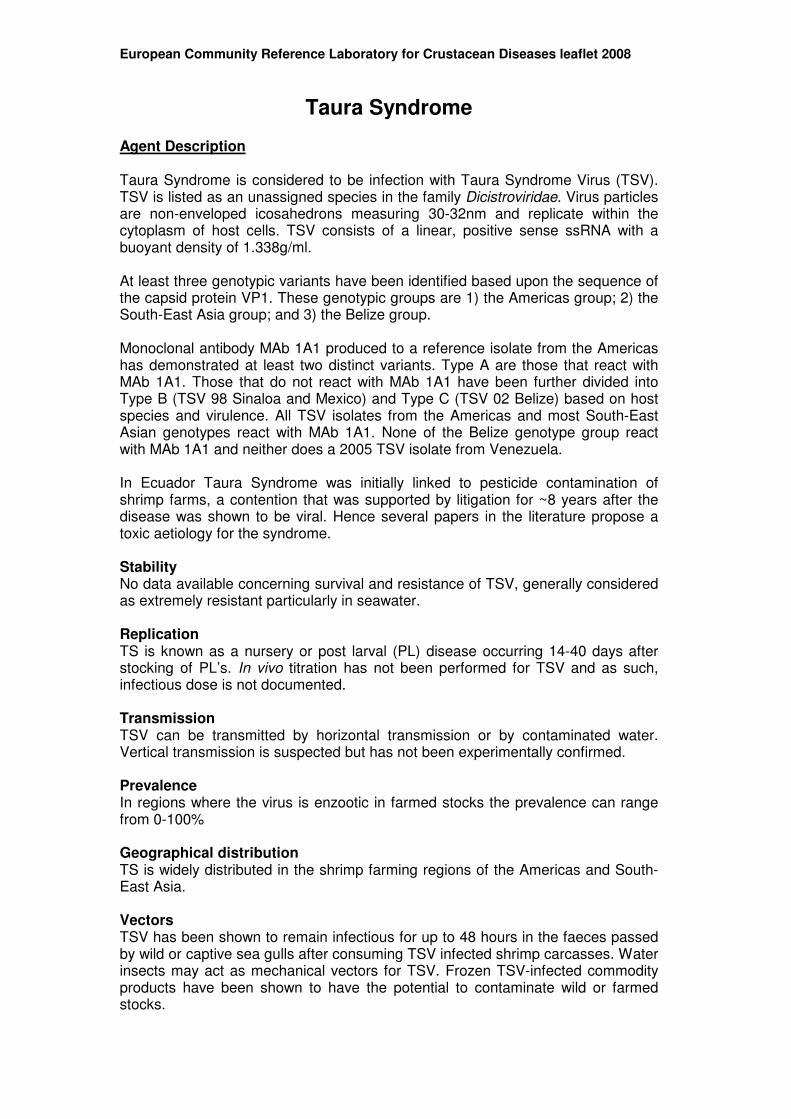
Taura Syndrome Agent Description Taura Syndrome is considered to be infection with Taura Syndrome Virus (TSV). TSV is listed as an unassigned species in the family Dicistroviridae. Virus particles are non-enveloped icosahedrons measuring 30-32nm and replicate within the cytoplasm of host cells. TSV consists of a linear, positive sense ssRNA with a buoyant density of 1.338g/ml. At least three genotypic variants have been identified based upon the sequence of the capsid protein VP1. These genotypic groups are 1) the Americas group; 2) the South-East Asia group; and 3) the Belize group. Monoclonal antibody MAb 1A1 produced to a reference isolate from the Americas has demonstrated at least two distinct variants. Type A are those that react with MAb 1A1. Those that do not react with MAb 1A1 have been further divided into Type B (TSV 98 Sinaloa and Mexico) and Type C (TSV 02 Belize) based on host species and virulence. All TSV isolates from the Americas and most South-East Asian genotypes react with MAb 1A1. None of the Belize genotype group react with MAb 1A1 and neither does a 2005 TSV isolate from Venezuela. In Ecuador Taura Syndrome was initially linked to pesticide contamination of shrimp farms, a contention that was supported by litigation for ~8 years after the disease was shown to be viral. Hence several papers in the literature propose a toxic aetiology for the syndrome. Stability No data available concerning survival and resistance of TSV, generally considered as extremely resistant particularly in seawater. Replication TS is known as a nursery or post larval (PL) disease occurring 14-40 days after stocking of PL’s. In vivo titration has not been performed for TSV and as such, infectious dose is not documented. Transmission TSV can be transmitted by horizontal transmission or by contaminated water. Vertical transmission is suspected but has not been experimentally confirmed. Prevalence In regions where the virus is enzootic in farmed stocks the prevalence can range from 0-100% Geographical distribution TS is widely distributed in the shrimp farming regions of the Americas and South-East Asia. Vectors TSV has been shown to remain infectious for up to 48 hours in the faeces passed by wild or captive sea gulls after consuming TSV infected shrimp carcasses. Water insects may act as mechanical vectors for TSV. Frozen TSV-infected commodity products have been shown to have the potential to contaminate wild or farmed stocks.

European Community Reference Laboratory for Crustacean Diseases leaflet 2008
Mortality On-farm epizootics of TS involving Penaeus vannamei (principle host species) typically result in cumulative mortalities of between 40 and 100 %. Survivors of TS outbreaks may carry the virus for life. Control and Prevention No effective vaccines for TSV are available. TSV-resistant stocks of L. vannamei and L. stylirostris have been reported. Use of specific pathogen free (SPF) or PCR-negative seed stocks and use of biosecure water and culture systems. Gross Pathology Penaeid shrimp TSV has three distinct phases, which are grossly distinguishable: Acute - shrimp have a general pale reddish colouration with the tail fan and pleopods appearing hyperpigmented (red) due to the expansion of chromatophores. Typically the cuticle is soft and the gut empty, and infected shrimps may not survive ecdysis. Moribund shrimp accumulate at the pond surfaces and edges. During outbreaks, birds may be seen feeding on moribund shrimp. Transition (Recovery) – Shrimp exhibit random, multifocal, irregularly shaped melanised cuticular lesions. These melanised spots are haemocytic aggregations indicating sites resolving from TSV infection. These shrimp may or may not have soft cuticle and red-chromatophore expansion, and may feed and behave normally. Chronic – Following successfully moulting, shrimp move into the chronic phase of TS in which animals show no obvious signs of disease. Chronically infected shrimp may however be less resistant to normal environmental stressors than uninfected shrimp and appear to become persistent carriers. Crabs, crayfish, freshwater prawns, spiny lobsters and clawed lobsters Unknown susceptibility and pathological outcome in the majority of species. Clinical Pathology Histology In penaeid shrimp, TSV has been shown to infect the cuticular epithelium, foregut, hindgut, gills, appendages, haematopoietic tissues, lymphoid organ and antennal gland. The hepatopancreas, midgut, midgut caeca, smooth cardiac muscle, striated muscle, ventral nerve cord and ganglia typically show no histological signs of infection. Acute – Multifocal areas of necrosis in the cuticular epithelium and epithelium of the gills, hindgut and foregut. Affected cells display increased eosinophillia of the cytoplasm and pyknotic or karyorrhectic nuclei. Cytoplasmic remnants of necrotic cells are often present in these acute phase lesions and appear as spherical bodies (1 - 20 µm diameter) that range in staining from eosinophilic to pale basophilic. These structures along with pyknotic and karyorrhectic nuclei give the lesions a “peppered” or “buckshot-riddled” appearance. In TSV infected tissues pyknotic or karyorrhectic nuclei stain positively with Feulgen stain distinguishing them from less basophilic to eosinophillic cytoplasmic inclusions that do not contain DNA.
European Community Reference Laboratory for Crustacean Diseases leaflet 2008
The absence of necrosis of the lymphoid organ in TSV infections distinguishes TS disease from acute Yellowhead disease. Transition (Recovery) – Cuticular lesions decline in abundance and severity and are replaced by infiltration and accumulation of haemocytes at the sites of necrosis. Haemocyte aggregates progress to melanised granulomatous lesions giving rise to irregular black spots on the cuticle that characterise the transition phase of the disease. Lesions may show erosion of the cuticle, surface colonisation and invasion of the affected cuticle and exposed haemocytes with secondary pathogens (e.g. Vibrio spp.). Chronic – Presence of numerous prominent lymphoid organ spheroids (LOS) which may remain associated with the main body of the lymphoid organ or which may detach and become ectopic LOS bodies that lodge in constricted areas of the haemocoel (heart, gills, subcuticular connective tissues etc). Representative images of gross and histopathological lesions associated with TS are shown in Annex 1. In situ hybridisation (ISH) Transition - Sections of lymphoid organ may appear normal when stained with H&E however when the same tissue is assayed for TSV by ISH, TSV aggregates are shown in peripheral parenchymal cells of the lymphoid organ tubules. Chronic – When assayed by ISH, some cells within the LOS give positive reactions for the virus while no other target tissues react. Susceptible Species A comprehensive assessment of host susceptibility to TS has recently been completed by EFSA (EFSA 2008). The assessment takes in to account the key susceptibility criteria of pathogen replication, bioassay, characteristic pathology and anatomical location of pathogen and has critically assessed the available literature on host-pathogen interaction with respect to TS. Gulf white shrimp (Penaeus setiferus), Pacific blue shrimp (P. stylirostris), and Pacific white shrimp (P. vannamei) are currently listed as species susceptible to TS virus in the Directive 2006/88/EC. However, the EFSA assessment identified scientific literature to confirm susceptibility of P. vannamei, P. duorarum, P. monodon, P. setiferus, P. chinensis, P. stylirostris, P. aztecus, and Metapenaeus ensis. Some information is available to also suggest susceptibility of P. schmitti and P. japonicus though not all of the EFSA criteria were met in these cases.
OIE Recommended Techniques for Surveillance and Confirmation The methods listed in the table below are the OIE recommended techniques for surveillance and confirmation testing:

European Community Reference Laboratory for Crustacean Diseases leaflet 2008
Pathogen Surveillance (Larvae, PLs, Juveniles and Adults)
Confirmatory Techniques
Taura Syndrome Virus
RT-Polymerase Chain Reaction (RT-PCR)
Histology, DNA probes in situ, RT-PCR and Sequencing
Surveillance Techniques An RT-PCR method that amplifies a 231bp fragment of the TSV genome is reported by Nunan et al. (1998) while a real time PCR assay with increased speed, sensitivity and specificity is reported by Tang et al. (2004). Both techniques are detailed in the most recent OIE Manual of Diagnostic Tests for Aquatic Animals. Confirmation of TSV can be achieved by nucleotide sequencing of the PCR product. Confirmatory Tests Histology Anaesthetise by immersing in ice until immobilised. Small animals (e.g. shrimp) can be fixed and prepared whole by injection of Davidson’s seawater fixative (for marine species) or neutral buffered formalin (for freshwater species), followed by transfer to a larger volume of the same fixative for 24-48 hrs. Fixed specimens should be transferred to 70% industrial methylated spirit (IMS) for storage or shipping prior to histological preparation. For larger animals, dissect the sub-cuticular epidermis, gut, gill, heart, gonad, nervous tissue, body musculature and lymphoid organ and place immediately into Davidson's seawater fixative for 24-48hrs followed by transfer to 70% IMS. Samples can then be infiltrated with paraffin under vacuum according to standard histology protocols and sections cut at a thickness of 3-5 µm, mounted onto glass slides, and stained with haematoxylin and eosin (H&E) or Feulgen stain. In situ hybridisation (ISH) A cDNA probe for TSV provides excellent diagnostic sensitivity via the non-radioactive, DIG-labelled probe used in dot blot and in situ hybridisation assays (see most recent OIE Manual of Diagnostic Tests for Aquatic Animals). RT-Polymerase Chain Reaction (RT-PCR) As for surveillance Sequencing For confirmation of TSV, the amplified product from the RT-PCR assay should undergo nucleotide sequencing. If a positive result is obtained, compare the sequences to available databases using the Basic Local Alignment Search Tool (BLAST) to determine approximate phylogenetic affiliations. If a negative result is obtained the sample should be tested again. EU-legislation Taura Syndrome is listed in the new EU Directive 2006/88/EC as an exotic pathogen.

European Community Reference Laboratory for Crustacean Diseases leaflet 2008
OIE Reference Laboratory Prof. Donald V. Lightner Aquaculture Pathology Laboratory, Department of Veterinary Science and Microbiology, University of Arizona Building 90, Room 202 Pharmacy/Microbiology, Tucson, AZ 85721 UNITED STATES OF AMERICA Tel: (1.520) 621.84.14 Fax: (1.520) 621.48.99 Email: [email protected] References Nunan, L.M., Poulos, B.T., Lightner, D.V. (1998) Reverse transcription polymerase chain reaction (RT-PCR) used for the detection of Taura Syndrome Virus (TSV) in experimentally infected shrimp. Dis. Aquat. Org., 34, 87-91 Tang, K.F.J., Wang, J. and Lightner, D.V. (2004) Quantitation of Taura Syndrome Virus by real-time RT-PCR with a TaqMan assay. J. Virol. Methods, 115, 109-114.
European Community Reference Laboratory for Crustacean Diseases leaflet 2008
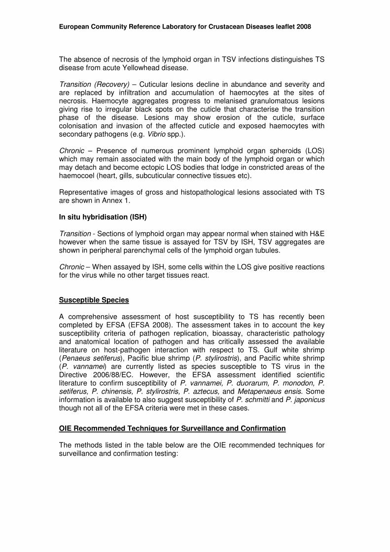
Annex 1 Gross signs and histopathology of TS
Fig. 1 A and B. Acute phase TSV lesions in subcuticular epidermis of P. vannamei
A
B
European Community Reference Laboratory for Crustacean Diseases leaflet 2008
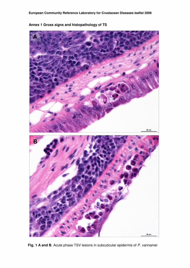
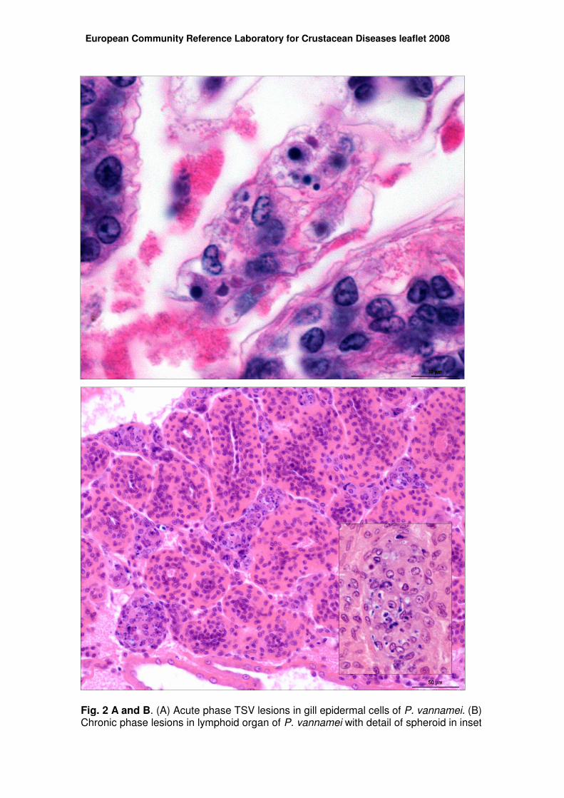
Fig. 2 A and B. (A) Acute phase TSV lesions in gill epidermal cells of P. vannamei. (B) Chronic phase lesions in lymphoid organ of P. vannamei with detail of spheroid in inset
European Community Reference Laboratory for Crustacean Diseases leaflet 2008
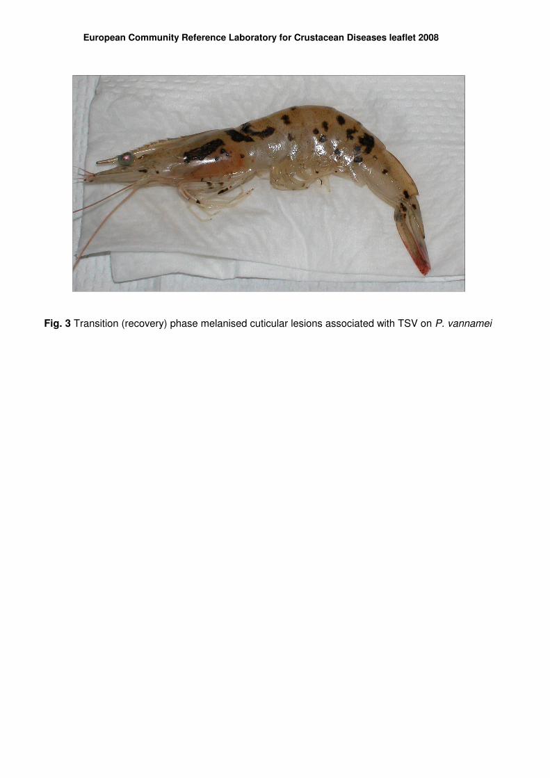
Fig. 3 Transition (recovery) phase melanised cuticular lesions associated with TSV on P. vannamei
European Community Reference Laboratory for Crustacean Diseases leaflet 2008