complex coronal shear fractures of the distal...
TRANSCRIPT
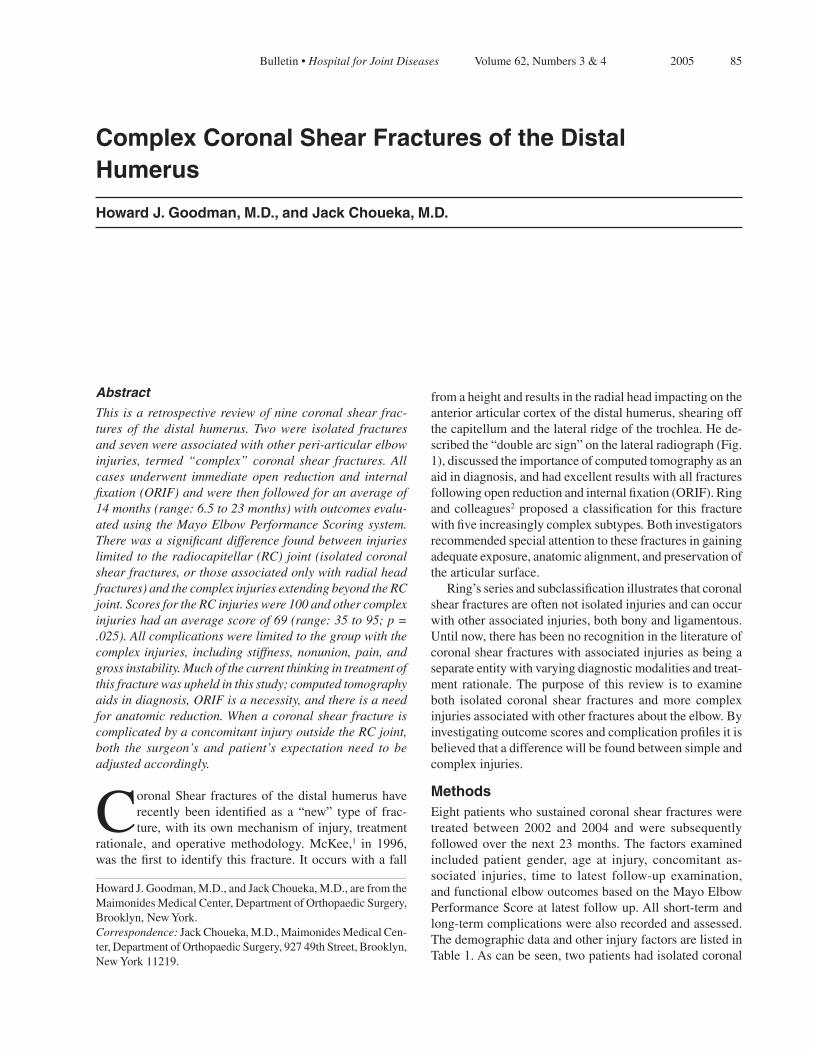
85 Bulletin• Hospital for Joint Diseases Volume62,Numbers3&4 2005
Abstract
This is a retrospective review of nine coronal shear frac-tures of the distal humerus. Two were isolated fractures and seven were associated with other peri-articular elbow injuries, termed “complex” coronal shear fractures. All cases underwent immediate open reduction and internal fixation (ORIF) and were then followed for an average of 14 months (range: 6.5 to 23 months) with outcomes evalu-ated using the Mayo Elbow Performance Scoring system. There was a significant difference found between injuries limited to the radiocapitellar (RC) joint (isolated coronal shear fractures, or those associated only with radial head fractures) and the complex injuries extending beyond the RC joint. Scores for the RC injuries were 100 and other complex injuries had an average score of 69 (range: 35 to 95; p = .025). All complications were limited to the group with the complex injuries, including stiffness, nonunion, pain, and gross instability. Much of the current thinking in treatment of this fracture was upheld in this study; computed tomography aids in diagnosis, ORIF is a necessity, and there is a need for anatomic reduction. When a coronal shear fracture is complicated by a concomitant injury outside the RC joint, both the surgeon’s and patient’s expectation need to be adjusted accordingly.
CoronalShear fracturesof thedistalhumerushaverecently been identified as a “new” type of frac-ture,withitsownmechanismofinjury,treatment
rationale, and operative methodology. McKee,1 in 1996,wasthefirsttoidentifythisfracture.Itoccurswithafall
fromaheightandresultsintheradialheadimpactingontheanteriorarticularcortexofthedistalhumerus,shearingoffthecapitellumandthelateralridgeofthetrochlea.Hede-scribedthe“doublearcsign”onthelateralradiograph(Fig.1),discussedtheimportanceofcomputedtomographyasanaidindiagnosis,andhadexcellentresultswithallfracturesfollowingopenreductionandinternalfixation(ORIF).Ringandcolleagues2proposedaclassificationfor thisfracturewithfiveincreasinglycomplexsubtypes.Bothinvestigatorsrecommendedspecialattentiontothesefracturesingainingadequateexposure,anatomicalignment,andpreservationofthearticularsurface. Ring’sseriesandsubclassificationillustratesthatcoronalshearfracturesareoftennotisolatedinjuriesandcanoccurwithotherassociatedinjuries,bothbonyandligamentous.Untilnow,therehasbeennorecognitionintheliteratureofcoronalshearfractureswithassociatedinjuriesasbeingaseparateentitywithvaryingdiagnosticmodalitiesandtreat-ment rationale.Thepurposeof this review is toexamineboth isolated coronal shear fractures and more complexinjuriesassociatedwithotherfracturesabouttheelbow.Byinvestigatingoutcomescoresandcomplicationprofilesitisbelievedthatadifferencewillbefoundbetweensimpleandcomplexinjuries.
MethodsEightpatientswhosustainedcoronalshearfracturesweretreated between 2002 and 2004 and were subsequentlyfollowedover thenext23months.The factorsexaminedincluded patient gender, age at injury, concomitant as-sociated injuries, time to latest follow-up examination,andfunctionalelbowoutcomesbasedontheMayoElbowPerformanceScoreatlatestfollowup.Allshort-termandlong-termcomplicationswerealsorecordedandassessed.ThedemographicdataandotherinjuryfactorsarelistedinTable1.Ascanbeseen,twopatientshadisolatedcoronal
Complex Coronal Shear Fractures of the Distal Humerus
Howard J. Goodman, M.D., and Jack Choueka, M.D.
HowardJ.Goodman,M.D.,andJackChoueka,M.D.,arefromtheMaimonidesMedicalCenter,DepartmentofOrthopaedicSurgery,Brooklyn,NewYork.Correspondence: JackChoueka,M.D.,MaimonidesMedicalCen-ter,DepartmentofOrthopaedicSurgery,92749thStreet,Brooklyn,NewYork11219.

86 Bulletin• Hospital for Joint Diseases Volume62,Numbers3&4 2005
shearfractures,andtwopatientshadassociatedradialheadfractures.These were considered “injuries limited to theradiocapitellar (RC) joint.”Theotherpatients’associatedfracturesarealsolisted. Allelbowswereimmediatelytestedforstabilityintheoperatingroomandimmobilizedinatemporarysplint.Re-habilitationwasbasedontheinjurytypeandstabilitywithrangeofmotionexercisesstartednolaterthantwoweekspostoperatively.
Surgical TechniqueOperativefixationistheonlyreportedmethodologyfortreatmentofthesefracturesandwastheonlytreatmentofferedinthisseries.Afterpresentingtotheemergencydepartment,patientsunderwentappropriateradiographicstudies,withaCTscanoftenusedtodelineatethefrac-
tureandassistinpreoperativeplanning.Assoonaswasmedicallyfeasible,thepatientsunderwentORIFforana-tomicrealignmentofthedistalhumeralarticularsurface.Inthemorecomplexinjuries,theconcomitantinjuriesmustalsoberigidlyfixed,allowingforearlymotion. WheninjuriesareisolatedtotheRCjoint,thelateralapproach to the elbow provides excellent exposure tovisualizethearticularsurfaces.Stamatis3andImatani4both approached all cases by this modified extensilelateralKocherapproach.Whilethismightbesufficientforfixationofasimplecapitellumfracture,thecoronalshearfracturerequiresamoreextensileexposureoftheanteriorhumeralarticularsurface.Whenusingthelateralapproach, the lateralcollateral ligament (LCL)canbereleasedfromitshumeralinsertionandreflectedposte-riorlyallowinglateralsubluxationoftheelbowjointinordertoprovideexcellentexposureofallarticularfrag-ments.With this approach, placement of the articularscrews(seeFig.4)andbonegraftingcanbemoreeasilyachieved. Upon completion of the articular reductionand repair withAcutrak™ articular screws (Acumed,Hillsboro, OR), the LCL is repaired to bone throughdrill holes, restoring this important elbow stabilizer.Alternatively,sutureanchorscanbeplacedinthelateralcondyleforrepairoftheligament. AposteriorapproachwasfoundusefulinthecomplexinjuriesoutsideoftheRCjoint,especiallyinreducingolecranon fractures and medial column injuries. If anolecranonfractureisnotpresent,anolecranonosteotomyispracticalforgettingtothejointfromtheposteriorap-proach.Posteriorapproachwithoutosteotomyallowingreflectiontothelateralormedialaspectsoftheelbowisalsoreasonableiffutureproceduresareanticipated. Becauseoftheshearingcomponentoftheinjury,therewasoften associatedbone loss foundon theposterior
Figure 1DoubleArcsign.Notetheradialheadfracture.
Table 1 PatientDemographicsandInjuryData,withComplications
TimetoLatest Patient Age Follow-up Mayo Number Gender (Years) OtherInjury (months) Score Complication
1 F 69 RadialHeadFracture 18 100 None 2 F 80 IsolatedCoronalShearFracture 23 100 None 3 F 76 RadialHeadFracture 19 100 None 4 F 64 Olecranonfracture*‡ 14.5 35 Ulnarnerveparesthesias,
severepain,instabilityandlossofADLs
5 M 33 LateralCondyleFracture 12.5 80 Mildpainandmoderatedecreasedarcofmotion
6 M 31 OlecranonandLateralCondyleFracture 9 95 Moderatedecreasedarcofmotion
7 F 76 MedialColumnFracture 9.5 65 Ulnarnerveparesthesias,painfulhardware
8 F 18 IsolatedCoronalShearFracture‡ 6.5 100 None
Average 75% 56 14 84.4 Female Years Months Points *Patientwasconvertedtoatotalelbowarthroplasty;‡Patientsunderwentreturntriptotheoperatingroom

87 Bulletin• Hospital for Joint Diseases Volume62,Numbers3&4 2005
aspect of the fracture fragment.With articular reduc-tionofthefragment,alargedefectwasoftenfoundinthis area. Bone graft was used to elevate the fracturefragment,withoutwhichthesubsidenceofthefragmentwouldhaveledtoalterationsinjointcongruityandelbowinstability.Figure4illustratestheneedforandlocationofthebonegraft. Followingreductionandfixation,allelbowsareex-aminedunderanesthesiaforstabilityandthenplacedinimmobilization.
Patient AssessmentAtthetimeofthefollow-upexaminations,theMayoElbowPerformanceScorewasusedtoquantifytheoutcomeofthesurgeryandfixation.TheScoreisbasedinfourdomains:Pain (none, mild, moderate, and severe for a total of 45points),Motion(totalarcoflessthan50º,50ºto100º,andmorethan100ºforatotalof20points),Stability(stable,moderately unstable, grossly unstable for a total of 10points),andFunction(25points).Thetotal(andhighest)scoreis100points.5-7
AnalysisRegressionanalysiswasthencarriedoutbetweenthecaseswithandwithoutconcomitant injuries.GroupswerealsocomparedbetweenthosewithinjurieslimitedonlytotheRCjointandinjuriesoutsideofthejoint.Student’st-testfortwo-variablesandANOVA(AnalysisofVariance)wasusedtocomparethetwogroups.
ResultsMayoscoringshowedthatpatientswithisolatedfracturesand those with concomitant radial head fractures hadscores of 100, with little to no pain, full functioning,normaltonearlynormalrangeofmotion,noinstability,andnodisabilityintheiractivitiesofdailyliving.Theotherpatientswithmorecomplexinjurieshadvaryingscores from 35 to 95 points. One patient, scoring 35points,hadgross instability, severepain,and inabilitytodoanyofheractivitiesofdaily living.Thispatientfailed initial ORIF and was later converted to a totalelbowarthroplasty. Betweenallofthepatients,theaverageelbowscorewas84points.ThoseinjurieslimitedtotheRCjointhadexcellentoutcomes,allscoring100,whiletheotherfourpatientsfaredworsewithameanof69points.RegressionshowedasignificantdifferencebetweenthefourpatientswithonlyRCinvolvementasopposedtothoseextend-ingbeyondtheRCjoint(p=.025).Figure2showstheindividualMayoscores. Complications were found only in the group withinjuries extending beyond the RC joint and are listedbypatientnumberinTable1.Twopatientsweretakenbacktotheoperatingroom,oneforremovalofpainfulhardwareassociatedwithparesthesias,andonewhowas
progressedtoatotalelbowreplacementtoaddressgrossinstabilityandpain.
DiscussionDiagnosisCoronalshearfracturesofthedistalhumerushavebeengainingincreasingrecognitionintheliterature.Differ-entiationfromtheclassiccapitellumfractureisessentialinbothdiagnosisandtreatment.Historically,capitellumfractureshavebeensubdividedintothreetypes8,9:TypeI,theHahn-Steinthalfracture,involvesalargepartofthecapitellumandrarelyextendsmediallyintothetrochlea;Type II, or theKocher-Lorenz fracture, hasvery littlesubchondralboneattachedtothefracturefragment;andType III is a fracture with severe comminution of thedistal fragment.Treatment options for the three typesincludeclosedreduction,excision,orORIF. McKeeproposedanewtypeoffractureatthedistalhumerus, the “Coronal Shear Fracture.”1This fracturehasrecentlybeensubdividedintofivecategories,withincreasingly complicated peri-articular injuries.2 Thepathognomoniclesionofthisfractureisextensionbeyondthecapitellumintothetrochlearridge,givingrisetobothofthedoublearcsseenonthelateralradiographsoftheelbow(seeFig.1).Thelargerarcisthesubchondralboneofthecapitellumandthesmalleristhelateralridgeofthetrochlea.Thedoublearcsignshouldbethesurgeon’sfirstindicatorofthisinjury,differentiatingitfromacapitel-lumfracture.Computed tomography isalsohelpful indiagnosis,especially in itsability tosee theextensionintothetrochleaandcomminutionofthefragments.ThiscurrentseriesalsodemonstratedtheuniqueabilityofCTscanningwiththree-dimensionalreconstructiontoshowfragmentmorphologyandpositioning(Fig.3). In addition to diagnostic differences, treatment forcoronalshearfracturescandifferfromcapitellumfrac-ture.10 While there is a role for fragment excision incertaincapitellumfractureswithverylittlesubchondral
Figure 2 GraphrepresentationofMayoElbowscores,separatedbyinjurytype.

88 Bulletin• Hospital for Joint Diseases Volume62,Numbers3&4 2005
boneremaining(asinTypeIIfractures),orevenclosedreductioninsomeTypeIfractures,coronalshearfrac-turesoffernosuchoption.Withextensionintothelateraltrochlea,failuretoobtainananatomicreductionwillleadtoinstabilityandmarkedjointincongruity.Furthermore,in our current series, the fragments were often founddisplaced and rotated beyond the joint, making this afracture-dislocation with its inherent instability andpotentialcomplications.Withoutfastidiousattentiontothetypeoffracture,misdiagnosisasasimplecapitellumfracture may lead to incorrect treatment planning andpoorpatientoutcome.
Patient OutcomesThecurrentseriesofpatientshadresultsthatweresimilartothosepreviouslypublished.Patients,ingeneral,hadgoodoutcomes.Allpatientswithisolatedcoronalshearfracturesorevencoronalshearfractureswithassociatedradial head fractures had Mayo scores of 100. Morecomplexinjuries,withperi-articularinjuriesextendingoutsidetheRCjoint,resultedinworseoutcomes,scor-ingameanof69.Furthermore,complicationsoccurredonlyinthecomplexgroupoutsideoftheRCjoint.Whilethereappeartobemanycomplications,itshouldbenotedthatpatient4,(withthelowestMayoscore)hadmanyofthecomplications,including,pain,paresthesias,andgrossinstability.Herlateralinjuryfilm(Fig.5)showstheextentofherinjuries.Thispatientwasreturnedtotheoperatingroomandconvertedtoanelbowarthroplasty. As with all other reports in the literature, this cur-rent series shows excellent outcomes associated withimmediateORIFofcoronalshearfractures,evenwhenassociatedwithradialheadfractures.However, italsoillustratesthatevenwithproperdiagnosisandtreatmentmorecomplexperi-articularinjuriesdonotachievetheoutcomesofthelowerenergyisolatedfractures.
One limitation of this study was the small numberof patient observed. With increasing recognition anddifferentiation of this type of fracture from capitel-lumfractures,largernumbersarelikelytobereported.Furthermore, the average follow-up in this series was14 months. While it is clear that complications suchaspainandinstabilityshowupearly,longerfollow-upisessentialtoassessthedevelopmentofposttraumaticarthritis. Withtherealizationthatthecoronalshearfracturepre-viouslydescribedcannowbethoughtofaseithersimpleorcomplex,severalconclusionsareevident.Concurringwithpreviousstudies,specialattentionneedstobegiventothe“doublearc”signonthelateralradiographs,whileinvestigatinganyconcomitantinjuriesabouttheelbow.Computed tomography is useful in both showing theextentofassociatedcomplexinjuriesaswellasshow-ingthemorphologyoftheintra-articularfracturefrag-ments.Accuratediagnosis isessential,becausesimpleorcomplex,thereisnorolefornonoperativetreatmentorfragmentexcisioninanycoronalshearfracture,andthisfracturecanhavedevastatingeffectsontheelbow.Surgical fixation of the inter-articular fragments may
Figure 4Intraoperativephotographofanatomicreduction,show-ing the need for bone graft posteriorly buttressing the fracturefragment.Notethereducedradialheadfracturetotheleft,andthelateralcollateralligamentthathasbeenreflectedposteriorlyforbettervisualizationofthearticularsurface.
Figure 3ApreoperativeCTscan.Thistransversecutatthedistalhumerusshowsthecoronalshear involvingboth thecapitellum(upper left) and the lateral part of the trochlea as a completefracturefragment.

89 Bulletin• Hospital for Joint Diseases Volume62,Numbers3&4 2005
Figure 5Alateralradiographofacomplexcoronalshearfractureaswellasatransverse,displacedolecranonfracture.
needtobesupportedbybonegraftposteriortothefrac-turefragment.Lastly,bothpatientandpractitionermusthavelowerexpectationsforthecomplexinjuriesextend-ingoutsideof theRC joint,withamind towardmorepotential complications.Accurate diagnosis, adequateradiography,anatomicsurgicalreconstruction,andap-propriateexpectationswillhelptheorthopaedicsurgeonandthepatientbesthandlethiscomplexinjury.
References1. McKeeMD,JupiterJB,BambergerHB:Coronalshearfrac-
turesofthedistalendofthehumerus.JBoneJointSurgAm1996;78(1):49-54.
2. RingD,JupiterJB,GulottaL:Articularfracturesofthedistalpartofthehumerus.JBoneJointSurgAm2003;85(2):232-8.
3. Stamatis E, Paxinos O:The treatment and functional out-come of type IV coronal shear fractures of the distal hu-merus:aretrospectivereviewoffivecases.JOrthopTrauma2003;17(4):279-84.
4. ImataniJ,etal:Internalfixationforcoronalshearfractureofthedistalendofthehumerusbytheanterolateralapproach.JShoulderElbowSurg2001;10(6):554-6.
5. MorreyBF,AnKN:Functionalevaluationoftheelbow.In:MorreyBF(ed):The Elbow and its Disorders.Philadelphia:W.B.SaundersCompany,2000,pp.74-83.
6. DeBoerYA,etal:Comparativeresponsivenessoffourelbowscoring instruments inpatientswithrheumatoidarthritis.JRheumatol2001;29:2616-23.
7. Turchin DC, Beaton DE, Richards RR:Validity of ob-server-based aggregate scoring systems as descriptors ofelbowpain,function,anddisability.JBoneJointSurgAm1998;80(2):154-62.
8. AlvarezE,etal:Fractureofthecapitulumhumeri.JBoneJointSurgAm1975;57(8):1093-6.
9. GranthamSA,NorrisTR,BushDC:Isolatedfractureofthehumeralcapitellum.ClinOrthop1981;(161):262-9.
10. O’DriscollSW,et al:Difficult elbow fractures:pearls andpitfalls.InstrCourseLect2003;52:113-34.