complications in complete denture wearers · e 1 lect.9 &10 prosthodontic dr. intisar j. ismail...
TRANSCRIPT

Pag
e1
Lect.9 &10 Prosthodontic Dr. Intisar J. Ismail
5th
class
Complications in complete denture wearers
To reduce the risk of mucosal damage and bone resorption in complete denture
wearers, a check should be made every year. It is important that the patient is
not under the mistaken belief that once the artificial substitute for the natural
teeth has been provided there will be no further problems, and no need for
further maintenance.
Treatment required at long-term recall appointments will be one, or a
combination, of the following:
Adjustment of the impression surface
Correction of denture base extension
Occlusal adjustment with or without a check record
Reline or rebase of the dentures
Construction of replacement dentures.
Long term recall appointments done because the following changes occurred
Mucosal changes
Bone resorption
Occlusal changes
Adaptation of patient
Following prosthetic complications have been recorded as a result of research
done by Hakan B. et al 2012 for complete denture wearers
1. Loss of retention (62.5%)
2. Existence of any denture irritation or ulceration (51.6%)
3. Existence of any debonded/fractured artificial teeth (26.6%)
4. Existence of any fracture in the denture base (31.3%)
Pag
e2
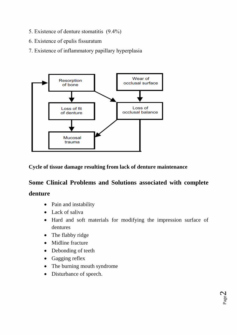
5. Existence of denture stomatitis (9.4%)
6. Existence of epulis fissuratum
7. Existence of inflammatory papillary hyperplasia
Cycle of tissue damage resulting from lack of denture maintenance
Some Clinical Problems and Solutions associated with complete
denture
Pain and instability
Lack of saliva
Hard and soft materials for modifying the impression surface of
dentures
The flabby ridge
Midline fracture
Debonding of teeth
Gagging reflex
The burning mouth syndrome
Disturbance of speech.
Pag
e3
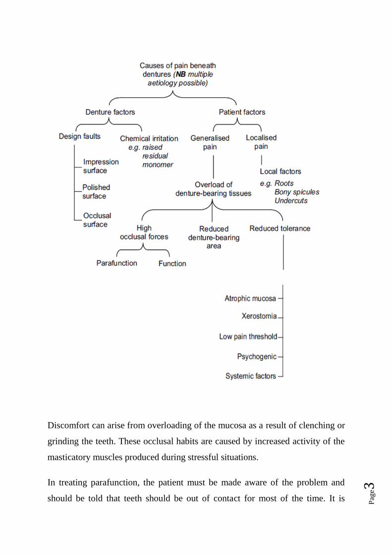
Discomfort can arise from overloading of the mucosa as a result of clenching or
grinding the teeth. These occlusal habits are caused by increased activity of the
masticatory muscles produced during stressful situations.
In treating parafunction, the patient must be made aware of the problem and
should be told that teeth should be out of contact for most of the time. It is

Pag
e4
important to reassure the patient, describe the link between stress, parafunction
and pain under dentures and point out that there is no change in the oral mucosa.
The importance of conscious relaxation should be emphasised and the patient
should be strongly encouraged to leave both dentures, or at least the lower
denture, out at night.
Another complication is Lack of saliva
Functions of saliva
Saliva possesses the following functions in the edentulous patient:
• It is responsible for the physical retention of complete dentures
• It prepares food for swallowing and facilitates the sense of taste
• It lubricates and protects the oral mucosa
• It helps to preserve a normal balance of the oral flora
• It promotes clear speech.
Problems of reduced salivary flow
A reduction, or absence of saliva (xerostomia), is likely to cause problems with
all the functions listed above so that a general, and significant, reduction in the
quality of life results. Reduced retention of dentures is a particular problem for
edentulous patients.
There may also be an increased susceptibility to denture trauma resulting in
complaints of pain and in some case the burning mouth syndrome
Aetiology of reduced salivary flow
MEDICAL HISTORY

Pag
e5
A full history is taken including a ‘I’m taking an anti-depressant and question
on current medication a diuretic’‘For how long have you been ‘One year’ DRY
MOUTH is a possible taking these tablets?’ contributory factor to the oral
complaint
SOCIAL HISTORY
The history has revealed a number of possible causes of the persistent pain. The
diagnosis can be established only after a careful examination of the patient, the
mouth and the various sets of dentures in order to confirm or deny the various
possibilities. The point should be made that unless a full history is obtained
some of the possible causes might never be revealed. The provision of new
dentures would do little to eliminate the problem if the persistent pain was due
to a dry mouth and to parafunction.
However, the condition is relatively common in middle-aged and older people,
the main candidates for complete dentures, with between 12% and 16%
complaining of a dry mouth
The commonest causes of dry mouth (Niedermeier et al. 2000; Field et al.
2001) are:
• Drugs, e.g. tricyclic antidepressants, beta-blockers
• Depression and chronic anxiety
• Dehydration
• Mouth breathing
• Sjögren’s syndrome
• Head and neck radiotherapy
• Poorly controlled diabetes

Pag
e6
• Smoking.
A complaint of dry mouth can occur in the absence of the clinical signs of
dryness (‘symptomatic xerostomia’) Under such circumstances the physical
retention of the dentures would not be expected to be diminished. In clinical
xerostomia there are intra-oral signs of dryness such as a dry, atrophic mucosa
and lack of saliva pooling in the floor of the mouth. The dentist can check the
dryness of the buccal mucosa simply and quickly during the examination of the
patient by carrying out the ‘mirror test’. For this the dentist lightly presses the
face of the mirror against the buccal mucosa and then tries to remove it. If the
mirror comes away easily the mucosa is still covered by a substantial film of
saliva; if the mucosa adheres to the mirror then it is dry.
Management of dry mouth
Close collaboration with the patient’s general medical practitioner or with a
specialist in oral medicine is often necessary. It might be possible, for example,
to change an existing xerostomic drug to one less liable to reduce salivary flow.
As there is a definite relationship between fluid intake and secretory
performance it is essential that the patient is kept well hydrated. Chewing and
energetic exercise improve salivary flow, possibly because of improved blood
circulation to the glands. Where some functional salivary tissue remains the
problem may be alleviated by sugar-free chewing gum or a scorbic acid. In
cases where flow rate cannot be improved limited relief may sometimes be
obtained by the use of artificial saliva.
It is very important for a denture patient with a dry mouth to maintain an
excellent level of denture hygiene. The likelihood of proliferation of Candida
albicans is increased in xerostomia and therefore unless denture hygiene is
maintained at a high level the denture is likely to be rapidly colonised by the
micro-organism, resulting in denture stomatitis.

Pag
e7
Motivation and instruction of the patient, followed by monitoring the quality of
denture hygiene are essential.
Cleaning dentures it should be done for:
Deposits form on dentures such as:
Microbial plaque
Calculus
Food debris.
These deposits may be responsible for a variety of problems including:
Denture stomatitis
Angular stomatitis
Unpleasant tastes
Odours
Unsightly appearance
Accelerated deterioration of some denture materials such as short-term
soft lining materials.
The effective cleaning of dentures is therefore of considerable importance to the
patient’s general well-being and oral health.
IPH-Inflammatory Papillary Hyperplasia
Candidiasis is contributing factor.
Wearing ill-fitting dentures 24h/7 days.
Resolve before making new dentures because. If left as is, new dentures will be
loose after placement, as inflammation resolves.
Leave dentures out at night.
Reline with tissue conditioner.
Nystatin mouth rinse

Pag
e8
In cases where an intractable dry mouth gives rise to a persistent problem of
loose dentures a denture adhesive will usually provide some improvement in
denture function.
Hard and soft materials for modifying the impression surface of dentures
Materials which can be used to modify the impression surface to overcome
some of these problems; these materials can either be applied by the dentist at
the chairside or by the dental technician in the laboratory.
The materials may be classified as follows:
• rigid materials
• short-term soft lining materials
• long-term soft lining materials.
Rigid materials
Recent years have seen the development of a group of useful materials,
frequently described as chairside reline materials, which can be used to modify
the impression surface of an existing denture.
Composition
Commonly these materials consist of a powder containing
polyethylmethacrylate together with a liquid monomer, butylmethacrylate. The
important point to make is that monomeric methylmethacrylate, a tissue irritant,
is avoided. Many of the products include a primer to enhance the adhesion of
the material to the existing denture polymer. The available materials vary in
working time, setting time and viscosity. These materials can be useful for
relining dentures. As they can be used at the chairside a ‘one-stop’ reline
technique can be employed.

Pag
e9
Short-term soft lining materials
Composition
Most materials are supplied in a powder/liquid form. An alternative presentation
is in a ready-to-use sheet form which can be found in one product available to
the dental profession and in several ‘over the counter’ products available
directly to the general public.
It is essential that traumatised tissue is examined by the dentist and that rational,
rather than empirical, treatment is prescribed.
The composition of the powder/liquid types is as follows:
(1) Powder. Polyethylmethacrylate, or copolymers of
polyethyl/methylmethacrylate.
(2) Liquid. A mixture of:
(a) an aromatic ester, such as dibutyl phthalate which acts as plasticiser
(b) ethyl alcohol.
Clinical applications
Short-term soft lining materials are placed in existing dentures for the following
reasons.
(1) Tissue conditioning. For tissue conditioning, the material is applied for a
period of a few days to the impression surface of a denture when the mucosa is
traumatised and inflamed. The tissue conditioner acts as a cushion absorbing the
occlusal loads, improving their distribution to the supporting tissues and
encouraging healing of the inflamed mucosa.
(2) Temporary soft reline. A short-term soft lining material can be used to
improve the fit of a denture, typically an immediate restoration.
Pag
e10
(3) Diagnosis. A short-term soft lining material can be used as a diagnostic aid
where the dentist wishes to check the reaction of the patient and the tissues to an
improvement in fit of a denture.
(4) Functional impression. A short-term soft lining material can be used as a
functional impression material applied to the impression surface of a denture for
the purpose of securing an impression under functional stresses.
(5) Recording the neutral zone. The ability of these materials to be moulded by
the oral musculature over an extended period of several minutes allows them to
be used to record the neutral zone
Long-term soft lining materials
Long-term soft lining materials distribute stress more evenly under dentures
than do the hard denture base materials. They also absorb impacts that can arise
from masticatory function .They can therefore be said to have a shock-
absorbing or cushioning effect. As a consequence it has been shown that the
addition of a long-term soft lining to a complete lower denture improves the
ability to bite and chew and provides general improvement in comfort when
compared with hard relines . the lining has also been shown to improve
masticatory performance
Indications for use
(1) Persistent pain under a denture.
(2) Thin atrophic mucosa.
(3) Parafunction.
It is useful to consider the first three indications together, as a complaint of
persistent pain may be due to the poor quality of the denture-bearing mucosa or
to the patient’s inability to regulate gripping or grinding habits. The whole
problem may be exacerbated by gross resorption of the mandible which results
in the normal masticatory forces being distributed over a reduced area. It is

Pag
e11
important to make two points; first, the problem is almost always found in the
lower jaw and, second, it is essential to ensure that all existing denture faults
have been eliminated before deciding to proceed with a long-term soft lining.
(4) Replacing an existing denture which has a soft lining. Once a patient has
successfully worn a lower denture with a soft lining and has got used to its
‘feel’ it is often wise to repeat the prescription. If this is not done and the new
denture is made with a hard base the patient may have problems in adapting to it
and reject the prosthesis as a result.
(5) Sharp bony ridges or spicules. The pattern of resorption of the mandible
may result in sharp ridges or spicules of bone on which the denture-bearing
mucosa. The problem might be overcome, at least in the short term, by
surgically smoothing the bone. However, there are often occasions where poor
health or a strong preference by the patient to avoid surgery are
contraindications to this approach. There is also the danger that surgical
interference with the mandible will speed up resorption of the bone. An
alternative, conservative approach is to provide a soft lining, which often
provides an acceptable level of comfort under these circumstances.
(6) Superficially placed mental nerve. Another consequence of advanced
resorption of the mandible is that the mental foramen and mental nerve may
become superficially placed within the denture-bearing area so that the nerve is
traumatised during function.
This typically gives rise to a complaint of a severe, sharp, stabbing pain from
the area of the mental foramen which is brought on by biting. A soft lining
restricted to the problem area may provide relief. However, it is not uncommon
to find that a superficial mental nerve requires greater pressure relief than can be
provided by a soft lining. If this is the case it may be necessary to cut the

Pag
e12
denture away in the area of the nerve to eliminate pressure on the nerve
altogether.
Types of long-term soft lining
Soft linings are made either of silicone rubber or soft acrylic. The silicone
materials may be cold-curing or heat-curing. The soft acrylics are heat-curing;
cold-curing soft acrylics have a very limited life span and are best thought of as
temporary soft linings.
The flabby ridge
This condition is most frequently seen in the upper anterior region. The bone
becomes grossly resorbed, often up to the level of the anterior nasal spine, and
is replaced by fibrous tissue. As a result of this mobile fibrous tissue, the
stability of a complete denture will be poor and both function and appearance
can be heavily compromised.
Aetiology
It has long been believed that the condition, sometimes called the ‘combination
syndrome’, is caused by the presence of lower natural teeth. This is probably
not surprising when the many factors that influence bone metabolism are
considered. Nevertheless it is probably wise to keep such patients under regular
review to ensure that a dramatic level of damage is not occurring.
Management
Approaches to treatment
The management of this condition is somewhat controversial, opinion falling
into two camps. In one, surgical removal of the fibrous tissue is favoured in
every case where the health of the patient allows. This approach produces a firm
ridge which is reduced in size. Advocates of the opposing view suggest that
surgical removal should rarely, if ever, be carried out because the fibrous tissue
may have a cushion effect which reduces trauma to the underlying bone. If the

Pag
e13
tissue is removed, it must be replaced by denture base material with consequent
increase in the bulk and weight of the prosthesis.
Denture breakages
Midline fracture of the complete upper denture accounts for 29% of all repair
work in dental laboratories, whilst teeth debonded from complete dentures
account for 33%These two common problems will be considered.
Midline fracture
The midline fracture of an acrylic denture may occasionally result from careless
handling by the patient; for example, accidental dropping of the denture while
cleaning can cause an impact fracture. Characteristically, however, a midline
fracture is due to fatigue of the acrylic resin produced by repeated flexing of the
denture by forces too small to fracture it directly. Failure of the denture base is
due to the progressive growth of a crack originating from a point on the surface
where an abrupt change in the surface profile causes a localised concentration of
stress many times that applied to the bulk of the denture.
Pag
e14
The crack often starts palatally to the upper central incisors, grows slowly at
first but undergoes an enormously increased rate of growth just before the
denture fractures. A failure of this type most commonly occurs in dentures that
are about 3 years old.
Debonding of teeth
The usual reasons for a weak bond between tooth and denture base are:
• The presence of tin-foil substitute on the ridge-lap surface of the tooth
• The presence of residual wax on the same surface
• The use of cross-linked teeth which are incompatible with the particular
denture base polymer.
Of the various recommendations that have been made for minimising the risk of
debonding the following have received fairly widespread support
(1) Choose artifi cial teeth and a denture base polymer which are compatible by
checking the information sheets provided with the products or by seeking
information from the manufacturers. Conventional denture teeth tend to achieve
a higher bond strength than cross-linked teeth.
(2) Ensure all traces of wax and tin-foil substitute are removed. The complete
removal of wax is not consistently achieved with boiling water alone and so for
optimum bond strength the use of a wax solvent is recommended .
(3) Drill small channels into the palatal surface of the teeth to increase the area
available for the polymerising denture base resin. However, it needs to be
remembered that such recesses in the ridge-lap surface of the teeth can make
complete wax removal more difficult. Therefore particular care needs to be
taken when removing the wax, otherwise the adjustments can result in a weaker,
rather than a stronger bond.

Pag
e15
(4) Apply a solvent such as dichloromethane to the ridge-lap surface of the
teeth. The solvent creates microscopic pores and channels which promote
diffusion of the polymerisable materials.
(5) Use a heat-curing denture base polymer. This material polymerises more
slowly than a cold-curing material and ensures better penetration into the tooth
substance.
Gagging reflex (retching)
is a protective reflex which guards the airway and posterior oropharynx. It may
occur during prosthetic procedures such as impression taking, or when dentures
are worn or, in extreme cases, when a mouth mirror is placed on the lips or tip
of the tongue.
Aetiology
There are a number of causes that may be conveniently grouped together as
follows.
(1) Somatic. The term ‘somatic’ covers those situations where the reflex is
triggered by tactile stimulation of the soft palate, posterior third of the tongue.
(2) Iatrogenic. Iatrogenic causes, which are related to the dentures, are
numerous. Some patients begin to retch after new dentures are inserted, but in
most cases this reflex soon disappears as they adapt to the dentures. However,
the reflex may persist if there are faults with the dentures such as an excessive
occlusal vertical dimension, or if the dentures are stimulating the sensitive areas
of the soft palate and tongue directly.
This stimulation may be caused by palatal over-extension, a posterior border
which is too thick or poorly adapted, the teeth encroaching on tongue space or
indeed by any factor producing denture instability.

Pag
e16
An upper denture whose posterior border is under-extended posteriorly can
provoke ggaging because as the edge of the denture terminates on relatively
incompressible mucosa a satisfactory post-dam cannot be produced. This result
in poor retention, which increases denture instability, stimulates the tongue and
palate, and causes apprehension in the patient. When this diagnosis is
established, it requires a very careful explanation by the dentist to convince the
patient that to cure the problem it will be necessary to cover more, rather than
less of the palate.
(3) Psychogenic. Psychogenic causes may arise from sight, sound or thought.
They include the sight of impression material being mixed or the sound of
another patient retching. The patient may be extremely apprehensive because of
an unhappy first experience of dental procedures or as a result of disturbing
stories from friends. In rare instances, retching may be a manifestation of a
psychological disturbance which is not primarily related to the patient’s dental
treatment.
(4) Systemic. Less frequently, the causative factor may be systemic disease,
particularly conditions affecting other regions of the gastrointestinal tract; for
example, the link between retching and alcoholism may be related to the
persistent gastritis found in such patients. Persistent catarrh will prevent nose
breathing and may contribute to the problem of retching.
Patient management
A carefully taken history will reveal the severity of the problem and provide
clues as to the cause. For example, a situation where a patient has been able to
tolerate the clinical stages of denture construction, but then has difficulty in
wearing the finished dentures, points to an iatrogenic cause which should be
treated relatively simply by correcting the error in denture design.
Impressions
Most of individuals find impression taking unpleasant. However, retching
during impression taking can usually be prevented by the following:
Pag
e17
(1) Reassurance and relaxation.
It is very important that the dentist has a confident and relaxed chairside
manner. It is essential that the anxious patient is reassured and encouraged to
relax both physically and mentally. The dental nurse can also play a major role
in creating an appropriate state of mind in the patient.
(2) Position of the patient.
The dental chair should be adjusted so that the patient is sitting comfortably in
the upright position.
(3) Breathing through the nose.
Instructing the patient to breathe through the nose while the tray is being tried in
the mouth or the impression is being taken is one of the most helpful methods of
preventing retching. During nasal breathing the soft palate remains stationary in
its low position and the tongue in its ‘guarding’ position, protecting the
nasopharynx from the threat of the foreign body in the mouth. If the patient
breathes through the mouth, this protection is lost and movement of the soft
palate results in intermittent contact with the setting impression material,
increasing stimulation.
(4) Impression technique.
Impression trays should be well fitting. As close-fitting special trays are less
bulky than spaced trays, they are better tolerated and should be used whenever
possible.
When trying trays in the mouth, firm, positive movements should be used. Most
patients tolerate the lower impression better than the upper one, so if the lower
impression is taken first, the success of the procedure is likely to reassure the
patient. The impression material should be mixed or prepared out of sight of the
patient and the amount placed in the tray kept to the minimum necessary to
record the relevant structures. A saliva ejector should be used if copious
amounts of saliva collect in the floor of the mouth.
(5) Distraction.

Pag
e18
It is during the insertion of the impression and while the material is setting that
it is particularly important to distract the patient’s attention from what is going
on.
This may be achieved by the dentist talking about something that is known to be
of particular interest to the patient, or by reinforcing the requirement that the
patient continues to breathe slowly and steadily through the nose. It has even
been suggested that the patient be asked to raise one leg and to concentrate on
not lowering it until the impression has set!
The severe retching reflex
The first challenge when trying to treat a patient who has this problem is to
obtain an accurate impression so that a well-fitting denture base can be
constructed.
The second challenge is to provide a prosthesis that can be worn by the patient
for a reasonable length of time.
The following approaches to the management of this difficult problem have
been found useful:
(1) Conscious sedation
(2) Acupuncture. The gag reflex has been shown to be capable of being
controlled by acupuncture . Although there is evidence to show that the
technique is of assistance when undertaking the various clinical stages of
denture construction there is, as yet, no evidence that it can be used by the
patient to allow the denture to be worn.
(3) Hypnosis. Hypnosis has been used in the treatment of severe cases its
success is dependent upon the patient being well motivated and being able to
practise self-hypnosis, thus enabling a denture to be worn outside the dental
surgery
(4) The training denture. The training denture approach may be of value when
treating any patient with a long history of difficulties which suggest frank
denture intolerance,including retching.

Pag
e19
The burning mouth syndrome
The burning mouth syndrome (BMS) can be very troublesome to the patient,
presents problems of diagnosis and often involves prolonged treatment. The
symptoms occur in 5–7% of the adult population. Of those who seek treatment,
there is a predominance of women, with a mean age of approximately 60 years.
The most common sites of the complaint are the tongue and the upper denture-
bearing tissues. Rather less common are the lips and lower denture-bearing
tissues. The oral mucosa appears normal.
Many of the BMS patients have consulted a number of health care professionals
before seeking help from the dentist or dental specialist. They know of no other
people with the complaint and therefore feel quite isolated. If several
professionals have stated that the mouth looks normal the patient may start to
feel as if ‘it is all in the mind’. The level of anxiety is consequently raised and
cancerophobia may well develop.
Aetiology
BMS has been attributed to a multitude of causes and these broadly fall into
three groups:
• Local irritants including denture faults
• Systemic factors
• Psychogenic factors.
Local irritation
Denture faults
Errors in denture design which cause a denture to move excessively over the
mucosa, which increase the functional stress on the mucosa or which interfere
with the freedom of movement of the surrounding muscles may initiate a
complaint of burning rather than frank soreness.
Denture design errors have been discovered in 50% of BMS patients.
Residual monomer
Pag
e20
High levels of residual monomer in the denture base have been reported and the
tissue damage produced is considered to be the result of chemical irritation
rather than a true allergy. It is possible that high levels of residual monomer,
which have ranged from three to ten times the normal value, are due to errors
inadvertently introduced into the short curing cycles which are popular with
manufacturers and dental laboratories. If the requisite curing temperature of
100°C is not achieved in the relevant part of the short curing cycle, there is a
marked increase in residual monomer content . Some authorities may not
consider this condition to be an example of BMS where, classically,
the mucosa looks normal. However, a patient who reacts to a high level of
residual monomer complains of a burning sensation and so we feel justified in
including it.
Micro-organisms
The role of micro-organisms in burning mouth syndrome is controversial and
studies have not shown a link between the presence of Candida albicans and the
complaint.
Smoking and mouthwashes
Smoking and the regular use of some mouthwashes are irritants that have been
implicated in BMS.
Systemic causes
Nutritional deficiencies
Contributions from nutritional deficiencies such as iron, vitamin B complex and
folic acid should be highlighted. An example of BMS caused by a deficiency is
Iron deficiencies have been found in 8% and folic acid deficiencies in 6% of
BMS patients. Low blood levels of vitamin B1 and B6 were found in 40% of
patients.
Endocrine disorders
What is apparent is the relative unimportance of the climacteric as a causative
factor, a modern viewpoint which is at variance with past clinical opinion. On

Pag
e21
rare occasions, the symptoms are found to be linked with an undiagnosed
diabetes mellitus. Treatment of the medical condition invariably results in
complete resolution of BMS.
Xerostomia, frequently associated with BMS .One that should be highlighted
here is drug-induced xerostomia. Recent investigations have produced evidence
of a link between BMS and reduced parotid gland function and ofantidepressant
medication reducing the salivary flow.
It should be recognised that the presence of a dry mouth is capable of
accentuating the symptoms initiated by any of the causes of local irritation. This
is an example of the multifactorial nature of BMS.
Hypersensitivity
True hypersensitivity to constituents of denture base polymer is rare and usually
results in local symptoms such as burning or itching. In one instance where
there were systemic symptoms of nausea, dizziness and general malaise the
patient was found to have reacted to dyes used to colour the polymer. Dentures
made of clear polymer proved successful
Parkinson’s disease
It has been reported that the prevalence of BMS was 24% in people suffering
from Parkinson’s disease;
Psychogenic causes
The more common disorders associated with BMS are anxiety, depression,
cancerophobia and hypochondriasis. The associated parafunctional activities
such as bruxism and abnormal and excessive tongue movements are capable of
inducing mucosal irritation.
Management
Faced with a multitude of causative factors, it will be recognised that the
process of diagnosis and treatment is usually a time-consuming affair.
• Initial assessment (history/examination/special tests).
• Provisional diagnosis.

Pag
e22
• Initial treatment (e.g. elimination of local irritants and investigating and
treating haematinic deficiencies).
• Assessment of initial treatment.
• Definitive diagnosis.
• Definitive treatment (local/systemic correction/psychological therapy).
• Follow-up.
With regard to outcome, analysis of various studies suggests that about two-
thirds of BMS patients are either cured or improved to such an extent that the
burning sensation is no longer an overwhelming problem. There remain a group
of patients for whom the current state of knowledge can offer relatively little
benefit. Some in this small group remain totally resistant to treatment. However,
it should be remembered that even in these refractory cases BMS is not
necessarily a life sentence as spontaneous remissions can eventually occur for
no apparent reason.
Disturbance of speech
The presence of complete dentures can modify speech by affecting articulation
and by altering the degree of oral resonance. A number of sounds are articulated
by contact of the tongue to the palate and to the teeth. A change in speech that
may be quite marked when the dentures are first inserted will usually disappear
completely within a few days. However, if the changes in the contact surfaces
require a modification of tongue behaviour that is beyond the adaptive
capability of an individual patient, a speech defect will persist. It should also be
remembered that the tongue of a patient who is wearing complete dentures has a
dual function – to take part in speech articulation and to control the dentures. If
the dentures are loose, the demands of this latter function may be so great that
there is a general deterioration in the quality of speech. As mentioned
The following relationships are particularly important to the production of clear
speech.
(1) Tip of the tongue to the palate.
Pag
e23
Contact between the tip of the tongue and the palate is required in the
production of /s/, /z/, /t/, /d/ and /n/. Consequently, a change in the shape or
thickness of the denture contact surface resulting from the fitting of new
dentures will require a modification of tongue behaviour in order to produce
sounds which are the same as before. In the vast majority of cases, the necessary
modification occurs without any difficulty in a relatively short period of time.
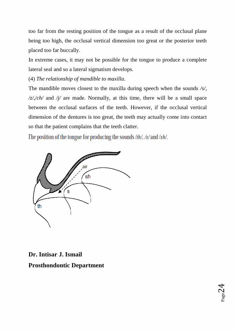
The sound most commonly affected in this way is /s/, a sound which isgenerally
produced with the tongue tip behind the upper anterior teeth. A narrow channel
remains in the centre of the palate through which air hisses .If the palate
is too thick at this point, or if the incisors are positioned too far palatally, the /s/
may become a /th/. If the denture is shaped so that it is diffi cult for the tongue
to adapt itself closely to the palate, a channel narrow enough to produce the /s/
sound will not be produced and a whistle or /sh/ sound may result. This is most
likely to be the consequence of excessive palatal thickening laterally in the
canine region
(2) Lower lip to incisal edges of upper anterior teeth.
The lower lip makes contact with the incisal edges of the upper anterior teeth
when the sounds /f/ and /v/ are produced. If the position of these teeth on a
replacement denture is dramatically different to that on the old denture there is
likely to be a disturbance in speech.
(3) Lateral margin of the tongue to posterior teeth.
Contact between the lateral margins of the tongue and the posterior teeth is
necessary to produce the English consonants /th/, /t/, /d/, /n/, /s/, /z/, /sh/, /zh/ (as
in measure), /ch/, /j/ and /r/ (as in red). Air is directed forwards over the dorsum
of the tongue and may be modifi ed by movement of the tongue against the
teeth or anterior slope of the palate to produce the final sound. If the contact can
only be achieved with difficulty,
movement of the tip of the tongue may be restricted with consequent
impairment of speech. This difficulty arises if the posterior contact surfaces are
Pag
e24
too far from the resting position of the tongue as a result of the occlusal plane
being too high, the occlusal vertical dimension too great or the posterior teeth
placed too far buccally.
In extreme cases, it may not be possible for the tongue to produce a complete
lateral seal and so a lateral sigmatism develops.
(4) The relationship of mandible to maxilla.
The mandible moves closest to the maxilla during speech when the sounds /s/,
/z/,/ch/ and /j/ are made. Normally, at this time, there will be a small space
between the occlusal surfaces of the teeth. However, if the occlusal vertical
dimension of the dentures is too great, the teeth may actually come into contact
so that the patient complains that the teeth clatter.
Dr. Intisar J. Ismail
Prosthondontic Department