concise histology- leslie p gartner & james l hiatt (1e)
DESCRIPTION
CONCISE HISTOLOGY, by Leslie P. Gartner, PhD and James L. Hiatt, PhD, thoroughly reviews all the histology knowledge required for the USMLE Step 1 in an easy-access outline format. Designed for students who need to learn a large amount of material in a limited time, it presents key information in a readable, concise manner, accompanied by full-color illustrations that clarify complex concepts.TRANSCRIPT


ConciseHistologyLESLIE P. GARTNER, PhDProfessor of Anatomy (Retired)Department of Biomedical SciencesBaltimore College of Dental SurgeryDental SchoolUniversity of MarylandBaltimore, Maryland
JAMES L. HIATT, PhDProfessor EmeritusDepartment of Biomedical SciencesBaltimore College of Dental SurgeryDental SchoolUniversity of MarylandBaltimore, Maryland

concise histology isBn: 978-0-7020-3114-4
Copyright © 2011 by Saunders, an imprint of Elsevier Inc. All rights reserved.
no part of this publication may be reproduced or transmitted in any form or by any means, electronic or mechanical, including photocopying, recording, or any information storage and retrieval system, without permission in writing from the publisher. Details on how to seek permission, further information about the Publisher’s permissions policies and our arrangements with organizations such as the copyright clearance center and the copyright licensing Agency, can be found at our website: www.elsevier.com/permissions.
this book and the individual contributions contained in it are protected under copyright by the Publisher (other than as may be noted herein).
Notices
Knowledge and best practice in this field are constantly changing. As new research and experience broaden our understanding, changes in research methods, professional practices, or medical treatment may become necessary.
Practitioners and researchers must always rely on their own experience and knowledge in evaluating and using any information, methods, compounds, or experiments described herein. in using such information or methods they should be mindful of their own safety and the safety of others, including parties for whom they have a professional responsibility.
With respect to any drug or pharmaceutical products identified, readers are advised to check the most current information provided (i) on procedures featured or (ii) by the manufacturer of each product to be administered, to verify the recommended dose or formula, the method and duration of administration, and contraindications. it is the responsibility of practitioners, relying on their own experience and knowledge of their patients, to make diagnoses, to determine dosages and the best treatment for each individual patient, and to take all appropriate safety precautions.
to the fullest extent of the law, neither the Publisher nor the authors, contributors, or editors, assume any liability for any injury and/or damage to persons or property as a matter of products liability, negligence or otherwise, or from any use or operation of any methods, products, instructions, or ideas contained in the material herein.
Library of Congress Cataloging-in-Publication Data
gartner, leslie P. concise histology / leslie P. gartner, James l. hiatt.—1st ed. p. ; cm. Based on: color textbook of histology / leslie P. gartner, James l. hiatt. 3rd ed. c2007. includes index. isBn 978-0-7020-3114-4 1. histology. i. hiatt, James l.,— ii. gartner, leslie P., 1943—color textbook of histology. iii. title. [DnlM: 1. histology—Atlases. Qs 517 g244c 2011] QM551.g366 2011 611’.018—dc22 2010013017
Printed in china
last digit is the print number: 9 8 7 6 5 4 3 2 1
Working together to grow libraries in developing countries
www.elsevier.com | www.bookaid.org | www.sabre.org
1600 John F. Kennedy Boulevardsuite 1800Philadelphia, PA 19103-2899
Acquisitions Editor: Kate DimockDevelopmental Editor: Barbara cicalese
Design Direction: lou ForgioneElectronic Media Manager: carol emery

To my wife, Roseann;
my daughter, Jennifer;
and my mother, Mary
LPG
To my grandchildren,
Nathan David,
James Mallary,
Hanna Elisabeth,
Alexandra Renate,
Eric James,
and Elise Victoria
JLH

This page intentionally left blank

v
once again, we are gratified to release a new histol-ogy textbook, one that is based on the third edition of our Color Textbook of Histology, a well-established textbook not only in its original language but also in several other languages.
in the past three decades, histology has evolved from the purely descriptive science of microscopic anatomy to a composite study integrating functional anatomy with both molecular and cell biology. this new textbook is designed in an unusual manner in that each even-numbered page tells the story in words and the facing odd-numbered page illustrates the textual story by beautiful four-color illustrations that are borrowed from the third edition of our Color Textbook of Histology. therefore, each set of facing pages may be thought of as individual learning units. to demonstrate the relevance of the information presented to the health professions, almost every learning unit is reinforced by clinical considerations pertinent to the topic. students and faculty alike will, no doubt, note the absence of photomicrographs and electron micrographs in Concise Histology. We made a deliberate decision to exclude that material from the hard copy and to place it, instead, on the student consult website that is associated with this book. We did that to reduce the size of the book, thereby making life easier for the student who has to learn material that a decade ago was taught in 16
Prefaceweeks and currently is done so in perhaps half that time. student consult houses not only all the illus-trations located on the right side of the facing pages of the book but also 150 photomicrographs and elec-tron micrographs, identified by chapter, with appro-priate examination questions and the answers to those questions so that the student can test his or her ability not only to recognize the organs/tissues/cells in question but also their functional characteristics. included on student consult are clinical scenarios with appropriate UsMle i-type questions that not only further demonstrate the relevance of histology to the health sciences but also prepare medical stu-dents for the histology component of the boards. the designs of the hard copy of this textbook, as well as that of the ancillary web-based material, intend to highlight the essential concepts underlying our pre-sentation of histology, namely that there is a close relationship between structure and function.
Although we have made every effort to present a complete and accurate account of the subject matter, we realize that there are omissions and errors in any undertaking of this magnitude. therefore, we con-tinue to encourage and welcome suggestions, advice, and criticism that will facilitate the improvement of future editions of this textbook.
leslie P. gartnerJames l. hiatt

This page intentionally left blank
vii
histology is a visual subject; therefore, excellent graphic illustrations are imperative. For that we are indebted to todd smith for his careful attention to detail in revising and creating new illustrations. We also thank our many colleagues from around the world and their publishers who generously permit-ted us to borrow illustrative materials.
AcknowledgmentsFinally, our thanks go to the project team at else-
vier for all their help, namely Kate Dimock, Barbara cicalese, lou Forgione, and carol emery. We also thank linnea hermanson for her painstaking effort in the production of this text book.

This page intentionally left blank

ix
Contents
1 Introduction to Histology . . . . . . . . . . . . . . . . . . . . . . . . . . . . . . . . . . . . . . . . . . . . . . . . . . . . . . 2
2 Cytoplasm . . . . . . . . . . . . . . . . . . . . . . . . . . . . . . . . . . . . . . . . . . . . . . . . . . . . . . . . . . . . . . . . . . . . . . . . . . . 8
3 Nucleus . . . . . . . . . . . . . . . . . . . . . . . . . . . . . . . . . . . . . . . . . . . . . . . . . . . . . . . . . . . . . . . . . . . . . . . . . . . . . .26
4 Extracellular Matrix . . . . . . . . . . . . . . . . . . . . . . . . . . . . . . . . . . . . . . . . . . . . . . . . . . . . . . . . . . . . .40
5 Epithelium and Glands . . . . . . . . . . . . . . . . . . . . . . . . . . . . . . . . . . . . . . . . . . . . . . . . . . . . . . . .48
6 Connective Tissue . . . . . . . . . . . . . . . . . . . . . . . . . . . . . . . . . . . . . . . . . . . . . . . . . . . . . . . . . . . . . .62
7 Cartilage and Bone . . . . . . . . . . . . . . . . . . . . . . . . . . . . . . . . . . . . . . . . . . . . . . . . . . . . . . . . . . . . .74
8 Muscle . . . . . . . . . . . . . . . . . . . . . . . . . . . . . . . . . . . . . . . . . . . . . . . . . . . . . . . . . . . . . . . . . . . . . . . . . . . . . . . .94
9 Nervous Tissue . . . . . . . . . . . . . . . . . . . . . . . . . . . . . . . . . . . . . . . . . . . . . . . . . . . . . . . . . . . . . . . . . 108
10 Blood and Hematopoiesis . . . . . . . . . . . . . . . . . . . . . . . . . . . . . . . . . . . . . . . . . . . . . . . . . 132
11 Circulatory System . . . . . . . . . . . . . . . . . . . . . . . . . . . . . . . . . . . . . . . . . . . . . . . . . . . . . . . . . . . . 152
12 Lymphoid (Immune) System . . . . . . . . . . . . . . . . . . . . . . . . . . . . . . . . . . . . . . . . . . . . . . 168
13 Endocrine System . . . . . . . . . . . . . . . . . . . . . . . . . . . . . . . . . . . . . . . . . . . . . . . . . . . . . . . . . . . . . 188
14 Integument . . . . . . . . . . . . . . . . . . . . . . . . . . . . . . . . . . . . . . . . . . . . . . . . . . . . . . . . . . . . . . . . . . . . . . . 204
15 Respiratory System . . . . . . . . . . . . . . . . . . . . . . . . . . . . . . . . . . . . . . . . . . . . . . . . . . . . . . . . . . . 218
16 Digestive System: Oral Cavity . . . . . . . . . . . . . . . . . . . . . . . . . . . . . . . . . . . . . . . . . . . 230
17 Digestive System: Alimentary Canal . . . . . . . . . . . . . . . . . . . . . . . . . . . . . . . . . . 238
18 Digestive System: Glands . . . . . . . . . . . . . . . . . . . . . . . . . . . . . . . . . . . . . . . . . . . . . . . . . 250
19 Urinary System . . . . . . . . . . . . . . . . . . . . . . . . . . . . . . . . . . . . . . . . . . . . . . . . . . . . . . . . . . . . . . . . . 260
20 Female Reproductive System . . . . . . . . . . . . . . . . . . . . . . . . . . . . . . . . . . . . . . . . . . . . 272
21 Male Reproductive System . . . . . . . . . . . . . . . . . . . . . . . . . . . . . . . . . . . . . . . . . . . . . . . 286
22 Special Senses . . . . . . . . . . . . . . . . . . . . . . . . . . . . . . . . . . . . . . . . . . . . . . . . . . . . . . . . . . . . . . . . . 304
Index . . . . . . . . . . . . . . . . . . . . . . . . . . . . . . . . . . . . . . . . . . . . . . . . . . . . . . . . . . . . . . . . . . . . . . . . . . . . . . . . . . . . . . . 325

This page intentionally left blank

1
Concise Histology

2
1 INTRoDucTIoN To HISToLoGy
histology is a study of the tissues of animals and plants, but the Concise Histology deals only with mammalian tissues, specifically, that of Homo sapiens. in addition to the structure of the tissues, cells, organs, and organ systems compose the theme of this textbook—hence, a better term for the subject matter presented in this book is microscopic anatomy. it is well known by the reader of this book that the body is a conglomerate of:
• Cells• Extracellular matrix (ECM), in
which the cells are embedded• Extracellular fluid that percolates
through the ecM to bring nutrients, oxygen, and signaling molecules to the cells and to take waste products, carbon dioxide, still more signaling molecules, hormones, and pharmacologic agents away from the cells• the extracellular fluid is derived from blood
plasma and released into the ecM at the arterial side of capillary beds, and most of the fluid is returned to the blood plasma at the venous ends of capillary beds.
• the remainder of the extracellular fluid enters the lower pressure lymphatic system of vessels to be returned to the bloodstream at the junction of the internal jugular vein and subclavian vein of the right and left sides.
Modern textbooks of histology discuss not only the microscopic morphology of the body, but also its function. the subject matter of this book also invokes cell biology, physiology, molecular biology, bio-chemistry, gross anatomy, embryology, and even a modicum of clinical medicine in the form of Clinical Considerations. it is hoped that the study of histology will illuminate for the reader the interrelationship of structure and function. Before all this could be real-ized, however, techniques had to be developed to permit the visualization of cells and tissues that, although dead, present an accurate representation of the living appearance.
Light Microscopy
TISSuE PREPARATIoN
A small block of tissue, harvested from an anesthe-tized or newly dead subject:
1. is fixed, usually with neutral buffered formalin that is treated in such a manner that the proteins in the tissue are rapidly cross-linked so that they remain in the same place where they were while the subject was alive.
2. once fixed, is dehydrated in a graded series of alcohols
3. immersed in xylene, which makes the tissue transparent.
4. to be able to view thin sections of the tissue under a microscope, the tissue has to be embedded
in melted paraffin that infiltrates the tissue. the tissue is placed into a small receptacle and allowed to cool, forming a paraffin block containing the tissue.
5. sliced into 5- to 10-µm thin sections using a microtome whose very sharp blade is capable of slicing thin increments of tissue from the block.
6. the sections are transferred to adhesive-coated glass slides, the paraffin is removed from the section by a xylene bath, and the tissue is rehydrated by the use of a graded series of alcohols (reversed in order when dehydration took place).
7. the rehydrated sections are stained with various water-soluble dyes (table 1.1); hematoxylin and eosin (H&E) are the most common stains used in normal histologic preparations. hematoxylin stains the acid components of cells and tissues a bluish color, and eosin stains the basic components of cells and tissues a pinkish color.
Modern light microscopes use a series of lenses arranged to provide the maximum magnification with the greatest clarity. Because more than one lens is used, this is known as a compound microscope (Fig. 1.1).
KEy WoRDS• Light microscopy• Immunocytochemistry• Autoradiography• confocal microscopy• Transmission electron
microscopy• Scanning electron
microscopy

Chapter
IntroduCtIon to HIstology
1
3Table 1.1 COMMON HISTOLOGIC STAINS AND REACTIONS
Reagent Result
hematoxylin Blue—nucleus; acidic regions of the cytoplasm; cartilage matrixeosin Pink—basic regions of the cytoplasm; collagen fibersMasson’s trichrome Dark blue—nuclei
Red—muscle, keratin, cytoplasmLight blue—mucinogen, collagen
orcein elastic stain Brown—elastic fibersWeigert’s elastic stain Blue—elastic fiberssilver stain Black—reticular fibersiron hematoxylin Black—striations of muscle, nuclei, erythrocytesPeriodic acid–schiff Magenta—glycogen and carbohydrate-rich moleculesWright’s and giemsa* Pink—erythrocytes, eosinophil stains
Blue—cytoplasm of monocytes of blood cells and lymphocytes
*Used for granules differential staining of blood cells.
Figure 1.1 comparison of light, transmission electron, and scanning electron microscopes. (From Gartner LP, Hiatt JL: Color Textbook of Histology, 3rd ed. Philadelphia, Saunders, 2007, p 4.)
Image in eye
Light microscope Transmission electronmicroscope
Scanning electronmicroscope
Ocular lens
Anode
Electronicamplifier
Condenser lens
Lamp Mirror
Specimen
Specimen
Condenserlens
Condenser lens
Scanning beam
Electron detector
Specimen
Viewing window
Image on viewing screen
Image on viewing screen
Projectionlens
Objectivelens
Scanning coil
Anode
Cathode
Television screen

Chapter
IntroduCtIon to HIstology
1
4red color indicates that glycogen was present at that particular location.
• other histochemical and cytochemical techniques can localize enzymes; however, it is not the enzyme that is visualized, but the presence of the reaction product that precipitated as a colored compound at the site of the reaction.
• Immunocytochemistry provides a more accurate localization of a particular macromolecule than does histochemistry or cytochemistry.• this is a more complex method, however,
because it involves the development of an antibody against the macromolecule of interest in the direct method, or
• Development of an antibody against a primary antibody in the indirect method (Fig. 1.3) and labeling the developed antibody with a fluorescing label, such as rhodamine or fluorescein. the indirect method is more sensitive and more accurate than the direct method because more fluorescent labeled antibodies bind to the primary antibody than in the direct method. Additionally, most of the time, primary antibodies are more expensive and more limited in their availability.
• immunocytochemistry can also be applied to electron microscopy by attaching the heavy metal ferritin instead of a fluorescent label.
• the method of autoradiography uses a radioactive isotope (usually tritium, 3h), which is integrated into the molecule that is being investigated.• if one wishes to follow the synthesis of a
particular protein, tritiated amino acid is fed into the system, and specimens are harvested at defined periods.
• sections are processed in a normal fashion, but instead of a coverslip, photographic emulsion is placed on the section, and the slide is stored in the dark for many weeks.
• the emulsion is developed and fixed as if it were a photographic plate, and a coverslip is placed over the section.
• Microscopic examination displays the presence of silver grains over the regions where the isotope labeled molecule was located.
• A method of autoradiography has been developed for electron microscopy.
TISSuE PREPARATIoN (cont.)
A high-intensity lightbulb provides the light, which is focused on the specimen from below by a condenser lens. the light that passes through the specimen is gathered by one of the objective lenses that sits on a rotatable turret, allowing a change in magnification from low to medium to high, and an oil lens, which in conventional microscopes magni-fies the image 4, 10, 20, 40, and 100 times. the first three are dry lenses, whereas the oil lens uses immer-sion oil to act as an interface between the glass of the slide and the glass of the objective lens. the light from the objective lens is gathered by the ocular lens, usually 10 times, for final magnification of 40, 100, 200, 400, and 1000 times, and the image is focused on the retina.
INTERPRETATIoN of MIcRoScoPIc SEcTIoNS
histologic sections are two-dimensional planes cut from a three-dimensional structure. initially, it is difficult for the student to reconcile the image seen in the microscope with the tissue or organ from which it was harvested. A simple demonstration of a coiled tube sectioned at various angles (Fig. 1.2) is instructive in learning how to reconstruct the three-dimensional morphology from viewing a series of two-dimensional sections.
ADvANcED vISuALIzATIoN PRocEDuRES
Various techniques were developed to use the micro-scope in elucidating functional aspects of the cells, tissues, and organs being studied. the most com-monly used techniques are histochemistry (and cyto-chemistry), immunocytochemistry, and autoradiogra phy.
• Histochemistry and cytochemistry use chemical reactions, enzymatic processes, and physicochemical processes that not only stain the tissue, but also permit the localization of extracellular and intracellular macromolecules of interest.• one of the most used histochemical methods
is the periodic acid–schiff (PAs) reagent, which stains glycogen and molecules rich in carbohydrates a purplish-red color. By treating consecutive sections with the enzyme amylase, to digest glycogen, the absence of the purplish-
-

Chapter
IntroduCtIon to HIstology
1
5
Figure 1.2 two-dimensional views of a three-dimensional tube sectioned in various planes. (From Gartner LP, Hiatt JL: Color Textbook of Histology, 3rd ed. Philadelphia, Saunders, 2007, p 4.)
Cross section Longitudinal
section
Oblique section
Diagram showing the different appearances of sections cut through a curved tube at different levels
Figure 1.3 Direct and indirect methods of immunocytochemistry. Left, An antibody against an antigen was labeled with a fluorescent dye and viewed with a fluorescent microscope. Fluorescence occurs only over the location of the labeled antibody. Right, Fluorescent labeled antibodies were prepared against an antibody that reacts with a particular antigen. When viewed with a fluorescent microscope, the fluorescence represents the location of the antibody that reacts with the antigen. (From Gartner LP, Hiatt JL: Color Textbook of Histology, 3rd ed. Philadelphia, Saunders, 2007, p 5.)
Fluoresceinated antibody
Antigen
Tissue section
Wash
Direct Indirect
Add fluoresceinated anti-antibody
Antigen
Antibody

Chapter
IntroduCtIon to HIstology
1
6confocal Microscopy
confocal microscopy uses a laser beam that is focused on the specimen impregnated with fluorescent dyes; the impinging laser beam that passes through a dichroic mirror excites the dyes, which then fluoresce (Fig. 1.4).
• the beam of laser light passes through a pinhole that is computer controlled so that the beam scans along the surface of the specimen, and the fluorescence originates as the specimen is being scanned.
• the emitted fluorescent light is captured as it passes through the pinhole in a direction opposite from that of the laser light. each emitted light represents only a single point on the specimen being scanned.
• the emitted light is captured by a photomultiplier tube; as each pixel is gathered, the pixels are compiled by a computer into an image of the specimen.
• Because each scan observes only a very thin plane within the specimen, multiple passes at different levels may be used to construct a three-dimensional image of the specimen.
Electron Microscopy
electron microscopes use a beam of electrons instead of photons as their light source, and, instead of glass lenses, they use electromagnets to spread and focus the electron beam (Fig. 1.5).
• the resolution of a microscope depends on the wavelength of the light source, and the wavelength of an electron beam is far shorter than that of visible light; the resolution of an electron beam is about 1000 times greater than that of visible light. the resolving power of a compound light microscope is about 200 nm, whereas that of a transmission electron microscope is 0.2 nm, providing a magnification of 150,000 times, which permits the visualization of a single macromolecule such as myosin.
• there are two types of electron microscopy: transmission electron microscopy (teM) and scanning electron microscopy (seM).• As the name implies, TEM (see Fig. 1.3, right)
requires the electrons to pass through a very thinly sliced specimen that was treated with a heavy metal stain (e.g., lead phosphate or uranyl acetate) and hit a phosphorescent plate, which absorbs the electron and gives off a point of light whose intensity is a function of the electron’s kinetic energy. As the electron interacts with the specimen, it loses some of its kinetic energy, and the more heavy metal is absorbed by a particular region of the specimen, the more energy the electron loses. in this fashion, the resultant image consists of points of light of different intensities ranging from light to dark gray. the image can be captured by placing an electron-sensitive photographic plate in the place of the phosphorescent plate. the photographic plate can be developed in the normal fashion, and the plate can be printed as a black-and-white photograph.
• SEM (see Fig. 1.5) does not require the electrons to pass through the specimen. instead, the surface of the specimen is bombarded with electrons and the resulting image is a three-dimensional representation of the specimen. to achieve this, the specimen is coated with a heavy metal, such as gold or palladium. As the electron beam bombards the surface of the specimen, the heavy metal coating scatters some of the electrons (backscatter electrons), whereas some of the impinging electrons cause the ejection of the heavy metal’s electrons (secondary electrons). Backscatter and secondary electrons are captured by electron detectors and are interpreted as a three-dimensional image that is projected onto a monitor. the digitized image can be saved as a file and printed as a photograph.

Chapter
IntroduCtIon to HIstology
1
7
Figure 1.4 confocal microscope displaying the pinhole through which the laser beam enters to scan the specimen and the path of the fluorescent light that subsequently is emitted by the specimen to be captured by the photomultiplier detector. (From Gartner LP, Hiatt JL: Color Textbook of Histology, 3rd ed. Philadelphia, Saunders, 2007, p 8.)
Specimen
Photomultiplierdetector
Pinhole aperture
Pinhole aperture
Laser withlaser light
Scanning mirror
Scanning mirror
Figure 1.5 comparison of light, transmission electron, and scanning electron microscopes. (From Gartner LP, Hiatt JL: Color Textbook of Histology, 3rd ed. Philadelphia, Saunders, 2007, p 4.)
Image in eye
Light microscope Transmission electronmicroscope
Scanning electronmicroscope
Ocular lens
Anode
Electronicamplifier
Condenser lens
Lamp Mirror
Specimen
Specimen
Condenserlens
Condenser lens
Scanning beam
Electron detector
Specimen
Viewing window
Image on viewing screen
Image on viewing screen
Projectionlens
Objectivelens
Scanning coil
Anode
Cathode
Television screen

8
2 cyToPLASMcomplex organisms are composed of cells and extra-cellular materials. Although there are more than 200 types of cells that constitute these organisms, each with various func-tions, the cells and the extracellular matrix are categorized into the four basic tissues: epithelium, connective tissue, muscle, and nervous tissue. tissues form organs, and combina-tions of organs form organ systems.
generally, a cell is a membrane-bound structure filled with proto-plasm that may be categorized into two components, the cytoplasm and the karyoplasms (Fig. 2.1).
• Karyoplasm constitutes the nucleus and is surrounded by the nuclear envelope.
• this chapter discusses the cell membrane and the cytoplasm of a generalized cell.• the main substance of the cytoplasm is
the cytosol, a fluid suspension in which the inorganic and organic chemicals, macromolecules, pigments, crystals, and organelles are dissolved or suspended.
• the cytosol is surrounded by a semipermeable, lipid bilayer cell membrane (plasmalemma, plasma membrane) in which proteins are embedded.
cell Membrane (Plasmalemma, Plasma Membrane)
the cell membrane is approximately 7 to 8 nm in thickness and is composed of a lipid bilayer com-prising amphipathic phospholipids, cholesterol, and embedded or attached proteins (Fig. 2.2). Viewed with the electron microscope, the plasmalemma appears to have two dense layers:
• An inner (cytoplasmic) leaflet• An outer leaflet, which sandwich between them
an intermediate clear, hydrophobic, layer
this tripartite structure is known as a unit mem-brane and forms not only the cell membrane, but also all other membranous structures of the cell. in the average membrane, the protein components con-stitute approximately 50% by weight. the arrange-ment of the phospholipid molecules is such that:
• the hydrophilic polar heads face the periphery, forming the extracellular and intracellular surfaces.
• the hydrophobic fatty acid chains of the two facing phospholipid sheets (inner and outer
leaflets) project toward the center of the membrane, forming the intermediate clear layer.
cholesterol is usually tucked away among the fatty acid tails of the phos-pholipid molecules. When the cell membrane is frozen and then frac-tured, it cleaves preferentially along the hydrophobic clear layer, making the two internal surfaces of the leaflets visible (Fig. 2.3).
• the surface of the inner leaflet (closest to the protoplasm) is the P-face.
• the surface of the outer leaflet (closer to the extracellular space) is known as the E-face.
Proteins of the cell membrane are integral pro-teins or peripheral proteins. integral proteins are:
• Transmembrane proteins, in that they occupy the entire thickness of the membrane, and they extend into the cytoplasm and into the extracellular space
• Peripheral proteins that are not embedded into the membrane; instead, they adhere either to the cytoplasmic or to the extracellular surface of the membrane. During freeze fracture, more proteins remain attached to the P-face than to the e-face.
• the extracellular surface of the cell membrane, which may have a glycocalyx (cell coat), composed of carbohydrates that form glycoproteins or glycolipids, depending on whether they form bonds with the integral proteins or with the phospholipids
the integral and peripheral proteins have some mobility in the two-dimensional phospholipid mem-brane and resemble a mosaic that is constantly changing. the movements of these proteins are restricted, and the membrane representation that used to be called the fluid mosaic model is now known as the modified fluid mosaic model. Regions of the membrane are slightly thickened because they possess a rich concentration of glycosphingolipids and cholesterol surrounding a cluster of membrane proteins. these specialized regions, lipid rafts, func-tion in cell signaling.
KEy WoRDS• cell• Ion channels• carrier proteins• organelles• Protein synthesis• Membrane
trafficking• cytoskeleton• Inclusions

Chapter
Cytoplasm
2
9
Microvilli
Plasmamembrane
Smoothendoplasmic
reticulum
Nuclearenvelope
Mitochondrion
Lysosome
Golgiapparatus
Roughendoplasmicreticulum
Nucleolus
Microfilaments
Microtubules
Secretion granule
Centrioles
Figure 2.1 A generalized cell and its organelles. (From Gartner LP, Hiatt JL: Color Textbook of Histology, 3rd ed. Philadelphia, Saunders, 2007, p 14.)
Figure 2.2 Fluid mosaic model of the cell membrane. (From Gartner LP, Hiatt JL: Color Textbook of Histology, 3rd ed. Philadelphia, Saunders, 2007, p 16.)
Extracellular space
Cytoplasm
Glycoprotein
Fattyacidtails
Polar head
Channel
Cholesterol
Peripheralprotein
Inner leaflet
Outerleaflet
Integralprotein
Glycolipid
Figure 2.3 the e-face and the P-face of the plasma membrane. (From Gartner LP, Hiatt JL: Color Textbook of Histology, 3rd ed. Philadelphia, Saunders, 2007, p 16.)
E-face
Outer leaflet
Inner leaflet
P-face
Integral protein

Chapter
Cytoplasm
2
10MEMbRANE TRANSPoRT PRoTEINS
the plasmalemma is permeable to nonpolar mole-cules, such as oxygen, and uncharged polar mole-cules, such as water and glycerol, and these may cross the membrane by simple diffusion following a con-centration gradient. ions and small polar molecules require assistance, however, from certain multipass integral proteins, known as membrane transport proteins, which function in the transfer of these sub-stances across the cell membrane.
• if the process does not require energy, the transfer across the plasmalemma is passive transport.
• if the process requires the expenditure of energy, it is known as active transport (Fig. 2.4).
Membrane transport proteins are of two types: channel proteins and carrier proteins.
• Channel proteins participate only in passive transport because they do not have the ability to use the expenditure of energy to work against a concentration gradient.• to be able to accomplish their function,
channel proteins are folded in such a fashion that they provide hydrophilic ion channels across the cell membrane.
• Most of these channels can control the entry of substances into their lumen by possessing barriers, known as gates, which block their entrance or exit. Various mechanisms control the opening of these gated channels.
• Voltage-gated channels, such as na+ channels of nerve fibers, are opened when the membrane is depolarized (see chapter 9).
• Ligand-gated channels open when a signaling molecule (ligand) binds to the ion channel. some ligand-gated channels respond to neurotransmitters and are known as neurotransmitter-gated channels (e.g., in skeletal muscle).
• others respond to nucleotides, such as cyclic adenosine monophosphate (cAMP) or cyclic guanosine monophosphate (cgMP), and are referred to as nucleotide-gated channels (e.g., in rods of the retina).
• Mechanically gated channels respond to physical contact for opening, as in the bending of the stereocilia of the hair cells of the inner ear.
• G protein–gated ion channels, such as the acetylcholine receptors of cardiac muscle cells,
require the activation of a g protein before the gate can be opened.• Ungated channels are always open. K+ leak
channels are the most common ungated channels, and these are responsible for the maintenance of the resting potentials of nerve cells. Aquaporins, channels designed for the transport of h2o, are also ungated channels.
• Carrier proteins are multipass proteins; however, they have the ability not only to be passive conduits that allow material to pass down a concentration gradient, but also to use adenosine triphosphate (AtP)–driven mechanisms to transport material against a concentration gradient. they also differ from ion channels because they have internal binding sites for the ions or molecules that they are designed to transfer. the transport may be of one molecule or ion in a single direction (uniport), or coupled—that is, two different ones in the:• same direction (symport) or• opposite direction (antiport)
the most common example of carrier proteins is the na+-K+ pump that uses Na+,K+-ATPase to cotrans-port three sodium ions against a concentration gradi-ent out of the cell and two potassium ions into the cell. some carrier proteins use the intracellular and extracellular Na+ concentration differential as a force to drive the movement of some ions or small mol-ecules or both against a concentration gradient. this process, performed by coupled carrier proteins, is known as secondary active transport, and glucose and na+ are frequently cotransported in this manner.
cELL SIGNALING
cells communicate with each other by releasing small molecules (signaling molecules, ligands) that bind to receptors of other cells. the cell that releases the signaling molecule is the signaling cell. the cell with the receptor is the target cell.
Frequently the roles of these cells may be reversed because often the communication is bidirectional. the receptors may be located on the cell membrane, and the ligand in this case is a polar molecule. if the receptor is intracellular or intranuclear, the ligand may be a nonpolar, hydrophobic molecule (e.g., steroid hormone), or the receptor on the cell surface transduces the signal by the activation of an intracel-lular second messenger system (e.g., g protein–linked receptors).

Chapter
Cytoplasm
2
11
cLINIcAL coNSIDERATIoNSThe amino acid cystine is removed from the lumen of the renal proximal tubule by a carrier protein. Some individuals who inherited two copies of the same mutation, one from each parent, that forms defective cysteine carrier proteins have a condition known as cystinuria. These individuals have a high enough concentration of this amino acid in their urine to form cystine stones. Cystinuria manifests between age 10 and 30 years, and the condition is responsible for recurrent kidney stones. Diagnosis is made on the basis of microscopic examination of the urine showing the presence of cystine crystals and by urinalysis showing abnormal levels of cystine. The condition can be very painful, but in many cases increased fluid intake dilutes the urine sufficiently to prevent the formation of stones.
Figure 2.4 types of transport. A, Passive transport that does not require the input of energy. B, Active transport is an energy-requiring mechanism. (From Gartner LP, Hiatt JL: Color Textbook of Histology, 3rd ed. Philadelphia, Saunders, 2007, p 18.)
A Passive Transport
B Active Transport
Facilitated diffusion
Coupled transport
Cytoplasm
Cytoplasm
Extracellular space
Extracellular space
Plasma membrane
Uniport
Antiport Symport
Simple diffusion of lipids
Ion channel-mediated diffusion
Carrier-mediated diffusion

Chapter
Cytoplasm
2
12G Protein–Linked Receptors and Secondary Messengers of the cell
G protein–linked receptors (guanine nucleotide–binding proteins) are transmembrane proteins whose extracytoplasmic aspects have binding sites for spe-cific signaling molecules (ligands), and their cyto-plasmic aspect is bound to a G protein on the inner leaflet of the plasmalemma. When the signaling molecule binds to the extracytoplasmic moiety of the receptor, the receptor’s cytoplasmic aspect undergoes a conformational change that activates the g protein (Fig. 2.5). there are several types of g proteins: stim-ulatory (gs), inhibitory (gi), pertussin-toxin sensitive and insensitive (go and gBq), and transducin (gt).
• Gs proteins are trimeric in that they are composed of α, β, and γ subunits. they are usually inactive, and in the inactive state they have a guanosine diphosphate (gDP) bound to their cytoplasmic aspect.
• When the gs protein is activated, it exchanges its gDP for a guanosine triphosphate (gtP); the α subunit dissociates from the other two components and contacts adenylate cyclase, activating it to catalyze the transformation of cytoplasmic AtP to cAMP.
• Uncoupling of the ligand from the g protein–linked receptor causes GTP of the α subunit to be dephosphorylated and to detach from the adenylate cyclase and rejoin its β and γ subunits.
• cAMP, one of the secondary messengers of cells, activates A kinase, which initiates the eliciting of a specific response from the cell.
• in other cells, cAMP enters the nucleus and activates CRE-binding protein, which binds to regulatory regions of genes, known as CREs (cAMP response elements), which permit the transcription of that particular gene effecting the specific response from the cell.
Protein Synthetic Machinery of the cell
A major function of most cells is the synthesis of proteins either for use by the cell itself or to be exported for use elsewhere in the body. Protein syn-thesis has:
• An intranuclear component, transcription, that is, the synthesis of a messenger RNA (mRNA) molecule, and
• Translation, the cytoplasmic component, which entails the assembly of the correct amino acid sequence, based on the nucleotide template of the mRnA to form the specific protein
the cytoplasmic component of protein synthesis uses ribosomes only if the protein to be formed is released free in the cytosol or ribosomes and the rough endoplasmic reticulum (ReR) (Fig. 2.6) if the protein is to be packaged for storage within the cell or to be released into the extracellular space.
• Ribosomes are small (12 nm × 25 nm), bipartite particles composed of a large and a small subunit. each subunit, manufactured in the nucleus, is composed of ribosomal RNA (rRNA) and proteins. the small subunit has binding sites for mRnA and three additional binding sites: one for binding peptidyl transfer RnA (tRnA) (P-site), another to bind aminoacyl tRnA (A-site), and an exit site (E-site) where the empty tRnA leaves the ribosome. the large subunit binds to the small subunit and has special rRnA that acts as an enzyme, known as ribozyme, which catalyzes the formation of peptide bonds that permit amino acids to bond to each other.
• there are two types of endoplasmic reticulum (ER): smooth endoplasmic reticulum (seR) and ReR. Although the former is not involved in protein synthesis, for the sake of completeness, its structure is discussed here.• SER consists of tubules and flat vesicles whose
lumina are probably continuous with those of the ReR. the seR functions in lipid and steroid synthesis, glycogen metabolism, and detoxification of noxious substances, and in muscle as an intracellular storage site for calcium.
• RER functions in the synthesis of proteins that are destined to be packaged either for storage within the cell or for release into the extracellular space. it is composed of flattened, interconnected vesicles, and its cytoplasmic surface is studded with ribosomes and polysomes that are actively translating mRnA and forming protein. the ReR possesses the integral proteins signal recognition particle receptor (docking protein), ribophorins i and ii, and translocators, proteins that bind ribosomes to the ReR and open as a pore through which nascent proteins can enter the cisternal (luminal) aspect of the ReR. the cisternal aspect of the ReR membrane houses the enzyme signal peptidase and dolichol phosphate, which functions in n-glycosylation. the cisterna of the ReR is continuous with the perinuclear cistern of the nuclear envelope.

Chapter
Cytoplasm
2
13
Figure 2.5 g protein–linked receptor. PPi, inorganic pyrophosphate. (From Gartner LP, Hiatt JL: Color Textbook of Histology, 3rd ed. Philadelphia, Saunders, 2007, p 21.)
Extracellular space
Cytoplasm
Signalingmolecule
Receptor
Adenylatecyclase
G protein
GTP
GDP
γ β α
αγβ GTP
cAMP+ PPi
Activated adenylate cyclase
Activated Gα-subunit
ATP
Figure 2.6 A generalized cell and its organelles. (From Gartner LP, Hiatt JL: Color Textbook of Histology, 3rd ed. Philadelphia, Saunders, 2007, p 14.)
Microvilli
Plasmamembrane
Smoothendoplasmic
reticulum
Nuclearenvelope
Mitochondrion
Lysosome
Golgiapparatus
Roughendoplasmicreticulum
Nucleolus
Microfilaments
Microtubules
Secretion granule
Centrioles

Chapter
Cytoplasm
2
14Protein Synthesis
the process of protein synthesis always begins when an mRnA is bound to a ribosome in the cytosol and, if the protein is not to be packaged, is then finished in the cytosol. if the protein is to be packaged, the mRnA contains the code for a signal peptide whose translation is the signal to move the ribosome-mRnA complex to the ReR.
SyNTHESIS of NoNPAcKAGED PRoTEINS
the synthesis of proteins that are not to be packaged occurs in the following manner (Fig. 2.7):
• An mRnA leaves the nucleus through a nuclear pore complex (see chapter 3), enters the cytosol, and binds a small ribosomal subunit, whose P-site is occupied by a methionine-bearing initiator tRNA. the anticodon of the tRnA matches the codon of the mRnA, aligning the system in the proper position. A large ribosomal subunit joins the complex, and translation begins as the ribosome moves the distance of a single codon along the mRnA in a 5′ to 3′ direction.
• An amino acid bearing tRnA (aminoacyl tRNA), if it possesses the correct anticodon, binds to the A-site of the small ribosomal subunit, and its amino acids form a peptide bond with the methionine in the P-site. the methionine is released by the tRnA located on the P-site, and the tRnA of the A-site now has two amino acids attached to it (methionine and the newly arrived amino acid). the empty tRnA moves from the P-site to the E-site, and the tRnA loaded with the two amino acids moves to the P-site. Finally, the entire ribosome moves the distance of a single codon along the mRnA in a 5′ to 3′ direction.
• A new acylated tRnA possessing the correct anticodon attaches to the A-site. it picks up the two amino acids from the t-RnA at the P-site and now has three amino acids attached to it. the tRnA at the e-site is ejected, and the empty tRnA at the P-site moves to the now vacant e-site. the tRnA with its three amino acids moves from the A-site to the P-site, and the entire ribosome moves the distance of a single codon in a 5′ to 3′ direction. A new acylated tRnA possessing the correct anticodon occupies the now vacant A-site.
• As this process continues, new small ribosomal subunits attach to the 5′ end of the mRnA; in this manner, several ribosomes are translating the same mRnA simultaneously. A single mRnA strand with several ribosomes is referred to as a polysome.
• the process of new acylated tRnA is added to the sequence until the stop codon is reached, which signals that the last amino acid of the protein has been incorporated into the nascent protein chain. the last empty tRnA is released at the e-site, no new tRnAs occupy the A-site, and the small and large ribosomal subunits dissociate from the mRnA.
SyNTHESIS of PRoTEINS THAT ARE To bE PAcKAGED
the synthesis of proteins to be packaged (Fig. 2.8) begins in the cytosol in the same fashion as previ-ously described.
• the peptide chain that is formed is the signal peptide that is recognized by the signal recognition particle (SRP), a molecule composed of protein and RnA that is freely floating in the cytosol. sRP binds to the signal peptide, protein synthesis ceases, and the ribosome-mRnA-sRP complex moves to the ReR.
• the sRP binds to the SRP receptor (docking protein) of the ReR membrane, and the ribosome binds to translocator proteins—integral proteins—of the ReR membrane. As the binding occurs, the sRP is released; translation continues, and the base of the translocator opens up, forming a pore into the ReR cistern. the nascent protein enters the ReR lumen through the pore.
• the signal peptide is cleaved off by the enzyme signal peptidase, and some of the elongating proteins are N-glycosylated by dolichol phosphate present in the luminal aspect of the ReR membrane. this process is assisted by the ReR-specific proteins ribophorin i and ribophorin ii in the ReR membrane. the process of translation is finished when the stop codon is reached.
• the newly synthesized protein is released into the ReR cistern, where it is modified further and folded in the proper fashion in the presence of chaperones.
• the completed proteins are packaged into transfer vesicles to leave the ReR and be transported to the Golgi apparatus for further modification and final packaging.
• Misfolded proteins are retrotranslocated through a translocator that is similar to the one that they used to enter the eR during synthesis. When in the cytoplasm, they are ubiquitylated and destroyed by proteasomes.

Chapter
Cytoplasm
2
15
Figure 2.7 synthesis of proteins that are not to be packaged occurs in the cytosol. (From Gartner LP, Hiatt JL: Color Textbook of Histology, 3rd ed. Philadelphia, Saunders, 2007, p 26.)
The P-site tRNA moves tothe E-site and the A-site tRNA, with the attached peptidyl chain, moves to the vacated P-site. As a new aminoacyl-tRNA bearing an amino acid occupies theA-site, the spent tRNA on theE-site drops off the ribosome. A peptide bond is formed, and the ribosome moves down the mRNA. The cycle of adding to the forming protein chain continues.
Polypeptide synthesis continues until the ribosomeencounters a “stop” or “non-sense codon” which signals the end of the polypeptide chain.
The terminal signal complex, a release factor which promotes polypeptide release, docks at the A-site.The polypeptide chain isreleased.
Once protein synthesis iscompleted, the two ribosomalsubunits dissociate from themRNA, and return to the cytosol.
A-siteA-sitemRNAP-site
P-site
E-site E-site
tRNAAminoacid
Initiation begins when the small ribosomal subunit binds with messenger RNA (mRNA). The initiator transfer RNA (tRNA) binds with its associated amino acid, methionine, to the P-site.
The large subunit joins theinitial complex. The empty A-site is now ready toreceive an aminoacyl-tRNA.
Polypeptidechain
Terminationsignal complex
A second aminoacyl-tRNA, bearing an amino acid, bindsto the empty A-site.
A peptide bond is formedbetween the two amino acids.This bond formation brings theacceptor end of the A-site tRNAinto the P-site as it picks up the peptidyl chain.
Large ribosomalsubunitSmall ribosomalsubunit
Figure 2.8 synthesis of proteins that are to be packaged occurs on the ReR surface. (From Gartner LP, Hiatt JL: Color Textbook of Histology, 3rd ed. Philadelphia, Saunders, 2007, p 27.)
mRNA 5′ 3′
Proteinsynthesisbegins
Proteinsynthesisinhibited
Proteinsynthesisresumes
Signalsequenceremoved
Protein synthesiscontinues to completion
Ribosomedissociates
Ribosome
Signalsequence
Signalrecognitionparticle
SRPreceptor
Signalpeptidase
Cleavedsignalsequence
Carbohydrate Completedprotein
C
N
NN
Rough endoplasmic reticulum
cLINIcAL coNSIDERATIoNSpresent glutamine in the sixth position of the β-chain is exchanged for valine, a condition known as sickle cell anemia. During low oxygen tension, such as after strenuous exercise, the modified β-chain causes the erythrocytes to become disfigured so that they appear sickle-shaped, and their ability to ferry oxygen is much reduced. These defective red blood cells are prone to fragmentation because they lose their normal pliability.
The amino acid sequence of a protein determines its primary structure. A minor alteration of the primary structure usually does not affect the functionality of the protein; however, there are cases where a point mutation—that is, the substitution of a single amino acid for another—makes a major difference in the ability of that protein to perform its intended function. An example of such a deleterious point mutation occurs in hemoglobin, where the normally

Chapter
Cytoplasm
2
16Golgi Apparatus
the Golgi apparatus (Golgi complex) is composed of clusters of preferentially oriented tubules and a series of flattened, convex membrane-bound vesicles stacked one above the other, where each vesicle resembles an uncut pita bread with a central lumen, the cistern (Fig. 2.9). A cell may have one to several golgi complexes, each of which has a:
• convex entry face near the nucleus, known as the cis-Golgi network (CGN)
• Cis-face, where newly synthesized proteins from the ReR enter the golgi complex
• concave exit face, oriented toward the cell membrane, known as the trans-face
• one to several intermediate faces, interposed between the cis-face and trans-face
• complex of vesicles and tubules, known as the vesicular-tubular cluster (VTC, formerly eRgic), located between the transitional region of the ReR and the cis-golgi network
• in association with the trans-face is another cluster of vesicles, the trans-Golgi network (TGN)
the functions of the golgi complex include car-bohydrate synthesis and the modification and sorting of proteins.
Protein Trafficking
Vesicles ferrying material (e.g., proteins or carbohy-drates) from one organelle to another or between regions of the same organelle are known as transport vesicles, and the material they transport is referred to as cargo. transport vesicles possess a protein coat (known as coated vesicles) on their cytosolic aspect that permits the vesicle to bud off and adhere to these organelles and to reach the proper target. there are three major types of proteinaceous coats (with some subtypes) that cells use to accomplish these goals:
• Coatomer I (COP I)• Coatomer II (COP II)• Clathrin
these coats ensure that the correct material becomes the cargo and that the membrane is formed into a vesicle of correct size and shape. each coat is used to encourage a specific type of transport (Fig. 2.10). As the coated vesicle reaches the membrane of its target organelle, it loses its coat and fuses with the target membrane. the ability of the vesicle and the target membrane to recognize each other depends on SNARE proteins (soluble attachment receptor n-
ethylmaleimide sensitive fusion proteins) and a group of gtPases specializing in target recognition known as Rabs. snARes allow binding only of the correct vesicle with the intended target. the initial docking of the vesicle is mediated in part by the Rabs protein. At the cell membrane, there are snARe-rich regions, known as porosomes, where vesicles dock to deliver their contents into the extracellular space.
Proteins leave the transitional ER, a region of the ReR that is devoid of ribosomes, packaged in small transport vesicles whose membrane, derived from the ReR, is covered by coP ii (see Fig. 2.10). these coP ii–coated vesicles travel to the vesicular-tubular cluster, lose their coP ii coat, and fuse with the Vtc. the delivered cargo is examined, and if it contains an escaped eR resident protein that protein is returned to the eR via coP i–coated vesicles (retro-grade transport), and the remaining, correct cargo is passed to the golgi apparatus also in coP i–coated vesicles (anterograde transport). the proteins are passed to the various faces of the golgi apparatus—again probably via coP i–coated vesicles—where they are modified in each face and sent to the tgn for final packaging. the modified proteins are pack-aged in clathrin-coated vesicles or coP ii–coated vesicles and are addressed to be sent to one of three places:
• the cell membrane, where they become inserted as membrane-bound proteins or where they fuse with the cell membrane to release their contents immediately into the extracellular space (continuous exocytosis)
• to be housed temporarily in the cytoplasm as storage (secretory) vesicles near the plasmalemma for eventual release of the cargo into the extracellular space (discontinuous exocytosis)
• late endosomes to become incorporated into lysosomes
the process of discontinuous exocytosis requires a clathrin coat and is said to follow the regulated pathway of secretory proteins, whereas the process of continuous exocytosis requires coP ii–coated vesicles and is said to follow the constitutive pathway of secretory proteins.
All of these protein-ferrying vesicles not only possess protein coats, but also have many membrane markers that allow them to be attached to microtu-bules and transported, by means of molecular motors, along these structures to their final destina-tions. the vesicles also possess markers that act as address labels, and the vesicles dock at their target by means of these molecules.

Chapter
Cytoplasm
2
17
Figure 2.9 Rough endoplasmic reticulum and the golgi complex. (From Gartner LP, Hiatt JL: Color Textbook of Histology, 3rd ed. Philadelphia, Saunders, 2007, p 28.)
ER
Transportvesicles
Transitional ER
ERGIC
trans-Golginetwork
Secretorygranules
Smooth andcoated vesicles
trans-face
cis-face
Medial face
Figure 2.10 Protein trafficking through the golgi complex and associated vesicles. (From Gartner LP, Hiatt JL: Color Textbook of Histology, 3rd ed. Philadelphia, Saunders, 2007, p 30.)
Protein synthesis
Phosphorylation of mannose
ER
TER (transitional ER)
Removal of mannose
Terminal glycosylation
Sulfation and phosphorylationof amino acids
Sorting of proteins
Clathrincoat
Secretorygranule
Clathrintriskelions
Non-clathrincoated vesicle
Mannose 6-phosphatereceptor
Late endosome
Lysosome
trans-Golgi network
Plasma membraneproteins
Lysosomalproteins
Secretoryproteins
COP Icoated transportvesicles
TER ERGICCis
GOLGI
MEDIAL Trans
COP IIcoatedvesicles
Plasma membrane

Chapter
Cytoplasm
2
18Membrane Trafficking
ENDocyToSIS: PHAGoSoMES AND PINocyToTIc vESIcLES
the transfer of material from the extracellular space into the cytoplasm is known as endocytosis.
• larger substances are phagocytosed into a vesicle known as a phagosome.
• smaller molecules (ligands) are pinocytosed into a pinocytotic vesicle.• Pinocytosis is a carefully controlled process
whereby the material to be engulfed is recognized via cargo receptor proteins located on the cell membrane that recognize the ligand extracellularly and clathrin intracellularly.
• the ability to recognize and bind to clathrin molecules causes the formation of a pinocytic vesicle that may contain hundreds of ligand molecules.
• cells can also transfer material from the cytoplasm into the intercellular space, a process known as exocytosis.
• During endocytosis, the plasmalemma loses membrane to the vesicles formed from it, and it gains the membranes of vesicles formed in the tgn during exocytosis. this continuous cycling of the membranes is known as membrane trafficking (Fig. 2.11).
ENDoSoMES (ENDoSoMAL coMPARTMENT)
Pinocytotic vesicles lose their clathrin coat and fuse with the:
• Early endosome, a membranous compartment located near the plasmalemma whose membrane possesses AtP-driven h+ pumps that acidify its lumen to a ph of 6.0
• in some early endosomes, recycling endosomes, the ligand and its receptor are dissociated from each other, the receptor is returned to the cell membrane, and the ligand is either released into the cytoplasm or transferred to
• Late endosomes, another membranous compartment located at a deeper level within the cytoplasm. the h+ pumps in the late endosomal membrane further acidify the lumen of this organelle, which continues to digest its luminal contents, and the partially degraded material is
transferred to lysosomes for complete degradation.
LySoSoMES (ENDoLySoSoMES)
Lysosomes are small, membrane-bound organelles housing dozens of hydrolytic enzymes that function at the low ph of 5.0, achieved by the presence of h+ pumps in their membrane. lysosomes degrade vari-ous substances whose useful components are re-leased into the cytoplasm, whereas their indi gestible substances remain enclosed by the lysosomal mem-brane, and the organelle becomes known as a resid-ual body.
PERoxISoMES
Peroxisomes are similar to lysosomes in morphol-ogy, but they house many oxidative enzymes that are synthesized on free ribosomes and then transported into these organelles by the assistance of peroxisome-targeting signals that recognize dedicated membrane-bound receptors on the peroxisomal surface.
• the most prevalent enzyme in peroxisomes is catalase, which decomposes h2o2 into water and oxygen. this organelle also participates in lipid biosynthesis, especially of cholesterol; lipid catabolism by β-oxidation of long-chained fatty acids; and, in hepatocytes, bile acid formation.
• in the central nervous system, kidneys, testes, and heart, peroxisomes possess enzymes that participate in synthesis of plasmalogen, membrane phospholipids that protect cells against singlet oxygen.
PRoTEASoMES
Proteasomes are small, barrel-shaped organelles that are responsible for:
• Degradation of proteins that are misfolded, damaged, denatured, or otherwise malformed
• cleaving of antigenic proteins into smaller fragments known as epitopes (see chapter 12)
Proteolysis via proteasomes is carefully managed by the cell through the energy-requiring attachment of multiple copies of ubiquinone to the candidate protein to form a polyubiquinated protein. the ubiquitin molecules and their degradation by- products are released in an energy-requiring process into the cytosol.

Chapter
Cytoplasm
2
19
Figure 2.11 endocytosis, endosomes, and lysosomes. cURl, compartment for uncoupling of receptor and ligand. (From Gartner LP, Hiatt JL: Color Textbook of Histology, 3rd ed. Philadelphia, Saunders, 2007, p 33.)
Early endosome / recycling endosome (CURL) pH = 6.0
Ligandin solution
Ligand attachesto receptors
Clathrin-coatedendocytotic vesicle
Clathrin triskelionsrecycle to plasmamembrane
Recycling of receptorsto plasma membrane
Uncoated endocytoticvesicle
Multivesicular body(type of lysosome)
Degradation productswithin residual body
Residual body fuses with cell membraneand contents eliminated from cell
Late endosomepH = 5.5
Clathrin-coated vesiclescontaining lysosomal hydrolasesor lysosomal membrane proteins
6
1
2
3
4
5
8
9
10
11
127
Clathrin-coated pit
Golgi
Roughendoplasmicreticulum
Nucleus
1
2
3
5
6
7 12
11
10
9
84
cLINIcAL coNSIDERATIoNSZellweger syndrome is a congenital, incurable, fatal disease of newborns; death occurs within 1 year after birth as a result of liver or respiratory failure or both. The disease is due to the inability of peroxisomes to incorporate peroxisomal enzymes because the requisite peroxisomal targeting signal receptors are missing from the membrane of the peroxisomes. This results in the inability of peroxisomes to perform β-oxidation of long-chain fatty acids to synthesize plasmalogens.

Chapter
Cytoplasm
2
20Mitochondria
Mitochondria are large organelles; some measure 7 µm long × 1 µm wide. the mean life span of a mitochondrion is about 10 days, after which the mito chondrion increases in length and then under-goes fission. each mitochondrion is composed of a:
• smooth outer membrane and• inner membrane that is folded into shelflike or
tubelike structures, known as cristae, increasing greatly the surface area of the inner membrane
the principal function of mitochondria is the syn-thesis of ATP via a process known as oxidative phos-phorylation. there are two spaces formed by the two membranes (Fig. 2.12B):
• Intermembrane space, located between the outer and inner membranes, and
• Matrix (intercristal) space, bounded by the inner membrane (see Fig. 2.12A), which houses the matrix, a viscous fluid with a high concentration of proteins, ribosomes, RnA, circular DNA (which codes for only 13 mitochondrial proteins), and dense granules of phospholipoproteins, known as matrix granules, which may have calcium-binding and magnesium-binding properties
the inner and outer membranes contact each other in regions, and here regulatory and transport proteins facilitate the movement of various molecules into and out of the mitochondrial spaces. the macromolecules targeted for the two mitochondrial membranes or the matrix use regions of the mitochon drial membranes where contact does not occur between them; however, these sites possess receptor molecules that recognize the targeted macromolecules.
• the outer membrane of the mitochondrion is smooth and quite permeable to small ions, and the presence of numerous porins permits the movement of h2o across it. the content of the intermembrane space is very similar to the content of the cytosol.
• the folded inner membrane is rich in cardiolipins, phospholipids that possess four instead of two fatty acyl chains and greatly reduce the permeability of the inner membrane to protons and electrons. the inner membrane is also rich in the enzyme complex ATP synthase, which is responsible for the generation of AtP from ADP and inorganic phosphate.• AtP synthase is composed of two major
portions, F0 and F1; the F0 portion is mostly embedded in the inner membrane, and the F1
portion (also referred to as the head) is suspended in the matrix and is connected to the F0 portion by the shaft and is kept stationary by several additional proteins (see Fig. 2.12B).
• each F0 portion possesses three sites for the phosphorylation of ADP to AtP. the F1 portion possesses a fixed outer sleeve and a freely movable inner sleeve composed of 10 to 14 subunits. the shaft also has a movable internal sleeve that extends into the F0 portion and a fixed outer sleeve.
• the movable sleeves of the shaft and of the F1 portion are together known as the rotor. the fixed outer sleeves are connected to the F0 portion, and these three components are known as the stator.
the matrix contains the enzymes, which, using pyruvate generated from glycolysis and fatty acids generated from fats and transported into the mito-chondrial matrix, convert them into acetyl coenzyme A (CoA), whose acetyl moiety is used by the enzymes of the citric acid cycle to reduce oxidized nicotin-amide adenine dinucleotide (nAD+) to NADH and flavin adenine dinucleotide (FAD) to FADH2. these reduced compounds accept high-energy electrons generated by the citric acid cycle and transfer them to a series of inner membrane integral proteins, known as the electron transport chain (Fig. 2.12c). the electron is passed along the chain, and its energy is used to transfer h+ (i.e., protons) from the matrix into the intermembrane space. As the concentration of h+ in the intermembrane space becomes greater than that of the matrix, the h+ ions are driven back into the matrix by this concentration gradient, the proton motive force, and the only path open to them is through the AtP synthase.
the movement of protons down the rotor com-ponent of the AtP synthase causes it to rotate and rub against the stator, creating energy that is used by the three sites of the F0 portion to phosphory-late ADP to the energy-rich compound AtP. some of the AtP formed is used by the mitochondria, but most is transported into the cytosol for use by the cell.
Brown fat is especially abundant in animals that hibernate. the mitochondria of these lipocytes possess thermogenins instead of AtP synthase. ther-mogenins have the ability to shunt protons from the intermembrane space into the matrix; however, oxi-dation in these cells is uncoupled from phosphoryla-tion, and, instead of AtP, heat is generated by the proton motive force. the heat is used to bring the animal out of hibernation.

Chapter
Cytoplasm
2
21
cLINIcAL coNSIDERATIoNSMitochondrial myopathies are disorders that are inherited from the mother because all mitochondria of an individual are derived from the ovum. These infrequently occurring myopathies do not have a gender-related disposition. The prognosis depends on the muscle groups involved. Myopathy may be evidenced only as muscle weakness and tiring after exercise, but in severe cases it may be fatal. The disorder usually manifests by the end of the second decade of life. Common myopathies are Kearns-Sayre syndrome, myoclonus epilepsy, and mitochondrial encephalomyopathy. There are no known treatments for these diseases.
Figure 2.12 A, three-dimensional view of a mitochondrion with shelflike cristae. B, Diagram of shelflike cristae at a higher magnification. C, Diagram of the electron transport chain and AtP synthase of the inner mitochondrial membrane. (From Gartner LP, Hiatt JL: Color Textbook of Histology, 3rd ed. Philadelphia, Saunders, 2007, p 39.)
H+
Cristae(folds)
Innermembrane
Outermembrane
Intermembranespace
A C
B
Matrixspace
Matrixspace
InnermembraneOuter
membrane
Intermembranespace
Matrix space
Intermembrane space
H+
H+ H+ H+
H+H+H+
H+
e–
H+
H+
H+H+
H+
H+ H+
H+ATP synthase
2H++1/2 O2
ATP synthase
ADP + Pi
ATP
ADP + Pi
ATP
H 2O

Chapter
Cytoplasm
2
22Inclusions and the cytoskeleton
INcLuSIoNS
inclusions are nonliving elements of the cell that are freely present within the cytosol and are not mem-brane bound. the major inclusions are glycogen, lipids, pigments, and crystals.
• Glycogen is usually stored in the cytosol in the form of rosettes of β particles that are located in the vicinity of seR elements. these particles are used as an energy deposit that undergoes glyco-genolysis to form glucose, which is converted to pyruvate for use in the citric acid cycle.
• Lipids are stored triglycerides that are catabolized into fatty acids that are fed into the citric acid cycle for the formation of pyruvate. lipids are much more efficient storage forms of energy than glycogen because 1 g of lipid provides twice the amount of AtP as does 1 g of glycogen.
• Usually, pigments are not active metabolically, but may serve protective functions, such as melanin of the skin, which absorbs ultraviolet radiation and serves to protect DnA of epidermal cells from chromosomal damage. Melanin also assists the retina in its function of sight. Another pigment, lipofuscin, is probably formed from fusion of numerous residual bodies, the membrane bound structures that are undigestible remnants of lysosomal activity.
• Crystals are not usually present in mammalian cells, although sertoli cells of the testis frequently contain crystals of Charcot-Bottscher, whose function, if any, is not understood.
cyToSKELEToN
the cytoskeleton, the three-dimensional structural framework of the cell, is composed of microtubules, thin filaments, and intermediate filaments. this framework not only functions in maintaining the morphologic integrity of the cell, but also permits cells to adhere to one another and to move along connective tissue elements, and facilitates exocytosis, endocytosis, and membrane trafficking within the cytosol. the cytoskeleton assists in the creation of compartments within the cell that localize intracel-lular enzyme systems so that specific biochemical reactions have a greater possibility of occurring.
• Microtubules are long, hollow-appearing, flexible, tubular structures, composed of a and b tubulin heterodimers (Fig. 2.13A). the tubulin
dimers are arranged in such a fashion that they form gtP-mediated linear assemblies known as protofilaments, and 13 of these protofilaments come together in a cylindrical array to form 25 nm–diameter microtubules whose hollow-appearing center is 15 nm in diameter. each microtubule has a growing, plus end and a minus end that, unless embedded in a cloud of ring-shaped structures composed of g tubulin molecules, would permit the shortening of the microtubule. the plus end is also stabilized by a removable cap that consists of specific microtubule-associated proteins (MAPs), which prevents the lengthening of the microtubule. it may be observed that microtubules have a specific polarity. Microtubules can become longer—a process known as rescue—or shorter—a process known as catastrophe—and this cyclic activity is referred to as dynamic instability.• Additional MAPs act as molecular motor
proteins, kinesin and dynein, that allow the microtubules to operate as cellular highways along which cargo is transported long distances toward either the plus end (kinesin) or the minus end (dynein).
• still other MAPs act as spacers between microtubules; some, such as MAP2, keep the microtubules farther apart from each other, whereas others, such as tau, permit microtubules to be bundled closer to each other.
• Usually, the minus ends of most microtubules of a cell originate from the same region of the cell, known as the centrosome, or the microtubule organizing center (MTOC) of the cell. Microtubules sustain cell morphology, assist in intracellular transport, form the mitotic and meiotic spindle apparatus, form the cores of cilia and flagella, and form centrioles and basal bodies.
• Centrioles are small, cylindrical structures composed of two pairs of nine triplet microtubules where the two centrioles are arranged perpendicular to each other (Fig. 2.13D). During the s-phase of the cell cycle, each component of the pair replicates itself. centrioles form the centrosome and, during cell division, act as nucleation sites of the spindle apparatus. they also form the basal bodies that direct the development of cilia and flagella.

Chapter
Cytoplasm
2
23cLINIcAL coNSIDERATIoNSGlycoGen StoraGe DiSorDerS
Some individuals have glycogen storage disorders as a result of their inability to degrade glycogen, resulting in excess accumulation of this substance in the cells. There are three classifications of this disease: (1) hepatic, (2) myopathic, and (3) miscellaneous. The lack or malfunction of one of the enzymes responsible for the degradation is responsible for these disorders.
Melanin conDitionS
Individuals who are unable to manufacture melanin, usually because of a genetic mutation involving the enzyme tyrosinase, have very light skin coloration and red eyes. This individuals have albinism. Individuals who produce more than the normal amount of melanin have darker than normal skin and exhibit scalelike patches of dark coloration. These individuals have a condition known as lamellar ichthyosis. Still other individuals may not possess melanocytes, the cells that manufacture melanin. These individuals have a condition known as vitiligo.
Figure 2.13 three-dimensional diagrams of the various components of the cytoskeleton. A, Microtubule. B, thin filament. C, intermediate filament. D, centriole. (From Gartner LP, Hiatt JL: Color Textbook of Histology, 3rd ed. Philadelphia, Saunders, 2007, p 43.)
A Microtubule
B Thin filaments (actin)
C Intermediate filaments
D Centriole
25 nm
6 nm
5 nmβ Tubulin
Cross section Longitudinal view
Actin monomer
Fibrous subunit
(+) End
Tubulin dimers(heterodimers)
8–10 nm
0.5 µm
α Tubulin

Chapter
Cytoplasm
2
24• Thin filaments (microfilaments) are composed
of G-actin monomers that have assembled (a process requiring AtP) in a polarized fashion into two chains of F-actin filaments coiled around each other, forming a 6-nm-thick filament (see Fig. 2.14B). Actin in its monomeric and filamentous forms constitutes approximately 15% of the protein content of most cells, making it one of the most abundant intracellular proteins. similar to microtubules, thin filaments have a plus end (barbed because of the presence of the myosin attachment site) and a minus end (pointed because of the absence of myosin attachment site). the lengthening of the filament occurs at a faster pace at the plus end.• When the thin filament achieves its required
length, the two ends are capped by capping proteins, such as gelsolin, which stabilizes both ends of the filament by preventing further polymerization or depolymerization. gelsolin has an additional role of cutting a thin filament in two and capping the severed ends.
• shortening of thin filaments can also occur by the action of cofilin, which induces depolymerization by the removal of g-actin monomers at the minus end. lengthening of thin filaments requires the presence of a pool of g-actin monomers. these monomers are sequestered by thymosin within the cytosol, and the protein profilin facilitates the transfer of g-actin from thymosin to the plus end of the thin filament.
• Branching of thin filaments is regulated by the protein complex, which functions in initiating the attachment of g-actin to an existing thin filament, and from that point on profilin increases the length of the branch. thin filaments form associations with each other that have been categorized into contractile bundles, gel-like networks, and parallel bundles. Actin also participates in the establishment and maintenance of focal contacts of the cell whereby the cell attaches to the extracellular matrix.
• Contractile bundles are associated with myosin i through myosin iX, and function in the contractile process, in muscle contraction or the intracellular movement of cargo.
• Gel-like networks are associated with the protein filamin to form high-viscosity matrices such as those of the cell cortex.
• Parallel bundles are thin filaments associated with the proteins villin and fimbrin, which maintain the thin filaments in a parallel array, such as those of the core of microvilli and microspikes and in the terminal web.
• Intermediate filaments, ropelike structures 8 to 10 nm in diameter, form the framework of the cell, anchor the nucleus in its position, secure integral membrane proteins to the cytoskeleton, and react to extracellular matrix forces. intermediate filaments (Fig. 2.14c) are composed of rodlike protein tetramers, eight of which form tightly bundled helices of protofilaments. two protofilaments aggregate to form protofibrils, and four of these structures bind to each other to form an intermediate filament. there are about 40 categories of intermediate filaments depending on their polypeptide components and cellular distribution. the principal classes of intermediate filaments are keratins, desmin, vimentin, glial fibrillary acidic protein, neurofilaments, and nuclear lamins. Intermediate filament binding proteins attach to and bind intermediate filaments to assist in the formation of the three-dimensional cytoskeleton. the best known of these binding proteins are filaggrin, synemin, plectin, and plakins.• Filaggrins attach keratin filaments to each
other to form them into bundles.• Synemin binds desmin, and plectin binds
vimentin to form a three-dimensional framework in the cytosol.
• Plakins attach keratin filaments to hemidesmosomes in epithelial cells and neurofilaments to thin filaments in dorsal ganglion neurons.
cyToSKELEToN (cont.)

Chapter
Cytoplasm
2
25
Figure 2.14 three dimensional diagrams of the various components of the cytoskeleton. A, Microtubule. B, thin filament. C, intermediate filament. D, centriole. (From Gartner LP, Hiatt JL: Color Textbook of Histology, 3rd ed. Philadelphia, Saunders, 2007, p 43.)
A Microtubule
B Thin filaments (actin)
C Intermediate filaments
D Centriole
25 nm
6 nm
5 nm β Tubulin
Cross section Longitudinal view
Actin monomer
Fibrous subunit
(+) End
Tubulin dimers (heterodimers)
8–10 nm
0.5 µm
α Tubulin

26
3 NucLEuSthe largest organelle in the cell, the nucleus, not only contains most of the cell’s DnA but also possesses the mechanisms for DnA and RnA syn-thesis. the nucleus contains three ma jor components: chromatin, the cell’s genetic material; nucleolus, where ri-bo somal RnA (rRnA) is synthesized, and ribosomal subunits are assembled; and nucleoplasm, a matrix containing various macromolecules and nuclear particles. the nucleus is surrounded by the nuclear envelope composed of two membranes. Although the nucleus may vary in shape, location, and number, in most cells it is centrally located and spherical in shape.
Nuclear Envelope
the nuclear envelope, composed of inner and outer nuclear membranes with an intervening perinuclear cisterna (10 to 30 nm in width) is perforated by nuclear pores, regions where the inner and outer nuclear membranes fuse with one another. Material is exchanged between the cytoplasm and the nucleus at these nuclear pores (Fig. 3.1).
• the 6-nm-thick inner nuclear membrane contacts the nuclear lamina, an interwoven meshwork of specialized intermediate filaments composed of lamins A, B, and C, located at the periphery in the nucleus. these lamins not only organize and support the perinuclear chromatin and the inner nuclear membrane, but they also assist in the reassembly of the nuclear envelope after cell division. transmembrane proteins of the inner nuclear membrane, usually in association with matrix proteins, present contact sites for nuclear RnAs and chromosomes.
• the 6-nm-thick, ribosome-studded outer nuclear membrane is continuous with the rough endoplasmic reticulum, and its cytoplasmic surface is enmeshed in a network of vimentin (intermediate filaments).
NucLEAR PoRES AND NucLEAR PoRE coMPLExES
Nuclear pores form where the outer and inner nuclear membranes fuse, permitting communication between the nucleus and the cytoplasm. glycopro-
teins stud the periphery of each nuclear pore and participate in the formation of the nuclear pore
complex. the nuclear lamina assists the nuclear pore complexes to com-municate with each other in their function of permitting substances to traverse their pores.
• three ringlike arrays of proteins, each displaying an eightfold symmetry and interconnected by vertical spokes and spanning both nuclear membranes, constitute a nuclear pore complex (100 to 125 nm in diameter).
• the three sets of rings layered above one another are named the cytoplasmic ring, luminal spoke ring, and nuclear ring. Additionally,
there is a nuclear basket on the nuclear aspect of the pore complex (Fig. 3.2).
• located on the rim of the cytoplasmic portion of the nuclear pore is the cytoplasmic ring composed of eight subunits, each possessing a cytoplasmic filament composed of a Ran-binding protein (gtP-binding protein) that assists in the import of materials from cytoplasm into nucleus.
• Another set of eight transmembrane proteins that project into the lumen of the pore and perinuclear cistern constitutes the luminal spoke ring (middle ring), whose central lumen is probably a gated channel that restricts passive diffusion. other proteins associated with the complex assist in regulated transport through the nuclear pore complex.
• An oblong structure, the transporter, is occasionally observed to be occupying the central lumen. the transporter probably repre-sents material that is being transported into or out of the nucleus.
• on the rim of the nucleoplasmic side of the pore complex is the nuclear ring (nucleoplasmic ring), also composed of eight subunits. this innermost ring assists in the export of RnA into the cytoplasm.
• suspended from the nuclear ring is the nuclear basket, a filamentous flexible basket-like struc-ture, and a smaller distal ring that is attached to the distal portion of the nuclear basket.
KEy WoRDS• Nuclear pore
complex• chromosomes• Deoxyribonucleic
acid (DNA)• Ribonucleic acid
(RNA)• cell cycle• Mitosis• Meiosis• Apoptosis
-

Chapter
nuCleus
3
27
Figure 3.1 Diagram of a typical nucleus. (From Gartner LP, Hiatt JL: Color Textbook of Histology, 3rd ed. Philadelphia, Saunders, 2007, p 52.)
Euchromatin
Nuclearenvelope
Nucleolus
Nuclear pore
Endoplasmicreticulum
Ribosomes
Nuclearlamina
Heterochromatin
Figure 3.2 nuclear pore complex. (From Gartner LP, Hiatt JL: Color Textbook of Histology, 3rd ed. Philadelphia, Saunders, 2007, p 54.)
Nuclear ring
Cytoplasmic ring
Scaffold
Cytoplasmic filaments
Luminal spoke ring
Outer nuclearmembrane
Inner nuclear membrane
Nuclear basket
Distal ring

Chapter
nuCleus
3
28Nuclear Pore function
the open channel of the nuclear pore complex seems to be reduced by proteins of the complex so that sub-stances larger than 11 nm cannot pass through the pore in either direction without being transported by the energy-requiring receptor-mediated transport.
• signal sequences on the material to be transported must be recognized by receptors, importins and exportins, on the nuclear pore complex, and the regulation of the transport depends on Ran and nuclear pore complex–associated nucleoproteins.
• the importins possess nuclear localization signals.
• Exportins possess nuclear export signals.
transport of protein subunits of ribosomes into the nucleus is an example of importin function, whereas transport of macromolecules such as RnA to the cytoplasm is an example of exportin function (Fig. 3.3).
chromatin
the genetic material (DNA) of the cell resides in the nucleus as an integral part of the chromosomes, structures that are so tightly wound during mitosis that they can be observed with the light microscope, but at other times the chromosomes are unwound into thin chromatin strands.
• Most of the nuclear chromatin is partially unwound, is transcriptionally inactive, and is located at the periphery of the nucleus and is known as heterochromatin.
• transcriptionally active chromatin, euchromatin, is completely unwound, exposing its 2-nm-wide
string of DnA, wrapped around beads of nucleosomes, to be transcripted into RnA.• each nucleosome is an octomer of proteins
known as histones (H2A, H2B, H3, and H4) wrapped with two complete turns of DnA representing about 150 nucleotide pairs.
• the linker DNA is about 200 base pairs that occupy the space between neighboring nucleosomes. nucleosomes support the DnA strand and assist in regulating DnA replication, repair, and transcription.
• chromatin is packaged into 30-nm threads as helical coils of six nucleosomes per turn and bound with histone H1 (see Fig. 3.4).
cHRoMoSoMES
As the cell prepares to undergo mitosis or meiosis, the chromatin fibers become extremely condensed forming chromosomes, reaching maximum conden-sation during metaphase (Fig. 3.4).
• each species has its own specific number of chromosomes, referred to as its genome or total genetic makeup.
• the human genome is made up of 46 chromosomes: 23 homologous pairs of chromosomes, one set of the pair from each parent.• there are 22 pairs of somatic chromosomes
(autosomes) and a single pair of sex chromosomes.
• the single pair of female sex chromosomes is represented by two X chromosomes (XX), whereas the single pair of male sex chromosomes is represented by an X chromosome and a y chromosome (XY).

Chapter
nuCleus
3
29
Figure 3.3 Role of Ran in nuclear import. gAP, gtPase-activating protein; gDP, guanosine diphosphate; nlss, nuclear localization signals. (From Gartner LP, Hiatt JL: Color Textbook of Histology, 3rd ed. Philadelphia, Saunders, 2007, p 54.)
Pi
GTP
GTP
GTP
GDPRan GAP
GTP
GTP
GTPGTP
GDP
GDP
Importin α
Importin β
Nuclear porecomplex
Ran
Cytoplasm
Nucleus
ProteinNLSs
Importin β
Figure 3.4 chromatin packaging to form a chromosome. (From Gartner LP, Hiatt JL: Color Textbook of Histology, 3rd ed. Philadelphia, Saunders, 2007, p 55.)
11 nm
300 nm
Metaphasechromosome
1400 nm
700 nm
Condensed section of chromosome
Extended section of chromosome
Chromatin fiber of packed nucleosomes “Beads-on-a-string”
form of chromatin
DNA double helix
30 nm
2 nm

Chapter
nuCleus
3
30DEoxyRIboNucLEIc AcID AND RIboNucLEIc AcID
two types of nitrogenous bases, purines (adenine and guanine) and pyrimidines (cytosine and thymine), bound to two chains of deoxyribose sugar backbones constitute the DNA molecule, forming a linear sequence of nucleotides. hydrogen bonds formed between facing complementary bases attach the two strands to each other to form the double helix.
the RNA molecule is similar to DnA, but instead of a double helix it is merely a single chain whose purines and pyrimidines are attached to a ribose sugar backbone (although in some RnA viruses it may be double chained). An additional difference is that one of the pyrimidine bases is uracil rather than thymine. the synthesis of RnA is called transcrip-tion because one of the DnA strands is used as a template, and a complementary chain of single-stranded RnA is the result. there are three different RnAs; the mode of transcription is the same for all three except that each type of RnA is synthesized by a specific RnA polymerase.
• Messenger RNA (mRNA), catalyzed by RnA polymerase ii, transports the genetic information transcribed from DnA that codes for a sequence of amino acids to the cytoplasm where protein synthesis occurs. the DnA molecule has transcribed to the RnA an exact copy of that particular region of the DnA molecule that constitutes one gene.
• Transfer RNA (tRNA), catalyzed by RnA polymerase iii, carries activated amino acids to the ribosome-mRnA complex so that protein synthesis can occur (see the section on protein synthesis in chapter 2).
• Ribosomal RNA (rRNA), catalyzed by RnA polymerase i, is synthesized in the nucleolus and is coupled to ribosomal proteins to be incorporated into the forming ribosomal subunits.
Transcription
cofactors assist the enzyme, polymerase ii, to unwind the DnA double helix two turns, thereby exposing the nucleotides of the DnA strands.
• one of the DnA strands is used by polymerase ii as the template on which to assemble the complementary mRnA molecule.
• the DnA double helix continues to be unwound as transcription proceeds, and the same single strand of DnA continues to be used as the template for mRnA transcription.
• As more nucleotides are polymerized, the mRnA chain grows and finally becomes separated from the DnA template strand permitting the DnA double helix to reform (Fig. 3.5).
the transcribed RnA (primary transcript) mole-cule separated from the DnA molecule is termed a precursor messenger RNA (pre-mRNA) possessing coding elements (exons) and noncoding elements (introns).
• the noncoding introns must be removed so that the exons can be spliced together.
• the splicing requires that pre-mRnA molecules form complexes with nuclear processing proteins called heterogeneous nuclear ribonucleoprotein particles (hnRNPs), and as splicing occurs the pre-mRnA molecule is reduced in length. other processing is in effect during the splicing.
• this process involves complexes of five small nuclear ribonucleoprotein particles (snRNPs) and many other non-snRNP splicing factors that form the core of splicosomes that assist in this process to produce messenger ribonucleoprotein (mRNP).
• When this task is completed and nuclear processing proteins are extracted, the remaining mRnA is ready to be transported through the nuclear pore complex and into the cytoplasm.
Although the intronic RnA segments stripped from the primary RnA strand represent a larger percent of the nuclear RnA than that in the spliced exons, it was believed that they had no function. More recent evidence indicates that these intronic RnA segments may perform regulatory functions in conjunction with regulatory proteins.

Chapter
nuCleus
3
31
Figure 3.5 DnA transcription into mRnA. (Modified from Alberts B, Bray D, Lewis J, et al: Molecular Biology of the Cell, 3rd ed. New York, Garland Publishing, 1994.)
TRANSCRIPTION
Nucleus
DNA strandPre-mRNA
Nucleotides aboutto join growing RNA strand
New RNA strand
DNA template strand
RNA processing
mRNA Ribosomes
Protein
Transport of mRNA
Translation of mRNA
Nuclearenvelope
Nuclear pores
DNA transcription

Chapter
nuCleus
3
32NucLEoPLASM
the nucleoplasm is composed of interchromatin gran-ules, perichromatin granules, snRnPs, and nuclear matrix (Fig. 3.6).
• Interchromatin granules (20 to 25 nm in diameter), found clustered among the chromatin material, contain RnPs and several enzymes, including adenosine triphosphatase (AtPase), guanosine triphosphatase (gtPase), β-glycerophosphatase, and nicotinamide adenine dinucleotide (nAD) pyrophosphatase. their function is not understood.
• Perichromatin granules (30 to 50 nm in diameter), surrounded by a 25-nm-wide halo of unknown composition, are situated in the vicinity of the heterochromatin and consist of hnRnP-like molecules. complexes of small RnAs and proteins, known as snRnPs, manipulate and transport hnRnP particles, which function in processing pre-mRnAs.
Nuclear Matrix
structurally, the components of the nuclear matrix include fibrillar elements, residual nucleoli, residual RnP networks, and nuclear pore–nuclear lamina complex. A nucleoplasmic reticulum has been dis-covered more recently in the nuclear matrix that appears to be continuous with the endoplasmic retic-ulum of the cytoplasm and is believed to store calcium that is used within the nucleus. Additionally, inositol 1,4,5-triphosphate receptors, which regulate certain nuclear calcium signals, particularly signals involved with protein transport and transcription of certain genes, have been discovered in the nuclear matrix.
the nuclear matrix may be subdivided into differ-ent interacting compartments that enable the regula-tion of specific gene expression at particular moments of time, tRnA and mRnA transcription and process-ing, and the binding of various signaling molecules and viral agents.
NucLEoLuS
the nucleolus, observed only during interphase, is a highly basophilic RnA and protein-rich structure present within the nucleus. each nucleus contains a single nucleolus, although some cells house three or more nucleoli, and during mRnA synthesis the nucle-oli enlarge in size and are associated with the portion of the chromosomes, the nucleolus-associated chro-matin, whose DnA is being transcribed into mRnA or rRnA. the nucleolus presents four discernible regions:
• Pale-staining fibrillar center, characterized by the presence of the tips of chromosomes 13, 14, 15, 21, and 22 (in humans), representing the location of genes that code for rRnA
• Pars fibrosa, representing the transcription of nucleolar RnA
• Pars granulosa, the region of the nucleolus where ribosomal subunit assembly is occurring
• Nucleolar matrix, an arrangement of fibers that is responsible for maintaining the organization of the nucleolus
the nucleolus functions in assembling and orga-nizing nonmitochondrial ribosomal subunits (Fig. 3.7), regulating certain processes of the cell cycle by sequestering or inactivating cyclic-dependent cyclases, facilitating the assembly of RnPs, assisting in the regulation of nuclear export, and perhaps participat-ing in the regulation of the aging process.

Chapter
nuCleus
3
33
Figure 3.6 nucleus. (From Gartner LP, Hiatt JL: Color Textbook of Histology, 3rd ed. Philadelphia, Saunders, 2007, p 52.)
Euchromatin
Nuclear envelope
Nucleolus
Nuclear pore
Endoplasmic reticulum
Ribosomes
Nuclear lamina
Heterochromatin
Figure 3.7 Ribosome formation. (Modified from Alberts B, Bray D, Lewis J, et al: Molecular Biology of the Cell, 3rd ed. New York, Garland Publishing, 1994.)
Immature ribosomal subunits composed of rRNA and ribosomal proteins
rRNA
Transcription
Ribosomal proteins synthesized in cytoplasm
mRNA
Small subunit
Subunits combine on mRNA to become functional ribosomes
Nucleus
Nucleolus
Large subunit

Chapter
nuCleus
3
34cell cycle
the cell cycle, a series of sequential cellular events in preparation for cell division, is composed of inter-phase, when the cell becomes larger and duplicates its genetic material, and mitosis, a process that results in the formation of two identical daughter cells. the cell cycle is usually described as beginning at the end of cell division when the cell is entering interphase (Fig. 3.8).
• certain cells that are highly differentiated (e.g., muscle cells and neurons) cease to continue to go through mitosis and remain in a resting stage G0 (G zero) phase.
• other cells, such as peripheral lymphocytes, enter the G0 phase temporarily and at a later time they may again enter the cell cycle.
events such as mechanical forces, ischemia, or death of cells in a particular cell line may induce sig-naling cells to release growth factors that induce the expression of proto-oncogenes, which prompt the proliferative pathways of the cell. this process acti-vates the release of a cascade of cytoplasmic protein kinases triggering a series of nuclear transcription factors regulating the expression of proto-oncogenes that result in cell division. Many cancers are the result of mutations in the proto-oncogenes that permit the uncontrolled proliferation of the mutated cell.
A group of proteins known as cyclins, by complex-ing with specific cyclin-dependent kinases (CDKs), not only activate them, but also guide them to target proteins and, in that fashion, control the entry and advance of the cell through the cell cycle. there are three principal checkpoints where the control system can prevent the cell from entering or continuing the cell cycle. At each checkpoint, the cell may commit to finish the cell cycle, pause temporarily, or withdraw completely. these checkpoints are the:
• Start/restriction point in gap 1, which permits chromosome duplication and the entry into gap 2;
• G2/M checkpoint, which initiates the condensation of chromosomes and other events necessary to permit the beginning of mitosis; and
• Metaphase/anaphase checkpoint, which permits the separation of sister chromatids, the comple-tion of the M phase, and the process of cytokinesis.
the four classes of cyclins and the cDKs with which they complex are as follows:
• G1 cyclins: cyclin D, early in the g1 phase, binds to cDK4 and to cDK6.
• G1/S cyclins: cyclin e is synthesized late in the g1 phase and binds to cDK2. these three complexes, along with other intermediaries, permit the cell to enter and progress through the s phase.
• S cyclins: cyclin A binds to cDK2 and cDK1 forming complexes that permit the cell to leave the s phase and enter the g2 phase and induce the formation of cyclin B.
• M cyclins: cyclin B binds to cDK1, and this complex allows the cell to leave the g2 phase and enter the M phase.
When the functions of the cyclins have been com-pleted, they are degraded to prevent their interfer-ence with the proper sequence of events.
INTERPHASE
interphase is subdivided into three phases: G1 (gap) phase, when the cell prepares to synthesize DnA; S (synthetic) phase, when DnA is replicated; and G2 phase, when the cell prepares for the mitotic event (see Fig. 3.8).
• G1 phase: At the conclusion of mitosis, the newly formed daughter cells enter the g1 phase of the cell cycle, a stage characterized by the synthesis of the regulatory proteins necessary for DnA replication, the restoration of the nucleoli and of the original cell volume of the daughter cell, and the initiation of centriole duplication.
• S phase: the s phase is the synthetic phase where the genome is duplicated. At this point, the cell’s normal complement of DnA has doubled from the normal (2n) to (4n) in preparation for the mitotic event.
• G2 phase: the interval between the end of DnA synthesis and the beginning of mitosis is known as the gap 2 phase; during this phase, the RnA, tubulin, and additional proteins required for cell division are synthesized. Additionally, adenosine triphosphate (AtP) reserves are increased, and the newly synthesized DnA is checked for possible errors and, if present, corrected.

Chapter
nuCleus
3
35
cLINIcAL coNSIDERATIoNSCancer chemotherapy has been enhanced by a more complete understanding of the cell cycle and mitosis. Certain drugs can be employed at specific times to arrest cell proliferation by disrupting certain stages of the cell cycle. Vincristine disrupts the mitotic spindle arresting the cell in mitosis. colchicine, a plant alkaloid, is used to produce the same effect and is used for individual chromosome studies and for karyotyping. Methotrexate, a drug that inhibits purine synthesis, and 5-fluorouracil, a drug that inhibits pyrimidine synthesis, act during the S phase of the cell cycle, preventing cell division, and are used in chemotherapy treatment.
Figure 3.8 the cell cycle in an actively dividing cell. nondividing cells such as neurons exit the cell cycle to enter the g0 phase (resting phase). other cells such as lymphocytes may return to the cell cycle. (From Gartner LP, Hiatt JL: Color Textbook of Histology, 3rd ed. Philadelphia, Saunders, 2007, p 61.)
Mitosis
I III II IV V VI
G2
S
G1
G0
Division
Interphase

Chapter
nuCleus
3
36MIToSIS
Mitosis is the component of the cell cycle that follows the g2 phase and results in the formation of two smaller but identical daughter cells from a single cell. the first event in this process is called karyokinesis, the division of the nuclear material, and is followed by cytokinesis, cytoplasmic division. Although the process of mitosis is a continuous one, for conve-nience of the student it is divided into five stages: prophase, prometaphase, metaphase, anaphase, and telophase (Fig. 3.9).
• the first phase of mitosis, prophase, is characterized by condensing chromosomes, the disappearance of the nucleolus, the beginning of the disruption of the nucleus, and the division of the centrosome into two halves migrating away from each other toward the opposite poles of the cell. each half of the centrosome has a centriole and a microtubule organizing center (MOC). As the chromosomes, each composed of two sister chromatids held together at the centromere by cohesin (a chromatin binding protein), continue to condense, another Moc, the kinetochore, develops, and the formation of the mitotic spindle apparatus is initiated. this mitotic spindle apparatus is responsible for directing the sister chromatids in their migration to the opposing poles of the nucleus.
• During prometaphase, the nuclear envelope disappears secondary to the phosphorylation of the nuclear lamins. the chromosomes continue to condense and are randomly oriented within the cytoplasm. the mitotic spindle apparatus becomes defined by microtubules attached to the kinetochores, known as mitotic spindle microtubules, and polar microtubules that extend between the two centrosomes, known as polar microtubules. the former function in directing the chromosomes to their proper orientation, and the latter are believed to maintain the correct space between the two centrosomes.
• At metaphase, the maximally condensed chromosomes become aligned on the equatorial
plate (metaphase plate) of the mitotic spindle in such a fashion that each chromatid lies parallel to the cell’s equator.
• Anaphase begins when the cohesion proteins that attach the sister chromatids to each other at the centromere disappear, and the sister chromatids (chromosomes) start to be pulled apart. the chromosomes seem to play a passive role in the process of migration to the opposite poles of the cell. the depolymerization of the mitotic spindle microtubules in association with dynein is the responsible agent in the chromosome migration. During the latter part of anaphase, a cleavage furrow develops indicating that the plasmalemma is beginning to anticipate cytokinesis.
• By telophase, the chromosomes have reached the opposite poles of the cell, and the nuclear envelope is reformed because of the dephosphorylation of the nuclear lamins. the chromosomes begin to uncoil, and the nucleolar organizing regions of five pairs of chromosomes are unfolded.• Although cytokinesis (the division of the
cytoplasm into two halves, forming two daughter cells) began during anaphase, it is completed in telophase.
• As the cleavage furrow deepens in 360 degrees around the periphery of the cell, the cell resembles a dumbbell where the two spheres are very close to each other.
• eventually, only the midbody, the polar microtubules surrounded by a very thin rim of cytoplasm, connects the cytoplasm of the two daughter cells to each other.
• Within each daughter cell, a contractile ring, composed of actin and myosin, is responsible for the constriction process, which is completed when the midbody’s microtubules are depolymerized.
• When the two daughter cells are completely separated from each other, the spindle apparatus also becomes depolymerized and cytokinesis is completed.
• the two diploid (2n) daughter cells are identical to each other.

Chapter
nuCleus
3
37
cLINIcAL coNSIDERATIoNSObservations of a karyotype may indicate aneuploidy—an abnormal number of chromosomes. An example of this condition is in Down syndrome, in which the individual has an extra chromosome 21 (trisomy 21). Individuals with this condition exhibit stubby hands, mental retardation, a malformed heart, and many other congenital malformations. Klinefelter syndrome is an example of aneuploidy of the sex chromosomes. These individuals are males but possess an extra X chromosome (XXY). They exhibit the male phenotype, but do not develop secondary sex characteristics and are usually sterile. Individuals possessing less than the normal number of chromosomes exhibit monosomy. Turner syndrome (XO) is an example. These individuals are mentally retarded females exhibiting undeveloped ovaries and breasts and a small uterus.
oncogenes are mutated forms of normal genes called proto-oncogenes, which code for proteins
that control cell division. Proto-oncogenes exhibit four regulatory mechanisms of cell growth, including growth factors, growth factor receptors, signal transduction molecules, and nuclear transcription factors. Oncogenes may result from a viral infection or random genetic accidents. When present in a cell, oncogenes dominate genes over the normal proto-oncogene alleles, causing unregulated cell division and proliferation. Examples of cancer cells arising from oncogenes include bladder cancer and acute myelogenous leukemia. Burkitt’s lymphoma develops from a proto-oncogene located on chromosome 8 that gets transformed onto chromosome 14, causing it to be detached from its normal regulatory element. Burkitt’s lymphoma is endemic in some parts of Africa, affecting children and young adults; it affects the maxilla and mandible. Burkitt’s lymphoma responds to chemotherapy.
Figure 3.9 stages of mitosis in a cell containing a diploid number of six chromosomes. (From Gartner LP, Hiatt JL: Color Textbook of Histology, 3rd ed. Philadelphia, Saunders, 2007, p 64.)
Interphase
Cytokinesis Telophase Anaphase
Prophase Prometaphase Metaphase

Chapter
nuCleus
3
38MEIoSIS
Meiosis is a special type of cell division in which a single diploid (2n) cell produces four haploid (1n) germ cells. in females, one of the four haploid cells is known as an ovum, and the other three haploid cells are polar bodies that disintegrate. in males, the four haploid cells are spermatozoa. Meiosis—divided into two separate events, meiosis i and meiosis ii—reduces the genetic complement of the germ cells, ensures genetic recombination by redis-tribution of genes, and introduces variability to the gene pool.
Meiosis I (Reductional Division)
During the cell cycle preceding meiosis, DnA in the germ cells is doubled to (4n) in the S phase, but the chromosome number remains at (2n) (Fig. 3.10).
• Prophase I is subdivided into the following five phases:• Leptotene: chromosomes begin condensing.• Zygotene: homologous chromosomes align in
gene-to-gene register to form synaptonemal complexes.
• Pachytene: homologous chromosomes continue to condense; crossing-over sites (chiasmata) are formed between nonsister chromatids resulting in random exchange of genetic material.
• Diplotene: chromosomes continue to condense, followed by disjunction of the homologous pairs.
• Diakinesis: As the chromosomes condense maximally, the synaptonemal complex disassembles, the nucleolus and the nuclear envelope disappear, and chromosomes are now free in the cytoplasm. A microtubule spindle begins to form.
• Metaphase I: homologous chromosomes align in random order on the equatorial plate, ensuring a shuffling of the maternal and paternal chromosomes. Kinetochore microtubules attach to the kinetochores.
• Anaphase I: homologous chromosomes, still composed of two sister chromatids, migrate to opposite poles.
• Telophase I: telophase i of meiosis i is similar to telophase of mitosis. chromosomes complete the migration to opposite poles, nuclei are reformed, and cytokinesis divides the one cell into two daughter cells, each with the (1n) number of chromosomes (23 chromosomes, but each composed of two chromatids, accounting for the (2n) amount of DnA) (see Fig. 3.10). the daughter cells now enter meiosis ii.
Meiosis II (Equatorial Division)
the equatorial division is not preceded by another s phase. Meiosis ii resembles mitosis and is subdi-vided into prophase II, metaphase II, anaphase II, and telophase II. chromosomes, still composed of sister chromatids, become arranged along the equa-tor, and attached kinetochore microtubules pull the sister chromatids apart and draw them to opposite poles of the cell. When the chromosomes reach the opposing poles, each daughter cell formed in meiosis i is subdivided into two new daughter cells via cyto-kinesis, resulting in the formation of four genetically unique haploid cells (Fig. 3.11).
Apoptosis
When cells die because they no longer receive nutri-ents or are exposed to sudden trauma, they undergo necrosis, a process that initiates an inflammatory response. Most cells kill themselves in a genetically determined manner, however, known as apoptosis, the best understood form of programmed cell death. some cells undergo apoptosis because of spe-cific environmental conditions, such as overcrowd-ing; others undergo apoptosis because of their age; and others, such as virally transformed cells, are forced into apoptosis by cells of the immune sys-tem. During apoptosis, cells undergo morphologic changes: the cell shrinks, there is breakdown of the cytoskeleton, the nuclear envelope disassembles, and nuclear chromatin breaks up into fragments. these events are followed by the cell remnants becoming membrane-enclosed apoptotic bodies that are phagocytosed by macrophages. the process of apoptosis is regulated by caspases, proteolytic enzymes that act at particular aspartate residues of their target proteins. each cell possesses the inactive form, procaspases, some of which become activated to form initiator caspases, which induce a cascade forming activated executioner procaspases and target proteins within the cell, initiating the mor-phological events listed earlier. signals external to the cell activate membrane bound death receptors, which activate the caspase system to drive the cell into the extrinsic pathway of apoptosis. the intrin-sic pathway of apoptosis is initiated by mitochon-dria that release cytochrome c into the cytosol. this molecule binds with apoptotic procaspase-activat-ing adaptor protein (Apaf1), which combines with other Apaf1 units to form a wheel-like apoptosome that induces a caspase cas cade resulting in pro-grammed cell death. Because the extrinsic pathway is unable to generate a sufficient caspase cascade by itself, it must activate the intrinsic pathway to induce a complete apoptosis cascade.

Chapter
nuCleus
3
39
Figure 3.10 stages of meiosis in a cell containing a diploid (2n) number of 4 chromosomes. Meiosis i. (From Gartner LP, Hiatt JL: Color Textbook of Histology, 3rd ed. Philadelphia, Saunders, 2007, p 66.)
Chromosomes that have been replicated condenseand pair with homologuesto form tetrads.
Tetrads are held together by chiasmata. Chromosomesarrange themselves on the equator of the spindle.
Homologous chromosomesseparate and migrate to opposite poles of the cell.
The chromosomes have formed two groups. The cell begins to constrict across the middle. Separates into two daughter cells.
Prophase I Anaphase IMetaphase I Telophase I
cLINIcAL coNSIDERATIoNSDuring meiosis I, when the homologous pairs of chromosomes normally separate and migrate to opposite poles (anaphase I), nondisjunction may occur—one daughter cell contains both homologous chromosomes resulting in 24 chromosomes, whereas the other daughter cell is totally without that chromosome, resulting in 22 chromosomes. At normal fertilization, one zygote has 47 chromosomes (trisomy), whereas the other zygote has 45 chromosomes (monosomy). Down syndrome is an example of trisomy 21. Nondisjunction occurs more frequently in chromosomes 8, 9, 13, 18, and 21, each producing unique characteristics. Nondisjunction occurs more frequently in women older than 35 years of age.
Figure 3.11 Meiosis ii. (From Gartner LP, Hiatt JL: Color Textbook of Histology, 3rd ed. Philadelphia, Saunders, 2007, p 67.)
The chromosomes of the two daughter cells condense again in preparation for a second meiotic division.
Prophase IIThe chromosomes thenmigrate to the equator.
Metaphase IIThe newly separated chromosomes of the twodaughter cells move to opposite poles of their spindle.
Anaphase IIThe cells constrict across the nuclear membrane. Fourhaploid nuclei are formed,each with one member of eachpair of chromosomes from the original nucleus.
Telophase II

40
4 ExTRAcELLuLAR MATRIx
cells with similar structural and functional charac-teristics assemble to form tissues that perform par-ticular functions. the four tissues of the mammalian body are epithelium, connective tissue, muscular tissue, and nervous tissue. each tissue is com-posed not only of cells, but also of non-living material, the extracellular matrix (ecM), whose two components are ground substance and fibers (Fig. 4.1). ecM is manufactured by cells and delivered by them into the extracellular space and was believed to function only in the capacity of physical support. ecM has been shown, however, to have numerous additional respon si-bilities, such as:
• influencing cell development, migration, mitosis, morphology, and function, and
• Permitting cells to migrate along it.
Fluid that escapes from blood vessels, known as extracellular fluid, carries nutrients, oxygen, and sig-naling molecules to the cells of the body, and the same fluid returns waste products, oxygen, and other cellular products to the bloodstream. cells also leave the bloodstream and make their way through ecM to eliminate toxic elements, antigens, microorgan-isms, debris of dead cells, and other unwanted mate-rial located in ecM.
Ground Substance
Ground substance is a gel that consists of glyco s-aminoglycans (gAgs), proteoglycans, and glycopro-teins.
• GAGs are long, unbranched, negatively charged polysaccharide chains composed of repeating units of disaccharides, one of which is always a uronic acid (iduronic acid or glucuronic acid) and the other an amino sugar (N-acetylglucosamine or N-acetylgalactosamine) (table 4.1).• their negative charge attracts na+ ions, which
attract water molecules from the extracellular fluid, making all gAgs highly hydrated.
Because these macromolecules are usually attached to protein cores, they tend to be very closely packed, which makes them not only resist compression but also quite slippery.
• With the exception of hyaluronic acid, a massive gAg composed of more than 10,000 disaccharides, all gAgs are sulfated.
• the most common gAgs (keratan sulfate, chondroitin 4-sulfate, chondroitin 6-sulfate, heparan sulfate, heparin, and dermatan sulfate) are composed of approximately 300 disaccharides,
are synthesized in the golgi apparatus, and are covalently bound to a linear protein core.
• hyaluronic acid is synthesized by the enzyme hyaluronan synthase on the cytoplasmic surface of the plasmalemma and is trans located into the extracellular space to become incorporated into ecM. A single hyaluronic acid molecule can be 20 µm long.
• Proteoglycans are very large macromolecules that are composed of a protein core to which sulfated gAgs are covalently bound (see Fig. 4.1). A proteoglycan resembles a bottlebrush, where the protein core is the wire stem and the gAgs comprise the bristles. these macromolecules vary in composition and in size:• Decorin is about 50 kDa with only a
single gAg bound to its protein core, whereas
• Aggrecan, with 200 gAgs, is 3 million Da.• Because all of the gAgs are hydrated, each
proteoglycan occupies a very large domain.• Many proteoglycans, such as aggrecan, are
attached to hyaluronic acid, forming enormous proteoglycans. these macromolecules may be several hundred million daltons and possess a huge domain that is responsible for the gel state of the ground substance.
• Functions of proteoglycans include resisting compression, binding signaling molecules and
KEy WoRDS• Ground substance• collagen• collagen synthesis• Elastic fibers• basement
membrane• basal lamina• Integrins

Chapter
extraCellular matrIx
4
41
Figure 4.1 Diagrammatic representation of ecM. Top, lower magnification showing the banded collagen fibers with the adherent proteoglycans. Bottom, gAgs attached to their protein core and the link proteins that attach them to hyaluronic acid, forming huge macromolecules that may be hundreds of million daltons in size. (Adapted from Fawcett DW: Bloom and Fawcett’s A Textbook of Histology, 11th ed. Philadelphia, Saunders, 1986.)
Hyaluronic acid
Hyaluronic acidmolecule
Collagen fibrils
Link protein
Core protein
Chondroitin sulfate
Proteoglycan
Collagen (type II)
Table 4.1 TYPES OF GLYCOSAMINOGLYCANS (GAGs)GAG Mass (Da) Repeating Disaccharides Location in Body
hyaluronic acid 107–108 D-glucuronic acid-β-1,3-N-acetyl-D-glucosamine Most connective tissue, synovial fluid, cartilage, dermis
Keratan sulfate i and ii
10,000–30,000 galactose-β-1,4-N-acetyl-D-glucosamine-6-so4 cornea (keratan sulfate i), cartilage (keratan sulfate ii)
heparan sulfate 15,000–20,000 D-glucuronic acid-β-1,3-N-acetyl galactosamine Blood vessels, lung, basal laminal-iduronic acid-2 or -so4-β-1,3-N-acetyl-D-galactosamine
heparin (90%) (10%)
15,000–20,000 l-iduronic acid-β-1,4-sulfo-D-glucosamine-6-so4 Mast cell granule, liver, lung, skinD-glucuronic acid-β-1,4-N-acetylglucosamine-6-so4
chondroitin 4-sulfate
10,000–30,000 D-glucuronic acid-β-1,3-N-acetylgalactosamine-6-so4 cartilage, bone, cornea, blood vessels
chondroitin 6-sulfate
10,000–30,000 D-glucuronic acid-β-1,3-N-acetylgalactosamine-6-so4 cartilage, Wharton’s jelly, blood vessels
Dermatan sulfate
10,000–30,000 l-iduronic acid-α-1,3-N-acetylglucosamine-4-so4 heart valves, skin, blood vessels
Adapted from gartner lP, hiatt Jl: color textbook of histology, 3rd ed. Philadelphia, saunders, 2007, p 71.

Chapter
extraCellular matrIx
4
42facilitating cell movement, impeding spread of infection, and assisting in the formation of collagen. When attached to the cell membrane, proteoglycans can assist in binding the cell to ecM and can act as a receptor molecule.
• Glycoproteins (cell adhesive glycoproteins) are large protein molecules with some carbohydrate moieties linked to them. they possess several binding domains that are specific for certain integrins and for ecM molecules, permitting the adherence of cells and elements of the ecM to each other. the best known glycoproteins are:• Fibronectin, a large V-shaped dimer of
approximately 440,000 Da manufactured by connective tissue cells such as fibroblasts, has binding sites for numerous ecM components and for integrin molecules, facilitating the adhesion of cells to ecM. Plasma fibronectin, a soluble form of fibronectin, is present in blood where it aids in coagulation, phagocytosis, and wound healing.
• Laminin, a very large epithelially produced glycoprotein (950,000 Da), consists of three polypeptide chains. it is almost always located on the epithelial aspect of the basal lamina and has binding sites for basal lamina components and for the integrins.
• Entactin (nidogen), binds to laminin and type iV collagen, facilitating an adherence between laminin and the basal lamina.
• Tenascin is a large glycoprotein (1700 kDa) composed of six polypeptides that resembles a spider with only six legs projecting outward from a central mass and has binding sites for fibronectin and syndecan, a transmembrane proteoglycan. it is usually limited to embryonic connective tissue, where it delineates pathways along which embryonic cells can migrate.
• Osteopontin is localized to bone where it aids calcification and binds to osteoclast integrins.
• Chondronectin and osteonectin resemble fibronectin but are present in cartilage and bone, respectively. they have binding sites for the cells of cartilage and bone and for the components of their particular ecMs.
fibers
historically, collagen, reticular, and elastic fibers have been described to constitute the fibers of ecM, although it is now known that reticular fibers are type ii of collagen fibers.
Collagen, constituting about 25% of the proteins of the body, is inelastic, and in noncalcified connec-tive tissue it resists tensile forces. Based on their amino acid sequences, there are about 25 different collagens, classified into three categories according to the manner in which they polymerize—fibril-form-ing, fibril-associated, and network-forming. some authors also recognize collagen-like proteins.
• Fibril-forming collagens (the most common ones are types i, ii, iii, V, and Xi) assemble into ropelike molecules that congregate to form flexible, cable-like structures, whose tensile strength exceeds that of stainless steel. Because they are white, they are also called white fibers.
• Fibril-associated collagens (types are iX and Xii) are located on the surface of collagen fibrils and facilitate collagen fibrils to be bound to other collagen fibrils and to elements of ecM.
• Network-forming collagens (types iV and Vii) do not form ropelike structures; instead they aggregate to form a feltlike meshwork that constitutes the major component of the lamina densa of the basal lamina (type iV) and anchoring fibrils (type Vii) that aid in anchoring the basal lamina to the lamina reticularis of the connective tissue.
• Collagen-like proteins include type XVii, associated with hemidesmosomes, and type XViii localized to the basal laminae of blood vessels.
STRucTuRE of fIbRIL-foRMING coLLAGEN
Unstained collagen fibers are colorless when viewed under the microscope and are very long, but only about 10 µm in diameter. Viewed with the electron microscope, these fibers exhibit a characteristic 67- nm cross-banding and longitudinal striations indi-cating that they are formed by collections of thinner fibrils that are 10 to 300 nm in diameter. these thin fibrils are composed of tropocollagen:
• Many tropocollagen subunits are aligned head to toe and side by side.
• A tropocollagen molecule, 280 nm long and 1.5 nm in diameter, is composed of three alpha chains coiled about one another (Fig. 4.2).
• An alpha chain is composed of about 1000 amino acids; every third amino acid in the chain is glycine.
• the alpha chains are rich in hydroxyproline to hold the three alpha chains together; hydroxylysine, holding adjacent tropocollagen molecules to each other; and proline, which usually follows glycine.
Ground Substance (cont.)

Chapter
extraCellular matrIx
4
43
cLINIcAL coNSIDERATIoNScollagenous colitis affects mostly middle-aged and elderly women, who present with loose, watery diarrhea with a relatively thick layer of acellular collagen just deep to the lining epithelium of the large intestine. Histologically, the epithelium of affected individuals is infiltrated by lymphocytes and neutrophils. The cause of collagenous colitis is unknown, although an autoimmune component has been postulated. The common treatment for this disease is administration of antidiarrheal or anti-inflammatory drugs or both. If infection is suspected, antibiotics may also alleviate the condition.
alcoholic hepatitis is frequently accompanied by collagen deposition in the region of the central vein of the hepatic lobule. If the condition is not alleviated by the cessation of alcohol abuse, the patient may progress to a more serious state, central hyaline sclerosis, in which the inlet venule and the perivenular sinusoids are surrounded by a dense collagenous connective tissue, reducing blood flow and portal hypertension results. Patients with this disease present with fever, pain in the upper right quadrant of the abdomen, and jaundice; in 20% to 25% of cases, the condition may progress to liver failure and death.
Figure 4.2 components of type i collagen fiber. the arrangement of the gap and overlap regions of the adjoining tropocollagen molecules gives rise to the characteristic 67-nm cross-banding noted in electron micrographs. (From Gartner LP, Hiatt JL: Color Textbook of Histology, 3rd ed. Philadelphia, Saunders, 2007, p 74.)
Fiber
Bundle
Tendon
Fibril
Overlapping region
Muscle
Tropocollagen triple helix
Gap region
Packing of tropocollagen molecules

Chapter
extraCellular matrIx
4
44collagen Synthesis
Collagen is synthesized on the rough endoplasmic reticulum (ReR) as individual preprocollagen chains coded for by individual mRnA molecules (Fig. 4.3).
• the amino and carboxyl terminals of these newly synthesized polypeptides possess extra propeptides.
• Within the ReR cisterna, not only is the signal peptide removed, but also some of the proline and lysine residues are hydroxylated by peptidyl proline hydroxylase and peptidyl lysine hydroxylase, respectively.
• Additional post-translational modifications include selective glycosylation of some lysine residues.
• After the modifications, the three preprocollagen molecules use the propeptides to align with each other and form a tight helical configuration, but the propeptides do not wrap around each other.
• the three preprocollagen chains together are known as procollagen, which resembles a short rope with two frayed ends. the procollagen molecules do not adhere to each other, probably because of the propeptides, but leave the ReR to enter the golgi apparatus where oligosaccharides are added.
• they are packaged into coatomer protein (coP) i–coated vesicles and leave the trans-golgi network and are transported out of the cell along the constitutive pathway.
• As the procollagen molecules are released into the extracellular space, their propeptides are cleaved by the membrane bound enzyme, procollagen peptidase, forming tropocollagen molecules (see Fig. 4.3).
• the absence of the propeptides permits the tropocollagen molecules to self-assemble and form type i collagen. the formation of type i collagen requires the presence of type Xi collagen, which forms the core of type i collagen. Additionally, types iii and V collagens are interspersed within the substance of the type i collagen fibrils. the alignment of the tropocollagen molecules and the shape of the collagen fiber that is being formed are determined by the cell that is synthesizing the collagen fiber.
• Network-forming collagens (types iV and Vii) retain the propeptides of their procollagen
molecules; they are unable to assemble into collagen fibers, and instead they form dimers that establish a feltlike meshwork.
• in some lymph nodes, the spleen, bone marrow, and thymus reticular fibers (type iii collagen) are synthesized by specialized reticular cells that form a cellular sheath around these thin, branching, argyrophilic fibers to isolate them from their environment. in most other areas of the body, they are manufactured by fibroblasts or smooth muscle cells (in blood vessels) and schwann cells (in peripheral nerves).
ELASTIc fIbERS
in contrast to collagen fibers, which are inelastic, elastic fibers may be stretched to 150% of their resting length, and when the tensile force is removed they return to their original length.
• Elastic fibers, also known as yellow fibers because of their color in their fresh state, are present in most noncalcified connective tissue elements of the body (manufactured by fibroblasts), and they are located in blood vessels (manufactured by smooth muscle cells) and elastic cartilage (synthesized by cartilage cells).
• these fibers may be present as very fine thin filaments, or they may be gathered into thick, coarse bundles. they are rarely visible in hematoxylin and eosin (h&e) dyed tissue sections, but become clearly evident with the use of special stains. elastic fibers are composed of an amorphous elastin core surrounded by microfibrils (Fig. 4.4).• Elastin is a glycine-rich protein (72 kDa) that
also has an abundance of alanine, lysine, proline, and valine, with a notable absence of hydroxylysine.
• Four lysine molecules of different chains of this protein form highly deformable covalent bonds, known as desmosine cross-links, with each other. these desmosine cross-links provide the elasticity inherent to elastic fibers.
• the microfibrils that surround the desmin core are composed of fibrillin, a 350-kDa glycoprotein.
• During the synthesis of elastic fibers, the cell produces the microfibrils first and then deposits the amorphous elastin component into the region surrounded by the microfibrils.

Chapter
extraCellular matrIx
4
45
Figure 4.3 type i collagen synthesis and assembly. types iii, V, and Xi are not shown in this diagram. (From Gartner LP, Hiatt JL: Color Textbook of Histology, 3rd ed. Philadelphia, Saunders, 2007, p 77.)
Transcription innucleus
Translation of preprocollagen in RER
Hydroxylation ( )in RER
Glycosylation ( )in RER
Formation of procollagen triple helix in RER
Secretion of procollagen via trans- Golgi network
Cleavage of propeptides to form tropocollagenmoleculeSpontaneous self- assembly of tropocollagen to form collagen fibril
NucleusDNA
mRNA
mRNA
1
2
3
4
5
6
7
8
cLINIcAL coNSIDERATIoNSSolar elastosis is a skin condition resulting from excess exposure to sun and ultraviolet rays in tanning salons. The sun-damaged skin is more wrinkled than normal, appears sagging, and looks and feels leathery. This condition is due to the damaged dermis, which has a decrease in collagen and increase in elastic fiber content. The elastic fibers lose some of their elasticity probably because their fibrillin components appear in disarray. This condition may progress to frank malignancy.
Scurvy is a condition that is due to the lack of vitamin C, a substance that is necessary for the hydroxylation of the proline moieties of preprocollagen. The paucity of hydroxyproline prevents tropocollagen molecules from assembling in a normal manner; tissues with a high turnover of collagen lead to loose teeth and bleeding gingivae. The consumption of vitamin C–rich foods corrects the problem.
Figure 4.4 elastic fiber. the amorphous elastin core is surrounded by microfibrils. (From Gartner LP, Hiatt JL: Color Textbook of Histology, 3rd ed. Philadelphia, Saunders, 2007, p 80.)
Elastin core
Microfibrils

Chapter
extraCellular matrIx
4
46basement Membrane
Viewed with the light microscope, connective tissue and epithelium are always separated from each other by a narrow acellular zone, known as the basement membrane. on electron micrographs, the basement membrane can be seen to have two components:
• Very narrow, epithelially produced basal lamina• thicker, connective tissue–derived lamina
reticularis (Fig. 4.5)
it is evident on electron micrographs that the basal lamina is also composed of two layers:
• A 50-nm-thick clear region, known as the lamina lucida, abutting the basal cell membranes of the epithelial sheet
• A 50-nm-thick dense, matlike layer, known as the lamina densa, which occupies the region between the lamina lucida and the lamina reticularis
some investigators suggest, however, that the lamina lucida is a fixation artifact, and that the basal lamina is composed only of the lamina densa. in addition to separating epithelium from connective tissue, basal laminae, referred to as external laminae, are also noted to surround schwann cells, skeletal and smooth muscle cells, and fat cells. A thickened basal lamina is present in the glomerulus of the kidney.
bASAL LAMINA AND LAMINA RETIcuLARIS
the lamina lucida component (whether or not pre-sent as a morphological entity) of the basal lamina (see Fig. 4.5) houses the extracellular portions of the integral cell membrane proteins, integrin and dystroglycan, both of which are laminin receptors. Additionally, laminin and entactin, two structural glycoproteins, form a thin sheath on the surface of the lamina densa, the dense-appearing matlike com-ponent of the basal lamina whose main constituent is type IV collagen (see Fig. 4.5). the lamina lucida and the lamina reticularis–facing surfaces of the lamina densa are coated with the heparan sulfate–rich proteoglycan, perlacan. Additionally, the lamina reticularis–facing surface of the lamina densa is rich in fibronectin.
Because laminin binds to integrins and dystrogly-cans of epithelial cells and to heparan sulfate and
type iV collagen of the lamina densa, the epithelium is securely anchored to the basal lamina. the lamina densa is firmly bound to the underlying lamina retic-ularis by means of fibronectin, type Vii collagen (anchoring fibrils), and microfibrils (fibrillin), ensur-ing the firm attachment of the epithelium not only to the basal lamina, but also to the lamina reticularis.
the basal lamina functions:
• in ensuring epithelial attachment• As a molecular filter owing to its type iV collagen
component and to the negative charge of its heparan sulfate molecules
• in enhancing mitotic activity of cells• in binding signaling molecules• in facilitating rearrangement of integral cell
membrane proteins• As an aid in the re-epithelialization of wounds
and in the regeneration of myoneural junctions
the lamina reticularis (see Fig. 4.5), composed of types i and iii collagens, is synthesized by fibroblasts. it is of variable thickness, depending on the abrasive forces acting on the epithelium superficial to it; it is thick deep to the epithelium of the palms of the hand and soles of the foot and thin beneath the epithelium of the lung tissue. the collagens of the lamina reticu-laris arise from and are continuous with the colla-gens of the connective tissue, forming a secure bond not only between the basal lamina and the lamina reticularis, but also between the connective tissue and the lamina reticularis, and in that fashion firmly securing the epithelium to the connective tissue.
Integrins and Dystroglycans
Integrins are transmembrane proteins whose extra-cellular moiety binds, in the presence of divalent cations, with certain ligands present in ecM, and their intracellular carboxyl ends bind to talins and a-actinins of the cytoskeleton. integrins are able to transduce extracellular signals into intracellular molecular events that result in cell division or regula-tion of gene expression or both.
Dystroglycans are also heterodimer transmem-brane proteins whose extracellular moiety binds to a particular site on laminin, whereas their intracellular moiety binds with dystrophin, an actin-binding pro-tein that forms a bond with the cytoskeleton.

Chapter
extraCellular matrIx
4
47
Figure 4.5 the basement membrane has two components, the basal lamina and the lamina reticularis. (Adapted from Fawcett DW: Bloom and Fawcett’s a Textbook of Histology, 12th ed. New York, Chapman & Hall, 1994.)
Epithelial cell
Lamina lucida
Basal lamina Lamina
densa
Reticular fibers (type III collagen)
Anchoring fibrils (type VII collagen) Lamina
reticularis Anchoring plaque (type IV collagen)
cLINIcAL coNSIDERATIoNSGoodpasture syndrome is an autoimmune condition that involves the kidneys and the lungs. If only the kidneys are involved, the condition is referred to as anti–glomerular basement membrane antibody glomerulonephritis. In either condition, the autoimmune reaction is against the type IV collagen of the basal lamina. The onset of Goodpasture syndrome is usually subsequent to a respiratory tract infection, and the lung involvement is related to smoking. Patients are usually young men, although both sexes and all ages have been affected. The disease can rapidly progress to renal failure and the need for renal transplant. Treatment in the early stages is with corticosteroids, the administration of cytotoxic compounds, and plasmapheresis. Frequently, the disease is fatal, and even with aggressive treatment the survival rate is only 50% within 2 years of the onset of the disease.
alport syndrome (hereditary nephritis) is a genetic disease caused by a mutation in the COL4A5 gene responsible for the coding for type IV collagen; these patients do not form normal basal laminae. The glomerular basal lamina of these patients is abnormally thick and appears to split into interweaving layers as if it were composed of blisters. The syndrome is more prevalent and severe in males, although both sexes are affected. The disease progresses to end-stage renal failure by the fifth decade of life in most men and in about 20% of women. Additionally, at least 50% of patients of both sexes experience progressive hearing loss and damage to the lenses of the eyes.
cLINIcAL coNSIDERATIoNSSome cases of osteoarthritis are treated by repeated injections of hyaluronic acid, a component of synovial fluid, directly into the joint to lubricate it providing relief for a prolonged period.

48
5 EPITHELIuM AND GLANDS
the human body consists of more than 200 cell types organized into the four basic tissues: epithelium, connective tissue, muscle, and nervous tissue. com-binations of these tissues form func-tional entities known as organs, which are combined into organ systems.
Epithelial Tissue
epithelial tissue can exist as sheets of adjoining cells covering or lining the body surface or as glands, secre-tory organs derived from epithelial cells. Most epithelia originate from ectoderm and endoderm, although mesoderm also gives rise to some epi-thelia.
• Ectoderm gives rise to the epidermis of the skin, lining of the mouth and nasal cavity, cornea, sweat and sebaceous glands, and mammary glands.
• Endoderm gives rise to the lining of the gastrointestinal and respiratory systems and to the glands of the gastrointestinal system.
• Mesoderm gives rise to the uriniferous tubules of the kidney, the lining of the reproductive and circulatory systems, and the lining of the body cavities.
epithelium, an avascular tissue organized into sheets, receives its nutrients from the vascular supply of the adjacent connective tissue. it is composed of
closely packed cells, held together by junctional com-plexes, with little intervening extracellular space and a scant amount of extracellular matrix. the two
tissues are separated from each other by the epithelially derived basal lamina. epithelial tissue functions in:
• Protection of the tissues that it covers or lines,
• Transcellular transport of molecules across epithelial sheets,
• Secretion of various substances by glands,
• Absorption (e.g., intestinal tract and kidney tubules),
• Control of movement of ions and molecules via selective permeability, and
• Detection of sensations (e.g., taste, sight, hearing).
cLASSIfIcATIoN of EPITHELIAL MEMbRANES
the epithelium can be classified based on the number of layers of cells between the basal lamina and the free surface and the morphology of the cells. A single layer of epithelial cells is called simple epithelium, whereas two or more layers constitute a stratified epithelium. the epithelial cells abutting the free sur-face may be squamous (flat), cuboidal, or columnar, giving rise to the various types of epithelia (table 5.1 and Fig. 5.1). two additional types of epithelia are pseudostratified columnar and transitional.
KEy WoRDS• Epithelium• Simple epithelium• Stratified
epithelium• Microvilli• Junctional complex• unicellular exocrine
glands• Multicellular
exocrine glands• Endocrine glands

Chapter
epItHelIum and glands
5
49
Table 5.1 CLASSIFICATION OF EPITHELIAType Shape of Surface Cells Sample Locations Functions
Simplesimple squamous Flattened Lining: pulmonary alveoli, loop of
henle, parietal layer of Bowman’s capsule, inner and middle ear, blood and lymphatic vessels, pleural and peritoneal cavities
limiting membrane, fluid transport, gaseous exchange, lubrication, reducing friction (aiding movement of viscera), lining membrane
simple cuboidal cuboidal Ducts of many glands, covering of ovary, form kidney tubules
secretion, absorption, protection
simple columnar columnar Lining: oviducts, ductuli efferentes of testis, uterus, small bronchi, much of digestive tract, gallbladder, and large ducts of some glands
transportation, absorption, secretion, protection
Pseudostratified All cells rest on basal lamina, but not all reach epithelial surface; surface cells are columnar
Lining: most of trachea, primary bronchi, epididymis and ductus deferens, auditory tube, part of tympanic cavity, nasal cavity, lacrimal sac, male urethra, large excretory ducts
secretion, absorption, lubrication, protection, transportation
Stratifiedstratified squamous
(nonkeratinized)Flattened (with nuclei) Lining: mouth, epiglottis, esophagus,
vocal folds, vaginaProtection, secretion
stratified squamous (keratinized)
Flattened (without nuclei)
epidermis of skin Protection
stratified cuboidal cuboidal Lining: ducts of sweat glands Absorption, secretionstratified columnar columnar conjunctiva of eye, some large
excretory ducts, portions of male urethra
secretion, absorption, protection
transitional Dome-shaped (relaxed), flattened (distended)
Lining: urinary tract from renal calyces to urethra
Protection, distensible
From gartner lP, hiatt Jl: color textbook of histology, 3rd ed. Philadelphia, saunders, 2007, p 86.
Figure 5.1 types of epithelia. (From Gartner LP, Hiatt JL: Color Textbook of Histology, 3rd ed. Philadelphia, Saunders, 2007, p 87.)
Simple
Stratified
Squamousnonkeratinized
Keratinized Columnar
Cuboidal Transitional(relaxed)
Transitional
Transitional(distended)
Squamous Cuboidal Columnar Pseudostratifiedcolumnar
Pseudostratified

Chapter
epItHelIum and glands
5
50PoLARITy AND cELL SuRfAcE SPEcIALIzATIoNS
epithelial cells generally possess specific regions—domains—that impart a distinct polarity to the cell. these domains are defined by their location at the apex or at the basolateral region of the cell. tight junctions, which are a specialization of the cell mem-brane, encircle the apex of the cell, separating the two domains from each other and imparting a polarity to the cell. each domain possesses specific modifications.
Apical Domain
the apical domain, the region of the epithelial cell facing the free surface, has an abundance of ion chan-nels, carrier proteins, h+-AtPase, aquaporins, glyco-proteins, and hydrolytic enzymes. Additionally, it serves as the region where regulated secretory prod-ucts leave the cell to enter the extracellular space. surface modifications of the apical domain, such as microvilli and associated glycocalyx, cilia, stereocilia, and flagella, assist in performing many of the cell’s functions.
• Microvilli (Fig. 5.2) are 1- to 2-µm-long membrane-bound, finger-like projections of the apical cell surfaces of simple cuboidal and simple columnar epithelia. they represent the striated and brush borders of light microscopy and, when closely packed, may increase the surface area as much as 20-fold.• the core of each microvillus is composed of
25 to 30 actin filaments that are held to each other by villin and fimbrin; those at the periphery of the bundle adhere to the plasmalemma via calmodulin and myosin I.
• the plus ends of the actin filaments reach the tip of the microvillus, where they are embedded in an amorphous substance.
• the cytoplasmic ends of the actin bundle are fixed to the terminal web and composed of intermediate filaments, spectrin, actin, and other cytoskeletal components.
• the extracellular aspect of the microvillar membrane is coated with a glycocalyx whose composition depends on the location and function of the cell.
• long, nonmotile, rigid microvilli, present only in the epididymis and on the sensory hair
cells of the cochlea (inner ear), are called stereocilia. they function in increasing surface to facilitate absorption in the epididymis, whereas in the ear they assist the hair cells in signal generation.
• Cilia (Fig. 5.3) are long (7 to 10 µm in length and 0.2 µm in diameter), finger-like structures projecting from the apical domain of the cell. they are highly conserved structures that are present in unicellular organisms, in plants, and in all members of the animal kingdom. cilia are contractile structures that allow unicellular organisms to move through water; in higher animals, where an epithelial sheet, such as that lining the respiratory tract, can have 2 billion cilia/cm2, their coordinated action can propel a fluid along an epithelial sheet.• the core of the cilium, known as the axoneme,
is a highly organized longitudinal arrangement of nine doublets surrounding two singlet microtubules, dynein, and associated elastic proteins.
• each doublet comprises a whole microtubule (subunit A), which is composed of 13 protofilaments, and a partial microtubule (subunit B), which has 10 protofilaments and shares 3 protofilaments of the whole microtubule.
• subunit A of each doublet possesses dynein arms located at prescribed intervals of 24 nm along its entire length, resembling the legs of a millipede. the free ends of these arms possess adenosine triphosphate (AtP)–dependent binding sites for subunit B.
• the elastic proteins associated with the axoneme are arranged in the following manner: the two central singlets are surrounded by a central sheath, and a radial spoke projects toward the central sheath from each subunit A.
• Additionally, subunit A of one doublet is connected to subunit B of the adjacent doublets by a nexin bridge.
• Viewed in three dimensions, the central sheet is a cylinder around the singlets; each nexin bridge and each radial arm is a quadrilateral sheet of elastic material.

Chapter
epItHelIum and glands
5
51
Figure 5.2 structure of a microvillus. (From Gartner LP, Hiatt JL: Color Textbook of Histology, 3rd ed. Philadelphia, Saunders, 2007, p 94.)
Villin
Actin filaments
Fimbrin
Lateral extension
Actin cortex linked by spectrin
Intermediate filaments
Linkage to cell membrane
Plasmalemma
Figure 5.3 structure of a cilium with its basal body. (From Gartner LP, Hiatt JL: Color Textbook of Histology, 3rd ed. Philadelphia, Saunders, 2007, p 95.)
Central sheath
RadialspokesNexin
Plasmalemma
ABSharedheterodimers
Dynein
Central microtubulepair
Peripheralmicrotubuledoublet
Plasmamembrane
Microtubuletriplet
Plasmamembrane
Basal body

Chapter
epItHelIum and glands
5
52ciliary Movement, basal body, and flagella
During ciliary movement in the presence of AtP, the dynein arms fleetingly attach to and detach from subunit B of the adjacent doublet climbing up toward the cilium tip. nexin and the radial spokes tend to restrain the climbing action, however, and the cilium bends instead.
• the bending of the cilium (a process that requires AtP) stretches the elastic proteins; however, when the dynein arms cease their climbing action, the elastic proteins return to their normal length, and the cilium resumes its straight position (an AtP-independent process).
• Alternating these two processes in rapid progression permits the cilia to propel substances along the epithelial surface.
the axoneme arrangement ceases at the base of the cilium where it is attached to the basal body (Fig. 5.4), a structure composed of nine triplet microtu-bules (subunits A, B, and c) with no central singlets. the basal body resembles a centriole and develops from procentriole organizers.
• Subunits A and B of the cilium are continuous with subunits A and B of the basal body.
• Subunit C of the basal body does not continue into the cilium.
certain cells, including fibroblasts, neurons, and certain epithelial cells such as those of kidney tu-bules, may possess a single nonmotile cilium whose axoneme has no dynein arms. these are known as primary cilia, and they are believed to function as sensory organs or signal receptors.
Flagella, present only on spermatozoa in humans, are modified cilia that possess an axoneme and a robust elastic protein complex that is designed to propel the spermatozoa along the female reproduc-tive tract. Flagella are described in chapter 21.
basolateral Domain
two regions constitute the basolateral domain of epi-thelia, the lateral and basal plasma membranes. spe-
cialized junctional complexes and signal receptors, ion channels, and na+,K+-AtPase abound in these regions, which also function as sites for constitutive secretion.
Lateral Membrane Specializations
Terminal bars, as viewed by light microscopy, are sites of apparent attachment of epithelial cells that have been shown to be structures that are continuous around the circumference of the entire cell. terminal bars occupy restricted regions of the cell located in the vicinity of its apex. When examined with the electron microscope, the terminal bars were resolved to be junctional complexes that facilitate the adher-ence of contiguous cells to each other (Fig. 5.5). three types of cell junctions constitute the terminal bar: the apicalmost zonula occludens and just basal to it, the zonula adherens, both of which are con-tinuous, beltlike junctions around the circumference of the cell, and the maculae adherentes (desmo-somes), which are spot junctions rather than con-tinuous around the cell’s perimeter. Additional types of cell junctions are located in regions of the cell other than at terminal bars and do not belong to the junctional complex. these are gap junctions, desmosomes, hemidesmosomes, and actin-linked cell-matrix adhesions. From a functional per spec-tive, there are three types of epithelial cell junc tions:
• Occluding junctions (zonulae occludentes) provide an impermeable, or selectively permeable, barrier that prevents material from traversing an epithelial membrane between adjoining cells (paracellular route).
• Anchoring junctions (zonulae adherentes, maculae adherentes, hemidesmosomes, actin-linked cell-matrix adhesions) permit epithelial cells to adhere to each other or to the basal lamina or both.
• Communicating junctions (gap junctions) permit the transcytoplasmic movement of ions and small molecules between adjacent cells, coupling them electrically and metabolically.

Chapter
epItHelIum and glands
5
53
Figure 5.4 structure of a cilium with its basal body. (From Gartner LP, Hiatt JL: Color Textbook of Histology, 3rd ed. Philadelphia, Saunders, 2007, p 95.)
Central sheath
RadialspokesNexin
Plasmalemma
ABSharedheterodimers
Dynein
Central microtubulepair
Peripheralmicrotubuledoublet
Plasmamembrane
Microtubuletriplet
Plasmamembrane
Basal body
Figure 5.5 Junctional complexes. (From Gartner LP, Hiatt JL: Color Textbook of Histology, 3rd ed. Philadelphia, Saunders, 2007, p 97.)
Zonulae occludentesExtend along entirecircumference of the cell. Prevent material from takingparacellular route in passing from the lumen into the connective tissues.
Zonulae adherentesBasal to zonulae occludentes. E-cadherins bind to each other in the intercellular space and to actin filaments, intracellularly.
Maculae adherentesE-cadherins are associated with the plaque; intermediate filaments form hairpin loops.
Gap junctionsCommunicating junctions for small molecules and ions to pass between cells. Couple adjacent cells metabolically and electrically.
HemidesmosomesAttach epithelial cells to underlying basal lamina.
Strands oftransmembraneproteinsExtracellularspace
Adjacent plasma membranes
Extracellularspace
Actin filaments
Intermediatefilaments
Plaque
Desmogleins
Adjacent plasma membranes
Extracellularspace
Connexons
Integrins(transmembranereceptor proteins)

Chapter
epItHelIum and glands
5
54Zonulae occludentes (tight junctions), the most
apical component of the junctional complex, are formed by the fusion of the outer leaflets of adjacent cell membranes (Fig. 5.6). the fusion extends along the entire circumference of epithelial cells and, when viewed with freeze fracture electron microscopy, fusion strands, linear arrangements of transmem-brane proteins, are evident on the P-face, and con-comitant linear grooves are noted on the e-face.
• Depending on the integrity of the tight junction, several instances of fusion may be present, resembling a diverging system of fusion strands, so that some zonulae occludentes are more leaky, having fewer fusion strands, or less leaky, possessing more fusion strands.
• these transmembrane proteins are present in the membranes of both cells, and they contact each other, in a calcium-independent manner, in the extracellular space, obliterating that space. there are three types of transmembrane proteins present in tight junctions:• Claudins are the most important of the three
components; this protein blocks the extracellular space when two cells contact one another.
• Tricellulin, instead of claudin, is present in regions where three cells contact each other.
• Occludins are the third type of protein; their function is not understood.
• tight junctions are reinforced by the other two components of the junctional complex, zonulae adherentes and maculae adherentes.• three cytoplasmic scaffolding proteins—tight
junction proteins (zonula occludens) Zo1, Zo2, and Zo3—ensure the proper alignment of the claudins, occludins, and tricellulins of cells facing each other, but the mechanism of their actions is not understood.
• Attached to the Zo1 protein is another complex of molecules, afadin-nectin complex, which is believed to meet its counterpart from the adjoining cell and reinforce the adherence of the claudins to each other.
tight junctions limit or prevent paracellular move-ment of material across the epithelial sheet, and pre-vent the migration of integral proteins between the apical and basolateral domains of the cell mem-brane.
Zonulae adherentes, similar to zonulae occlu-dentes, are beltlike junctions that encircle the cell
(see Fig. 5.6). these adhesion junctions rely on cal-cium-dependent transmembrane linker proteins, cadherins, to hold adjacent cells to one another. the calcium-sensitive moiety of cadherins is extra-cellularly located and is a flexible, hingelike struc-ture.
• in the presence of ca++, the hinge region is unable to flex, and as it extends, it contacts and binds to the extended moiety of the cadherin of the adjacent cell, but the two membranes cannot be more than 15 to 20 nm apart. the intracellular moieties of cadherins are affixed to actin filament bundles that course parallel to the cell membrane.
• the links to the actin filaments occur via catenins, vinculin, and α-actinin. in this fashion, the transmembrane linker cadherins attach the cytoskeleton of one cell to the cytoskeleton of its neighboring cell. As in the zonulae occludentes, an afadin-nectin complex reinforces this adhesion junction.
• Adherens junctions may also be ribbonlike attachments, as in capillary endothelia, where they do not encircle the perimeter of the cell; here these junctions are known as fasciae adherentes.
other weldlike cell junctions, known as desmo-somes (approximately 400 × 250 × 10 nm), appear to be haphazardly located on the basolateral plasma-lemmae of cells of simple epithelia and on the adja-cent cell membranes of stratified squamous epithelia, such as that of the epidermis (see Fig. 5.6). each half of a desmosome pairs up on the intracellular surfaces of the membranes of adjoining epithelial cells.
• Desmoplakins and pakoglobins function as attachment proteins composing each plaque.
• cytokeratin filaments (intermediate filaments) are thought to reduce the shearing forces on the cell as they penetrate the plaque and turn back on themselves to reenter the cytoplasm.
• the intercellular space between opposing desmosome plaques (approximately 30 nm in width) contains filamentous ca++-dependent transmembrane linker proteins of the cadherin family, desmoglein and desmocollin.
• if ca++ is present, the transmembrane linker proteins of each cell form a bond with each other. When calcium is unavailable, the bond is broken, and the two halves of the desmosome are unable to maintain their firm contact, and the cells become detached from each other.
Lateral Membrane Specializations (cont.)

Chapter
epItHelIum and glands
5
55
Figure 5.6 Junctional complexes. (From Gartner LP, Hiatt JL: Color Textbook of Histology, 3rd ed. Philadelphia, Saunders, 2007, p 97.)
Zonulae occludentesExtend along entirecircumference of the cell. Prevent material from takingparacellular route in passing from the lumen into the connective tissues.
Zonulae adherentesBasal to zonulae occludentes. E-cadherins bind to each other in the intercellular space and to actin filaments, intracellularly.
Maculae adherentesE-cadherins are associated with the plaque; intermedi-ate filaments form hairpin loops.
Gap junctionsCommunicating junctions for small molecules and ions to pass between cells. Couple adjacent cells metabolically and electrically.
HemidesmosomesAttach epithelial cells to underlying basal lamina.
Strands oftransmembraneproteinsExtracellularspace
Adjacent plasma membranes
Extracellularspace
Actin filaments
Intermediatefilaments
Plaque
Desmogleins
Adjacent plasma membranes
Extracellularspace
Connexons
Integrins(transmembranereceptor proteins)
cLINIcAL coNSIDERATIoNSPemphigus vulgaris is an autoimmune disease of the skin in which antibodies are produced against desmosomal proteins. Antibodies bind to the desmosomal proteins disturbing cell adhesion. This disturbance leads to blistering of the epidermis causing loss of tissue fluids. If this condition is left untreated, death occurs. Systemic steroids and immunosuppressants are used to control this disease.

Chapter
epItHelIum and glands
5
56Gap JunctionS
the most abundant of the junctional complexes, gap junctions, are present in most epithelial tissues and neurons and in cardiac and smooth muscle cells. gap junctions are sites of intercellular communication because they permit small molecules to pass through the narrow (2 to 4 nm) wide intercellular space. these gap junctions couple cells chemically and elec-trically to each other, facilitating intercellular com-munications in adult and embryonic tissues.
• six connexins, transmembrane channel–forming proteins, gather to form aqueous channels, known as connexons, in the cell membrane (Fig. 5.7).
• the number of connexons that are present in a gap junction varies from a few to several thousands.
• connexons of one side of the gap junction that are in register with connexons on the opposing side of the gap junction bind to each other forming a hydrophilic communication channel, 1.5 to 2 nm in diameter, through which molecules less than 1 kDa in size can pass between adjoining cells.
• Although the manner in which passage of material through gap junctions is not understood, it is known that an increase in cytosolic ca++ concentrations or a decrease in cytosolic ph closes gap junctions, whereas gap junctions open if the cytoplasmic ph is high, or ca++ concentration is low.
Basal Surface Specializations
Basal lamina, cell membrane plications, and hemides-mosomes are the three principal specializations of the basal surfaces of epithelial cells (see Fig. 5.7). hemidesmosomes, located on the basal surface of the cell, contribute to anchoring the basal plasma membrane to the underlying basal lamina.
• the basal lamina, a product of the epithelium located at the interface between the epithelium
and the underlying connective tissue, was discussed in chapter 4.
• Basal plasma membrane enfoldings of epithelial cells, especially those concerned with ion transport, increase plasmalemma surface area and compartmentalize the basal cytoplasm into mitochondria-housing segments. the presence of mitochondria coupled with the plicated plasma membrane makes the cell appear striated when viewed by light microscopy.
• Hemidesmosomes appear to be half of a desmosome and are located on the basal plasma membrane. they assist in the attachment of the basal plasmalemma to the basal lamina, facilitating the anchoring of the cell to the underlying connective tissue.• located on the cytoplasmic side of the plasma
membrane, hemidesmosomes display attachment plaques composed of desmoplakins, plectin, and other minor proteins, into which the terminal ends of keratin intermediate filaments (tonofilaments) are embedded.
• Transmembrane linker proteins, which are integrins, a family of extracellular matrix receptors, penetrate the plaque on the cytoplasmic side and pass through the cell membrane; their extracellular moiety binds to laminin and type IV collagen present in the basal lamina.
Renewal of Epithelial cells
there is a high replacement rate for cells of an epi-thelium, but this rate is faster in some organs, as in the lining of the gastrointestinal tract, and slower in other regions, as in the epidermis of skin. the renewal rate for a particular organ is generally constant, however. in the event that numerous cells are lost because of infection or injury, mitotic activity is increased to restore the cell population to normal levels.

Chapter
epItHelIum and glands
5
57
Figure 5.7 Junctional complexes. (From Gartner LP, Hiatt JL: Color Textbook of Histology, 3rd ed. Philadelphia, Saunders, 2007, p 97.)
Zonulae occludentesExtend along entirecircumference of the cell. Prevent material from takingparacellular route in passing from the lumen into the connective tissues.
Zonulae adherentesBasal to zonulae occludentes. E-cadherins bind to each other in the intercellular space and to actin filaments, intracellularly.
Maculae adherentesE-cadherins are associated with the plaque; intermedi-ate filaments form hairpin loops.
Gap junctionsCommunicating junctions for small molecules and ions to pass between cells. Couple adjacent cells metabolically and electrically.
HemidesmosomesAttach epithelial cells to underlying basal lamina.
Strands oftransmembraneproteinsExtracellularspace
Adjacent plasma membranes
Extracellularspace
Actin filaments
Intermediatefilaments
Plaque
Desmogleins
Adjacent plasma membranes
Extracellularspace
Connexons
Integrins(transmembranereceptor proteins)
cLINIcAL coNSIDERATIoNSnonsyndromic deafness and the skin disease erythrokeratodermia variabilis are the result of connexin gene mutations. Mutations of connexin genes are also associated with aberrant migration of neural crest cells leading to developmental defects in the pulmonary vessels of the heart.
Diverse populations of epithelial cells display their own distinctive characteristics that are based on many factors, including location and environment, but all are related to function. Under certain pathological conditions, epithelial cells may be transformed into another epithelial type via a process called metaplasia. The respiratory epithelium (pseudostratified ciliated columnar
epithelium) of a heavy smoker may undergo squamous metaplasia resulting in an epithelial transformation to a stratified squamous epithelium, restricting function. This transformation may be reversed after the environmental insult is removed.
Epithelial cell tumors may be benign or malignant. The malignant tumors that arise from epithelia are known as carcinomas. Malignant tumors of glands are called adenocarcinomas. Cancers in children younger than 10 years are least likely to be derived from epithelia, whereas adenocarcinomas are most prominent in adults. About 90% of all cancers in adults older than 45 years originate in epithelia.

Chapter
epItHelIum and glands
5
58
cLINIcAL coNSIDERATIoNSSjögren syndrome is a chronic inflammatory autoimmune disease of the salivary and lacrimal glands in which the secretory units of these exocrine glands are rendered unable to release their secretions, resulting in dry mouth and dry eyes. This condition may occur in isolation, or it may be associated with underlying disorders, such as rheumatoid arthritis, lupus, and scleroderma; it is also associated with the development of lymphoma. Sjögren syndrome affects women nine times more frequently than men. Currently, this disease is incurable.
Glands
During the development of certain regions of the body, epithelial cells invade the underlying connec-tive tissue, form the parenchyma (secretory units and ducts) of glands, and surround themselves with a basal lamina that they secrete. the surrounding con-nective tissue, referred to as the stroma, supports the parenchyma of the gland by providing vascular and neural supplies, and its structural elements such as capsules, which envelop the entire gland, and septa, which subdivide the gland into lobes and lobules. the individual cells of the gland’s secretory units synthesize secretory products and store them in intra-cellular compartments known as secretory granules until the secretion is released. Depending on the gland, these secretory products may be as varied as:
• A hormone, such as insulin from the islets of langerhans;
• An enzyme, such as salivary amylase from the parotid gland, or a bicarbonate-rich fluid from Brunner’s glands of the duodenum; or
• A tear, a watery secretion from the lacrimal gland.
two principal categories of glands exist based on the manner of delivery of their secretory products:
• Exocrine glands possess ducts through which their secretory products are delivered onto an epithelial surface.
• Endocrine glands are ductless; consequently, their secretory product is delivered directly into the bloodstream or lymphatic vessels.
Frequently, cells communicate with each other by releasing cytokines, which are signaling molecules designed to act on specific cells known as target cells. cells secreting cytokines are known as signaling cells, and their signaling molecules bind to receptors inducing these target cells to perform a specific func-tion (see chapter 2). the effects of cytokines may be classified into three categories, based on the distance between the signaling cell and the target cell:
• Autocrine: the signaling cell and the target cell are the same—the cell stimulates itself.
• Paracrine: the target cell and signaling cell are near each other, so the cytokine can diffuse to the target cell.
• Endocrine: A great enough distance separates the signaling cell from the target cell so that the cytokine has to enter the blood or lymphatic system to reach its destination.
ExocRINE GLANDS
exocrine glands may be classified by the number of cells that compose the gland:
• Unicellular—a single cell is the entire gland (e.g., goblet cell)
• Multicellular—the gland is composed of more than just a single cell (e.g., submandibular gland).
Additional classifications are based on the type of secretion the gland produces:
• Serous—watery (e.g., parotid gland)• Mucous—viscous (e.g., minor salivary glands of
the palate)• Mixed—serous and mucous (e.g., sublingual
gland)
still other classifications are based on the mechanism whereby the cells of the gland release their secretory products (Fig. 5.8):
• Merocrine—only the secretory product is released (as in the parotid gland)
• Apocrine—a small piece of the cell’s cytoplasm accompanies the secretory product (as, perhaps, in the lactating mammary gland)
• Holocrine—the entire cell dies and becomes the secretion (as in the sebaceous gland)
unicellular Exocrine Glands
the goblet cell, located in the epithelial lining of the small and large intestines and of the conducting portion of the respiratory tract, is the principal example of a unicellular exocrine gland (Fig. 5.9). the narrow base of the goblet cell, known as the stem, contacts the basal lamina. the theca, the apical portion of the cell, expanded by the numerous mucinogen-containing secretory granules, abuts the lumen of the intestine or that of the conducting portion of the respiratory sys-tem. Mucinogen, released as a result of noxious chem-ical stimulation or by neurotransmitter substances derived from the parasympathetic nervous system, is hydrated to form the viscous slippery substance known as mucin, which, when mixed with other com-ponents located in the lumen, is known as mucus.

Chapter
epItHelIum and glands
5
59
Figure 5.8 Modes of glandular secretion. A, holocrine. B, Merocrine. C, Apocrine. (From Gartner LP, Hiatt JL: Color Textbook of Histology, 3rd ed. Philadelphia, Saunders, 2007, p 105.)
A
Disintegrating cell and its contents (secretion)
New cell
B C
Secretion
Intact cell
Pinched off portion of cell (secretion)
Figure 5.9 Ultrastructure of a goblet cell. (From Lentz TL: Cell Fine Structure: An Atlas of Drawings of Whole-Cell Structure. Philadelphia, Saunders, 1971.)
Theca
Stem
Microvilli
Mucinogendroplets
Nucleus

Chapter
epItHelIum and glands
5
60
cLINIcAL coNSIDERATIoNScarcinoid tumors originate from DNES cells mostly in the digestive system. Before 2000, this name was applied to benign and malignant forms of DNES growths. Since that year, benign DNES growths are known as neuroendocrine tumors, but if they migrate to other parts of the body, they are called carcinoids. The following terms apply to cancers: neuroendocrine cancers (carcinomas), well differentiated (less aggressive), and poorly differentiated (more aggressive). Many physicians still prefer to use the term carcinoid for benign and well-differentiated cancers. These DNES tumors and cancers release hormone-like substances as they grow and spread causing face flushing, wheezing, diarrhea, and rapid heartbeat; these symptoms are called carcinoid syndrome. Additionally, these tumors and cancers can cause symptoms throughout the body.
Multicellular Exocrine Glands
secretory cells that are grouped together and orga-nized to act as secretory organs are multicellular exocrine glands. some multicellular glands display a simple structure (e.g., gastric mucosa and in the uterus), or they may be complex structures that ex-hibit assorted types of secretory units along with com pound branching (e.g., submandibular gland). Multicellular glands are classified according to the shape and organization of their secretory units and their duct components. they may be classified as:
• Simple, where the ducts do not branch, or• Compound, where the ducts branch.
the morphology of the secretory units on the com-pound ducts is classified as acinar (alveolar), tubular, or tubuloalveolar (Fig. 5.10).
collagenous connective tissue forms capsules that encase large multicellular glands and form strands called septa that add structural support to the gland by subdividing the gland into lobes and lobules (Fig. 5.11). nerves, blood vessels, and ducts access and exit the glands via the passageways of the septa.
Myoepithelial cells—cells of epithelial origin that possess the ability to contract—are present in major salivary glands and sweat glands, where they share the same basal lamina as the glandular acini. glan-dular acini and small ducts are wrapped by fibrillar strands of cytoplasm that extend from these myoepi-thelial cells. contractions of these cells squeeze the acini and small ducts, assisting them in delivering their secretory product.
ENDocRINE GLANDS
the endocrine glands include the suprarenal (adre-nal), thyroid, pituitary, parathyroid, ovaries, testes, placenta, and pineal glands. Because these glands are ductless, they must release their secretions (hor-mones) into the blood or into the lymphatic vessels so that they can be distributed to the target organs. certain of these endocrine glands (e.g., the islets of langerhans of the pancreas and the interstitial cells of leydig in the testes) are simply composed of clus-ters of cells embedded within the connective tissue stroma of those organs.
hormones secreted by endocrine glands include proteins, peptides, steroids, modified amino acids, and glycoproteins (see chapter 13). endocrine secre-tory cells are arranged as cords or as follicles. cords, the most common, frequently anastomose around capillaries or blood sinusoids. their hormone, stored within the cell, is released on receiving a neural stim-
ulation or a signaling molecule. endocrine glands of the cord arrangement include the parathyroid and suprarenal glands and the anterior lobe of the pitu-itary gland. endocrine glands of the follicle arrange-ment possess follicular cells (secretory cells) that surround a depression or a cavity, and because they do not store the secretory product, they release it into the cavity where it is stored. on receiving the proper signal, the stored hormone is resorbed from the cavity by the follicular cells and then released into blood capillaries located within the associated con-nective tissue (e.g., the thyroid gland).
other glands of the body are mixed—that is, they contain exocrine and endocrine secretory units. the pancreas, ovaries, and testes each possess both kinds of glands. the exocrine portion empties its secretion into a duct, and the endocrine portion empties its secretion into the bloodstream.
Diffuse Neuroendocrine System
endocrine cells are also scattered among the epithe-lial cells lining the digestive tract and the respiratory system. these particular endocrine cells represent the diffuse neuroendocrine system (DNES). certain paracrine and endocrine hormones are products of these Dnes cells. the Dnes designation has replaced the terms argentaffin cells, argyrophil cells, and APUD cells (see chapter 17).

Chapter
epItHelIum and glands
5
61
Figure 5.10 classification of multicellular exocrine glands. (From Gartner LP, Hiatt JL: Color Textbook of Histology, 3rd ed. Philadelphia, Saunders, 2007, p 107.)
Simple tubular
Secretoryportion
Simple branched tubular
Compound tubular Compound acinar Compound tubuloacinar
Simple coiledtubular
Simple acinar Simple branchedacinar
Duct
Mixed salivary gland
Serous cell
Mucous acinus
Main duct
Lobar duct Mucous cell
Serous demilunes
Intralobularduct
Intralobularduct
Acinus
Intercalatedduct
Multicellular gland
Myoepithelialcell
Striated duct
Striatedduct cell
Intercalatedduct cell
Intercalated duct
Serous acinus
Lobule
Figure 5.11 salivary gland: its organization, secretory units, and system of ducts. (From Gartner LP, Hiatt JL: Color Textbook of Histology, 3rd ed. Philadelphia, Saunders, 2007, p 108.)

62
6 coNNEcTIvE TISSuEConnective tissue, one of the four basic tissues of the body, is derived mostly from mesoderm, and serves to connect those tissues and different types of con-nective tissues to each other. During embryonic development, multipoten-tial mesenchymal cells of the primitive embryonic connective tissue known as mesenchyme migrate throughout the body to differentiate into mature cells of specialized connective tissue, such as tissues of cartilage, bone, and blood. Mesenchymal cells also give rise to cells of connective tissues that are not specialized—connective tissue proper, including fibroblasts, adipo-cytes, and mast cells.
the various types of connective tissues have diverse and far-ranging functions:
• cartilage, bone, tendons, ligaments, and capsules of organs provide structural support.
• Blood, lymph, and connective tissue proper act as a medium for exchange by delivering nutrients, waste products, and signaling molecules to and from cells of the body.
• certain cells that travel in the bloodstream leave the blood and enter connective tissue proper to defend and protect the body from potentially deleterious agents.
• Adipose cells store lipids and congregate to form adipose tissue serving as local storage depots of fat.
connective tissue proper is composed of extracel-lular matrix and cells, some of which function in manufacturing the matrix in which they and other cells are embedded. Depending on the function of a particular connective tissue, cells or the extracellular matrix predominates and forms the essential compo-nent. Fibers are more important than their cells, the fibroblasts, for the function of tendons and liga-ments, whereas in loose connective tissue, fibroblasts serve a more important function than do the fibers. in other instances, such as during immunological responses, the function of the ground substance
supersedes the functions of cells and fibers because the defense of the body depends on the characteris-tics of the ground substance.
Extracellular matrix, the nonliv-ing component of connective tissue, composed of ground substance and fibers, is described in chapter 4, but its salient features are reviewed here. ground substance is composed of:
• Glycosaminoglycans, either sulfated (e.g., keratan sulfate, heparin, chondroitin sulfates, dermatan sulfate, and heparan sulfate) or nonsulfated (e.g., hyaluronic acid).
• Proteoglycans, which, by being covalently bound to hyaluronic
acid, form macromolecules of aggrecan aggregates, producing the gel state of the extracellular matrix.
• some adhesive glycoproteins, such as fibronectin, which is dispersed throughout the extracellular matrices, and laminin, which is also widespread as it is localized in the basal lamina. others, such as chondronectin, are located in cartilage, and osteonectin is located in bone.
Fibers, also nonliving substances, are of two types:
• Collagen fibers are of 25 different types depending on the amino acid sequence of their three alpha chains, but only 6 are of major importance for the purpose of this textbook (table 6.1). Most collagen fibers have great tensile strength. glycine, proline, hydroxyproline, and hydroxylysine are the most common amino acids of collagen.
• elastin and microfibrils compose elastic fibers. the amorphous protein elastin, composed mostly of glycine and proline, is responsible for their elasticity (e.g., elastic fibers may be stretched 150% of their length), whereas microfibrils are responsible for their stability. elastin also contains a high concentration of lysine, responsible for the formation of desmosine bonds that are elastic and deformable.
KEy WoRDS• Extracellular matrix• cells of connective
tissue• Lipid storage by fat
cells• Inflammatory
response• connective tissue
types

Chapter
ConneCtIve tIssue
6
63
cLINIcAL coNSIDERATIoNSehlers-Danlos syndrome is a group of rare genetic disorders affecting humans caused by defective collagen synthesis. Symptoms vary widely based on the type of Ehlers-Danlos syndrome the patient has. In each case, the symptoms are ultimately due to faulty or reduced amounts of collagen, the most common of which include unstable joints that are easily dislocated and hypermobile because of overstretchable ligaments that are composed of defective collagen. Some forms affect the skin, and others affect the walls of blood vessels. The severity of the syndromes of this incurable disease can vary from mild to life-threatening.
Marfan syndrome is an autosomal dominant disorder in which the elastic tissue is weakened because of a mutation in the fibrillin gene. This disorder affects the elastic fibers of the cardiovascular, ocular, and skeletal systems. Individuals with Marfan syndrome are unusually tall, with very long arms, fingers, legs, feet, and toes. Cardiovascular problems are life-threatening and include valvular problems and dilation of the ascending aorta. Ocular disorders include myopia and detached lens. Skeletal disorders include abnormally weak periosteum because of defects in the elastic fibers being unable to provide an appositional force in bone development.
Table 6.1 MAJOR TYPES AND CHARACTERISTICS OF COLLAGEN
Molecular TypeMolecular Formula Synthesizing Cells Function Location in Body
I (fibril-forming); most common of all collagens
[α(i)]2α2(i) Fibroblasts, osteoblasts, odontoblasts, cementoblasts
Resists tension Dermis, tendon, ligaments, capsules of organs, bone, dentin, cementum
II (fibril-forming) [α1(ii)]3 chondroblasts Resists pressure hyaline cartilage, elastic cartilage
III (fibril-forming); also known as reticular fibers; highly glycosylated
[α1(iii)]3 Fibroblasts, reticular cells, smooth muscle cells, hepatocytes
Forms structural framework of spleen, liver, lymph nodes, smooth muscle, adipose tissue
lymphatic system, spleen, liver, cardiovascular system, lung, skin
IV (network-forming); do not display 67-nm periodicity, and alpha chains retain propeptides
[α1(iV)]2α2(iV) epithelial cells, muscle cells, schwann cells
Forms meshwork of lamina densa of basal lamina to provide support and filtration
Basal lamina
V (fibril-forming) [α1(V)]2α2(V) Fibroblasts, mesenchymal cells
Associated with type i collagen, also with placental ground substance
Dermis, tendon, ligaments, capsules of organs, bone, cementum, placenta
VII (network-forming); form dimers that assemble into anchoring fibrils
[α1(Vii)]3 epidermal cells Forms anchoring fibrils that fasten lamina densa to underlying lamina reticularis
Junction of epidermis and dermis
From gartner lP, hiatt Jl: color textbook of histology, 3rd ed. Philadelphia, saunders, 2007, p 76.

Chapter
ConneCtIve tIssue
6
64connective Tissue cells
the cells of connective tissue proper are classified into two categories: fixed (resident), referring to cells that do not migrate, and transient, referring to cells that use the blood and lymph vascular system(s) to relocate to regions of connective tissue proper where they have a particular function to perform, and then they either die there or leave to go to a different loca-tion (table 6.2).
fIxED cELLS of coNNEcTIvE TISSuE
fibroblasts
Fibroblasts (Figs. 6.1 and 6.2), the most abundant cells of connective tissue, are derived from mesen-chymal cells and are responsible for synthesizing the extracellular matrix. Fibroblasts are either active or quiescent; myofibroblasts are a subcategory of fibro-blasts:
• Active fibroblasts lie parallel to the long axis of collagen bundles as elongated, fusiform cells with pale-staining cytoplasm and a dark, large ovoid nucleus. During matrix production, the golgi apparatus and rough endoplasmic reticulum (ReR) are well developed. Myosin is located throughout the cytoplasm, and actin and α-actinin are localized at the cell periphery.
• Inactive fibroblasts are smaller, display acidophilic cytoplasm, and have a denser, deeply stained nucleus. ReR and the golgi apparatus are reduced in these cells, but ribosomes are abundant.
• Fibroblasts may be modified to become myofibroblasts in regions of wound healing. they possess characteristics of fibroblasts and smooth muscle cells, but in contrast to smooth muscle, they do not have an external lamina. Myofibroblasts function in wound contraction, and as resident cells of the periodontal ligament they may assist in tooth eruption.
Pericytes
Pericytes, derived from mesenchymal cells, partially surround endothelial cells of capillaries and small venules (see Fig. 6.2). they possess their own basal lamina, which may fuse with that of adjacent endo-thelial cells. Pericytes share some of the characteris-tics of smooth muscle and endothelial cells, and they may give rise to fibroblasts, endothelial cells, or vas-cular smooth muscle cells in response to injury.
Adipose cells
Fat cells (adipocytes) are amitotic and function in the synthesis and storage of triglycerides (see Fig. 6.2). there are two types of adipose cells—unilocu-lar and multilocular.
• Unilocular fat cells, large round cells (≤120 µm in diameter) filled with a single drop of lipid, constitute the principal population of white adipose tissue. electron microscopy shows a thin peripheral cytoplasm rich in ribosomes with a small golgi complex, few mitochondria, ReR, and numerous pinocytotic vesicles along the cytoplasmic aspect of the cell membrane.
• Multilocular fat cells are polygonal in shape, are smaller than white fat cells, and store fat in small droplets throughout the cytoplasm. electron micrographs show abundant mitochondria, which are responsible for the cell’s darker coloration—hence their being called brown fat cells, the principal component of brown adipose tissue.
cLINIcAL coNSIDERATIoNSAlthough fibroblasts are considered to be fixed cells, they are able to display some limited movement. These cells may undergo cell division under special conditions, such as in wound healing. Additionally, when tendons are stressed because of overuse, fibroblasts may be stimulated to become chondrocytes and form cartilage matrix around themselves and transform the tendon into fibrocartilage. Additionally, fibroblasts may differentiate into adipose cells; under pathological conditions, fibroblasts may even differentiate into osteoblasts.
Table 6.2 FIXED AND TRANSIENT CELLSFixed Cells Transient Cells
Fibroblasts Plasma cellsAdipose cells lymphocytesPericytes neutrophilsMast cells eosinophilsMacrophages Basophils
MonocytesMacrophages

Chapter
ConneCtIve tIssue
6
65
Lymphocyte precursor
Undifferentiatedmesenchymal cell
Chondroblast
Chondrocytes
Adipocyte
Fibroblast Mesothelialcell
Endothelialcell Osteoblast
Osteocyte
Hematopoieticstem cell
MonocyteMast cell
Neutrophil
Eosinophil
Basophil
Macrophage
MegakaryocyteOsteoclast
Plasma cell
Red bloodcell
B lymphocyte
T lymphocyte
Figure 6.1 origins of connective tissue cells. (From Gartner LP, Hiatt JL: Color Textbook of Histology, 3rd ed. Phila-delphia, Saunders, 2007, p 112.)
Collagen
Endothelialcell
Elastic fiber
Fat cells
Pericyte
Macrophages
Mastcell
Plasma cells
FibroblastFigure 6.2 cell types and fiber types in loose connective tissue. (From Gartner LP, Hiatt JL: Color Textbook of Histology, 3rd ed. Philadelphia, Saunders, 2007, p 113.)

Chapter
ConneCtIve tIssue
6
66Storage and Release of Fat by adipose cells
During digestion, fats in the lumen of the small intes-tine are catabolized by pancreatic lipase into fatty acids and glycerol, substances that are absorbed by surface absorptive cells of the epithelial lining. When in the cytoplasm of these cells, the fatty acids and glycerol enter the smooth endoplasmic reticulum, where they are re-esterified and conveyed to the golgi apparatus, where they are invested with a pro-tein coat. these proteinated triglycerides, called chy-lomicrons, are released into the lamina propria of the small intestine to enter lymph channels, known as lacteals, and eventually are released into the blood-stream.
capillaries that vascularize adipose tissue have an enzyme, lipoprotein lipase, manufactured by adipo-cytes, on the luminal surface of their endothelial cells (Fig. 6.3). this enzyme catabolizes chylomicrons and other blood-borne lipids, such as very-low-density lipoproteins (VlDl), into glycerol and fatty acids. the fatty acids leave the capillaries; penetrate the adipocyte plasmalemma; and within the cytoplasm of fat cells are formed into triglycerides, which are stored in the pool of lipid droplets, an efficient and low weight method of energy storage. When norepi-nephrine and epinephrine bind to their respective receptor sites on the fat cell membrane, the adipo-cyte’s adenylate cyclase system is activated to form cyclic adenosine monophosphate (AMP), which induces the cytoplasmic enzyme hormone-sensitive lipase to degrade triglycerides of the lipid droplet. the fatty acids and glycerol leave the adipocyte to enter the surrounding capillaries (see Fig. 6.3).
Mast cells
Mast cells (20 to 30 µm in diameter) are derived from precursors in the bone marrow and enter the connective tissue compartment where they mature, live for a few months, and only seldom enter the cell cycle. these ovoid cells with a centrally placed nucleus have membrane bound granules (Fig. 6.4 and table 6.3) that are responsible for their meta-chromasia. Mast cells store some pharmacologic agents, known as primary or preformed mediators, in granules and synthesize others, known as second-ary mediators, as they are required.
• Primary mediators are histamine and heparin (in connective tissue mast cells) or histamine and chondroitin sulfate (in mucosal mast cells of the mucosa of the respiratory tract and
alimentary canal), neutral proteases (tryptase, chymase, and carboxypeptidases), aryl sulfatase, β-glucuronidase, kininogenase, peroxidase, superoxide dismutase, eosinophil chemotactic factor, and neutrophil chemotactic factor.
• Secondary mediators, synthesized from membrane arachidonic acid precursors, include leukotrienes (c4, D4, and e4), thromboxanes (thromboxane A2 and thromboxane B2), and prostaglandins (prostaglandin D2).
• Secondary mediators that are not derived from arachidonic acid precursors include platelet-activating factor, bradykinins, interleukins (il-4, il-5, and il-6), and tumor necrosis factor-α. (see table 6.3 for a list of the major primary and secondary mediators released by mast cells.)
Mast cell activation and Degranulation
the plasma membranes of mast cells possess high-affinity cell surface Fc receptors (FcεRi) for ige mol-ecules that project into the extracellular space. these cells have the ability to release pharmacologic agents that set off a localized response known as immedi-ate hypersensitivity reaction or, in extreme cases, a wide spread, possibly fatal response known as an ana-phylactic reaction. certain drugs, venoms of some insects, various pollens, and other antigens may elicit these responses in the following manner (see Fig. 6.4):
1. Mast cells become sensitized when they bind ige antibodies against a particular antigen to their FcεRi receptors, but the mast cells do not respond to the first exposure to the antigen.
2. if the same antigens enter the connective tissue for a second time, the antigens bind to the ige on the mast cell surface, causing the immunoglobulin molecules to be linked to each other and the receptors to be crowded together, stimulating receptor coupling factors to activate adenylate cyclase and phospholipase A2.
3. Adenylate cyclase is responsible for the formation and increased concentration of cyclic AMP within the plasma cell cytosol, inducing the release of Ca++ ions from sequestered storage compartments, which induces the exocytosis of preformed mediators by degranulation.
4. Phospholipase A2 induces the synthesis of arachidonic acid, which is transformed into secondary mediators that are immediately released into the extracellular space.

Chapter
ConneCtIve tIssue
6
67
Figure 6.3 transport of lipid between a capillary and an adipocyte. (From Gartner LP, Hiatt JL: Color Textbook of Histology, 3rd ed. Philadelphia, Saunders, 2007, p 119.)
FAT CELL
Cleavage oftriglycerides to glycerol and fattyacids by hormone-sensitive lipase
Breakdown bylipoprotein lipaseto free fatty acidswithin the capillary
Glycerol
Fatty acids
Albumin
Transport in blood
Glucose
Glycerolphosphate
Free fatty acids
Triglyceridestored in droplet
Chylomicrons
VLDLparticles
CAPILLARY
Figure 6.4 Binding of antigens and cross-linking of ige receptor complexes on the mast cell plasma membrane. ecF, eosinophil chemotactic factor; ncF, neutrophil chemotactic factor. (From Gartner LP, Hiatt JL: Color Textbook of Histology, 3rd ed. Philadelphia, Saunders, 2007, p 120.)
1 Binding of antigen to IgE-receptorcomplex causes cross-linking of IgEand consequent clustering of receptors
2 Activation ofadenylate cyclase
3 Activation ofprotein kinase
4 Phosphorylationof protein
5 Release of Ca2+
6 Fusion of granules
7 Release of granules' contents
5a Activation of phospholipases
6a Conversion of arachidonic acidin membrane
7a Secretion of: Leukotrienes Thromboxanes Prostaglandins
Chondroitin sulfateHistamineHeparinECFNCFAryl sulfatase
Antigen
Receptorcoupling factor
IgE
Fcreceptor

Chapter
ConneCtIve tIssue
6
68Mast cells and the inflammatory Response
the release of mediators (both primary and second-ary) by mast cells (see table 6.3) in response to the binding of antigens to their surface ige results in the following sequence of events:1. Histamine is a vasodilator, and its effects are
to increase vascular permeability; it also is a bronchoconstrictor, and it not only reduces the luminal diameter of bronchioles, but also causes an increase in mucus production.
2. the leakage of plasma from the blood vessels brings complement into the connective tissue spaces, which is catabolized by neutral proteases into macromolecules that contribute to the inflammatory process.
3. Neutrophil and eosinophil chemotactic factors recruit neutrophils and eosinophils to the site of inflammation; neutrophils kill microorganisms, and eosinophils phagocytose antigen-antibody complexes and kill parasites.
4. Bradykinins also increase vascular permeability and elicit pain in the area of inflammation.
5. Leukotrienes C4, D4, and E4 have similar functions as histamine, but are much more potent in their action; they do not affect mucus production, however.
6. Prostaglandin D2 causes contraction of bronchiolar smooth muscles and increases mucus production.
7. Platelet-activating factor attracts neutrophils and eosinophils to the site of inflammation, increases the permeability of blood vessels, and is a bronchoconstrictor.
8. Thromboxane A2, although it is rapidly inactivated by being converted into thromboxane B2, is a vasoconstrictor and induces aggregation of platelets.
Macrophages
Macrophages, irregularly shaped cells about 10 to 30 µm in diameter, are phagocytes, belonging to the mononuclear phagocyte system, all of whose mem-bers are derived from common bone marrow pre-cursor cells. they travel in the bloodstream as mono cytes, but when they enter connective tissue, they mature and become macrophages. some macro-phages remain in the area of the body that they enter and are known as resident (fixed) macrophages
(e.g., Kupffer cells, langerhans cells, dust cells, microglia), whereas others are transient (free, elic-ited) macrophages that perform their function and then either die or migrate from the area of their activity.
some macrophages that have to eliminate larger substances fuse with each other to be able to perform their duties; examples of such cells are osteoclasts and foreign body giant cells. the macrophage cell membranes have a smooth outline, unless they are actively moving or phagocytosing foreign substances or cellular debris, and then they develop folds and pleats on their plasmalemma. to be able to perform their functions, some macrophages have to be acti-vated by signaling molecules released by lympho-cytes that are participating in an immune response (see chapter 12). As macrophages mature, their cyto-plasm possesses numerous vacuoles, a prominent golgi apparatus, a copious amount of lysosomes, many microtubules, and numerous profiles of ReR. their nuclei are dense and characteristically kidney shaped. the principal functions of macrophages, other than phagocytosis of invading microorganisms and cellular and extracellular debris, are to synthesize and release signaling molecules, such as tumor necro-sis factor-a and IL-1, and to act as antigen presenting cells that display antigenic fragments on their mem-brane bound receptors to lymphocytes inducing them to initiate an immune response.
TRANSIENT coNNEcTIvE TISSuE cELLS
Plasma cells
Plasma cells, derived from a subcategory of lympho-cytes (B cells) that have been activated by contact with an antigen, are large (approximately 20 µm in diameter), oval cells, the heterochromatin of whose acentric, dense nucleus displays a characteristic clock-face or cartwheel configuration (Fig. 6.5). the cyto-plasm of these cells is richly endowed with golgi apparatus and ReR because they are responsible for the manufacture of antibodies in response to anti-genic challenges. these cells live for approximately 2 to 3 weeks. they are present throughout the connec-tive tissue compartment of the body, but they are especially numerous in regions of chronic inflamma-tion and areas that are susceptible to antigenic or microbial invasions, such as the lamina propria of the alimentary canal and respiratory tract.
-

Chapter
ConneCtIve tIssue
6
69
Table 6.3 PRINCIPAL PRIMARY AND SECONDARY MEDIATORS RELEASED BY MAST CELLS
SubstanceType of Mediator Source Action
histamine Primary granule increases vascular permeability, vasodilation, smooth muscle contraction of bronchi, mucus production
heparin Primary granule Anticoagulant binds and inactivates histaminechondroitin sulfate Primary granule Binds to and inactivates histamineAryl sulfatase Primary granule inactivates leukotriene c4, limiting inflammatory responseneutral proteases Primary granule Protein cleavage to activate complement (especially c3a);
increases inflammatory responseeosinophil chemotactic
factorPrimary granule Attracts eosinophils to site of inflammation
neutrophil chemotactic factor
Primary granule Attracts neutrophils to site of inflammation
leukotrienes c4, D4, and e4
secondary Membrane lipid Vasodilator; increases vascular permeability; causes contraction of bronchial smooth muscle
Prostaglandin D2 secondary Membrane lipid causes contraction of bronchial smooth muscle; increases mucus secretion; vasoconstriction
thromboxane A2 secondary Membrane lipid causes platelet aggregation, vasoconstrictionBradykinins secondary Formed by activity of
enzymes located in granules
causes vascular permeability and is responsible for pain sensation
Platelet-activating factor secondary Activated by phospholipase A2
Attracts neutrophils and eosinophils; causes vascular permeability and contraction of bronchial smooth muscle
From gartner lP, hiatt Jl: color textbook of histology, 3rd ed. Philadelphia, saunders, 2007, p 121.
Figure 6.5 Drawing of a plasma cell as observed on an electron micrograph. (From Lentz TL: Cell Fine Structure: An Atlas of Drawings of Whole-Cell Structure. Philadelphia, Saunders, 1971.)
Golgi apparatus
Mitochondrion
Rough endoplasmic reticulum
Heterochromatin

Chapter
ConneCtIve tIssue
6
70
cLINIcAL coNSIDERATIoNSHay fever victims experience localized edema and swelling of the nasal mucosa, which hinders breathing and results in the stuffed up feeling. These symptoms result from histamine being released by the mast cells of the nasal mucosa, increasing permeability of the small blood vessels and localized edema. Difficulty in breathing also accompanies patients with asthma resulting from leukotrienes being released in the lungs that brings about bronchospasm.
Mast cell degranulation is normally a localized condition bringing on a typical mild inflammatory response. Hyperallergic individuals are at risk, however, because they may experience systemic anaphylaxis after a second exposure to the allergen (e.g., bee sting). This exposure, characterized by systemic and severe immediate hypersensitivity reaction, is called anaphylactic shock. The symptoms occur almost immediately to within a few minutes, and if they are left
untreated, death may occur within a few hours. Symptoms include sudden decrease in blood pressure and shortness of breath. Wearing a medical emergency bracelet is suggested for hyperallergic individuals because it informs an emergency health provider of the need for immediate medical attention.
Normally, the extracellular fluid within the tissues is returned to capillaries directly or to lymph vessels and then to the bloodstream. During an inflammatory response, there is an accumulation of extracellular fluid within loose connective tissue that prevents the return of extracellular fluid to the bloodstream. This condition results in edema (gross swelling), which may be due to the excessive release of histamine and leukotrienes C4 and D4, products of mast cells that increase capillary permeability. Edema can also be caused by venous or lymphatic vessel obstructions.
Leukocytes
Leukocytes, or white blood cells, circulate in the blood and enter the connective tissue compartments to which they are recruited by cytokines or that they recognize by their own homing receptors (see Fig. 6.4). these cells are discussed in detail in chapters 10 and 12.
• Monocytes are discussed in the previous section on Macrophages.
• Neutrophils respond to neutrophil chemotactic factor released by mast cells to act in acute inflammation, where they phagocytose and digest bacteria. After they degranulate and destroy the bacteria, they die and become a component of pus.
• Eosinophils are recruited to the site by eosinophil chemotactic factor released by mast cells to act in acute inflammation. they kill parasites and phagocytose antibody-antigen complexes.
• Basophils are similar to mast cells and perform the same function as mast cells.
• Lymphocytes are most numerous at sites of chronic inflammation.
classification of connective Tissue
there are three categories of connective tissue. Em-bryonic connective tissue exists only during the embryonic and fetal stages of development, although
some authors consider it to belong to the category of connective tissue proper, which is distributed throughout the body. Specialized connective tissue consists of cartilage, bone, and blood. table 6.4 sum-marizes the various categories and subcategories of the connective tissues.
EMbRyoNIc coNNEcTIvE TISSuE
there are two types of embryonic connective tissues:
• Mesenchymal connective tissue is widespread throughout the embryo and fetus and is composed of a gelatinous ground substance rich in hyaluronic acid in which reticular fibers (type III collagen fibers) and mesenchymal cells are embedded. Mesenchymal cells are multipotential cells whose relatively long processes extend in various directions away from the cell body. each mesenchymal cell has a single, pale, ovoid nucleus displaying a well-defined nucleolus surrounded by a slender array of fine chromatin threads. With the exception of a few regions of the body, mesenchymal cells are not present in the adult.
• Mucoid connective tissue, located only deep to the embryonic skin and in the umbilical cord, is composed of a hyaluronic acid–rich ground substance in which fibroblasts and slender type i and type iii collagen fibers are embedded. Within the umbilical cord, the mucoid connective tissue is known as Wharton’s jelly.

Chapter
ConneCtIve tIssue
6
71cLINIcAL coNSIDERATIoNSadult obesity develops in two forms:
• Hypertrophic obesity develops from an imbalance between energy intake and energy expenditure resulting in accumulation and storage of fat in unilocular fat cells, quadrupling their size.
• Hypercellular (hyperplastic) obesity develops as a result of an excessive number of ad i-pocytes. Mature adipocytes do not undergo cell division. Their precursors do undergo cell division for a short time postnatally, however. Significant evidence exists that overfeeding newborns even for a few weeks increases the adipocyte precursor population, resulting in abnormally increasing the adipocyte population and leading to hyperplastic obesity that may begin in childhood. Infants who are overweight are three or more times likely to develop obesity as adults compared with infants of average weight.
Adipose tissue tumors may be either benign or malignant. lipomas are benign tumors of adipocytes, whereas liposarcomas are malignant tumors of adipocytes or their precursors Liposarcoma is common with approximately 2000 cases per year in the United States. There are three types of liposarcoma:
• Well-differentiated or dedifferentiated liposarcoma (approximately 50%; the most frequent type) develops in the abdominal cavity or in an extremity, as a large painless mass. Primary therapy is surgical, with a 70% to 80% recurrence risk in the abdomen. The
“dedifferentiated” version is the more aggressive form, but still not a high-grade sarcoma.
• Myxoid or round cell liposarcoma (approximately 40%).
• Pleomorphic liposarcoma (10%; the least common) affects an extremity and is aggressive and may spread to other sites, including the lung and soft tissue.
Fat accumulation is regulated by two different sets of hormones—those responsible for short-term weight control, ghrelin and peptide YY, and those for long-term weight control, leptin and insulin.
• Ghrelin is manufactured by P/D1 cells of the gastric epithelium and of the pancreas and induces a feeling of hunger, whereas peptide yy, manufactured by l cells of the epithelial lining of the ileum and colon, induces a feeling of satiation. It is interesting to note that sleep deprivation increases ghrelin levels and induces a feeling of hunger.
• leptin, manufactured by white adipocytes and by the ovary and muscle cells, binds to receptors of cells in the “appetite center” of the hypothalamus and induces a feeling of satiety. Some individuals are resistant to leptin and, even though they have high serum leptin levels, may be morbidly obese. Recombinant human leptin has been very effective in treating these patients. insulin increases the amount of fat stored in unilocular white blood cells by inducing the conversion of glucose to triglycerides in these cells.
Table 6.4 CLASSIFICATION OF CONNECTIVE TISSUE
A. embryonic connective tissues1. Mesenchymal connective tissue2. Mucous connective tissue
B. connective tissue Proper1. loose (areolar) connective tissue2. Dense connective tissue
a. Dense irregular connective tissueb. Dense regular connective tissue
(1) collagenous(2) elastic
3. Reticular tissue4. Adipose tissue
c. specialized connective tissue1. cartilage2. Bone3. Blood
From gartner lP, hiatt Jl: color textbook of histology, 3rd ed. Philadelphia, saunders, 2007, p 126.

Chapter
ConneCtIve tIssue
6
72coNNEcTIvE TISSuE PRoPER
connective tissue proper may be divided into four major categories: loose connective tissue, dense con-nective tissue, reticular tissue, and adipose tissue. each of these different types possesses specific char-acteristics and functions.
Loose (Areolar) connective Tissue
the extracellular matrix of loose (areolar) connec-tive tissue is composed of a loose, apparently hap-hazard arrangement of types I and III collagen fibers interspersed with long, slender elastic fibers embed-ded in a gelatinous ground substance. cells of con-nective tissue proper are also present in healthy loose connective tissue, and they are nourished by the extracellular fluid that percolates through the ground substance as it leaves the abundance of arterioles and the arterial side of capillary beds to return to the venous side of capillary beds and to the profusion of venules and lymph vessels. loose connective tissue is present deep to the skin and envelopes neurovas-cular bundles.
Reticular Tissue
Reticular tissue, composed mostly of type iii colla-gen fibers, constitutes the netlike framework of some organs such as liver, spleen, bone marrow, smooth muscle, adipose tissue, and lymph nodes. Usually, reticular fibers are manufactured by fibroblasts, al-though the smooth muscle cells manufacture type iii collagen fibers in smooth muscle.
Dense connective Tissue
Dense connective tissue is much richer in fibers and much poorer in cells than loose connective tissue. Depending on the precision in the orientation of the fibers, dense connective tissue may be classified as irregular or regular:
• the type i collagen fiber bundles of dense irregular collagenous connective tissue are arranged in an apparently random orientation that provides this tissue a great deal of flexibility and elasticity, but at the same time imparts to it the ability to resist tensile forces. Dense irregular collagenous connective tissue forms the dermis of skin, the capsules of many organs, and the connective tissue sheaths that surround nerves and larger blood vessels.
• Dense regular connective tissue is of two types—collagenous and elastic—
depending on the majority of the fiber type composing it.• Dense regular collagenous connective tissue
forms tendons, aponeuroses, and ligaments. it is composed mostly of thick, coarse, parallel bundles of type i collagen fibers packed so closely that only a scant amount of ground substance and compressed fibroblasts are present among the fiber bundles.
• Dense regular elastic connective tissue is composed of densely packed elastic fiber bundles disposed parallel to each other with fibroblasts among the bundles. Dense regular elastic connective tissue is arranged in perforated sheets, as in the fenestrated membrane of the aorta, or as thick, short bundles, as in the ligamentum nuchae of the spinal column.
Adipose Tissue
there are two categories of adipose tissue depending on the type of fat cells that compose it—the unilocu-lar fat cells of white adipose tissue or the multilocular adipocytes of brown fat. Brown fat develops prena-tally, whereas white fat develops postnatally.
• Brown (multilocular) adipose tissue is even more lobular than white adipose tissue, and in contrast to in white fat, the nerve fibers serve blood vessels and the multilocular adipocytes. in appearance, brown fat is present only in embryos and neonates in humans; after birth the fat droplets coalesce to appear as if they were unilocular. in some older individuals with wasting diseases, multilocular adipocytes may reappear.
• White (unilocular) adipose tissue consists of unilocular adipocytes (Fig. 6.6) arranged in lobules that are incompletely separated from each other by connective tissue septa that convey nerve fibers and blood vessels to the tissue. the rich vascular supply forms extensive capillary beds within the lobules so that each adipocyte is closely associated with nearby capillaries. in addition to its location in the subcutaneous connective tissue, omentum, mesenteries, and buttocks, there is also a gender-specific accumulation of white adipose tissue. in women, it is prominent in the breasts, hips, and thighs, whereas in men it is prominent in the neck, shoulder, and hips.

Chapter
ConneCtIve tIssue
6
73
cLINIcAL coNSIDERATIoNSMultilocular adipocytes are prominent in animals that hibernate. These cells, on the proper signal—the release of norepinephrine from nerve fibers synapsing with multilocular fat cells—begin to generate heat and bring the animal out of hibernation. Brown adipocytes are able to generate heat because the inner membranes of their mitochondria possess a transmembrane protein, known as uncoupling protein-1 (UPc-1), also known as thermogenin, that is specific only to these cells. UPC-1, instead of permitting the
flow of protons through the enzyme complex adenosine triphosphate (ATP) synthase, directs a reverse flow of the protons, uncoupling oxidation from phosphorylation. The proton flow, instead of generating ATP, is dissipated and generates heat. The blood in the rich vascular supply of brown adipose tissue is dispersed throughout the animal’s body, and the increasing body temperature brings the animal out of its hibernating state.
Lymphocyte precursor
Undifferentiatedmesenchymal cell
Chondroblast
Chondrocytes
Adipocyte
Fibroblast Mesothelialcell
Endothelialcell Osteoblast
Osteocyte
Hematopoieticstem cell
Monocyte
Mast cell
Neutrophil
Eosinophil
Basophil
Macrophage
MegakaryocyteOsteoclast
Plasma cell
Red bloodcell
B lymphocyte
T lymphocyte
Figure 6.6 origins of connective tissue cells. (From Gartner LP, Hiatt JL: Color Textbook of Histology, 3rd ed. Philadelphia, Saunders, 2007, p 112.)

74
7 cARTILAGE AND boNEcartilage and bone are the two specialized connec-tive tissues discussed in this chapter. cartilage is a smooth, firm structure containing a flexible matrix, whereas the matrix of bone is cal-cified, making it inflexible. As the cells of cartilage and bone secrete their re-spective matrices, they be come en-trapped in that matrix. car tilage and bone are intimately related through their function in resisting stresses and in supporting various elements of the body; also, during embryonic devel-opment, hyaline cartilage is elaborat-ed first to form the template on which long bone develops. As bone is being elaborated, the cartilage is resorbed in a process called endochondral bone formation. Most of the remaining bones of the skel-eton are formed by another method called intra-membranous bone formation, in which bone is formed within a membranous sheath in the absence of a cartilage template.
cartilage
cells of cartilage known as chondroblasts and chon-drocytes secrete an extracellular matrix composed of glycosaminoglycans and proteoglycans reinforced by collagen and elastic fibers. During their secretory process, the chondroblasts become entr apped within the matrix they secreted and become known as chon-drocytes, occupying small cavities called lacunae. Because cartilage does not possess a vascular supply, nourishment must reach these cells by diffusion through the matrix from the vascular supply located in the connective tissue, perichondrium, surround-ing the cartilage. the flexible nature of cartilage and its resistance to compression enable it to:
• Absorb shock• cover the surfaces of most bony joints; its
smooth surface eliminates frictional forces during articulation
there are three types of cartilage, and they are defined by the fibers present in their matrix (Fig. 7.1 and table 7.1):
• Elastic cartilage resembles hyaline cartilage except for the presence of coarse elastic fibers in its matrix that impart an opaque yellowish tinge to it and a greater degree of flexibility. there are additional subtle differences between them: the
chondrocytes of elastic cartilage are more numer-ous and larger than the chondrocytes of its hyaline counterpart, and the matrix of elastic cartilage is
less abundant than the matrix of hyaline cartilage. elastic cartilage is present in the pinna of the ear, larynx, epiglottis, and external and internal auditory tubes.• Fibrocartilage has no
perichondrium and resembles tendon because it is composed of thick parallel bundles of type i collagen fibers with very little matrix. Fibrocartilage matrix, composed mostly of dermatan sulfate and chondroitin sulfates, surrounds small chondrocytes
lodged between these coarse collagen fiber bundles. Frequently, fibrocartilage is formed when the tensile forces placed on tendons become excessive; fibroblasts differentiate into chondrocytes that form matrix, transforming the tendon into fibrocartilage because it is better able to resist the powerful tensile forces placed on the tendon. Fibrocartilage is present in articular disks, intervertebral disks, pubic symphysis, and at sites where tendons and ligaments attach to bone.
• Hyaline cartilage is located throughout the body, including the rib/sternum joints, the skeleton of the air passageways in the respiratory system, and the skeleton of the larynx and much of the nose. it also covers the articular surfaces of bony joints. epiphyseal plates located at the ends of developing long bones are composed of hyaline cartilage. hyaline cartilage formed during embryonic development becomes the template on which bone is elaborated during endochondral bone formation.
KEy WoRDS• Hyaline cartilage• Elastic cartilage• bone matrix• cells of bone• Lamellar systems• bone formation• bone remodeling• bone repair
cLINIcAL coNSIDERATIoNSAlthough fibrocartilage is very strong, sometimes the forces on the vertebral column may be excessive causing the intervertebral disk to herniate or to rupture. These conditions may often be very painful because the slipped disk may impinge on spinal nerves in its vicinity. Slipped and ruptured disks occur most often on the posterior aspects of the lumbar region.

Chapter
CartIlage and Bone
7
75
Figure 7.1 types of cartilage. (From Gartner LP, Hiatt JL: Color Textbook of Histology, 3rd ed. Philadelphia, Saunders, 2007, p 132.)
HYALINE CARTILAGE
Perichondrium
Perichondrium
Interterritorialmatrix
TerritorialmatrixLacunae withoutchondrocytesIsogenous group
Chondrocytesin lacunae
Chondrocytes
Elastic fibers
Chondrocyte
Collagen fibers
ELASTIC CARTILAGE FIBROCARTILAGE
Table 7.1 TYPES OF CARTILAGEType Characteristics Perichondrium Location
hyaline type ii collagen, basophilic matrix, chondrocytes usually arranged in groups
Perichondrium present in most places
Articular ends of long bones, nose, larynx, trachea, bronchi, ventral ends of ribs
elastic type ii collagen, elastic fibers Perichondrium present Pinna of ear, walls of auditory canal, auditory tube, epiglottis, cuneiform cartilage of larynx
Fibrocartilage type i collagen, acidophilic matrix, chondrocytes arranged in parallel rows between bundles of collagen fibers
Perichondrium absent intervertebral disks, articular disks, pubic symphysis, insertion of some tendons
From gartner lP, hiatt Jl: color textbook of histology, 3rd ed. Philadelphia, saunders, 2007, p 133.

Chapter
CartIlage and Bone
7
76HISToGENESIS AND GRoWTH of HyALINE cARTILAGE
Mesenchymal cells located in the vicinity where cartilage is to form assemble into chondrification centers, express the gene Sox9, and differentiate into chondroblasts. these newly formed cells secrete car-tilage matrix, which surrounds chondroblasts, trap-ping each in a small space known as a lacuna (Fig. 7.2). these cells still possess the ability to divide; they are now known as chondrocytes.
• During interstitial growth of cartilage, a chondrocyte divides to form a collection of two or four cells, called isogenous groups, within the lacuna. As each cell of the isogenous group secretes matrix, the lacuna is separated into two or four compartments, the chondrocytes move farther away from each other, and the cartilage increases in size.
• other mesenchymal cells surrounding the forming cartilage differentiate into fibroblasts that form a dense, vascular connective tissue—the two-layered perichondrium.• the outer fibrous layer, composed of fibrous
tissue, houses fibroblasts and a rich vascular supply.
• the inner cellular layer possesses mitotically active chondrogenic cells that become chondroblasts.
• Chondroblasts of the inner layer of the perichondrium form cartilage matrix on the periphery of the cartilage, a process known as appositional growth. Appositional growth is the major method of growth of cartilage except in locations where a perichondrium is absent (e.g., at articular joints and epiphyseal plates of long bones).
chondrogenic cells differentiate into not only chon-droblasts, but also, under high oxygen tension, into bone precursors known as osteoprogenitor cells. the growth and development of hyaline cartilage are affected by various hormones and vitamins (table 7.2).
MATRIx of HyALINE cARTILAGE
type ii collagen is the major component of the matrix (40% of dry weight and slight amounts of types iX, X, and Xi collagens), whereas the remainder is composed of glycoproteins (chondroitin 4-sulfate, chondroitin 6-sulfate, and heparan sulfate), proteo-glycans (chondronectin), and extracellular fluid. two specialized regions of the matrix exist:
• Territorial matrix, immediately surrounding each lacuna.
• Interterritorial matrix, constituting the matrix between territorial matrices. the territorial matrix is collagen-poor, but rich in chondroitin sulfates, whereas interterritorial matrix is rich in collagen, but possesses fewer proteoglycans.
• A narrow band of the territorial matrix, the pericellular capsule, resembles a basal lamina and is in direct contact with the chondrocytes, perhaps protecting them from physical insults.
cartilage matrix possesses abundant aggrecans, large proteoglycan molecules of protein cores to which glycosaminoglycans are covalently linked. Many of these aggrecan molecules bind to hyaluronic acid, forming giant, negatively charged aggrecan composites that are 3 to 4 µm in length and, due to their negative charge, attract na+ ions. Water mole-cules are attracted to this sheath of positively charged elements, forming a sheath of hydration around the aggrecan composites responsible for the high water content of hyaline cartilage (approximately 80%), and permitting hyaline cartilage to resist compres-sion. Type II collagen fibers embedded in this matrix not only form electrostatic bonds with the matrix, but also resist tensile forces. the adhesive glycopro-tein chondronectin has binding sites to the cells of cartilage, type ii collagen, and components of the aggrecans composite, and in this manner facilitates the adhesion among the various cellular and extracel-lular elements of hyaline cartilage. the smoothness of this cartilage surface and its ability to resist forces of compression and tension make hyaline cartilage an ideal substance to cover the articular surfaces of long bones.
cLINIcAL coNSIDERATIoNSChondrocytes of hyaline cartilage that undergo hypertrophy and die leave behind a calcified matrix that results in degeneration of the cartilage. This sequence of hyaline cartilage deterioration represents the normal course of events during endochondral bone formation; however, these same events accompany the normal pattern in aging with the consequence of acute and chronic joint pain and limited mobility. Regeneration of cartilage is mostly confined to children with only limited restoration in older adults, and when it does occur, chondrogenic cells migrate from the perichondrium to the lesion. If the defect is not too large, new cartilage fills the lesion; otherwise, dense collagenous tissue is formed to fill it.

Chapter
CartIlage and Bone
7
77
Table 7.2 EFFECTS OF HORMONES AND VITAMINS ON HYALINE CARTILAGESubstance Effects
Hormonesthyroxine, testosterone, and somatotropin (via
insulin-like growth factors)stimulate cartilage growth and matrix formation
cortisone, hydrocortisone, and estradiol inhibit cartilage growth and matrix formation
Vitaminshypovitaminosis A Reduces width of epiphyseal plateshypervitaminosis A Accelerates ossification of epiphyseal plateshypovitaminosis c inhibits matrix synthesis and deforms architecture of epiphyseal plate;
leads to scurvyAbsence of vitamin D, resulting in deficiency in
absorption of calcium and phosphorusProliferation of chondrocytes is normal, but matrix does not become
calcified properly; results in rickets
From gartner lP, hiatt Jl: color textbook of histology, 3rd ed. Philadelphia, saunders, 2007, p 135.
Figure 7.2 Diagrammatic representation of the extracellular matrix. Top, lower magnification showing the banded collagen fibers with the adherent proteoglycans. Bottom, glycosaminoglycans attached to their protein core and the link proteins that attach them to hyaluronic acid, forming huge macromolecules that may be hundreds of millions of daltons in size. (From Gartner LP, Hiatt JL: Color Textbook of Histology, 3rd ed. Philadelphia, Saunders, 2007, p 72.)
Hyaluronic acid
Hyaluronic acidmolecule
Collagen fibrils
Link protein
Core protein
Chondroitin sulfate
Proteoglycan
Collagen (type II)

Chapter
CartIlage and Bone
7
78bone
Bone, the third hardest tissue, is always being remod-eled by responding to pressure by undergoing resorp-tion and to tension by adding more bone.
• the bony skeleton not only supports the body, but also forms a defensive armor to protect vital organs, such as the brain and spinal cord.
• skeletal muscles attach to bones across joints permitting movement of parts of the body against each other and locomotion of the entire body.
• Almost all of the body’s calcium is stored in the bony skeleton; acting as a reservoir, the calcium can be liberated from the skeleton to maintain the proper blood calcium level.
• Bone also houses bone marrow in its marrow cavity, which is responsible for hematopoiesis.
the outer surface of bone is covered by a soft con-nective tissue, the two-layered periosteum:
• the outer fibrous layer is composed of dense irregular collagenous connective tissue.
• the inner cellular layer is osteogenic, housing osteoprogenitor cells (osteogenic cells), some osteoblasts, and, occasionally, osteoclasts.
the marrow cavities are lined by endosteum, a thin cellular layer composed of osteoprogenitor cells, osteoblasts, occasionally osteoclasts, and slender con-nective tissue elements.
Bone matrix is composed of inorganic and organic components:
• calcium and phosphorus constitute most of the inorganic components (about 65% of the dry weight). Most of the calcium and the phosphorus are present as hydroxyapatite crystals [ca10(Po4)6(oh)2] that are inserted into the gap regions of and are aligned along the length of type i collagen fibers. the crystals attract water, forming a hydration shell that facilitates ion exchange with the extracellular fluid.
• type i collagen, the principal constituent of the organic component, forms approximately 80% to 90% of the organic portion of bone. the bulk of the remaining organic component is in the form of aggrecan composites, whereas osteocalcin, osteopontin, bone sialoproteins, and adhesive glycoproteins complete the organic component of bone matrix. glycoproteins facilitate the adherence of bone matrix proteins to hydroxyapatite crystals and to integrins present in the plasma membranes of bone cells.
cELLS of boNE
Bone possesses four classes of cells, the first three of which—osteoprogenitor cells, osteoblasts, and osteocytes—belong to the same cell lineage, whereas the fourth class, osteoclasts, are derived from mono-cyte precursors (Fig. 7.3).
• Osteoprogenitor cells populate the inner cellular layer of the periosteum; they line haversian canals and the marrow cavities. they proliferate, forming more osteoprogenitor cells and osteoblasts when oxygen tension is high, or chondrogenic cells when oxygen tension is low.
• Osteoblast formation requires the presence of bone morphogenetic proteins and transforming growth factor-b. osteoblasts form a layer of cells that secrete the organic components of bone matrix, as well as the signaling molecules, receptor for activation of nuclear factor kappa β ligand (RANKL) and macrophage colony-stimulating factor (M-CSF). As the osteoblasts secrete their matrix, they form slender processes that contact the processes of adjacent osteoblasts and form gap junctions with them. As the bone matrix accumulates around the osteoblasts, these cells become incarcerated in the matrix that they formed; they become known as osteocytes, and the space that they occupy in their matrix is known as a lacuna. osteoblasts not only manufacture bone matrix and become osteocytes, but they also participate in the calcification of the matrix. As bone formation is completed, its outer surface retains a layer of inactive osteoblasts that no longer manufacture bone matrix, but become flattened, and are known as bone-lining cells. these cells and osteocytes are separated from the calcified bone matrix by a thin layer of noncalcified matrix, known as osteoid. if the need arises, bone-lining cells can be activated to form bone matrix. the cell membranes of osteoblasts possess integrins and parathyroid hormone (PTH) receptors; the former permit osteoblasts to adhere to bone matrix components, and the latter, when they bind Pth, prompt the secretion of RAnKl and osteoclast-stimulating factor by osteoblasts. RAnKl facilitates the transformation of preosteoclasts to osteoclasts, and osteoclast-stimulating factor induces osteoclasts to resorb bone. Before they can do that, however, osteoblasts have to remove the osteoid from the bone surface, permitting the osteoclasts to gain access to the calcified bone.

Chapter
CartIlage and Bone
7
79
Lymphocyte precursor
Undifferentiatedmesenchymal cell
Chondroblast
Chondrocytes
Adipocyte
Fibroblast Mesothelialcell
Endothelialcell Osteoblast
Osteocyte
Hematopoieticstem cell
Monocyte
Mast cell
Neutrophil
Eosinophil
Basophil
Macrophage
MegakaryocyteOsteoclast
Plasma cell
Red bloodcell
B lymphocyte
T lymphocyte
Figure 7.3 origins of connective tissue cells (see osteoblasts, osteocytes, and osteoclasts). (From Gartner LP, Hiatt JL: Color Textbook of Histology, 3rd ed. Philadelphia, Saunders, 2007, p 112.)
cLINIcAL coNSIDERATIoNSalkaline phosphatase is richly represented in the cell membranes of osteoblasts. When bone is being elaborated, these cells secrete high levels of alkaline phosphatase, elevating the blood level of this enzyme. By evaluating alkaline phosphatase blood levels, it is possible to monitor bone formation.

Chapter
CartIlage and Bone
7
80• Osteocytes occupy lacunae in bone, and their
slender osteocytic processes extend through canaliculi, narrow channels in the calcified bone matrix, to contact the osteocytic processes of adjacent osteocytes, where they form gap junctions with one another and with the processes of osteoblasts, thus facilitating communications among the cells of bone. extracellular fluid infiltrates the periosteocytic spaces and the canaliculi, providing nutrients and signaling molecules for these cells, and removing waste products and signaling molecules released by these cells. these spaces house more than 1 l of extracellular fluid into which osteocytes can release as much as 20 g of calcium in a short time.
• Osteoclasts, derived from the mononuclear-phagocyte system, are large (150 µm in diameter), multinucleated cells (≤50 nuclei) that resorb bone and possess numerous cell surface receptors, which include osteoclast-stimulating factor-1 receptor, calcitonin receptor, and RANK. osteoclasts are activated to resorb bone by osteoblasts that have been stimulated by PTH, and are inhibited by calcitonin that binds to calcitonin receptors on their plasmalemma. M-CSF, secreted by osteoblasts, bind to M-CSF receptors on osteoclast precursor cells, stimulating them to proliferate and to express cell membrane RANK receptors. simultaneously, osteoblasts express receptors for RAnK, RANKL, allowing osteoclast precursors to bind to osteoblasts.• the RAnK-RAnKl interaction induces the
trimerization of RAnK on the surface of the osteoclast precursor cell, activating its adaptor molecules to trigger nuclear transcription.
• the nuclear factors that are produced convert the mononuclear osteoclast precursor into an inactive multinuclear osteoclast, which detaches from the osteoblast.
• osteoblasts also manufacture osteoprotegerin (OPG), a ligand that possesses a strong affinity for RAnKl, blocking its availability for RAnK and preventing the binding of osteoclast precursor to an osteoblast, preventing osteoclast formation.
• in the presence of PTH, osteoblasts manufacture more RAnKl than oPg, and in this manner they facilitate osteoclastogenesis (development of osteoclasts).
• inactive osteoclasts express avb3 integrins allowing these cells to adhere to the bone surface.
• After osteoblasts remove osteoid from the bone surface, they leave, and their previous location becomes populated by inactive osteoclasts that, by adhering to the bone surface, become active osteoclasts. shallow depressions located on the bone surface, called Howship’s lacunae, house these active osteoclasts.
osteoclasts have four recognizable regions when resorbing bone (Fig. 7.4):
• the basal zone contains most of the organelles of the osteoclast except for the mitochondria, which preferentially concentrate at the ruffled border.
• the ruffled border is located at the osteoclast-bone interface where resorption occurs. the osteoclast exhibits motile finger-like cytoplasmic extensions whose plasma membrane is thickened to protect the cell as it is resorbing bone forming a subosteoclastic compartment.
• the clear zone, the organelle-free region at the periphery of the ruffled border, expresses avb3 integrins whose extracellular aspect binds with osteopontin on the bone surface to form a sealing zone, isolating the microenvironment of the subosteoclastic compartment. intracellularly, the integrin molecules contact actin filaments that form an actin ring.
• the vesicular zone, the region of the osteoclast located between the basal zone and the ruffled border, is rich in exocytotic and endocytotic vesicles. the former transport cathepsin K, which degrades collagens and other proteins of the bone matrix, into the subosteoclastic compartment, whereas the latter transport degraded bone products into the osteoclast.
MEcHANISM of boNE RESoRPTIoN
the acidic environment leaches the inorganic compo-nents from the bone matrix, and the dissolved miner-als enter the osteoclast cytoplasm, where they are exocytosed for delivery into the local capillaries in the vicinity of the basal zone. osteoclasts secrete cathep-sin K into the subosteoclastic compartment to degrade the organic components of the bone matrix. the re-sultant partially degraded materials are endocytosed by the osteoclasts, where they undergo fur ther degra-dation before their release at the basal re gion (see Fig 7.4).
cELLS of boNE (cont.)

Chapter
CartIlage and Bone
7
81
Figure 7.4 osteoclastic function. ReR, rough endoplasmic reticulum. (From Gartner LP, Hiatt JL, Strum JM: Cell Biology and Histology [Board Review Series]. Philadelphia, Lippincott Williams & Wilkins, 1998, p. 100.)
_
Actin filaments
Ruffled border
Bone
Endocyticvesicle
CO2 + H2O H2CO3 H+ HCO3
Lysosomes
RER
Golgi
Nucleolus
NucleusOSTEOCLAST
Microenvironment of low pHand lysosomal enzymes
Section ofcircumferentialclear zone
CapillaryMitochondria
+
cLINIcAL coNSIDERATIoNSosteopetrosis results from a genetic defect in which osteoclasts are formed that are unable to resorb bone because they cannot form a ruffled border. Patients with osteopetrosis present with very dense bones and possibly anemia because of a reduced volume of marrow cavity. These individuals are also susceptible to blindness, deafness, and cranial nerve anomalies as a consequence of narrowing of the foramina through which cranial nerves exit the skull.

Chapter
CartIlage and Bone
7
82GRoSS obSERvATIoN of boNE
Based on their external morphology, bones are cat-egorized as:
• Long bones—composed of a slender shaft, diaphysis, and two heads, epiphyses
• Short bones—length and width are similar• Flat bones—composed of two flat plates of
compact bone sandwiching a layer of spongy bone
• Irregular bones—no definitive morphology• Sesamoid bones—formed within the substance
of tendons
Based on density, bone may be dense, as in com-pact bone, or spongelike, as in cancellous (spongy) bone. spongy bone is always surrounded by com-pact bone. the marrow cavity of long bones, lined by a thin layer of cancellous bone, houses red mar-row in young individuals, but it accumulates fat deposits as one ages and becomes known as yellow marrow in adults. Red marrow produces blood cells, whereas yellow marrow does not produce blood cells, but does retain its hematopoietic potential. cancellous bone has resting osteoblast-lined marrow spaces that contain red marrow, and the bone tissue that forms the perimeter of the marrow spaces has smaller and larger irregular lamellae of bone— spicules and trabeculae.
the articulating surfaces of the epiphyses are com-posed of a thin layer of compact bone that overlies spongy bone and is covered by hyaline cartilage. in individuals still growing, an epiphyseal plate of hyaline cartilage is interposed between the epiphysis and the diaphysis. the metaphysis is a flared zone of the shaft, located between the diaphysis and the epiphyseal plate.
the external surface of the diaphysis and the non-articulating surfaces of the epiphyses are covered by a two-layered periosteum that is inserted into the bone via collagen fibers, Sharpey’s fibers (Fig. 7.5).
• the outer fibrous layer of the periosteum is composed of dense irregular fibrous connective tissue whose neurovascular elements serve the outer region of compact bone.
• the inner cellular layer possesses osteoprogenitor cells and osteoblasts.
Bones of the calvaria (skull cap) are composed of the outer and inner tables of compact bone with a layer of spongy bone known as the diploë interposed
between them. the periosteum covering the outer table of the bones of the cranium is known as the pericranium, but the periosteum covering the inner table of the bones of the calvaria is the dura mater, the outermost layer of the meninges covering and protecting the brain. the dura also serves as the peri-osteum of the inner table.
boNE TyPES bASED oN MIcRoScoPIc obSERvATIoNS
two types of bone may be observed from micro-scopic studies—primary bone and secondary bone.
• Primary bone (immature or woven bone) is the first bone to be formed and it is the bone formed initially during bone repair. Primary bone is more cellular, it is less calcified, and its collagen fiber arrangement is haphazard. it is replaced by secondary bone except in the alveoli of teeth and tendon insertions.
• Secondary bone (mature or lamellar bone) is highly organized into concentric bony lamellae (3 to 7 µm thick), and because it is more calcified and has a precise arrangement of collagen fiber bundles, it is stronger than primary bone. Osteocytes housed in lacunae are distributed at regular intervals between, or infrequently within, lamellae (see Fig. 7.5). these cells communicate with one another via their osteocytic processes that form gap junctions with each other in narrow channels known as canaliculi.
Lamellar Systems of compact bone
compact bone consists of very thin bony layers called lamellae, arranged in four lamellar systems—outer and inner circumferential lamellae, interstitial lamellae, and osteons (haversian canal systems)—that are readily observable in long bones (see Fig. 7.5).
• the outermost calcified layer of the diaphysis, located just deep to the periosteum, is the outer circumferential lamellar system, into which sharpey’s fibers insert.
• lamellae of bone that encircle the marrow cavity are known as the inner circumferential lamellar system. spongy bone lining this lamellar system extends trabeculae and spicules into the marrow cavity.

Chapter
CartIlage and Bone
7
83
Figure 7.5 Diagram of bone illustrating compact cortical bone, osteons, lamellae, Volkmann’s canals, haversian canals, lacunae, canaliculi, and spongy bone. (From Gartner LP, Hiatt JL: Color Textbook of Histology, 3rd ed. Philadelphia, Saunders, 2007, p 144.)
CanaliculiConcentriclamellae
Osteon
Lacuna
Haversian canal
Volkmann’s canal(with blood vessel)
Haversiancanal
Sharpey’s fibers
Periosteum
Blood vessels
Outer circumferentiallamellae
Compact bone
Inner circumferentiallamellae
Cancellous bone(spongy bone)
Marrow cavity

Chapter
CartIlage and Bone
7
84• Haversian canal systems (osteons), about 20 to
100 µm in diameter, constitute the predominant lamellar system in compact bone. osteons are composed of wafer-thin lamellae of calcified bone that form concentric cylinders whose central, haversian canal contains a neurovascular supply and are lined by osteoprogenitor cells and osteoblasts (Fig. 7.6). As the vascular supply branches and bifurcates, osteons mirror this organization. osteons are delimited by a boundary, known as a cementing line, composed of calcified ground substance containing only few collagen fibers.• the helical arrangement of collagen fibers is
strictly organized, so that, when viewed in cross section, the fibers parallel each other within a particular lamella, but are perpendicular to collagen fibers of adjacent lamellae. this pattern is created by varying the pitch of the helix, lessening the chances of bone fracture.
• haversian canals are connected to the canals of their neighboring osteon via oblique channels, Volkmann’s canals, which allow blood vessels access to other haversian canals (see Fig. 7.6).
• osteons are formed as follows: the outermost lamella, the one bordering the cementing line, is formed first; succeeding lamellae line the last one that was formed; and the innermost lamella, bordering the haversian canal, is the last one to be formed. Because osteocytes depend on the inefficient canaliculi for their sustenance, the thickness of each osteon is limited to approximately 20 lamellae.
• Bone is being continually remodeled as osteons are resorbed by osteoclasts and are replaced by osteoblasts. this process leaves remnants of the old osteons, which appear as arc-shaped fragments of lamellae, known as interstitial lamellae, trapped among unresorbed osteons.
HISToGENESIS of boNE
Bone develops in the embryo either by intramem-branous bone formation or by endochondral bone formation. Although these two methods are grossly different, histologically, the final products are indistin-
guishable from each other. Regardless of the mode of development, primary bone is the first to form; this is resorbed and replaced by secondary bone, which is mature bone that continues to be resorbed and remodeled as it responds to environmental forces placed on it throughout life (Fig. 7.7).
• Intramembranous bone formation is the method by which most of the flat bones develop.• Formation begins in a highly vascular
environment of mesenchymal tissue in which mesenchymal cells maintain contact with each other.
• these mesenchymal cells express the osteogenic master regulators, transcription factors Cbfa1/Runx2 and the zinc finger transcription factor osterix, and differentiate into osteoblasts, which secrete bone matrix.
• in the absence of osterix, the mesenchymal cells differentiate into preosteoblasts, but cannot make the transition into fully competent, matrix-secreting osteoblasts.
• osteogenesis begins as the initial matrix forms trabecular complexes whose surfaces are occupied by osteoblasts. this area now represents a primary ossification center forming primary bone.
• When osteoid is secreted, calcification begins trapping osteoblasts in lacunae. these cells, surrounded by their matrix, are now known as osteocytes. the matrix calcifies, and canaliculi are formed around processes of osteocytes.
• Trabeculae enlarge and increase in number forming networks around the vascular elements, which become transformed into bone marrow.
• Additional ossification centers are necessary in the larger flat bones, such as those of the skull. As bone formation continues, these ossification centers fuse forming a single bone. An exception is in the fontanelles of the newborn skull, where the ossification centers of the frontal and parietal bones do not fuse until after birth when the membranous soft spots are replaced by bone.
• Regions of the mesenchymal connective tissue that do not participate in bone formation become transformed into the periosteum and endosteum.
Lamellar Systems of compact bone (cont.)

Chapter
CartIlage and Bone
7
85
Figure 7.6 Diagram of bone illustrating compact cortical bone, osteons, lamellae, Volkmann’s canals, haversian canals, lacunae, canaliculi, and spongy bone. (From Gartner LP, Hiatt JL: Color Textbook of Histology, 3rd ed. Philadelphia, Saunders, 2007, p 144.)
CanaliculiConcentriclamellae
Osteon
Lacuna
Haversian canalVolkmann’s canal(with blood vessel)
Haversiancanal
Sharpey’s fibersPeriosteumBlood vessels
Outer circumferentiallamellae
Compact bone
Inner circumferentiallamellae
Cancellous bone(spongy bone)
Marrow cavity
Figure 7.7 intramembranous bone formation. (From Gartner LP, Hiatt JL: Color Textbook of Histology, 3rd ed. Philadelphia, Saunders, 2007, p 146.)
SkinConnective
tissue
Spongy bone
Collagenfiber
Primary bonetissue (trabeculae)
OsteoidMesenchyme Osteoblasts Osteocytes
Connectivetissue

Chapter
CartIlage and Bone
7
86Endochondral bone formation
With the exception of the flat bones, most of the bones of the body are developed via endochondral bone formation, a method employing several phases. these processes are presented graphically in Figure 7.8 and summarized in table 7.3.
• A hyaline cartilage model becomes a scaffold for development of bone.
• As the bone being formed becomes stable enough to support the body, the cartilage model is resorbed and replaced by the forming bone.
• the first area of the cartilage model to be replaced is the diaphysis, the primary center of ossification, to be followed by bone formation in the epiphyses, the secondary centers of ossification.
the process of endochondral bone formation is represented by a dynamic series of interrelated events that begin in fetal life and continue into adulthood and beyond as bone may need repair. even in adult-hood, these processes are in action as bone is in a dynamic state and must be remodeled constantly to accommodate environmental forces.
Table 7.3 EVENTS IN ENDOCHONDRAL BONE FORMATIONEvent Description
hyaline cartilage model formed Miniature hyaline cartilage model formed in region of embryo where bone is to develop. some chondrocytes mature, hypertrophy, and die. cartilage matrix becomes calcified
Primary Center of OssificationPerichondrium at midriff of diaphysis becomes
vascularizedVascularization of perichondrium changes it to periosteum.
chondrogenic cells become osteoprogenitor cellsosteoblasts secrete matrix, forming subperiosteal bone
collarsubperiosteal bone collar is formed of primary bone
(intramembranous bone formation)chondrocytes within diaphysis core hypertrophy, die,
and degeneratePresence of periosteum and bone prevents diffusion of
nutrients to chondrocytes. their degeneration leaves lacunae, opening large spaces in septa of cartilage
osteoclasts etch holes in subperiosteal bone collar, permitting entrance of osteogenic bud
holes permit osteoprogenitor cells and capillaries to invade cartilage model, now calcified, and begin elaborating bone matrix
Formation of calcified cartilage/calcified bone complex Bone matrix laid down on septa of calcified cartilage forms this complex. histologically, calcified cartilage stains blue, calcified bone stains red
osteoclasts resorbing calcified cartilage/calcified bone complex
Destruction of calcified cartilage/calcified bone complex enlarges marrow cavity
subperiosteal bone collar thickens, begins growing toward epiphyses
this event, over time, completely replaces diaphyseal cartilage with bone
Secondary Center of Ossificationossification begins at epiphysis Begins in same way as primary center except there is no bone
collar. osteoblasts lay down bone matrix on calcified cartilage scaffold
growth of bone at epiphyseal plate cartilaginous articular surface of bone remains. epiphyseal plate persists—growth added at epiphyseal end of plate. Bone added at diaphyseal end of plate
epiphysis and diaphysis become continuous At end of bone growth, cartilage of epiphyseal plate ceases proliferation. Bone development continues to unite diaphysis and epiphysis

Chapter
CartIlage and Bone
7
87
Figure 7.8 endochondral bone formation. Blue represents the cartilage model on which bone is formed. Bone then replaces cartilage. A, hyaline cartilage model. B, cartilage at the midriff (diaphysis) is invaded by vascular elements. C, subperiosteal bone collar is formed. D, Bone collar prevents nutrients from reaching cartilage cells, so they die leaving confluent lacunae. E, calcified bone/calcified cartilage complex at the epiphyseal ends of the growing bone. F, enlargement of the epiphyseal plate at the end of the bone where bone replaces cartilage. (From Gartner LP, Hiatt JL: Color Textbook of Histology, 3rd ed. Philadelphia, Saunders, 2007, p 147.)
A
E
F
B C D

Chapter
CartIlage and Bone
7
88Bone Growth in Length
the proliferation of chondrocytes located in the epiphyseal plate is responsible for bone elongation. the epiphyseal side of the plate is cartilaginous, whereas at the diaphyseal side of the plate, bone is replacing cartilage. the epiphyseal plate presents five distinct zones beginning at the epiphyseal side of the epiphyseal plate as follows (Fig. 7.9):
• Zone of reserve cartilage: Mitotically active chondrocytes are haphazardly arranged.
• Zone of proliferation: chondrocytes secrete the protein Indian hedgehog, which hinders hypertrophy of chondrocytes and induces the release of PTH-related protein (PTH-RP), which promotes cell division among the chondrocytes of the zone of proliferation. the proliferating chondrocytes form parallel rows aligned in the direction of bone growth.
• Zone of maturation and hypertrophy: Maturing chondrocytes amass glycogen and express the transcription factors Cbfa1/Runx2, which permits them to hypertrophy. these chondrocytes also release type X collagen and vascular endothelial growth factor, which promotes vascular incursion.
• Zone of calcification: hypertrophied cells attract macrophages to destroy the calcified walls between their adjacent, enlarged lacunae; chondrocytes undergo apoptosis and die.
• Zone of ossification: osteoprogenitor cells enter the zone of ossification and form osteoblasts, which deposit bone matrix that becomes calcified on the surface of the calcified cartilage. the calcified cartilage/calcified bone complex becomes resorbed and is replaced by bone.
Bone continues to grow in length as long as there is a balance between the zone of proliferation and the rate of resorption in the zone of ossification. By the time the individual reaches 20 or so years of age, the mitotic rate in the proliferation zone is sur-passed by the resorption rate in the zone of ossifica-tion depleting the zone of reserve cartilage. When the last calcified cartilage/calcified bone complex is resorbed, the epiphyseal plate no longer separates the epiphysis from the diaphysis, the marrow cavities of the two regions become continuous, and the bone is unable to continue growing in length.
Bone Growth in Width
Bone growth in length occurs by interstitial growth of the cartilage of the epiphyseal plate, whereas bone growth in width is accomplished by appositional growth occurring deep to the periosteum. osteo-blasts derived from osteoprogenitor cells of the periosteum secrete bone matrix on the bone surface, a process known as subperiosteal intramembran-ous bone formation, which continues during bone development and growth.
throughout life, the processes of bone resorption and bone deposition must be in balance. Bone for-mation on the external surface of the diaphysis must be balanced with osteoclastic activity resorbing the internal aspect to enlarge the marrow cavity.
cALcIfIcATIoN of boNE
Although the process of calcification is not fully understood, it is known that proteoglycans, osteo-nectin, and bone sialoprotein stimulate calcification. the calcification theory currently accepted involves release of membrane-bound matrix vesicles (100 to 200 nm in diameter) by osteoblasts.
• Matrix vesicles contain high concentrations of ca++ and Po4
3− ions, adenosine triphosphate (AtP), alkaline phosphatase, cyclic adenosine monophosphate, AtPase, pyrophosphatase, calcium-binding proteins, and phosphoserine.
• Matrix vesicle membranes have calcium pumps that transport ca++ ions into the vesicle; increased concentrations of ca++ ions cause the formation of calcium hydroxyapatite crystals that grow in size and eventually puncture the vesicle membrane causing it to disperse its contents.
• Freed calcium hydroxyapatite crystals serve as nidi of crystallization within the matrix.
• Enzymes released from matrix vesicles liberate phosphate ions that combine with calcium ions and calcify the matrix around the nidi of crystallization.
• Water is resorbed from the matrix, and hydroxyapatite crystals are deposited within the gap regions of the collagen molecules.
• the various nidi of mineralization enlarge and fuse with each other, and the entire matrix becomes calcified.

Chapter
CartIlage and Bone
7
89
cLINIcAL coNSIDERATIoNSChildren who are deficient in somatotropin exhibit dwarfism, whereas individuals possessing an excess of somatotropin during their years of growth exhibit pituitary gigantism. Adults who produce excess somatotropin display increased bone deposition without normal bone resorption. This condition, called acromegaly, causes thickening of the bones particularly in the face, causing disfigurement of the overlying soft tissues.
Figure 7.9 Zones of the epiphyseal plate.
Zone of reserve cartilage
Zone of proliferation
Zone of maturation and hypertrophy
Zone of calcification
Zone of ossification

Chapter
CartIlage and Bone
7
90boNE REMoDELING
Adult bone is remodeled constantly, and haversian systems are continuously being replaced to counter the changing environmental stresses placed on it as some bone is resorbed from one area, and some bone is added to another area.
• the generalized shape of bones continues to mirror the form of the embryonic cartilage templates even though they are many times larger than their embryonic counterparts. this ability is a product of surface remodeling because bone deposition and bone resorption act in concert on the periosteal and on the endosteal surfaces of bone.
• cells of compact bone respond to the systemic factors calcitonin and PTH, whereas cancellous bone is remodeled in response to local bone marrow–derived factors, such as colony-stimulating factor-1, tumor necrosis factor, interleukin-1, osteoprotegerin (OPG, a RAnK homolog), osteoprotegerin ligand (OPGL, a RAnK l homolog), and transforming growth factor-b.
• When haversian systems are replaced, the osteocytes die, and some portions of the old haversian systems are resorbed by osteoclasts, whose activities create resorption cavities.
• As osteoclastic activity is continued, the resorption cavities enlarge and become invaded by blood vessels.
• Bone formation begins as osteoblasts manufacture successive lamellae surrounding the blood vessels, forming a new haversian system. this resorption followed by bone replacement is known as coupling; the remnants of resorbed osteons remain as the interstitial lamellar system.
boNE REPAIR
When bone injury is severe, the ends of a broken bone may be displaced or bone fragments may be detached from the injured bone or both. Addition-ally, blood vessels are severed near the break, causing localized hemorrhaging, forming a blood clot that fills the injury site (Fig. 7.10).
• Blood supply to the region ceases retrograde from the injury site back to where anastomosing vessels may later develop collateral circulation to the region.
• Many haversian systems lose their vascular supply, which causes their osteocytes to die,
resulting in empty lacunae and enlarging the zone of injury.
• the periosteum and the bone marrow are less affected by this loss of a vascular supply because their tissues are exceptionally well vascularized from many areas.
• Within 2 days of injury, small capillaries and fibroblasts invade the blood clot that fills the injury site and form granulation tissue.
• the osteogenic layer of the periosteum, endosteum, and undifferentiated cells of the bone marrow proliferate, forming osteoprogenitor cells, which differentiate into osteoblasts.
• the newly formed osteoblasts secrete bone matrix cementing dead bone in the injury site to the healthy bone, beginning the formation of a collar of bone—the external callus.
• concurrently, the clot within the marrow cavity is invaded by multipotential cells of the bone marrow and by osteoprogenitor cells originating from the endosteum to form, within less than 1 week after injury, the internal callus composed of bony trabeculae.
Proliferation of osteoprogenitor cells in the region of the external callus outpaces the growth of capillar-ies; some of the osteoprogenitor cells located farther away from the capillary bed are exposed to a reduced oxygen tension. these cells become chondrogenic cells that differentiate into chondroblasts and secrete cartilage matrix on the surface of the bone collar. osteoprogenitor cells that are still in the presence of capillaries continue to proliferate forming more osteoprogenitor cells. the external callus is com-posed of three layers:
• layer of bone collar cemented to bone• layer of cartilage forming an intermediate layer• surface layer containing osteogenic cells
cartilage matrix adjacent to the woven bone of the collar becomes calcified and is ultimately replaced with primary bone via endochondral bone forma-tion. eventually, all of the bone fragments become united by cancellous bone. Finally, the injury site is remodeled by replacing the primary bone with sec-ondary bone and resorbing the callus. the injury zone ultimately is restored to its original shape and strength. intramembranous and endochondral bone formation are necessary for successful repair of bone fractures.

Chapter
CartIlage and Bone
7
91
cLINIcAL coNSIDERATIoNSWhen bone is damaged so severely that it may be necessary to remove some of its fragments, the distance between the remaining bone segments may be too great to form a callus bridge and form a bony union. In this instance, viable bone for grafting is secured from a bone bank where bone is harvested and frozen to maintain its osteogenic potential. Three different kinds of bone for grafting are available. autographs are grafts of bone from the recipient and are the most successful. Homografts are grafts of bone from another individual of the same species. These pose risks of immunological rejection. Heterografts are grafts of bone from a different species and are the least successful.
androgens and estrogens produced by the male and female gonads influence skeletal maturation by affecting the closure of the epiphyseal plates. Skeletal development is stunted when sexual maturation occurs early because this stimulates the epiphyseal plates to close prematurely. The opposite is true in individuals whose sexual maturity is retarded. Skeletal growth in these
individuals continues for a longer time because their epiphyseal plates remain functional beyond the normal time frame.
osteoporosis is related to decreasing bone mass and affects about 10 million Americans, especially postmenopausal women and women older than 40 years. The decreased estrogen production by these women reduces the number of osteoblasts recruited to secrete bone matrix. Additionally, osteoclastic activity is increased beyond that of bone deposition, resulting in a decreased bone mass. The severity of the reduction may be great enough that the affected individual’s bones become fragile. To ameliorate the disease process, estrogen replacement therapy was initiated in these women. It was discovered, however, that estrogen replacement therapy increased the risk for heart disease, breast cancer, stroke, and blood clots. Consequently, instead of estrogen replacement therapy, a new group of drugs (bisphosphonates) are employed to reduce the incidence of osteoporosis-induced fractures.
Figure 7.10 events in bone fracture repair. (From Gartner LP, Hiatt JL: Color Textbook of Histology, 3rd ed. Philadelphia, Saunders, 2007, p 153.)
A
Periosteum Periosteum proliferation Bone
Endosteum
Hyaline cartilage
Newly formed primary bone
Newly formed secondary bone
Callus
Healed fracture
B
C
D

Chapter
CartIlage and Bone
7
92MAINTENANcE of bLooD cALcIuM LEvELS
calcium levels in the blood are carefully controlled and maintained at a blood plasma concentration of 9 to 11 mg/dl. the body’s reservoir for calcium is in bone, where about 99% of the body’s calcium is stored in hydroxyapatite crystals; the remaining (1%) is available for rapid recruitment from newly formed osteons whose labile calcium ions are more readily available. A constant exchange occurs between cal-cium ions present in bone and those in blood.
HoRMoNAL EffEcTS
When blood calcium levels decrease, the parathyroid glands secrete Pth, activating osteoblasts to secrete osteoclast-stimulating factor and OPGL and to cease bone formation. As a result, dormant osteo-clasts are activated, and the formation of new osteo-clasts is induced, initiating bone resorption and ultimately leading to calcium ions being released from the bone and transferred to the bloodstream.
Plasma calcium ion levels are monitored by para-follicular cells (c cells) of the thyroid gland. increased levels of calcium ions in the plasma prompt these cells to secrete calcitonin, a hormone that sensitizes receptors on the osteoclasts, restraining them from resorbing bone. At the same time, osteoblasts are signaled to increase osteoid synthesis, and calcium is recruited and deposited in newly forming bone. the anterior lobe of the pituitary gland secretes somato-tropin that controls bone development by encou-raging insulin-like growth factors, formerly called soma tomedins, which stimulate epiphyseal plate growth. Additional factors affecting bone metabolism are:
• Interleukin-1, derived from osteoblasts, activates proliferation of osteoclast precursors and indirectly stimulates osteoclasts.
• Interleukin-6, derived from bone cells, induces the formation of new osteoclasts.
• OPG restrains osteoclast differentiation.• Tumor necrosis factor, formed by activated
macrophages, resembles interleukin-1 in function.
• Interferon-g, formed by t lymphocytes, prevents the formation of osteoclasts.
• Colony-stimulating factor-1, formed by stromal cells, stimulates osteoclast formation.
• Transforming growth factor-b, released from bone matrix during bone degradation,
induces osteogenesis and inhibits osteoclast formation.
in addition to hormones, vitamins also aftect skel-etal development (table 7.4).
JoINTS
A bone may articulate with another bone at a movable joint, or two bones may closely approximate each other in a nonmovable joint. classification of joints is based on whether there is a lack of movement (synarthrosis joints) or there is freedom of move-ment (diarthrosis joints) between the two bones of the joint (Fig. 7.11).
Synarthrosis joints are of three types:
• Synostosis: Minimal or no movement; bone is joint-uniting tissue (e.g., right and left parietal bones of the adult skull).
• Synchondrosis: only a limited amount of movement; hyaline cartilage is joint-uniting tissue (e.g., sternocostal joint).
• Syndesmosis: little movement; dense connective tissue is the joint-uniting tissue (e.g., inferior tibiofibular articulation joined by the interosseous ligament.
Diarthrosis joints are the most common joints of the extremities (see Fig. 7.10). the articulating sur-faces of the bones of these joints are permanently covered by hyaline cartilage (articular cartilage). contact between the bony members of the joint is usually maintained by ligaments that are attached to both bones of the joint. A joint capsule encloses and seals the joint. the outer fibrous layer of the cap-sule is composed of dense connective tissue, which becomes continuous with the periosteum of both bones of the joint. the inner layer of the capsule, the cellular synovial layer (synovial membrane), covers all of the joint surfaces except the articulating sur-faces. the synovial layer is composed of two cell types:
• Type A cells are macrophages that phagocytose debris present in the joint cavity.
• Type B cells secrete synovial fluid.
synovial fluid supplies nutrients and oxygen to the chondrocytes of the articular cartilage. it pos-sesses leukocytes, a high concentration of hyaluronic acid, and lubricin, a glycoprotein that is combined with plasma filtrate to lubricate the joint.

Chapter
CartIlage and Bone
7
93
cLINIcAL coNSIDERATIoNSosteoarthritis is a degenerative disease of the synovial joints related to wear and tear on the articular cartilage of the condyles of one or both bony members of the joint. The hyaline cartilage begins to degenerate and eventually erodes, and the cortical bones of the condyles contact each other during articulation, causing pain severe enough to restrict movement at the joint and debilitate the individual.
rheumatoid arthritis is a disease of synovial joints related to destruction of the synovial membrane. The synovial membrane becomes thickened and infiltrated with plasma cells and lymphocytes. The articular cartilage is eventually destroyed and replaced with fibrovascular connective tissue resulting in severe pain during movement at the joint.
rickets is a disease of infants and children caused by vitamin D deficiency. When vitamin D is absent, the mucosa of the intestines cannot absorb calcium even when the diet is adequate. Without calcium, there are bone ossification
disorders in the epiphyseal cartilages and confused orientation of metaphysis cells resulting in insufficiently calcified bone matrix. Rickets causes a child’s bones, especially those of the legs, to be deformed and weakened because the bones can no longer bear the body weight.
osteomalacia is a prolonged vitamin D deficiency disease in adults (adult rickets). When vitamin D has been absent for an extended time, newly formed bone in the remodeling process does not calcify in a proper fashion. During pregnancy, this condition may become severe for the woman because the fetus requires calcium, and the only source for the fetus is from the mother.
Scurvy is a vitamin C deficiency disease. When intake of vitamin C is inadequate, there is deficient collagen production resulting in inadequate formation of bone matrix and bone development. This condition is also problematic because healing is delayed in the absence of proper levels of collagen.
Figure 7.11 Anatomy of a diarthrodial joint. (From Gartner LP, Hiatt JL: Color Textbook of Histology, 3rd ed. Philadelphia, Saunders, 2007, p 156.)
Periosteum
Fibrous layer of capsule
Synovial membrane
Articular cavity
Articular cartilage
Spongy bone
Compact bone
Marrow cavity
Table 7.4 VITAMINS AND THEIR EFFECTS ON SKELETAL DEVELOPMENT
Vitamin Effects on Skeletal Development
Vitamin A deficiency
inhibits proper bone formation as coordination of osteoblast and osteoclast activities fails. Failure of resorption and remodeling of cranial vault to accommodate the brain with serious damage to central nervous system
hypervitaminosis A
erosion of cartilage columns without increases of cells in proliferation zone. epiphyseal plates may become obliterated, ceasing growth prematurely
Vitamin c deficiency
Mesenchymal tissue affected as connective tissue is unable to produce and maintain extracellular matrix. Deficient production of collagen and bone matrix results in retarded growth and delayed healing. scurvy
Vitamin D deficiency
ossification of epiphyseal cartilages disturbed. cells become disordered at metaphysis, leading to poorly calcified bones, which become deformed by weight bearing. in children—rickets. in adults—osteomalacia

94
8 MuScLEAnimals are able to move and have the capacity of moving blood and other material along the lumina of tubular structures because of elongated muscle cells that specialize in the ab ility to contract. these muscle cells are of two types—striated, which dis-play alternating light and dark bands, and smooth, which lack such striations. there are two types of striated muscle:
• Skeletal, for voluntary movements, and
• Cardiac, for pumping blood (Fig. 8.1).
these specialized cells have their own nomenclature. their cell mem-branes are known as sarcolemma, their smooth endoplasmic reticulum is referred to as sarco-plasmic reticulum, and their mitochondria are some-times referred to as sarcosomes. Because their length far exceeds their girth, they are frequently referred to as muscle fibers. All three are mesodermal derivatives.
Skeletal Muscle
Skeletal muscle cells are formed by hundreds of myoblasts that line up end to end and coalesce into a myotube. each myotube manufactures its own con-tractile elements, myofilaments, which are distinc-tively arranged to form myofibrils, and cytoskeletal components and organelles. skeletal muscle cells:
• May be several centimeters long and 10 to 100 µm in diameter, and
• Are arranged so that they not only are parallel to each other, but also the dark and light bands of adjacent cells are aligned with each other.
the extracellular spaces between neighboring cells are occupied by continuous capillaries.
skeletal muscle strength is a function of the number and diameter of the muscle fibers compos-ing a particular muscle.
• White fibers (e.g., chicken breast) are designed for fast contractility but are easily fatigued.
• Red fibers (e.g., dark meat) contract slower but are not fatigued easily.
• Fibers that are in between red and white are intermediate fibers.
White fibers have a poorer vascular supply, fewer mitochondria, fewer oxidative enzymes, and less of
the oxygen-transporting protein myoglobin than red fibers, but their diameters are larger, and their sarco-
plasmic reticulum is more extensive. the nerve supply determines whether a muscle fiber is red or white, and switching the fiber of one muscle cell type to that of the other switches the characteristic of the muscle cell to the modality of its new innervation.
the connective tissue elements of skeletal muscle not only harness the contraction-derived energy of the mus cle but also conduct neurovascular ele-ments to each muscle cell and subdi-vide the muscle mass into smaller units, known as fascicles. each fasci-cle, enveloped by its perimysium (see
Fig. 8.1), is composed of numerous skeletal muscle fibers, each with its own, slender connective tissue investment—the endomysium, whose reticular fibers interweave with those of adjacent cells. the connec-tive tissue surrounding the entire muscle, the epimy-sium (see Fig. 8.1), is continuous with the tendons and aponeuroses of the whole muscle and is inti-mately related to the reticular fibers of the endomy-sium that interdigitate with the fluted ends of the muscle cell; this relationship is the myotendinous junction.
LIGHT MIcRoScoPy of SKELETAL MuScLE
Along the length of the skeletal muscle fiber, small regenerative cells, known as satellite cells and pos-sessing a single nucleus, are present, sharing the external lamina of the muscle fiber. occasional fibro-blasts are also noted in the endomysium. the cytoplasm of skeletal muscle cells is packed with cylindrical myofibrils.
• Myofibrils are precisely arranged so that their dark and light bands are aligned with those of their neighbors; these bands are aligned along the length of the muscle fiber.
• I bands are transected by a thin Z disk (line).• Dark bands, A bands, are bisected by a light area,
the H band, which is transected by a thin M line.• the contractile unit of skeletal muscle, the
sarcomere, extends from Z disk to Z disk.• During muscle contraction, the sarcomere
shortens; the Z disks are closer to each other, the h band disappears, and the i bands become narrower, but the A band does not change.
KEy WoRDS• Skeletal muscle• Myofibrils• Sarcomere• Myofilaments• Muscle contraction• Neuromuscular
junction• cardiac muscle• Smooth muscle
-

Chapter
musCle
8
95
Figure 8.1 Diagram of the three types of muscle: skeletal (top), smooth (middle), and cardiac (bottom). (From Gartner LP, Hiatt JL: Color Textbook of Histology, 3rd ed. Philadelphia, Saunders, 2007, p 159.)
Epimysium
Total muscle
Perimysium
SKELETAL MUSCLE
SMOOTH MUSCLE
CARDIAC MUSCLE
Endomysium
FascicleSarcolemma
Sarcoplasm
Nucleus
Nucleus in centralsarcoplasm
Endomysium
Intercalated disk
Endomysium
Myofibril
Nucleus
Sarcoplasm
Endomysium
Fiber
cLINIcAL coNSIDERATIoNtemporary myositis is a mild to severe inflammation of skeletal muscles that results from accidental injury, infection, strenuous exercise, viral infection, or certain prescription drugs. Symptoms include muscle pain, muscle weakness, tenderness of the area over the region of the muscle, warmth, and reduced or impaired function. As its name suggests, the condition is not serious; it is temporary, and the problem resolves itself when the offending condition is removed.
Myositis can be a very serious condition that includes numerous inflammatory myopathies—dermatomyositis, inclusion body myositis, the juvenile form of myositis, and polymyositis. All of these diseases are idiopathic, although they may be autoimmune diseases. The general symptoms for all of these myopathies are painful, weak muscles; general malaise; reduced mobility (especially in climbing stairs and standing up after falling down); and frequently difficulties in deglutition (dysphagia).

Chapter
musCle
8
96ELEcTRoN MIcRoScoPy of SKELETAL MuScLE
the sarcolemma is similar in most respects to other cell membranes except that in skeletal muscle it forms numerous deep, tubular invaginations.
• T tubules (Fig. 8.2) extend into the cytoplasm and interweave, always at the junction of the i and A bands, throughout the interior of the muscle fiber. two t tubules for each sarcomere spread waves of depolarization into the interior of the muscle fiber.
• two terminal cisternae, expanded regions of the sarcoplasmic reticulum that store calcium, flank each t tubule at the i–A junctions (known as a triad) around every myofibril.
• Voltage-gated calcium release channels (ryanodine receptors) of the terminal cisternae are in close association with the voltage-sensitive dihydropyridine-sensitive receptors (DHSR) of the t tubules (this complex is known as junctional feet). As the wave of depolarization is conducted into the interior of the muscle cell, the DhsR causes calcium release channels to open, and calcium leaves the terminal cisternae to enter the sarcoplasm (Fig. 8.3; see Fig. 8.2).
the A and i bands of adjacent myofilaments are closely aligned with each other.
• this relationship is maintained by desmin, which wraps around the Z disks of adjacent myofibrils, fastening them to each other and to Z disks via the assistance of plectin.
• the heat shock protein aB-crystallin protects desmin from stresses placed on it.
• Actin-binding protein dystrophin fixes desmin to the costamere regions of the sarcolemma.
• long, tubular mitochondria occupy spaces among myofilament bundles and the periphery of the sarcoplasm deep to the cell membrane. the sarcoplasm is rich in myoglobin.
STRucTuRAL oRGANIzATIoN of MyofIbRILS
the dark and light bands seen in light microscopy are due to the presence of parallel, interdigitating:
• Thin myofilaments (1 µm in length, 7 nm in diameter, and composed mainly of actin) and
• Thick myofilaments (1.5 µm long, 15 nm in diameter, and composed principally of myosin II).
thin filaments extend from each side of the Z disk in opposite directions toward the middle of successive sarcomeres. the two Z disks of a single sarcomere have thin filaments pointing toward the center of that sarcomere and pointing toward the center of the sarcomeres to its right and left sides.
if the skeletal muscle cell is not contracted, neither the thin nor the thick filaments extend the entire length of the sarcomere, and the area on either side of a particular Z disk, composed only of thin fila-ments, is the i band of light microscopy.
• An I band is composed of two halves, each belonging to adjacent sarcomeres.
• the area of a particular relaxed sarcomere that is composed of the entire length of the thick filament is the A band. the center of the A band of a relaxed sarcomere is void of thin filaments, and this represents the H band, an area rich in creatine kinase, the enzyme that catalyzes the transfer of high-energy phosphate from creatine phosphate to form adenosine triphosphate (ATP).
• in the center of the h band is the M line, composed mainly of C protein and myomesin, macromolecules that interconnect the thick filaments to each other and assist in maintaining their proper position to permit the interdigitation of the thick filaments with the thin filaments.
When a muscle cell contracts, the thin filaments slide past the thick filaments and drag the Z disks closer to each other, shortening the sarcomere by approximately 0.4 µm. Because a single skeletal mus-cle cell may have 100,000 sarcomeres in sequence, a change in length of 0.4 µm per sarcomere means that the contracted muscle becomes 4 cm shorter. For the thin filaments to be able to interact with the thick filaments as they slide past them, the morphologic arrangements must be very precise.
in mammalian skeletal muscle, each thick fila-ment is surrounded by six thin filaments at 60-degree intervals so that in cross section the thin filaments form a hexagon with a thick filament in the center (Fig. 8.4). Five proteins are responsible for maintain-ing the correct relationships among the sarcomere components:
• two titin molecules, large, elastic proteins extend from each Z disk of the same sarcomere to the M line, ensure that the thick filaments remain in the correct position.
• a-Actinins anchor thin filaments to the Z disk.• two nebulin molecules extend from the Z disk
to the end of each thin filament, ensuring that the thin filaments are in their proper positions, and that they are exactly the correct length.
• the length of the thin filament is also controlled by Cap Z and tropomodulin, molecules that prevent the addition to or deletion of g actin to or from the thin filament. cap Z acts at the barbed plus end (at the Z disk), whereas tropomodulin acts at the pointed minus end of the thin filament (see Fig. 8.4).

Chapter
musCle
8
97
Figure 8.4 A–D, Myofilaments of a sarcomere. (From Gartner LP, Hiatt JL: Color Textbook of Histology, 3rd ed. Philadelphia, Saunders, 2007, p 164.)
Sarcomere
Z disk
A
B
C
D
A bandH bandM band Nebulin Titin
Tropomodulin Tropomodulin
TropomyosinMyofilaments
Myosin II molecule
Light chain
Heavy meromyosinS1 S2 Light meromyosin
ActinTroponin
Myosin II
Figure 8.2 organization of sarcomeres and myofibrils of a skeletal muscle cell. (From Gartner LP, Hiatt JL: Color Textbook of Histology, 3rd ed. Philadelphia, Saunders, 2007, p 161.)
Bundle of muscle fibers
One muscle fiber
Z disk
One myofibrilSarcomere
A band
I band
H band
Figure 8.3 organization of triads and sarcomeres of skeletal muscle fibers. (From Gartner LP, Hiatt JL: Color Textbook of Histology, 3rd ed. Philadelphia, Saunders, 2007, p 162.)
Nucleus
Z lineA band I band
Z line
Transversetubule
Terminalcisternae ofsarcoplasmicreticulum Sarcolemma
Myofibril
Mitochondrion

Chapter
musCle
8
98Thick filaments
Approximately 300 myosin II molecules, each 2 to 3 nm in diameter and 150 nm long, are present in a thick filament. Myosin ii molecules are composed of:
• two heavy chains• two pairs of light chains; each pair consists of an
essential light chain and a regulatory light chain (Fig. 8.5), and the regulatory light chain can be phosphorylated by myosin light chain kinase (MlcK)
each of the two identical heavy chains resembles a golf club, and the polypeptide chains (handles) of the two form an α-helix as they wrap around each other. each heavy chain can be enzymatically cleaved by trypsin into:
• Rodlike light meromyosin• heavy meromyosin, two globular heads with a
short stalk, composed of two polypeptide chains wrapped around each other; papain cleaves heavy meromyosin into two globular regions (s1) and the short stalk (s2)
each S1 subfragment has three binding sites—AtP, light-chain myosin, and F actin binding sites. Myosin molecules are arranged head to tail in a thick filament so that the center of the thick filament is smooth, and the two ends appear barbed because of the projection of the S1 subfragments. Myosin mol-ecules possess two pliant regions—one at the junc-tion of the s1 and s2 moieties, and one at the junction of the heavy and light meromyosins—that allow myosin ii to contact and drag the thin filament toward the center of the sarcomere.
Thin filaments
thin filaments, composed of F actin, tropomyosin, and troponin, have a barbed plus end attached to the Z disk and a pointed minus end capped by tropo-modulin (Fig. 8.6).
• F actin consists of two chains of G actin polymers, which resemble two strands of pearls twisted around each other. the two shallow grooves formed in this fashion are each occupied by 40-nm-long linear tropomyosin molecules arranged head to toe.
• the tropomyosin molecules mask the active site of each g actin molecule so that it is unavailable for contact by the s1 subunit of the myosin ii molecule.
• A tripartite troponin molecule is bound to each tropomyosin. the three components are
troponin C (TnC), which binds free calcium; troponin T (TnT), which binds the troponin molecule to tropomyosin; and troponin I (TnI), which inhibits the interaction of the s1 subunit with g actin.
• if free calcium ions are available, they bind to tnc causing a conformational change in the troponin molecule that pushes the tropomyosin molecule deeper into the groove of the F actin filament and, by unmasking the active site, allows temporary binding with the s1 subunit.
MuScLE coNTRAcTIoN
Muscle contraction usually occurs after a nervous impulse, and for each individual muscle cell, it follows the all-or-none law, which is that either the cell contracts or it does not. the amount of shorten-ing is a function of the number of sarcomeres in a particular myofibril, and the strength of contraction of an entire muscle depends on the number of muscle cells that are contracting. Myofilaments do not contract; instead, according to the Huxley sliding filament theory, the thin filaments slide past the thick filaments as follows:
• t tubules convey the impulse generated at the myoneural junction to the terminal cisternae. Voltage-gated calcium release channels of the terminal cisternae open, and ca++ ions, released into the sarcoplasm, bind to tnc, altering its conformation and pushing the tropomyosin deeper into the groove, unmasking the myosin binding site of g actin molecules.
• hydrolysis of AtP on the s1 moiety of myosin ii results in the formation of adenosine diphosphate (ADP) and inorganic phosphate (Pi), both of which remain attached to the s1 moiety. the myosin head swivels, and the entire complex becomes bound to the myosin binding site of g actin (see Fig. 8.6).
• Pi leaves the complex; this not only results in a stronger bond between the myosin and the actin, but also the s1 moiety alters its conformation and releases ADP, and the conformation of the myosin head alters and pulls the thin filament toward the center of the sarcomere. this movement is referred to as the power stroke of muscle contraction.
• the s1 moiety accepts a new AtP, releasing the bond between actin and myosin (see Fig. 8.6).
• For muscle contraction to be complete, the attachment and release cycles must be repeated approximately 200 to 300 times, and each cycle necessitates the hydrolysis of an AtP.

Chapter
musCle
8
99
Figure 8.6 Role of AtP in muscle contraction. (Modified from Alberts B, Bray D, Lewis J, et al: Molecular Biology of the Cell. New York, Garland Publishing, 1994.)
Actin
MyosinATP present on the S1 subfragment is hydrolyzed, and the complex bindsto the active site on actin.
Pi is released, resulting in a conformational alteration ofthe Si subfragment.
A new ATP molecule binds to the S1 subfragment, which causes the release of the bond between actin and myosin.
ADP is also released and the thin filament isdragged toward the center of the sarcomere.
Power Stroke
ATP
ATP
ADPP
ADP
ADP
P
Figure 8.5 A–D, thick and thin filaments within a sarcomere. (From Gartner LP, Hiatt JL: Color Textbook of Histology, 3rd ed. Philadelphia, Saunders, 2007, p 164.)
Sarcomere
Z disk
A
B
C
D
A bandH bandM band Nebulin Titin
Tropomodulin Tropomodulin
TropomyosinMyofilaments
Myosin II molecule
Light chain
Heavy meromyosinS1 S2 Light meromyosin
ActinTroponin
Myosin II
cLINIcAL coNSIDERATIoNMutations in some of the structural proteins that are responsible for the integrity of the myofibrillar organization of skeletal muscle can be devastating. If the primary structure of the intermediate filament desmin or of the heat shock protein αB-crystallin is altered, the myofibrils cannot be fixed in their proper position in three-dimensional space, and the myofibrils become destroyed under conditions of stressful contractile forces.
rigor mortis is a condition that occurs after death. During muscle contraction in a living individual, ATP on the S1 moiety (myosin head) of myosin II is hydrolyzed into ADP and Pi, but neither ADP nor Pi leaves the myosin head. A change in conformation of myosin II allows the head to contact the myosin binding site of G actin of the thin filament. This contact is followed by the release of Pi and a stronger bond between myosin and actin, and then ADP is released from the
myosin head resulting in the power stroke. New ATP binds to the myosin head releasing the bond between the S1 moiety of myosin II and the G actin of the thin filament. In a dead individual, ATP is not regenerated, and after a while the muscle’s ATP supply becomes exhausted; the sarcoplasmic reticulum can no longer sequester calcium, and muscle contraction continues until ATP is no longer available to detach the S1 moiety of myosin II from the thin filament, and a sustained muscle contraction (i.e., muscle rigidity) ensues. This rigidity is known as rigor mortis. Depending on the ambient temperature, a little while later, lysosomal enzymes escape from the lysosomes and break down the actin and myosin, resolving rigor mortis. During late spring in temperate zones, rigor mortis begins 3 to 8 hours after death, and the stiffness lasts 16 to 24 hours; by 36 hours after death, the muscles are no longer rigid.

Chapter
musCle
8
100MuScLE RELAxATIoN
the process of muscle contraction requires the pres-ence of free calcium ions in the sarcoplasm. When the neural stimulus ceases, and the t tubules no longer convey the wave of depolarization into the interior of the muscle cell, the voltage-gated calcium release channels of the terminal cisternae close.
• the sarcoplasmic calcium is driven back into the sarcoplasmic reticulum by the action of calcium pumps to be sequestered by calsequestrin.
• Because calcium is no longer abundant, tnc releases its calcium ions and regains its relaxed conformation; the tropomyosin molecule occupies its previous position, hiding the active site of the g actin molecule, and myosin and actin are unable to bind to each other.
INNERvATIoN of SKELETAL MuScLE
skeletal muscle cells receive motor nerve fibers, which induce muscle contraction; sensory nerve fibers, which supply muscle spindles and golgi tendon organs that protect the muscle from injury; and auto-nomic fibers, which control the vascular supply of the muscle. Depending on the degree of fine coordina-tion of a particular muscle, it may have a:
• Rich nerve supply, as in the muscles of the eyes, in which a single motoneuron may control only five muscle cells, or
• Crude nerve supply, as in the muscles of the back, in which a single motoneuron may control several hundred muscle cells.
the motoneuron and all of the muscle cells that it controls are known as a motor unit. All the muscle fibers of a particular motor unit either contract simul-taneously or do not contract at all.
Impulse Transmission at the Neuromuscular Junction
skeletal muscle cells are innervated by the myelin-ated axons of a-motoneurons. these axons use the connective tissue elements of the muscle as they arborize to reach each skeletal muscle cell of their motor unit. As an axon branch reaches its muscle cell, it loses its myelin sheath, but retains its Schwann cell cover, and forms an expanded axon terminal (presynaptic membrane) over the motor end plate (postsynaptic membrane), a modified region of the sarcolemma. the combination of the motor end plate, (primary) synaptic cleft (the space between the presynaptic and postsynaptic membranes), and
axon terminal is known as a neuromuscular junc-tion (Fig. 8.7).
the postsynaptic membrane has numerous folds, and the spaces between these folds are referred to as secondary synaptic clefts (junctional folds). the folds and secondary synaptic clefts are lined by an external lamina. the axon terminal is covered by schwann cells, and it houses mitochondria, sarco-plasmic reticulum, and several hundred thousand syn aptic vesicles that contain the neurotransmitter acetylcholine, proteoglycans, ATP, and various other substances. the presynaptic membrane displays dense bars in the vicinity of which the membrane houses voltage-gated calcium channels. the trans-mission of a stimulus occurs in the following manner:
• A stimulus, traveling along the axon, reaches and depolarizes the presynaptic membrane, causing an opening of the voltage-gated calcium channels and the influx of calcium into the axon terminal.
• With each impulse, approximately 120 synaptic vesicles fuse with the active sites of the presyn-aptic membrane along the dense bars, releasing a quantum of acetylcholine (approximately 20,000 molecules), proteoglycans, and AtP into the primary synaptic cleft (Fig. 8.8).
• Acetylcholine receptors of the postsynaptic (muscle) membrane bind the released acetylcholine, opening ligand-gated sodium channels of the postsynaptic membrane, and the influx of sodium causes depolarization of the sarcolemma and t tubule. the wave of depolarization reaches the terminal cisternae, and calcium is released at the i–A junction to initiate muscle contraction.
• in less than 500 msec, the enzyme acetylcholinesterase, located in the external lamina of the primary and secondary synaptic clefts, degrades acetylcholine into choline and acetate; the resting membrane potential of the postsynaptic membrane is re-established, preventing a single release of acetylcholine from precipitating multiple contractions.
• the sodium concentration gradient powers a sodium-choline symport to ferry the choline back into the axon terminal where activated acetate, derived from mitochondria, combines with the choline facilitated by the action of the enzyme choline O-acetyltransferase. the acetylcholine is conveyed into synaptic vesicles by a proton gradient powered by antiport carrier proteins.
• the surface area of the presynaptic membrane remains constant because of the membrane-trafficking mechanism.

Chapter
musCle
8
101
cLINIcAL coNSIDERATIoNClostridium tetani is a common, spore-forming bacterium that lives in the soil and, under anaerobic conditions, forms a toxin that blocks glycine, an inhibitory neurotransmitter produced by certain neurons of the central nervous system. Usually, the infection occurs when the bacterium is introduced by soil or contaminant into a penetrating wound. The proliferating bacteria release the toxin, which enters the spinal cord and inhibits the release of glycine, resulting in spasmodic muscle contraction, known as tetanus. The initial symptoms, stiffness of the muscles of mastication, may be noted 2 to 50 days after the infection. The initial stiffness may develop into a lack of ability to open the mouth, commonly referred to as lockjaw. Additional symptoms
include stiffness of other muscles; in severe conditions, the muscles of the neck, abdomen, and back can go into violent spasms causing the forward arching of the thorax and abdomen and the posteriorward stretching of the head and lower extremities, a typical position in late tetanus referred to as opisthotonos. The global death toll is approximately 50,000 people per year. The best prevention is the administration of tetanus vaccination followed by a booster shot every 10 years. Treatment involves an antibiotic regimen with accompanying tetanus immunoglobulin to inactivate the toxin. Analgesics, sedation, muscle relaxants, and ventilation may be required to assist the patient in breathing.
Figure 8.7 neuromuscular junction. (From Gartner LP, Hiatt JL: Color Textbook of Histology, 3rd ed. Philadelphia, Saunders, 2007, p 171.)
Myofibril
Muscle nucleus
Junctionalfolds
Synapticvesicles
Terminal nervebranches
Schwann cellnucleus
Axon
MyelinsheathSchwanncellsheath
Figure 8.8 events occurring at a synapse of a motoneuron with a skeletal muscle cell. AccoA, acetyl coenzyme A; Ach, acetylcholine; Ache, acetylcholinesterase; eR, endoplasmic reticulum; Pg, prostaglandin. (Modified from Katzung BG: Basic and Clinical Pharmacology, 4th ed. East Norwalk, CT, Appleton & Lange, 1989.)
ACETATE
CHOLINE
CHOLINE
CHOLINE
ACETATE
AChE
AcCoA
ACh
Smooth ER
Synapticcleft
Synapticvesicle
Voltage-gatedCa2+ channel
Membrane isreclaimed asclathrin coated vesicle
Voltage-gatedNa+ channel
Cholineacetyltransferase
Musclecell Acetylcholine
receptors
ACh
PG
ACh
ACh
ACh
ACh
PG
PG
+
AChE
H+
ATP
ATP
ATP
Na+
Na+
Ca2+
Ca2+

Chapter
musCle
8
102SENSoRy SySTEM of SKELETAL MuScLE
the activity of a muscle has to be monitored to ensure that the muscle or its tendons are not injured.
• Muscle spindles monitor the alteration and its rate in the length of a muscle.
• the Golgi tendon organ monitors the tensile forces and the rate at which the tensile forces develop in a tendon as the muscle shortens.
information gathered by these two sensory organs reaches the spinal cord for processing. the informa-tion is also transmitted to the cerebellum for subcon-scious processing and to the cerebral cortex where the information may reach conscious levels so that the individual can become aware of the position of his or her muscles.
Muscle Spindles
Muscle spindles (Fig. 8.9) are encapsulated sensory receptors interspersed among skeletal muscle fibers that cause stretched muscles to contract automati-cally, a proprioceptive response known as the stretch reflex. these encapsulated muscle spindles are com-posed of a few modified skeletal muscle cells, known as intrafusal fibers, located within the fluid contain-ing periaxial space; they are arranged parallel to the longitudinal axis of the muscle. Although the skeletal muscle cells that surround the muscle spindle are ordinary muscle cells, they are referred to as extra-fusal muscle fibers.
there are two types of intrafusal fibers: nuclear bag fibers and nuclear chain fibers. the nuclear bag fibers are wider and fewer in number than the nuclear chain fibers. Both fiber types have their nuclei located in the center of the cell, and their contractile regions are limited to their polar regions.
• nuclei of the nuclear bag fibers form a clump in the expanded region in the middle of the cell.
• nuclei of the nuclear chain fibers, aligned in a row, do not form a clump in the middle of these cells.
nuclear bag fibers are of two types, dynamic and static. Although the nerve supply of the intrafusal fibers seems to be complex, it is really very straight-forward because they receive two types of sensory fibers, which innervate the nuclear regions, and two types of motor fibers, which innervate the contractile regions.
• the nuclear regions of nuclear chain and both types of nuclear bag fibers of a muscle spindle are innervated by branches of a single, large,
myelinated group Ia (also referred to as Ia or dynamic sensory ending) nerve fiber that wraps around this region in a spiral fashion.
• the nuclear areas of all nuclear chain fibers and only static nuclear bag fibers of a muscle spindle are innervated by branches of a single, sensory group II nerve fiber (also referred to as static or II sensory nerve endings) that wrap around this area of the cells (see Fig. 8.9).
• Motor innervation to the polar (contractile) regions of all nuclear chain fibers and only static nuclear bag fibers is by axons of static g-motoneurons, whereas the polar regions of dynamic nuclear bag fibers receive their motor innervation from axons of dynamic g-motoneurons.
• All extrafusal fibers are innervated by myelinated axons of g-motoneurons (see Fig. 8.5A and B).
stretching of a skeletal muscle stretches the muscle spindle and stimulates group ia (dynamic) and group ii (static) sensory nerve fibers. these fibers fire more often with increased stretching of the muscle. Also, group ia fibers respond to a change in the rate at which the muscle fiber is stretched; a muscle spindle provides information not only about how rapidly a muscle is stretched, but also about unex-pected stretching of the muscle. the γ-motoneurons induce contraction of the two polar regions of the intrafusal fibers, stretching and sensitizing them to even minute changes in the stretching of a muscle.
Golgi Tendon organs
in contrast to muscle spindles, Golgi tendon organs monitor the tensile forces (and the rate at which these forces develop) placed on tendons due to the shortening (contraction) of a skeletal muscle. golgi tendon organs, situated at the muscle-tendon inter-face, are about 1 mm long and 0.1 mm in diameter and are parallel to the longitudinal axis of the muscle. they are composed of wavy collagen fibers whose interstices house nonmyelinated branches of type Ib axons. As the muscle contracts and places tensile forces on the tendon, the wavy collagen fibers straighten out and compress the free nerve endings. the rate of impulse generation in these nerve fibers is a function of the tensile forces that the tendon is experiencing. if the force approaches critical values so that the tendon, muscle, and bone can be damaged, the golgi tendon organ acts to inhibit further con-traction of the muscle. Muscle spindles monitor the stretching and golgi tendon organs monitor the con-traction of the same muscle to coordinate spinal control over skeletal muscle reflexes.

Chapter
musCle
8
103
cLINIcAL coNSIDERATIoNMyaStHenia GraViS
Myasthenia gravis is an autoimmune disease that has highest prevalence among women 20 to 40 years old, but can affect individuals of both genders and all ages. Approximately 10% of these patients have tumors of the thymus; the antibodies can cross the placental barrier, and in 10% to 12% of pregnant women, infants are born with a temporary myasthenia gravis that spontaneously resolves before 2 months of age. Patients with myasthenia gravis form antibodies against their acetylcholine receptors, reducing the ability of the muscle to contract properly. Although the blocked receptors are internalized and replaced by the muscle cell, the disease overpowers the ability of the system to repair itself. The disease affects especially the muscles of the face, particularly the extrinsic muscles of the eyes. Additionally, muscles of the throat and the rest of the body become affected resulting in difficulties in speech and swallowing and generalized muscle weakness involving most of the muscles of the body. The degree of weakness fluctuates from mild to severe. The severe condition is known as myasthenia crisis, and it may involve the muscles of respiration with fatal consequences. Immunosuppressants and drugs that increase the production of acetylcholine can frequently control the condition.
SiMPle reflex arc
Muscle spindles, such as the patellar reflex, are designed as two neuron reflexes, which react to stretching of their parent muscle by initiating the contraction of that muscle. An example of the importance of such a reflex is shown by the following scenario: As a person is standing at ease, someone approaches the person from the back and kicks him or her in the right popliteal fossa (behind the right knee). That action causes the right leg to bend; the right knee moves forward, and the right leg begins to buckle. As the knee moves forward, the large quadriceps muscle (four muscles in the front of the thigh) of the right leg is stretched; the sensory nerve fibers of the muscle spindles fire, and the wave of depolarization enters the spinal cord. Neurotransmitters are released at the synapse to stimulate the α-motoneurons of the ventral horn of the spinal cord that serve the extrafusal muscle fibers of the right quadriceps muscle and cause them to contract. As the quadriceps muscle of the right leg contracts, the right leg straightens and prevents the person from falling down. This system was designed to be activated when an individual trips and the reflex arc protects the individual from falling down.
Figure 8.9 A, schematic diagram showing components of a muscle spindle. B, Various fiber types of a muscle spindle and their innervation are presented in a spread-out fashion. (A, Modified from Krstic RV: Die Gewebe des Menschen und der Saugertiere. Berlin, Springer-Verlag, 1978. B, Modified from Hulliger M: The mammalian muscle spindle and its central control. Rev Physiol Biochem Pharmacol 101:1–110, 1984.)
External capsule
Primary endingof group Ia afferent fiber
Static γ motorneuron
Internal capsule
Subcapsular space
α motorfiber
Nuclear chain fiber
Nuclear bag fiber
Group II sensoryfibers
Nuclei
Extrafusal fiber
A
II
Nuclear chainfiber
Static nuclearbag fiber
Dynamicnuclearbag fiber
Ia
B
Static - γ
Dynamic - γ

Chapter
musCle
8
104cardiac Muscle
Cardiac muscle is also a striated muscle, but it differs from skeletal muscle in many respects. it is not under voluntary control; it is located in the heart and the beginnings of the great vessels of the heart. the myo-cardium, the bulk of the heart, is composed of lam-inae, or overlapping sheaths, of cardiac muscle cells. slender connective tissue elements, carrying blood vessels and neural components, separate laminae from each other. A rich capillary network supplies individual cardiac muscle cells.
• Cardiac muscle cells are short cylindrical, branching cells about 80 µm in length and 15 µm in diameter with a single, centrally placed nucleus (or occasionally two nuclei). At either end of the nucleus, the cell possesses glycogen deposits and triglycerides.
• Approximately 50% of the sarcoplasm is occupied by mitochondria that are arranged parallel to the longitudinal axis of the cell interspersed among the myofibrils of the cardiac muscle cell. there is also a copious amount of the oxygen-bearing protein myoglobin.
in contrast to skeletal muscle fiber, cardiac muscle cells are able to contract spontaneously and possess an inherent rhythmicity. Modified heart muscle cells (sinoauricular node, atrioventricular node, bundle of his, and Purkinje fibers), discussed in chapter 11, function as the neural elements of the heart that regulate and coordinate its pumping action.
• Ventricular muscle cells are larger than atrial muscle cells.
• Atrial muscle cells possess atrial granules that contain atrial natriuretic factor and brain natriuretic factor, diuretic substances that inhibit the release of aldosterone by the adrenal cortex and inhibit the release of renin by the juxtaglomerular cells of the kidney, decreasing the ability of the kidney to conserve sodium and water, and decreasing blood pressure.
the muscle cells of the heart display fluted ends that interdigitate with each other as they line up end to end and form specialized interdigitating junctions, known as intercalated disks (Fig. 8.10). each inter-calated disk has:
• A lateral portion that is well endowed with gap junctions
• A transverse portion that has abundant desmosomes and fasciae adherentes
• Thin myofilaments are attached via a-actinin and vinculin to the fasciae adherentes, which acts as if it were a Z disk.
• gap junctions facilitate the passage of information between cardiac muscle cells, coordinating the process of contraction in such a fashion that the ventricles twist on themselves so that the blood is pumped efficiently out of the ventricles and into the aorta and pulmonary trunk.
similar to skeletal muscle fibers, cardiac muscle cells exhibit alternating A and i banding, and the sarcomere arrangements of the two types of striated muscle are identical to each other. the huxley sliding filament theory applies to cardiac muscle as well. there are differences, however:
• the T tubules have a wider diameter (a little more than twice that of skeletal muscle), and they are lined by a negatively charged external lamina that stores calcium ions by loosely binding them. instead of being positioned at the junction of the i and A bands, in cardiac muscle the t tubules are located at the Z disk of the sarcomere.
• the sarcoplasmic reticulum of cardiac muscle cells is less abundant and consequently is unable to sequester enough ca++ ions to initiate contraction.
• Additionally, instead of having dilated cisternae that are placed on either side of a t tubule to form triads, only one sarcoplasmic reticulum profile adjoins a t tubule located at each Z disk, forming diads.
• the sarcolemma of cardiac muscle cells possesses fast sodium channels (sodium channels of skeletal muscle cells) and slow sodium channels (calcium-sodium channels) that remain open for several tenths of a second. During depolarization, the slow sodium channels of the t tubules open, and calcium and sodium ions enter the sarcoplasm in the vicinity of the sarcoplasmic reticulum.
• ca++ ions open the calcium release channels of the sarcoplasmic reticulum, and even more calcium enters the sarcoplasm. Muscle contraction is initiated in a fashion that is similar to contraction in skeletal muscle. the contraction lasts longer than in a skeletal muscle cell because K+ cannot readily leave the cardiac muscle cell, retarding the repolarization of the sarcolemma.

Chapter
musCle
8
105
Mitochondria
I band A band
Z disk
Intercalated disk
A Fasciaadherens
Desmosome Gap junctionsB
cLINIcAL coNSIDERATIoNMyocarDial infarction
Myocardial infarction refers to damage to the cardiac muscle caused by the lack of blood flow to the affected area. The stoppage of blood flow may be due to an atherosclerotic coronary artery that was completely occluded or a dislodged thrombus that lodged in a vessel whose lumen was small enough to be blocked by the clot. The damage is reversible if blood flow resumes within 20 minutes; after that time, the injury is irreversible and the cardiac muscle cells that are not being perfused die. Dead cardiac muscle cells release cardiocyte-specific troponin i (cardiocyte-specific tni), a marker that is characteristic of myocardial infarction, within 3 to 10 hours after the injury, and the TnI remains in circulation for approximately 2 to 3 weeks. A less specific marker that is indicative of dead cardiac muscle cells is the presence of creatine kinase and creatine kinase-MB isozyme in the patient’s bloodstream.
artificial PaceMakerS
When an individual’s slow arrhythmia cannot be controlled by medications, an artificial pacemaker is implanted. Pacemakers are electronic devices that are placed just under the skin below the clavicle and are connected to a wire that is threaded via the venous system into the right atrium and right ventricle. Depending on the type of pacemaker used, it may continuously control the rate of heartbeat, or it may act on demand and control the heart rate when the biologic pacemakers are not functioning properly. Still other pacemakers can adjust the heart rate depending on the activity of the individual. The batteries of pacemakers last for about 15 years, and battery replacement is a simple process. Implanting a pacemaker may be done on an outpatient basis; the entire procedure lasts 1 to 2 hours or less.
Figure 8.10 A, three-dimensional representation of cardiac muscle at the level of its intercalated disk. B, two-dimensional representation of an intercalated disk showing its transverse and lateral portions. (From Gartner LP, Hiatt JL: Color Textbook of Histology, 3rd ed. Philadelphia, Saunders, 2007, p 178.)

Chapter
musCle
8
106Smooth Muscle
Smooth muscle has neither striations nor t tubules. it is located in the walls of viscera; it is not under voluntary control; it is regulated by local factors, hor-mones, and the autonomic nervous system; and it may be:
• Multiunit smooth muscle, in which each cell is innervated individually, or
• Unitary (single-unit, or vascular) smooth muscle, in which only some of the cells have their own nerve supply, and the information is transmitted to other smooth muscle cells via gap junctions.
smooth muscle cells not only function in contrac-tion, but they also synthesize extracellular matrix macromolecules.
LIGHT MIcRoScoPy of SMooTH MuScLE fIbERS
smooth muscle fibers are short, elongated, fusiform cells that are generally 200 µm or less in length with an average diameter of 5 to 6 µm. the single, oval nucleus is located in the center of the cell from a longitudinal perspective, but is acentric from the perspective of the cell’s diameter. During contrac-tion, the entire cell twists on itself, and the nucleus resembles a corkscrew (Fig. 8.11). smooth muscle cells have an external lamina, but the lamina is absent at the sites of gap junctions. Reticular fibers are enmeshed within the substance of the external lam-ina, and they harness the contractile forces.
Using special stains, such as iron hematoxylin, reveals the slender longitudinal striations that repre-sent aggregates of myofilaments. Additionally, dense bodies that act as Z disks are located intracellularly and along the cytoplasmic aspect of the sarcolemma. smooth muscle cells usually aggregate in a sheet arranged so that individual muscle cells are packed tightly and their tapered ends fit in almost precisely among the wider regions of their neighbors.
ELEcTRoN MIcRoScoPy of SMooTH MuScLE
the sarcoplasm of smooth muscle cells at each pole of the nucleus houses mitochondria, sarcoplasmic reticulum, golgi apparatus, and glycogen deposits. Myofilaments are also present, although they are not associated in the paracrystalline configuration as in striated muscle.
• the thin filaments are similar to those of striated muscle; however, instead of troponin, caldesmon, a protein that masks the active site of g actin, is associated with the thin filament.
• the thick filaments are composed of myosin II molecules, but instead of being lined up head to tail, the myosin heads protrude along the entire length of the thick filament. this arrangement allows contractions to occur for a longer time than in striated muscle, and the all-or-none law does not apply. it is possible for only a portion of the smooth muscle cell to contract.
• intermediate filaments vimentin and desmin in unitary smooth muscle and desmin only in multiunit smooth muscle harness contractile forces generated by the myofilaments.
• intermediate filaments and thin filaments are anchored into the dense bodies, structures composed of a-actinin and additional Z disk–associated molecules, that are located abutting the sarcolemma and interspersed within the sarcoplasm. Dense bodies form an interconnected complex that is responsible for the twisting of the smooth muscle cell on itself during contraction.
• A system of cholesterol and sphingolipid-rich lipid rafts abounds in the sarcolemma of smooth muscle cells. the lipid rafts, in association with caveolin proteins, form caveolae, small vesicles that function as primitive t tubules and induce the sarcoplasmic reticulum to release calcium into the sarcoplasm.
coNTRoL of SMooTH MuScLE coNTRAcTIoN
smooth muscle contraction depends on calcium levels within the sarcoplasm and the arrangement of myosin II whose light meromyosin portion contacts and masks the actin binding site of the s1 moiety. calcium ions enter the sarcoplasm via caveolae, from the sarcoplasmic reticulum, and through gated cal-cium channels of the sarcolemma. Four ca++ ions bind to each calmodulin, and the calcium-calmodu-lin complex activates MLCK to phosphorylate the regulatory myosin light chain, which allows the s1 moiety to release the light meromyosin; the myosin ii molecule straightens out (see Fig. 8.11B) and aggregates with other myosin ii molecules to form a thick filament. calcium also binds to caldesmon to unmask the active site of the thin filament, the thin and thick filaments slide past each other, and muscle contraction occurs. Because in smooth muscle AtP hydrolysis occurs slowly, contraction is prolonged and uses less energy. When calcium is removed from the sarcoplasm, the calmodulin is no longer active, and MlcK also becomes inactive. the enzyme myosin phosphatase dephosphorylates the myosin light chain, myosin ii folds on itself, and the thick myofilament becomes disassembled.

Chapter
musCle
8
107
cLINIcAL coNSIDERATIoNleioMyoMa
leiomyoma is a benign tumor of smooth muscle cells that occurs usually in blood vessels or in the wall of the digestive system, especially in the esophagus and small intestine, where it forms small nodules of interlaced smooth muscle cells. Leiomyomas of the gastrointestinal tract usually affect adults 30 to 60 years old and are of no major concern, unless they are painful or grow to be large enough to cause inability to swallow, obstruction of the lumen of the gastrointestinal tract, or intestinal strangulation. They usually can be treated by electrocautery or, if necessary, by surgical excision.
leioMyoSarcoMa
leiomyosarcoma is an infrequent malignant tumor of smooth muscle cells that occurs in the walls of blood vessels. They are usually larger and not nearly as hard as leiomyomas, and may display necrotic and hemorrhagic regions. The smooth muscle cells are actively undergoing mitosis and form numerous fascicles. In most cases, the tumor spreads; metastasis may occur 10 to 15 years after the excision of the primary tumor, and prognosis for long-term survival of leiomyosarcoma patients is unfavorable.
Figure 8.11 A, smooth muscle cell in the relaxed and contracted states. B, Activation of the myosin molecule of smooth muscle. P, myosin light chain–bound phosphate. (A, From Gartner LP, Hiatt JL: Color Textbook of Histology, 3rd ed. Philadelphia, Saunders, 2007, p 182. B, Modified from Alberts B, Bray D, Lewis J, et al: Molecular Biology of the Cell. New York, Garland Publishing, 1994.)
Dense bodies
NucleusRelaxed
Contracted
AP
ATP
ADP
Myosin lightchain kinase
Myosin tail released
Myosin heavy chains
Actin-bindingsite
Active state(light chains phosphorylated)
Inactive state(light chains not phosphorylated)
Myosin light chains
P
B

108
9 NERvouS TISSuEthe nervous system, with its hundreds of billions of neurons forming myriad intricate and complex inter-connections among themselves and with an abundance of non–nervous system cells, functions as the commu-nications and database center of the body. this communication center is based on the presence of receptors that receive information from outside and from inside the body and convey the data to processing centers. here the newly received information is pro cessed and compared with infor-mation stored in the database, and responses are formulated and con-veyed to effector organs to perform the requisite actions. neurons are sup-ported physically and metabolically by non–nervous system cells, known as neuroglia.
the anatomic organization of the nervous system is as follows:
• the central nervous system (CNS) consists of the brain and spinal cord.
• the peripheral nervous system (PNS), composed of 12 pairs of cranial and 31 pairs of spinal nerves and their respective ganglia, facilitates the ability of the nervous system to perform its plethora of functions.
the Pns is divided functionally into sensory (afferent) components, which perceive a stimulus and transmit it to higher centers for processing, and motor (efferent) components, which originate in either the brain or the spinal cord and transmit a motor nerve impulse to an effector organ (e.g., skel-etal muscle, cardiac muscle, smooth muscle, gland). the motor component of the nervous system is sub-divided further into the:
• Somatic nervous system, serving motor impulses exclusively to skeletal muscles of the body via a single neuron.
• Autonomic nervous system, serving motor impulses to cardiac muscle, smooth muscles, and
glands via a two-neuron system with an autonomic ganglion interposed between the preganglionic neuron originating from the cns and the postganglionic neuron originating in the autonomic ganglion. Additional neurons known as neuroglial cells serve in a supporting capacity to the impulse-transmitting neurons (Fig. 9.1).
Development of Nervous Tissue
cytokines originating from the noto-chord stimulate ectoderm positioned
above it to differentiate into neuroepithelium, which thickens into the neural plate. the margins of the neural plate initially fold to form the neural groove and eventually fuse with each other to form the cylin-drical neural tube. the brain forms from the rostral end of the neural tube, whereas the spinal cord develops from its caudal end. other structures of the nervous system, including neuroglia, neurons, choroid plexus, and ependyma, also arise from the neural tube. Arising from the right and left margins of the neural plate before their fusion, a thin strip of cells (neural crest cells) migrates away from the neural plate to give rise to the following structures:
• sensory ganglia and autonomic ganglia and neurons originating in them
• Most of the mesenchyme and its derivatives in the head and anterior neck
• odontoblasts• Melanocytes• Adrenal medulla chromaffin cells• Arachnoid and pia mater cells• Peripheral ganglia satellite cells• schwann cells
KEy WoRDS• Neurons• Neuroglia• Nerve impulses• Synapse• Neurotransmitters• Somatic nervous
system• Autonomic nervous
system• Meninges

Chapter
nervous tIssue
9
109
cLINIcAL coNSIDERATIoNSBecause the nervous system develops early and is so complex, many abnormalities and congenital malformations may occur during embryogenesis. Spina bifida is a malformation resulting from an incomplete fusion of the neural tube in which the spinal cord and the spinal meninges may extend through the defect. Spina bifida anterior results from incompletely closed vertebrae. When severe, thoracic and abdominal viscera may be malformed.
When the anterior neuropore fails to close, there is an open cranial vault with an undeveloped brain. This developmental defect is known as anencephaly and is lethal.
Cortical cells that do not undergo proper migration may disrupt the normal functioning of nerve tissue called interneurons. This disruption may be responsible for epilepsy.
Hirschsprung’s disease, or congenital megacolon, results from failure of neural crest cells to migrate into the wall of the forming distal colon. auerbach’s plexus of the enteric nervous system, which is responsible for innervating the distal colon, is absent, causing the colon to enlarge.
Figure 9.1 comparison of somatic and autonomic reflexes. (From Gartner LP, Hiatt JL: Color Textbook of Histology, 3rd ed. Philadelphia, Saunders, 2007, p 207.)
Somatic afferent fibers
Somatic efferent fibers
Visceral afferent fibers
Visceral preganglionicefferent fibersVisceral postganglionicefferent fibers
Gut
Prevertebralganglion
Spinalnerve
Sympatheticchain ganglion
Lateral hornVentralhorn
Dorsal rootDorsal root
Somatic reflex Visceral reflex
Ventralroot
Spinalnerve
Dorsalrootganglion
Dorsal hornInterneuron

Chapter
nervous tIssue
9
110Nervous System cells
two separate groups of cells compose the nervous system. Neurons are functional nerve cells; they range in size from the smallest (5 µm) to the largest (150 µm) cell of the body and are responsible for conveying information to and away from the cns. Neuroglial cells provide physical and metabolic support for the neurons.
STRucTuRE AND fuNcTIoN of NEuRoNS
the typical neuron is composed of a cell body (peri-karyon or soma) that consists of a nucleus sur-rounded by the perinuclear cytoplasm and two types of processes, several dendrites, and a single axon (Fig. 9.2).
• Cell bodies may be of different sizes and shapes, but in the cns, most tend to be polygonal shaped, whereas cell bodies of the sensory ganglia are spherical. the cell body houses the nucleus, as well as various organelles, the most prominent of which are the rough endoplasmic reticulum (ReR) (Nissl body of light microscopy), the large perinuclear golgi apparatus, abundant mitochondria; and a well-developed system of microtubules, microfilaments, and neurofilaments. the microtubules sport microtubule-associated protein 2 (MAP-2). the soma also houses inclusions such as lipofuscin, an age-related substance believed to be the indigestible remnants of lysosomal degradation; melanin, a dark brown pigment that may be the remnant of the synthesis of certain neurotransmitters (e.g., noradrenaline and dopamine); secretory granules, probably containing neurotransmitter substances; and lipid droplets.
• Dendrites, cell processes that receive stimuli originating from outside and inside the body, often form branches and may arborize to receive stimuli from multiple sources at the same time, which they transmit as an impulse toward the cell body. neurons usually have several dendrites, each of which possesses organelles, but not golgi, in their proximal regions. these processes are usually broader near the soma, but begin to taper at a distance. the neurofilaments of dendrites usually contact microtubules, which have MAP-2 associated proteins. As dendrites branch, they form numerous synapses and the dendrites of some neurons form small bulges, or spines, on their surface that provide larger surface areas for synapse formation.
• the cell body of a neuron possesses only a single axon that arises from a specialized region on the
cell body called the axon hillock. An axon may extend long distances to provide motor supply to muscles and glands. the axon diameter varies and is related to the conduction velocity (i.e., as axon diameter increases, conduction velocity increases). the diameter is specific for the type of neuron, however. Although there is only one axon, it may give off branches at right angles, known as collateral axons, and as it approximates its target, it may arborize. Axons end in axon terminals (end bulbs, end-foot, terminal boutons) where they form synaptic junctions (synapses) with other cells.• the axon hillock is a specialized region of the
cell body that occupies the opposite side of the cell body from where dendrites originate. the cytoplasm within the region of the axon hillock is devoid of ReR, golgi, ribosomes, and nissl bodies but is rich in microtubules and neurofilaments perhaps regulating axon diameter.
• on exiting the cell body, the axon’s initial segment is without myelin and is termed the spike trigger zone where excitatory and inhibitory impulses are summed and evaluated to decide whether or not the impulse is to be transmitted.
• Because the axoplasm (cytoplasm within the axon) is devoid of ReR and polyribosomes, its maintenance is provided by the cell body. the axoplasm does possess, however, smooth endoplasmic reticulum (seR), abundant elongated mitochondria, microtubules with their associated protein MAP-3, and neurofilaments at the distal end.
• Oligodendroglia in the cns and Schwann cells in the Pns form a myelin sheath (white in color) that surrounds some axons. the cns is divided into white matter, where most of the axons are myelinated, and gray matter, where most axons are not myelinated.
• Materials within the axoplasm and the cell body are ferried by a process called axonal transport, which occurs in two directions:• Anterograde transport conveys materials such
as organelles, vesicles, actin, myosin, clathrin, and enzymes required for the synthesis of neurotransmitters in the axon terminal, toward the end-foot. the axon uses the motor protein kinesin for anterograde transport.
• Retrograde transport conveys material, such as tubulin monomers and dimers, neurofilament subunits, enzymes, viruses, and molecules to be degraded, to the soma. the axon uses the motor protein dynein for retrograde transport.

Chapter
nervous tIssue
9
111
Figure 9.2 Ultrastructure of a neuron cell body. (From Gartner LP, Hiatt JL: Color Textbook of Histology, 3rd ed. Philadelphia, Saunders, 2007, p 190.)
Lipofuscingranule
Dendrite
Synapse
Synaptic vesicleGolgi
Nissl substance
Lysosomes
Ribosomes
Microtubule
Axon
Smoothendoplasmicreticulum
cLINIcAL coNSIDERATIoNSCertain viruses, such as herpes simplex and the rabies virus, employ retrograde axonal transport as a means of spreading from neuron to neuron within a chain. Also, toxins, such as Clostridium tetani—which causes tetanus—are spread in the same manner from the periphery to the CNS.
Most intracranial tumors are of neuroglial origin, and only rarely result from CNS neurons.
Neuroglial tumors include benign oligodendrogliomas and fatal malignant astrocytomas. Other intracranial tumors that arise from the connective tissues of the nervous system include benign fibroma and malignant sarcoma. neuroblastoma, an extremely malignant tumor that attacks mainly infants and young children, is a PNS tumor located within the suprarenal gland.

Chapter
nervous tIssue
9
112NEuRoN cLASSIfIcATIoN
the three categories of neurons are based on their morphology and the organization of their processes (Fig. 9.3):
• Unipolar neurons (pseudounipolar neurons) are located in the dorsal root ganglion and some ganglia of the cranial nerves. they possess only one process; however, that single process bifurcates into a peripheral branch that continues until it reaches the site it services and a central branch that gains entry to the cns. the peripheral branch arborizes with receptor endings similar to a dendrite, and it functions as a receptor. the impulse passes to the central process, but bypasses the cell body.
• Bipolar neurons are found in the olfactory epithelium and in the ganglia of the vestibulocochlear nerve. they possess two processes—a dendrite and an axon.
• Multipolar neurons are ubiquitous, are generally motoneurons, and are located in the spinal cord and in the cerebral and cerebellar cortices. they possess several dendrites and one axon.
there are also three categories of neurons based on their function:
• Sensory (afferent) neurons are stimulated at their dendritic receptors at the periphery where they respond to external environmental stimuli, and from within the body where they respond to internal environmental stimuli and transmit the information to the cns for processing.
• Motor (efferent) neurons originate in the cns and transmit their impulses to other neurons, muscles, and glands.
• Interneurons, present solely within the cns, function as intermediaries between sensory neurons and motoneurons; they establish and integrate the activities of neuronal circuits.
NEuRoGLIAL cELLS
Neuroglial cells (Fig. 9.4) are at least 10 times more abundant than neurons, and although they cannot transmit nerve impulses, they have the essential func-tion of providing support and protection for the neurons whose soma, dendrites, and axons they envelop. in contrast to neurons, neuroglial cells can undergo cell division. neuroglial cells that function
within the cns include oligodendrocytes, microglia, astrocytes, and ependymal cells; schwann cells are neuroglia cells in the Pns.
• Oligodendrocytes are of two types:• Interfascicular oligodendrocytes produce
myelin, insulating axons of the cns. A single oligodendrocyte may wrap several axons together in myelin.
• Satellite oligodendrocytes surround the soma of large neurons and probably function to insulate them from unwanted contact.
• Microglial cells are small cells that originate in the bone marrow and serve as macrophages, belonging to the mononuclear phagocyte system. they reside in the cns where they phagocytose debris and damaged cells and mount protection against viruses, microorganisms, and tumors. Additionally, they serve as antigen-presenting cells and secrete cytokines.
• there are two types of astrocytes—protoplasmic astrocytes located in the gray matter of the cns and fibrous astrocytes located in the white matter. it has been proposed, however, that there is only a single type of astrocyte, and the presence of astrocytes in two different locations is responsible for their dissimilar characteristics. Both types of astrocytes possess intermediate filaments whose unique glial fibrillar acidic protein is a distinguishing characteristic of these cells. Astrocytes scavenge accumulated products, including ions and neurotransmitters and their metabolic remnants in their immediate area. Additional functions of astrocytes include repairing damage in the cns, where they form scar tissue composed solely of cells; releasing glucose to nourish neurons of the cerebral cortex; and participating with the endothelial cells of blood vessels in the formation of the blood-brain barrier (BBB).• Protoplasmic astrocytes possess pedicels
(vascular feet) contacting blood vessels. others located adjacent to the pia of the brain or spinal cord possess pedicles that touch each other to form a thin layer that contact the pia mater, establishing the pia-glial membrane.
• Fibrous astrocytes possess long processes that associate with blood vessels and pia mater, but contact is prevented by their basal lamina.

Chapter
nervous tIssue
9
113
Figure 9.3 types of neurons. (From Gartner LP, Hiatt JL: Color Textbook of Histology, 3rd ed. Philadelphia, Saunders, 2007, p 189.)
Cell body
Axon
Pyramidal (hippocampus)
Purkinje (cerebellum)
Axon Axon
Axon
Dendrites Dendrites
Cell body
Cell body
Bipolar (retina)
Unipolar (pseudounipolar)
Multipolar (motor)
Dendrites
Figure 9.4 types of neuroglial cells. (From Gartner LP, Hiatt JL: Color Textbook of Histology, 3rd ed. Philadelphia, Saunders, 2007, p 193.)
Blood vessel
Perivascular foot
Protoplasmic astrocyte
Fibrous astrocyte
Microglia Oligodendrocyte

Chapter
nervous tIssue
9
114NEuRoGLIAL cELLS (cont.)
• Ependymal cells are cuboidal cells that line the ventricles of the brain and the central canal of the spinal cord. they also contribute to the formation of the choroid plexus, the structure responsible for the production of the cerebrospinal fluid (CSF). certain ependymal cells are ciliated, assisting in circulating the csF, and others, known as tanycytes, have been implicated in the transfer of csF to neurosecretory cells of the hypothalamus.
• Schwann cells arise from neural crest cells, and although they are considered neuroglial cells, they are located exclusively in the Pns (Fig. 9.5). similar to oligodendrocytes, schwann cells form a myelinated or unmyelinated sheath around axons, insulating them; however, in contrast to oligodendroglia, a single schwann cell can myelinate only a single axon; however, several unmyelinated axons can be ensheathed by a single schwann cell. the myelin sheath is the plasmalemma of the schwann cell that is wrapped around the axon as many as 50 times. thousands of schwann cells line up side by side, and each wraps its plasma membrane around a small length of the axon. the region of the axon wrapped by one schwann cell is known as the internodal segment. the region between two adjoining internodal segments lacks myelin and is referred to as the node of Ranvier. Because each schwann cell has its own basal lamina, the axon at the node of Ranvier is covered by interdigitations of the schwann cell processes and by the schwann cell’s basal lamina; thus, the axon is not exposed directly to its surrounding environment. oligodendroglia do not form processes at the nodes of Ranvier; instead, the region of the node is occupied by the process of an astrocyte. (Fig. 9.6).• Although the axons of many neurons are
myelinated in adults, not all axons are myelinated at the same time during development. sensory nerves are not myelinated completely until several months after birth, whereas motor axons are almost completely myelinated at birth. in the cns, the axons of some of the fiber tracts are not myelinated for the first few years of life.
• Myelination is a complex and as yet incompletely understood process. the schwann cell (or oligodendroglion in the
cns) membrane wraps around the axon, and during the wrapping process the cytoplasm is squeezed back into the cell body. the inner aspect of the plasmalemma comes very close to the inner aspect of the plasmalemma, and the outer aspect comes very close to the outer aspect, and this relationship is repeated with each turn of the wrapping.
• Viewed with the electron microscope, the spiraling membrane presents a wider, darker line—the major dense line that indicates the contact between the two cytoplasmic aspects of the schwann cell plasma membrane. the contact between the outer surfaces of the plasma membrane is noted as a thinner, intraperiod line. the major dense line and the intraperiod line alternate with one another. At very high resolution, a narrow gap is visible within the intraperiod line, known as the intraperiod gap; this is a very narrow extracellular space that permits communication between the axon and the milieu outside the myelin sheath. naturally, only small ions are capable of traversing the intraperiod gap.
• certain regions of the myelin sheath have residual cytoplasm, and they appear as bleblike areas known as Schmidt-Lanterman incisures.
• the schwann cell membrane that forms the myelin sheath is rich in glycoproteins and sphingomyelin and two essential protein components, myelin protein zero (MPZ) and myelin basic protein (MBP). MPZ not only facilitates the process of myelin formation, but also assists in stabilizing the myelin sheath. MBP is also believed to help in maintaining the stability of the myelin sheath. MPZ is not present in myelin of the cns; instead, another protein, proteolipid protein (PLP), assumes its functions.
• the external aspects of the cell membranes (intraperiod lines) are held to each other by tight junctions that not only contain the usual proteins, claudins and zonula occludens proteins, but also contain connexin 32 (Cx32).
• the region of the myelin sheath where the myelin wrapping ends farthest from the axolemma (axon membrane) is the external mesaxon.
• the region of the myelin sheath where the myelin wrapping ends closest to the axolemma is the internal mesaxon.• the intraperiod gap extends from the external
to the internal mesaxon.

Chapter
nervous tIssue
9
115
cLINIcAL coNSIDERATIoNSMultiple sclerosis, a disease of demyelination within the CNS, is common. Individuals 15 to 45 years old are affected, and it is approximately 1.5 times more common in females. Regions of the CNS that are demyelinated include the cerebellum, white matter of the cerebrum, spinal cord, and cranial and spinal nerves. There are periods of multifocal inflammation accompanied by edema with demyelination of CNS axons. Each episode may lead to severe deterioration or malignancy or both within the affected nerves, and depending on areas affected, death may result within months. These attacks are followed by remissions lasting several months or decades. Each episode causes the patient to lose vitality. Multiple sclerosis is believed to be an inflammatory autoimmune disease resulting from the presence of an infectious agent.
Immunosuppressants combined with corticosteroids and anti-inflammatory treatment are the therapies of choice.
radiation therapy involving the brain or spinal cord can lead to demyelination of the nerves in the pathway of the radiation beam. Also, the toxic substances used in chemotherapy can lead to demyelination of axons of the nervous system that may cause neurologic problems.
Guillain-Barré syndrome is an immune disorder resulting from recent respiratory or gastrointestinal infection. It produces inflammation and demyelination of peripheral nerves causing muscle weakness in the extremities. The onset is early and peaks within a few weeks. Early diagnosis with autoimmune globulin treatments and physical therapy are usually recommended.
Figure 9.5 the fine structure of a myelinated nerve fiber and its schwann cell. (From Gartner LP, Hiatt JL: Color Textbook of Histology, 3rd ed. Philadelphia, Saunders, 2007, p 192.)
Mesaxon
Basallamina
Schwanncell
Figure 9.6 Diagrammatic representation of the myelin structure at the node of Ranvier of axons in the cns and the Pns (inset). (From Gartner LP, Hiatt JL: Color Textbook of Histology, 3rd ed. Philadelphia, Saunders, 2007, p 197.)
Oligodendrocyte
Myelinated nerve fibers
Node of RanvierAxon
Schwann cell
Plasmalemmaof Schwann cell
Axon
Myelin sheath

Chapter
nervous tIssue
9
116Generation and conduction of Nerve Impulses
the membranes of all cells are polarized electrically in such a fashion that the inner aspect of the mem-brane is less positive than the outer aspect because of the differential in ion concentrations, namely con-centration of na+ and cl− ions is greater outside the cell than inside, and the concentration of K+ ions is higher inside than outside the cell. this characteris-tic of cell membranes is accentuated in mammalian nerve cells, where the resting potential is −90 mV in large neurons, although it is less negative in smaller neurons and muscle fibers (Figs. 9.7 and 9.8). neurons communicate by modulating the mem-brane potential by depolarizing and repolarizing the membrane, and in this fashion a wave of depo-larization spreads along the processes of the neuron and is transmitted to another neuron, muscle cell, or the cell of a gland across a specialized junction known as the synapse. the axon plasma membrane possesses at least the following three ion channels and a na+-K+ pump:
• K+ leak channels, which permit K+ to exit the cell along a gradient of potassium concentration resulting in a buildup of positive charges along the external aspect of the cell membrane. the K+ leak channel establishes the resting membrane potential, although it is assisted in this to a very limited extent by Na+-K+ pumps.
• Na+-K+ pumps in the cell membrane, which pump three na+ ions out for every two K+ ions it pumps into the cell.
• Voltage-gated Na+ channels, which, if they are open, permit na+ ions to enter the cell. these channels open if the membrane is depolarized,
but the open state is unstable, and the channel becomes inactivated (i.e., it closes and cannot be opened again until the membrane is repolarized to its resting potential). this capability of this particular ion channel is due to its having two gates—a gate on its extracytoplasmic surface, the activation gate, and a second gate on its cytoplasmic surface, the inactivation gate. Although the activation gate remains open because of the voltage change, the inactivation gate closes, and na+ cannot pass through the ion channel, and the ion channel is said to be in its refractory period. Voltage-gated na+ channels can be:• closed (activation gate closed, inactivation
gate open),• open (activation gate open, inactivation gate
open), or• inactivated (refractory period—activation gate
open, inactivation gate closed).• Voltage-gated K+ channels, which open—but do
so slowly—when the membrane is depolarized, permitting an efflux of K+ ions out of the neuron. these channels close when the membrane is repolarized.
Usually a neuron is stimulated at the axon’s spike trigger zone. When this occurs, the membrane potential alters at that particular point, and the fol-lowing sequence of events occurs:
1. Voltage-gated na+ channels open at the spike trigger zone, na+ ions enter the axon, and the preponderance of positive na+ ions at the internal aspect of the membrane reverses the membrane potential, and the membrane becomes depolarized.
Continued on p. 118

Chapter
nervous tIssue
9
117
Figure 9.7 schematic diagram of the establishment of the resting potential in a typical neuron. the K+ leak channels outnumber the na+ and cl− channels; consequently, more K+ can leave the cell than na+ or cl− can enter. Because there are more positive ions outside than inside the cell, the outside is more positive than the inside, establishing a potential difference across the membrane. ion channels and ion pumps not directly responsible for the establishment of resting membrane potential are not shown. (From Gartner LP, Hiatt JL: Color Textbook of Histology, 3rd ed. Philadelphia, Saunders, 2007, p 199.)
Extracellular space
K+ K+ K+ K+ K+ K+
Axoplasm
Na+ channels Na+ Cl–
Cl– channel K+ leak channels
Figure 9.8 schematic diagram of the propagation of an action potential in an unmyelinated (A) and myelinated (B) axon. (From Gartner LP, Hiatt JL: Color Textbook of Histology, 3rd ed. Philadelphia, Saunders, 2007, p 200.)
+–
+–
+–
+–
+–
+–
+–
+– +
+ + ++
–– –
– – – –+ + + +
+–
+–
+–
+–
+–
+–
+–
+–
+–
+–
+–
+–
+–
+–
+–
+–
+–
+
+ + + + + + + + + + + + + + + + + + + + + + + + + + + + + + + + +
–
– – – – – – – – – – – – – – – – – – – – – – – – – – – – – – – – –
+–
+–
+–
+–
+–
+–
Na+ K+
Axon
Propagation
Propagation
+ +– –– –– – – –
– –
– –
– –– –– –– –
++ ++
+ ++
+ +
+ + + + +
+
+
A
B

Chapter
nervous tIssue
9
118
2. the voltage-gated Na+ channels that opened at that point become inactivated for about 2 msec. the voltage-gated K+ channels open, K+ ions leave the axon at the spike trigger zone, and the region of the spike trigger zone is repolarized and even hyperpolarized for a fraction of a millisecond (Fig. 9.9).
3. Many of the na+ ions that entered the axon in step 1 flow in both directions and would cause depolarization of the adjacent regions of the axon. this wave of depolarization would spread in both directions toward the soma and away from the soma; however, the voltage-gated Na+ channels toward the soma are in their refractory period and cannot open. Propagation of the impulse (wave of depolarization) cannot proceed in the direction of the soma (retrograde propagation); however, it can and does propagate away from the soma, toward the axon terminals.
4. the membrane voltage changes just described are known as an action potential; this is an all-or-none process that can occur 1000 times every second.
SyNAPSES
Synapses, specialized junctions where nerve cells communicate with other nerve cells or with effector cells (i.e., muscle cells or cells of glands), are of two types—electric and chemical. the former are gap junctions, but rarely occur in mammals with the exception of some regions in the cns. the latter involve the release of a neurotransmitter substance into a specially adapted intercellular space known as a synaptic cleft, located between the plasmalemma of the end-foot of an axon (the presynaptic mem-brane) and a specialized region of the cell membrane (the postsynaptic membrane) of another neuron, muscle cell, or cell of a gland. Various types of syn-apses between two neurons are listed in table 9.1 and are illustrated in Figure 9.10. the neurotransmit-ter substance released at the presynaptic membrane binds to receptors on the postsynaptic membrane, resulting in the opening of receptor-associated ion channels, which in turn results in the movement of ions through the lumen of the channel. if the ion movement causes a:
• large enough depolarization of the postsynaptic membrane so that an action potential commences, the stimulus is known as an excitatory postsynaptic potential, or
• hyperpolarization of the postsynaptic membrane so that an action potential does not commence, the stimulus is known as an inhibitory postsynaptic potential.
the presynaptic terminus, the end-foot, houses profiles of seR; mitochondria; and small, neurotrans-mitter-containing vesicles known as synaptic vesi-cles, 40 to 60 µm in diameter. these synaptic vesicles are clustered near the presynaptic membrane at and near the regions known as active sites because it is at these locations that the vesicles fuse with the pre-synaptic membrane and release their contents into the synaptic cleft. synaptic vesicles that are at the active site are ready to release their contents, whereas vesicles near the active site are held in reserve by:
• the vesicle’s transmembrane proteins synapsin-I and synapsin-II, which bind and immobilize the vesicles to actin filaments
• Phosphorylation of these two proteins, which release the synaptic vesicles from their attachment to the actin filaments allowing them to move to the active site.
Fusion of the synaptic vesicles at the active site with the presynaptic membrane is facilitated by the:
• entry of ca++ ions into the end-foot via voltage-gated ca++ channels that opened because the action potential reached the end-foot plasmalemma
• Presence of ca++ ions in the cytoplasm that permit transmembrane proteins of the synaptic vesicle and presynaptic membrane rab3A, synaptotagmin, synaptobrevin, syntaxin, SNAP-25 (soluble N-ethylmaleimide-sensitive fusion protein attachment protein-25), and synaptophysin, to interact with each other to complete the fusion process and allow the release of the neurotransmitter substances into the synaptic cleft
• Vesicle membrane that was added to the presynaptic membrane and is retrieved by endocytosis mediated by clathrin coat, a process facilitated by integral proteins vesicle coat protein AP-2 and synaptotagmin. the retrieved membrane is ferried to the seR to be recycled.
the postsynaptic membrane, located across the synaptic gap from the presynaptic membrane, is thicker than the remaining membrane of the post-synaptic cell and houses receptors for the neurotrans-mitter released at the active site of the presynaptic neuron end-foot. the thickness of the postsynaptic membrane is usually indicative of its response to the neurotransmitter released.
Generation and conduction of Nerve Impulses (cont.)

Chapter
nervous tIssue
9
119
Figure 9.9 schematic diagram of the propagation of an action potential in an unmyelinated (A) and myelinated (B) axon. (From Gartner LP, Hiatt JL: Color Textbook of Histology, 3rd ed. Philadelphia, Saunders, 2007, p 200.)
+–
+–
+–
+–
+–
+–
+–
+– +
+ + ++
–– –
– –+ + + +
+–
+–
+–
+–
+–
+–
+–
+–
+–
+–
+–
+–
+–
+–
+–
+–
+–
+
+ + + + + + + + + + + + + + + + + + + + + + + + + + + + + + + + +
–
– – – – – – – – – – – – – – – – – – – – – – – – – – – – – – – – –
+–
+–
+–
+–
+–
+–
Na+ K+
Axon
Propagation
Propagation
+ +– –– –– – – –
– –
– –
– –– –– –– –
++ ++
+ ++
+ +
+ + + + +
+
+
A
B
– –
Figure 9.10 schematic diagram of types of synapses. (From Gartner LP, Hiatt JL: Color Textbook of Histology, 3rd ed. Philadelphia, Saunders, 2007, p 201.)
Synaptic vesicles
Spineapparatus
Spinesynapse
Shaftsynapse
AxosomaticAxodendritic
Synaptic cleft
Presynaptic dense projection
Postsynaptic density
cLINIcAL coNSIDERATIoNSThe bacterium Clostridium botulinum releases botulinum toxin, a neurotoxin that is exceptionally lethal in very small quantities (LD50 for intravenous administration is approximately 1 ng/kg). Although the toxin is heat sensitive and is denatured at 140° F, the bacterial spores remain viable and germinate under anaerobic conditions. The vegetative microorganisms release the toxin and usually, in improperly handled food or damaged canned food, the bacteria thrive. The toxin is a protease that specifically cleaves one of the fusion
proteins (SNAP-25, syntaxin, or synaptobrevin) at myoneural junctions. The presence of cleaved fusion proteins prevents the fusion of synaptic vesicles with the presynaptic membrane and thwarts the release of acetylcholine, resulting in flaccid paralysis of the affected muscles. Death is usually due to the paralysis of the muscles of respiration, but the toxin takes effect over several days, and if recognized early enough death can be averted by artificial ventilation and the administration of available botulinum antitoxins.
Table 9.1 TYPES OF SYNAPSES BETWEEN TWO NEURONS
Type of SynapseRegions of Neurons Involved
Axodendritic Between axon and dendriteAxosomatic Between axon and somaAxoaxonic Between two axonsDendrodendritic Between two dendrites

Chapter
nervous tIssue
9
120Neurotransmitters (Signaling Molecules)
Neurotransmitters contact receptors on their target cells to initiate a specific response. Receptors are:
• Fast-acting (the process takes ≤1 msec) because they are coupled with ion channels, and the signaling molecules (first messenger system) activating them are known as neurotransmitters
• Slow-acting (the process can take several minutes) because they are coupled with g proteins, and the signaling molecules (activating a second messenger system) are known as neuromodulators or neurohormones
the more than 100 neurotransmitters/neurohor-mones may be categorized into three groups (table 9.2):
• small molecule transmitters (acetylcholine, amino acids, biogenic amines)
• neuropeptides (opioid peptides, gastrointestinal peptides, hypothalamic releasing hormones, hormones stored in the neurohypophysis)
• gases (nitric oxide and carbon monoxide)
neurotransmitters may elicit different responses under different conditions, and the configuration of the postsynaptic receptor may dictate the effect of the neurotransmitter on the postsynaptic cell. inter-neuronal synaptic communication usually requires multiple neurotransmitters or volume transmission, especially between brain cells, where neurotransmit-ters are located in the intercellular fluid between brain cells, resulting in activation of groups of cells that possess the proper receptors as opposed to acti-vation of a single cell. Volume transmission is slow acting and is thought to apply to alertness, auto-nomic function, sensitivity to pain, and moods. in contrast, synaptic communication is fast acting.
Peripheral Nerves
Peripheral nerves containing sensory and motor nerve fibers are bundled together by nerve invest-ments that permit observation with the unaided eye.
these bundles, known as fascicles, appear whitish because of the presence of myelin on many of those fibers.
coNNEcTIvE TISSuE INvESTMENTS
three separate, distinct connective tissue investments surround the nerves within the fascicle (Fig. 9.11):
• Epineurium, the outermost layer of the investments, completely surrounds the entire nerve and is continuous with the dura mater of the cns. it is thickest at the origin of the nerve where it leaves the cns, and becomes thinner as it gives off branches and eventually disappears. it is composed of dense, irregular collagenous connective tissue intermingled with thick elastic fibers. collagen fibers of the sheath are organized in such a fashion as to prevent stretching.
• Perineurium, the middle layer of the connective tissue investments, surrounds individual nerve fascicles. it is composed of a thin, dense, irregular connective tissue with a few collagen fibers mixed with elastic fibers. the internal surface of the perineurium is lined by layers of epithelioid cells and a basal lamina separating the neuronal compartment from the connective tissue.
• Endoneurium, the innermost layer of the connective tissue investments, surrounds each nerve fiber individually. the endoneurium contacts schwann cell basal lamina, isolating it from the perineurium and the schwann cells. near the terminus, it is only a few type iii collagen fibers.
fuNcTIoNAL cLASSIfIcATIoN of NERvES
nerves are composed of sensory or motor fibers or both. the former, known as afferent nerve fibers, convey nerve signals from sensory receptors to the cns for processing. the latter, known as efferent nerve fibers, originate in the cns and convey motor impulses to effector organs. Mixed nerves are the most common type, and they carry afferent nerve fibers (sensory fibers) and efferent nerve fibers (motor fibers).

Chapter
nervous tIssue
9
121
cLINIcAL coNSIDERATIoNSHuntington’s chorea, a hereditary disease, begins as painful joints, then flicking of the joints of the extremities. It progresses to flinging of the joints, including distortions accompanied by dementia and motor dysfunction. The onset of the disease is in the third and fourth decades. It is thought to be the result of the loss of the cells producing γ-aminobutyric acid (GABA), an inhibitory neurotransmitter. The dementia is thought to be related to loss of the cells secreting acetylcholine.
Parkinson’s disease, the second most common neurodegenerative disease, is defined by resting tremor, slow voluntary movements, rigidity, and a
mask-like face. The disease is due to the loss of dopaminergic neurons from the substantia nigra, resulting in the absence of dopamine in the brain. Several therapies have been developed and administered, but most provide only temporary relief without checking the death of dopaminergic neurons. Grafting of genetically modified cells to secrete dopamine that would establish new connections to certain cells in the brain where dopamine is needed is presently under study. One current therapy, deep brain stimulation (a pacemaker type of therapy), involves implanting electrodes in the thalamus and the globus pallidus, which reduces rigidity and tremors and increases balance.
Table 9.2 COMMON NEUROTRANSMITTERS AND FUNCTIONS ELICITED BY THEIR RECEPTORNeurotransmitter Compound Group Function
Acetylcholine small molecule transmitter; not derived from amino acids
Myoneural junctions, all parasympathetic synapses, and preganglionic sympathetic synapses
norepinephrine small molecule transmitter; biogenic amine; catecholamine
Postganglionic sympathetic synapses (except for eccrine sweat glands)
glutamic acid small molecule transmitter; amino acid Presynaptic sensory and cortex: most common excitatory neurotransmitter of cns
gABA small molecule transmitter; amino acid Most common inhibitory neurotransmitter of cns
Dopamine small molecule transmitter; biogenic amine; catecholamine
Basal ganglia of cns; inhibitory or excitatory, depending on receptor
serotonin small molecule transmitter; biogenic amine inhibits pain; mood control; sleepglycine small molecule transmitter; amino acid Brainstem and spinal cord; inhibitoryendorphins neuropeptide; opioid peptide Analgesic; inhibit pain transmission?enkephalins neuropeptide; opioid peptide Analgesic; inhibit pain transmission?
From gartner lP, hiatt Jl: color textbook of histology, 3rd ed. Philadelphia, saunders, 2007, p 204.

Chapter
nervous tIssue
9
122coNDucTIoN vELocITy
Nerve conduction velocity is directly related to the degree of myelination (table 9.3). ions may access the plasma membrane only at the nodes of Ranvier in myelinated nerves because myelin present at the internodes insulates the plasma membrane from being available for ion exchange, and voltage-gated na+ channels are concentrated at the nodes of Ran-vier. Action potentials jump from one node to the next, a process called saltatory conduction. Unmy-elinated fibers, covered by only one layer of schwann cell plasma membrane, are essentially uninsulated from the outward movement of excess na+ ions, and voltage-gated na+ channels are distributed along the entire length of the axonal plasma membrane. the conduction process, known as continuous conduc-tion, is not only slower, but also requires more energy.
Somatic Motor and Autonomic Nervous Systems
skeletal muscles receive somatic motor innervation via single efferent neurons whose cell bodies lie within the cns. smooth muscles, cardiac muscle, and glands receive autonomic motor innervation via a two-neuron chain where the soma of the first neuron is in the cns, and the soma of the second neuron is located in an autonomic ganglion in the Pns.
• the somatic motor nervous system (Fig. 9.12) is composed of spinal motor nerves from the ventral horn of the spinal cord and cranial motor nerves serving skeletal muscles from motor nuclei of certain cranial nerves. As spinal motor nerves leave the cns, they travel in spinal nerves to the muscle and synapse at the motor end plate. cranial nerves leave the cranial vault and pass via branches of a cranial nerve to synapse on the motor end plate of the skeletal muscle.
• the autonomic nervous system (see Fig. 9.12) is an involuntary motor system serving smooth muscle, cardiac muscle, and glands. in contrast to the somatic motor system, the autonomic nervous system requires two neurons to reach the effector organs. the first motoneuron in the chain, the preganglionic neuron, originates in the cns, and its axon seeks an autonomic ganglion located outside the cns, where it synapses on multipolar cell bodies of postganglionic neurons located within the ganglion. Axons of the postganglionic neurons exit the ganglion and terminate on an effector organ (smooth muscle, cardiac muscle, or gland). Postganglionic synapses on the effector organs are more generalized than that of the somatic motor system because the neurotransmitter spreads out over a wider area with a more extensive effect. Additionally, muscles activated to contract may convey the stimulation to adjacent muscles via gap junctions.
Table 9.3 CLASSIFICATION OF PERIPHERAL NERVE FIBERS
Fiber Group Diameter (µm)Conduction Velocity (m/sec) Function
type A fibers—heavily (myelinated)
1–20 15–120 high-velocity somatic efferent fibers; also those that register acute pain, temperature, touch, pressure, and proprioception
type B fibers—less myelination
1–3 3–15 Moderate-velocity fibers: visceral afferents; preganglionic autonomic fibers
type c fibers—no myelination
0.5–1.5 0.5–2 slow-velocity fibers: postganglionic autonomics; chronic pain
From gartner lP, hiatt Jl: color textbook of histology, 3rd ed. Philadelphia, saunders, 2007, p 206.

Chapter
nervous tIssue
9
123
Figure 9.11 structure of a nerve bundle. (From Gartner LP, Hiatt JL: Color Textbook of Histology, 3rd ed. Philadelphia, Saunders, 2007, p 205.)
Epineurium
Perineurium
Endoneurium
Schwann cells
Axon
Figure 9.12 comparison of somatic and visceral reflexes. (From Gartner LP, Hiatt JL: Color Textbook of Histology, 3rd ed. Philadelphia, Saunders, 2007, p 207.)
Somatic afferent fibers
Somatic efferent fibers
Visceral afferent fibers Visceral preganglionic efferent fibers Visceral postganglionic efferent fibers
Gut
Prevertebral ganglion
Spinal nerve
Sympathetic chain ganglion
Lateral horn Ventral horn
Dorsal root Dorsal root
Somatic reflex Visceral reflex
Ventral root
Spinal nerve
Dorsal root ganglion
Dorsal horn Interneuron

Chapter
nervous tIssue
9
124
Autonomic innervation is subdivided into two functionally different divisions: the sympathetic ner-vous system and the parasympathetic nervous system. Broadly defined, the sympathetic system is regarded to be vasoconstrictor in function, whereas the para-sympathetic system is regarded to be secretomotor in function.
• the sympathetic nervous system functionally prepares the body for flight or fight by slowing down visceral activity; dilating the pupils; increasing blood pressure, heart rate, and respiration; and increasing blood flow to skeletal muscles (Fig. 9.13).
• the parasympathetic nervous system functionally prepares the body for rest and digest by increasing visceral functions; constricting the pupils; decreasing blood pressure, heart rate, and respiration; and decreasing blood flow to skeletal muscles (see Fig. 9.13).
the neurotransmitter between the preganglionic and postganglionic neurons in the sympathetic and parasympathetic nervous systems is acetylcholine, and acetylcholine is the neurotransmitter between the postganglionic neuron and the effector organ in the parasympathetic nervous system. Norepineph-rine is the neurotransmitter between the postgangli-onic neuron and the effector organ in the sympathetic nervous system.
Ganglia
An accumulation of nerve cell bodies located outside the cns with the same general function is known as a ganglion (see Fig. 9.13). two categories of ganglia exist:
• Sensory ganglia are associated with all of the sensory nerves originating from the spinal cord
and with cranial nerves V, Vii, iX, and X. sensory ganglia associated with the spinal cord are called dorsal root ganglia, whereas sensory ganglia associated with the cranial nerves are identified by specific names related to the nerve. sensory ganglia contain unipolar neurons. the endoneurium of the axon becomes continuous with the connective tissue surrounding the ganglion. specialized receptors surrounding the terminals of peripheral nerves are able to transduce the various stimuli, initiating an action potential, which is passed directly to the brain or spinal cord for processing.
• Autonomic ganglia are associated with purely motor function. Preganglionic cell bodies of parasympathetic neurons are located in the brain and sacral spinal cord, whereas cell bodies of the sympathetic neurons are located in certain segments of the thoracic and lumbar spinal cord. the axons of the preganglionic motoneurons seek their ganglia where they synapse on postganglionic motor cell bodies. Axons of postganglionic neurons may rejoin the peripheral nerve of their origin to reach their effector organs. Many postganglionic parasympathetic fibers located in the head, on exiting the ganglia, join branches of the trigeminal nerve (cn V) for distribution to effector organs. Postganglionic parasympathetic neurons arising from Meissner’s or Auerbach’s plexus located within the gut wall simply synapse on effector organs that lie in close proximity.
sympathetic ganglia are confined to the sympa-thetic chain ganglia along the spinal column or to the collateral ganglia located along the abdominal aorta. Parasympathetic ganglia associated with cra-nial nerves are located within the head (except for gan-glia belonging to cn X), whereas parasympathetic ganglia associated with sacral nerves are located in the organ that they serve.
Somatic Motor and Autonomic Nervous Systems (cont.)

Chapter
nervous tIssue
9
125
Figure 9.13 Autonomic nervous system. Left, sympathetic division. Right, Parasympathetic division. (From Gartner LP, Hiatt JL: Color Textbook of Histology, 3rd ed. Philadelphia, Saunders, 2007, p 209.)
Preganglionic cholinergic fibersInferiormesentericganglion
Large andsmallintestine
Large intestineand rectum
Kidney
Bladder andgenitalia
Pelvic nerveSuperiormesentericganglion
Sacral
Lumbar
Thoracic
Adrenal
Celiacganglion Stomach
Pancreas
Liver
Heart
Lungs
Trachea
Larynx
Lacrimal gland
Pterygopalatine ganglion
Ciliary ganglionCiliary body
Otic ganglion
III
VII
IX
X
Submandibular ganglion
Parotid gland
Sublingual gland
Submandibulargland
Cervical ganglia
Cervical
Sacral
Lumbar
Thoracic
Cervical
Sympathetic division Parasympathetic division
Postganglionic cholinergic fibers
Postganglionic adrenergic fibers
1
1
2
3
4
5
6
7
8
9
10
11
12
1
2
3
4
5
1
2
3
4
5
C
2345678
1
1
2
3
4
5
6
7
8
9
10
11
12
1
1
2
3
4
5
C
2
3
4
5
2345678

Chapter
nervous tIssue
9
126central Nervous System
the brain and spinal cord are composed of:
• White matter, which consists mostly of myelinated nerve fibers along with some unmyelinated fibers and neuroglial cells. the abundant myelin covering the axons gives it the white color.
• Gray matter, which consists of accumulations of neuronal cell bodies and their dendrites along with unmyelinated axons. the gray color indicates the absence of myelin.
the twisted, intertwined collection of axons, den-drites, and processes of neuroglial cells composes the neuropil. localized collections of nerve bodies in the white matter are known as nuclei. Within the brain, gray matter is located on the periphery, whereas white matter is located deeper; in the spinal cord, gray matter is located deep to the white matter. in cross section of the spinal cord, the gray matter forms the letter H, and in its center is the central canal, a small foramen lined with ependymal cells that con-tains CSF. central processes of sensory neurons ter-minate on interneuron cell bodies in the dorsal horns, the superior aspects of the vertical bars of the h. interneuron axons terminate on motoneuron cell bodies in the inferior vertical bars of the h, the ventral horns. Axons of the motoneurons exit the spinal cord by passing out the ventral roots.
MENINGES
the meninges represent the three connective tissue coverings of the brain and spinal cord identified as the outer layer, the dura mater; an intermediate arach-noid; and the innermost layer, the pia mater (Fig. 9.14).
1. Dura mater, the outermost layer of the meninges, is different in the brain than in the spinal cord. the cranial dura mater is composed of dense connective tissue consisting of two separate components:
• An outer periosteal layer closely adhered to the bony cranium, serving also as the periosteum of the inner aspect of the skull. it is highly vascularized and contains osteoprogenitor cells, fibroblasts, and bundles of type i collagen.
• the innermost layer of the dura, the meningeal layer, which presents dark-staining fibroblasts possessing long processes, fine collagen fibers organized in sheets, and is vascularized by small arteries. the innermost
region of the meningeal layer, the border cell layer, consists of a thin layer of fibroblasts enveloped by an unstructured extracellular matrix lacking collagen fibers that extends into the meningeal layer.
the spinal dura mater is not represented in layers because it does not adhere to the vertebral canal as a periosteal layer. Rather, the spinal dura mater forms a complete tube surrounding the spinal cord begin-ning at the foramen magnum and ending at the second sacral segment. Along this tract, spinal nerves pierce the spinal dura, and the space between the bony vertebral canal and the dura, the epidural space, is filled with epidural fat and a venous plexus.
2. the avascular arachnoid consists of two layers:• one is a flat sheetlike layer that lies against the
dura mater.• the second layer is formed of sparse, loosely
organized modified fibroblasts (arachnoid trabecular cells) interspersed with a few fibers of collagen and some elastic fibers from trabeculae that contact the pia mater.
• the space between the flat sheet contacting the dura and the trabeculae contacting the pia is known as the subarachnoid space.
• Blood vessels course through the arachnoid as they progress from the dura on their way to the pia mater, but they are isolated from the arachnoid and from the subarachnoid space by a sheet of fibroblasts derived from the arachnoid. the subarachnoid space is a real space filled with csF, but the subdural space, located between the dura and the sheetlike layer of the arachnoid that contacts the dura, is only a potential space. specialized regions of the arachnoid, known as arachnoid villi, extend into the dural venous sinuses and translocate csF from the subarachnoid space into these dural sinuses.
3. the innermost layer of the meninges, the pia mater, is composed of flattened fibroblasts, mast cells, macrophages, and lymphocytes, and is described as being closely apposed to the brain and spinal cord. the pia is separated from the actual brain tissue, however, by a thin membrane composed of neuroglial processes that adhere to the thin reticular and elastic fibers of the pia and form a physical barrier at the periphery of the cns. A sheath of pial cells covers the rich vascular supply of the pia, which is replaced by neuroglial cells as these vessels penetrate the nervous tissue.

Chapter
nervous tIssue
9
127
cLINIcAL coNSIDERATIoNSTumors of the meninges, known as meningiomas, are most often slow-growing and benign. They produce possibly serious clinical manifestations, however, such as brain compression or increasing intracranial pressure.
Meningitis, an inflammation of the meninges, may be caused by bacteria or by viruses that have gained access to the CSF. Viral meningitis is not very dangerous; however, bacterial meningitis is not only a very hazardous condition, but it is also highly contagious. The pathogen may gain initial entry through the nose, ear, or throat, and can be spread by the exchange of respiratory secretions via coughing and kissing. The onset of meningitis is characterized by fever, stiff neck, nausea, and vomiting. Meningitis is diagnosed by the examination of the CSF obtained by lumbar puncture, and an antibiotic regimen is used to treat the disease. Vaccines are now available for protecting against some of the common bacteria that cause meningitis.
The blood-brain barrier (BBB) is exceedingly discriminating in permitting passage of substances from the bloodstream into the CNS. It prevents
most therapeutic drugs, many antibiotics, toxins, and certain neurotransmitters including dopamine from entering the neural tissue. Perfusion of a hypertonic solution of mannitol may alter the tight junctions of the BBB sufficiently for a short time permitting the passage of therapeutic drugs. Another method of bypassing the BBB is the binding of the therapeutic drug to antibodies against transferrin receptors located in the endothelial cells of the capillaries, facilitating their transport into the CNS.
Certain diseases or conditions that affect the CNS, such as stroke, tumors, and infections, alter the BBB by reducing its functionality and permitting the entry of toxic substances and unwanted metabolites into the neural tissue.
The subarachnoid space is a real space filled with CSF, but the subdural space, located between the dura and the sheetlike layer of the arachnoid that contacts the dura, is only a potential space. It may become a real space after injury, however, when bleeding forces the two layers apart; this condition is called a subdural hemorrhage.
Figure 9.14 the skull and the layers of the meninges covering the brain. (From Gartner LP, Hiatt JL: Color Textbook of Histology, 3rd ed. Philadelphia, Saunders, 2007, p 212.)
Scalp
Skull
Dura mater
Subdural space
Arachnoid membrane
Subarachnoid space
Vein
Artery
Pia mater
Brain

Chapter
nervous tIssue
9
128bLooD-bRAIN bARRIER
endothelial cells of the continuous capillaries located in neural tissues form tight junctions with each other, establishing the blood-brain barrier (BBB), which limits the ability of blood-borne mate-rial to enter the confines of the cns.
• certain molecules, such as o2, co2, water, and small lipids, can easily pass through the BBB.
• Most other substances, such as glucose, nucleosides, and amino acids, have to be transported by carrier proteins and ion channels that are specific for them.
• still other materials pass through this barrier by the use of receptor-mediated transport.
the BBB is reinforced by astrocytes whose pro-cesses form end-feet that completely surround the basal lamina of the capillaries located in the cns. the cylindrical sheath fashioned by these end-feet form the perivascular glia limitans. Astrocytes also function in transporting metabolites from the capil-laries to the neurons and in scavenging K+ ions and neurotransmitters from the extracellular spaces sur-rounding the neurons and their processes.
cHoRoID PLExuS
the choroid plexus, composed of tufts of highly vascularized pia mater surrounded by cuboidal epen-dymal cells, project into the ventricles of the brain and produce approximately 50% of the csF. it is unknown where in the brain the remaining half of the csF is produced. csF fills the ventricles of the brain, the central canal of the spinal cord, and the subarachnoid spaces.
cerebrospinal fluid
the CSF is a clear, protein-poor but electrolyte-rich fluid that has a scant amount of lymphocytes and other cells (table 9.4). Because csF is formed on a continuous basis of 0.2 to 0.6 ml/min and is trans-ferred into the dural venous sinuses by the arachnoid villi at the same rate, its formation and resorption acts as a pump that facilitates its circulation through the ventricles of the brain, central canal of the spinal cord, and subarachnoid spaces. this fluid functions in supporting the metabolic activities of the cns, and by acting as a shock absorber diminishes sudden forces that may act on the brain and spinal cord.
cEREbRAL coRTEx
the cerebrum consists of two hemispheres whose periphery is composed of gray matter, the cerebral cortex, which overlies the thick layer of white matter located deeper within the cerebrum. the cerebral cortex is folded into elevated areas called gyri that are separated from each other by depressions called sulci.
• the cerebrum has a plethora of functions, including memory, learning, integration of sensory input, analysis of information, initiation of motor response, and thought processing.
• the cerebral cortex is composed of six horizontally arranged layers; the neurons in each layer possess distinct morphologic characteristics specific to that layer (table 9.5).
• the outermost layer lies just deep to the overlying pia mater, and the sixth layer contacts the white matter of the cerebral cortex.
• All of the layers contain specific neurons and neuroglia.

Chapter
nervous tIssue
9
129Table 9.4 COMPARISON OF SERUM AND CEREBROSPINAL FLUID (CSF)
Constituent Serum CSF
White blood cells (cells/ml) 0 0–5Protein (g/l) 60–80 negligibleglucose (mMol/l) 4–5.5 2.1–4na+ (mMol/l) 135–150 135–150K+ (mMol/l) 4–5.1 2.8–3.2cl− (mMol/l) 100–105 115–130ca++ (mMol/l) 2.1–2.5 1–1.4Mg++ (mMol/l) 0.7–1 0.8–1.3ph 7.4 7.3
From gartner lP, hiatt Jl: color textbook of histology, 3rd ed. Philadelphia, saunders, 2007, p 215.
Table 9.5 LAYERS OF THE CEREBRAL CORTEXLayer Layer Name Neuron Cell Types*
1 Molecular horizontal cells2 external granular tightly packed granule
cells3 external
pyramidallarge pyramidal cells
4 internal granular Pyramidal cells, small granule cells; thin layer, high cell density
5 internal pyramidal large pyramidal cells; low cell density
6 Multiform Martinotti cells
*All layers house neuroglia.
cLINIcAL coNSIDERATIoNSBecause CSF is produced in a continuous fashion, blockage of the ventricles or less than optimal functioning of the arachnoid villi results in enlargement of the ventricles, a condition known as hydrocephalus. This disorder has severe consequences because the excess CSF in the enlarged ventricles exerts pressure on the brain. Because the fontanelles and the bony sutures in the skull are not yet fused in fetuses and neonates, this condition results in enlargement of the head, mental impairment, malfunctioning muscles, and eventual death without treatment.
alzheimer’s disease, the most common neurodegenerative disease, affecting about 5 million individuals in the United States, results in dementia that is progressive and terminal. It is usually diagnosed in individuals older than 65
years, although the onset may have occurred years earlier. The most common early symptom is memory loss followed by confusion, irritability, aggression, and mood swings. Later symptoms include breakdown of language, long-term memory loss, decline of senses, general withdrawal, loss of bodily functions, and finally death. Although the cause is not clearly understood, it is characterized by the loss of neurons and synapses mainly within the cerebral cortex followed by gross atrophy of the individual cerebral lobes. Autopsies have shown that patients with Alzheimer’s disease develop amyloid plaques and neurofibrillary tangles within the brain that, as they continue to expand, involve a greater number of neurons, rendering them nonfunctional.

Chapter
nervous tIssue
9
130cEREbELLAR coRTEx
the cerebellum comprises two lateral hemispheres and a central, connecting portion, known as the ver-mis. the peripheral layer of the cerebellum, the cer-ebellar cortex, is composed of gray matter that over lies the deeper white matter. the cerebellar cortex is responsible for maintaining balance during all phases of posture and coordinates voluntary muscle activ ity and muscle tone. the cerebellar cortex has three separate layers:
• Molecular layer—composed mainly of dendrites of Purkinje cells and unmyelinated axons from the granular layer, and some stellate cells and basket cells
• Purkinje cell layer—composed of large Purkinje cells (unique to the cerebellum) whose arborized dendrites are observed in the molecular layer, whereas their myelinated axons project into the white matter
• Granular layer—composed of crowded nuclei of small granule cells and glomeruli (cerebellar islands) representing synapses between axons entering the cerebellum and the resident granule cells.
Purkinje cells have only an inhibitory output, and they process and integrate simultaneous infor-mation from hundreds of thousands of excitatory and inhibitory synapses before forming a response. Purkinje cells release only gABA as their neurotrans-mitter substance, and they are the only cells of the cerebellum whose processes and responses extend outside the cerebellum.
Nerve Regeneration
Although there is some evidence that certain neurons may undergo proliferation, it is generally believed that most neurons and nerves within the cns that have been destroyed by trauma cannot regenerate because they do not undergo proliferation. Damage to neurons and their processes within the cns is per-manent. Peripheral nerves that have been damaged are able to repair the damage, however, via a series of events known as the axon reaction (Fig. 9.15).
AxoN REAcTIoN
Reaction to the damage involves changes in three specific areas of the neuron: local changes, antero-grade changes, and retrograde changes. Although some of the reactions to the damage occur quickly, most of the changes, repair, regeneration, and restora-tion of function take weeks to months to complete.
• Local reaction occurs at the site of the injury. if the axon is severed, the cut ends draw back from
each other, and the axolemma covers the cut ends, diminishing the escape of axoplasm. Macrophages and fibroblasts invade the site of injury and release signaling molecules. the macrophages, aided by schwann cells, begin to phagocytose the damaged tissue.
• Anterograde reaction involves the degeneration that occurs to the part of the axon that is between the site of the injury and the end-feet.• Within 7 days of the injury, the end-foot
becomes swollen, and as it begins to degenerate, it loses its contact with the postsynaptic membrane. the remains of the end-foot become phagocytosed by the proliferating schwann cells that migrate into the region of the former synapse.
• the region of the axon and its myelin sheath, located between the injury site and the former synaptic cleft, become fragmented, a process known as wallerian degeneration. schwann cells cease to form myelin, and instead begin to phagocytose the remnants of the distal axon and its myelin sheath; the basal lamina of the endoneurium remains intact
• schwann cells continue to reproduce, and the newly produced cells form a Schwann tube enveloped by the endoneurially derived basal lamina.
• Retrograde reaction and regeneration involves the portion of the neuron located between the site of injury and the soma within the cns.• the injured neuron undergoes chromatolysis:
its nissl bodies diffuse, its nucleus shifts location, its soma enlarges, and the neuron manufactures macromolecules that are used for the regeneration of the damaged axon.
• the proximal axon end begins to develop numerous axon sprouts, one of which finds the endoneurium to travel down the lumen of the Schwann tube; the other axon sprouts degenerate, and their remnants are phago-cytosed by macrophages and schwann cells.
• the axon sprout lengthens within the schwann tube, guided and promoted by factors produced by the schwann cells, fibroblasts, and macrophages; growing at 3 to 4 mm/day, the axon sprout reaches the postsynaptic membrane and re-establishes synaptic contact.
if the schwann tube is blocked by scar tissue, and the growing axon cannot penetrate its lumen, regen-eration most likely would not occur. consequently, the postsynaptic cell also atrophies in a development known as transneuronal degeneration, indicating that the neuron exerts a trophic influence on the cell with which it synapses.

Chapter
nervous tIssue
9
131
Figure 9.15 schematic diagram of nerve regeneration. A, normal neuron. B–D, Appearance 2 weeks (B), 3 weeks (C), and 3 months (D) after injury. E, Appearance several months after injury of neuron with unsuccessful nerve regeneration. (From Gartner LP, Hiatt JL: Color Textbook of Histology, 3rd ed. Philadelphia, Saunders, 2007, p 217.)
A Normal neuron
B 2 weeks after injury
C 3 weeks after injury
D 3 months after injury
E Months after injury
Injury
Fewer Nissl bodies Degenerating fiber and myelin sheath
Atrophied muscle
Axon penetratingSchwann cells
Successful nerveregeneration
Muscleregeneration
ProliferatingSchwann cells
Peripheral nucleus Macrophage
Normal muscle
Atrophied muscle
Disorganizedaxon growth
Unsuccessful nerve regeneration
Cord of Schwann cells
cLINIcAL coNSIDERATIoNSUntil more recently, it was thought that regeneration within the CNS was impossible for many reasons, including the presence of macrophages called microglia, which phagocytose injured cells very quickly, and the cleared space is rapidly filled with a mass of glial cells forming a glial scar. There are neuronal stem cells in the CNS that may be stimulated to proliferate, however, giving rise to new neurons that assume the functions of the cells that were lost because of injury. More recent investigations involving stem cells, neuronal plasticity, nerve growth factor, nerve growth inhibitors, and neurotrophins offer hope of repairing and reversing the results of spinal cord injuries.

132
10 bLooD AND HEMAToPoIESIS
the average human possesses approximately 5 l of blood, a red, somewhat thick fluid with a ph of 7.4. Blood is pumped by the heart through the vessels of the circulatory system, transporting nutrients; signaling mol-ecules, electrolytes, and oxygen to the cells of the body; and ferrying waste products and carbon dioxide away from those same cells to be eliminated by the organs designed for those tasks. Additionally, specific cells and formed elements of the blood travel in the bloodstream to perform their func-tions inside the bloodstream, or when they reach their destination, they leave the circulatory system and enter the connective tissue compartment to carry out their particular duties. Blood also functions in regulating the osmotic and acid-base balance and temperature of the body. Because blood is a fluid, it has a protective mechanism of coagulation, driven by platelets, which minimizes blood loss in case of damage to blood vessels.
When 100 ml of blood is centrifuged in a hepa-rinized test tube, it separates into its cells and formed elements and its fluid component, the plasma.
• the bottom 44 ml is composed of packed erythrocytes (red blood cells [RBcs]).
• the buffy coat, 1 ml composed of leukocytes (white blood cells [WBcs]), sits on top of the erythrocytes.
• on top of the leukocytes lies 55 ml of plasma.
the 44% of RBcs represents the hematocrit, the total erythrocyte volume.
Blood cells and platelets have natural life spans and must be replenished daily to maintain a constant number of each particular cell type in the circulating population. the process of this continuous renewal is known as hematopoiesis (hemopoiesis).
blood
PLASMA
Plasma constitutes 55% of blood volume, of which:
• 90% is water,• 9% is proteins, and
• 1% is electrolytes, nutrients, and dissolved gases.
the protein composition of plasma is presented in table 10.1. the pro-tein-poor fluid component of plasma leaves small venules and capillary beds to enter the connective tissue compartment where it is known as extracellular fluid (interstitial tissue fluid). the protein albumin exerts a colloid osmotic pressure within the vascular system that is responsible for keeping fluid within the vascular system and limiting the extracellular fluid volume.
When blood coagulates, some of the proteins and factors that are present in plasma are depleted during the clotting process. the straw-colored fluid that remains, serum, is a protein-rich remnant of plasma lacking fibrinogen.
foRMED ELEMENTS
the formed elements of blood are erythrocytes, leu-kocytes, and cell fragments known as platelets (Fig. 10.1). special techniques and stains are used for the microscopic study of blood cells.
• Usually a drop of blood or bone marrow is spread on a glass slide and permitted to air dry.
• the slide is dipped in absolute methanol and air dried again.
• the slide is stained with Wright or Giemsa modification of the Romanovsky-type stain, which was originally composed of a combination of eosin and methylene blue.
• the slide is rinsed quickly in water, air dried again, and may be placed under a coverslip or observed without a coverslip.
in this book, all descriptions of blood cells (with the exception of reticulocytes, discussed in the section on erythropoiesis) are based on colors achieved by the use of these stains.
KEy WoRDS• Plasma• Erythrocytes• Agranulocytes• Granulocytes• Stem cells• Progenitor cells• Precursor cells• Hematopoietic
growth factors

Chapter
Blood and HematopoIesIs
10
133
Table 10.1 PROTEINS PRESENT IN PLASMAProtein Size Source Function
Albumin 60,000–69,000 Da liver Maintains colloid osmotic pressure and transports certain insoluble metabolites
globulins α-globulins and β-globulins 80,000–1 × 10⁶ Da liver transport metal ions, protein-bound
lipids, and lipid-soluble vitamins γ-globulin Plasma cells Antibodies of immune defenseclotting proteins (e.g., prothrombin,
fibrinogen, accelerator globulin)Varied liver Formation of fibrin threads
complement proteins c1–c9 Varied liver Destruction of microorganisms and initiation of inflammation
Plasma lipoproteins chylomicrons 100–500 µm intestinal epithelial
cellstransport of triglycerides to liver
Very-low-density lipoprotein 25–70 nm liver transport of triglycerides from liver to body cells
low-density lipoprotein 3 × 10⁶ Da liver transport of cholesterol from liver to body cells
From gartner lP, hiatt Jl: color textbook of histology, 3rd ed. Philadelphia, saunders, 2007, p 221.
Figure 10.1 Formed elements of circulating blood. (From Gartner LP, Hiatt JL: Color Textbook of Histology, 3rd ed. Philadelphia, Saunders, 2007, p 220.)
Neutrophil Lymphocyte Eosinophil Monocyte
Basophil Platelets Erythrocytes (red blood cells)

Chapter
Blood and HematopoIesIs
10
134Erythrocytes
Erythrocytes (RBCs) resemble biconcave disks and are the most numerous and smallest (7.2 µm in diameter) of the blood cells (see Fig. 10.1). there is a sex difference in the number of erythrocytes per unit volume: Women possess 4.5 × 10⁶ RBcs per mm3, and men possess 5 × 10⁶ RBcs per mm3. this number increases in both sexes if they live at higher elevations. in contrast to all other cells of the body, erythrocytes no longer possess organelles or a nucleus because they were extruded during their formation and before the time they entered circulation. Because of their shape, these cells have a high surface area-to-volume ratio, which assists in the performance of their function—carrying and exchanging o2 for co2 and vice versa. to facilitate their ability to perform this function, these cells are packed with hemoglo-bin, and they possess the enzyme carbonic anhy-drase. these cells preferentially:
• Release O2 and pick up co2 in regions of low o2 and high co2 tension—the tissues of the body
• Pick up o2 and release CO2 in regions that are oxygen-rich and carbon dioxide–poor—the lungs.
Erythrocyte cell Membrane
the cell membrane of RBcs (Fig. 10.2) has the nor mal membrane composition of 40% phospholipids in the form of a bilayer, 10% carbohydrates, and 50% protein, which consists mostly of:
• Glycophorin A, one of the two transmembrane proteins, which forms part of the junctional complex of proteins that binds to spectrin
• ion channels• carrier proteins, including the ion transporter
band 3 protein that exchanges cl− and hco3−
ions across the cell membrane, permitting the erythrocyte to discharge co2 in the lungs
• Peripheral protein band 4.1, which binds glycophorin A to actin and tropomyosin
supporting the plasmalemma is a network of spectrin tetramers, proteins that form hexagonal scaffolding beneath the plasma membrane with the assistance of ankyrin, which binds spectrin to band 3 protein. Additional support of the spectrin scaf-folding is provided by the junctional complex of proteins composed of band 4.1 protein, actin,
adducin, tropomyosin, and glycophorin. the sup-porting network of proteins provides not only a great degree of flexibility for the erythrocyte, but also an amazing stability and capability to resist shearing forces. these cells live for approximately 120 days, and during that time they pass tens of thousands of times through narrow capillaries where they become distorted and subjected to powerful shearing forces, but when out of the confines of these channels they resume their normal shape.
the extracellular aspects of RBc plasmalemma sport inherited carbohydrate groups that are anti-genic and must be taken into consideration during blood transfusions. the two principal antigens are A and B antigens, giving rise to four blood groups (table 10.2). Additionally, 85% of the U.s. popula-tion has one of the three principal Rh antigens (C, D, and E), and these individuals are said to be Rh-positive, whereas the other 15% are Rh-negative (see clinical considerations).
carbon Dioxide and oxygen transport
RBcs transport co2 and o2 by two different mecha-nisms. Most of the CO2 is carried as hco3
− ions (formed by the action of carbonic anhydrase on h2o and co2, forming h2co3 that immediately dis-sociates into h+ and hco3
−). in the lungs, where the co2 tension is low, hco3
− ions leave the RBc cyto-plasm, and cl− ions enter via the ion exchanger band 3 proteins (the exchange is known as the chloride shift).
Oxygen is carried by the protein hemoglobin, a large tetramer. each of the four polypeptide chains of hemoglobin is tightly bound to an iron-carrying heme group. table 10.3 lists the major types of hemoglobin.
• the globin moiety of hemoglobin carries some co2 and is known as carbaminohemoglobin; it releases its co2 in areas of low co2 tension (the lungs).
• o2, picked up in the oxygen-rich lungs, binds to the heme portion, and hemoglobin becomes known as oxyhemoglobin and is carried to oxygen-poor regions where it is easily released.
• the place of o2 becomes occupied by 2,3-diphosphoglycerate, and hemoglobin changes its name to deoxyhemoglobin.

Chapter
Blood and HematopoIesIs
10
135
cLINIcAL coNSIDERATIoNSerythroblastosis fetalis, a possibly deadly condition for the fetus, is due to an attack by the mother’s immune system against the fetus’ erythrocytes. During late pregnancy and at birth, it is possible for fetal blood to enter the mother’s circulation. If the mother is Rh negative and the fetus is Rh positive, the mother forms antibodies against the Rh antigen. Initially, the mother produces IgM antibodies that are too large to cross the placental barrier, and there are no consequences for the fetus. Subsequent pregnancies may present complications if the fetuses are also Rh positive because the mother’s immune system has switched isotypes; instead of continuing to form IgM, it forms IgG antibodies against the Rh antigen. IgG antibodies are smaller and can cross the placental barrier; they bind to the Rh antigens on the fetal erythrocytes and cause them to undergo hemolysis, killing the fetus. To circumvent this condition, the mother is
given anti-Rh agglutinins after the birth of the first Rh-positive infant to mask the antigenic sites on the fetal blood cells and prevent the mother from mounting a full-fledged antigenic response against the Rh antigen.
The morphology of the erythrocyte is intimately related to its function; mutations that cause alterations in the normal shape of RBCs may cause some types of anemia. Alteration in the polypeptide chain of spectrin may reduce its binding capacity to band 4.1 protein, and the spectrin molecules are unable to form a proper support for the erythrocyte plasmalemma, resulting in a condition known as hereditary spherocytosis. These RBCs cannot carry enough oxygen, and they are fragile and have the propensity to be destroyed by the spleen, resulting in anemia.
Table 10.2 ABO BLOOD GROUP SYSTEMBlood Group Antigens Present Miscellaneous
A Antigen AB Antigen B
AB Antigens A and B Universal acceptoro neither antigen A
nor BUniversal donor
From gartner lP, hiatt Jl: color textbook of histology, 3rd ed. Philadelphia, saunders, 2007, p 224.
Table 10.3 MAJOR TYPES OF HEMOGLOBINPolypeptide Chains Hemoglobin Type Designation
ααγγ Fetal hemoglobin hbFααββ Adult hemoglobin
(most common, 96%)
hbA1
ααdd Adult hemoglobin (rare, 2%)
hbA2
Figure 10.2 erythrocyte cell membrane and associated proteins. (From Gartner LP, Hiatt JL: Color Textbook of Histology, 3rd ed. Philadelphia, Saunders, 2007, p 224.)
Glycophorin C
Actin
Ankyrin Band 3
Membrane
Band 4.2
Band 4.1
Actin
Band 4.9
Spectrin
a chain
b chain

Chapter
Blood and HematopoIesIs
10
136Leukocytes
Leukocytes (WBCs) number only 6500 to 10,000 per mm3 of blood. they have no function within the confines of the circulatory system; they merely use it to reach their destination. When leukocytes arrive at their destination, they leave the capillaries or venules via diapedesis (migration between endothe-lial cells), enter the connective tissue compartment, and perform their function. there are two major cat-egories of leukocytes (table 10.4):
• Agranulocytes, leukocytes that do not possess specific granules• lymphocytes• Monocytes
• Granulocytes, leukocytes that possess specific granules• neutrophils• eosinophils• Basophils
Lymphocytes
Lymphocytes, round cells with an acentric nucleus that occupies most of the cytoplasm, are only slightly larger than erythrocytes and form 20% to 25% of the WBc population (see table 10.4). Although they are agranulocytes, meaning that they do not possess specific granules, they have some azurophilic gran-ules, which on electron micrographs were shown to be lysosomes. there are three types of lympho-cytes:
• B cells constitute 15% of the lymphocyte population and are responsible for the humorally mediated immune response. they enter an unknown region of the bone marrow to become immunologically competent. When antigenically stimulated, they become antibody-producing plasma cells.
• T cells form 80% of the lymphocyte population and are responsible for the cell-mediated immune response. they have to go to the thymic cortex to become immunologically competent.
• Null cells compose 5% of the lymphocyte population and are of two types: stem cells and natural killer (nK) cells.• circulating stem cells can differentiate to form
all blood cells and platelets of the blood.• NK cells are cytotoxic cells that do not require
interaction either with the thymus or with t cells to perform their function.
chapter 12 discusses the functions of B cells, t cells, and nK cells. stem cells are discussed in detail later in this chapter.
Monocytes
Monocytes, round cells with a kidney-shaped nucleus, are the largest blood cells in the circulation (see table 10.4). electron microscopy shows that these cells are rich in lysosomes and that they have a small golgi apparatus, usually ensconced in the nuclear indentation. A few days after they are released from the bone marrow into the circulation, they leave the bloodstream, enter the connective tissue compartment, and differentiate into macrophages, constituents of the mononuclear phagocytic system. Macrophages:
• Preferentially phagocytose dead and disrupted cells and invading pathogens, whether they are nonliving antigens or microorganisms
• Release signaling molecules that induce inflammatory responses and the proliferation of cells that act in the immune process
• can fuse with each other to form a very large foreign body giant cell that can phagocytose the large substance if the particulate matter is too large for a single macrophage
• Become antigen-presenting cells that phagocytose antigens, break them down into smaller antigenic units known as epitopes, place them on their membrane bound major histocompatibility complex antigens II (MHC II, also known as class II human leukocyte antigens [class II HLA]), and present these protein fragments to immunocompetent cells
cLINIcAL coNSIDERATIoNSInflammation is the body’s response to noxious stimuli, which may include physical or chemical insults or the invasion of the body by pathogens. The initial vascular response is known as acute inflammation, and it serves to eliminate the harmful agents and begin the process of healing. If the inflammation is protracted, it is referred to as chronic inflammation and entails the recruitment of monocytes, lymphocytes, plasma cells, and fibroblasts, which attempt to ameliorate the conditions causing the inflammation.

ChapterBlood and HematopoIesIs 10 137
Table 10.4 LEUKOCYTES: FEATURES, CATEGORIES, AND FUNCTIONSGRANULOCYTES AGRANULOCYTES
Features Neutrophils Eosinophils Basophils Lymphocytes Monocytes
no./mm3 3500–7000 150–400 50–100 1500–2500 200–800% WBcs 60–70 2–4 <1 20–25 3–8Diameter (µm) section 8–9 9–11 7–8 7–8 10–12 smear 9–12 10–14 8–10 8–10 12–15nucleus three to four lobes two lobes (sausage-shaped) s-shaped Round Kidney-shapedspecific granules 0.1 µm, light pink* 1–1.5 µm, dark pink* 0.5 µm, blue-black* none nonecontents of specific
granulestype iV collagenase,
phospholipase A2, lactoferrin, lysozyme, phagocytin, alkaline phosphatase, vitamin B12 binding protein
Aryl sulfatase, histaminase, β-glucuronidase, acid phosphatase, phospholipase, major basic protein, eosinophil cationic protein, neurotoxin, ribonuclease, cathepsin, peroxidase
histamine, heparin, eosinophil chemotactic factor, neutrophil chemotactic factor, peroxidase, neutral proteases, chondroitin sulfate
none none
surface markers Fc receptors, platelet-activating factor receptor, leukotriene B4 receptor, leukocyte cell adhesion molecule-1
ige receptors, eosinophil chemotactic factor receptor
ige receptors T cells: t cell receptors, cD molecules, il receptors
class ii hlA, Fc receptors
B cells: surface immunoglobulins
life span <1 wk <2 wk 1–2 yr (in mice) Few months to several years
Few days in blood, several months in connective tissue
Function Phagocytosis and destruction of bacteria
Phagocytosis of antigen-antibody complex; destruction of parasites
similar to mast cells to mediate inflammatory responses
T cells: cell-mediated immune response
Differentiate into macrophage: phagocytosis, presentation of antigens
B cells: humorally mediated immune response
*Using Romanovsky-type stains (or their modifications).
From gartner lP, hiatt Jl: color textbook of histology, 3rd ed. Philadelphia, saunders, 2007, p 226.cD, cluster of differentiation; hlA, human leukocyte antigen; il, interleukin.

Chapter
Blood and HematopoIesIs
10
138neutrophils
Neutrophils (polymorphonuclear leukocytes, polys) form approximately 60% to 70% of the WBc count (see table 10.4). the nucleus of a young neutrophil has only two or three lobules, but nuclei of older cells become more lobulated, with each lobule con-nected to the main body nucleus (or to each other) by slim threads of chromatin. Frequently, nuclei of females display a small pouchlike bleb, or “drum-stick,” housing the Barr body—the second X chro-mosome that is believed to be inactive. the cell membrane of neutrophils displays Fc receptor and the complement receptor for C3b, and L-selectin and integrins that facilitate adhesion to the endothe-lial lining in preparation for diapedesis. the cyto-plasm of neutrophils possesses three types of granules (see table 10.4):
• Specific granules, which are small and house pharmacologic agents the cell uses to destroy microorganisms
• Azurophilic granules (lysosomes), which contain hydrolytic and oxidative enzymes
• Tertiary granules, which house gelatinase, cathepsins, and certain glycoproteins
chemotactic agents released by certain cells, such as mast cells, attract neutrophils to sites of acute inflammation, where they attack invading bacterial pathogens. to recognize the exit site from the blood vessel, endothelial cells, in response to interleukin (IL)-1 and tumor necrosis factor (TNF) produced by connective tissue cells, display intercellular adhe-sion molecules 1 and 2 (ICAM-1 and ICAM-2), to which the neutrophil integrins bind, stopping their travel in the bloodstream, and leave the blood vessel. to be able to enter the connective tissue and phago-cytose the pathogenic bacteria:
• tertiary granules release the enzyme gelatinase, which digests the basal lamina to ease diapedesis.
• neutrophils release enzymes from their specific granules to kill bacteria that invaded the connective tissue.
• neutrophils phagocytose bacteria using their membrane bound Fc and c3b receptors (Fig. 10.3A and B).
• Azurophilic granules release their enzymes into the bacteria-laden phagosomes (Fig. 10.3c).
• Specific granules release their enzymes into the bacteria-laden phagosomes that not only degrade
bacteria enzymatically, but also kill them by the formation of superoxides as reduced nicotinamide adenine dinucleotide phosphate (nADPh) oxidase acts on o2, initiating a respiratory burst that includes the formation of hypochlorous acid and hydrogen peroxide (Fig. 10.3D).
neutrophils die after they kill the invading patho-gens and, with the dead bacteria and extracellular fluid, form pus.
Eosinophils
eosinophils, composing 2% to 4% of the WBc popu-lation, are large round cells with a bilobed nucleus. their cytoplasm is packed with large, eosinophilic specific granules and azurophilic granules that resemble the lysosomes of neutrophils (see table 10.4). their cell membrane displays igg, ige, and complement receptors, and receptors for eosinophil chemotactic factor, histamine, and leukotrienes. eosinophils function in destroying parasites and phagocytosing and degrading antigen-antibody com-plexes. the specific granules of eosinophils have two regions:
• Externum houses various hydrolytic enzymes and cathepsins, peroxidase, and histaminase, which limits the inflammatory response (see table 10.4).
• Internum houses major basic protein (MBP), eosinophil cationic protein (ECP), and eosinophil-derived neurotoxin. MBP and ecP form pores in the pellicles of parasites to permit hydrogen peroxide and superoxides formed by eosinophilic enzymes to reach and kill the parasites.
Basophils
Basophils form only 1% of the population of WBcs (see table 10.4). their cell membrane sports high-affinity receptors for ige; they have an s-shaped nucleus, and their cytoplasm is packed with dark specific granules whose contents include histamine, heparin, eosinophil chemotactic factor, neutrophil chemotactic factor, peroxidase, neutral proteases, and chondroitin sulfate. Basophils also have azuro-philic granules similar to those of eosinophils.
Although basophils probably are not related to mast cells, they have very similar functions because they both initiate inflammatory responses. Mast cell function is discussed in detail in chapter 6.

Chapter
Blood and HematopoIesIs
10
139
cLINIcAL coNSIDERATIoNSMutations in the genes coding for ligands binding to selectins prevent leukocytes, such as neutro-phils, from becoming marginated and from slowing down as they contact the luminal surfaces of endothelial cells. Normally, these ligands contact selectins displayed on the surface of endothelial cells; because these contacts are reversible, the leukocytes do not stop but merely slow down. This condition is known as leukocyte adhesion deficiency i. Macrophages in the connective tissue release IL-1 and TNF-α, which cause endothelial cells in the region of inflammation to express icaM-1 and icaM-2. Two of the leukocyte integrins, leukocyte function-associated antigen 1 (lfa-1) and macrophage-1 (Mac-1), adhere to ICAM-1 and ICAM-2, and the leukocytes come to a halt. il-8, produced by inflammatory cells in the connective tissue, prompts the leukocytes to commit to diapedesis and leave the blood vessel to enter the
connective tissue compartment. Individuals who present with mutations in the genes coding for the integrin molecules of the leukocytes have the condition known as leukocyte adhesion deficiency i, and some of their leukocytes cannot participate in the inflammatory response.
Mutations involving the gene that codes for the enzyme naDPH oxidase affect the ability of certain cells, such as neutrophils, to undergo respiratory burst response to pathogenic bacterial invasion of the connective tissue compartment. Although the neutrophils reach the area of infection and phagocytose the bacteria, they cannot destroy them quickly and efficiently enough to reduce their numbers to safe levels. Patients with this condition are mostly children, and they are said to have hereditary deficiency of naDPH oxidase and present with frequent bacterial infections.
Figure 10.3 A–D, Process of bacterial phagocytosis using c3b and Fc receptors and their destruction by neutrophils. h2o2, hydrogen peroxide; hocl, hypochlorous acid; MPo, myeloperoxidase; o2
−, superoxide; PlA2, phospholipase A2. (From Gartner LP, Hiatt JL: Color Textbook of Histology, 3rd ed. Philadelphia, Saunders, 2007, p 228.)
antibody
A
C
B
D
Neutrophil
C3b receptor
C3b complements
Bacterium
Fc region of
Fc receptor
Azurophilic granulereleasing its contents into endolysosome
Lysozyme, lactoferrin,PLA2 released from specific granule
O2
O2–
H2O2HOCl
H2O2
HOClMPO
Cationicproteins
Endocytosis

Chapter
Blood and HematopoIesIs
10
140Platelets
Platelets, cell fragments 2 to 4 µm in diameter (Fig. 10.4; see table 10.4), are derived from megakaryo-cytes of the bone marrow and participate in the coagulation of blood and in the protection of dam-aged blood vessels. Platelets stay in the circulating blood for approximately 2 weeks and are then de -stroyed. each platelet is discoid in shape and poss-esses a lighter periphery, the hyalomere, and a darker central region, the granulomere. Viewed with the electron microscope, platelets present a ring of 10 to 15 parallel microtubules, flanked by monomers of actin and myosin that encircle the platelet’s perime-ter. two hyalomeres present two systems of cytoplas-mic channels (Fig. 10.4):
• one that opens to the surface, a surface opening tubular system, that is an extension of the outer surface of the platelet, enhancing the surface area by a factor of 7 or 8
• one that does not open to the surface, the dense tubular system
the granulomere viewed with the electron micro-scope presents peroxisomes, mitochondria, glyco-gen granules, enzyme systems that facilitate platelet metabolism and adenosine triphosphate (AtP) pro-duction, and three types of granules whose contents are detailed in table 10.5:
• Alpha granules• Delta granules• Gamma granules (lysosomes)
the function of platelets is to quench blood loss from damaged vessels by forming a clot to plug the defect in the vessel wall. When the endothelial lining is violated and platelets contact collagen, they become activated and attach to the damaged area, a process known as platelet adhesion, and to each other, known as platelet aggregation. the process of clot formation depends on the interaction of vari-ous factors derived from the plasma, platelets, and damaged tissue. An undamaged endothelial lining releases nitric oxide and prostacyclins to prevent platelet aggregation and heparin-like molecule and thrombomodulin to prevent coagulation. the pro-cess of blood clotting involves a sequence of fac-tors that act in a cascade where the formation or activation of one factor induces the formation or
activation of another factor. the entire complex sequence of these events is not presented in this textbook, but is well described in many textbooks of pathology. the principal steps in blood clotting are as follows:
• Damaged endothelium ceases to produce factors that prevent platelet aggregation and coagulation and releases tissue thromboplastin, von Willebrand factor, and the vasoconstrictor and endothelial cell mitosis stimulator endothelin.
• Platelets become activated because von Willebrand factor induces them to adhere to the collagen and laminin protruding into the lumen from the injured vessel wall, adhere to other platelets, and release the contents of their granules.
• Adenosine diphosphate (ADP) and thrombo-plastin released by the platelets increase their pro pensity to adhere to each other and cause the newly adhered platelets to degranulate.
• Plasmalemma of activated platelets produces arachidonic acid–derived thromboxane A2, which constricts blood vessels and activates platelets.
• Platelets aggregate and acquire platelet factor 3 on their plasma membrane, facilitating the precipitation of coagulation factors.
• in the presence of coagulation factors, prothrombin is converted to thrombin, a reaction catalyzed by thromboplastin derived from platelets and the damaged tissue.
• thrombin converts fibrinogen to fibrin, a calcium requiring reaction.
• Fibrin monomers are cross-linked by factor XIII to form a reticulum of clot that, as it traps more blood cells and platelets, is transformed into a thrombus (blood clot).
• Within 1 hour, actin and myosin mono mers released by platelets coalesce to form myofilaments whose contraction reduces the size of the clot and pulls the cut edges of the vessel closer to each other, reducing leakage of blood from the damaged vessel further.
• When the endothelial lining is restored to normal, plasminogen activator converts plasminogen to plasmin, a protease that, in concert with hydrolytic enzymes derived from platelet lambda granules, dissolves the fibrin clot.

Chapter
Blood and HematopoIesIs
10
141
cLINIcAL coNSIDERATIoNSVitamin K, a cofactor in the production of prothrombin and certain clotting factors, is frequently present at insufficient levels in newborns, and they are usually administered an injection of this vitamin to prevent them from dying of hemorrhagic disease of the newborn. Infants who are breastfed and have not been injected with vitamin K are particularly at risk for lacking sufficient levels of this vitamin. Adults who have conditions that impede fat absorption may also be subject to vitamin K deficiency, which manifests as excessive bleeding and frequent bruising. The administration of supplemental vitamin K usually alleviates the disorder.
Table 10.5 PLATELET TUBULES AND GRANULESStructure (Size) Location Contents Function
surface-opening tubule system
hyalomere expedites rapid uptake and release of molecules from activated platelets
Dense tubular system hyalomere Probably sequesters calcium ions to prevent platelet “stickiness”
Alpha granules (300–500 nm)
granulomere Fibrinogen, platelet-derived growth factor, platelet thromboplastin, thrombospondin, coagulation factors
contained factors facilitate vessel repair, platelet aggregation, and coagulation of blood
Delta granules (dense bodies) (250–300 nm)
granulomere calcium, ADP, AtP, serotonin, histamine, pyrophosphatase
contained factors facilitate platelet aggregation and adhesion and vasoconstriction
gamma granules (lysosomes) (200–250 nm)
granulomere hydrolytic enzymes contained enzymes aid clot resorption
From gartner lP, hiatt Jl: color textbook of histology, 3rd ed. Philadelphia, saunders, 2007, p 236.
Figure 10.4 Anatomy of a platelet. (From Gartner LP, Hiatt JL: Color Textbook of Histology, 3rd ed. Philadelphia, Saunders, 2007, p 233.)
Microtubules
Dense tubular system
Dense tubular system
Glycogen
Lysosomes (lambda granules)
Alpha granules
Plasma membrane
Microtubules
Delta granules
Surface-opening tubule
Mitochondrion

Chapter
Blood and HematopoIesIs
10
142bone Marrow
Bone marrow (see Fig. 10.5), a gelatinous, exten-sively vascular, cell-rich connective tissue, occupies the marrow cavities of long bones and the intertra-becular spaces of spongy bones and provides a microenvironment that is conducive for hematopoi-esis, formation of blood cells and platelets. in infants and young individuals, all marrow is hematopoieti-cally active, and because most of the forming cells are erythrocytes, it is known as red marrow. As an individual approaches 20 years of age, much of the marrow housed in the diaphysis of long bones accu-mulates so much fat that it becomes hematopoieti-cally inactive and is known as yellow marrow. the marrow’s vascular supply arises from:
• Arteries that enter the marrow cavity via nutrient canals
• A system of large sinusoids that eventually deliver their blood into the central longitudinal vein, which delivers its blood into many veins that exit the marrow through nutrient canals.
in contrast to most veins, the veins of the mar row are smaller than their arterial counterparts, and hydro-static pressure within the marrow is high enough to maintain the patency of the sinusoids. the marrow’s
vascular compartment comprises the blood vessels and sinusoids, and the interstices are populated by clusters of hematopoietic cells (islands of hemato-poietic cells), constituting the hematopoietic com-partment. the adluminal surfaces of the endothelial cells of the sinusoids are surrounded by a:
• Basal lamina• Fine mesh of reticular fibers• Adventitial reticular cells that contact the basal
lamina, covering most of the sinusoidal surfaces
cytoplasmic extensions of these adventitial reticu-lar cells extend away from the sinusoids and estab-lish contacts with cytoplasmic extensions of other adventitial reticular cells enclosing spaces that house hematopoietic islands (hematopoietic cords). these clusters of hematopoietic cells are present in various stages of their development but most commonly are composed only of one specific cell lineage. in addi-tion to the various maturing cells, macrophages are also present to destroy extruded nuclei, phagocytose discarded cytoplasm, and provide iron to cells of the erythrocytic series. Adventitial reticular cells control how much of the bone marrow volume is available for hematopoiesis; as they amass lipid in their cyto-plasm, they increase in size and decrease the volume of the hematopoietic compartment.

Chapter
Blood and HematopoIesIs
10
143
Figure 10.5 Diagram of rabbit bone marrow that was injected with lithium carmine and india ink. (Modified from Fawcett DW: Bloom and Fawcett A Textbook of Histology, 12th ed. New York, Chapman & Hall, 1994, p 238.)
Primitivereticularcell
Small lymphocyte
Polychromatophilicerythroblasts
Polychromatophilicerythroblasts in
mitosis
Orthochromatophilicerythroblast
Erythrocytes
Orthochromatophilicerythroblast
Heterophilicleukocyte
Lining cells
Migratingmacrophage
Fixedmacrophage
Freemacrophage
Basophilicerythroblasts
Fixed macrophage(lining cell)
Venous sinus
Arteriole
Plasma cell
Small lymphocyte
Eosinophilicmyelocytes
Orthochromatophilicerythroblast
Adventitialreticular
cells
Megakaryocyte
Primitivereticular
cell
Small lymphocyte
Megakaryocyte

Chapter
Blood and HematopoIesIs
10
144HEMAToPoIESIS
Hematopoiesis has a prenatal and a postnatal com-ponent. Prenatal hematopoiesis begins around the 14th day of development and has four phases:
• the mesoblastic phase is initiated when blood islands form in the yolk sac; cells at the border of the blood island become endothelial cells, forming blood vessels, whereas most of the cells differentiate into erythroblasts that form nucleated RBCs.
• the hepatic phase replaces the mesoblastic phase at the end of the fifth week after fertilization; RBcs are still nucleated, and leukocytes begin to be formed at the eighth week after fertilization.
• the splenic phase begins during the fourth month of development, and the spleen and liver continue their hematopoietic function until parturition.
• the myeloid phase (bone marrow phase) starts around the sixth month of development and increases in importance as the fetus reaches
parturition; after birth, all hematopoiesis occurs in the bone marrow, although the liver and the spleen can resume hematopoiesis if necessary.
Postnatal hematopoiesis begins at birth and con-tinues throughout the individual’s life and produces an inordinate number of cells. An individual’s bone marrow manufactures and replaces 1 billion blood cells every day through the process of hematopoiesis. Stem cells, the least differentiated of the hematopoi-etic cells, undergo cell division to form more differ-entiated cells, known as progenitor cells, which also proliferate to form precursor cells (table 10.6). stem cells and progenitor cells do not have histologic char-acteristics that differentiate them from one another. Precursor cells can be identified as belonging to a specific cell line, however, and each cell is identified by a specific name. some precursor cells are able to proliferate, whereas others are postmitotic cells even though they continue to mature to become a circulat-ing blood cell. the process of hematopoiesis is closely monitored and controlled by cytokines and growth factors.

ChapterBlood and HematopoIesIs 10 145
Table 10.6 CELLS OF HEMOPOIESIS
Modified from gartner lP, hiatt Jl, strum J: histology. Baltimore, Williams & Wilkins, 1988.
CFU-Ly
PHSC
CFU-GEMM
Stem cells
Progenitor cells
Precursor cells
BFU-E
CFU-E
Proerythroblast
Basophilic erythroblast
Polychromatophilic erythroblast
Orthochromatophilic erythroblast
Reticulocyte
ErythrocyteMature cells
CFU-Meg
Megakaryoblast
Megakaryocyte
CFU-Eosinophil
Myeloblast
Promyelocyte
Eo. myelocyte
Eo. metamyelocyte
Eo. stab
Eosinophil
CFU-Basophil
Myeloblast
Promyelocyte
Ba. myelocyte
Ba. metamyelocyte
Ba. stab
Basophil
CFU-GM
CFU-G
Myeloblast
Promyelocyte
Neutro. myelocyte
Neutro. metamyelocyte
Neutro. stab
Neutrophil
CFU-M
Promocyte
Monocyte
CFU-LyT
Tlymphocyte
Tlymphoblast
Tlymphocyte
CFU-LyB
Blymphocyte
Blymphoblast
Blymphocyte
Ba., basophil; BFU, burst-forming unit (e, erythrocyte); cFU, colony-forming unit (e, erythrocyte); eo., eosinophil; g, granulocyte; geMM, granulocyte, erythrocyte, monocyte, mega-karyocyte; gM, granulocyte-monocyte; ly, lymphocyte; lyB, B cell; lyt, t cell; M, monocyte; Meg, megakaryoblast; neutro., neutrophil; Phsc, pluripotential hemopoietic stem cell.

Chapter
Blood and HematopoIesIs
10
146Stem cells, Progenitor cells, and Precursor cells
the antecedents of all blood cells and platelets are stem cells, known as pluripotential hematopoietic stem cells (PHSCs), that reside in the bone marrow, forming approximately 0.1% of the entire nucleated cells of the marrow, and resemble lymphocytes. Although Phscs seldom enter the cell cycle, occa-sionally they experience sudden spurts of mitotic activity, forming more Phscs. Although stem cells are morphologically indistinguishable, they possess differing cell membrane markers that permit them to be recognized. Phscs differentiate into two catego-ries of stem cells (see table 10.6) known as multi-potential hematopoietic stem cells (MHSCs):
• colony-forming unit–granulocyte, erythrocyte, monocyte, and megakaryocyte cells (cFU-geMMs) are responsible for the formation of progenitor cells that give rise to the myeloid cell lines.• BFU-E (burst-forming unit–erythrocytes)
gives rise to CFU-E and then to erythrocytes.• CFU-Meg give rise to megakaryocytes that
form platelets.• CFU-Eosinophil give rise to eosinophils.
• CFU-Basophil give rise to basophils.• CFU-GM give rise to CFU-G and CFU-M,
cells that give rise to neutrophils and monocytes.
• Colony-forming unit–lymphocyte cells (CFU-Ly) are responsible for the formation of progenitor cells that give rise to the lymphoid cells lines, cFU-lyt (t lymphocytes) and cFU-lyB (B lymphocytes).
• in contrast to stem cells and progenitor cells, precursor cells cannot regenerate themselves (i.e., they cannot produce more precursor cells), but they do possess definite histologic features permitting their identification as the predecessor of specific circulating blood cells (Fig. 10.6, see table 10.6). the first precursor cell of each cell lineage is:• Proerythroblasts, which give rise to erythrocytes• Megakaryoblasts, which give rise to platelets• Myeloblasts—an exception to the rule because
they are recognizable only as the precursors of neutrophils, eosinophils, or basophils
• Promonocytes, which give rise to monocytes• Naïve T lymphocytes• Naïve B lymphocytes

Chapter
Blood and HematopoIesIs
10
147
cLINIcAL coNSIDERATIoNSMyelofibrosis is a condition in which fibroblasts of the bone marrow manufacture an abundance of fibrous connective tissue, instead of producing just slender collagen fibers to support the blood vessels, sinusoids, and blood islands of bone marrow. As more and more fibrous tissue is formed, the marrow becomes heavily inundated with this fibrous material, and the volume formerly available for hematopoiesis is reduced to such an extent that erythrocyte formation is reduced, and the patient becomes anemic. WBC formation may decrease or increase, whereas platelet formation decreases. This rare disorder, affecting 1 out of 50,000 people in the United States, is usually limited to individuals 50 to 70 years old.
Frequently, this disease has no known cause (idiopathic myelofibrosis), or it may accompany other bone marrow disorders or bone marrow infections. Many patients with this disease have been exposed to ionizing radiation or benzene. In
its early stages, myelofibrosis is asymptomatic and remains so until the patient becomes so anemic that he or she experiences a decline in energy levels, loses weight, and has weakness and general malaise. If the leukocyte and platelet counts are also depressed, the individual becomes susceptible to infection, petechiae, and hematomas. Because the bone marrow is incapable of sustaining normal hematopoiesis, the spleen and liver begin to assume the function of blood cell formation, and they both increase in size, causing abdominal pain. The only way to confirm that the patient has myelofibrosis is by obtaining a bone marrow biopsy specimen. Patients with this disorder may live for 10 years or more, but in certain cases the disease progresses rapidly (acute or malignant myelofibrosis). There is no cure for this condition, although bone marrow transplants have been successful in ameliorating the disease.
Figure 10.6 Precursor cells of the erythrocytic and granulocytic series. (From Gartner LP, Hiatt JL: Color Textbook of Histology, 3rd ed. Philadelphia, Saunders, 2007, p 240.)
ERYTHROCYTIC
EOSINOPHILIC
NEUTROPHILIC
BASOPHILIC
Proerythroblast Basophilicerythroblast
Polychromatophilicerythroblast
Eosinophilicmyelocyte
Myeloblast Promyelocyte
Neutrophilicmyelocyte
Basophilicmyelocyte
Basophilicmetamyelocyte
Neutrophilicmetamyelocyte
Neutrophilic stab cell
Basophilic stab cell Basophil
Neutrophil
Eosinophilicmetamyelocyte
Eosinophilic stab cell Eosinophil
Orthochromatophilicerythroblast
Reticulocyte Erythrocyte

Chapter
Blood and HematopoIesIs
10
148Hematopoietic Growth Factors (colony-Stimulating Factors)
certain cells of the body produce numerous gly-coproteins that stimulate hematopoiesis. these hem atopoietic growth factors (colony-stimulating fac tors) reach their target cells as endocrine hor-mones, paracrine hormones, or via cell-to-cell contact. each of these factors stimulates a specific stem cell, progenitor cell, or precursor cell to proliferate or dif-ferentiate or both so that the level of a particular blood cell attains its normal concentration in the circulating blood (table 10.7):
• Steel factor (stem cell factor), granulocyte-monocyte colony-stimulating factor, IL-3, and IL-7 induce Phsc, cFU-geMM, and cFU-ly to undergo mitosis to maintain their population density
• Granulocyte colony-stimulating factor, monocyte colony-stimulating factor, IL-2, IL-5,
IL-6, IL-11, IL-12, macrophage inhibitory protein-a, and erythropoietin induce Phsc, cFU-geMM, and cFU-ly to give rise to progenitor cells (see table 10.7).
Additionally, colony-stimulating factors induce uni-potential precursor cells to form neutrophils, eosin-ophils, basophils, and monocytes; erythropoietin induces the formation of erythrocytes; and thrombo-poietin induces the formation of platelets.
steel factor, produced by stromal cells of the bone marrow, is expressed on the plasma membrane of these cells. For Phsc, cFU-geMM, and cFU-ly cells to become activated, they must contact the steel factor in the stromal cell plasmalemma. hematopoi-esis can occur only in areas where stromal cells express steel factor on their membranes. if hemato-poietic cells are not contacted by hematopoietic growth factors, they enter into apoptosis, die, and are eliminated by macrophages.

Chapter
Blood and HematopoIesIs
10
149Table 10.7 HEMATOPOIETIC GROWTH FACTORS
Factors Principal Action Site of Origin
stem cell factor Promotes hematopoiesis stromal cells of bone marrowgM-csF Promotes cFU-gM mitosis and differentiation;
facilitates granulocyte activityt cells; endothelial cells
g-csF Promotes cFU-g mitosis and differentiation; facilitates neutrophil activity
Macrophages; endothelial cells
M-csF Promotes cFU-M mitosis and differentiation Macrophages; endothelial cellsil-1 in conjunction with il-3 and il-6, it promotes
proliferation of Phsc, cFU-geMM, and cFU-ly; suppresses erythroid precursors
Monocytes; macrophages, endothelial cells
il-2 stimulates activated t cell and B cell mitosis; induces differentiation of nK cells
Activated t cells
il-3 in conjunction with il-1 and il-6, it promotes proliferation of Phsc, cFU-geMM, and cFU-ly and all unipotential precursors (except for lyB and lyt)
Activated t cells and B cells
il-4 stimulates t cell and B cell activation and development of mast cells and basophils
Activated t cells
il-5 Promotes cFU-eo mitosis and activates eosinophils t cellsil-6 in conjunction with il-1 and il-3, promotes
proliferation of Phsc, cFU-geMM, and cFU-ly; also facilitates ctl and B cell differentiation
Monocytes and fibroblasts
il-7 Promotes differentiation of cFU-ly; enhances differentiation of nK cells
stromal cells
il-8 induces neutrophil migration and degranulation leukocytes, endothelial cells, and smooth muscle cells
il-9 induces mast cell activation and proliferation; modulates ige production; promotes t helper cell proliferation
t helper cells
il-10 inhibits cytokine production by macrophages, t cells, and nK cells; facilitates ctl differentiation and proliferation of B cells and mast cells
Macrophages and t cells
il-12 stimulates nK cells; enhances ctl and nK cell function
Macrophages
γ-interferons Activate B cells and monocytes; enhance ctl differentiation; augment the expression of class ii hlA
t cells and nK cells
erythropoietin cFU-e differentiation; BFU-e mitosis endothelial cells of peritubular capillary network of kidney; hepatocytes
thrombopoietin Proliferation and differentiation of cFU-Meg and megakaryoblasts
Unknown
BFU, burst-forming unit (e, erythrocyte); ctl, cytotoxic t cell; cFU, colony-forming unit (eo, eosinophil; g, granulocyte; geMM, granulocyte, erythrocyte, monocyte, megakaryocyte; gM, granulocyte-monocyte; ly, lymphocyte; s, spleen); csF, colony- stimulating factor (g, granulocyte; gM, granulocyte-monocyte; M, monocyte); il, interleukin; nK, natural killer; Phsc, pluri-potential hematopoietic stem cell.
From gartner lP, hiatt Jl: color textbook of histology, 3rd ed. Philadelphia, saunders, 2007, p 242.

Chapter
Blood and HematopoIesIs
10
150
Table 10.8 CELLS OF THE ERYTHROPOIETIC SERIES
Cell Size (µm)Nucleus* and Mitosis Nucleoli Cytoplasm*
Electron Micrographs
Proerythroblast 14–19 Round, burgundy-red; chromatin network is fine; mitosis
3–5 gray-blue, peripheral clumping
scant ReR; many polysomes, few mitochondria; ferritin
Basophilic erythroblast
12–17 same as above, but chromatin network is coarser; mitosis
1–2? similar to above but slight pinkish background
similar to above; some hemoglobin is present
Polychromatophilic erythroblast
12–15 Round and densely staining; very coarse chromatin network; mitosis
none yellowish pink in bluish background
similar to above but more hemoglobin is present
orthochromatophilic erythroblast
8–12 small, round, dense; eccentric or is being extruded; no mitosis
none Pink in a slight bluish background
Few mitochondria and polysomes; much hemoglobin
Reticulocyte 7–8 none none similar to mature RBc, but stained with cresyl blue; display bluish reticulum
clusters of ribosomes; cell is filled with hemoglobin
erythrocyte 7.5 none none Pink cytoplasm only hemoglobin
*colors as appear using Romanovsky-type stains.
From gartner lP, hiatt Jl: color textbook of histology, 3rd ed. Philadelphia, saunders, 2007, p 246.ReR, rough endoplasmic reticulum.
Erythropoiesis, Granulocytopoiesis, Monocytopoiesis, and Lymphopoiesis
the formation of erythrocytes, erythropoiesis, requires two forms of progenitor cells to produce 2.5 × 10¹¹ RBcs on a daily basis. if the number of RBcs in blood is less than the normal amount, endothelial cells of the kidney’s peritubular capillary network and hepatocytes of the liver release erythropoietin. this factor, in concert with steel factor, il-3, il-9, and granulocyte-macrophage colony-stimulating factor, stimulates cFU-geMM to differentiate into numer-ous BFU-e, which proliferate to form even more cFU-e cells. When these cells are formed, the kidney and liver cells cease the production of erythropoietin, and the low level of this factor induces the cFU-e to form proerythroblasts. the cells of the erythro-blastic series and their properties are presented in table 10.8.
the formation of granulocytes, granulocytopoie-sis, depends on cFU-gM, which gives rise to two other progenitor cells: cFU-M, responsible for the monocyte formation, and cFU-g, responsible for neutrophil formation. eosinophils and basophils arise from cFU-eo and cFU-Ba. the factors il-1, il-6, and tnF-α induce the release of the growth factors granulocyte colony-stimulating factor, granu-locyte-monocyte colony-stimulating factor, and il-5, which function in stimulating the formation of neu-trophils, eosinophils, and basophils. the first mor-
phologically recognizable cell of the granulocytic precursors is the myeloblast, and the second is the promyelocyte. neither myeloblasts nor promyelo-cytes possess specific granules, however, and all three granulocytes share these precursors. the next cell in the lineage has specific granules and can be recog-nized as a neutrophilic, eosinophilic, or basophilic myelocyte. the cells of the neutrophilic series are presented in table 10.9.
the progenitor cell of monocytopoiesis is the bipotential CFU-GM, which gives rise to the uni-potential CFU-M, from which promonocytes are derived. these give rise to monocytes that enter the circulation.
Platelets are derived from CFU-Meg, which give rise to megakaryoblasts that enlarge by undergoing endomitosis, where the cell undergoes mitosis with-out cytokinesis and gives rise to very large cells, known as megakaryocytes. these large cells lie next to sinusoids and extend their cytoplasm into the sinusoidal lumen. the cytoplasmic projections un-dergo fragmentation along demarcation channels and release proplatelets into the sinusoids. the proplate-lets disperse into individual platelets and enter the circulation.
Lymphopoiesis begins with the stem cell CFU-Ly, which gives rise to the progenitor cells cFU-lyt and cFU-lyB. these cells give rise to naïve t cells (cFU-lyt) and naïve B cells (cFU-lyB).

ChapterBlood and HematopoIesIs 10 151
Table 10.9 CELLS OF THE NEUTROPHILIC SERIESCell Size (µm) Nucleus* and Mitosis Nucleoli Cytoplasm* Granules Electron Micrographs
Myeloblast 12–14 Round, reddish blue; chromatin network is fine; mitosis
2–3 Blue clumps in pale blue setting; cytoplasmic blebs at cell periphery
none ReR, small golgi, many mitochondria and polysomes
Promyelocyte 16–24 Round to oval, reddish blue; chromatin network is coarse; mitosis
1–2 Bluish cytoplasm; no cytoplasmic blebs at cell periphery
Azurophilic granules
ReR, large golgi, many mitochondria, numerous lysosomes
neutrophilic myelocyte
10–12 Flattened, acentric; chromatin network is coarse; mitosis
0–1 Pale blue cytoplasm Azurophilic and specific granules
ReR, large golgi, numerous mitochondria, lysosomes, and specific granules
neutrophilic metamyelocyte
10–12 Kidney-shaped, dense; chromatin network is coarse; no mitosis
none Pale blue cytoplasm Azurophilic and specific granules
organelle population is reduced, but granules are as above
neutrophilic band (stab; juvenile)
9–12 horseshoe-shaped; chromatin network is very coarse; no mitosis
none Pale blue cytoplasm Azurophilic and specific granules
same as above
neutrophil 9–12 Multilobed; chromatin network is very coarse; no mitosis
none Pale bluish pink Azurophilic and specific granules
same as above
*colors as appear using Romanovsky-type stains (or their modifications).
From gartner lP, hiatt Jl: color textbook of histology, 3rd ed. Philadelphia, saunders, 2007, p 248.ReR, rough endoplasmic reticulum.

152
11 cIRcuLAToRy SySTEMcardiovascular System
the cardiovascular system is composed of a four-chambered heart divided into right and left atrial (receiving) chambers and right and left ventricular (discharging) cham-bers. the right side of the heart, con-taining the right atrium and right ventricle, comprises the pulmonary cir-cuit delivering blood to the lungs for oxygenation and release of car-bon dioxide. the oxygenated blood is returned to the left side (systemic circuit) of the heart and is pumped out of the left ventricle to be distrib-uted to the tissues of the body.
the vessels constituting the cardio-vascular system are:
• Arteries that originate at the heart and convey blood away from the heart; as these vessels arborize, their branches diminish in size the farther they are from the heart.
• Veins whose vessels return blood to the heart; the smallest vessels are farthest from the heart, and the largest vessels are closest to the heart.
• Capillaries, the smallest vessels with the thinnest walls, are interposed between the arterial and venous systems; they function in permitting the exchange of materials between cells and the bloodstream. capillaries receive blood from the smallest arteries, the arterioles (and metarterioles), and deliver blood to the smallest veins, the venules.
bLooD vESSEL TuNIcS
the wall of arteries and veins is composed of three layers: tunica intima, tunica media, and tunica adventitia (Fig. 11.1).
• the innermost layer of the tunics, the tunica intima, is composed of a simple squamous epithelium and endothelium that lines the lumen of the vessel.
• interposed between the endothelium and the subendothelial connective tissue is the basement membrane.
• in muscular arteries, the subendothelial connective tissue houses a few smooth muscle cells.
• the subendothelial connective tissue is surrounded by the internal elastic lamina, a perforated elastic membrane composed mostly of elastin.
• in cross sections of small vessels, such as a capillaries, one or two endothelial cells are able to encircle the lumen, whereas in large vessels, dozens of endothelial cells may be required to do the same.
• endothelial cells provide a smooth, friction-free surface and secrete many substances, such as lamin; endothelin; types ii, iV, and V collagen; nitric oxide (no); and von Willebrand factor (vWF).
• on their luminal aspect, endothelial cell membranes sport angiotensin-converting enzyme and other enzymes that incapacitate numerous blood-borne agents, such as bradykinin, thrombin, prostaglandin, and serotonin.
• lipoprotein lipase binds to the luminal aspect of endothelial cell membranes and cleaves lipoproteins.
• the thickest of the three coats, especially in arteries, is the tunica media, composed of multiple layers of smooth muscle cells, arranged in a helical configuration. the extracellular matrix of the tunica media contains elastic fibers formed by smooth muscle cells, types i and iii collagen fibers, and ground substance. the outermost layer of the media, at least in large muscular arteries, houses slender elastic fibers composing the external elastic lamina. instead of a tunica media, capillaries possess contractile cells known as pericytes.
KEy WoRDS• vessel tunics• Arteries• Arterioles• Regulation of blood
pressure• capillaries• veins• Heart• Lymph vessels

Chapter
CIrCulatory system
11
153
cLINIcAL coNSIDERATIoNSA specific protein, von Willebrand factor (vWf), which is a clotting factor, is produced by all endothelial cells; however, it is stored only within Weibel-Palade bodies of arteries. vWF facilitates the coagulation of blood as it attaches to platelets during the clotting process. von Willebrand’s disease is an inherited bleeding disorder affecting clotting of the blood. It is usually caused by deficient or defective vWF.
An aneurysm is a ballooning out of the wall of an artery (or infrequently a vein) as a result of a
weakness in the vessel wall. Aneurysms are usually related to aging as in atherosclerosis, or they may result from other conditions, such as Marfan syndrome, Ehlers-Danlos syndrome, or syphilis. Although aneurysms may occur in many arteries, the abdominal aorta is the most frequent site. If diagnosed in time, aneurysms may be repaired, but if an aneurysm is not discovered and it ruptures, a massive loss of blood occurs leading to death of the patient.
Figure 11.1 A typical artery. (From Gartner LP, Hiatt JL: Color Textbook of Histology, 3rd ed. Philadelphia, Saunders, 2007, p 252.)
Vasa vasorum
Nerve
Smooth muscle
External elastic lamina
Adventitia
Internal elastic lamina
Subendothelial connective tissue
Variable basal lamina of endothelium
Lumen Endothelium of tunica intima
Tunica intima
Tunica media
Tunica adventitia

Chapter
CIrCulatory system
11
154• the outermost coat, the tunica adventitia, is a
fibroelastic connective tissue that affixes blood vessels to the surrounding structures (Fig. 11.2).• in large blood vessels, the nutrients and
oxygen present in the bloodstream are unable to percolate throughout the wall of the vessel; vasa vasorum, small arteries, enter the tunica adventitia, ramify throughout the wall of the vessel, and provide nutrients and oxygen for the cells located in the adventitia and the media. Vasa vasorum are more prominent in veins than in arteries.
• the nerve supply of blood vessels also enters the tunica adventitia; the vasomotor nerves release the neurotransmitter norepinephrine, which diffuses to the smooth muscle cells of the tunica media. these are sympathetic vasomotor fibers that cause the smooth muscle cells to contract, and the wave of contraction is spread via gap junctions between neighboring smooth muscle cells, eliciting vasoconstriction.
ARTERIES
Arteries (table 11.1) are large muscular blood vessels that gradually decrease in diameter as they carry blood away from the heart and deliver it into capil-lary beds. Although the definitions are not clear cut, there are three categories of arteries determined by their diameter, wall thickness, and other histologic features:
• elastic (conducting) arteries are the largest.• Arterioles are the smallest.• Muscular (distributing) arteries range in size
between the other two types.
Specialized Arterial Sensory Structures
Muscular arteries house specialized sensory organs, the carotid sinus and the carotid body, and the arch of the aorta houses a similar sensory structure, the aortic body.
• the carotid sinus, situated in the tunica adventitia of the internal carotid artery, is innervated by cranial nerve iX (glossopharyngeal
nerve), and because it monitors blood pressure, it acts as a baroreceptor. information from the carotid sinus enters the vasomotor center where a response is formulated to preserve normal blood pressure.
• the carotid body, a small chemoreceptor organ well supplied with capillary beds, is situated at the bifurcation of the common carotid artery and is supplied by cranial nerves iX and X (glossopharyngeal and vagus nerves). it responds to changes in blood levels of co2, o2, and h+. electron microscopic examination displays two types of cells that compose the carotid body:• the cytoplasm of glomus cells (type I cells)
houses granules containing catecholamines and possesses cell processes that contact capillary endothelial cells and neighboring glomus cells.
• Processes of sheath cells (type II cells) envelop the glomus cell processes and replace the schwann cell sheath of naked nerve fibers that penetrate the glomus cell groups.
• the aortic bodies, present in the arch of the aorta, resemble the carotid bodies in morphology and function.
Regulation of Arterial blood Pressure
Blood pressure is controlled by the neural pathway and by biochemical pathways.
• the vasomotor center of the brain, by controlling the neural pathway, is responsible for maintaining the proper blood pressure of 90–119/60–79 mm hg, and it does so by causing the smooth muscle cells of the tunica media of blood vessels to be under a constant tonus.• if blood pressure decreases, the sympathetic
nervous system increases muscle contraction by releasing the neurotransmitter norepinephrine.
• if the blood pressure is too high, the parasympathetic nervous system decreases the tonus by releasing the neurotransmitter acetylcholine, which prompts the endothelial cells of the blood vessel to release NO. the smooth muscle cells of the tunica media relax when the no reaches them.
bLooD vESSEL TuNIcS (cont.)

Chapter
CIrCulatory system
11
155
Table 11.1 CHARACTERISTICS OF VARIOUS TYPES OF ARTERIESArtery Tunica Intima Tunica Media Tunica Adventitia
elastic artery (conducting) (e.g., aorta, pulmonary trunk and arteries)
endothelium with Weibel-Palade bodies, basal lamina, subendothelial layer, incomplete internal elastic lamina
40–70 fenestrated elastic membranes, smooth muscle cells interspersed between elastic membranes, thin external elastic lamina, vasa vasorum in outer half
thin layer of fibroelastic connective tissue, vasa vasorum, lymphatic vessels, nerve fibers
Muscular artery (distributing) (e.g., carotid arteries, femoral artery)
endothelium with Weibel-Palade bodies, basal lamina, subendothelial layer, thick internal elastic lamina
≤40 layers of smooth muscle cells, thick external elastic lamina
thin layer of fibroelastic connective tissue, vasa vasorum not prominent, lymphatic vessels, nerve fibers
Arteriole endothelium with Weibel-Palade bodies, basal lamina, subendothelial layer not prominent, some elastic fibers instead of a defined internal elastic lamina
1–2 layers of smooth muscle cells
loose connective tissue, nerve fibers
Metarteriole endothelium, basal lamina
smooth muscle cells form precapillary sphincter
sparse loose connective tissue
From gartner lP, hiatt Jl: color textbook of histology, 3rd ed. Philadelphia, saunders, 2007, p 254.
Figure 11.2 A typical artery. (From Gartner LP, Hiatt JL: Color Textbook of Histology, 3rd ed. Philadelphia, Saunders, 2007, p 252.)
Vasa vasorum
Nerve
Smooth muscle
External elastic lamina
Adventitia
Internal elastic lamina
Subendothelial connective tissue
Variable basal lamina of endothelium
Lumen Endothelium of tunica intima
Tunica intima
Tunica media
Tunica adventitia

Chapter
CIrCulatory system
11
156• the kidneys and pituitary gland control the
biochemical pathways.• the kidneys release the enzyme renin into the
bloodstream. this enzyme cleaves circulating angiotensinogen into angiotensin I, which is converted into angiotensin II, a powerful constrictor of tunica media smooth muscles, by angiotensin-converting enzyme, present on the luminal plasma membrane of capillary endothelia.
• the pituitary releases the potent vasoconstrictor vasopressin (antidiuretic hormone).
Blood pressure is also modulated by the presence of elastic membranes in the large, muscular arteries, but especially by the ones in the elastic arteries.
• As the ventricles of the heart contract, they pump a large volume of blood into the aorta and pulmonary arteries, whose walls are richly endowed with elastic fibers and elastic membranes (fenestrated membranes). the vessel wall bulges, the elastic stretches and slowly returns to its normal size, and in this way the velocity of blood flow and blood pressure are not allowed to undergo rapid changes.
cAPILLARIES
Capillaries (Fig. 11.3) are the smallest blood vessels with the thinnest walls. they are composed of a simple squamous epithelium fashioned into a tube usually less than 50 µm in length and 8 to 10 µm in diameter. Where the endothelial cell meets itself, or other endothelial cells, in forming the tube, it over-laps itself and other cells forming a slight flap, the marginal fold that projects into the lumen. endothe-lial cells also form fascia occludentes (tight junc-tions). interposed between arterioles and venules, capillaries form an anastomosing complex known as a capillary bed.
• capillary endothelial cells are highly attenuated; they are less than 0.2 µm thick and their nuclei form bulges that project into the vessel’s lumen.
• the cytoplasm possesses a scant amount of the normal organelles and intermediate filaments composed of desmin or vimentin or both.
• the abundance of pinocytotic vesicles associated with capillary plasmalemma is a distinguishing feature of capillaries.
• capillaries form a basal lamina that coats their abluminal surface.
• Pericytes, contractile cells associated with capillaries and small venules, share the capillary’s basal lamina, form gap junctions with the endothelial cells, and may act to regulate blood flow. Pericytes may also function as regenerative cells that assist in repairing damaged vessels.
Viewed with the electron microscope, three types of capillaries may be distinguished:
• Continuous capillaries are located in connective tissue, muscle, and nerve tissue, and modified continuous capillaries are located in the brain. continuous capillaries contain numerous pinocytic vesicles, and their cell junctions are sealed with fasciae occludentes, so carrier-mediated transport is required for passage of amino acids, glucose, nucleosides, and purines. Although endothelial cells regulate the blood-brain barrier, astrocytes also have been shown to exert some influence.
• Fenestrated capillaries, located in endocrine glands, pancreas, and the intestines, possess fenestrae (pores, 60 to 80 nm in diameter) in their walls that are covered by a diaphragm. these pore/diaphragm complexes are situated at 50-nm intervals from each other, although they may be organized in clusters.
• Sinusoidal capillaries, located in bone marrow, spleen, liver, lymph nodes, and certain endocrine glands, are formed into amorphous channels (sinusoids) lined by endothelial cells that possess numerous large fenestrae without diaphragms. in some instances, the basal lamina and the endothelial wall may be discontinuous, facilitating a much freer exchange of materials between the blood and tissues.
Regulation of Arterial blood Pressure (cont.)

Chapter
CIrCulatory system
11
157
cLINIcAL coNSIDERATIoNSVaScUlar cHanGe
The largest arteries continue their growth to about age 25 with elastic laminae being continually added to the walls. Muscular arteries, beginning at middle age, display thickened walls with collagen and proteoglycan deposits resulting in reduced flexibility. Coronary vessels are the first to display aging signs, especially in the tunica intima. Changes are similar to those observed in arteriosclerosis.
arterioScleroSiS
Arteriosclerosis is often associated with hypertension and diabetes. It is characterized by deposits of hyaline substance in the media walls of small arteries and arterioles (especially of the
kidneys). Vessel rigidity results as the blood vessel walls become calcified.
atHeroScleroSiS
Atherosclerosis is the most common cause of morbidity in vascular disease, characterized by deposits of noncellular yellowish lipid plaques (atheromas) in the intima, reducing the luminal diameter in the walls of the coronary arteries as well as in the walls of the largest arteries (e.g., carotid arteries), and also of the large arteries of the brain. Continued deposits can reduce luminal diameter and restrict blood flow to the region involved by 25 years of age. When this restricted blood flow occurs in the coronary vessels, referred pain may be the forerunner of heart attack and stroke.
Figure 11.3 A–C, three types of capillaries. (From Gartner LP, Hiatt JL: Color Textbook of Histology, 3rd ed. Philadelphia, Saunders, 2007, p 262.)
C Sinusoidal (discontinuous) capillary
B Fenestrated capillary
A Continuous capillary

Chapter
CIrCulatory system
11
158Regulation of blood flow into a capillary bed
the regulation of blood flow into capillary beds is accomplished by arteriovenous anastomoses (AVA) and central channels (Fig. 11.4).
• AVAs bypass capillary beds; instead, there is a direct connection between the arterial and venous sides. the connecting vessel possesses three regions—an arterial end, a venous end, and an intermediate segment. the intermediate segment has a:• thickened tunica media and modified smooth
muscle cells in the subendothelial layer and• Rich adrenergic and cholinergic nerve supply
controlled directly by the thermoregulatory center in the brain
• Blood flow is controlled by opening or closing these AVA shunts.• When the AVA shunt is closed, blood flows
normally through the capillary bed.• When the shunt is open, blood bypasses the
capillary bed.
Although AVAs are located throughout the body, they are especially common in the skin, where they function in thermoregulation.
• Central channels are composed of a metarteriole and its continuation, the thoroughfare channel.• Metarterioles, arising from arterioles, possess
precapillary sphincters that, when open, allow the flow of blood into the capillary bed.
• Blood from the capillary beds enters the thoroughfare channels; because these channels do not have sphincters, blood can always enter them, and from there blood is delivered into small venules.
Histophysiology of capillaries
Physiologic studies of capillary permeability showed the presence of two types of pores in the walls of capillaries (Fig. 11.5): small pores, which probably represent slight gaps between epithelial cell junctions (9 to 11 nm in diameter), and large pores, which probably represent fenestrae and transport vesicles (50 to 70 nm in diameter).
• small molecules can diffuse either through the entire thickness of the endothelial cell or through the intercellular junctions.
• larger molecules are transported from the extracellular space into the lumen (or vice versa) via the use of pinocytotic vesicles, a process known as transcytosis.
• other substances, such as those packaged in the golgi apparatus of the endothelial cells, are delivered to the luminal aspect of the plasmalemma in clathrin-coated vesicles, where the cargo is exchanged for different cargo, which is transported to the abluminal aspect of the cell membrane to be released into the extracellular matrix.
• White blood cells leave the lumen via diapedesis: they penetrate either the endothelial cell or the endothelial cell junctions to enter the extracellular space. Frequently, diapedesis is facilitated by the presence of adhesion molecule receptors on the luminal aspect of the endothelial cells that are recognized by adhesion molecules expressed on leukocyte membranes.
the pharmacologic factors histamine and brady-kinin increase capillary permeability, facilitating the egress of fluid from the vessel lumen and increasing the extracellular fluid volume. if the increase in extra-cellular fluid is substantial, it is referred to as edema.
the capillary endothelium also produces:
• Macromolecules destined for the extravascular environment, such as laminin, fibronectin, and collagen (types II, IV, and V)
• substances that participate in the clotting mechanism, in the regulation of tunica media smooth muscle tone, and in diapedesis of neutrophils
• Pharmacologic agents, such as the vasodilator prostacyclin, which also impedes platelet aggregation
• Enzymes that degrade and inactivate norepinephrine, prostaglandins, serotonin, thrombin, and bradykinin
• Enzymes, such as lipoprotein lipase, that cleave lipoproteins and triglycerides into glycerol and fatty acids for storage in adipocytes and angiotensin-converting enzyme that converts the weak vasoconstrictor angiotensin i to the potent vasoconstrictor angiotensin II.

Chapter
CIrCulatory system
11
159
Figure 11.4 control of blood flow through a capillary bed. the central channel, composed of the metarteriole on the arterial side and the thoroughfare channel on the venous side, can bypass the capillary bed by closure of the precapillary sphincters. (From Gartner LP, Hiatt JL: Color Textbook of Histology, 3rd ed. Philadelphia, Saunders, 2007, p 264.)
Arteriole
Muscle fiber (cell)
Precapillary sphincter
Metarteriole
True capillaries
Thoroughfare channel
Venule
A Lumen
Connective tissue
Lumen
Connective tissue
Lumen
Connective tissue
Cytoplasm of endothelial cell
B
C
Figure 11.5 A–C, Methods of transport across capillary endothelia. (Adapted from Simionescu N, Simionescu M: In Ussing H, Bindslev N, Sten-Knudsen O [eds]: Water Transport Across Epithelia. Copenhagen, Munksgaard, 1981.)

Chapter
CIrCulatory system
11
160vEINS
capillary beds deliver their blood to venules, from which the blood drains into veins of increasing size until it enters the atria of the heart. Because veins are low-pressure blood vessels, there are more veins than arteries, and their luminal diameter is greater such that they contain approximately 70% of the total blood volume.
• Veins and arteries are usually side by side, but the walls of veins are flattened because their walls are thinner, less elastic, and much less muscular.
• Although veins possess the same three tunics as arteries, the boundary between their tunica media and tunica intima is relatively indeterminate; the tunica media is reduced, but the tunica adventitia is increased in thickness.
• Veins are classified into three groups: venules, medium and small veins, and large veins (table 11.2).
to thwart the reversal of blood flow, low-pressure, medium-sized veins—especially the veins of the lower extremity—possess valves that ensure a unidi-rectional flow of blood. Venous valves are:
• composed of two leaflets derived from the tunica intima that project into the lumen
• Flimsy, but are reinforced by elastic and collagen fibers derived from the tunica intima
• Pressed against the luminal aspect of the vessel wall as blood flows toward the heart
• Flipped back into and blocking the lumen, like two hands cupped to hold water in the palms of the hands, resisting blood flow in the opposite direction

Chapter
CIrCulatory system
11
161
cLINIcAL coNSIDERATIoNSVaricose veins are superficial veins that have become enlarged and tortuous. Varicose veins are usually the result of aging as the walls of the veins have degenerated, or the muscles within the vein have lost their tone, or the venous valves have become incompetent. Varicose veins may also develop in the terminal end of the esophagus (esophageal varices) and at the terminal end of the anal canal (hemorrhoids).
Table 11.2 CHARACTERISTICS OF VEINSType Tunica Intima Tunica Media Tunica Adventitia
large veins endothelium, basal lamina, valves in some, subendothelial connective tissue
connective tissue, smooth muscle cells
smooth muscle cells oriented in longitudinal bundles, cardiac muscle cells near their entry into the heart, collagen layers with fibroblasts
Medium and small veins
endothelium, basal lamina, valves in some, subendothelial connective tissue
Reticular and elastic fibers, some smooth muscle cells
collagen layers with fibroblasts
Venules endothelium, basal lamina (pericytes, postcapillary venules)
sparse connective tissue and a few smooth muscle cells
some collagen and a few fibroblasts
From gartner lP, hiatt Jl: color textbook of histology, 3rd ed. Philadelphia, saunders, 2007, p 265.

Chapter
CIrCulatory system
11
162HEART
the heart (Fig. 11.6), a highly modified blood vessel, possesses three layers: endocardium (corresponds to tunica intima); myocardium (corresponds to tunica media), composed of cardiac muscle; and epicar-dium (corresponds to tunica adventitia).
• Endocardium lines the lumen of the heart; because it is a continuation of the tunica intima of the blood vessels, it is composed of a simple squamous epithelium, which overlies a fibroelastic connective tissue with a scattered collection of fibroblasts. A deeper layer of dense connective tissue is richly supplied with elastic fibers and intermingled with smooth muscle cells. the deepest layer, the subendocardial layer, separating the endocardium from the myocardium, is composed of loose connective tissue with blood vessels, nerve fibers, and Purkinje fibers.
• Myocardium, the middle and most robust layer of the heart wall, is composed of cardiac muscle cells organized in spirals surrounding each of the four chambers of the heart. cardiac muscle cells have various functions:• Joining the myocardium to the fibrous
skeleton of the heart• synthesizing and secreting hormones, such as
atrial natriuretic polypeptide, cardionatrin, and cardiodilatin; these hormones function in maintaining fluid and electrolyte balance and reducing blood pressure
• generating and conducting impulses
• the generating and conducting impulses are performed by:• A specialized group of modified cardiac cells
that form the sinoatrial (SA) node located in the right atrial wall at its junction with the superior vena cava. these nodal cells spontaneously depolarize, generating impulses to initiate a heart beat at approximately 70 beats/min.
• the impulses generated spread over the atrial chambers of the heart and along pathways to the atrioventricular (AV) node located in the septal wall just superior to the tricuspid valve.
• the modified cardiac muscle cells located in the AV node receive the impulses from the sA node and transmit the signals via the AV bundle (bundle of His) to the apex of the ventricular walls and branches of the AV bundles, known as Purkinje fibers, large, modified cardiac muscle cells, to transmit the impulses to cardiac muscle cells.
• Although the heartbeat is generated by these specialized cardiac muscle cells, the heart rate and stroke volume are moderated by the autonomic nervous system:• sympathetic fibers increase the heart rate.• Parasympathetic innervation decreases the
heart rate.

Chapter
CIrCulatory system
11
163
cLINIcAL coNSIDERATIoNSrheumatic heart disease results from being stricken with rheumatic fever during childhood. Rheumatic fever scars the valves resulting from fibrotic healing, causing them to lose their elasticity so that the valves can neither close properly (incompetence) nor open properly (stenosis). The most common valve affected is the bicuspid AV valve followed by the aortic valve.
Infections that engage the pericardial cavity are called pericarditis, and these may be severe enough to restrict the normal heartbeat as the pericardial cavity becomes burdened with fluid along with adhesions that develop between the serous layer of the pericardium and the epicardium.
raynaud’s phenomenon is a condition resulting in discolorations of the fingers or toes or both
after exposure to changes in temperature (cold or hot) or emotional events. Skin discoloration results from abnormal spasms of the blood vessels and from a diminished blood supply to the local tissues. Initially, the digits involved turn white because of the diminished blood supply. The digits then turn blue because of prolonged lack of oxygen. Finally, the blood vessels reopen, causing a local “flushing” phenomenon, which turns the digits red. This three-phase color sequence occurs most often on exposure to cold temperature and is characteristic of Raynaud’s phenomenon. Raynaud’s phenomenon most frequently affects women, especially in the second, third, or fourth decades of life. Individuals can have Raynaud’s phenomenon alone or as a part of other rheumatic diseases. The cause is unknown.
Figure 11.6 Diagram of the heart illustrating locations of the sA and VA nodes, Purkinje fibers, and bundle of his. (From Gartner LP, Hiatt JL: Color Textbook of Histology, 3rd ed. Philadelphia, Saunders, 2007, p 267.)
SA node
Superior vena cava
Right bundle branch
Left bundle branch
Purkinje fibers
Bundle of His
AV node Right atrium
Right ventricle
Left atrium
Left ventricle
Aorta

Chapter
CIrCulatory system
11
164• Epicardium, representing the outermost layer of
the heart (visceral pericardium), consists of the mesothelium, a simple squamous epithelium, which overlies the subepicardial layer of loose, fat-laden connective tissue with its coronary vessels, nerves, and ganglia. enclosing the entire heart and becoming continuous with the visceral pericardium on the great vessels entering and leaving the heart is the parietal pericardium, composed of an inner serous layer and an outer fibrous layer. the pericardial cavity located between visceral and parietal pericardium contains serous fluid to reduce friction between the two surfaces of the pericardium during the movement of the heart (Fig. 11.7).
the heart is the pump responsible for the circula-tion of blood throughout the body, and to accom-plish that task it has four chambers—the two atria, which receive blood from the venous system, and the two ventricles, which propel the blood from the
HEART (cont.) heart to circulate throughout the body. the four chambers are divided into two circuits: a pulmonary circuit and a systemic circuit (see Fig. 11.7).
• Blood received from the tissues of the body enters the right atrium and passes through the right AV valve (tricuspid valve) to enter the right ventricle.
• Blood is discharged from the right ventricle through the semilunar valve to enter the pulmonary trunk, and from here the deoxygenated blood goes to the lungs to be oxygenated.
• oxygenated blood returning from the lungs enters the left atrium, and after passing through the left AV valve (bicuspid valve, also known as the mitral valve), it enters the left ventricle.
• From the left ventricle, the blood is discharged through another semilunar valve to enter the aorta for distribution to the tissues of the body.
Valves prevent the flow of blood back into the originating chamber.

Chapter
CIrCulatory system
11
165
cLINIcAL coNSIDERATIoNScoronary heart disease affects about 14 million individuals in the United States. It develops when calcium and scar tissue build up in the coronary arteries that serve the myocardium. Over time, the plaque and calcium buildup results in atherosclerosis giving rise to narrowing of the coronary artery lumina so that the heart muscle does not receive enough blood. This condition causes chest pain and angina (referred pain down the left arm). When the artery becomes completely blocked, it may cause a myocardial infarction (heart attack) or cardiac arrest. Angioplasty is presently the treatment of choice for partially occluded arteries.
Figure 11.7 Diagram of the heart illustrating locations of the sA and VA nodes, Purkinje fibers, and bundle of his. (From Gartner LP, Hiatt JL: Color Textbook of Histology, 3rd ed. Philadelphia, Saunders, 2007, p 267.)
SA node
Superior vena cava
Right bundle branch
Left bundle branch
Purkinje fibers
Bundle of His
AV node Right atrium
Right ventricle
Left atrium
Left ventricle
Aorta

Chapter
CIrCulatory system
11
166Lymphatic vascular System
Lymph, the extracellular tissue fluid that bathes the interstitial tissue spaces of the body, is collected by blind-ended lymphatic capillaries (Fig. 11.8) located within the connective tissue compartments and is delivered to larger and larger vessels, eventually to be returned to the cardiovascular system via the two lymphatic ducts into veins at the root of the neck. tributaries of the lymphatic system are located throughout the body except in the central nervous system, orbit, cartilage and bone, internal ear, and epidermis. the lymphatic vascular system is an open system; lymph does not circulate, and it is not pro-pelled by a pump. interposed at various intervals along the routes of the lymphatic vessels are lymph nodes through which the lymph is filtered.
• Afferent lymphatic vessels dispense the lymph to the lymph nodes containing abundant channels lined with endothelium and copious macrophages that clear the lymph of particulate matter.
• As the filtered lymph exits the lymph node, lymphocytes are introduced into the lymph, which is returned to the lymphatic vessel via efferent lymphatic vessels.
LyMPHATIc cAPILLARIES AND vESSELS
the blind-ended lymphatic capillaries, formed by a highly attenuated simple squamous epithelium, possess an incomplete basal lamina, and in the absence of tight junctions intercellular spaces are commonly present between the adjoining endothe-lial cells. the lumina of these delicate vessels are maintained open by lymphatic anchoring filaments (5 to 10 nm in diameter) that are inserted into the abluminal plasma membranes.
lymph from the lymphatic capillaries drains into small and then medium-sized lymphatic vessels whose composition is similar to small veins but with larger lumina and thinner walls. still larger lym-phatic vessels possess a thin layer of elastic fibers and smooth muscle covered by elastic fibers blending into surrounding connective tissue. the two largest of the lymphatic vessels, the right lymphatic duct and the thoracic duct, which empty their contents into the venous system within the neck, are similar in composition to large veins, having the three defined tunics and possessing nutrient vessels similar to the vasa vasorum of arteries and veins.

Chapter
CIrCulatory system
11
167
cLINIcAL coNSIDERATIoNSlymphedema is an abnormal buildup of interstitial fluid that causes swelling, most often in the arms or legs. Lymphedema develops when lymph vessels or lymph nodes are missing, impaired, damaged, or removed. Primary lymphedema is rare and is caused by the absence of certain lymph vessels at birth, or it may be caused by abnormalities in the lymphatic vessels. Secondary lymphedema occurs as a result of a blockage or interruption that alters the lymphatic system. Secondary lymphedema can develop from an infection, malignancy, surgery, scar tissue formation, trauma, deep vein thrombosis, radiation, or other cancer treatment.
cancerous tumor cells gain entry to the lymphatic system from the site of the primary tumor. During their travel within the lymphatic vessels, these tumor cells enter a lymph node where their spread may be hindered. The tumor cells may proliferate in the lymph node, however, and eventually leave to metastasize at a secondary site. It is incumbent on the surgeon to remove not only the cancerous growth but also to remove enlarged lymph nodes in the pathway and associated lymphatic vessels in an effort to prevent secondary spread of the cancerous cells by metastatic growth.
Figure 11.8 Diagram of ultrastructure of a lymphatic capillary. (From Gartner LP, Hiatt JL: Color Textbook of Histology, 3rd ed. Philadelphia, Saunders, 2007, p 270.)
Lymphatic anchoring filaments
Basal lamina

168
12 LyMPHoID (IMMuNE) SySTEM
the lymphoid system protects against foreign inva-sions, such as macromolecules and microorganisms, and against virally altered cells. this system is composed of collections of nonencapsulated cells, known as the diffuse lymphoid system, and encap-sulated collections of cells, lymph nodes, tonsils, thymus, and spleen.
overview of the Lymphoid System
there are three lines of defense that the body has: the epithelium, which isolates the body from the external environment; the epidermis; and the various mucosae. these form physical obstacles that usually prevent for eign pathogens from gaining access to the sterile body compartments. these relatively thin barriers can be damaged by trauma, and some pathogens are able to penetrate them even if intact. two additional lines of defense are innate (nonspe-cific) and adaptive (acquired) immune systems. in most cases, these systems can protect the body when these barriers have been violated.
INNATE IMMuNE SySTEM
the more primitive and evolutionarily older but faster-responding innate (natural) immune system consists of complement, antimicrobial peptides, cyto-kines, macrophages, neutrophils, natural killer (nK) cells, and toll-like receptors. this system is nonspe-cific and does not establish an immunologic memory of the agent that elicited its attack. table 12.1 lists acronyms used in this chapter.
• Complement, an assortment of macromolecules circulating in the blood, precipitates in a specific sequence and forms a membrane attack complex on the cell membranes of pathogens that entered the bloodstream. neutrophils and macrophages possess c3b receptors that induce these cells to phagocytose microorganisms bearing c3b on their surface.
• Antimicrobial peptides, such as lysozyme and defensin, not only kill microorganisms but also attract t cells and dendritic cells.
• there are several categories of signaling molecules, collectively known as cytokines,
based on their origin and functions:
• Molecules manufactured by lymphocytes are interleukins.
• chemoattractants are chemokines.
• Molecules that induce prolifer-ation and differentiation are colony-stimulating factors (CSFs).
• Antiviral cytokines are known as interferons.
• Macrophages are phagocytes that can recognize Fc portions of antibodies, c3b portions of
complement, and carbohydrates that belong to microorganisms. they interact with t cells and B cells presenting antigens to them. Macrophages also induce proliferation of cFU-gM and cFU-g.
• Because NK cells participate in antibody-dependent cellular cytotoxicity, they resemble cytotoxic t lymphocytes (ctls). in contrast to ctls, nK cells do not have to go to the thymus to become cytotoxic cells. nK cells possess killer-activating receptors and killer-inhibitory receptors. the former, by recognizing the Fc portion of igg antibodies, kill the cells to which the variable portion of igg antibodies are attached, unless there are major histocom-patibility complex type i molecules on the cell membranes of these cells.
• Toll-like receptors, integral proteins present in the plasmalemma of cells of the innate immune system, function when arranged in pairs. some of these receptors are transmembrane proteins, whereas others are associated only with the cytoplasmic aspect of the cell membrane. Almost all toll-like receptors induce the nuclear factor-κB pathway to initiate an intracellular response sequence culminating in the release of specific cytokines. toll-like receptors also may activate an inflammatory response and launch a response involving t and B cells of the acquired immune system. table 12.2 presents the putative functions of the various toll-like receptors.
KEy WoRDS• Innate immune
system• Adaptive immune
system• Immunoglobulins• T cells• b cells• MHc molecules

Chapter
lympHoId (Im
mune) system
12
169Table 12.1 ACRONYMS AND ABBREVIATIONS
Acronym/Abbreviation Definition
ADDc Antibody-dependent cellular cytotoxicityAPc Antigen-presenting cellBAlt Bronchus-associated lymphoid tissueB lymphocyte Bursa-derived lymphocyte (bone marrow–derived lymphocyte)c3b complement 3bcD cluster of differentiation molecule (followed by an Arabic numeral)cliP class ii associated invariant proteincsF colony-stimulating factorctl cytotoxic t lymphocyte (t killer cell)Fab Antigen-binding fragment of an antibodyFc crystallized fragment (constant fragment of an antibody)gAlt gut-associated lymphoid tissueg-csF granulocyte colony-stimulating factorgM-csF granulocyte-macrophage colony-stimulating factorheV high endothelial venuleiFn-γ interferon-γil interleukin (followed by an Arabic numeral)M cell Microfold cellMAc Membrane attack complexMAlt Mucosa-associated lymphoid tissueMhc i and Mhc ii Major histocompatibility class i molecules and class ii moleculesMiic vesicle Mhc class ii–enriched compartmentnK cell natural killer cellPAls Periarterial lymphatic sheathsigs surface immunoglobulinstAP transporter protein (1 and 2)tcM central memory t celltcR t cell receptorteM effector t memory cellth cell t helper cell (followed by an Arabic numeral)tlRs toll-like receptorst lymphocyte thymus-derived lymphocytetnF-α tumor necrosis factor-αt reg cell Regulatory t celltsh thyroid-stimulating hormone
From gartner lP, hiatt Jl: color textbook of histology, 3rd ed. Philadelphia, saunders, 2007, p 274.
Table 12.2 TOLL-LIKE RECEPTORS AND THEIR PUTATIVE FUNCTIONSDomains Receptor Pair Function
intracellular and extracellular (on cell membrane)
tlR1–tlR2 Binds to bacterial lipoprotein; binds to certain proteins of parasites
tlR2–tlR6 Binds to lipoteichoic acid of gram-positive bacterial wall and to zymosan
tlR4–tlR4 Binds to lPs of gram-negative bacteriatlR5–?* Binds to flagellin of bacterial flagellatlR11–?* host recognition of Toxoplasmosis gondii
intracellular only tlR3–?* Binds to double-stranded viral RnAtlR7–?* Binds to single-stranded viral RnAtlR8–?* Binds to single-stranded viral RnAtlR9–?* Binds to bacterial and viral DnAtlR10–?* UnknowntlR12–?* Unknown
*currently, tlR partner is unknown.
From gartner lP, hiatt Jl: color textbook of histology, 3rd ed. Philadelphia, saunders, 2007, p 275.lPs, lipopolysaccharide.

Chapter
lympHoId (Im
mune) system
12
170ADAPTIvE IMMuNE SySTEM
the adaptive (acquired) immune system is specific and composed of t and B lymphocytes (t and B cells) and antigen-presenting cells (APcs), although they also use the components of the innate immune system to perform their task of protecting the body. these cells not only release cytokines to communi-cate with each other, but also contact one another, and by recognizing particular membrane bound mole-cules, they induce specific responses in the other cells to combat foreign substances known as antigens. By definition:
• All antigens can interact with an antibody whether or not they can induce an immune response.
• An immunogen is a foreign substance that has the ability to initiate an immune response.
the cells of the adaptive immune system release cytokines, recruiting cells of the innate immune system to assist in the response against the invading anti-gens. the adaptive immune system is typified by the following four characteristics: specificity, diversity, memory, and ability to distinguish between self and nonself. there are two types of immune reac-tions mounted by the adaptive immune system:
• Humoral immune response uses immunoglobulins (antibodies) manufactured by differentiated B cells, known as plasma cells. Antibodies bind to and either inactivate the antigens or mark them for destruction by macrophages.
• In cell-mediated immune response, a specific category of t cells, CTLs, is induced to contact the foreign or virally altered cell and drive it into apoptosis.
the cells of the adaptive immune system develop in the bone marrow where B cells mature and develop into immunocompetent cells. t cells have to leave the bone marrow and enter the thymic cortex, however, to develop into immunocompetent cells. immunocompetent B and t cells leave their primary lymphoid organs (bone marrow and thymus) to enter diffuse lymphoid tissue, lymph nodes, and the spleen—collectively known as secondary lymphoid organs. here they search out and contact antigens.
cLoNAL SELEcTIoN AND ExPANSIoN
to be able to recognize and eliminate all the possi-ble antigens and pathogens that one may contact in a lifetime, during embryogenesis about 1015 lym-phocytes are established. each lymphocyte has the property of recognizing a particular foreign antigen,
and each proliferates to form a cluster of identical cells, where each cluster is known as a clone. the members of each clone possess the same mem-brane-bound antibodies (surface immunoglobu-lins [sIgs]) or the same T cell receptor (TCR) for B cells and t cells, respectively. if the sig or the tcR is against the macromolecules of the self, that clone is either eliminated during embryonic development (clonal dele tion) or inactivated so that it cannot initiate an immune response (clonal anergy), pro-tecting the individual from autoimmunity.
• First contact with a particular antigen elicits a slow, weak adaptive immune system response, the primary immune response, because the B and t cells have never met this antigen before and are considered to be naïve (virgin) cells.
• After contact, naïve cells proliferate and form effector cells (plasma cells for humoral response, and CTLs, t-helper [th] cells TH1, TH2, TH17, and CD regulatory T cells [T reg cells] for cell-mediated immune response) that respond to and eliminate the antigen and memory cells that resemble naïve cells. effector cells live for a long time (years), respond faster and more vigorously to a new challenge by the same antigen (secondary immune response, anamnestic response), and greatly increase the size of their clone (clonal expansion).
Immunoglobulins (Antibodies)
A special family of glycoproteins, known as anti-bodies (immunoglobulins), is manufactured in enormous numbers by plasma cells and in small quantities by B cells (that place them on their cell membranes as sigs, B cell receptors). A representative antibody (igg) resembles the letter Y and is com-posed of four polypeptide chains (Fig. 12.1).
• two long, identical heavy chains, secured to each other by disulfide bonds, form the stem and arms of the y (where the arm and stem are held to each other by a hinge region).
• two short, identical light chains participate in the formation of the arms of the y, each held to its heavy chain by disulfide bonds.
enzymatic cleavage of an antibody by papain occurs at the hinge region and forms an Fc fragment, the stem, whose amino acid sequence is constant, and two Fab fragments (antigen binding), each com-posed of a light chain and part of a heavy chain, whose distal portions are specific in their ability to bind only one particular epitope (the antigenic deter-minant region of an antigen). there are five different classes of immunoglobulins depending on various characteristic differences (table 12.3).

Chapter
lympHoId (Im
mune) system
12
171
Table 12.3 IMMUNOGLOBULIN ISOTYPESClass and No. Units* Cytokines† Binds to Cells Biological Characteristics
igA 1 or 2 tgF-β temporarily to epithelial cells during secretion
secreted into tears, saliva, lumen of the gut, and nasal cavity as dimers; individual units of the dimer are held together by J protein manufactured by plasma cells and protected from enzymatic degradation by a secretory component manufactured by the epithe-lial cell; combats antigens and microorganisms in lumen of gut, nasal cavity, vagina, and conjunctival sac; secreted into milk, protecting neonate with passive immunity; monomeric form in bloodstream; assists eosinophils in recognizing and killing parasites
igD 1 B cell plasma membrane
surface immunoglobulin; assists B cells in recognizing antigens for which they are specific; functions in the activation of B cells after antigenic challenge to differentiate into plasma cells
ige 1 il-4, il-5 Mast cells and basophils
Reaginic antibody; when several membrane-bound antibodies are cross-linked by antigens, ige facilitates degranulation of basophils and mast cells, with subsequent release of pharmacological agents, such as heparin, histamine, eosinophil and neutrophil chemotactic factors, and leukotrienes; elicits immediate hypersensitivity reactions; assists eosinophils in recognizing and killing parasites
igg 1 iFn-γ, il-4, il-6
Macrophages and neutrophils
crosses placenta, protecting fetus with passive immunity; secreted in milk, protecting neonate with passive immunity; fixes complement cascade; functions as opsonin; that is, by coating microorganisms, facilitates their phagocytosis by macrophages and neutrophils, cells that possess Fc receptors for the Fc region of these antibodies; participates in antibody-dependent cell-mediated cytotoxicity by activating nK cells; produced in large quantities during secondary immune responses
igM 1 or 5 B cells (in monomeric form)
Pentameric form maintained by J-protein links, which bind Fc regions of each unit; activates cascade of the complement system; is the first isotype to be formed in the primary immune response
†cytokines responsible for switching to this isotype.Fc, crystallizable fragment; iFn, interferon; il, interleukin; nK, natural killer; tgF, transforming growth factor.
*A unit is a single immunoglobulin composed of two heavy and two light chains; igA exists as a monomer and as a dimer.
NH2
COOH
HOOC
COOH
COOH
Constant regions
Disulfide bonds
Heavy chain
NH2 NH2
Variable regions
Hingearea
NH2
Light chain
Figure 12.1 Drawing of a typical igg. (From Gartner LP, Hiatt JL: Color Textbook of Histology, 3rd ed. Philadelphia, Saunders, 2007, p 278.)

Chapter
lympHoId (Im
mune) system
12
172cELLS of THE ADAPTIvE AND INNATE IMMuNE SySTEMS
the adaptive and innate immune systems rely on the following cells: B cells, t cells, macrophages and their subtype APcs, and nK cells.
b Lymphocytes (b cells)
B cells develop and become immunocompetent in the bone marrow. these cells manufacture igM and igD antibodies and insert their Fc end into their plas-malemma (sIgs) so that the Fab end projects into the external milieu. the Fc portion is affixed to the cell membrane by two transmembrane proteins, igβ and igα, that, when the sig contacts an epitope, transduce that information intracellularly, starting a sequence of steps whose consequence is:
• Activation of the B cell, whose responsibility is the humorally mediated immune system.
• Activated B cells proliferate to form plasma cells and B memory cells.• Memory cells are responsible for clonal
expansion.• Plasma cells manufacture igM and then switch
to a different isotype (table 12.4).
certain polysaccharides, such as peptidoglycans of bacterial membranes, are thymic-independent antigens because they can initiate a humoral immune response without t cell intermediaries. only igM antibodies are produced, however, and B memory cells are not formed.
T Lymphocytes (T cells)
T cells develop in the bone marrow but have to enter the cortex of the thymus to express the necessary plasmalemma-bound molecules to become immu-nocompetent (see later in the section on the thymus). in contrast to B lymphocytes, t cells:
• Possess TCRs rather than sigs.• tcRs resemble antibodies in that their
constant region is embedded in the plasmalemma, and their variable region, projecting into the intercellular space, binds to epitopes.
• Do not recognize epitopes unless APcs proffer it to them.
• express cluster of differentiation proteins (CD molecules) on their plasmalemma (table 12.5).• About 200 different cD molecules have been
identified. the TCR complex, consisting of tcR, cD3, and either cD4 or cD8, recognizes and binds to epitopes presented by APcs.
• Are able to act only in their immediate vicinity.• ignore nonprotein antigens.• Recognize epitopes only if they are associated
with one of the two classes of MHC molecules of APcs. these molecules are genetically determined and are unique to each individual, characterizing the self.• Mhc class i are on the cell membranes of
nucleated cells.• Mhc class ii (and Mhc class i) are on the cell
membranes of APcs.
t cells can become activated only if they recognize not only the epitope but also the Mhc molecule. if the t cell does not recognize the Mhc molecule, it cannot mount an immune response; therefore, t cells are said to be MHC-restricted. t lymphocytes are classified into three broad categories:
• naïve t cells• Memory t cells• effector t cells
Naïve T cells are immunologically competent and have cD45RA molecules on their plasmalemma, but have not as yet been challenged immunologically. When they are challenged, they proliferate to form memory and effector t lymphocytes.
Memory T cells possess cD45R0 molecules on their plasmalemma and are of two types: central memory T cells (TCMs), whose cell membrane sports cR7+ molecules, and effector memory T cells (CR7− cells, TEMs), which do not have cR7 mole-cules on their surface. these cells establish the immu-nologic memory of the immune system. tcMs reside in the paracortex of lymph nodes where they bind to APcs, inducing the APcs to release il-12. this cyto-kine causes tcMs to proliferate and form teMs. the newly formed teMs travel to the site of inflamma-tion, differentiate into effector T cells, and respond to the antigenic challenge.

Chapter
lympHoId (Im
mune) system
12
173
cLINIcAL coNSIDERATIoNSIgM is the first antibody to be formed by B cells until TH cells instruct them to switch to IgG synthesis. Individuals who have defective cD40 ligands are unable to switch isotypes and have excess blood levels of IgM, a condition known as hyper-igM syndrome, resulting in humoral immunodeficiency–induced chronic infections.
All nucleated cells possess MHc i molecules, and these have to be recognized by CTLs to
mount an immune response. Many tumor cells and virally altered cells stem the synthesis of MHC I molecules to avoid being recognized and destroyed by CTLs. NK cells are able to destroy these cells, however, because they do not need to recognize MHC I molecules.
Table 12.4 ISOTYPE SWITCHING FROM IgMSwitch to Cytokine from TH Cell Microorganism Function
ige il-4, il-5 Parasitic worms Attach to mast cellsigg il-6, iFn-γ Bacteria and viruses opsonizes bacteria, fixes complement, induces
nK cells to kill virally altered cells (ADcc)igA tgF-β Bacteria and viruses secreted onto mucosal surface
ADcc, antibody-dependent cellular cytotoxicity; il, interleukin; iFn, interferon; nK, natural killer; tgF, transforming growth factor.
Table 12.5 SELECTED SURFACE MARKERS INVOLVED IN THE IMMUNE PROCESSProtein Cell Surface Ligand and Target Cell Function
cD3 All t cells none transduces epitope–Mhc complex binding into intracellular signal, activating t cell
cD4 t helper cells Mhc ii on APcs coreceptor for tcR binding to epitope–Mhc ii complex, activation of t helper cell
cD8 cytotoxic t cells and suppressor t cells
Mhc i on most nucleated cells
coreceptor for tcR binding to epitope-Mhc i complex; activation of cytotoxic t cell
cD28 t helper cells 7 on APcs Assists in the activation of t helper cellscD40 B cells cD40 receptor molecule
expressed on activated t helper cells
Binding of cD40 to cD40 receptor permits t helper cell to activate B cell to proliferate into B memory cells and plasma cells
APc, antigen-presenting cell; cD, cluster of differentiation molecule; Mhc, major histocompatibility complex; tcR, t cell receptors.
From gartner lP, hiatt Jl: color textbook of histology, 3rd ed. Philadelphia, saunders, 2007, p 281.

Chapter
lympHoId (Im
mune) system
12
174Effector t cells
effector memory t cells give rise to effector T cells, three different groups of immunocompetent cells that have the ability to mount an immune response. the three categories are TH cells, CTLs and T killer cells, and T reg cells.
All TH cells display CD4 molecules on their plas-malemma and have the ability to work with cells that belong to the innate and the adaptive immune systems. th cells also function in activating ctls to kill foreign and virally altered cells and in activating B cells to differentiate into plasma cells to form anti-bodies. there are four subcategories of th cells (a fifth one was placed into the t reg cell category), and they all secrete various cytokines (table 12.6):
• TH0 cells, precursors of the other three classes of th cells, are able to release many cytokines.
• TH1 cells:• Direct responses against pathogens that invade
the cytosol.• initiate cell-mediated immune responses.• secrete il-2, which induces mitosis in cD4
and cD8 t cells and ctl cytotoxicity.• secrete iFn-γ, which induces macrophages to
destroy phagocytosed microorganisms and activates nK cells. Macrophages secrete il-12, which causes formation of more th1 cells and restrains production of th2 cells.
• secrete tumor necrosis factor-β, which promotes acute inflammation by neutrophils.
• TH2 cells function in prompting humoral responses against parasites and infection of the mucosa and secrete:• il-4, which encourages B cells to switch to ige
production for allergic responses and, with il-10, impedes the development of th1 cells.
• il-5, which prompts eosinophil formation.• il-6, which encourages formation of t and B
cells to battle asthma and systemic lupus erythematosus.
• il-9 which augments mast cell responses and th2 cell proliferation
• il-13, which encourages B cell formation and retards formation of th1 cells.
• TH17 cells secrete IL-17 and boost neutrophil response by facilitating their recruitment; they also develop from naïve t cells if il-6 and transforming growth factor-β are present.
• CTLs, in contrast to th cells, have CD8 molecules on their plasmalemma. the tcRs of ctls binds to epitopes on the plasma membranes of foreign, virally altered tumor cells; additionally, ctls:• insert perforins into the target cell
plasmalemma, inducing creation of pores in the membrane.
• secrete granzymes that enter the target cell’s cytosol through the newly formed pores, driving the cell into apoptosis.
• Possess CD95L (death ligand) on their plasmalemma and bind to and activate CD95 (death receptor) on the target cell membrane, inducing the cascade of apoptotic death in the target cell.
• T reg cells also have cD4 molecules on their plasmalemma and function in suppressing the immune response. the two categories of t reg cells, which may function together to curtail autoimmune responses, are:• Natural T reg cells, which stem an immune
response in a non–antigen-specific fashion by binding to APcs.
• Inducible T reg cells (previously known as th3 cells), which secrete il-10 and tgF-β to prevent the formation of th1 cells.
• in contrast to the other t cells, natural T killer cells are able to respond against lipid antigens that APcs with cD1 molecules on their cell surface present to them. natural t killer cells are similar to nK cells in that they can be activated without intermediate steps, although only after they spent time in the thymic cortex where they become immunocompetent. these cells release il-4, il-10, and iFn-γ.

Chapter
lympHoId (Im
mune) system
12
175
cLINIcAL coNSIDERATIoNSOccasionally, the immune system develops a dysfunction, as in Graves’ disease, in which the thyroid follicular cells’ receptors for thyroid- stimulating hormone are no longer recognized as part of the self. Instead, these receptors become viewed as if they were antigens. Conditions where the self is viewed as if it were foreign are known as autoimmune diseases. Antibodies bind to the TSH receptors, causing the follicular cells to secrete an overabundance of thyroid hormone. Patients with Graves’ disease present with an enlarged thyroid gland and exophthalmos (protruding eyeballs).
Table 12.6 ORIGIN AND SELECTED FUNCTIONS OF SOME CYTOKINESCytokine Cell Origin Target Cell Function
il-1a and il-1b Macrophages and epithelial cells
t cells and macrophages Activate t cells and macrophages
il-2 th1 cells Activated t cells and activated B cells
Promotes proliferation of activated t cells and B cells
il-4 th2 cells B cells Promotes proliferation of B cells and their maturation to plasma cells; facilitates switch from production of igM to igg and ige
il-5 th2 cells B cells Promotes B cell proliferation and maturation; facilitates switch from production of igM to ige
il-6 Antigen-presenting cells and th2 cells
t cells and activated B cells
Activates t cells; promotes B cell maturation to igg-producing plasma cells
il-10 th2 cells th1 cells inhibits development of th1 cells and inhibits them from secreting cytokines
il-12 B cells and macrophages
nK cells and t cells Activates nK cells and induces the formation of th1-like cells
tnF-α Macrophage Macrophages self-activates macrophages to release il-12
th1 cells hyperactive macrophages stimulates hyperactive macrophages to produce oxygen radicals, facilitating bacterial killing
iFn-α cells under viral attack nK cells and macrophages
Activates macrophages and nK cell
iFn-β cells under viral attack nK cells and macrophages
Activates macrophages and nK cells
iFn-γ th1 cells Macrophages and t cells Promotes cell killing by cytotoxic t cells and phagocytosis by macrophages
il, interleukin; iFn, interferon; nK, natural killer; th, t helper; tnF, tumor necrosis factor.From gartner lP, hiatt Jl: color textbook of histology, 3rd ed. Philadelphia, saunders, 2007, p 284.

Chapter
lympHoId (Im
mune) system
12
176Major Histocompatibility complex Molecules
Mhcs, located on the surface of APcs, including virally attacked and virally altered cells, function in holding short peptides cleaved from antigens, known as epitopes, that are presented to t cells. Mhc mol-ecules of every individual differ from Mhc mole-cules of other individuals; t cells can recognize the self. there are two types of Mhc molecules:
• MHC I presents epitopes (8 to 12 amino acids long) cleaved from proteins made by the cell (endogenous protein); all nucleated cells, including APcs, manufacture Mhc i molecules.
• MHC II presents epitopes (13 to 25 amino acids long) cleaved from phagocytosed proteins (exogenous proteins); only APcs manufacture Mhc ii molecules.
Loading Major Histocompatibility complex i Molecules
Proteasomes cleave endogenous proteins into epi-topes 8 to 12 amino acids in length. the epitopes, transferred into the rough endoplasmic reticulum by transporter proteins, tAP1 and tAP2, are bound to Mhc i, and the complex is transferred to the golgi apparatus for packaging and transport. the Mhc i–epitope complex is transported to the plasma membrane of the cell to be presented to ctls, which determine whether or not the cell has to be destroyed. if the cell is producing viral protein, it is driven into apoptosis; if the cell is producing self proteins, the cell is allowed to live.
Loading Major Histocompatibility complex ii Molecules
• Exogenous proteins phagocytosed by macrophages and APcs are cleaved into increasingly smaller fragments in early and late endosomes (13 to 25 amino acids long).
• simultaneously, these cells synthesize MHC II molecules on their rough endoplasmic reticulum in whose lumen the Mhc ii molecule temporarily binds class II–associated invariant protein (CLIP).
• Mhc ii–cliP complex enters the golgi apparatus to be packaged and delivered to Miic vesicles (Mhc ii–enriched compartment) that also receives epitopes from late endosomes.
• Within the Miic vesicle, cliP is exchanged for the epitope, and the Mhc ii–epitope complex is delivered to the cell membrane for insertion.
• APcs and macrophages present the Mhc ii–epitope complex to th cells, which determine whether to mount an immune response.
Antigen-Presenting cells
there are two types of APCs:
• Members of the mononuclear phagocyte system, such as macrophages and dendritic cells
• B cells and thymic epithelial reticular cells
APcs phagocytose and process antigens, load the epitopes on Mhc ii molecules, place the complex on their plasma membrane, and present the complex to t cells. APcs release cytokines such as il-1, il-6, il-12, and tnF-α, which affect the immune response and a host of other signaling molecules that function outside the immune system.
interaction among Lymphoid cells
to mount an immune response, lymphoid cells interact with one another and examine each other’s surface molecules. if the molecules of the presenter cell are not recognized, the lymphocyte to which they are presented is driven into apoptosis. if the mole-cules are recognized, the lymphocyte that recognizes them becomes activated—that is, it proliferates and differentiates. For activation to occur:
• the epitope must be recognized.• A costimulatory signal (either released or
membrane bound) must be recognized.
TH2 cell–Mediated Humoral Immune Response
For all thymus-dependent antigens, B cells internal-ize and disassemble their antigen-sig complex, load the Mhc ii, and place the Mhc ii–epitope complex on its plasmalemma to present it to a th2 cell (Fig.12.2).
• Step 1: th2 cell recognizes the epitope with its tcR and the Mhc ii with its cD4 molecule.
• Step 2: th2 cell’s cD40 receptor and cD28 molecule have to bind to the B cell’s cD40 molecule and cD80 molecule, resulting in the formation of B memory cells and plasma cells.

Chapter
lympHoId (Im
mune) system
12
177
cLINIcAL coNSIDERATIoNSAcquired immunodeficiency syndrome (AIDS) is caused by human immunodeficiency virus (HIV), which has the ability to bind to the CD4 molecules of TH cells. After binding to the CD4 molecules, the virus introduces its core into the TH cell, debilitating it. As the virus increases in number and infects additional TH cells, the number of TH cells diminishes, and the patient is unable to mount an immune response and succumbs to opportunistic infections.
Figure 12.2 Activation of B cells by th2 cells to produce B memory cells and antibody-forming plasma cells. the humoral response to thymus-independent antigens and the interaction with th2 cells are not required. (From Gartner LP, Hiatt JL: Color Textbook of Histology, 3rd ed. Philadelphia, Saunders, 2007, p 285.)
TH2 cell recognizes the MHC II–epitope complex presented by theB cell, using its TCR and CD4molecules. Additionally, the TH2CD40 receptor binds to the CD40molecule on the B cellplasmalemma and CD28 binds to CD80.
Binding of CD40 to CD40 receptorcauses proliferation of B cells.The TH2 cell releases cytokinesIL-4, IL-5, IL-6, and IL-10. Binding of CD28 of B cell to CD80 of TH2 cellactivates more TH2 cells.
B cell becomes activatedby the cross-linking of surface antibodies by the antigen. B cell places MHC II–epitope complexon its surface.
IL-4, IL-5, and IL-6 facilitate theactivation and differentiation ofB cells into B memory cells andantibody-forming plasma cells.IL-10 inhibits the proliferation ofTH1 cells.
B memory cells
AntibodiesPlasma cellsCytokines IL-4, IL-5,IL-6, and IL-10
CD4 molecule
T cell receptor
CD28CD40
CD28 CD80
TH2 cellB cellB cell
Antigen
Antibody
MHC II–epitope complex
CD40 receptor

Chapter
lympHoId (Im
mune) system
12
178TH1 cell–Mediated Killing of virally Transformed cells
the ability of a CTL to kill a virally transformed cell depends on two conditions:
1. it must receive signaling molecules from an activated th1 cell.
2. it must be bound to the same APC that is in the process of activating the th1 cell (Fig. 12.3).
• Activation of the TH1 cell occurs when the following two steps are achieved:• Step 1: th1 cell tcR and cD4 molecules must
be able to bind to the epitope–Mhc ii complex of the APc, inducing the APc to place a B7 molecule on its plasmalemma.
• Step 2: th1 cell’s cD28 molecule has to bind to the APc’s B7 molecule, and the th1 cell releases il-2, iFn-γ, and tnF.
• Activation of the CTL occurs when the following two steps are achieved:• Step 1: ctl’s cD8 molecule and tcR must
recognize the APc’s epitope–MHC II complex, and the ctl’s cD28 molecule must bind to the APc’s B7 molecule.
• Step 2: th1 cell releases il-2, which must bind to the il-2 receptor of the ctl. the activated ctl proliferates because of the influence of iFn-γ.
the activated ctls bind, via tcR and cD8, to the epitope–Mhc i complex of the virally transformed cells and kill the transformed cells by:
• inserting perforins into the transformed cells’ plasmalemma, which cause the formation of large pores through which the components of the cytosol leak out of the cell
• inserting perforins into the transformed cells’ plasmalemma and releasing granzymes into the cytosol, driving the cell to apoptosis
• Alternatively, the ctl’s Fas ligand (CD95L molecule, death ligand) can bind with the transformed cells’ Fas protein (CD95, death receptor), which drives the transformed cells to apoptosis.
TH1 cells Assist Macrophages in Killing Phagocytosed bacteria
Macrophages have to be activated by th1 cells before they can destroy bacteria that they phagocytosed. this process requires that first the th1 cell become activated; the activated th1 cell then instructs the macrophage to destroy the bacteria in its phago-somes (Fig. 12.4). the activation of the th1 cell requires two steps:
• Step 1: th1 cell’s cD4 molecule and tcR have to recognize the epitope–Mhc ii complex of the macrophage.
• Step 2: th1 cell activates itself by expressing il-2 receptors and releasing il-2, which binds to the newly expressed receptors and induces mitotic activity of the th1 cells.
the newly formed, activated TH1 cells bind to the macrophages with bacteria in their phagosomes.
• Step 1: th1 cell’s cD4 molecule and tcR have to recognize the epitope–Mhc ii complex of the macrophage, and the th1 cell releases iFn-γ.
• Step 2: Macrophage is activated by iFn-γ and releases tnF-α, which also binds to the macrophage; these two signaling molecules initiate the destruction of the phagocytosed bacteria by the formation of oxygen radicals.
Lymphoid organs
Lymphoid organs are of two types:
• Primary (central) lymphoid organs (fetal liver, postnatal bone marrow, and thymus), where lymphocytes become immunocompetent
• Secondary (peripheral) lymphoid organs (lymph nodes, spleen, postnatal bone marrow, and mucosa-associated lymphoid tissue [MAlt]), where immunocompetent cells can interact with other cells and with antigens to initiate an immune response against pathogens and antigens

Chapter
lympHoId (Im
mune) system
12
179
Figure 12.3 Activation of ctls by th1 cells. the th1 cell and the ctl must be complexed to the same APc. (From Gartner LP, Hiatt JL: Color Textbook of Histology, 3rd ed. Philadelphia, Saunders, 2007, p 286.)
TH1 cell TCR binds to MHC II–epitope complex of antigen-presenting cell. The CD4 molecule of the TH1 cell recognizes MHC II. These two events cause the APC to express B7 molecules on its surface, which bind to CD28 of the TH1 cell, causing it to release IL-2,IFN-γ, and TNF.
The same APC also has MHC I–epitopecomplex expressed on its surface thatis bound by a CTL’s CD8 molecule andT-cell receptor. Additionally, the CTL hasCD28 molecules bound to the APC’s B7 molecule. The CTL also possesses IL-2 receptors, which bind the IL-2 released by the TH1 cell, causing the CTL to undergo proliferation, and IFN-γ causes its activation.
The newly formed CTLs attach to the MHC I–epitope complex via their TCRand CD8 molecules and secrete perforins and granzymes, killing the virus-transformed cells. Killing occurs when granzymes enter the cell throughthe pores established by perforins and act on the intracellular components to drive the cell into apoptosis.
B7 molecule
IL-2
MHC II–epitope complex
MHC I–epitope complex
CD8 molecule
CD4 moleculeT cellreceptor
CD28 molecule
TH1 cell
Cytotoxic Tlymphocyte
B7CD28
PerforinsGranzymes
Virus-transformedcell
Antigen- presenting cell
CTL
IFN-γ
TNF
Figure 12.4 Activation of macrophages by th1 cells. (From Gartner LP, Hiatt JL: Color Textbook of Histology, 3rd ed. Philadelphia, Saunders, 2007, p 287.)
TH1 cell
IL-2
TH1 cell's TCR and CD4 molecules recognize the MHC II–epitope complex presented by a macrophage that was infected by bacteria. The TH1 cell becomes activated, expresses IL-2 receptors on its surface, and releases IL-2. Binding of IL-2 results in proliferation of the TH1 cells.
The newly formed TH1 cells contact infectedmacrophages (TCR and CD4 recognition of MHC II–epitope complex) and release inter-feron-γ (IFN-γ). IFN-γ activates the macrophage to express TNF-α receptors on its surface aswell as to release TNF-α. Binding of IFN-γand TNF-α on the macrophage cell membranefacilitates the production of oxygen radicalsby the macrophage resulting in killing of bacteria.
Bacteria proliferatingin phagosomes
LysosomesMacrophageBacteria
IFN-γ
TH1 cell
MacrophageActivated lysosome
MHC II–epitope complexCD4 moleculeT-cellreceptor
TNF-α
TNF-αreceptor

Chapter
lympHoId (Im
mune) system
12
180THyMuS
the thymus, a small endodermally derived organ lo-cated in the superior mediastinum, is divided into two lobes by its connective tissue capsule and functions in educating t cells to become immunocompetent. Although around the time of puberty the thymus begins to involute (degenerate) and becomes infil-trated by adipocytes, it is still functional in adults. each lobe of the thymus is subdivided into incomplete lobules so that each lobule has its individual cortex, but shares the medulla with other lobules (Fig. 12.5).
the thymic cortex is occupied by numerous lym-phocytes whose large nuclei and scant cytoplasm impart a dark, basophilic image in histologic sec-tions. immunoincompetent T cell precursors from the bone marrow enter the cortex of the thymus to proliferate and become immunocompetent t cells. to do this, they must contact various epithelial retic-ular cells of the cortex and develop some and elimi-nate other surface markers.
• T cell precursors from the bone marrow enter the corticomedullary junction of the thymus and migrate into the outer cortex, where they are known as thymocytes.
• Notch-1 receptors on the thymocyte plasmalemma receive signaling molecules from the cortical epithelial reticular cells, causing them to become committed to the T cell lineage.
• thymocytes begin to express some T cell markers—cD2, but not cD3-tcR complex and not cD4 or cD8—therefore, they are known as double negative thymocytes.
• As the double negative thymocytes move deeper into the cortex (nearer the medulla), they express, and then suppress, other proteins on their surface.
• these double negative thymocytes express pre–T cell receptors (pre-TCRs) that cause the cells to proliferate.
• these newly formed thymocytes express cD4 and cD8 molecules and become known as double positive thymocytes.
• the double positive thymocytes rearrange their genes coding for the variable region of their TCR and express a low level of the cD3-tcR complex on their surface.
• the double positive thymocytes that express low levels of cD3-tcR on their surface are tested by cortical epithelial reticular cells to see if they can recognize self-MHC–self-epitope complexes.• Most double positive thymocytes (about 90%)
do not recognize these complexes and are driven into apoptosis, and cortical macrophages phagocytose the dead cells.
• some double positive thymocytes (10%) recognize these complexes and are allowed to
mature, express higher levels of tcRs, and stop expressing both cD4 and cD8 molecules.
• When the t cells express either cD4 or cD8, they are known as single positive thymocytes, and they leave the cortex to enter the thymic medulla.
• single positive thymocytes contact medullary epithelial reticular cells and dendritic cells that challenge them to see if the thymocytes recognize self-epitopes that were not presented to them in the cortex.• Single positive thymocytes that would mount
an attack against the self are driven into apoptosis in the medulla, and the dead cells are eliminated by medullary macrophages (clonal deletion).
• Single positive thymocytes that would not initiate an immune response against the self are allowed to leave the thymus to populate secondary lymphoid organs as naïve T cells.
Epithelial Reticular cells
there are six types of epithelial reticular cells, three in the cortex and three in the medulla:
• type i cells isolate the cortex from the connective tissue capsule and trabeculae and form a sheath around blood vessels of the cortex.
• type ii cells are located in the midcortex and surround islands of thymocytes; they present self-antigens, Mhc i molecules, and Mhc ii molecules to thymocytes.
• type iii cells are located at the corticomedullary junction, they present self-antigens, Mhc i molecules, and Mhc ii molecules to thymocytes.
• type iV cells are located in the medulla at the corticomedullary junction; they assist type iii cells in isolating the cortex from the medulla.
• type V cells form the architectural framework of the medulla.
• type Vi cells form thymic (Hassall’s) corpuscles, release thymic stromal lymphopoietin that promotes clonal deletion, and assist in driving single positive t cells into apoptosis.
some individuals who are born without a thymus, a condition known as DiGeorge’s syndrome, are unable to generate t cells and are incapable of mounting a cell-mediated immune response. Because th cells are required in the initiation of most humor-ally mediated immune responses, these patients are mostly immunoincompetent. As long as patients with Digeorge’s syndrome are protected from infec-tion, they can survive; however, most die of infec-tions, or because many of these patients are also born without parathyroid glands, they die of calcium tetani (severe hypocalcemia).

Chapter
lympHoId (Im
mune) system
12
181
cLINIcAL coNSIDERATIoNSThe blood supply of the thymus first gains entry into the medulla and forms a capillary bed at the junction of the cortex and the medulla. Branches of these capillaries enter the cortex and immediately become surrounded by a sheath of type I epithelial reticular cells that are held to one another by fasciae occludentes. These epithelial reticular cells form the blood thymus barrier in the thymic cortex, which ensures that macromolecules carried in the bloodstream
cannot enter the cortex and interfere with the immunologic development of T cells. The endothelial cells of the cortical capillaries and the type I epithelial reticular cells possess their own basal lamina, which adds support to the barrier. The space between the epithelial sheath and the endothelium is patrolled by macrophages that destroy macromolecules that manage to escape from the capillaries. The cortex of the thymus drains into the venous network of the medulla.
Medulla
Medulla
Cortex
Cortex
Capsule
Capsularvessels
in capsule
Hassall’scorpuscle
Epithelialreticular
cells
Capillaries in cortexLymphocytes
SeptumSeptal vessels
Figure 12.5 Diagram of the thymus depicting its histology and vascular supply. (From Gartner LP, Hiatt JL: Color Textbook of Histology, 3rd ed. Philadelphia, Saunders, 2007, p 288.)

Chapter
lympHoId (Im
mune) system
12
182LyMPH NoDES
Lymph nodes are usually small, bean-shaped struc-tures (≤3 cm in diameter) with a convex surface and a concave surface (hilum) invested by a connective tissue capsule (Fig. 12.6) that is usually embedded in adipose tissue. Deep to the capsule, the paren-chyma is subdivided into:
• An outer cortex, housing B cells that form primary and secondary lymphoid nodules
• A middle paracortex, housing th cells• Deeper medulla, whose predominant cells are
lymphocytes, plasma cells, and macrophages
the capsule on the convex aspect sends trabeculae into the cortex, subdividing it into incomplete com-partments; as the trabeculae continue into the para-cortex and the medulla, they become more tortuous and less definite (see Fig. 12.6). lymph nodes house t cells, B cells, dendritic cells, macrophages, and APcs, and function in clearing lymph and initiating immunologic reactions against foreign antigens. lymph enters the lymph node via afferent lymph vessels that pierce the convex surface and whose valves prevent the lymph from flowing out of the node. the lymph percolates through the node and exits, via efferent lymph vessels, which also have valves to prevent the lymph from reentering the node at the hilum. Arteries enter and veins leave the lymph node at the hilum; these vessels use trabeculae to penetrate the parenchyma of the node. in the paracortex, the veins form high endothelial venules (HEVs).
the incomplete compartments of the cortex of a lymph node are bounded superiorly by the con-nective tissue capsule and laterally by trabeculae derived from the capsule (see Fig. 12.6). As the affer-ent lymph vessels pierce the capsule, they deliver their lymph into the subcapsular sinus, from which the lymph travels into paratrabecular sinuses that follow the trabeculae and deliver their lymph into the very tortuous medullary sinuses that are drained by efferent lymph vessels. these lymphatic sinuses are lined by simple squamous endothelial cells, and their lumina are spanned by an interdigitating complex of stellate reticular cells that not only slow the flow of lymph but also are used as scaffoldings by macrophages that phagocytose antigenic particu-late matter.
the cortical compartments display dark, spherical secondary or primary lymphoid nodules.
• Secondary nodules (see Fig. 12.6) are formed as a reaction to an antigenic stimulation, and they actively produce B cells (centroblasts) that have not as yet expressed sigs. Proliferation of these cells occurs initially in the dark zone and later in the light zone of the central, clear area (the germinal center); the centroblasts displace the resting B cells, pushing them away to form the dense mantle (corona) that fashions a cap over the germinal center toward the subcapsular sinus. Additional cells that are located in a secondary follicle are:• Migrating dendritic cells, such as Langerhans
cells of the skin, are bone marrow–derived and are distributed throughout the body; when they detect foreign antigens, they migrate to the nearest lymph node to initiate an immune response.
• Follicular dendritic cells are not derived from bone marrow and reside in the lymph node; they present antigens to centrocytes, newly formed B cells that have expressed sigs. Follicular dendritic cells force B cells with improper sigs into apoptosis and permit the other B cells to differentiate into B memory cells and plasma cells, which enter the medulla and leave the lymph node.
• Reticular cells synthesize type iii collagen (reticular fibers), which forms the architectural framework of lymph nodes.
• Macrophages destroy apoptotic cells.• Primary nodules (see Fig. 12.6) are resting
nodules in that they do not have germinal centers or a mantle until B cells that were activated by t helper cells at the border of the cortex and paracortex migrate into the primary nodule to form a germinal center, transforming the primary into a secondary nodule.
the paracortex (see Fig. 12.6) is the t cell–rich region of the lymph node. here heVs permit the entry of B and t cells into the lymph node. B cells migrate to the cortex, and t cells remain in the paracortex.
the medulla (see Fig. 12.6) is composed of med-ullary sinusoids, trabeculae, and medullary cords, structures formed by reticular fibers, reticular cells, and macrophages, and B cells and plasma cells that were formed in secondary lymphoid follicles.

Chapter
lympHoId (Im
mune) system
12
183
cLINIcAL coNSIDERATIoNSIn a healthy individual, lymph nodes are too soft to be able to be palpated. If the patient has a regional infection, however, the lymphocytes of the node draining that particular area proliferate; the node swells, becomes hard and painful, and may be palpated with ease. Each area of the body is drained by a series of lymph nodes that are connected to one another by lymph vessels. This formation of chains of lymph nodes is frequently responsible for the spread of infections or the metastasis of malignancy from one part of the body to another. As lymph percolates throughout the sinusoids of the lymph node, macrophages remove approximately 99% of foreign or undesirable particulate matter by phagocytosing it.
aPcs that contacted antigens make their way to the lymph node nearest to their location, present the MHC-epitope complex to T helper cells, and initiate an immune response. When in the lymph node, these APCs are known as migrating dendritic cells.
antigens that enter the lymph node via the afferent lymph vessels are picked up by follicular dendritic cells, which present the epitope to
resident lymphocytes. When the antigen is recognized, a B cell becomes activated at the interface of the paracortex and cortex, it migrates into a primary lymphoid nodule, and begins to undergo rapid mitosis, forming a germinal center, transforming the primary into a secondary lymphoid nodule. If the activated B cells express improper sIgs, they are driven into apoptosis by the follicular dendritic cells; if they present proper sIgs, they are permitted to continue to differentiate into B memory cells and plasma cells. The newly differentiated cells migrate into the medulla of the lymph node and form medullary cords. Approximately 90% of the plasma cells leave the lymph node via the efferent lymph vessels and migrate to the bone marrow, where they manufacture and release antibodies until they die. The remaining 10% of plasma cells stay in the medullary cord and manufacture antibodies until they also die. Most B memory cells also leave their lymph node of origin to seed other secondary lymphatic organs, where they set up small clones in case the same antigen invades the body again. A few B memory cells remain in their lymph node of origin and establish a small clone there.
Figure 12.6 Diagram of a typical lymph node. (From Gartner LP, Hiatt JL: Color Textbook of Histology, 3rd ed. Philadelphia, Saunders, 2007, p 291.)
Afferent lymph vessel
Lymphoid nodule
Cortex
Capsule
Subcapsular sinus
Medulla
Paracortex
Medullary sinus
Lymph
Lymph
Arterial blood
Venous blood
Artery
Vein
Efferent lymphaticvessels
Subcapsular sinus
Capillary bed
Postcapillary venules
Trabecula
Trabecular sinus

Chapter
lympHoId (Im
mune) system
12
184SPLEEN
the spleen has a dense, irregular, and collagenous connective tissue capsule that is covered by the peri-toneum, a simple squamous epithelium. the largest lymphoid organ, the spleen has a convex surface and a concave area, the hilum, where the capsule sends connective tissue trabeculae, bearing blood vessels and nerve fibers into the substance of the spleen. Attached to the capsule and the trabeculae is a three-dimensional complex of type III collagen fibers with their associated reticular cells that form the physical framework of the spleen. in contrast to lymph nodes, the spleen is not divided into a cortex, paracor tex, and medulla; instead, it comprises white pulp, the marginal zone, and red pulp (sporting an abundance of tortuous sinusoids) that are inter-mingled (Fig. 12.7) to serve the functions of the spleen:
• Filtering blood and destroying senescent erythrocytes
• Forming t and B cells and mounting immune responses
• hematopoiesis in the fetus and, if the need arises, in adults
vascular Supply of the Spleen
the large artery supplying the spleen, the splenic artery, forms several branches before it enters the substance of the spleen at its hilum (Figs. 12.7 and 12.8).
• the vessels travel via trabeculae as trabecular arteries that provide numerous, ever smaller branches in correspondingly smaller trabeculae.
• When the arteries are 200 µm or smaller in diameter, they leave their respective trabeculae, and their tunica adventitia unravels and becomes mired in a sheath of t cells, known as the periarterial lymphatic sheath (PALS). the artery occupying the center of the PAls is referred to as the central artery.
• As the central artery becomes smaller in diameter, it loses its PAls, and it forms a series of small, straight arterioles that parallel each other as they enter the red pulp, known as the penicillar arteries, each of which has three sections:• Pulp arteriole• sheathed arteriole that possesses a coat of
macrophages (schweigger-seidel sheath)• terminal arterial capillary, which delivers
blood directly into a sinusoid (closed
circulation) or into the red pulp tissue in the vicinity of a sinusoid (open circulation) or, as believed by most investigators, in open and closed circulations.
• Veins of the pulp (see Fig. 12.8) receive blood from the sinusoids and are drained by larger veins that accompany arteries of corresponding sizes in trabeculae that lead the larger veins to the hilum, where they form the large splenic vein.
White Pulp, Marginal zone, and Red Pulp
the three components of the spleen are white pulp, marginal zone, and red pulp.
• White pulp is the sheath of T lymphocytes, the PALS, whose center is delineated by the central artery. often a lymphoid nodule, composed of B cells, is formed within the PAls so that the t cells surround a spherical accumulation of B cells. if the nodule is responding to an immunologic challenge, a germinal center is also present. in the spleen, as in lymph nodes, t and B cells occupy prescribed regions (see Figs. 12.7 and 12.8).
• Marginal zone, a region approximately 110 mm
wide, is the interface between the white pulp and red pulp (see Fig. 12.7). the cells of the marginal zone are interdigitating dendritic cells (APCs), macrophages, plasma cells, T cells, and B cells. Additionally, small sinusoids, marginal sinuses, abound in this region. capillaries, derived from the central artery, enter the red pulp for a short distance, recur, and empty into the marginal sinuses.
• the red pulp (see Figs. 12.7 and 12.8) is composed of vascular spaces, the sinusoids, surrounded by the stroma of the red pulp, the splenic cords, consisting of a network of reticular fibers that are invested by stellate reticular cells to prevent the collagen fibers from contacting the extravasated blood that percolates through its interstices and precipitating the coagulation cascade. the endothelial cells of the sinusoids are unusual in that they are fusiform cells whose longitudinal axes parallel the long axis of the sinusoids. the endothelium is quite leaky with wide spaces between adjacent cells through which blood cells can easily escape from the lumen into the splenic cords. sparse, threadlike reticular fibers, coated with discontinuous basal lamina–like material, wrap around the endothelial lining of the sinusoids.

Chapter
lympHoId (Im
mune) system
12
185
Figure 12.7 Diagram of the spleen. (From Gartner LP, Hiatt JL: Color Textbook of Histology, 3rd ed. Philadelphia, Saunders, 2007, p 294.)
Periarterial lymphatic sheath
PENICILLAR ARTERY
Venous sinusoid
Terminal arterial capillary
Sheathed arteriole
Pulp arteriole
Marginal zone
Marginal zone
Periarterial lymphaticsheath
Lymphoid nodule
Capsule
RED PULP
WHITE PULP
Pulp cords
Venous sinusoids
Venous sinusoid
Terminal arterial capillary
Sheathed arteriole
Lymphocytes
Germinal center
Corona
Central artery
Marginal sinusoid
LYMPHOID NODULE
Germinal center
Corona
Trabecula
Trabecular vein
Figure 12.8 Diagram of the closed and open circulation in the spleen.
Red pulp
Trabecula
Sinusoid
Marginal sinus
White pulp
Pulp cord
Pulp vein
Closed circulation
Opencirculation
Terminal arterial capillary
Germinal center
Capsule
Trabecular artery
Trabecular vein

Chapter
lympHoId (Im
mune) system
12
186the functions of the spleen are intimately intercon-nected with the design of its vascular supply.
• the first region where blood entering the spleen contacts the splenic parenchyma is at the marginal sinusoids, where APCs search for antigens, and macrophages attack microorganisms traveling in the bloodstream. T and B cells leave the bloodstream through the walls of the marginal sinusoids and enter the PALS and the lymphoid nodules.
• At the marginal zone, interdigitating dendritic cells present their Mhc-epitope complex to T cells. B cells recognize thymus-independent antigens to initiate an immune response; they differentiate into plasma cells, most of which migrate to the bone marrow and make antibodies.
• Material that is not eliminated in the marginal zone enters the sinusoids of the red pulp to be eliminated there by macrophages. this material includes old platelets and senescent erythrocytes. old erythrocytes are recognized because they lose sialic acid residues and have galactose moieties on their cell membranes.
MucoSA-ASSocIATED LyMPHoID TISSuE
the mucosae of the respiratory, digestive, and urinary tracts display nonencapsulated clusters of lymphoid nodules and lymphocyte infiltrations known as MALT; examples are gut-associated lymphoid tissue (GALT), bronchus-associated lymphoid tissue (BALT), and tonsils.
• Lymphoid follicles located all along the mucosa of the alimentary canal, known as GALT, are composed of B cells with a peripheral association of t cells. the most prominent gAlt is located in the mucosa of the ileum, known as Peyer’s patches (Fig. 12.9A). Arterioles supplying Peyer’s patches are drained by veins, some of which are HEVs that permit the exit of lymphocytes and macrophages from their lumina (Fig. 12.9B–D).
M cells (microfold cells), associated with Peyer’s patches, trap antigens from the lumen of the gut and transfer these unprocessed antigens to APcs present in Peyer’s patches.
• BALT is similar in morphology and function to gAlt except that these follicles are located in the mucosa of the respiratory tract.
Tonsils
tonsils, a collection of partially encapsulated lym-phoid nodules (palatine, pharyngeal, lingual, and numerous very small tonsils), are located at the entrance of the oral pharynx, protecting it from inhaled antigens. in the presence of an antigenic challenge, lymphocytes become activated and prolif-erate, enlarging the affected tonsil.
• the paired palatine tonsils, ensconced between the palatoglossal and palatopharyngeal folds, are covered by a stratified squamous epithelium and present about a dozen deep crypts that may house food and other debris and microorganisms and desquamated epithelial cells. the parenchyma of the palatine tonsils has numerous lymphoid nodules, some with germinal centers. the deep aspect of the palatine tonsils possesses a dense fibrous capsule.
• the unpaired pharyngeal tonsil, located in the nasal pharynx, is similar to the palatine tonsil except it has a respiratory epithelium covering it and shallow infoldings, called pleats instead of crypts, and its capsule is thinner. When the pharyngeal tonsil is inflamed, it is known as the adenoid.
• the lingual tonsils, located on the dorsal aspect of the posterior one third of the tongue, are covered by a stratified squamous epithelium that dips into the numerous crypts whose floor receives the posterior mucous minor salivary glands. the deep aspect of the lingual tonsils possesses a thin capsule. the parenchyma of the lingual tonsils is composed of lymphoid nodules, many of which display germinal centers.
SPLEEN (cont.)

Chapter
lympHoId (Im
mune) system
12
187
Figure 12.9 transmission electron micrographs. A, AlPA vessel (l) of the interfollicular area full of lymphocytes that has an intraendothelial channel that includes lymphocytes (arrow) in the endothelial wall (×3000). B–D, Ultrathin serial sections that document various stages through an intraendothelial channel composed of one (1) and two (2) endothelial cells (×9000). l, lymphocyte. (From Azzali G, Arcari MA: Ultrastructural and three-dimensional aspects of the lymphatic vessels of the absorbing peripheral lymphatic apparatus in Peyer’s patches of the rabbit. Anat Rec 258:76, 2000.)

188
13 ENDocRINE SySTEMthe maintenance of homeostasis and the control of the metabolic activity of certain organs and organ systems are under the control of the autonomic nervous system and of the endocrine system. the former acts rapidly by releasing neurotrans-mitter substances in the immediate environment of the organ system being controlled, whereas the latter acts more slowly and at a distance by releasing hormones—messenger molecules that use the bloodstream to reach their destination. none-theless, these two separate systems function together in orchestrating the body’s metabolic activities. the endocrine system is com posed of:
• Richly vascularized glands—the pituitary, thyroid, parathyroid, and suprarenal glands and the pineal body
• clusters of endocrine cells, such as the islets of Langerhans in the pancreas
• individual endocrine cells scattered among the epithelial lining of the gastrointestinal tract and respiratory tract (diffuse neuroendocrine system cells).
Hormones
Hormones are classified into three categories based on their chemical nature:
• Proteins and polypeptides, such as insulin and luteinizing hormone (lh), are hydrophilic and bind to cell surface receptors on the extracellular surface of the plasma membrane.
• Amino acid derivatives, such as thyroxine and norepinephrine, are hydrophilic and bind to cell surface receptors on the extracellular surface of the plasma membrane.
• Steroid and fatty acid derivatives, such as estrogens and androgens, are hydrophobic and bind to intracellular receptors in the cytosol.
the binding of a hormone to its receptor (either to cell surface receptors or to intracellular recep-tors) initiates signal transduction, the process of
cellular reaction to the hormone. signal transduction by cell surface receptor binding activates:
• Protein kinase, which activates regulatory proteins, such as adenylate cyclase, to form the second messenger, cyclic adenosine monophosphate. other systems form different second messengers, such as cyclic guanosine monophos-phate, phosphatidylinositol derivatives, calcium ions, and sodium ions
• G proteins, which activate a second messenger system
• catalytic receptors, which activate protein kinases to
initiate a phosphorylation cascade
signal transduction by intracellular receptor binding is achieved by entry into the nucleus of the hormone receptor complex, where the complex binds to the DnA in the vicinity of a promoter site, initiating mes-senger RnA (mRnA) transcription with eventual translation of the mRnA to form the requisite protein.
if the amount of hormone released is insufficient to initiate signal transduction, a positive feedback is generated by the target cell to ensure the release of a larger quantity of the hormone. Activation of a target cell occurs, however, that initiates not only the requisite response but also an inhibitory response, whereby a signaling molecule is generated that acti-vates a feedback mechanism that shuts down the endocrine gland/cell, preventing it from releasing more of the hormone.
Pituitary Gland (Hypophysis)
the pituitary gland (hypophysis), responsible for the production of numerous hormones, is suspended from the hypothalamus of the brain and is housed in the sella turcica of the cranial vault (Fig. 13.1). this small gland, the size of a pea, is derived from two separate sources:
• the neurohypophysis is an evagination of the diencephalon.
• the adenohypophysis is an outpocketing of the oral cavity (Rathke’s pouch).
KEy WoRDS• Hormones• Pituitary gland• Hypothalamohypophyseal
tract• Thyroid gland• Parathyroid glands• Suprarenal cortex• Suprarenal medulla• Pineal body

Chapter
endoCrIne system
13
189
Figure 13.1 the pituitary gland and its target organs. Acth, adrenocorticotropic hormone; ADh, antidiuretic hormone; Fsh, follicle-stimulating hormone; tsh, thyroid-stimulating hormone. (From Gartner LP, Hiatt JL: Color Textbook of Histology, 3rd ed. Philadelphia, Saunders, 2007, p 305.)
Hypothalamus
Paraventricularnuclei (oxytocin)
Neurosecretory cellslocated in hypothalamussecrete releasing andinhibitory hormones
Median eminence
Hypophysealstalk
Parsnervosa
Adrenalcortex
Secretion
Secretion
Spermato-genesis
Folliculardevelopment:estrogensecretion
Ovulation:progesteronesecretion
Androgensecretion
Parsdistalis
Kidney
Uterus
Waterabsorption
Oxytocin Contraction
Myoepithelialcontraction
Growth
Adiposetissue
Mammarygland
BoneMuscle
HyperglycemiaElevation of freefatty acids
Milksecretion
MammaryGland
Prolactin
LH
FSH
TSH Basophil
AcidophilThyroid
Testis
Ovary
ACTH
Growth hormonevia somatomedins
ADH
Portal system
Supraopticnuclei (ADH)
Table 13.1 DIVISIONS OF THE PITUITARY GLANDAdenohypophysis (Anterior Pituitary)
Neurohypophysis (Posterior Pituitary)
Pars distalis (pars anterior) Median eminencePars intermedia infundibulumPars tuberalis Pars nervosa

Chapter
endoCrIne system
13
190nerve fibers and neurotransmitter substances
derived from the hypothalamus enter the pituitary and its vascular supply, respectively, to coordinate the release of the hormones produced by or stored in the pituitary. the hypophysis is subdivided into the adenohypophysis (anterior pituitary) and the neu-rohypophysis (posterior pituitary), each of which has its own subdivision (table 13.1). Residual cells of Rathke’s pouch remain inserted between the ade-nohypophysis and the neurohypophysis as colloid-filled vesicles. the infundibulum is enveloped by a sheath of endocrine cell, known as the pars tubera-lis. the pituitary receives its blood from superior and inferior hypophyseal arteries, branches of the inter-nal carotid arteries.
the two superior hypophyseal arteries vascularize the infundibulum and the pars tuberalis, and arbo-rize to form the primary capillary plexus (composed of fenestrated capillaries) of the median eminence. the inferior hypophyseal arteries predominantly serve the posterior pituitary. the primary capillary bed is drained by the hypophyseal portal vein, which deliv-ers its blood into the secondary capillary bed (also composed of fenestrated capillaries) that permeates the anterior pituitary.
Axons derived from neurons of the hypothalamus terminate in the region of the primary capillary bed and release their hypothalamic neurosecretory hor-mones (releasing or inhibitory hormones), which find their way into the primary capillary bed. the hypophyseal portal veins deliver the neurosecretory hormones into the secondary capillary bed, which permeates the substance of the anterior pituitary. the hypothalamus is able to regulate the activity of the anterior pituitary by releasing hormones (factors), listed in table 13.2.
ADENoHyPoPHySIS (ANTERIoR PITuITARy)
the adenohypophysis, arising from Rathke’s pouch, has three regions—the pars distalis, pars intermedia, and pars tuberalis (Fig. 13.2).
• the capsule of the pars distalis sends reticular fibers into the substance of the gland fibers that support the parenchymal cells and the sinusoidal capillaries of the secondary capillary bed. the
parenchymal cells of the pars distalis are of two types: (1) cells whose secretory granules take up histologic stains, known as chromophils, and (2) cells whose secretory granules do not take up histologic stains, known as chromophobes. the granules of certain chromophils are preferentially stained by acidic dyes, acidophils, whereas the granules of other chromophils stain with basic dyes, basophils.• Acidophils, the most abundant cells of the
pars distalis, are of two types: somatotrophs, which secrete somatotropin, a growth hormone, and mammotrophs, which secrete prolactin, the hormone that fosters the development of mammary glands in a gravid woman and lactation to nourish the newborn.
• Basophils are located at the periphery of the pars distalis. three subtypes are represented: (1) corticotrophs, which secrete adrenocor-ticotropic hormone (ACTH) and lipotropic hormone; (2) thyrotrophs, which secrete thyrotropin; and (3) gonadotrophs, which secrete follicle-stimulating hormone (FSH) and luteinizing hormone (LH).
• Chromophobes possess little cytoplasm, possess few secretory granules, and do not take up histologic stains. these cells are probably chromophils that have released the contents of their secretory granules, although some investigators suggest that they may be stem cells. the most prominent cells of the pars distalis are the folliculostellate cells, whose function is unknown.
• the pars intermedia (zona intermedia), located between the pars anterior and the pars nervosa, houses colloid-filled cysts derived from Rathke’s pouch and clusters of basophils that produce pro-opiomelanocortin. the hormones a-melanocyte-stimulating hormone (a-MSH), b-endorphin, corticotropin, and lipotropin all are formed by the cleaving of this prohormone. in contrast to lower animals, in humans, α-Msh induces prolactin release and is known as prolactin-releasing factor.
• the pars tuberalis partially envelops the stalk of the pituitary. Although it is not described as secreting any hormones, some of its cells contain Fsh and lh.
Pituitary Gland (Hypophysis) (cont.)

Chapter
endoCrIne system
13
191
Figure 13.2 the pituitary gland and its target organs. (From Gartner LP, Hiatt JL: Color Textbook of Histology, 3rd ed. Philadelphia, Saunders, 2007, p 305.)
Hypothalamus
Paraventricularnuclei (oxytocin)
Neurosecretory cellslocated in hypothalamussecrete releasing andinhibitory hormones
Median eminence
Hypophysealstalk
Parsnervosa
Adrenalcortex
Secretion
Secretion
Spermato-genesis
Folliculardevelopment:estrogensecretion
Ovulation:progesteronesecretion
Androgensecretion
Parsdistalis
Kidney
Uterus
Waterabsorption
Oxytocin Contraction
Myoepithelialcontraction
Growth
Adiposetissue
Mammarygland
BoneMuscle
HyperglycemiaElevation of freefatty acids
Milksecretion
MammaryGland
Prolactin
LH
FSH
TSH Basophil
AcidophilThyroid
Testis
Ovary
ACTH
Growth hormonevia somatomedins
ADH
Portal system
Supraopticnuclei (ADH)
Table 13.2 RELEASING HORMONES OF THE HYPOTHALAMUS AND THEIR FUNCTIONSReleasing Hormone Function
thyroid-stimulating hormone (tsh)–releasing hormone Release of thyroid stimulating hormonecorticotropin-releasing hormone Release of adrenocorticotropinsomatotropin-releasing hormone Release of somatotropin (growth hormone)luteinizing hormone (lh)–releasing hormone Release of lh and FshProlactin-releasing hormone Release of prolactinProlactin-inhibitory factor inhibits prolactin secretion

Chapter
endoCrIne system
13
192NEuRoHyPoPHySIS
the neurohypophysis (posterior pituitary gland) de-velops from the hypothalamus and is divided into three regions (Figs. 13.3 and 13.4): median emin-ence, infundibulum, and pars nervosa. the entire neurohypophysis may be considered to be a pro-longed extension of the hypothalamus. the hypothal-amohypophyseal tract is composed of unmyelinated axons of neurosecretory cells located in the two nuclei of the hypothalamus:
• Supraoptic• Paraventricular
the neurosecretory cells of these nuclei manufacture antidiuretic hormone (ADH, vasopressin) and oxy-
tocin and the carrier protein neurophysin to which these hormones are bound (Fig. 13.5).
Pars Nervosa
the hypothalamohypophyseal tract terminates in the pars nervosa, and these axons are supported by pituicytes, glia-like cells characteristic of this re gion of the pituitary gland. the hormones ADH and oxytocin are stored in their active state in vari cosities of the axons, known as Herring bodies, and are released, on demand, in the vicinity of the fenestrated capillary bed established by the two inferior hypophyseal arteries (tables 13.2 and 13.3).
cLINIcAL coNSIDERATIoNSPituitary adenomas represent the common tumors of the anterior pituitary gland. Because the pituitary gland is confined within the hypophyseal fossa of the sphenoid bone, its growth and enlargement impinges on its normal function of hormone production in the pars distalis. When left untreated, these tumors may erode the bone and other neural tissues.
Diabetes insipidus may be related to lesions in the hypothalamus or pars nervosa or both that reduce production of ADH, leading to renal dysfunction in which the urine cannot be concentrated. As a result, an individual with diabetes insipidus drinks enormous quantities of water and may secrete 20 L of urine per day (polyuria).

Chapter
endoCrIne system
13
193
Figure 13.3 the pituitary gland and its circulatory system. (From Gartner LP, Hiatt JL: Color Textbook of Histology, 3rd ed. Philadelphia, Saunders, 2007, p 306.)
Hypothalamic neurosecretorycells: producing vasopressinand oxytocin
Primary capillaryplexus
Superior hypophysealartery
Portal system of veinscarrying releasing and inhibiting hormonesreleased in the medianeminence
Secondary capillaryplexus
Chromophil
Pars distalis
Median eminence
Pars tuberalis
Infundibulum (stalk)
Inferior hypophysealartery
Pars nervosa
Herring bodies (storingADH and oxytocin)
Hypophyseal veins
Hypothalamic neurosecretorycells: releasing and inhibitinghormone production
Hypothalamohypo-physeal tract
Figure 13.4 the pituitary gland and its target organs. (From Gartner LP, Hiatt JL: Color Textbook of Histology, 3rd ed. Philadelphia, Saunders, 2007, p 305.)
Hypothalamus
Paraventricularnuclei (oxytocin)
Neurosecretory cellslocated in hypothalamussecrete releasing andinhibitory hormones
Median eminence
Hypophysealstalk
Parsnervosa
Adrenalcortex
Secretion
Secretion
Spermato-genesis
Folliculardevelopment:estrogensecretion
Ovulation:progesteronesecretion
Androgensecretion
Parsdistalis
Kidney
Uterus
Waterabsorption
Oxytocin Contraction
Myoepithelialcontraction
Growth
Adiposetissue
Mammarygland
BoneMuscle
HyperglycemiaElevation of freefatty acids
Milksecretion
MammaryGland
Prolactin
LH
FSH
TSH Basophil
AcidophilThyroid
Testis
Ovary
ACTH
Growth hormonevia somatomedins
ADH
Portal system
Supraopticnuclei (ADH)

Chapter
endoCrIne system
13
194Table 13.3 PHYSIOLOGIC EFFECTS OF PITUITARY HORMONES
Hormone Releasing/Inhibiting Function
Pars Distalissomatotropin (growth
hormone)Releasing—sRh/Inhibiting—
somatostatingeneralized effect on most cells is to increase
metabolic rates; stimulate liver cells to release somatomedins (insulin-like growth factors i and ii), which increases proliferation of cartilage and assists in growth in long bones
Prolactin Releasing—PRh/Inhibiting—PiF Promotes development of mammary glands during pregnancy; stimulates milk production after parturition (prolactin secretion is stimulated by suckling)
Adrenocorticotropic hormone (Acth, corticotropin)
Releasing—cRh stimulates synthesis and release of hormones (cortisol and corticosterone) from suprarenal cortex
Fsh Releasing—lhRh/Inhibiting—inhibin (in males)
stimulates secondary ovarian follicle growth and estrogen secretion; stimulates sertoli cells in seminiferous tubules to produce androgen-binding protein
lh Releasing—lhRh Assists Fsh in promoting ovulation, formation of corpus luteum, and secretion of progesterone and estrogen, forming a negative feedback to the hypothalamus to inhibit lhRh in women
interstitial cell–stimulating hormone (icsh) in men
stimulates leydig cells to secrete and release testosterone, which forms a negative feedback to the hypothalamus to inhibit lhRh in men
tsh (thyrotropin) Releasing—tRh/Inhibiting—negative feedback suppresses via cns
stimulates synthesis and release of thyroid hormone, which increases metabolic rate
Pars Nervosaoxytocin stimulates smooth muscle contractions of uterus
during orgasm; causes contractions of pregnant uterus at parturition (stimulation of cervix sends signal to hypothalamus to secrete more oxytocin); suckling sends signals to hypothalamus, resulting in more oxytocin, causing contractions of myoepithelial cells of the mammary glands, assisting in milk ejection
Vasopressin (antidiuretic hormone [ADh])
conserves body water by increasing resorption of water by kidneys; thought to be regulated by osmotic pressure; causes contraction of smooth muscles in arteries, increasing blood pressure; may restore normal blood pressure after severe hemorrhage
cns, central nervous system.From gartner lP, hiatt Jl: color textbook of histology, 3rd ed. Philadelphia, saunders, 2007, p 307.

Chapter
endoCrIne system
13
195
Figure 13.5 the pituitary gland and its target organs. (From Gartner LP, Hiatt JL: Color Textbook of Histology, 3rd ed. Philadelphia, Saunders, 2007, p 305.)
Hypothalamus
Paraventricularnuclei (oxytocin)
Neurosecretory cellslocated in hypothalamussecrete releasing andinhibitory hormones
Median eminence
Hypophysealstalk
Parsnervosa
Adrenalcortex
Secretion
Secretion
Spermato-genesis
Folliculardevelopment:estrogensecretion
Ovulation:progesteronesecretion
Androgensecretion
Parsdistalis
Kidney
Uterus
Waterabsorption
Oxytocin Contraction
Myoepithelialcontraction
Growth
Adiposetissue
Mammarygland
BoneMuscle
HyperglycemiaElevation of freefatty acids
Milksecretion
MammaryGland
Prolactin
LH
FSH
TSH Basophil
AcidophilThyroid
Testis
Ovary
ACTH
Growth hormonevia somatomedins
ADH
Portal system
Supraopticnuclei (ADH)
cLINIcAL coNSIDERATIoNSnontoxic goiter refers to enlargement of the thyroid gland that is not associated with overproduction of thyroid hormone or malignancy. Numerous factors may cause the thyroid to become enlarged. A diet deficient in iodine can cause goiter, but this is rarely the case because of the iodine available in the diet. A more common cause of goiter is an increase in thyroid-stimulating hormone (TSH) in response to a defect in normal hormone synthesis within the thyroid gland. In this situation, TSH causes the thyroid to
enlarge over several years. Most small to moderate-sized goiters can be treated with thyroid hormone in the form of a pill. This treatment reduces TSH production from the pituitary gland, which should result in stabilization in size of the gland. This treatment often does not cause the size of the goiter to decrease, but usually keeps it from growing any larger. Patients who do not respond to thyroid hormone therapy are often referred for surgery if it continues to grow.

Chapter
endoCrIne system
13
196Thyroid Gland
the thyroid gland is a bilobed gland located in the neck, anteroinferior to the larynx (Fig. 13.6). the right and left lobes are connected across the midline by the isthmus. occasionally, ascending from the isthmus, there is a pyramidal lobe, a remnant of the thyroglossal duct from which the thyroid develops in the posterior region of the embryonic tongue. A thin cap sule surrounds the gland, and embedded in its posterior aspect are the parathyroid glands. the cap-sule sends septa into the substance of the gland, subdividing it into lobes, and conveys the gland’s vascular, neural, and lymphatic supply to its paren-chyma, which is arranged in cystlike follicles (≤1 mm in diameter) whose lumen contains a colloid that is surrounded by simple cuboidal follicular cells and occasional parafollicular cells.
each follicle is surrounded by the basal lamina, manufactured by the follicular cells (Fig. 13.7).
• Binding of TSH, produced by the anterior pituitary, to tsh receptors on the basal cell membranes of follicular cells and the presence of iodide, which enters the cells via iodide pumps of the basal plasmalemmae of follicular cells, stimulate these cells to synthesize the hormones tetraiodothyronine (thyroxine, T4) and triiodothyronine (T3).• iodination of the hormones is preceded by the
oxidation of iodide at the follicular cell–colloid interface by the enzyme thyroid peroxidase.
• tyrosine residues, bound to the secretory glycoprotein, thyroglobulin, are iodinated by the attachment of one or two oxidated iodides, forming monoiodinated tyrosine (MIT) or diiodinated tyrosine (DIT).
• the active hormones t3 and t4 are produced by combining one Mit and one Dit or two Dits.
• When formed, t3 and t4, bound to the secretory glycoprotein thyroglobulin, are released into the colloid for storage.
• Release of t3 and t4 occurs in response to tsh, occupying tsh receptor sites on the follicular cell basal plasmalemma. Follicular cells:
• Form filopodia that extend into the colloid capturing and endocytosing a small amount of it in endocytic vesicles.
• have colloid-filled endocytic vesicles that deliver their content into the endosomal compartment where Mit, Dit, t3, and t4 are stripped from the thyroglobulin and are released into the cytosol.
• secrete t3, but predominantly t4, and are exocytosed into the capillary beds of the richly vascularized connective tissue stroma of the thyroid gland.
• Within the bloodstream are bound to plasma proteins and are delivered to their target cells throughout the body.
• t3 binds less avidly to the plasma proteins than t4, and t3 is more likely to be endocytosed by its target cell than is t4.
• When in the cytosol, t3 complexes much more readily than does t4 to nuclear thyroid receptor protein, but both complexes enter the nucleus to initiate transcription (table 13.4); t3 is more physiologically active than is t4.
• t3 and t4 boost the metabolic rates of their target cells, promote the rate of growth in growing individuals, enhance mental acuity, stimulate carbohydrate and lipid metabolism, and increase heart rate, respiration, and muscle action.
• t3 and t4 decrease the production of fatty acids, cholesterol, and triglycerides, and facilitate weight loss.
Parafollicular cells (C cells, clear cells) stain lightly and are located at the periphery of follicles but share the basal lamina of the follicle. these cells produce the peptide hormone calcitonin, which is released directly into the capillary beds of the thyroid connective tissue stroma, attaches to calcitonin re-ceptors of osteoclasts, and inhibits them from resorb-ing bone (see table 13.4). calcitonin is released by parafollicular cells if the plasma calcium levels are greater than normal.

Chapter
endoCrIne system
13
197
Figure 13.6 the thyroid and parathyroid glands. (From Gartner LP, Hiatt JL: Color Textbook of Histology, 3rd ed. Philadelphia, Saunders, 2007, p 313.)
Oxyphil cell
Chief cell
CapsuleBlood vessel
Parafollicularcell
Follicularcell
THYROID GLAND
PARATHYROID GLAND
Figure 13.7 the synthesis and iodination of thyroglobulin (A) and release of thyroid hormone (B). (From Gartner LP, Hiatt JL: Color Textbook of Histology, 3rd ed. Philadelphia, Saunders, 2007, p 315.)
A
Colloid
Apical vesiclecontaining thyroglobulin
Mannoseincorporation
Thyroglobulinsynthesis
Amino acids Iodide
B Uptake of colloidby endocytosis
Lysosomes
Lysosome and colloid droplet fuse
Digestion by enzymes releasesthyroid hormones(T3, T4)
Lysosomal enzymesynthesis
Thyroid-stimulating hormone bound to receptor
Iodideoxidation
Iodinated thyroglobulinin colloid
T3, T4

Chapter
endoCrIne system
13
198
cLINIcAL coNSIDERATIoNSPrimary hyperparathyroidism, a condition most prevalent in women, is an overproduction of PTH. The word primary in this case indicates that overproduction is due to a nonmalignant hyperplasia of one or more of the parathyroid glands. Excess plasma levels of PTH cause an overabundance of calcium and decreased phosphate levels in the blood and interstitial fluid. This condition results in bone mineral loss; bone pain and fractures; muscle weakness; paresthesia; fatigue; development of kidney stones, nausea, vomiting, confusion, and depression.
Hypoparathyroidism results from a deficiency in secreting PTH. A common cause is injury to one or more of the parathyroid glands during thyroid surgery. Hypoparathyroidism is characterized by low blood calcium levels, retention of bone calcium, and increased phosphate resorption in the kidneys. Symptoms include muscle spasms, paresthesia, numbness, tingling, muscle tetany in facial and laryngeal muscles, cataract formation, mental confusion, and loss of memory. Intravenous doses of calcium gluconate, vitamin D, and oral calcium are the only treatment for survival.
Parathyroid Glands
the parathyroid glands (Fig. 13.8) are represented as four small (5 × 4 × 2 mm) individual glands located on the posterosuperior and posteroinferior poles of the thyroid gland. each parathyroid gland is enveloped in its own connective tissue capsule, which may become infiltrated by adipose cells in an adult. con-nective tissue septa entering the substance of the glands convey nerves, blood vessels, and lymph vessels, and support the cords of parenchymal cells and the rich capillary network. the parathyroid glands produce parathyroid hormone (PTH), which (see table 13.4):
• increases blood calcium levels and, in concert with calcitonin, produced by the parafollicular cells of the thyroid, maintains optimal concen-trations of calcium within the bloodstream and the interstitial fluid.
• Binds to Pth receptors of osteoblasts, prompting them to release osteoclast-stimulating factor to increase the number and activity of osteoclasts.
• Acts on the kidneys to conserve calcium and to increase the production of vitamin D, which
enhances the ability of the alimentary canal to increase the amount of calcium absorption.
the parenchyma of the parathyroid gland is composed of two cell populations, chief cells and oxyphil cells.
• Chief cells, small, round, eosinophilic cells that form clusters of cells throughout the richly vascularized substance of the parathyroid glands, manufacture preproparathyroid hormone on their rough endoplasmic reticulum. this prohormone is cleaved within the rough endoplasmic reticulum to form proparathyroid hormone, which is transported to the golgi complex where it is cleaved to form PTH. the packaged hormone is stored in secretory granules until its release via exocytosis.
• Oxyphil cells are larger, stain darker, and are much fewer in number than chief cells. they appear in small clusters, and their function is unknown, although some investigators suggest that they are inactive chief cells.

Chapter
endoCrIne system
13
199
cLINIcAL coNSIDERATIoNSGraves’ disease is the most common form of hyperthyroidism, resulting from the immune system attacking the thyroid gland, causing an overproduction of the hormone thyroxine. When severe, it attacks the tissues behind the eyes, producing exophthalmos and skin lesions around the shins and tops of the feet. Additionally, Graves’ disease can increase the body’s metabolic rate, leading to a number of health problems, including increased heart rate. It is most common in women older than 20 years. Treatments do not stop the immune attacks, but they can ease symptoms and decrease thyroxine production.
Simple goiter is an enlargement of the thyroid gland resulting from an insufficient intake of iodine. Simple goiter is associated with neither hyperthyroidism nor hypothyroidism and can be treated with supplemental intake of iodine in the diet.
Hypothyroidism, or underactive thyroid, is a condition in which the thyroid gland does not
produce enough hormones. It is most common in women older than 50 years. When left untreated, it upsets the normal balance in the body and can cause many health problems, including fatigue, obesity, joint pain, heart disease, mental sluggishness, loss of hair, and failure of body functions. Synthetic thyroid hormone is the effective treatment of choice.
Myxedema is an extreme form of hypothyroidism resulting in several health problems, including depression, mental slowness, weakness, bradycardia, and fatigue. Additional symptoms indude a swollen face, bagginess under the eyes, and nonpitting edema of the skin as a result of excesses of glycosaminoglycans and proteoglycans infiltrating the extracellular matrix. Patients with myxedema need immediate medical attention.
cretinism is a severe form of hypothyroidism occurring in fetal life through childhood as a result of the congenital absence of a thyroid gland. Patients with cretinism display severely stunted physical and mental growth.
Figure 13.8 the thyroid and parathyroid glands. (From Gartner LP, Hiatt JL: Color Textbook of Histology, 3rd ed. Philadelphia, Saunders, 2007, p 313.)
Oxyphil cell
Chief cell
CapsuleBlood vessel
Parafollicularcell
Follicularcell
THYROID GLAND
PARATHYROID GLAND

Chapter
endoCrIne system
13
200Suprarenal Glands (Adrenal Glands)
the paired suprarenal glands are surrounded by an abundance of adipose tissue in their position on the superior pole of each kidney. each of these small glands weighs less than 10 g and is invested by its capsule that provides slender connective tissue ele-ments that convey neural elements and a profuse blood supply into the substance of the gland. the glands are subdivided into an outer cortex and a small, inner medulla (Fig. 13.9), each with a differ-ent embryonic origin; the cortex is derived from mesoderm, whereas the medulla arises from neural crest. each suprarenal gland has three arteries supply-ing it: the superior, middle, and inferior suprarenal arteries. these vessels perforate the capsule and form the subcapsular plexus from which short and long cortical arteries arise.
• short cortical arteries give rise to:• Fenestrated sinusoidal capillaries, whose
fenestrae increase in diameter as the capillaries penetrate deeper into the cortex.
• sinusoidal capillaries, which are drained by small venules that pass through the medulla and deliver their blood into the suprarenal vein
• Long cortical arteries have no branches in the cortex; they enter the medulla and form a capillary plexus, which is drained by small venules that deliver their blood into the suprarenal vein.
the suprarenal cortex is composed of three over-lapping concentric zones: the outermost zona glo-merulosa; the middle and widest region, the zona fasciculata; and the innermost zone, the zona reticularis (see Fig. 13.9). these regions secrete the cholesterol-based hormones, mineralocorticoids, gluco corticoids, and androgens, in response to the binding of Acth to their Acth receptors (see table 13.4).
• the parenchymal cells of the zona glomerulosa, the outermost of the three concentric regions of the suprarenal cortex, display occasional lipid droplets and a wealth of smooth endoplasmic reticulum. these cells manufacture, in response to Acth and angiotensin ii, aldosterone and a limited quantity of deoxycorticosterone. Mineralocorticoids help regulate electrolyte and water balance by acting on distal convoluted tubules of the kidneys.
• the widest region of the cortex is the zona fasciculata, whose large cells, arranged in
longitudinal columns, are so well-endowed by lipid droplets that in histologic sections they resemble sponges—hence they are called spongiocytes. in response to the presence of Acth, these cells secrete the glucocorticoids cortisol and corticosterone, hormones that control the metabolism of lipids, proteins, and carbohydrates. they enhance gluconeogenesis and glycogen synthesis in the liver, and lipolysis and proteolysis in adipocytes and muscle cells. in excess levels, they suppress the immune system and have anti-inflammatory properties.
• the thinnest and innermost region of the cortex is the zona reticularis, whose cells resemble the spongiocytes of the zona fasciculata but with smaller lipid droplets. the parenchymal cells of this zone are arranged in networks of anastomosing cords and manufacture androgens, predominantly dehydroepiandrosterone and androstenedione; neither dehydro-epiandrosterone nor androstenedione exerts any significant effects in a healthy individual.
the suprarenal medulla (see Fig. 13-9 and table 13.4) is quite small, constituting approximately 10% of the suprarenal gland in weight. the richly vascu-larized medulla has an ample neural supply and is composed of two types of parenchymal cells, the more populous chromaffin cells and the large, sym-pathetic ganglion cells.
• Chromaffin cells received their name because they have a great affinity to chromaffin salts, indicating that their cytoplasm is well endowed with catecholamines, specifically epinephrine and norepinephrine. these cells are ubiquitous throughout the suprarenal medulla and are arranged in cordlike clusters. chromaffin cells are innervated by preganglionic sympathetic neurons. When these neurons release their neurotransmitter, acetylcholine, it binds to the acetylcholine receptors of chromaffin cells, depolarizing their plasmalemma and resulting in the release of epinephrine (if the stimulus is physiologic) or norepinephrine (if the stimulus is emotional) into the capillary beds.• Epinephrine increases blood pressure and
heart rate and depresses gastrointestinal smooth muscle motility.
• Norepinephrine increases blood pressure by causing vascular smooth muscle contraction.
• Sympathetic ganglion cells are scattered throughout the suprarenal medulla and modified so that they are without dendrites and axons.

Chapter
endoCrIne system
13
201
Figure 13.9 the suprarenal gland and its cell types. (From Gartner LP, Hiatt JL: Color Textbook of Histology, 3rd ed. Philadelphia, Saunders, 2007, p 319.)
Capsule
Zona glomerulosa
Zona fasciculata
Zona reticularis
Medulla
Cortex
Hormones:
Mineralocorticoids(e.g.,aldosterone)
Glucocorticoids(e.g.,cortisone)
and
Sex hormones(e.g.,dehydroepi-androsterone)
Preganglionicsympathetic terminal
Preganglionicsympathetic terminal
Medullaryvein
Medulla
Zonareticularis
Zonafasciculata
Zonaglomerulosa
Capsule
Noradrenaline
Adrenaline
Capsularartery
cLINIcAL coNSIDERATIoNScushing’s syndrome (hyperadrenocorticism) results from adenomas located in the anterior pituitary gland leading to an increase in ACTH production. Excess ACTH causes the adrenal glands to be enlarged, the suprarenal cortex to be hypertrophied, and the overproduction of cortisol. Patients are obese, especially in the face, neck, and trunk. They exhibit muscle wasting and osteoporosis. Men become sterile, and women have amenorrhea.
addison’s disease is an adrenocortical insufficiency resulting from destruction of the adrenal cortex from some diseases. It is most often caused by an autoimmune process. It can be caused by tuberculosis and some other infec tious diseases. Symptoms develop over several months and include fatigue, muscle weakness, low blood pressure, nausea, vomiting, joint pains, decreased blood glucose, weight loss, and depression. Treatment is by replacement hormones.

Chapter
endoCrIne system
13
202
cLINIcAL coNSIDERATIoNSThe central nervous system may be protected to some degree by the action of melatonin in scavenging and eliminating free radicals resulting from oxidative stress. Some individuals use melatonin as a supplement to combat mood and sleep disorders and depression. It has been reported that exposure to bright artificial light may inhibit the production of melatonin, easing depression. Additionally, many individuals suggest that doses of melatonin taken at the proper time may reduce jet lag.
Pineal Gland (Pineal body)
the pineal gland, an evagination of the roof of the diencephalon (see table 13.4), is a small endocrine gland weighing less than 150 mg. it is covered by pia mater, which, acting as a capsule, sends blood vessel–bearing septa into the substance of the gland, subdividing it into partial lobules. two cell types compose the parenchyma of this gland—pinealo-cytes and interstitial cells.
• Pinealocytes, the principal cells of the pineal gland, possess one or two long tortuous processes whose terminals are flattened and dilated as they approach the capillaries. these cells possess a well-developed cytoskeleton and specialized tubular structures of unknown function, called synaptic ribbons, whose numbers increase during the dark segment of the diurnal cycle. Postganglionic sympathetic fibers form synapses with pinealocytes, stimulating them to release melatonin at night but not during the day, establishing the body’s diurnal rhythm. By inhibiting the release of growth hormone and gonadotropin, they regulate certain bodily functions. levels of melatonin in the blood are highest before bedtime.
• the glia-like interstitial cells are more prominent in the pineal stalk than in the bulk of the gland. they stain deeply and possess long cellular processes containing intermediate filaments, microfilaments, and microtubules. these cells, along with connective tissue, provide support to the pinealocytes.
• the pineal gland contains calcified structures known as corpora arenacea (brain sand) of unknown function or origin. calcification begins early in childhood and increases throughout life.

Chapter
endoCrIne system
13
203Table 13.4 HORMONES AND FUNCTIONS OF THE THYROID, PARATHYROID, ADRENAL, AND PINEAL GLANDS
Hormone Cell SourceRegulating Hormone Function
thyroid glandthyroxine (t4) and
triiodothyronine (t3)Follicular cells tsh Facilitate nuclear transcription of genes
responsible for protein synthesis; increase cellular metabolism, growth rates; facilitate mental processes; increase endocrine gland activity; stimulate carbohydrate and fat metabolism; decrease cholesterol, phospholipids, and triglycerides; increase fatty acids; decrease body weight; increase heart rate, respiration, muscle action
calcitonin (thyrocalcitonin) Parafollicular cells
Feedback mechanism with parathyroid hormone
Decreases plasma calcium concentration by suppressing bone resorption
Parathyroid GlandParathyroid hormone (Pth) chief cells Feedback
mechanism with calcitonin
increases calcium concentration in body fluids
Suprarenal (Adrenal) Glands and Suprarenal CortexMineralocorticoids:
aldosterone and deoxycorticosterone
cells of zona glomerulosa
Angiotensin ii and Acth
control body fluid volume and electrolyte concentrations by acting on distal tubules of the kidney, causing excretion of potassium and resorption of sodium
glucocorticoids: cortisol and corticosterone
cells of zona fasciculata (spongiocytes)
Acth Regulate metabolism of carbohydrates, fats, and proteins; decrease protein synthesis, increasing amino acids in blood; stimulate gluconeogenesis by activating liver to convert amino acids to glucose; release fatty acid and glycerol; act as anti-inflammatory agents; reduce capillary permeability; suppress immune response
Androgens: dehydroepiandrosterone and androstenedione
cells of zona reticularis
Acth Provides weak masculinizing characteristics
Suprarenal Medullacatecholamines:
epinephrine and norepinephrine
chromaffin cells Preganglionic, sympathetic, and splanchnic nerves
Epinephrine—operates “fight or flight” mechanism preparing the body for severe fear or stress; increases cardiac heart rate and output, augmenting blood flow to organs and release of glucose from liver for energy
Norepinephrine—causes elevation in blood pressure by vasoconstriction
Pineal GlandMelatonin Pinealocytes norepinephrine May influence cyclic gonadal activity
From gartner lP, hiatt Jl: color textbook of histology, 3rd ed. Philadelphia, saunders, 2007, p 312.

204
14 INTEGuMENTthe integument, the largest and heaviest organ in the body (weighing approximately 15% of total body weight and having an average surface area of about 2 m2), comprises the skin, hair, sebaceous glands, nails, and sweat glands. it covers the entire surface of the body and becomes continuous with the mucous membranes of the digestive, respira-tory, and urogenital systems at their external orifices. skin lines the outer ear canal, covers the eardrums, and is continuous with the conjunctiva of the eye at the eyelid.
Skin
Skin is composed of two layers: the outer stratified squamous keratinized epithelium, known as the epidermis, which overlies the connective tissue layer, called the dermis (Fig. 14.1). the epidermis is separated from the dermis by a basement membrane. the junction is not a flat plane; instead, the dermis forms conelike and ridge-like elevations—dermal ridges (dermal papillae). the dermal ridges are precisely matched by the con-tours of the epidermis—the epidermal ridges (epi-dermal papillae). the epidermal ridges and dermal ridges together are known as the rete apparatus. Deep to the dermis is a fascial layer, the hypodermis (superficial fascia), which may contain a consider-able amount of adipose tissue in overweight indi-viduals, but the hypodermis is not considered to be a component of skin.
skin has a plethora of functions. the most preva-lent are:
• Forming a supple cover for the body• Protecting against impact and abrasion injury,
bacterial assault, and dehydration• Absorbing ultraviolet (UV) radiation for vitamin
D production• Receiving information from the external milieu
(e.g., touch, pain, temperature)• Regulating temperature• excreting sweat• Producing melanin (protecting the deeper layers
from excessive UV radiation)
the presence of raised ridges with intervening grooves in the forms of loops, whorls, and arches—dermatoglyphs (fingerprints)—on the pads of the
fingertips and toes provides for a less slippery surface so that smaller objects may be held more securely and provides sensory input for the identification of the object being handled.
EPIDERMIS
the epidermis, the outer layer of skin, is avascular and receives its nutrients via diffusion from the capillary net-works of the dermis. the epidermis is composed of a stratified squamous keratinized epithelium whose average thickness is less than 0.1 mm, although on the palm of the hand it may be almost 1 mm in thickness, and on the sole of the foot it may be 1.4 mm thick. there are two types of skin (table 14.1; see Fig. 14.1):
• Thick skin, present on the palm of the hand and the sole of the foot, is hairless, has no arrector pili muscles, and has no sebaceous glands, although it does have sweat glands.
• Thin skin, present on the remainder of the body, possesses hair follicles, arrector pili muscles, sebaceous glands, and sweat glands.
Four different cell types compose the epidermis—keratinocytes, Langerhans cells, melanocytes, and Merkel cells—of which keratinocytes are the most populous and are the ones that are derived from ectoderm. the other three cell types are distributed among the keratinocytes.
Because the cells on the epithelial surface are des-quamated, the lost cells are replaced by mitotic activity of keratinocytes occupying the deeper layers of the epidermis. it is believed that epidermal growth factor and interleukin-1a induce mitotic activity of keratinocytes, and transforming growth factor is believed to inhibit such activity. cell divi-sion occurs only at night, and the newly formed cells push the cells above them toward the surface, eventually to be sloughed off. it takes approximately 1 month for a newly formed cell to reach the free surface and be desquamated. As keratinocytes move toward the free surface, they undergo cytomorpho-sis, which permits the epidermis to be divided into five layers. only three of the five layers are evident in thin skin, whereas all five layers are observable in thick skin.
KEy WoRDS• Skin• Keratinocytes• Nonkeratinocytes of
the epidermis• Dermis• Glands of skin• Hair• Nails

Chapter
Integument
14
205
Figure 14.1 comparison of thick skin and thin skin. (From Gartner LP, Hiatt JL: Color Textbook of Histology, 3rd ed. Philadelphia, Saunders, 2007, p 328.)
Epidermis
Dermis
Hypodermis
Hair follicle
Eccrine sweatgland
Artery
Vein
Hair rootSebaceous glandArrector pili muscleNerve fiber
Dermis
Epidermis
Stratum corneum
Stratum lucidum
Stratum granulosum
Hair shaftSweat poreStratum corneum
Stratum spinosum
Stratum basaleMalpighianlayer
Melanocyte
Meissner’scorpuscle
StratumspinosumMerkel cellLangerhans cell
MelanocyteStratum basale
Basement membraneBlood vessel
Adipose tissue
THIN SKINTHICK SKIN
Table 14.1 CHARACTERISTICS OF THICK AND THIN SKINSkin Type Examples Thickness (µm) Strata Appendages
thick skin Palms, soles 400–600 All five strata Without hair follicles, arrector pili muscles, and sebaceous glands
thin skin Remainder of the body
75–150 Without distinct stratum lucidum and granulosum
With hair follicles, arrector pili muscles, and sebaceous and sweat glands

Chapter
Integument
14
206Layers of the Epidermis
the five layers of the epidermis of thick skin are stratum basale (stratum germinativum), sitting di-rectly on the basement membrane; stratum spinosum; stratum granulosum; stratum lucidum; and stratum corneum (Fig. 14.2 and table 14.2). Keratinocytes of the five layers adhere to adjacent cells via desmosomal contacts. isolated cells of the strata granulosum and lucidum are present in thin skin, but their cells do not form distinct layers as they do in thick skin. thin skin has only three of the five strata.
• the stratum basale (stratum germinativum), composed of a single layer of cuboidal to low columnar-shaped cells, sits on the basement membrane. these cells undergo cell division, and the newly formed cells push the older cells lying above them toward the free surface. stratum basale cells form hemidesmosomes with the underlying basal lamina and desmosomes with their adjacent cells. the desmosomal and hemidesmosomal plaques have bundles of intermediate filaments (tonofilaments) associated with them. their cytoplasm has a limited organelle content but is rich in ribosomes.
• the stratum spinosum is a substantial region composed of several layers of cells that are polyhedral in shape in the vicinity of the stratum basale but become flatter as the cells migrate away from the basement membrane. the polyhedral cells display mitotic activity, but cells in the more superficial layers of the stratum spinosum no longer divide. the organelles of these cells resemble those of the stratum basale; however, their tonofilaments are better developed, especially in the more superficially located flattened cells, forming thicker bundles known as tonofibrils. in the same region, the flattened cells house secretory granules called
membrane-coating granules (lamellar granules), which are less than 0.5 µm in diameter and contain lamellar deposits of lipid. cytoplasmic extensions of these cells resemble spines—hence the name of this layer. the spines of adjacent cells interdigitate with each other, and by forming desmosomes these cells adhere to each other and to cells of the strata basale and granulosum.
• cells of the stratum granulosum house membrane-coating granules and non–membrane-bound deposits of keratohyalin in which bundles of tonofilaments are embedded. the contents of the membrane-coating granules are exocytosed into the extracellular space superficial to the stratum spinosum so that there is a pool of lipid barrier that accumulates between the stratum granulosum and the stratum lucidum that prevents aqueous fluid from penetrating in either direction. the presence of this lipid makes the epidermis impermeable to water, preventing fluid loss from the underlying dermis and the entry of water into the dermis from outside the body.
• the stratum lucidum is a transparent layer of cells whose organelles, including its nucleus, have been eliminated by lysosomal action. these are dead cells, but they are packed with a significant amount of tonofilaments enveloped by eleidin, a derivative of keratohyalin. the cell membranes of these cells are coated on their cytoplasmic aspect by the protein involucrin, whose function is not understood.
• the stratum corneum, the most superficial layer, is usually the thickest layer of the epidermis of thick skin. the plasma membranes of these dead cells, known as squames, are thickened, and they are filled with keratin filaments. cells of the most superficial layers of the stratum corneum cannot maintain desmosomal contact with their neighbors and are sloughed off.

Chapter
Integument
14
207
cLINIcAL coNSIDERATIoNSPsoriasis is a chronic, noncontagious autoimmune disease that affects the skin and joints. It is characterized by patchy lesions especially around the joints called psoriatic plaques, which are brought about by an increase in the number of proliferating cells of the stratum basale, resulting in an accumulation of cells of the stratum corneum. Plaques frequently occur on the skin of the elbows and knees but can affect any area, including the scalp and genitals; even the fingernails and toenails may be affected. Psoriasis
can also cause inflammation of the joints, which is known as psoriatic arthritis. Of individuals with psoriasis, 10% to 15% have psoriatic arthritis.
epidermolysis bullosa, one of a group of hereditary diseases, is characterized by blistering of the skin after minor trauma. It is caused by defects in the intermediate filaments of the keratinocytes that prevent stability in these cells and defects in anchoring fibrils between the dermis and epidermis.
Table 14.2 STRATA AND HISTOLOGIC FEATURES OF THICK SKINLayer Histologic Features
Epidermis Derived from ectoderm; composed of stratified squamous keratinized epithelium (keratinocytes)
stratum corneum numerous layers of dead flattened keratinized cells, keratinocytes, without nuclei and organelles (squames, or horny cells) that are sloughed off
stratum lucidum* lightly stained thin layer of keratinocytes without nuclei and organelles; cells contain densely packed keratin filaments and eleidin
stratum granulosum* three to five cell layers thick. these keratinocytes still retain nuclei; cells contain large, coarse keratohyalin granules and membrane-coating granules
stratum spinosum thickest layer of epidermis, whose keratinocytes, known as prickle cells, interdigitate with one another by forming intercellular bridges and numerous desmosomes; prickle cells have numerous tonofilaments and membrane-coating granules and are mitotically active; this layer also houses langerhans cells.
stratum basale (germinativum)
single layer of cuboidal to low columnar, mitotically active cells, separated from the papillary layer of the dermis by a well-developed basement membrane; Merkel cells and melanocytes are also present in this layer.
Dermis Derived from mesoderm; composed mostly of type i collagen and elastic fibers, subdivided into two regions—papillary layer and reticular layer, a dense, irregular collagenous connective tissue
Papillary layer interdigitates with epidermis, forming the dermal papilla component of the rete apparatus; type iii collagen and elastic fibers in loose arrangement and anchoring fibrils (type Vii collagen); abundant capillary beds, connective tissue cells, and mechanoreceptors are located in this layer; occasionally, melanocytes are also present in the papillary layer.
Reticular layer Deepest layer of skin; type i collagen, thick elastic fibers, and connective tissue cells; contains sweat glands and their ducts, hair follicles and arrector pili muscles, and sebaceous glands and mechanoreceptors (e.g., pacinian corpuscles)
*Present only in thick skin. All layers are usually thinner in thin skin.From gartner lP, hiatt Jl: color textbook of histology, 3rd ed. Philadelphia, saunders, 2007, p 329.

Chapter
Integument
14
208Nonkeratinocytes in the Epidermis
there are three types of nonkeratinocytes in the epi-dermis (see Fig. 14.2):
• Langerhans cells, antigen-presenting cells derived from the bone marrow, are scattered throughout the stratum spinosum; there may be 800 langerhans cells per square millimeter. the nuclei and cytoplasm are not unusual except for the presence of cytoplasmic Birbeck granules (vermiform granules), which resemble table tennis paddles in section and whose function is unknown. these cells appear clear with the light microscope and may be differentiated from surrounding keratinocytes by the absence of tonofilaments. similar to other antigen-presenting cells, langerhans cells possess Fc and c3 receptors, phagocytose antigens, form epitope–major histocompatibility complexes, and migrate to nearby lymph nodes, where they present their epitope–major histocompatibility complexes to t cells.
• Merkel cells, derived from neural crest, are clear cells located in the stratum basale, especially in the oral mucosa, hair follicles, and tips of the fingers. the nuclei of these cells have deep grooves, their cytoskeleton is rich in cytokeratins, and they are closely linked with myelinated sensory fibers, forming Merkel cell–neurite associations. Merkel cells function as mechanoreceptors responsible for light touch.
• Melanocytes also are neural crest derivatives and are located in the stratum basale, but they have long, slender, finger-like processes that extend into the stratum spinosum, where their tips are surrounded by cytoplasmic extensions of keratinocytes. Melanocytes possess oval-shaped granules (except in individuals with red hair these granules are spherical) containing the enzyme tyrosinase, known as melanosomes. in these melanosomes, the tyrosinase converts tyrosine into the dark pigment melanin. Melanosomes migrate into the tip of the melanocyte processes accumulating more and more melanin along the way, a process stimulated by UV radiation. the tips of these melanocyte processes are nipped off by keratinocytes, a process known as cytocrine secretion, and the melanosomes located within the keratinocytes of the stratum spinosum are attacked by lysosomal enzymes, to be degraded within a few days. Meanwhile, the melanin acts to protect the keratinocytes from UV irradiation. Although the population density of melanocytes varies with regions of the body of a single individual, the numbers are essentially the same across the races. the differences in skin color are due not to a greater number of melanocytes but to the greater production and slower degradation of melanin.

Chapter
Integument
14
209
cLINIcAL coNSIDERATIoNSIn the presence of UV light, tyrosinase activity is increased, resulting in acceleration of melanin production. Also, melanin is darkened by the presence of UV light. Pigmentation is also influenced by adrenocorticotropic hormone of the pituitary gland. In certain instances, such as in patients with addison’s disease, the production of cortisol is insufficient, causing an excess of adrenocorticotropic hormone that results in hyperpigmentation.
Vitiligo is a disease in which certain areas of the skin (often the face and hands) are devoid of pigmentation. This autoimmune disease destroys the melanocytes, resulting in an area devoid of pigmentation, although keratinocytes are unaffected. Vitiligo is usually associated with other autoimmune disorders.
albinism is a genetic defect resulting in the complete lack of melanin production. Individuals
with albinism possess melanosomes but fail to produce tyrosinase, and so they are devoid of melanin.
Moles (nevi) are benign accumulations of melanocytes in the epidermis. They vary in size from small dots to more than 1 inch in diameter. They may be flat or raised, may be smooth or rough (wartlike), and may have hairs growing from them. Although they are usually brown or dark brown, some moles are flesh-colored.
UV rays are of two types. UVB rays are responsible for sunburn, whereas UVA rays tan the skin. It has been shown that UV radiation may be an important factor in photoaging and in the development of basal cell carcinoma and melanoma later in life.
Figure 14.2 Melanocytes and their function. ReR, rough endoplasmic reticulum. (From Gartner LP, Hiatt JL: Color Textbook of Histology, 3rd ed. Philadelphia, Saunders, 2007, p 334.)
Sunlight:
Stratumspinosum
Golgi
Pinchedoff Melanin granule
(no tyrosinaseactivity)
Melanosome(tyrosinaseand melanin)
MelanocyteStratum basalecell
Tyrosinaseis synthesized in RER
Increases production and changes chemical characteristics of melanin.

Chapter
Integument
14
210DERMIS
the connective tissue layer deep to the epidermis, known as the dermis, is composed of two regions: the superficial papillary layer and the deeper reticu-lar layer. Both layers are composed of a dense, irreg-ular fibroelastic connective tissue. the papillary layer is loose, with slender bundles of type i collagen, whereas the reticular layer is much denser, housing thick, coarse bundles of type i collagen. Deep to the dermis, but not a part of skin, is the hypodermis, a superficial fascia of gross anatomy, which frequently houses a variable layer of adipose tissue, the pan-niculus adiposus, which can be several centimeters thick in obese individuals. the dermis is thin in certain regions, as in the eyelids, where it is about 0.6 mm thick. in other regions, such as the sole of the foot, it may be 3 mm thick.
• the papillary layer abuts the basement membrane, forming evaginations known as dermal ridges (dermal papillae) that interdigitate with epidermal ridges. the fibers of this loose connective tissue are composed of type iii collagen and slender elastic fibers that intertwine with one another. Additionally, anchoring fibrils, composed of type Vii collagen fibers, attach to the reticular fibers to assist in securing the basement membrane to the papillary layer and, in this fashion, affixing the epidermis
to the dermis. the cells of the papillary layer are the normal cells of connective tissue proper, but this region also houses capillary loops to provide nutrients for the avascular epidermis and aid in regulating body temperature. Additionally, encapsulated neural nerve endings, such as Meissner’s corpuscles for mechanoreception and Krause’s end bulbs, which may be thermoreceptors, are located in the papillary layer. naked nerve endings penetrate the papillary layer to enter the epidermis, where they serve as pain receptors.
• the reticular layer is a much denser connective tissue than the papillary layer, and its fibers are composed mostly of coarse bundles of type i collagen interspersed with thick elastic fibers embedded in a matrix of ground substance rich in dermatan sulfate. the cellular composition is similar to that of the papillary layer but not quite as rich. the deep aspects of sweat glands, sebaceous glands, and hair follicles, with their associated arrector pili muscles, are located in the dermis. A rich plexus of blood and lymph vessels, which give rise to smaller vessels that supply the papillary layer, also is located in the dermis. encapsulated neural elements such as pacinian corpuscles and Ruffini’s corpuscles respond to deep pressure and tensile forces.

Chapter
Integument
14
211cLINIcAL coNSIDERATIoNSThe three types of malignant tumors of the skin are basal cell carcinoma, squamous cell carcinoma, and malignant melanoma. Basal cell carcinoma, the most common malignancy in humans, affects approximately 1 million Americans each year. Almost all basal cell carcinomas occur on parts of the body excessively exposed to the sun, especially the face, ears, neck, scalp, shoulders, and back. Individuals at highest risk have fair skin and light-colored hair. It most often affects older individuals, but younger individuals have become more affected in recent years. Individuals who work or spend their leisure time in the sun are particularly susceptible. Basal cell carcinoma arises in the cells of the stratum basale. A lesion forms at the affected site, which may appear as psoriasis or eczema or as a small sore (e.g., on the face) that bleeds and does not heal. Only a trained physician can diagnose basal cell carcinoma, and it must be confirmed by biopsy. Surgical removal is the usual treatment. Although basal cell carcinomas normally do not metastasize, individuals who have experienced one episode are at risk for recurrence.
Squamous cell carcinoma is the second most common skin cancer. More than 250,000 new cases are diagnosed each year in the United States. Middle-aged and older individuals with fair
complexions who have been exposed to the sun for a prolonged period are most likely to be affected. The keratinocytes of the skin are affected, and the lesions appear as crusted or scaly patches on the skin with a red, inflamed base or a nonhealing ulcer. They are generally found in sun-exposed areas, but they may occur on the lips, inside the mouth, on the genitalia, or anywhere on the body. Any lesion, especially lesions that enlarge, bleed, change in appearance, or do not heal, should be evaluated by a dermatologist. Early diagnosis and treatment are important because lesions can increase in size and metastasize. Surgical intervention is the usual treatment.
Malignant melanoma is a very serious malignant tumor of melanocytes. These transformed cells multiply, invade the dermis, enter the lymphatic and circulatory systems, and metastasize to many organs. Melanoma affects fair-skinned individuals more frequently, especially when these individuals are exposed to excessive UV rays. Evidence suggests that UV radiation used in indoor tanning equipment may cause melanoma. The risk may also be inherited. Malignant melanoma is curable when detected early, but can be fatal if allowed to progress and spread. The usual treatment after early detection is surgical excision.

Chapter
Integument
14
212Glands of the Skin
Although skin has four different types of glands (Fig. 14.3), only three of them are described in this chapter: eccrine sweat glands, apocrine sweat glands, and sebaceous glands. the fourth type of skin gland, the mammary gland, which is a highly modified sweat gland, is described with the female reproduc-tive system in chapter 20.
• Almost 4 million eccrine sweat glands are distributed throughout most of the skin covering the body. each of these simple coiled tubular glands is an ectodermal derivative (invested by a basement membrane) that grows down through the epidermis and dermis and frequently enters the hypodermis. there it forms the highly coiled, merocrine secretory portion of the gland. Arising from the secretory portion is the narrower, corkscrew-shaped duct that pierces the tip or crest of a dermal ridge and enters the epidermis to end at its free surface as a sweat pore.
• the simple cuboidal to low columnar epithelium of the secretory portion is composed of dark cells and clear cells. Myoepithelial cells, rich in actin and myosin filaments, surround the cells of the secretory portion, assisting in the expression of sweat from its lumen.• Dark cells (mucoid cells) viewed with the
electron microscope are seen to be pyramidal in shape, where the base is at the lumen and the apex of the cell may or may not reach the basal lamina. these cells manufacture and release a mucous type of secretion.
• Clear cells are similar in shape to dark cells, with their bases abutting the basal lamina and their apex barely reaching the lumen. these cells exhibit a rich glycogen content and an intricately folded basal plasmalemma on electron micrographs, indicative of participating in epithelial transport. these cells manufacture and release into the lumen a serous secretory product.
the stratified cuboidal epithelium of the eccrine sweat gland duct is composed of a basal layer
housing numerous mitochondria and a luminal layer with a scant amount of cytoplasm and an irreg-ularly shaped nucleus.
sweat produced by the secretory portion is more or less iso-osmotic with plasma, but the cells of the duct portion conserve sodium, chloride, and potas-sium, and excrete lactic acid, urea, and some ingested material, such as certain drugs and the essence of garlic.
• Apocrine sweat glands are similar to, but are much larger than, eccrine sweat glands and are located in the armpit (axilla), areola of the nipple, and circumanal area. Despite their name, they most probably secrete via the merocrine mode. they begin secretion only after puberty, are associated with and deliver their secretory product into the canals of hair follicles, and, in some women, undergo periodic alteration associated with the menstrual cycle. Although their secretion is odorless, bacterial metabolism converts it into an odoriferous substance, 3-methyl-1,2-hexanic acid, which may have pheromonal properties. there are certain modified apocrine sweat glands in the external ear canal, wax-producing ceruminous glands, and the glands of Moll in the eyelids.
• Sebaceous glands are holocrine glands that are associated with hair follicles. the ducts of these glands empty their secretory product, the oily sebum, into the canals of hair follicles; they are located only in glabrous (hairy) skin. the most peripheral cells of these globular glands are flat; they sit on a basement membrane and undergo cell division to produce more flat cells and larger, round cells. the larger cells are centrally located, and they accumulate lipid droplets that eventually displace the organelles of the cells, causing their degeneration, necrosis, and transformation into sebum that coats the hair shaft and skin surface. sebum makes the hair less brittle and the skin more supple. sebaceous glands, similar to apocrine sweat glands, are under hormonal control and become more active after puberty.

Chapter
Integument
14
213
Figure 14.3 An eccrine gland and a sebaceous gland and their constituent cells. (From Gartner LP, Hiatt JL: Color Textbook of Histology, 3rd ed. Philadelphia, Saunders, 2007, p 337.)
Myoepithelialcell
Clear cell
Dark cell
Eccrine sweat gland
Excretory duct
Sebaceous gland
Sebaceous gland cell(early stage)
Sebaceous glandcell (late stage)

Chapter
Integument
14
214Hair
the surface of thin skin is covered with hair (Fig. 14.4), a keratinous filament whose amino acid com-position determines whether it is soft and supple or coarse and wiry. humans have three types of hair: lanugo, present only on fetuses and newborns; vellus, a soft, short, very fine hair, such as that present on the eyelids; and terminal hairs, the coarse, hard, dark hair that is located on the scalp and eyebrows and face in men. humans appear to be much less hairy than other primates; however, that is because most human hair is vellus, whereas most primate hair is terminal hair. the number of hairs per square centimeter is the same in humans as in other primates.
hair develops from hair follicles, epidermal invag-inations that frequently extend into the hypodermis. they are enveloped in a basement membrane, known as the glassy membrane, which is surrounded by dermally derived connective tissue membrane. there are several components of a hair follicle:
• the hair root is an enlarged, hollow terminus whose concavity is occupied by vascular connective tissue elements known as the dermal papilla; the two together are known as the hair bulb.
• the core of the hair root consists of cells known as the matrix; the mitotic activity of these cells is responsible for hair growth.
• immediately deep to the glassy membrane is a single layer of cells at the hair bulb that increase in number in the vicinity of the stratum corneum; this layer of cells is known as the external root sheath.
• the internal root sheath, surrounded by the external root sheath, is composed of three layers of cells: Henley’s layer; Huxley’s layer; and the deepest layer, the cuticle of the internal root sheath. the internal root sheath develops from the most peripheral cells of the matrix; it extends from the matrix to where the duct of the sebaceous gland enters the hair canal. the absence of the internal root sheath from that point leaves a space known as the canal of the hair follicle.
• the hair shaft, the part of the hair follicle that extends through the epidermis, has three layers:• Most peripheral is the cuticle of the hair,
which arises from peripheral cells of the matrix.
• slightly peripheral, the cortex arises from the cells of the matrix peripheral to the center. During their migration away from their site of
origin, the cells of the cortex manufacture and accumulate keratin filaments that become embedded in a matrix of trichohyalin, a substance similar to keratohyalin of the stratum granulosum, and form the hard keratin characteristic of the hair shaft.
• the central core of the hair shaft, the medulla, arises from the most central cells of the matrix. the medulla is displaced by the cells of the cortex as the hair shaft extends above the skin surface.
HAIR coLoR
Hair color is due to the production of melanin by melanocytes that occupy a position in the matrix along the basal lamina adjacent to the dermal papilla. the tips of the dendritic processes of the melanocytes become engulfed and are pinched off by cells of the cortex; depending on the quantity of melanin that the cells of the cortex carry with them, hair color ranges from light blond to dark black. As mentioned earlier, individuals with red hair have spherical rather than oval melanosomes. gray hair of older individu-als is due to the reduced activity of tyrosinase that prevents melanocytes from producing an adequate quantity of melanin pigment.
ARREcToR PILI
Arrector pili are smooth muscle bundles that insert into the papillary layer of the dermis and, at an oblique angle, into the connective tissue sheet sur-rounding the external root sheet of the hair follicle. When these smooth muscle cells contract, they raise the hair shaft and depress the skin at the site of muscle attachment. the nondepressed regions of the epidermis seem to be elevated, giving the appearance of goose bumps.
HAIR GRoWTH
the growth of hair, about 2 to 3 mm per week, occurs in three phases: the anagen phase, when the growth period may be 6 years for hair on the scalp but only a few months for hair in the underarm; the catagen phase, when the hair bulb involutes for a short time; and the telogen phase, when the hair follicle is at rest until the hair shaft falls out and a new hair shaft is formed in its place. hair follicles in specific regions of the body alter from vellum hair to terminal hair in response to the presence of hor-mones. At puberty, pubic hairs and underarm hairs develop in boys and girls, and facial hair becomes coarse in boys.

Chapter
Integument
14
215
Figure 14.4 An eccrine gland and a sebaceous gland and their constituent cells. (From Gartner LP, Hiatt JL: Color Textbook of Histology, 3rd ed. Philadelphia, Saunders, 2007, p 337.)
Myoepithelialcell
Clear cell
Dark cell
Eccrine sweat gland
Excretory duct
Sebaceous gland
Sebaceous gland cell(early stage)
Sebaceous glandcell (late stage)
cLINIcAL coNSIDERATIoNSacne, a disease of the skin, is the most common disease seen by dermatologists. Acne is the term for plugged pores (blackheads and whiteheads), pimples, and deeper lumps (cysts or nodules) that occur on the face, neck, chest, back, shoulders, and upper arms. It affects nearly 100% of teenagers to some extent. Acne is not restricted to any age group, however; adults in their 40s can get acne. When severe, acne can lead to serious and permanent scarring. Several factors contribute to the development of acne. Acne is a result of obstructions causing an impacting of sebum within the hair follicle. The bacteria Propionibacterium acnes produce substances that cause redness and
inflammation, and they produce enzymes, which dissolve the sebum into irritating substances that exacerbate the inflammation. Androgens, male hormones that are present in both sexes, enlarge the sebaceous glands and increase sebum production (during puberty), which may lead to plug formation progressing to acne. estrogens, female hormones, improve acne in girls. The monthly menstrual cycle is due to changes in the estrogen levels, which is why acne in a girl may get better and then get worse as she goes through her monthly cycle. It is also believed that there is a genetic factor in acne, but the factor has not been identified.

Chapter
Integument
14
216Nails
Nails (nail plates) (Fig. 14.5), composed of thick plates of horny keratin, are located on the dis tal phalanx of each of the 20 digits. each nail plate lies on the epidermal nail bed and grows from the nail matrix that is located in that part of the nail root that is directly deep to the proximal nail fold, a doubling over of the epidermis. the stratum corneum of the nail fold, known as the eponych-
ium (cuticle), overlies the lunula, the white region of the nail plate. on the lateral aspects of the nail plate, the epidermis folds down to form the lateral nail walls, where each nail wall borders a longitu-dinal depression, the nail groove. Under the free end of the nail plate, the epidermis folds down, and its stratum corneum forms the cuticle-like hyponychium. Fingernails grow very slowly, about 2 mm per month, and toenails grow even more slowly.

Chapter
Integument
14
217
cLINIcAL coNSIDERATIoNSonychomycosis is a common fungal disease, affecting mostly adults and particularly elderly adults, that attacks fingernails or toenails, causing them to thicken, discolor, disfigure, or split. The toenails are more likely to be affected. Without treatment, the nails can become so thickened that they may rub against the shoe, causing pain and inflammation.
Figure 14.5 structure of the thumbnail. (From Gartner LP, Hiatt JL: Color Textbook of Histology, 3rd ed. Philadelphia, Saunders, 2007, p 343.)
Dermal papillae
Nail body
Lunula
Nail root
Dermis
Cuticle
Capillaries
Epidermalridges

218
15 RESPIRAToRy SySTEMthe respiratory system—the lungs and the airways leading to and from the lungs—distributes oxygen (o2) to and removes carbon dioxide (co2) from the cells of the body. the ability of the respiratory system to accomplish these essential responsibilities depends on:
• Ventilation (breathing) that propels air to and from the lungs
• External respiration—transferring, in the bloodstream, the inhaled o2 for the co2 released by cells
• O2 and CO2 delivery• Internal respiration—exchange of o2
for co2 in the cellular environment.
Ventilation and external respira-tion are the domains of the respira-tory system, the delivery of o2 and co2 is the function of the circulatory system, and internal respiration is a cellular event that occurs in the vicin-ity of all living cells. Proper function of the respiratory system requires that the inspired air be delivered by the conducting portion to the respiratory portion, where exchange of gases (exter-nal respiration) can occur.
conducting Portion of the Respiratory System
the conducting portion of the respiratory system consists of the nasal cavity, mouth, nasopharynx, pharynx, larynx, trachea, primary bronchi, secondary bronchi, bronchioles, and terminal bronchioles. this system of conduits is kept patent by bone, cartilage, and fibroelastic connective tissue. As the passageways branch and get closer to the respiratory portion, they decrease in diameter but increase in number; the total cross-sectional areas increase at deeper levels, causing a decrease in the velocity of airflow as the inspired air approaches its final destination, the alve-olus. concomitantly, the velocity of the expired air increases as it approaches the nares and the lips.
the nasal cavity begins at the nostrils (nares), ends at the choanae, and is divided into two halves by the bony and cartilaginous nasal septum.
• the anterior aspect is lined by thin skin (table 15.1) with vibrissae, which filter larger particulate matter present in the inspired air.
• the pseudostratified columnar epithelium of the posterior aspect of the nasal cavity is rich in goblet cells (see table 15.1). the underlying connective tissue has an abundant vascular
supply with large venous sinusoids, seromucous glands, a rich supply of lymphoid cells and antibodies.• the olfactory region, located in
the posterosuperior aspect of the nasal cavity, is yellowish and houses the olfactory epithelium that perceives odors (Fig. 15.1). cells of the olfactory epithelium include basal, sustentacular, and olfactory cells.
• Basal cells are small regenerative cells of two types: horizontal, which give rise to the second type, globose, which divide to form sustentacular and olfactory cells.
• Sustentacular cells make the yellow pigment that gives the
olfactory epithelium its color. these cells establish junctional complexes with the sustentacular and olfactory cells that adjoin them, and provide support and electrical insulation to olfactory cell. sustentacular cells live for 12 months.
• Olfactory cells, bipolar neurons of cranial nerve i (the olfactory nerve), are responsible for perception of odors. the dendrites of these cells extend to form a slight bulge, the olfac tory vesicle, from which nonmotile olfactory cilia extend into the mucous layer of the nasal cavity. the axoneme of the cilia have the nine doublets surrounding the two singlets configuration, but distally the nine doublets degenerate into nine singlets that surround the pair of singlets in the middle. the opposite end of the olfactory cell body is its axon, which joins axons of other olfactory cells to form olfactory nerve fiber bundles, which pass through foramina in the cribriform plate and synapse with cells in the olfactory bulb.
the vascularized lamina propria possesses lym-phatic elements and Bowman’s glands that secrete a watery fluid containing odorant binding protein, igA, and antimicrobial agents.
KEy WoRDS• Nasal cavity• olfactory epithelium• conducting portion
of the respiratory system
• Respiratory epithelium
• bronchial tree• Respiratory portion
of the respiratory system
• Alveolus• Gas exchange
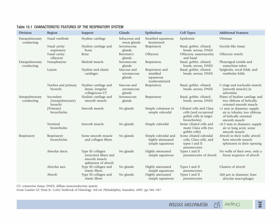
ChapterrespIratory system 15 219
Table 15.1 CHARACTERISTIC FEATURES OF THE RESPIRATORY SYSTEMDivision Region Support Glands Epithelium Cell Types Additional Features
extrapulmonary conducting
nasal vestibule hyaline cartilage sebaceous and sweat glands
stratified squamous keratinized
epidermis Vibrissae
nasal cavity: respiratory
hyaline cartilage and bone
seromucous glands
Respiratory Basal, goblet, ciliated, brush, serous, Dnes
erectile-like tissue
nasal cavity: olfactory
Bone Bowman’s glands
olfactory olfactory, sustentacular, and basal
olfactory vesicle
extrapulmonary conducting
nasopharynx skeletal muscle seromucous glands
Respiratory Basal, goblet, ciliated, brush, serous, Dnes
Pharyngeal tonsils and eustachian tubes
larynx hyaline and elastic cartilages
Mucous and seromucous glands
Respiratory and stratified squamous nonkeratinized
Basal, goblet, ciliated, brush, serous, Dnes
epiglottis, vocal folds, and vestibular folds
trachea and primary bronchi
hyaline cartilage and dense, irregular collagenous ct
Mucous and seromucous glands
Respiratory Basal, goblet, ciliated, brush, serous, Dnes
c-rings and trachealis muscle (smooth muscle) in adventitia
intrapulmonary conducting
secondary (intrapulmonary) bronchi
hyaline cartilage and smooth muscle
seromucous glands
Respiratory Basal, goblet, ciliated, brush, serous, Dnes
Plates of hyaline cartilage and two ribbons of helically oriented smooth muscle
(Primary) bronchioles
smooth muscle no glands simple columnar to simple cuboidal
ciliated cells and clara cells (and occasional goblet cells in larger bronchioles)
<1 mm in diameter; supply air to lobules; two ribbons of helically oriented smooth muscle
terminal bronchioles
smooth muscle no glands simple cuboidal some ciliated cells and many clara cells (no goblet cells)
<0.5 mm in diameter; supply air to lung acini; some smooth muscle
Respiratory Respiratory bronchioles
some smooth muscle and collagen fibers
no glands simple cuboidal and highly attenuated simple squamous
some ciliated cuboidal cells, clara cells, and types i and ii pneumocytes
Alveoli in their walls; alveoli have smooth muscle sphincters in their opening
Alveolar ducts type iii collagen (reticular) fibers and smooth muscle sphincters of alveoli
no glands highly attenuated simple squamous
types i and ii pneumocytes of alveoli
no walls of their own, only a linear sequence of alveoli
Alveolar sacs type iii collagen and elastic fibers
no glands highly attenuated simple squamous
types i and ii pneumocytes
clusters of alveoli
Alveoli type iii collagen and elastic fibers
no glands highly attenuated simple squamous
types i and ii pneumocytes
200 µm in diameter; have alveolar macrophages
ct, connective tissue; Dnes, diffuse neuroendocrine system.From gartner lP, hiatt Jl: color textbook of histology, 3rd ed. Philadelphia, saunders, 2007, pp 346–347.

Chapter
respIratory system
15
220HISToPHySIoLoGy of THE NASAL cAvITy
the richly vascularized nasal mucosa has an abun-dance of glands that produce mucus and watery fluid that keep the surface of the epithelium moist.
• the mucous layer traps particulate matter suspended in the inspired air.
• the cilia of the columnar cells of the pseudostratified epithelium sweep the mucus, with its entrapped particulate matter, toward the back of the throat to be eliminated.
• the rich vascular supply is arranged in such a fashion that a countercurrent mechanism is established that humidifies and warms the inspired air.
• the lymphoid elements present in the lamina propria capture allergens and antigens to ensure that foreign antigens are eliminated, although frequently symptoms associated with hay fever and colds are induced.
the olfactory epithelium (see Fig. 15.1), by sens ing various odors, provides most of the information that is recognized as taste. Although the system of olfac-tion is not well understood, it is believed to be similar to the immune system, in that odorants—molecules that dissolve in the mucus—bind to odor receptor molecules on the surface of the olfactory cilia.
• When odorants have bound to the required number of odor receptor molecules, the olfactory cell membrane depolarizes, and the action potential travels along its axon to stimulate collections of mitral cells, located in small regions known as glomeruli within the olfactory bulb.
• the 1000 or so glomeruli located in the olfactory bulb receive input from about 2000 olfactory neurons.
• the particular permutations and combinations of possible inputs permit humans to recognize approximately 10,000 different odors, which requires each glomerulus to participate in the recognition of numerous different scents.
• the odorants are quickly washed away from the olfactory cilia by the copious fluid flow from
Bowman’s glands, preventing multiple responses from a single odorant.
LARyNx
the larynx is a short musculocartilaginous segment of the conducting portion of the respiratory system interposed between the pharynx and the trachea. it permits phonation and averts the entry of food or drink material into the respiratory system during the process of deglutition.
• the larynx comprises several cartilages that are connected to one another by ligaments and by extrinsic and intrinsic skeletal muscles.
• the aditus, the superior opening of the larynx, is guarded by the epiglottis, a cartilaginous flap that stays open during phonation and breathing but closes over the aditus during swallowing.
the laryngeal lumen displays two pairs of folds:
• the superior vestibular folds, which do not move
• the inferior vocal folds, which contain a dense regular elastic connective tissue, the vocal ligament, and its adhering vocalis muscle; these assist intrinsic muscles in tensing the vocal ligaments and moving them in a medial direction, narrowing the rima glottidis, the space between the right and left vocal folds.
As the exhaled air rushes past the vocal folds, it vibrates them and produces a sound that, by being modulated by the tongue and lips, creates speech. the length of the vocal fold and the degree of tension placed on it modulates the pitch of the sound that is being produced; the shorter the fold and the greater the tension on the vocal cord, the higher the pitch of the sound that is being produced.
the superior aspects of the vocal folds and of the epiglottis are covered by stratified squamous non-keratinized epithelium, and the remainder of the laryngeal lumen is lined by pseudostratified ciliated columnar epithelium. the cilia of the columnar cells of this epithelium transport mucus toward the pharynx to be eliminated.

Chapter
respIratory system
15
221
cLINIcAL coNSIDERATIoNSA common problem with many individuals is a nosebleed (epistaxis). In children, nosebleeds are usually the result of nasal drying, whereas in adults, they may be a warning sign of high blood pressure. Nosebleed usually occurs in the anteroinferior region of the nasal septum (kiesselbach’s area) an area rich in blood vessels. Bleeding may be stopped by applying pressure or packing the nasal cavity with cotton. Any nasal bleeding that persists, recurs, or is severe needs to be evaluated by a physician.
Within the lamina propria of the respiratory epithelium covering the conchae are large venous plexuses known as swell bodies. Every 20 to 30 minutes, the swell bodies on one side of the nasal fossa become engorged with blood that distends the mucosa slightly, impeding airflow. At this time, most of the airflow passes through the opposite nasal fossa. This cycling assists the respiratory mucosa in maintaining a hydrated state.
Acute or chronic laryngitis is an inflammation of the larynx resulting from overuse, irritation, or infection of the vocal folds (the mucous membrane
covering of the vocal cords). Normally, the vocal folds open and close smoothly, forming sounds as air passes by them, causing them to vibrate. During laryngitis, the vocal folds become inflamed or irritated and they swell, resulting in the creation of hoarse sounds. Most cases of laryngitis are triggered by temporary viral infections or vocal strain and are not serious. Persistent hoarseness can sometimes signal a more serious underlying medical condition, however.
cough reflex is initiated by particulate matter or irritants in the upper passageways of the respiratory system, including the trachea and the bronchi. The reflex begins with a large volume of air being inhaled. This inhalation is followed by closing the glottis and epiglottis, followed by a powerful contraction of the muscles of expiration (abdominal and intercostal muscles) and then an immediate opening of the glottis and epiglottis. This last event permits a powerful rush of air (velocity of which may reach 100 mph) forcing the irritant out of the upper respiratory passages.
Figure 15.1 the olfactory epithelium and its various cell types—basal, sustentacular, and olfactory cells. (From Gartner LP, Hiatt JL: Color Textbook of Histology, 3rd ed. Philadelphia, Saunders, 2007, p 348.)
Schwann cell
Olfactory vesicle
Dendrite
Olfactory cilia Microvilli
Duct of Bowman's gland
Sustentacularcell
Olfactoryreceptor cell
Basal cell
Connective tissue
Bowman's gland

Chapter
respIratory system
15
222TRAcHEA
the trachea, a 12-cm-long tube, is always patent because of the 10 to 12 c-shaped hyaline cartilage rings, called C-rings, that support it along its entire length. each c-ring, positioned in such a fashion that its closed portion faces in an anterior direction, has its own perichondrium. the perichondria of neigh-boring c-rings are connected to each other by fibro-elastic connective tissue. the posteriorly facing open ends of the c-rings are connected to each other by slips of smooth muscle—the trachealis muscle—whose contraction reduces the diameter of the trachea, accelerating the flow of air through this tubular structure. the lumen of the trachea resembles the shape of the capital letter D. the trachea is com-posed of three layers: the innermost mucosa, the middle submucosa, and the outermost adventitia. the mucosa of the trachea is composed of respira-tory epithelium, subepithelial connective tissue (lam-ina propria), and a thick sheet of elastic fibers that separates the mucosa from the submucosa. the res-piratory epithelium, pseudostratified ciliated col-umnar epithelium (Fig. 15.2), is composed of goblet cells, ciliated columnar cells, basal cells, brush cells, serous cells, and diffuse neuroendocrine system (Dnes) cells. All cells of this epithelium sit on the basement membrane, but not all of them reach the lumen.
• Almost one third of the cells of this epithelium are goblet cells (Fig. 15.3), unicellular glands that manufacture the slippery secretion known as mucinogen, which they store in secretory vesicles in the expanded apical region of their cytoplasm, known as the theca. Most of the organelles, including the nucleus of goblet cells, are located in the thin, basal part of the cell known as the stem. When the mucinogen is released into the lumen of the trachea, it becomes hydrated and is known as mucin. When mucin is mixed with particulate matter in the lumen, it becomes known as mucus.
• there are approximately as many ciliated columnar cells as there are goblet cells. these tall
cells have numerous cilia that propel the mucus in the lumen of the trachea toward the larynx.
• Basal cells are short, regenerative cells; they constitute slightly less than one third of the cell population of the respiratory epithelium. these cells undergo mitosis and replace the dead and dying goblet cells, ciliated cells, and brush cells.
• Brush cells, also known as small granule cells, constitute less than 3% of the total cell population of the respiratory epithelium. these narrow cells may be goblet cells that have released their mucinogens, or they may have some neurosensory function because they have long microvilli that extend into the lumen of the trachea.
• there are approximately as many serous cells as there are brush cells. serous cells are also tall columnar cells that display apical secretory granules containing a serous secretion whose function is not understood.
• DNES cells compose about 3% of the total cell population. these cells manufacture and release paracrine or endocrine hormones in response to stimuli such as hypoxia. Dnes cells are frequently innervated, and the nerve fiber Dnes cell complex is known as pulmonary neuroepithelial bodies, which, under hypoxic conditions, may be able to activate neurons in the respiratory center of the hypothalamus.
the lamina propria of the trachea is a fibroelastic connective tissue housing lymphoid elements and serous and mucous glands that deliver their secretion into the lumen. A thick elastic sheet, the outermost layer of the lamina propria, separates the mucosa from the submucosa.
A richly vascularized dense irregular fibroelastic connective tissue forms the submucosa of the trachea. it houses numerous seromucous glands and an abundance of lymphoid elements. the c-rings of the trachea are located in the fibroelastic connective tissue of the adventitia. this outermost layer binds the trachea to the surrounding structures, such as the esophagus.
cLINIcAL coNSIDERATIoNSIndividuals who are chronically exposed to irritants such as tobacco smoke or coal dust may display alteration of their respiratory epithelium, known as metaplasia, so that instead of the normal pseudostratified ciliated columnar morphology, their ciliated cells are greatly reduced in height, goblet cells become abundant, and a thicker layer of mucus is produced attempting to remove the irritants. The
decrease in the number of ciliated cells hinders the clearance of mucus, however, increasing congestion. Additionally, seromucous glands of the lamina propria and the submucosa become enlarged and produce more secretory products. If the environmental conditions are changed and the irritants are eliminated, the respiratory epithelium returns to its previously normal, healthy state.

Chapter
respIratory system
15
223
Figure 15.2 types of epithelia. (From Gartner LP, Hiatt JL: Color Textbook of Histology, 3rd ed. Philadelphia, Saunders, 2007, p 87.)
Simple
Stratified
Squamous nonkeratinized
Keratinized Columnar
Cuboidal Transitional (relaxed)
Transitional
Transitional (distended)
Squamous Cuboidal Columnar Pseudostratifiedcolumnar
Pseudostratified
Theca
Stem
Microvilli
Mucinogendroplets
Nucleus
Figure 15.3 Ultrastructure of a goblet cell. (From Lentz TL: Cell Fine Structure: An Atlas of Drawings of Whole-Cell Structure. Philadelphia, Saunders, 1971.)

Chapter
respIratory system
15
224bRoNcHIAL TREE
the bifurcation of the trachea signals the beginning of the bronchial tree, the conducting portion of the respiratory system, which extends all the way into the lungs down to the level of the terminal bronchioles. As the branches of the bronchial tree bifurcate, their diameter decreases, and their numbers increase, and, as stated earlier, the total cross-sectional area of the bronchial tree increases in size, slowing the velocity of the inspired airflow and increasing the velocity of the expired airflow. the bronchial tree comprises primary bronchi, secondary and tertiary bronchi, bronchioles, and terminal bronchioles (Fig. 15.4). general comments concerning the histology of the bronchial tree as it progresses from primary bronchi to terminal bronchioles are that:
• the size of cartilage and glands decreases.• the percent of goblet cells and the epithelial
thickness decrease.• the amount of elastic tissue and smooth muscle,
relative to the thickness of its wall, increases.
the trachea bifurcates to form the left and right primary (extrapulmonary) bronchi, which resemble the trachea except that they are smaller in diameter and their walls are thinner. the left primary bron-chus is not as straight as the right bronchus, and it bifurcates. the right one trifurcates before entering the lung tissue to become intrapulmonary (second-ary) bronchi. each branch of the left and right primary bronchi serves one of the five lobes of the lung.
Secondary bronchi (intrapulmonary bronchi, lobar bronchi) resemble primary bronchi, but the supporting hyaline cartilage is no longer in the shape of a c-ring. instead, it is composed of small cartilage pieces that surround the circumference of the lumen, giving the cross-sectional circumference of the lumen a round shape.
• Because the cartilage no longer has open arms facing in a posterior direction, the smooth muscle migrates toward the lumen, occupying a position between the lamina propria and the submucosa, and is arranged in two helically disposed layers.
• the fibroelastic adventitia has elastic fibers radiating from it in such a fashion that the fibers are positioned more or less perpendicularly to a tangent placed at their contact with the wall of the secondary bronchus.
• these elastic fibers contact other elastic fibers that ramify throughout the substance of the lungs.
secondary bronchi, when in the lung, arborize to form tertiary (segmental) bronchi, each of which serves 1 of the 10 relatively large, bronchopulmo-nary segments of each lung. As these tertiary bron-chioles arborize, they form smaller and smaller cylindrical conduits that eventually have no hyaline cartilage in their wall, and possess, relative to the thickness of their walls, more smooth muscle cells disposed in two helically oriented bundles. the smallest of these segmental bronchi serve two or more pulmonary lobules, small subdivisions of a bronchopulmonary segment, and arborize to form bronchioles.
A convenient, but not universally accepted, work-ing definition of a bronchiole is that it is less than 1 mm in diameter and serves a single pulmonary lobule. Bronchioles possess:
• no cartilage in their walls• A relatively thick smooth muscle coat compared
with the thickness of their wall• elastic fibers that emanate from the connective
tissue, enveloping the smooth muscle bundles in:• such a fashion that they are positioned more
or less perpendicular to a tangent placed at their contact with the wall of the bronchiole
• contact with other elastic fibers arising from various sources, placing tension on the entire circumference of the bronchiole and maintaining its patency
• no glands in their lamina propria• A simple columnar ciliated to a simple cuboidal
ciliated epithelial lining• no goblet cells in the epithelial lining of smaller
bronchioles• columnar Clara cells in bronchioles of all sizes
that:• secrete a surfactant-like substance that assists
in maintaining bronchiolar patency• Destroy inhaled toxins and act as regenerative
cells to maintain the epithelial lining
Bronchioles arborize to give rise to terminal bronchioles (see Fig. 15.4), the smallest segments of the conducting portion of the respiratory system (<0.5 mm in diameter), each of which serves a small segment of a lung lobule, the lung acinus. terminal bronchioles resemble bronchioles but are much more slender and are lined by a simple epithelium composed of cuboidal cells (some with cilia) and clara cells. their narrow lamina propria is sur-rounded by some smooth muscle cells and kept patent by elastic fibers radiating from the fibroelastic connective tissue wall. terminal bronchioles give rise to respiratory bronchioles.

Chapter
respIratory system
15
225
Figure 15.4 the conduits of the respiratory system. (From Gartner LP, Hiatt JL: Color Textbook of Histology, 3rd ed. Philadelphia, Saunders, 2007, p 355.)
Smooth musclefibers
Intra-alveolarseptum
Respiratorybronchiole
Alveolarpore
Alveolar capillary network
Pulmonary vein(carryingoxygenatedblood)
Alveolar duct
Respiratorybronchiole
Alveoli
Pulmonaryartery (carryingdeoxygenatedblood)
Alveolar elastinnetwork

Chapter
respIratory system
15
226
cLINIcAL coNSIDERATIoNSchronic obstructive pulmonary disease (coPD) is a combination of lung diseases that includes emphysema and bronchitis. Emphysema is a chronic disease in which the alveoli become damaged, resulting in difficulty breathing; chronic bronchitis is a disease in which a bronchial obstruction impedes breathing. It becomes chronic, resulting in swelling, inflammation, wheezing, coughing, and increased difficulty in breathing. It can begin as a mild condition, and over time it can lead to a greatly reduced quality of life and respiratory failure resulting in death of the individual.
Respiratory Portion of the Respiratory System
the respiratory portion of the respiratory system consists of respiratory bronchioles, alveolar ducts, alveolar sacs, and alveoli. Respiratory bronchioles (Fig. 15.5) resemble terminal bronchioles with the exception of the occasional alveoli that balloon out of their walls permitting the exchange of gases to occur, which does not happen in terminal bronchi-oles. Respiratory bronchioles undergo branching and eventually give rise to alveolar ducts (see Fig. 15.5), which are simply a linear sequence of alveoli that branch to form many additional alveolar ducts.
• each of these alveolar ducts ends as a cul-de-sac formed by two or three groups of alveoli.
• each alveolar group is referred to as an alveolar sac.• the common airspace from which these groups
of alveolar sacs originate is known as the atrium.• Alveolar ducts, alveolar sacs, and alveoli possess
their own basal lamina, and they maintain their patency by the presence of slender elastic fibers that attach to the slight connective tissue elements of these structures and to other elastic fibers in their vicinity.
Alveoli (see Fig. 15.5) are small airspaces lined by two types of cells: highly attenuated type I pneumo-cytes (type I alveolar cells) and type II pneumocytes (septal cells). the very thin wall of the alveolus permits the exchange of o2 for co2. A smooth muscle cell and its surrounding reticular fibers encircle the opening of each alveolus controlling the size of its aperture. it has been estimated that there are 300 million alveoli in the two lungs, where each alveolus is approximately 0.002 mm3 providing a total surface area of 140 m2 devoted to the exchange of o2 for co2.
• Type I pneumocytes are highly attenuated cells that form greater than 95% of the alveolar surface. Most of the organelles of this cell are crowded around the region of the nucleus, whereas the remaining regions of the cell are approximately 80 nm wide and house mostly the fluid cytosol. Adjacent type i pneumocytes form occluding junctions with each other to avoid the entry of extracellular fluid from the interalveolar connective tissue compartment into the alveolus.
• Type II pneumocytes (Fig. 15.6) are cuboidal-shaped cells that form only 5% of the alveolar surface even though they outnumber the type i pneumocytes. these cells house lamellar bodies that they discharge into the lumen of the alveolus as pulmonary surfactant, a substance composed of dipalmitoyl phosphatidylcholine and surfactant apoproteins sP-A, sP-B, sP-c, and sP-D that coats the wall of the alveolar airspace
and, by reducing surface tension, assists in maintaining alveolar patency. these cells not only manufacture and resorb surfactant in a continuous manner, but they also have the ability to enter the cell cycle and form more type ii and type i pneumocytes.the region between two neighboring alveoli is
known as an interalveolar septum, which is a slender, continuous, capillary-rich (see Figs. 15.5 and 15-6) connective tissue element that provides a degree of stability to these delicate structures. Mono-cytes, derived from bone marrow, populate inter-alveolar septa and enter the lumina of alveoli, becoming known as alveolar macrophages (dust cells), which phagocytose not only particulate matter and microorganisms that gain access to the alveoli with the inhaled air but also surfactant to ensure a constant exchange of old for newly formed surfac-tant. Dust cells, engorged with phagocytosed mate-rial, either reenter the interalveolar septum to leave the lung via lymph vessels or migrate up the bron-chial tree to enter the pharynx to be eliminated with the mucus by being swallowed or expectorated.
interalveolar septa may be very narrow, housing only a continuous capillary whose basal lamina is fused with the basal lamina of the type i pneumo-cyte; or a capillary and connective tissue elements, including lymphoid cells, fibroblasts, and macro-phages surrounded by reticular fibers, elastic fibers, ground substance, and extracellular fluid. the exchange of co2 in the capillaries for oxygen in the alveolus occurs best in the region of the narrowest possible interalveolar septum, known as the blood-gas barrier, a structure composed of:
• capillary endothelium• Fused basal laminae of the capillary endothelium
and type i pneumocytes• Attenuated type i pneumocyte• surfactant

Chapter
respIratory system
15
227
Figure 15.5 A, Respiratory bronchiole, alveolar duct, alveolar sac, and alveoli. B, Relationship between an alveolus and continuous capillaries. (From Gartner LP, Hiatt JL: Color Textbook of Histology, 3rd ed. Philadelphia, Saunders, 2007, p 358.)
Alveolar poreRespiratorybronchiole
Alveolar duct
Alveolus
Oxygenated blood to heart
Plasma
Interior of alveolus
Deoxygenated blood from heart
Red blood cell
Cell ofalveolus
O2CO2
Alveolar capillary
A
B
Figure 15.6 type ii pneumocyte. (From Gartner LP, Hiatt JL: Color Textbook of Histology, 3rd ed. Philadelphia, Saunders, 2007, p 361.)
Surfactant extruded fromlipoprotein vesicle
Aqueous hypophase
Lipid monolayerSurfactant
Small lamellar body fusing to lipoprotein vesicle
Multivesicular body
Smalllamellar body(phospholipid)
Proteinsynthesis
Golgi
Phosphatidylcholine synthesis
Amino acids
Choline

Chapter
respIratory system
15
228ExcHANGE of GASES bETWEEN THE TISSuES AND THE LuNGS
cellular respiration in the entire body results in the formation of approximately 200 ml of co2 per minute that enters the capillaries to be transported to the lungs (Fig. 15.7A and B), where it is exchanged for o2. Because the partial pressure of co2 is greater in the tissues than in blood, the gas enters the capil-laries via simple diffusion. the 200 ml of co2 that enters the capillaries every minute is distributed in the following manner:
• 20 ml becomes dissolved gas in the plasma.• 40 ml binds to globin component of hemoglobin.• 140 ml enters the erythrocyte where carbonic
anhydrase catalyzes its reaction with water to form h2co3, which dissociates to form h+ and hco3
−. the bicarbonate ion diffuses out of the red blood cell (RBc) cytosol into the plasma; to compensate for the change in ionic balance, cl− enters the RBc cytosol from the plasma, a process known as the chloride shift (Fig. 15.7c).
in the alveoli of the lungs, the partial pressure of oxygen is greater than that of the oxygen entering blood. the two gases are again exchanged by simple diffusion, a process that does not require the expen-diture of energy. the release of co2 and the uptake of o2 occur in the following manner (Fig. 15.7D):
• Bicarbonate ions of the plasma enter the RBc cytosol necessitating the exit of cl− ions from the cytosol, a reversal of what happened before (constituting another chloride shift).
• carbonic acid forms by binding bicarbonate ions with h+ ions.
• oxygen enters the RBc and binds to the heme portion of hemoglobin.
• the enzyme carbonic anhydrase cleaves carbonic acid to form water and co2.
• the 200 ml of co2 that enters the bloodstream in the tissues per minute is released from the bloodstream, diffuses across the blood-gas barrier, enters the alveolar airspaces, and is exhaled.
Because of its ability to bind to two separate sites on the hemoglobin molecule, nitric oxide (NO) plays an important role in the process of gas exchange.
• in the tissues, no is released by endothelial cells in the lung and binds to one site on the
hemoglobin molecule; when it reaches the tissues, it is released, causing relaxation of the vascular smooth muscles with a resultant dilation of the vessel, facilitating the release of oxygen.
• no occupies the second binding site on hemoglobin by binding to the site of the released oxygen; in this fashion, no is conveyed to the lungs, where it is released and enters the alveolar airspaces to be exhaled with carbon dioxide.
MEcHANISM of vENTILATIoN
the processes of inhalation and exhalation depend on the anatomic relationship of the lungs, the pleural membranes, the pleural cavities, and the elastic fiber components of lung tissue. each lung is covered by visceral pleura, which is continuous, at the root of each lung, with the parietal pleura.
• the parietal pleura adheres to the walls of the thoracic cage and to the connective tissue components of the mediastinum.
• the visceral pleura adheres to the surface the lung.
• the space between the parietal pleura and the visceral pleura is a serous cavity known as the pleural cavity, an empty space lubricated by a thin, serous fluid whose function is to reduce friction caused by the movements of the lung.
Inhalation is facilitated by the contraction of the muscles of chest wall and the diaphragm (respiratory muscles). As these muscles contract, an energy-requiring process, the thoracic cage enlarges and pulls on the adhering parietal pleura, enlarging the pleural cavities and consequently reducing the pres-sure inside the pleural cavities.
• the atmospheric pressure is now greater than that of the pleural cavities, causing the influx of air into the lungs.
• the entry of air into the lungs stretches the lung, including its elastic fibers, and reduces the formerly enlarged volume of the pleural cavities, increasing the pressure inside these cavities.
Exhalation is facilitated by the relaxation of the respiratory muscles, permitting:
• elastic fibers that were stretched to begin to return to their normal length
• increased pressure within the pleural cavities to drive air out of the lungs

Chapter
respIratory system
15
229
cLINIcAL coNSIDERATIoNSPatients with pulmonary congestion and congestive heart failure have lungs that are congested with extravasated blood. RBCs gain entrance into the alveoli, where they are phagocytosed by alveolar macrophages. In these instances, these macrophages are called heart failure cells when they are present in the lung and sputum.
lung cancer is the most common cause of cancer-related death among men and women in the United States. It claims more lives each year than colon, prostate, and breast cancers combined. Lung cancer is a disease of
uncontrolled cell growth in lung tissue that may lead to metastasis with invasion of adjacent tissue and other organ systems. Most lung cancers are carcinomas of the lung, derived from epithelial cells. Almost 90% of lung cancer is caused by long-term exposure to tobacco smoke. The most common symptoms are shortness of breath, coughing, coughing up blood, and weight loss. There are two main types of lung cancer: small cell lung carcinoma, which responds to chemotherapy, and non–small cell lung carcinoma, which responds better to surgery. Radical radiation is often used in the treatment of small cell lung carcinoma.
Figure 15.7 A, Respiratory bronchiole, alveolar duct, alveolar sac, and alveoli. B, Relationship between an alveolus and continuous capillaries. C, co2 uptake from body tissues by erythrocytes and plasma. D, co2 release by erythrocytes and plasma in the lung. (compare A with the alveolar duct shown in Fig. 15.4.) (From Gartner LP, Hiatt JL: Color Textbook of Histology, 3rd ed. Philadelphia, Saunders, 2007, p 358.)
CO2
Alveolus
Diffusion of CO2 out of blood into alveolus
Alveolusof lung
Diffusion of CO2 into blood and conversion to HCO3–
Body tissue
Hb
CO2+H2O
Carbonic anhydrase H2CO3
HCO3–+H+
HCO3–
Cl–
CO2
CO2
Produced by tissue cells
Capillary
Cl–
CO2+H2OH2CO3
HCO3–+H+
Hb
Carbonic anhydrase
CO2
Capillary
Alveolarpore
Respiratorybronchiole
Alveolar duct
Alveolus
Oxygenated blood to heart
Plasma
Interior of alveolus
Deoxygenated blood from heart
Red bloodcell
Cell ofalveolus
O2 CO2
Alveolar capillary
C D
A
B

230
16 DIGESTIvE SySTEM: oRAL cAvITy
the digestive system is a complex, continuous tube that includes the functions of modified ingestion, swallowing (deglutition), digestion, absorption of nutrients and fluids, and elimination of indigestible residues and gases. the glandular portion of the digestive system may be intramural or extramural.
oral cavity
the oral cavity (mouth), lined by a wet stratified squamous epithelium, is subdivided into two spaces—the vestibule and the oral cavity proper. the subepithelial connective tissue and the epithelium together are known as the oral mucosa.
• When epithelium is keratinized or parakeratinized because of friction, the mucosa is referred to as masticatory mucosa, located on the gingiva, hard palate, and dorsal tongue.
• Most of the oral cavity has a lining mucosa.• the dorsum of the tongue and areas of the soft
palate and pharynx possess taste buds, and those regions are referred to as specialized mucosa.
the paired major salivary glands produce saliva, which possesses salivary amylase, the antimicrobial agents lactoferrin and lysozyme, and IgA and main-tains a moist environment. During eating, flow of saliva allows macerated food to be formed into bolus that can be swallowed.
Lips
each lip (Fig. 16.1A) has three surfaces: the hairy external skin aspect, the red vermilion zone, and the wet mucosal (internal) aspect. the tall rete appara-tus of the vermilion zone brings capillaries near the surface imparting a pink color to it. the muco-sal aspect is always wet and has a lining mucosa whose richly vascularized connective tissue possesses mucous (but some serous) minor salivary glands.
Teeth
humans have 20 deciduous teeth that are replaced and supplanted by the permanent dentition.
each tooth (Fig. 16.1B) is hollow and has an enamel-covered crown and cementum-covered root, which meet at the cervix. the hollow pulp cavity is divided into the root canal and the pulp chamber
housing the pulp, and is surrounded by a mineralized dentin. the root is suspended in the bony alveolus by a dense collagenous periodontal liga-ment (PDL).
the pulp has a neurovascular core surrounded by three concentric layers:• the cell-rich zone, which is
surrounded by• the cell-poor zone and• the outermost zone, the
odontoblastic layerA plexus of sensory nerve fibers, Raschkow’s plexus, located at the interface of the pulp core and the cell-rich zone, conveys pain sensation to the brain. nerve fibers and vascular supply enter the pulp through the apical foramen of the root tip.
Enamel, the hardest tissue in the body, is translu-cent and covers the crown. it is composed of 4% organic matrix and water and 96% calcium hydroxy-apatite, large crystals coated by an organic matrix (enamelins and tuftelins), formed into enamel rods (enamel prisms). each enamel prism is manufac-tured by specialized cells, ameloblasts, which die after tooth eruption; enamel cannot repair itself.
Dentin, yellowish in color, is located in the crown and in the root. it is composed of a type i collagen–based organic matrix and 65% to 70% calcium hydroxyapatite. its elastic property protects the over-lying enamel from being fractured easily. Dentin is manufactured by cells called odontoblasts, whose long processes occupy the tunnel-like spaces, dentinal tubules, in the substance of dentin.
Cementum, composed of 50% to 55% of type i collagen–based organic matrix and bound water and 45% to 50% calcium hydroxyapatite, is located only on the root. cementum of the apical aspect of the root possesses lacunae occupied by cementocytes (cellular cementum); cementum of the coronal aspect lacks cementocytes (acellular cementum). Both types of cementum are covered by a single layer of cementoblasts, which manufacture cementum. Ce-mentoclasts (odontoclasts) resorb cementum.
KEy WoRDS• Lips• Teeth• Tooth development• Supporting tissues
of teeth• Tongue• Taste buds
-

Chapter
dIgestIve system: oral CavIty
16
231
Lower lip EnamelDentin
CrownGingiva
AlveolusRoot of tooth
CementumPeriodontalligament
Root canal
Apical foramenMandible
M. orbicularis orisLabial glands in connective tissueVestibule
Pulp
A
cLINIcAL coNSIDERATIoNSliP
Angular cheilitis is a painful condition in which the corners of the lips become dry and cracked; it is frequently due to dietary deficiency of vitamin B2, zinc deficiency, or iron deficiency anemia. The lesions are most common in elderly individuals who have poorly fitting dentures, and the area becomes infected with pathogens such as Candida albicans.
oral caVity
The lining mucosa of the lips and cheeks may become ulcerated, small areas that are characterized by small, red-rimmed, white, painful spots known as canker sores (or aphthous ulcers). These lesions are usually stress-related and resolve within 7 to 10 days. The pain can be relieved by the application of a local anesthetic ointment.
Squamous cell carcinoma is the most common oral cancer. It is initially painless and appears as a smooth or rough-surfaced red or white lesion that may be in the form of a hard lump or an ulcerated depression that bleeds occasionally. In almost half
of affected individuals, these carcinomas occur on the lining mucosa of the lip, whereas in the remaining individuals the affected area is the tongue or the floor of the mouth. Frequently, squamous cell carcinoma is caused by smoking, alcohol use, or the use of smokeless tobacco, but individuals who neither drink alcohol nor use tobacco products may have the disease. The treatment is usually a combination of surgery and radiation therapy.
aGe-relateD cHanGeS in teetH
Because enamel is nonvital, and the cells manufacturing enamel are no longer present after completion of tooth eruption, enamel cannot be regenerated. The frictional and attritional forces of mastication continuously remove enamel from the occlusal and incisal surfaces, reducing the total amount of enamel. To compensate for the reduction in the size of the crown, cementum is added onto the apical surface, causing the tooth to continue to erupt. This posteruptive movement of the tooth insures that it remains in contact with the tooth opposing it in the other arch.
Figure 16.1 A, A tooth in situ, presenting the lower lip, vestibule, and part of the oral cavity. B, tooth and its surrounding tissues. the enamel of the crown meets the cementum of the root at the cervix of the tooth. Dentin is located in the crown and in the root. (A and B, From Gartner LP, Hiatt JL: Color Textbook of Histology, 3rd ed. Philadelphia, Saunders, 2007, pp 368 and 369.)

Chapter
dIgestIve system: oral CavIty
16
232oDoNToGENESIS bEfoRE THE bELL STAGE
Odontogenesis, or tooth development, begins between the sixth and seventh weeks of development when the ectodermal oral epithelium proliferates to form a horseshoe-shaped dental lamina, one on the maxillary arch and one on the mandibular arch (Fig. 16.2). the dental lamina is separated from the neural crest–derived ectomesenchyme by a basement membrane.
• in 10 different regions of each dental lamina, a bud forms, beginning the bud stage of odontogenesis. each of the 20 buds presages a specific deciduous tooth. the ectomesenchyme at the tip of each bud condenses to form dental papilla.
• each bud enlarges by mitotic activity and forms a three-layered enamel organ—the cap stage of odontogenesis. the simple squamous epithelium, the outer enamel epithelium (OEE), is continuous at the rimlike cervical loop with the concave simple squamous-cuboidal inner enamel epithelium (IEE). the stellate-shaped cells of the stellate reticulum are located between the iee and oee. the basement membrane completely surrounds the enamel organ whose concavity is filled with the dental papilla, a well-vascularized ectomesenchyme. the enamel organ and dental papilla together are known as the tooth germ. the later stage of the enamel organ alters its morphology to form a template that is incisiform, caniniform, or molariform. this ability of the enamel organ depends on the enamel knot, a cluster of cells located among the cells of the stellate reticulum. it is the principal signaling center of tooth formation. the dental papilla manufactures fibroblast growth factor 4 (FGF-4) and epidermal growth factor (EGF), both necessary for the survival of the enamel knot, which synthesizes FgF-4, Sonic Hedgehog, and various bone morphogenetic proteins that direct the transformation of the enamel organ into a molariform template. When the transformation is complete, the dental papilla ceases to express egF and FgF-4, and the enamel knot undergoes apoptosis. the dental papillae of incisiform and caniniform enamel organs never express FgF-4 or egF, and their enamel knots undergo apoptosis during the cap stage. the absence of the enamel knot results in the formation of a default noncusped tooth. the dental papilla forms the dentin and the pulp of the tooth. the ectomesenchyme surrounding the tooth germ forms a thin, dense connective tissue layer, the dental sac, which forms the alveolus, PDL, and
cementum of the tooth. Enamel is synthesized by ameloblasts, cells that are differentiated from the iee. the permanent teeth arise from the succedaneous laminae of the 20 tooth buds. the 12 permanent molars arise from the extensions of the two dental laminae that begin to elongate.
• As the cap grows in size, a fourth layer, the stratum intermedium, appears that is characteristic of the bell stage (stage of histodifferentiation and morphodifferen-tiation). As development continues, the cells of the iee at the region farthest from the cervical loop become elongated cells known as preameloblasts as they begin to manufacture enamel matrix; these cells mature into ameloblasts (Fig. 16.3). in response to the initial formation of enamel, the layer of dental papilla cells that abut the basal lamina differentiate into preodontoblasts, and when they begin to manufacture dentin matrix, they mature into odontoblasts (see Fig. 16.3).
• the dentinoenamel junction is established, and the appositional stage of tooth development begins. initially, the dentinoenamel junction is just a microscopic region that continues to spread along the concavity of the enamel organ and eventually reaches the cervical loop. While that is taking place, ameloblasts and odontoblasts continue to manufacture enamel and dentin, respectively. Both hard tissues become thicker, and the two cell types are displaced farther and farther from each other.
• When enamel formation is completed, the cervical loop elongates, forming a cylindrical sheet, Hertwig’s epithelial root sheath (HERS), composed only of oee and iee that encloses, and is surrounded by, ectomesenchymal cells. the enclosed ectomesenchymal cells are continuous with the dental papilla and form radicular pulp and radicular dentin. the older regions of heRs begin to disintegrate, and some ectomesenchymal cells surrounding heRs migrate onto the radicular dentin surface, differentiate into cementoblasts, and manufacture cementum. As heRs continues to lengthen, more and more of the root is formed, and finally the last region of the root housing the apical foramen is produced. As the root becomes longer, the tooth is erupting into the oral cavity. the eruptive motion is independent of root elongation even though the two processes occur concurrently. eruption is effected by the activity of specialized myofibroblasts of the dental sac that, by tugging on the type i collagen fibers of the dental sac (future PDl) attached to the cementum, drag the developing tooth into occlusion.

Chapter
dIgestIve system: oral CavIty
16
233
Figure 16.2 A–G, odontogenesis. (From Gartner LP, Hiatt JL: Color Textbook of Histology, 3rd ed. Philadelphia, Saunders, 2007, p 372.)
Oralepithelium
Dental lamina
BudEnamelorgan
Dentallamina
EnamelDentin
BoneBonycrypt
Condensedmesenchyme
A Bud stage B Cap stage C Bell stage D Apposition
E Early root formation
Enamel
Alveolar bone
F Late root formation G Eruption
PulpDentin
Cementum
Figure 16.3 Ameloblast and odontoblast. the long odontoblastic process of the odontoblast was shortened by cutting out a long portion of it (white space). (From Lentz TL: Cell Fine Structure: An Atlas of Drawings of Whole-Cell Structure. Philadelphia, Saunders, 1971.)
Ameloblast Odontoblast

Chapter
dIgestIve system: oral CavIty
16
234STRucTuRES ASSocIATED WITH TEETH
the alveolus, PDl, and gingiva are associated with teeth and assist each tooth in maintaining its proper position in the oral cavity (Fig. 16.4).
• the PDL, composed of a cellular, neurovascular, dense, irregular collagenous connective tissue, occupies the narrow PDl space (width ≤0.5 mm in a healthy mouth) between the cementum of the root and the alveolus (see Fig. 16.4). the type i collagen fibers of the PDl are arranged in principal fiber groups, which resist and accommodate the forces of mastication. they suspend the tooth in its alveolus via Sharpey’s fibers embedded in the cementum and in the alveolar bone. the most numerous cells of the PDl are fibroblasts, which not only synthesize the extracellular matrix but also degrade its collagen, accounting for its exceptionally high turnover rate in this tissue. the PDl possesses autonomic, sensory, and proprioceptive nerve fibers, the last of which provide information about spatial orientation.
• the bony housing of the root of each tooth, known as the alveolus (see Fig. 16.4), is composed of three regions: the cone-shaped thin compact bone that has numerous perforations and is in contact with the PDl, known as the alveolar bone proper; surrounded by cancellous bone, the spongiosa; and the outermost thick compact bone—the cortical plate—which is disposed lingually and labially. neurovascular supply of the alveolus resides in tunnel-shaped nutrient canals. the blood vessels and nerve fibers of the alveolus pass through the perforations in the alveolar bone proper, serving not only the alveolus but also the PDl.
• the stratified squamous orthokeratinized or parakeratinized epithelium of the gingiva (gum) attests to the harsh frictional forces to which it is exposed (see Fig. 16.4). similar to the PDl, the type i collagen fiber bundles of its dense irregular collagenous connective tissue are arranged in principal fiber groups. As the gingival
epithelium reaches the enamel, it makes a sharp bend and attaches, via hemidesmosomes, to the enamel surface as an epithelial band around the entire circumference of the tooth, which is known as the junctional epithelium. this thin, wedge-shaped, 1-mm-long epithelial collar that is no more than 50 cells wide coronally and less than 7 cells broad apically prevents the abundant population of microorganisms of the oral cavity from invading the sterile connective tissue of the gingiva.
Palate
the palate, composed of the anterior, immovable hard palate and posterior, movable, muscular soft palate, separates the nasal and oral cavities from each other.
• on the oral surface, the hard palate is lined by a masticatory mucosa whose connective tissue has adipose tissue anteriorly and minor mucous salivary glands posteriorly. the connective tissue of the hard palate adheres to the bony shelf in its core. the nasal side of the hard palate possesses a dense, irregular collagenous connective tissue covered by a pseudostratified ciliated columnar epithelium with an abundance of goblet cells.
• the oral surface of the soft palate is covered by a lining mucosa. the connective tissue is rich in minor mucous salivary glands that are continuous with the glands of the hard palate. the core of the soft palate is composed of skeletal muscles, some of which originate from the anterior edge of the bony shelf of the hard palate. the nasal aspect of the soft palate is identical to the nasal aspect of the hard palate.
• the soft palate ends in the conical uvula, which is covered by a lining mucosa on all of its surfaces with some minor mucous salivary glands interspersed among the connective tissue elements. the core of the uvula contains skeletal muscle fibers that function in elevating the uvula during swallowing.

Chapter
dIgestIve system: oral CavIty
16
235
Figure 16.4 tooth and its surrounding tissues. the enamel of the crown meets the cementum of the root at the cervix of the tooth. Dentin is located in the crown and in the root. (From Gartner LP, Hiatt JL: Color Textbook of Histology, 3rd ed. Philadelphia, Saunders, 2007, p 369.)
cLINIcAL coNSIDERATIoNSThe jaw-jerk reflex is responsible for the opening of the mouth when one unexpectedly encounters a hard object while chewing one’s food. This reflex is initiated when the sudden force encountered by the PDL causes the proprioceptive fibers to inhibit the muscles of mastication from continuing to contract, protecting the teeth from being fractured.
occlusal trauma from atypical activities such as bruxing (grinding the teeth at night) or excessive clenching of the teeth may result in thrombosis or, in the worst case, ischemic necrosis of the PDL. Such lesions are responsible for the widening of the PDL space (i.e., the space between the cementum of the tooth and the bony alveolus) with a concomitant increase in the mobility of the tooth and, if untreated, the loss of that tooth.
alveolar damage may occur because of excessively rapid orthodontic forces placed on the tooth. The forces placed on the tooth become transferred to the PDL causing it to become inflamed, and in response osteoclasts are recruited to the PDL, where they resorb the alveolus to a greater extent than intended by the dental practitioner. The greater than anticipated
widening of the PDL space may result in a possible loss of the tooth owing to irreversible motility.
Halitosis, or bad breath, is usually caused by food particles that have not been removed from between the teeth, from the crevices of the tongue, or from the pits of the palatine tonsils where this debris begins to decompose and emit an unpleasant odor. Additionally, individuals with poor oral hygiene or endodontics problems that have resulted in abscess formation usually have halitosis. The ingestion of certain foods, such as raw garlic or onion, gives the breath an unpleasant odor that disappears when the volatile oils present in these substances clears the bloodstream. Infections with certain bacteria, such as Haemophilus influenzae, produce characteristic sweet, mousy odors that a physician well trained in microbiology should be able to recognize. Less frequently, organic problems may also impart a specific odor to the breath; the breath has an acetone odor in diabetes, smells like urine in kidney failure, and smells mousy in liver disease. Certain esophageal and gastric tumors can impart a foul odor to the breath as well.

Chapter
dIgestIve system: oral CavIty
16
236Tongue
the tongue (Figs. 16.5 and 16.6) is a large, excep-tionally mobile, muscular organ that not only assists in mastication by positioning food on the occlusal plane but also functions in the formation and swal-lowing of the bolus. the tongue also possesses four types of lingual papillae, most of which jut above the surface and have a masticatory mucosa whose highly keratinized stratified squamous epithelium allows the papillae to scrape food off a surface. other lingual papillae are covered by a nonkeratinized stratified squamous epithelium that houses taste buds to determine the taste of food.
the muscles of the tongue are voluntary and divided into two categories:
• Extrinsic muscles originate outside, but insert into, the tongue and move it.
• Intrinsic muscles are contained wholly within the tongue and alter its shape.
the tongue has three surfaces: dorsal, ventral, and lateral. the dorsal surface is separated into an ante-rior two thirds and a posterior one third by the V- shaped sulcus terminalis, whose posteriorly posi -tioned apex is marked by the pitlike foramen cecum. the posterior one third is characterized by a lining mucosa whose surface is irregular because its subep-ithelial connective tissue is rich in lymph nodes, collectively termed the lingual tonsil. the root of the tongue attaches this muscular organ to the floor of the mouth and to the pharynx.
LINGuAL PAPILLAE
three of the four types of lingual papillae are located on the dorsum of the anterior two thirds of the tongue.
• the most numerous of these, the long, finger-like, highly keratinized filiform papillae, have no taste buds. they project above the surface of the tongue and function in scraping food off a surface.
• Fungiform papillae are much fewer in number, resemble a mushroom, project above the surface, and are dispersed in an apparent random fashion among the filiform papillae. Because fungiform papillae are covered by a nonkeratinized stratified squamous epithelium, they appear as red dots on the surface of the tongue. the epithelium of their dorsal surface houses three or four taste buds.
• the 12 or so circumvallate papillae are located in front of the sulcus terminalis. they are depressed into the surface and are surrounded by a furrow whose epithelial lining possesses taste buds. the floor of this furrow accepts small ducts from the glands of von Ebner.
• the lateral surface of the posterior aspect of the anterior two thirds of the tongue has longitudinally disposed groove-like regions, the foliate papillae, that resemble leaves of a book. the taste buds of these papillae degenerate by the third year of age. the depth of the furrows receives small ducts of the minor serous salivary glands of Ebner.
TASTE buDS
Taste buds (see Figs. 16.5 and 16.6) are an intraepi-thelial collection of neural crest–derived cells that form a barrel-shaped structure whose opening, the taste pore, has microvilli—known as taste hairs—protruding from it. the taste bud is composed of approximately 60 to 80 spindle-shaped cells that are constantly being shed and replaced by new cells. the 3000 or so taste buds function in the sensation of the five (or perhaps six) primary taste sensations: bitter, sweet, salty, sour, umami (delicious), and, for some people, fat. each taste bud is completely intraepithelial and is composed of four types of cells, three of which have a life span of 10 days. the fourth cell type, the basal cell (type IV cell), is a regenera-tive cell whose mitotic activity is responsible for gen-erating new cells. the other three cell types are:
• Type I cell (dark cell)• Type II cell (light cell)• Type III cell (intermediate cell)
it is believed that basal cells give rise to type i cells that differentiate into type ii cells that begin to degenerate and become type iii cells and then die. types i, ii, and iii all possess microvilli (taste hairs), structures that have the ability to respond to tastants, chemicals present in food that become dissolved in saliva. some of these tastants activate ion channels (salt and sour), others activate g protein–linked receptors (umami, sweet, and bitter), and still others activate fatty acid transporters (lipids). Most of the taste that people associate with food depends on the odor of food rather than on the taste that is perceived by taste buds.

Chapter
dIgestIve system: oral CavIty
16
237
Figure 16.5 tongue in the oral cavity and a section from the posterior aspect of its anterior two thirds showing the various types of lingual papillae. (From Gartner LP, Hiatt JL: Color Textbook of Histology, 3rd ed. Philadelphia, Saunders, 2007, p 377.)
Geniohyoid muscle Uvula
Palatoglossal fold
Palatine tonsil
Foramen cecum
Lingual tonsil
Epiglottis
Hyoid bone
Fungiformpapilla
Taste budson circumvallatepapilla
Serous glands
Genioglossus muscle
Circumvallate papilla
Filiform papillae
Taste buds
Intrinsic muscle
Figure 16.6 section of the tongue showing the various types of lingual papillae and a diagram of a taste bud. (From Gartner LP, Hiatt JL: Color Textbook of Histology, 3rd ed. Philadelphia, Saunders, 2007, p 378.)
Taste pore
Microvilli
Nerve
Type I cell
Basal cell (Type IV)
Sensorynervefiber
Connective tissue
Fungiform papillaSerous gland
Wall oftaste pore

238
17 DIGESTIvE SySTEM: ALIMENTARy cANAL
the 9-meter-long alimentary canal is a tubular struc-ture composed of the esophagus, stomach, small and large intestines, and anal canal. it digests food, absorbs nutrients and water, and compacts and eliminates the indigestible components of the ingested food.
General organization of the Alimentary canal
the digestive tract is composed of con centric cylinders around a lumen. these layers are modified along the canal, but before discussing the mo-difications, the general pattern is de-scribed (Fig. 17.1):
• the lumen is lined by an epithelial layer and a subepithelial connective tissue, the lamina propria, which houses glands and lymphatic nodules that constitute the mucosa-associated lymphoid tissue. the lamina propria is surrounded by the muscularis mucosae, two smooth muscle layers arranged in a helical fashion: an inner circular and an outer long i-tudinal layer. the epithelium, lamina propria, and muscularis mucosae together are the mucosa.
• surrounding the mucosa is a dense, collagenous connective tissue, the submucosa, which is a vascularized region that houses glands but only in the esophagus and duodenum. Meissner’s submucosal plexus, a component of the enteric nervous system, occupies the most peripheral layer of the submucosa, serving intramural glands, vascular supply, and muscularis mucosae, and acts locally.
• the muscularis externa is composed of smooth muscle in two helically disposed layers—an inner circular and an outer longitudinal layer. Auerbach’s (myenteric) plexus lies between these two smooth muscle layers and controls peristalsis. Auerbach’s plexus acts locally and also over long stretches of the alimentary canal.
• the alimentary canal is covered by either connective tissue (adventitia), which affixes
regions of the gut to the body wall, or by a moist simple squamous epithelium (serosa), which
reduces friction as the gut moves during peristalsis.
the enteric nervous system can act completely on its own; however, it is modulated by the sympathetic and parasympathetic nervous systems.
Esophagus
the esophagus, a 25-cm-long mus-cular tube whose wall is collapsed unless it is transmitting a bolus into the stomach, closely follows this gen-eral plan.
• the esophageal mucosa is composed of a stratified squamous nonkeratinized epithelium, a lamina propria whose esophageal cardiac glands produce mucus that aids in swallowing the bolus. these glands are located in the regions near the pharynx and near the stomach. the muscularis mucosa is composed of a longitudinally disposed smooth muscle layer.
• the vascular submucosa has the esophageal glands proper, which produce mucous and serous secretions. the serous component of this gland manufactures pepsinogen (a proenzyme) and lysozyme, an antibacterial agent. the mucous component lubricates the epithelium. the esophagus has glands in its submucosa.
• the muscularis externa, composed of an inner circular and an outer longitudinal layer, is unusual because in the upper one third of the esophagus, near the pharynx, both layers are composed of skeletal muscle; in the middle third, they are composed of skeletal and smooth muscles; and in the lower third, near the stomach, they are composed solely of smooth muscle.
• the outermost layer of the esophagus is the adventitia in the thorax and a serosa once it enters the abdominal cavity.
KEy WoRDS• Esophagus• Stomach• Gastric glands• DNES cells• Hydrochloric acid
production• Small intestine• Large intestine• Rectum

Chapter
dIgestIve system: alIm
entary Canal
17
239
cLINIcAL coNSIDERATIoNSThe most common symptoms that are indicative of disorders of the digestive system include dysphagia (problems with swallowing), regurgitation, constipation, diarrhea, and bleeding from the digestive tract. Dysphagia may have numerous causes, including physical obstructions in the pharynx or esophagus, muscular or neural problems, and psychogenic problems. regurgitation is not accompanied by nausea or by violent constriction of the abdominal musculature that occurs during vomiting. The process of regurgitation may be due to neural or muscular problems associated with the esophageal sphincter or due to stenosis of the esophagus as a result of a malignancy or untreated acid reflux. Regurgitation in the absence of a physical cause is known as rumination; in this condition, the swallowed food returns to the mouth, where it may be chewed and swallowed again. Rumination is common in infants; it may occur in adults also and is usually stress-related.
constipation is a condition in which bowel movements occur less than three times per week. It is more common in women than in men, in adults older than 65 years, and in women who are pregnant. Individuals who are constipated usually produce small, dark, dry, hard stools that are difficult to eliminate. The causes of constipation may be dietary (e.g., low fiber consumption, eating too much dairy, not drinking enough fluids);
sedentary lifestyle; certain medications, including overuse of laxatives; disruption of the daily routine as in traveling; and ill health, such as stroke and intestinal disorders. Diarrhea refers to the production of loose, watery stool at least three times in one day. There are two types of diarrhea—acute and chronic. acute diarrhea is quite common, lasts for 1 or 2 days, and resolves itself; if it lasts for more than 2 days, it is considered to be chronic diarrhea, and a physician should be consulted to prevent dehydration and rule out organic disease. Most cases of acute diarrhea are due to bacterial, viral, or parasitic infections, whereas chronic diarrhea is usually due to problems with the alimentary canal, such as irritable bowel syndrome.
Bleeding from the digestive tract may occur from the mouth as bloody vomiting or from the anus as bloody discharge or bloody stool. The blood may be fresh or coagulated. Usually, if the blood is fresh, it is red in color and originates near the oral cavity or the anus. If the blood is coagulated, it appears as black particulate matter that resembles coffee grounds in the vomit or it stains the stool black (melena). Bleeding from the alimentary canal may be caused by peptic ulcers, varices that leak blood, use of certain anti-inflammatory agents, inflammatory bowel disease, and cancer.
Figure 17.1 general plan of the alimentary canal. (From Gartner LP, Hiatt JL: Color Textbook of Histology, 3rd ed. Philadelphia, Saunders, 2007, p 382.)

Chapter
dIgestIve system: alIm
entary Canal
17
240Stomach
the stomach has an inlet, where the esophagus joins it, and an outflow, where it is joined by the duode-num. it can expand from a 50-ml volume when empty to approximately 1500 ml when distended. As the stomach receives a bolus from the esophagus, it secretes gastric juices to liquefy the bolus into an acidic fluid known as chyme and to begin to digest it via hydrochloric acid and its enzymes, rennin, pepsin, and gastric lipase. the hormone ghrelin maintains a constant intraluminal pressure by allow-ing the muscularis externa to adapt to the expanding volume and sustains the feeling of hunger as the stomach is being distended. the acidic chyme is released in 1- to 2-ml aliquots into the duodenum through the pyloric sphincter, the modified inner circular layer of the muscularis externa. the stomach has:
• A cardiac region at the concave lesser curvature• A pyloric region at the greater curvature• two additional anatomic regions, the fundus
and the body, which are identical histologically and are referred to as the fundic region (Fig. 17.2)
the lumen of the empty stomach presents rugae, folds of the mucosa and submucosa, which disap-pear when the stomach is distended. the stomach lining displays numerous epithelially lined depres-sions, gastric pits (foveolae), whose floor is perfo-rated by many tubular gastric glands populating the highly vascular lamina propria. the muscularis mucosae has three layers of smooth muscle—inner circular, outer longitudinal, and a poorly defined outermost oblique layer. the submucosa is unre-markable. the muscularis externa has inner circular, outer longitudinal, and innermost oblique layers.
• the simple columnar epithelium of the fundic stomach is composed of tightly packed surface lining cells and regenerative cells; the lateral cell membranes of these cells form tight junctions with each other. surface lining cells produce the thick, visible mucus that guards the epithelium from the acidic chyme, and regenerative cells proliferate to replace the stomach’s epithelial lining.
• the gastric pits of the fundic and cardiac regions extend one third of the way into the lamina propria, which is crowded with gastric glands.
• each gland possesses six cell types, distributed disproportionately, in its three regions: the isthmus, which pierces the gastric pit; the neck; and the base, which abuts the muscularis mucosae. the gastric pits of the pyloric region extend halfway into the lamina propria.
cELLuLAR coMPoSITIoN of fuNDIc GLANDS
see Figure 17.2.
• Mucous neck cells manufacture soluble mucus that becomes part of the chyme and lubricates the alimentary canal. the plasmalemmae of these cells form tight junctions with their neighbors.
• the regenerative cells’ rapid mitotic rate replaces the entire epithelial lining every 5 to 7 days.
• Parietal (oxyntic) cells are usually not present in the base of the gland. they have deep intracellular canaliculi that are lined by microvilli. An intracellular vesicular network, the tubulovesicular system whose membrane component is rich in the proton pump H+,K+-ATPase, parallels the intracellular canaliculi. Parietal cells produce hydrochloric acid and gastric intrinsic factor. During hcl production, the tubulovesicular system is reduced in size with a concomitant increase in the number of microvilli, suggesting that the vesicular network is used to store membranes destined for microvillus production, enabling the cell to increase its surface area during its secretory activity. With the cessation of hcl production, the microvillus membranes are returned to the tubulovesicular system. this energy-requiring process is fueled by the abundant mitochondrial content of parietal cells. the glycoprotein gastric intrinsic factor is released into the lumen of the stomach where it complexes with vitamin B12, to be absorbed by cells of the ileum.
• Chief (zymogenic) cells, located mostly in the base of fundic glands and not at all in cardiac or pyloric glands, manufacture pepsinogen, rennin, and gastric lipase. the proenzyme pepsinogen is converted to the proteolytic enzyme pepsin in the acidic milieu of the stomach; rennin (chymosin) is a proteolytic enzyme that curdles milk, and the enzyme gastric lipase degrades lipids. these enzymes are present within vesicles in the apical cytoplasm of chief cells and are released because of the interaction of acetylcholine and secretin as they bind to their respective receptors on the basal plasma membrane of chief cells to activate their second messenger systems.
• Diffuse neuroendocrine system (DNES) cells are of two types—open and closed—where the former reach the lumen, and the latter do not. each Dnes cell produces a particular hormone that it releases into the lamina propria. these hormones are autocrine if destined for the releasing cell, paracrine if destined for a cell nearby, and endocrine if they have to travel via the bloodstream.

Chapter
dIgestIve system: alIm
entary Canal
17
241
cLINIcAL coNSIDERATIoNSToo much HCl production in the stomach may be due to many causes, one of which is a malignancy involving G cells, the DNES cells that produce the paracrine hormone gastrin. Individuals with this type of cancer have Zollinger-ellison syndrome, and the tumor may be located in various areas of the digestive system, such as the bile duct, duodenum, or pancreas. These patients have recurring ulcers that do not respond to normal ulcer treatments, such as antibiotics against Helicobacter pylori or histamine2 blockers. Proton pump inhibitors are effective, but cure involves surgical excision of the tumor.
The normal position of the stomach and the gastroesophageal junction is below the diaphragm in the abdominal cavity. In some cases, however, the stomach may protrude into the thoracic cavity, a condition known as hiatal hernia. This is a very common condition, and the incidence increases with the age of the individual. There are two types of hiatal hernia. In sliding hiatal hernia, which is more common, the gastroesophageal junction and part of the stomach slide up and down through weakness in the esophageal hiatus of the diaphragm. The second type, paraesophageal
hiatal hernia, is less common but may be more serious. In this condition, the gastroesophageal junction remains in the abdominal cavity, but a part of the stomach protrudes into the thoracic cavity through the esophageal hiatus and lies alongside the esophagus; if it becomes trapped in that location, possible strangulation of that portion of the stomach may result. If the stomach does not retract on its own, surgical intervention becomes necessary.
Sliding hiatal hernia is usually asymptomatic, although it may manifest with gastric reflux, heartburn, and indigestion, especially in individuals who lie down after eating. The same symptoms accompany paraesophageal hiatal hernia; however, if strangulation occurs, the patient experiences substernal pain. Because this symptom resembles a possible heart attack, an individual with substernal pain should seek immediate medical attention. In most cases, surgery is not required. Treatment of hiatal hernias involve changes in dietary habits; the patient should eat more frequently and smaller quantities at each meal. Also, antacids and elevating the head of the bed usually relieve the symptoms.
Figure 17.2 graphic representation of the mucosa of the fundic region of the stomach displaying the fundic glands of the lamina propria. the various cell types composing the epithelium of the stomach and fundic glands are displayed. (From Gartner LP, Hiatt JL: Color Textbook of Histology, 3rd ed. Philadelphia, Saunders, 2007, p 386.)

Chapter
dIgestIve system: alIm
entary Canal
17
242GASTRIc HISToPHySIoLoGy
on a daily basis, the glands of the stomach produce about 2 to 3 l of secretions whose main constituents are water, hcl, enzymes, intrinsic factor, and soluble and visible mucus. By coating the lining of the stomach, the neutral ph of the visible mucus protects the stomach lining from the acidic gastric juices. the visible mucus also affords a beneficial environment for H. pylori, the bacterium that resides within it.
• the muscularis externa of the stomach functions in churning and liquefying ingested food, forming a thick chyme that resembles split pea soup.
• the muscularis mucosae ensure that the entire epithelial surface of the stomach comes into contact with the chyme. When the chyme is of proper consistency, and depending on its acidity, osmolality, and lipid and caloric content, DNES cells of the duodenum release the hormone gastrin, which induces the pyloric sphincter to open and the longitudinal muscles of the pylorus to contract, injecting 1 to 2 ml of chyme into the duodenum. if the chyme is not ready to be discharged, Dnes cells release the hormones cholecystokinin and gastric inhibitory peptide, which inhibit the pyloric sphincter from opening, and the chyme remains in the stomach.
hcl production in the stomach has three phases: cephalic, referring to the thought, smell, or sight of food; gastric, the presence of food in the lumen of the stomach; and intestinal, the presence of food in the duodenum. the mechanism of hcl production by parietal cells is the same, however, regardless of the phase that induced it. the basal aspect of the cell membrane of parietal cells possesses receptors for the neurotransmitter acetylcholine and for the paracrine hormones gastrin and histamine2. When all three sites have bound their respective signaling molecules, hcl production and its secretion into the intra-cellular canaliculi occurs in the following manner (Fig. 17.3):
1. Carbonic anhydrase, present in the cytosol, catalyzes the production of h2co3, which dissociates into H+ and HCO3
−.2. Active transport exchanges intracellular H+ for
extracellular K+ located in the intracellular canaliculi. Although K+ is also actively transported into the parietal cell at its basal cell membrane, it also leaves the parietal cell via K+ channels located in the basal plasmalemma.
3. K+ and Cl− are actively transported out of the cell and into the intracellular canaliculi, where h+ and cl− combine to form HCl.
4. the circulation of ions between the parietal cell and the extracellular fluid of the lamina propria alters osmotic pressures, causing the flow of H2O into the parietal cell.
5. the movement of ions between the parietal cell and the intracellular canaliculi alters the osmotic pressure within the parietal cell, causing the movement of H2O into the intracellular canaliculi.
6. hcl and water, the main components of the gastric juice, are released into the intracellular canaliculi, spaces that are continuous with the lumen of the stomach.
the presence of tight junctions formed by the epi-thelial lining of the gastric mucosa prevents the entry of hcl from the gastric lumen into the lamina propria. Additionally, the HCO3
− that is produced by the parietal cell is released into the lamina propria and, by its buffering action, protects the lamina propria from the accidental leakage of hcl from the gastric lumen. Another possible protection is afforded by the release of prostaglandins in areas whose epi-thelial barrier is accidentally breached. Prostaglan-dins in the lamina propria amplify blood flow to the area, facilitating the elimination of h+ from the affected region.
the inhibition of hcl production and secretion is controlled by four hormones:
• Prostaglandin, gastric inhibitory peptide, and urogastrone act directly on parietal cells, inhibiting their secretory activity.
• the fourth hormone, somatostatin, inhibits the release of gastrin by g cells and histamine by enterochromaffin-like cells, eliminating the signaling molecules necessary for inducing parietal cells to make and secrete hcl.
Small Intestine
the small intestine (Fig. 17.4) is approximately 7 m in length and is said to have three regions. the first portion is the very short duodenum, about 25 cm long; the middle portion is the jejunum, whose wall is relatively thick and is a little less than 3 m long; and the third portion is the ileum, which is the nar-rowest, has the thinnest walls, and is about 4 m in length. the small intestine receives digestive enzymes from the pancreas and bile from the gallbladder, which assist it in digesting the food in its lumen and absorbing water and the nutrients generated. histo-logically, the three regions are quite similar to each other. After a general description of their common features, the differences among the three regions are described in detail.

Chapter
dIgestIve system: alIm
entary Canal
17
243
Figure 17.3 graphic representation of hydrochloric acid production by parietal cells. A, Resting cell. B, Mechanism of hcl release. C, numerous microvilli in an active cell. (From Gartner LP, Hiatt JL: Color Textbook of Histology, 3rd ed. Philadelphia, Saunders, 2007, p 397.)
Tubulovesicles
RESTINGA ACTIVESTIMULATION
H+
K+
K+
ADP + Pi
ATP
KCl
Cl–
H Cl Canaliculus
H 2O
B C
Figure 17.4 graphic representation of the small intestine and the cell types composing its epithelial lining. (From Gartner LP, Hiatt JL: Color Textbook of Histology, 3rd ed. Philadelphia, Saunders, 2007, p 399.)
Crypt ofLieberkühn
Small intestine
Lacteal
Lamina propria
Lymphoid nodule
VilliVillus Villus
Surface absorptive cell
Enteroendocrine cell
Regenerative cell
Paneth cell
Goblet cell
Muscularis mucosae Crypt of Lieberkühn

Chapter
dIgestIve system: alIm
entary Canal
17
244coMMoN HISToLoGIc fEATuRES
to increase the luminal surface area of the small intestine, its submucosa and mucosa have:
• transverse folds, plicae circulares (valves of Kerckring), begin in the duodenum and extend into the ileum, retard the velocity of chyme, and augment the surface area two- to threefold.
• Finger-like extensions of the lamina propria, villi, are covered by a simple cuboidal epithelium and enlarge the luminal surface 10-fold; these villi are 1.5 mm tall in the duodenum, 1 mm tall in the jejunum, and 0.5 mm tall in the ileum; each villus possesses a vascular, loose connective tissue core with a blindly ending lymph capillary, the lacteal.
• simple columnar epithelial cells covering each villus possess abundant microvilli that augment the luminal surface area 20-fold.
• intervillous spaces of the small intestine display openings of the crypts of Lieberkühn, which increase the luminal surface area by a factor of 3 to 4.
HISToLoGy of THE SMALL INTESTINE
the mucosa of the small intestine comprises the epithelial lining, lamina propria, and muscularis mucosae (Fig. 17.5). the simple columnar epithe-lium covering the villus is composed of:
• Surface absorptive cells, the most abundant of the cells, function in the end stage of digestion and in absorbing amino acids, lipids, and sugars. these cells possess 3000 microvilli covered with a glycocalyx; the glycocalyx comprises mostly enterokinases, aminopeptidases, and oligosaccharidases, enzymes that digest oligopeptides and oligosaccharides. the lateral aspects of the cell membranes of surface absorptive cells adhere to the membranes of adjacent cells by forming junctional complexes.
• Goblet cells manufacture mucinogen, a complex protein polysaccharide that, when in contact with water, becomes mucin. When released into the lumen, it mixes with the luminal content and becomes a slippery substance known as mucus.
• DNES cells constitute about 1% of the villous epithelial cells, each producing a specific paracrine/endocrine hormone.
• Microfold cells (M cells), located where lymphoid nodules of the lamina propria contact the epithelium, have deep folds—intercellular pockets. M cells phagocytose intraluminal antigens and transfer them to lymphocytes present in their intercellular pockets, which then transfer the antigens to APcs of the lamina
propria to initiate an immune response. some of the igA manufactured by plasma cells is endocytosed by epithelial cells that couple secretory component to it and release the complex into the lumen. Most of the igA travels to the liver; hepatocytes complex it with secretory component and release it into the bile to be transported to the gallbladder (Fig. 17.6).
the lamina propria of the mucosa is a loose con-nective tissue that is rich in lymphoid and capillary elements, and, in the core of the villus, possesses lacteals (see Fig. 17.5). the deeper aspect of the lamina propria, between the base of the villi and the muscularis mucosae, is quite vascular, although it is mostly displaced by the abundance of intestinal intramural glands, the crypts of Lieberkühn. these glands extend from the intervillous spaces to the muscularis mucosae, and their epithelium consists of the same cell types as those covering the villus, and:
• Regenerative cells proliferate to form new cells of the epithelial lining. the new cell migrates along the basal lamina to the tip of the villus, where it is sloughed off into the lumen 5 to 7 days after its formation; the epithelial lining of the small intestine is replaced once every week.
• Paneth cells live longer (20 days), are located at the base of the crypts of lieberkühn, and house large eosinophilic granules that contain lysozyme and defensins, antimicrobial agents, and tumor necrosis factor-a.
the muscularis mucosa is composed of an inner circular and an outer longitudinal layer of smooth muscle. occasional smooth muscle cells enter the core of the villus extending to its tip. the submucosa of the small intestine is unremarkable, and its mus-cularis externa is composed of an inner circular and an outer longitudinal layer. Meissner’s and Auer-bach’s plexuses occupy their normal positions. the outermost layer is a serosa except in parts of the duodenum where it is an adventitia.
cLINIcAL coNSIDERATIoNSDefensins are produced by Paneth cells, in response to tumor necrosis factor-α and because of the presence of metabolic by-products released by microorganisms. Defensins insert into the phospholipid membranes of microorganisms, where they form ion channels that permit the leakage of ions from the invading organism, killing it. lysozyme, a proteolytic enzyme also manufactured by Paneth cells, breaks down the bacterial membrane component—peptidoglycan—killing the organism.

Chapter
dIgestIve system: alIm
entary Canal
17
245
Figure 17.5 graphic representation of the small intestine and the cell types composing its epithelial lining. (From Gartner LP, Hiatt JL: Color Textbook of Histology, 3rd ed. Philadelphia, Saunders, 2007, p 399.)
Figure 17.6 graphic representation of the role of M cells and the circulation of igA in the small intestine. (From Gartner LP, Hiatt JL: Color Textbook of Histology, 3rd ed. Philadelphia, Saunders, 2007, p 405.)
Bacteria
M cells
Lymphocytes
Lymph node
Antigen presenting
cell Thoracic duct
B cell Bcells
B cells
IgA in bileoutput
Lamina propriaPlasma cells
IgA
IgA
B cells
IgAIgA
IgA
Liver

Chapter
dIgestIve system: alIm
entary Canal
17
246Regional Differences in the Histology of the Small Intestine
the three regions of the small intestine may be dif-ferentiated from one another by minor variations in their histologic appearance.
• the villi are tallest in the duodenum (1.5 mm), shorter in the jejunum (1 mm), and shortest in the ileum (0.5 mm).
• the major difference is the presence of Brunner’s glands in the submucosa of the duodenum.
• Peyer’s patches, which are collections of lymphoid nodules, are present in the lamina propria of the ileum. the jejunum has neither glands in its submucosa nor Peyer’s patches in its lamina propria.
the duodenum receives the bile duct and pancre-atic duct at the duodenal papilla (papilla of Vater). Brunner’s glands (duodenal glands) are tubuloal-veolar, branched glands that open into the base of the crypts of lieberkühn. Brunner’s glands manufacture a seromucous fluid rich in bicarbonates that acts to buffer the acidic chyme delivered from the stomach. these glands also manufacture the hormone urogas-trone (human epidermal growth factor), which inhibits parietal cells from manufacturing hcl and stimulates the mitotic activities of epithelial cells.
HISToPHySIoLoGy of THE SMALL INTESTINE
the motility of the small intestine has two phases—mixing contractions, which are localized events that function in exposing the luminal contents to the epithelial lining of the gut, and propulsive contrac-tions (peristaltic waves), which move the luminal contents along the length of the small intestine. this movement is slow, about 1 to 2 cm/min, and chyme entering the duodenum from the stomach spends 6 to 12 hours in the small intestine. the propulsive contractions are controlled by Auerbach’s plexus and by the Dnes-derived hormones cholecystoki-nin, gastrin, motilin, substance P, and serotonin, which increase motility, and secretin and glucagon, which retard motility (see table 17.1).
• Glands of the small intestine secrete 2 l of a seromucous secretion on a daily basis. the secretory process is mostly under the control of Meissner’s submucosal plexus, but also is influenced by the Dnes-derived hormones secretin and cholecystokinin.
• Digestion of the food material present in the lumen of the small intestine is due to the presence of enzymes, mostly derived from the pancreas and bile from the liver and concentrated in and delivered from the
gallbladder. the process of digestion begins in the oral cavity and stomach and continues in the duodenum, where pancreatic enzymes degrade the various components of the chyme into oligopeptides and dipeptides and oligosaccharides and disaccharides. Enterokinases and aminopeptidases, located in the glycocalyx of the microvilli, complete protein digestion into amino acids and dipeptides and tripeptides that are absorbed into the surface absorptive cells to be converted into amino acids by cytoplasmic peptidases. the glycocalyx also contains oligosaccharidases (lactase, maltase, sucrase, and a-dextrinase) that complete the digestion of dietary carbohydrates by degrading oligosaccharides into monosaccharides that are absorbed by the surface absorptive cells. lipids present in the chyme are emulsified by the bile salts into micelles, and the pancreatic lipase breaks down the lipids into fatty acids and monoglycerides, which diffuse through the plasmalemma of the microvilli.
• Absorption (Fig. 17.7) of the end products of digestion and electrolytes and water occurs mostly in the small and large intestines, although certain substances, such as alcohol, are also absorbed in the stomach. enormous quantities enter the surface absorptive cells of the small intestine, including 1 kg of fat, 0.5 kg of proteins and carbohydrates, about 35 g of sodium, and 7 l of fluid per day. Amino acids and monoglycerides are released into the core of the villus, enter the tributaries of the hepatic portal vein, and go to the liver for further action. Monoglycerides and long-chain fatty acids bind to intracellular fatty acid–binding proteins, and enter the smooth endoplasmic reticulum to be esterified by acyl CoA synthetase and acyltransferases into triglycerides. these triglycerides, coupled to proteins, are transported to the basolateral membrane to be released into the core of the villus as chylomicrons that enter the lacteals, which become filled with the lipid-rich fluid, chyle. the lacteal is emptied because of the rhythmic contractions of the slips of smooth muscle derived from the inner circular layer of the muscularis mucosae, discharging the chyle into the submucosal lymphatic plexus. the chyle is transported to the thoracic duct to be delivered into the junction of the subclavian vein with the left internal jugular vein. Fatty acids that are less than 12 carbons long avoid the re-esterification process; instead, they pass to the basolateral cell membrane to be released into the core of the villus to travel to the hepatic portal vein and from there to the liver.

Chapter
dIgestIve system: alIm
entary Canal
17
247
Figure 17.7 graphic representation of fat absorption by the surface absorptive cells in the small intestine. (From Gartner LP, Hiatt JL: Color Textbook of Histology, 3rd ed. Philadelphia, Saunders, 2007, p 407.)
Bile
LipidsMonoglycerides andlong-chain fatty acids
Lipids in the lumen of the smallintestine are broken down by pancreatic lipase to fatty acidsand monoglycerides.
Monoglycerides and fatty acids are emulsified by bile, formingmicelles that move intosurface absorbing cells. Glycerol diffuses directly into surface absorbing cells.
Monoglycerides and fatty acids are esterified intotriglycerides within the smooth ER.
Triglycerides are complexedwith protein within the Golgiapparatus, forming chylomicrons that are releasedinto the lacteals.
Glycerol and short- andmedium-chain fatty acidsare absorbed directly intothe blood.
Micelle
Triglyceridesynthesis
Protein
Golgi
Blood capillary
Lymphatic capillary(lacteal)
Chylomicron
Lipoprotein andglycoproteinsynthesis
SERRER
Glycerol, short-, medium-chain fatty acids
Glycerol, short-, medium-chain fatty acids
Lipase
1
2 1
2
3
3
4
5
4
5
Table 17.1 DIFFUSE NEUROENDOCRINE SYSTEM CELLS OF THE GASTROINTESTINAL TRACTCell Location Hormone Produced Function
A stomach, small intestine glucagon elevates blood glucoseD stomach, intestines somatostatin inhibits hormone release by Dnes cellsec stomach, intestines serotonin, substance P increases peristaltic movementecl stomach histamine stimulates hcl secretiong stomach, small intestine gastrin stimulates hcl secretion; gastric motilitygl stomach, intestines glicentin elevates blood glucose levelsi small intestine cholecystokinin stimulates release of pancreatic enzymes
and contraction of gallbladderK small intestine gastric inhibitory peptide inhibits hcl secretionMo small intestine Motilin increases intestinal peristalsisn small intestine neurotensin Decreases intestinal peristalsisPP stomach, large intestine Pancreatic polypeptide stimulates enzyme release by chief cells;
inhibits release of pancreatic enzymess small intestine secretin stimulates release of pancreatic bufferViP stomach, intestines Vasoactive intestinal peptide increases peristalsis of intestines; stimulates
elimination of water and ions
ec, enterochromaffin cell; ecl, enterochromaffin-like cell; g, gastrin-producing cell; gi, gastrointestinal; gl, glicentin-producing cell; hcl, hydrochloric acid; Mo, motilin-producing cell; n, neurotensin-producing cell; PP, pancreatic polypeptide–producing cell; ViP, vasoactive intestinal peptide–producing cell.
From gartner lP, hiatt Jl: color textbook of histology, 3rd ed. Philadelphia, saunders, 2007, p 392.

Chapter
dIgestIve system: alIm
entary Canal
17
248Large Intestine
the large intestine, approximately 1.5 m long, com-prises the cecum, appendix, colon, rectum, and anus. histologically, except for the appendix and the anus, these regions are very similar and are referred to as the colon. the colon functions in the absorption of water, electrolytes, and gases and in the compaction of chyme it receives from the ileum into feces.
the colon resembles the small intestine except that it has a greater diameter and possesses no villi.
• the crypts of Lieberkühn resemble their counterparts in the small intestine, but they lack Paneth cells, possess few Dnes cells, and have a greater number of goblet cells (Fig. 17.8). Although it would appear at first glance that most of the epithelial cells are goblet cells, actually more than half of them are surface absorptive cells. As in other regions of the alimentary canal, the entire epithelial lining is replaced via mitotic activity of the regenerative cells at least once per week.
• the lamina propria, muscularis mucosae, and submucosa are unremarkable.
• the muscularis externa is modified in that many of the smooth muscle fibers of the outer longitudinal layer are collected into three slim bands of muscle, the taeniae coli, that are almost in constant tonus, making them shorter than the colon.
• the colon forms a linear sequence of pouches, called haustra coli, along its length.
• the entire colon is invested by a serosa except at the anus, where it is attached to the body wall by a connective tissue adventitia. Along the length of the colon, the serosa forms fat-filled pouches known as appendices epiploicae.
the function of the colon is the secretion of bicarbonate-rich mucus; it also absorbs more fluid and electrolytes from the intestinal contents, accom-plishing the compaction of the feces. every day, the colon reclaims about 1.4 l of electrolyte-containing fluids and reduces the daily feces volume to approxi-mately 100 ml. the colon absorbs approximately 6 to 9 l of gases a day, releasing only about 0.5 to 1 l of gas as flatus. the crypts of lieberkühn of the rec-tum are sparsely distributed and deeper than those of the colon; otherwise, the rectum greatly resembles the colon.
• the 3- to 4-cm-long anal canal is narrower than the rectum, and in its lower half it does not possess even the shallow crypts of lieberkühn present in its upper half.
• the anal mucosa presents longitudinal folds, anal columns (of Morgagni), that converge at the pectinate line to form the anal valves that house the pocket-like anal sinuses.
• the anal canal is lined by a simple cuboidal epithelium that becomes stratified squamous nonkeratinized below the pectinate line. the fibroelastic lamina propria houses circumanal glands at the anus; hair follicles and their attendant sebaceous glands are present here.
• the muscularis mucosae is present but does not extend past the pectinate line. the fibroelastic connective tissue of the submucosa of the anal canal has an internal hemorrhoidal venous plexus above and external hemorrhoidal venous plexus below the pectinate line, just above the anal orifice.
• the muscularis externa is unremarkable except that the inner circular layer is thickened at the pectinate line to form the internal anal sphincter muscle, and the external longitudinal layer is replaced by a fibroelastic membrane that surrounds the internal anal sphincter. the external anal sphincter muscle is formed by thickenings of the skeletal muscle of the floor of the pelvis and surrounds the internal anal sphincter and the fibroelastic sheath. this skeletal muscle sphincter muscle permits voluntary control over the anus.
• the outermost layer of the colon is a serosa.
the appendix is a narrow 5- to 6-cm-long out-pocketing of the cecum whose stellate lumen is lined by simple columnar epithelium composed of surface absorptive cells, goblet cells, and M cells that adjoins the lymphoid nodules present in the lamina propria. the crypts of lieberkühn are sparse and shallow and are composed of surface absorptive cells, goblet cells, regenerative cells, numerous Dnes cells, and occasional Paneth cells. the lamina pro-pria is a loose connective tissue richly endowed with lymphoid cells and lymphoid nodules. the muscu-laris mucosae, submucosa, and muscularis externa follow the common organization of the diges-tive tract. the outermost layer of the appendix is a serosa.

Chapter
dIgestIve system: alIm
entary Canal
17
249
Figure 17.8 graphic representation of the colon and the cell types composing its epithelial lining. (From Gartner LP, Hiatt JL: Color Textbook of Histology, 3rd ed. Philadelphia, Saunders, 2007, p 408.)
cLINIcAL coNSIDERATIoNScrohn’s disease is a chronic inflammation of the wall of the alimentary canal that occurs most frequently in the ileum and the colon. It is believed to be an immune-related disorder that may have genetic and environmental components. It occurs in equal distribution among men and women, making its first appearance usually between ages 15 and 25 years, but almost always before age 35. The usual symptoms are fever, chronic diarrhea that may or may not be accompanied by bleeding, abdominal cramps of varied severity, weight loss, and lack of appetite. The symptoms may last several weeks and then resolve by themselves, only to recur at various indeterminate intervals with variable degrees of severity. The location of the inflammation may be the same as before, or it may spread to other regions of the alimentary canal. Common complications are the formation of fistulas and obstruction of the digestive canal, and in the colon it may lead to colorectal cancer. Although Crohn’s disease is incurable, palliative treatments include the use of antidiarrheal and anti-inflammatory agents, antibiotics, immunomodulators, dietary
modifications, and, if necessary, surgical resection of the affected areas.
Individuals with cholera have ingested water or food infected with Vibrio cholerae, an organism that produces cholera toxin. Although cholera is a very easily treated disease, it is widespread in the tropical and subtropical regions in developing countries, where it is responsible for a high degree of fatality. Cholera is prevented successfully by employing proper sanitary conditions and is treated by antibiotics and the rapid administration of electrolyte-balanced fluids to replace fluid and electrolyte losses, which may be 10 L/day owing to uncontrolled diarrhea.
intestinal gas is a by-product of bacterial metabolism and some swallowed air. The odoriferous components of feces are mercaptans, indole, and hydrogen sulfide, and the odoriferous component of flatus is methane. Because the methane gas is mixed with O2, CO2, and H2, it is quite flammable, and occasionally during cauterization as part of sigmoidoscopy, small, localized explosions may occur.

250
18 DIGESTIvE SySTEM: GLANDS
the glands of the digestive system are located within the wall of the alimentary canal—the intramural glands and outside the wall the extra-mural glands, including the major salivary glands (parotid, submandibul-ar, and sublingual glands), pancreas, and liver (and gallbladder), whose secre tions gain access to the lumen of the alimentary canal by a system of ducts.
Major Salivary Glands
the major salivary glands (Fig. 18.1)—parotid, submandibular, and sublingual—are compound tubuloal-veolar glands that secrete saliva.
• each major salivary gland is surrounded by a connective tissue capsule that sends connective tissue septa into the substance of the gland dividing it into lobes and lobules.
• the neurovascular elements travel in these connective tissue septa to supply the parenchyma of the gland. the parenchyma is the secretory portion, consisting of acini or tubules or both, and a duct portion that culminates in the principal duct of the gland.
• the functional unit of a salivary gland, the salivon, is composed of an acinus and its intercalated and striated ducts.
three types of cells form the secretory portion of a salivary gland: serous, mucous, and myoepithelial.
• serous cells resemble a truncated pyramid, and they produce a watery fluid composed mostly of water, electrolytes, and enzymes (salivary amylase and lipase) that begin digestion in the oral cavity. other secretions include kallikrein and the antibacterial agents lysozyme and lactoferrin. the secretions are stored in apically located zymogen granules (secretory granules) until their release is prompted.
• Mucous cells resemble serous cells, but their apical cytoplasm houses secretory granules filled with mucinogen, a proteoglycan that, when released, becomes hydrated to form mucin. When mucin mixes with the secretion, it becomes mucus.
• Acini are composed of serous cells only, mucous cells only, or mucous cells only but capped
by a few serous cells as serous demilunes. each acinus is surrounded by a basal lamina, and myoepithelial cells whose contraction assists in delivering the secretory product of the acinus into the lumen to enter the ducts.
the ducts of salivary glands begin as very slender conduits lined by simple cuboidal epithelium known as intercalated ducts.
• the secretion entering these ducts and isotonic with blood is known as the primary saliva. larger, striated ducts, lined with low colum nar epithelial cells, receive the primary saliva.
• the basal cell membranes of these cells display numerous mitochondria-rich folds that actively transport Na+ out of the lumen and K+ and HCO3
− into the lumen of the duct, modifying the primary saliva into hypotonic secondary saliva. several striated ducts unite to form intralobular ducts, which unite to form larger excretory ducts.
• the principal excretory duct that delivers the saliva into the oral cavity is usually lined by a stratified cuboidal to pseudostratified epithelium.
Plasma cells of the connective tissue stroma make IgA dimers that are held to each other by a J chain. these dimers are taken up by the acinar cells and by striated duct cells where the secretory component is added, forming secretory igA that is transferred into the lumen of the acinus and striated duct.
in contrast to the minor salivary glands, the major salivary glands secrete on demand, and their secre-tion is controlled by the process of smelling food, chewing, and vomiting. saliva output is about 1 l/day and is reduced with fear and fatigue and while sleeping. Parasympathetic innervation induces flow mainly of watery saliva, whereas sympathetic inner-vation induces the release of more viscous saliva.
of the major salivary glands, the parotid gland produces a serous secretion, about 30% of the saliva, whereas the submandibular gland produces 60%, and the sublingual gland produces only 5% of saliva. the latter two glands release a mixed saliva.
KEy WoRDS• Salivary glands• Exocrine pancreas• Endocrine pancreas• Liver• Liver lobules• Hepatocytes• Gallbladder

Chapter
dIgestIve system: glands
18
251
cLINIcAL coNSIDERATIoNSThe flow of saliva is essential for the maintenance of a healthy mouth because saliva cleanses the teeth, keeps the oral mucosa moist, and offers the first line of defense against invading microorganisms. Also, by moistening the food, it allows the formation of a compact, but pliable and slippery bolus that can be swallowed easily. Salivary flow is normal when one is relaxed; however, when an individual is scared or nervous, the mouth becomes dry. This condition was well known during the time of the Inquisition and was used by the court in trying the individual. The accused was given flour to swallow with the assumption that if the individual was not guilty, he or she would not be nervous or scared and could produce enough saliva to swallow the flour. Naturally, the accused was always nervous and scared, and because of the limited salivary flow could not swallow the flour; this was taken as undeniable evidence that the accused was guilty as charged.
Mumps is a viral disease that most commonly occurs in children 5 to 15 years old. It is spread by droplets of virus-containing saliva that become airborne after an infected individual coughs near an uninfected individual or if an uninfected individual has contact with an object on which saliva from an infected individual has landed. The incubation period for mumps is 2 to 3 weeks, after which the patient becomes lethargic, has a headache, and experiences a lack of appetite. The most frequent symptom is painful swelling of the parotid glands that is accompanied by a high fever of 103°F to 104°F. Most children in the United States are immunized against mumps, so incidence of this disease is very low. Mumps is much more serious in men because it may spread to one or both testes, where it may cause sterility, or to the meninges and pancreas, but usually the infection resolves itself.
Figure 18.1 generalized depiction of a major salivary gland. (From Gartner LP, Hiatt JL: Color Textbook of Histology, 3rd ed. Philadelphia, Saunders, 2007, p 414.)
Myoepithelialcell
Striated duct
Intercalatedduct
Serous acinus
Mucous acinus
Serousdemilunes
Serouscell
Intercalatedcell
Mucouscell
Striated ductcell
Chapter
dIgestIve system: glands
18
252Pancreas
the pancreas is a 25-cm-long gland that weighs about 150 g and has an exocrine and an endocrine component. its insubstantial connective tissue cap-sule sends septa into the substance of the gland, which not only subdivide it into lobes and lobules but also convey a system of ducts and neurovascular elements to serve the gland. the exocrine portion occupies most of the gland, and the endocrine com-ponent, the islets of langerhans, is distributed as richly vascularized spherical clusters of endocrine cells among the secretory acini (Fig. 18.2).
the exocrine pancreas, composed of tubuloaci-nar units with its associated system of ducts, manu-factures and releases:
• Approximately 1.2 l of a buffered fluid that is designed to neutralize the acidic chyme released by the stomach into the duodenum
• Proenzymes that are activated when in the lumen of the duodenum to break down the nutrient-rich chyme
each acinus is composed of 40 to 50 acinar cells. the lumen of the acinus houses a few centroacinar cells, the beginning elements of the pancreatic duct system. the presence of centroacinar cells is charac-teristic of the pancreas.
• Acinar cells resemble truncated pyramids whose apex is packed with zymogen granules containing proenzymes. the basal plasmalemma of each acinar cell possesses receptors for the hormone cholecystokinin and the neurotransmitter acetylcholine.
• the centroacinar cells of each acinus are continuous with the intercalated ducts, several of which join to form intralobular ducts, which unite with others to form interlobular and larger ducts that eventually drain into the main pancreatic duct. the common bile duct of the gallbladder and the main pancreatic duct join each other to pierce the wall of the duodenum, forming the papilla of Vater.
the acinar cells function in the synthesis of diges-tive proenzymes and enzymes that are stored and released when prompted by the binding of acetyl-choline from parasympathetic postganglionic fibers in con junction with cholecystokinin released from
diffuse neuroendocrine system (Dnes) cells of the duodenum.
• the enzymes released by the pancreatic acinar cells are Rnase, Dnase, lipase, and amylase, and the proenzymes released are elastase, chymotrypsinogen, trypsinogen, and procarboxypeptidase.
• the acinar cells protect themselves by synthesizing trypsin inhibitor to prevent the activation of trypsinogen while in the cytosol.
• the bicarbonate-rich buffer is released by cells of intercalated ducts and centroacinar cells in response to the binding to receptors of their basal plasmalemmae of acetylcholine, derived from postganglionic parasympathetic fibers and secretin, derived from Dnes cells of the duodenum. the bicarbonate is manufactured within the striated duct cells that combine co2 and h2o, which form h2co3. this molecule dissociates into h+ and hco3
−. the bicarbonate is released into the lumen of the duct along with na+ to maintain neutrality. h+ is released into the connective tissue to enter the periacinar capillary bed.
• Because the mechanism of release of the enzymes and buffer depends on different signaling molecules, the enzymes and buffer are released independently, although sometimes simultaneously.
ENDocRINE PANcREAS
the endocrine pancreas is composed of approxi-mately 1 million islets of Langerhans, each encased in a thin, reticular fiber sheath that sends fibers into each islet to support its separate, rich vascular supply, the insuloacinar portal system. Veins leaving each islet meander by neighboring acini and bring signal-ing molecules released by the cells of the islets to control the function of the acini.
Five cell types constitute the 3000 or so cells of each islet of langerhans. each cell type manufactures a particular hormone: α cells manufacture glucagon, β cells synthesize insulin, d cells form somatostatin, PP cells manufacture pancreatic polypeptide, and g cells manufacture gastrin. the frequency of these cells in the islets of langerhans, the hormones that they produce, and the functions of the hormones are presented in table 18.1.

Chapter
dIgestIve system: glands
18
253
cLINIcAL coNSIDERATIoNSDiabetes mellitus is a condition in which the blood glucose level is higher than normal. There are two types of diabetes mellitus: type 1, which begins at a young age because the patient does not manufacture enough insulin, and type 2, which begins later in life when the patient produces enough insulin, but the body becomes resistant to its effects. The primary cause of type 2 diabetes mellitus is obesity, although chronic increased levels of corticosteroids and pancreatitis are also factors in the development of this disease. With the increasing incidence of obesity in children and in adults in the United States, the incidence of type 2 diabetes has been increasing. Initially, there are no overt symptoms of type 2
diabetes, but after years of living with this condition symptoms begin to be noted. The symptoms include increased urinary output, feeling of thirst, fatigue, dehydration, dizziness, confusion, blurred vision, and seizures. It is usually at this time that the patient sees a physician, and blood tests show very high blood glucose levels. The long-term sequelae may be very serious, involving circulatory problems, elevated blood pressure, damage to the heart, gangrene of the extremities, kidney failure, and blindness. The treatment is the reduction of blood glucose levels, which in type 2 diabetes can be accomplished with diet and exercise and medication in some cases.
Table 18.1 CELLS AND HORMONES OF THE ISLETS OF LANGERHANSCells Incidence Hormone Produced Function of Hormone
β cell 70% insulin Decreases blood glucose levelsα cell 20% glucagon increases blood glucose levelsd cell 5% D cell somatostatin inhibits release of hormones and exocrine products of
pancreas D1 cell Vasoactive intestinal peptide induces glycogenolysis; regulates intestinal motility;
controls secretion of ions and h2o by intestinesg cell 2%–3% gastrin stimulates hcl production by parietal cells of stomachPP cell 2%–3% Pancreatic polypeptide inhibits exocrine secretion by pancreas
Modified from gartner lP, hiatt Jl: color textbook of histology, 3rd ed. Philadelphia, saunders, 2007, p 421.
Figure 18.2 the pancreas displaying its tubuloacinar units and system of ducts and its endocrine components, the islets of langerhans. eR, endoplasmic reticulum. (From Gartner LP, Hiatt JL: Color Textbook of Histology, 3rd ed. Philadelphia, Saunders, 2007, p 418.)

Chapter
dIgestIve system: glands
18
254Liver
the parenchymal cells of the liver, the largest gland of the body, are the hepatocytes, which manufacture the exocrine secretion—bile—and form myriad endocrine products that they release into the blood. the liver is almost entirely invested by the perito-neum, deep to which is a loosely adhering fibro-elastic connective tissue called Glisson’s capsule. connective tissue elements, derived from the capsule, enter the substance of the liver at the porta hepatis and convey vascular, lymphatic, and bile duct ele-ments into and out of the liver. the right and left hepatic arteries provide about 25% of the liver’s oxy-gen supply, whereas the remaining 75% is received from the nutrient-laden blood carried by the hepatic portal vein that brings blood to the liver from the entire gastrointestinal tract and from the spleen. the hepatic veins carry blood away from the liver at its back, not at the porta hepatis, to deliver it into the inferior vena cava (Fig. 18.3).
cLASSIc LIvER LobuLE
the liver acts as a central depot, receiving blood that carries all the nutrients, except for chylomicrons, absorbed in the gastrointestinal tract. the liver also receives blood from the spleen bearing iron and deg-radation products of old red blood cells destroyed by that organ. hepatocytes not only process these nutri-ents, store them, and convert them into products usable by the cells of the body, but also they elimi-nate toxic substances.
• the liver is organized into richly vascularized hexagonal solids, classic lobules that are 2 mm high and less than 1 mm across (see Fig. 18.3). in some animals, classic lobules are bounded by connective tissue, but in humans the connective tissue is too slim to define their borders clearly.
• these connective tissue elements are thickened, however, even in human livers, at the junction of three classic lobules into a portal area (triad) that houses slender branches of the portal vein, the hepatic artery, the interlobular bile duct, and a lymph vessel (see Fig. 18.3).
• only three of the portal areas associated with the six longitudinal edges of the classic lobule are well defined. they are disposed so that they occupy every alternate edge of each lobule.
• A cylindrical limiting plate, composed of modified hepatocytes, surrounds each portal area but is isolated from the connective tissue by the space of Moll.
• each branch of the hepatic artery at the portal area gives rise to numerous smaller branches, distributing arterioles that resemble the legs of
centipedes as they wrap around the adjacent walls of the hexagonal lobule, reaching toward the distributing arterioles from the adjacent portal area.
• even smaller inlet arterioles arise from the distributing arterioles to serve the substance of each classic lobule. the branches of the portal vein emulate those of the hepatic artery, forming distributing veins and inlet venules.
• interlobular bile ducts are supplied by the peribiliary capillary plexus. Bile, released into bile ducts, is delivered into the gallbladder for storage and eventual release.
the center of each classic lobule displays a longi-tudinally disposed central vein, the beginning of the hepatic vein (see Fig. 18.3). Anastomosing plates of liver cells radiate from the central vein, forming open vascular channels between them, known as hepatic sinusoids, which open into the central vein. inlet arterioles and inlet venules deliver their blood into the hepatic sinusoids and then into the central vein. on leaving the lobule, the central vein drains into the sublobular vein, which receives numerous addi-tional central veins from other classic lobules. sub-lobular veins join each other to form collecting veins that eventually form the hepatic vein, taking blood away from the liver and into the inferior vena cava.
THREE coNcEPTS of LIvER LobuLES
• As noted earlier, the classic liver lobule is a hexagonal solid in which the blood flows from the periphery of the lobule to the center, and bile flows in the opposite direction (Fig. 18.4).
• generally, the flow of exocrine secretory product is toward the center of a lobule—hence another lobule was imagined in which, in a two-dimensional view of the liver, three adjoining central veins were imagined to form the apices of a lobule and the portal area then became the center of the lobule, called the portal lobule, where bile flows toward the center, as expected of an exocrine gland.
• the third model is based on blood flow. there are three concentric, diamond-shaped zones of hepatocytes observed, called the acinus of Rappaport: the zones closest to the central vein are zone 3, the zones closest to the periphery of the acinus of Rappaport are zone 1, and the zones in the region in between are zone 2. the boundaries of the acinus are formed by four imaginary lines, extending from a central vein to portal area to the adjoining central vein to the opposite portal area and back to the original central vein. the center of the acinus is the distributing arteriole.

Chapter
dIgestIve system: glands
18
255
Figure 18.3 liver. A, gross anatomy. B, classic lobules indicating their vascular supply, drainage, and bile ducts. C, Portion of a classic lobule displaying its various components. (From Gartner LP, Hiatt JL: Color Textbook of Histology, 3rd ed. Philadelphia, Saunders, 2007, p 424.)
Figure 18.4 the three concepts of the liver lobule: classic lobule, portal lobule, and the acinus of Rappaport. (From Gartner LP, Hiatt JL: Color Textbook of Histology, 3rd ed. Philadelphia, Saunders, 2007, p 425.)
PAPortal area (PA)Hepatic artery
Portal vein
CV
Centralvein (CV)
Bile duct
CV CV
PA
PA PA PA
Portallobule
Liver lobule
Hepaticacinus
Classic lobule
CV
CV

Chapter
dIgestIve system: glands
18
256HEPATIc SINuSoIDS, PLATES of LIvER cELLS, AND HEPATocyTES
the plates of liver cells of an adult human, composed of a single layer of hepatocytes, anastomose with each other as they extend from the central vein, similar to spokes of a wheel, to the borders of a classic lobule. the spaces between the plates are occupied by hepatic sinusoids bounded by fenestrated sinusoidal lining cells. this endothelium is leaky because its cells have intercellular gaps as large as 0.5 µm. Kupffer cells (resident macrophages) are located on the sinusoidal aspect of the endothelial cells (Fig. 18.5).
• the sinusoidal lining cells are separated from the plate of hepatocytes by a 0.2- to 0.5-µm-wide space, the perisinusoidal space of Disse, where exchange of material between the basolateral domain of hepatocytes and the blood occurs, preventing hepatocytes from contacting the bloodstream. to increase their surface area, hepatocytes form microvilli on their surface that abuts the space of Disse.
• Additionally, this space contains collagen fibers, mostly type iii but also some types i and iV, and two types of cells: pit cells, believed to be natural killer cells, and slender Ito cells (also known as fat-storing cells or hepatic stellate cells).• ito cells store fats and vitamin A, manufacture
type iii collagen and other extracellular matrix components to be released into the space of Disse, and manufacture growth factors.
• in response to tumor growth factor-b (TGF-b) activation by hepatocytes and Kupffer cells, hepatic stellate cells not only increase their production and release of collagen, which reduces the leakiness of the endothelium, but they also transform into myofibroblasts, cells that reduce blood flow into the hepatic sinuses and facilitate cirrhosis-induced portal hypertension.
Hepatocytes are large, polygonal cells 20 to 30 µm in diameter that are closely packed within indi-vidual plates of liver cells. each hepatocyte has:
• Lateral domains, where the hepatocyte comes in contact with other hepatocytes and forms narrow intercellular channels, bile canaliculi, into which hepatocytes deliver primary bile via active transport.
• Sinusoidal domains, where the hepatocyte comes in contact with the space of Disse to deliver its endocrine secretion and to endocytose material from the hepatic sinusoids (Fig. 18.6).
three out of four hepatocytes have a single nucleus, whereas the other 25% of the cells possess
two nuclei. nuclei of 50% of hepatocytes are small, diploid nuclei, but some are larger and evidence polyploidy, attaining even 64 n.
the bile that hepatocytes manufacture is primary bile, which becomes concentrated and modified within the gallbladder to become the bile that is released into the duodenum.
• Because hepatocytes synthesize myriad proteins for their own use and for export, their cytoplasm is rich in Golgi apparatus, ribosomes, and rough endoplasmic reticulum (ER).
• these cells also possess an abundance of mitochondria to serve their enormous adenosine triphosphate (AtP) needs. the mitochondria of zone 3 of the acinus of Rappaport are smaller but more abundant than the mitochondria of zone 1.
• hepatocytes are also richly endowed by smooth ER because this organelle serves the detoxifying function of hepatocytes.
• hepatocytes are also rich in inclusions such as lipid deposits, especially in the form of very-low-density lipoproteins (VLDLs), and glycogen (beta particles) in large clumps surrounded by smooth eR in zone 1 hepatocytes and sparse deposits in zone 3 liver cells.
• hepatocytes are also rich in peroxisomes, organelles that house oxidases that form h2o2 and catalase that breaks down h2o2. these organelles function in detoxification, β-oxidation of fatty acids, purine metabolism, and gluconeogenesis.
if the liver is injured either by toxic substances or because of mechanical injury (as by excision of a portion of the liver), ito cells release various growth factors, such as tgF-α, tgF-β, hepatocyte growth fac-tor, interleukin-6, and epidermal growth factor, which induce existing hepatocytes to undergo rapid mitotic activity. if the extent of the lesion is great, cholangioles and canals of hering also participate in the regen-eration of the liver.
Most of the myriad functions of the liver are carried out by hepatocytes.
• these functions include the formation of bile; metabolism, storage, and timely release of nutrients absorbed by the alimentary canal; detoxification of noxious substances; transfer of cholesterol and secretory igA into bile; synthesis of albumins, nonimmune globulins, prothrombin, fibrinogen, factor Viii, complement proteins, and binding proteins for signaling molecules; and formation of urea.
• other functions of the liver occur in ito cells (storage of vitamin A) and Kupffer cells (phagocytosis).

Chapter
dIgestIve system: glands
18
257
Figure 18.5 liver. A, gross anatomy. B, classic lobules indicating their vascular supply, drainage, and bile ducts. C, Portion of a classic lobule displaying its various components. (From Gartner LP, Hiatt JL: Color Textbook of Histology, 3rd ed. Philadelphia, Saunders, 2007, p 424.)
Figure 18.6 Diagram of a hepatocyte displaying its sinusoidal and lateral domains. eR, endoplasmic reticulum. (From Gartner LP, Hiatt JL: Color Textbook of Histology, 3rd ed. Philadelphia, Saunders, 2007, p 428.)

Chapter
dIgestIve system: glands
18
258bILE, bILIARy DucTS, AND GALLbLADDER
Bile is composed of water, phospholipids, choles-terol, bile salts, bile pigments, lecithin, igA, and elec-trolytes. Bile salts (bile acids) are formed in the hepatocyte smooth eR by conjugating choline, the metabolic by-product of cholesterol, to glycine or taurine, forming glycocholic acid or taurocholic acid, respectively. Biliverdin, a by-product of the conversion of heme derived from hemoglobin of erythrocytes destroyed by splenic macrophages, is reduced to the water-insoluble bilirubin (bile pig-ment) and released into the bloodstream, where it is bound to albumin. the albumin-bilirubin complex is uncoupled in hepatocytes, and the free bilirubin, complexed to the cytosolic carrier protein ligandin, enters the smooth eR, where it is uncoupled again. the free bilirubin enters the cytosol to be conjugated by the enzyme glucuronyl transferase to the water-soluble form bilirubin glucuronide (conjugated bilirubin), which enters the bile canaliculi to reach the gallbladder or be released into the bloodstream (Fig. 18.7). From the gallbladder, it is released into the duodenum to become eliminated in the feces, and from the bloodstream, it enters the kidneys to be eliminated in urine.
HEPATIc DucTS
the intercellular spaces bounded by adjacent hepa-tocytes form an anastomosing system of bile cana-liculi that deliver their bile into cholangioles at the periphery of the classic lobules. these cholangioles are formed by hepatocytes contacting low cuboidal cells. cholangioles empty into canals of Hering, slight branches of bile ducts composed of low cuboi-dal cells that parallel the inlet arterioles. Bile ducts, composed of a simple cuboidal epithelium, join other bile ducts to form larger and larger ducts ter-minating in the right and left hepatic ducts. the cuboidal cells of cholangioles, canals of hering, and bile ducts form, in response to secretin released by Dnes cells of the duodenum, a bicarbonate-rich buffer that is stored in the gallbladder for release into the duodenum along with the bicarbonate-rich buffer formed by the centroacinar cells and striated ducts of the exocrine pancreas.
the right and left hepatic ducts join each other to form the common hepatic duct, which joins the cystic duct of the gallbladder to form the 7- to 8-cm-long common bile duct. the pancreatic duct joins the common bile duct in the wall of the duodenum to form the duodenal papilla (papilla of Vater), the common opening of the gallbladder and the pan-creas into the lumen of the duodenum. this opening
is controlled by a group of smooth muscle fibers, the sphincter of Oddi, which can open the two separate ducts independently of each other. Unless bile or pancreatic secretions are to be released into the duo-denum, both ducts are closed. this closure of the common bile duct permits the entry of bile into the gallbladder because as the bile flows down the common bile duct and cannot enter the duodenum, the bile backs up, and at the junction of the common hepatic and cystic ducts the bile backs up into the cystic duct (flow of bile into the common hepatic duct is opposed by flow in the opposite direction from the right and left hepatic ducts).
Gallbladder
the gallbladder, attached to glisson’s capsule on the inferior aspect of the liver, can hold about 70 ml of bile; it is composed of a body that resembles a duffle bag whose opening, the neck, is continuous with the cystic duct. the function of the gallbladder is to concentrate the bile that it stores. the lumen is lined by a mucosa that is highly plicated when empty but smooth when the gallbladder is filled. its simple columnar epithelium is composed mostly of clear cells, with numerous microvilli, whose function is to concentrate the bile by absorbing water via the Na+,K+-ATPase pump located in the basolateral plas-malemma of the cell. By actively pumping sodium out of the cell into the underlying connective tissue, cl− and h2o follow. the loss of these ions from the cell causes the same ions to enter the cell from the lumen, and the osmotic change drives water from the lumen into the cell, reducing the volume of the luminal content and concentrating bile.
the epithelium also has a few brush cells that may produce mucinogen. the fibroelastic lamina propria is a vascular connective tissue that houses small mucous glands, but only in the neck of the gallbladder, whose secretion lubricates the narrowed lumen of this region. the gallbladder has a two-lay-ered, ill-defined smooth muscle coat, composed of an internal oblique layer and an outer longitudi-nal layer. the hormone cholecystokinin is released rhythmically by DNES cells (I cells) of the duode-num, and acetylcholine derived from the vagus nerve causes contraction of these smooth muscle fibers and intermittent emptying of bile from the gallbladder. Additionally, cholecystokinin and ace-tylcholine cause a concomitant relaxation of the sphincter of oddi so that bile can enter the duode-num. the gallbladder has an adventitia where it adheres to glisson’s capsule and a serosa on its nonadherent aspect.

Chapter
dIgestIve system: glands
18
259
Figure 18.7 secretion of bile acids and bilirubin by hepatocytes. seR, smooth endoplasmic reticulum. (From Gartner LP, Hiatt JL: Color Textbook of Histology, 3rd ed. Philadelphia, Saunders, 2007, p 432.)
SER Cholic acid isconjugated with taurineand glycine in SERBile
canaliculus
Bile acids reabsorbedin the intestines
Bilirubin from the break-down of hemoglobin enters the cell
Water-solublebilirubinglucuronide
Glucuronyl transferase(conjugates water-insoluble bilirubin forming water-soluble bilirubin glucuronide)
cLINIcAL coNSIDERATIoNSfatty liver (steatohepatitis) may be of two types in the United States and Western countries: alcoholic and nonalcoholic. The first type is more common and is due to the excessive use of alcohol. The second type, nonalcoholic steatohepatitis, is due to syndromes such as diabetes mellitus, obesity, or elevated triglyceride levels. Both conditions result in an enlarged liver and can result in cirrhosis of the liver. Steatohepatitis in itself is not serious but should be controlled to prevent scarring of the liver and subsequent cirrhosis. The treatment is to control the underlying causes—eliminate excess alcohol consumption, dietary modification to reduce triglyceride levels, and lose weight in the case of obesity. The control of diabetes mellitus, when present, with insulin therapy or dietary regimen or both is essential.
The most common condition that affects the gallbladder and the biliary tract is the presence of cholesterol crystals, gallstones, that accumulate in that viscus (cholelithiasis) or along the extrahepatic bile ducts (choledocholithiasis) and obstruct the normal flow of bile. The presence of gallstones is gender related and age related:
More women and individuals older than 65 years have this condition. Usually, stones in the gallbladder are asymptomatic, but they can enter the biliary ducts and cause obstruction with associated inflammation and infection. When the extrahepatic bile ducts are obstructed, the patient experiences excruciating pain in the upper right of the abdomen and nausea and vomiting. Shortly after the obstruction, inflammation and infection can develop with fever and chills, and shortly thereafter the patient becomes jaundiced. The obstructed bile duct is treated either surgically or by an endoscopic procedure.
cancers usually do not originate in the extrahepatic bile ducts, but occasionally the junction between the common bile duct and the hepatic duct develops a malignant tumor. When the mass obstructs the flow of bile, the patient becomes jaundiced without the presence of fever or chills, but with nausea, vomiting, abdominal tenderness or pain, weight loss, and generalized itching. Endoscopic treatment may permit the placement of stents to open the bile duct, but the chances of survival are not favorable.

260
19 uRINARy SySTEMthe two kidneys function to remove toxins from the bloodstream and to conserve water, salts, proteins, glucose, amino acids, and other essen-tial substances. they also assist in regulating blood pressure, hemody-namics, and acid-base balance of the body fluids. Additionally, the kidneys produce hormones, such as erythro-poietin and prostaglandins, and assist in the formation of vitamin D.
the hemisected view of the kidney in Figure 19.1A shows that the kidney is surrounded by a connective tissue capsule that covers the outer region of the substance of the kidney, known as the cortex, deep to which is the medulla with its renal pyramids and the intervening cortical columns. each renal pyramid drains its urine into a:
• Minor calyx, and several minor calyces deliver their urine into a
• Major calyx, whose confluence forms• the renal pelvis, the expanded region of the
ureter located at the hilum.
Also at the hilum, the branches of the renal artery enter the kidney, and tributaries of the renal vein leave the kidney.
the basic unit of the kidney, known as the urinif-erous tubule (Fig. 19.1B), is completely epithelial in structure and is separated from the connective tissue elements of the kidney by its basal lamina. it is composed of a nephron (cortical or juxtamedul-lary) and a collecting tubule. several nephrons drain into a collecting tubule, and several collecting tubules join each other to form larger and larger collecting tubules. each nephron has several component parts:
• Bowman’s capsule• Proximal tubule• Henle’s loop• Distal tubule
About 15% of the nephrons—the juxtamedullary nephrons—have a long henle’s loop, and their renal corpuscle is located at the junction of the cortex and the medulla. About 85% of the nephrons—the corti-
cal nephrons—have a short henle’s loop, and their renal corpuscle is located closer to the kidney capsule.
bowman’s capsule
Bowman’s capsule (Fig. 19.1c), the expanded portion of the nephron, resembles a balloon during embry-onic development and is composed of a simple squamous epithelium that is invaded by a cluster of fenestrated capillaries, the glomerulus, whose fenestrae have no diaphragms and are 70 to 90 nM in diameter. in this fashion, the space within Bowman’s capsule is reduced and forms a narrow cavity, Bowman’s space (urinary
space), located between the outer and inner layers of Bowman’s capsule (known as the parietal and vis-ceral layers of Bowman’s capsule, respectively). the glomerulus becomes invested by the visceral layer, all of whose cells become modified in shape and are known as podocytes. the glomerulus and Bowman’s capsule collectively are known as the renal corpus-cle. Where the podocytes and the endothelial cells of the glomerulus contact each other, the two basal laminae fuse. Podocytes (see Fig. 19.1c) bear numer-ous long, tentacle-like cytoplasmic extensions—pri-mary (major) processes—each of which possesses many secondary processes (pedicels), arranged in an orderly fashion. Pedicels completely envelop most of the glomerular capillaries by interdigitating with pedicels from neighboring major processes of different podocytes.
KEy WoRDS• uriniferous tubules• Nephrons• Renal corpuscles• Henle’s loop• Juxtaglomerular
apparatus• collecting tubules• ureter• urinary bladder
cLINIcAL coNSIDERATIoNSDuring nephrogenesis and even at birth, the kidneys display signs of lobulations, but as the nephrons develop, a smooth, convex shape is formed. Occasionally, the lobes are recognizable externally in an adult, however, and this condition is known as a lobated kidney. This condition has no apparent functional consequences.

Chapter
urInary system
19
261
Figure 19.1 A, hemisected kidney. B, Uriniferous tubules. C, Renal corpuscle. (A–C, From Gartner LP, Hiatt JL: Color Textbook of Histology, 3rd ed. Philadelphia, Saunders, 2007, pp 438 [A and B] and 441 [C].)

Chapter
urInary system
19
262fILTRATIoN PRocESS
As the fluid leaves the capillary bed to enter Bowman’s space, it has to pass through the filtration barrier, composed of the:
• glomerular endothelium• Fused basal laminae (which trap molecules
>69,000 Da)• Filtration slit diaphragm (Fig. 19.2A and inset)
the filtered fluid entering Bowman’s space is called the glomerular ultrafiltrate. Because the basal lamina traps larger macromolecules, it would become clogged if it were not continuously phagocytosed by intraglomerular mesangial cells and replenished by the combined efforts of the visceral layer of Bow-man’s capsule (podocytes) and glomerular endothe-lial cells.
Proximal Tubule
the proximal tubule has two regions:
• long, highly tortuous pars convoluta (proximal convoluted tubule) located near the renal corpuscle
• shorter, straight pars recta (descending thick limb of Henle’s loop) that dips into the medulla, where it joins the descending thin limb of henle’s loop (Fig. 19.2B)
Both regions of the proximal tubule are composed of a simple columnar epithelium with a well- developed apical striated border of densely packed microvilli, an endocytic apparatus richly endowed with endocytic vesicles, and intricately interleaved and interlocking lateral cellular processes.
the proximal tubule is responsible for resorbing 60% to 80% of the water, sodium, and chloride; 100% of the proteins, amino acids, and glucose; and toxins from the ultrafiltrate that enters its lumen from Bowman’s space of the renal corpuscle. An
adenosine triphosphate (AtP)–powered sodium pump, located in the basal cell membrane, drives sodium into the connective tissue stroma, and chlo-ride follows passively to maintain electrical neutral-ity; water follows to maintain osmotic balance via aquaporin-I channels, reducing the volume but not affecting the osmolarity of the ultrafiltrate. the endo-cytic apparatus is responsible for the resorption of the larger macromolecules.
Thin Limb of Henle’s Loop
the thin limb of henle’s loop, composed of a simple squamous epithelium, has three regions:
• straight, descending thin limb• hairpin-shaped henle’s loop• straight ascending thin limb that joins the distal
tubule (see Fig. 19.2B)
Although the thin limb may be absent in cortical nephrons, in juxtamedullary nephrons it is almost 1 cm long and may extend far into the medulla, reaching the renal papilla. the descending thin limb is completely permeable to water, urea, sodium, chloride, and other ions, whereas the ascending thin limb is relatively impermeable to water, but is per-meable to urea and most ions.
Distal Tubule
the distal tubule, composed of a simple cuboidal epithelium, does not have as rich a supply of micro-villi or as complex lateral interdigitations as do the cells of its proximal tubule. the distal tubule has three regions (see Fig. 19.2B):
• Pars recta (ascending thick limb), which is the continuation of the ascending thin limb of henle’s loop
• Very short macula densa• Distal convoluted tubule
cLINIcAL coNSIDERATIoNSMinimal change disease is the most common kidney disorder in children. Adjacent pedicels seem to fuse with each other, resulting in proteinuria. In most cases, corticosteroid therapy successfully treats the condition.
Heroin-associated focal segmental glomerulosclerosis occurs subsequent to long-term intravenous use of heroin, resulting in significant proteinuria with irreversible uremia in 2
years. The syndrome occurs mostly in African American men younger than 50 years old. The disease attacks podocytes, causing some of them to degenerate and to lose contact with the basal lamina. The best treatment is elimination of heroin use, but in most patients progression to end-stage renal disease that would possibly require dialysis or renal transplant occurs.

Chapter
urInary system
19
263
Figure 19.2 A, segment of the glomerulus. B, Uriniferous tubule and cross sections of its component parts. (A and B, From Gartner LP, Hiatt JL: Color Textbook of Histology, 3rd ed. Philadelphia, Saunders, 2007, pp 443 [A] and 446 [B].)

Chapter
urInary system
19
264the pars recta of the distal tubule is almost 1 cm
long, and its cells form very effective zonulae occlu-dentes with their adjacent neighbors, forming an efficient barrier between the lumen and the sur-rounding connective tissue stroma and thus prevent-ing material from taking the paracellular route. the pars recta is highly impermeable to water and urea, but its cuboidal cells possess basally located chloride (and perhaps sodium) pumps that deliver na+ and cl− into the connective tissue, reducing the na+ and cl− concentration of the ultrafiltrate in the lumen of the pars recta of the distal tubule to such an extent that by the time it reaches the corticomedullary junc-tion, it is quite hypo-osmotic, but the concentration of urea remains high (see below).
the macula densa, located between the afferent and efferent glomerular arterioles in the vicinity of the distal tubule’s own renal corpuscle, is part of the juxtaglomerular apparatus. the distal convoluted tubules (Fig. 19.3A) are less than 5 mm in length, are impermeable to water, and drain their ultrafil-trate into the collecting tubules. the columnar cells of the distal convoluted tubules possess aldosterone receptors and Na+,K+-ATPase sodium-potassium exchange pumps, both basally located. Binding of aldosterone to its receptors activates these cells to transfer sodium (and, passively, chloride) into the renal interstitium, reducing the osmolarity of the ultrafiltrate even further.
Juxtaglomerular Apparatus
the juxtaglomerular apparatus (function is de-scribed later) is composed of three parts: the macula densa, juxtaglomerular cells, and extraglomerular mesangial cells (Fig. 19.3B):
• nuclei of the narrow, pale cells of the macula densa are very close to each other and appear as a dense spot—hence the name. the basal lamina is absent between the macula densa and the juxtaglomerular cells.
• Juxtaglomerular cells are modified smooth muscle cells of the afferent (and frequently) efferent glomerular arterioles. these cells synthesize and store renin, a proteolytic enzyme that converts angiotensinogen into angiotensin i. these cells also contain angiotensin-converting enzyme, angiotensin i, and angiotensin ii.
• Extraglomerular mesangial cells occupy the space between the afferent and efferent
glomerular arterioles. they may also enter the renal corpuscle, where they are known as intraglomerular mesangial cells.
collecting Tubules
the second part of the uriniferous tubules, the col-lecting tubules, are approximately 2 cm long and have a different embryologic origin than the neph-rons. the two become connected during embryonic development. collecting tubules are composed of a simple cuboidal epithelium; they have three regions— cortical, medullary, and papillary—and during cer-tain conditions, they modify the ultrafiltrate that they receive from nephrons.
• Cortical collecting tubules are located in the medullary rays, and their cuboidal epithelium is composed of principal and intercalated cells.
• Principal cells possess only a few, short microvilli, and their lateral cell membranes are smooth with only a few interdigitations with neighboring cells. Principal cells possess aquaporin-2 channels that are sensitive to antidiuretic hormone (ADh, vasopressin).
• Intercalated cells are of two types, type A and type B; both cell types possess numerous apically located microplicae and vesicles.• Type A cells transport h+ into the lumen via
apically located h+-AtPase and acidify urine.• Type B cells have basolaterally located h+-
AtPase and resorb h+ and secrete hco3−.
• several cortical collecting tubules join each other to form larger, medullary collecting tubules that increase in diameter as they progress deeper into the medulla. the tubules in the outer zone of the medulla have principal and intercalated cells, whereas tubules in the inner zone have only principal cells.
• several medullary collecting tubules join each other to form the large (200 to 300 µm in diameter) papillary collecting tubules (ducts of Bellini) that open at the area cribrosa of the renal papilla to deliver their urine into the minor calyx. Papillary collecting tubules are formed by a simple columnar epithelium composed of principal cells only. these cells possess ADh receptors, and if ADh binds to these receptors, the cells place aquaporin-2 channels into their membrane and become permeable to water and to urea; as water leaves the lumina of these tubules and enters the renal interstitium, the urine becomes hyperosmotic and low in volume.
Distal Tubule (cont.)

Chapter
urInary system
19
265
Cortical connecting tubuleProximal convoluted tubule
Distal convoluted tubule
Ascending thick segmentof loop of Henle
Ascending thin segmentof loop of Henle
Collecting tubule
A B
Juxtaglomerularcells
Extraglomerularmesangialcells
Intraglomerularmesangial cells
Macula densa
Distal tubule
Afferentarteriole
Podocyte
Efferentarteriole
Bowman'sspace
Glomerularcapillaries
Figure 19.3 A, Uriniferous tubule and cross sections of its component parts. B, Renal corpuscle and juxtaglomerular apparatus. (A and B, From Gartner LP, Hiatt JL: Color Textbook of Histology, 3rd ed. Philadelphia, Saunders, 2007, pp 446 [A] and 450 [B].)
cLINIcAL coNSIDERATIoNSacute tubular necrosis, evidenced by engorged kidneys and focal necrosis of the kidney tubules, may be caused by nephrotoxins or ischemia, resulting in acute renal failure. Prompt correction of the insult can lead to rapid recovery as indicated by increased production of urine and reduction in serum creatinine.
Progressive, rapid, irreversible renal failure has been shown to be caused by the use of a weight reduction regimen that includes the Chinese herb Aristolochia fangchi, a family of plants that contain aristocholic acid. Renal cancers have also been
documented in patients using this herb to lose weight. Most of the patients involved were overweight, middle-aged women. Many health-conscious individuals use herbal supplements or remedies without informing their physician or other health professionals because the compounds they are ingesting are available over-the-counter, and they wrongly believe these are “natural.” It is always wise for health professionals to inquire if patients are taking any over-the-counter supplements.

Chapter
urInary system
19
266Renal Interstitium
the uriniferous tubules and the rich vascular supply of the kidney are completely surrounded by slender elements of connective tissue, known as the renal interstitium. only 7% of the cortical volume and no more than 30% of the medullary volume is com-posed of connective tissue.
Renal circulation
the renal artery (Fig. 19.4), a branch of the abdomi-nal aorta, bifurcates into the anterior and posterior divisions, which in turn branch to form the five seg-mental arteries that enter the kidney at the hilum. segmental arteries do not anastomose with each other; in a case of blockage, blood flow ceases to the region of the kidney supplied by that vessel.
each segmental artery gives rise to lobar arteries, which branch to form two or three interlobar arter-ies that pass between the renal pyramids to ascend to the corticomedullary junction, where they form arcuate arteries. these remain at the junction of the cortex and the medulla as they distribute over the base of the renal pyramids to give rise to numerous interlobular arteries. the terminal branches of the arcuate arteries also become interlobular arteries.
interlobular arteries ascend into the cortex about halfway between neighboring medullary rays and provide many branches that serve renal corpuscles. these branches are the afferent glomerular arteri-oles that are responsible for the formation of the capillary bed, or the glomerulus, of the renal cor-puscle (see Fig. 19.4). the terminal branches of some interlobular arteries become afferent glomerular arte-rioles, whereas some terminate just deep to the renal capsule to participate in the formation of the capsu-lar plexus. the glomerulus is drained by the efferent glomerular arterioles (EFGs), which is why the blood pressure is higher within the glomerulus than in most other capillary beds (see Fig. 19.4).
• EFGs of cortical nephrons are responsible for the formation of the peritubular capillary network that supplies the tubules of the cortical labyrinth. these capillaries, whose endothelial cells manufacture erythropoietin, are drained by the arcuate vein (see Fig. 19.4).
• EFGs of juxtamedullary nephrons each branch to form 25 long, hairpin-like capillaries that extend into the medulla as far as the renal papilla. the descending limbs of these capillaries, the arteriolae rectae, have narrow lumina, whereas the ascending limbs, the venae rectae, have wider lumina, and they drain their blood into the arcuate veins. together, the arteriolae
and venae rectae are known as the vasa recta, and they envelop the medullary regions of the uriniferous tubules. their role in urine concentration is described subsequently.
Arcuate veins return their blood into the inter-lobular veins, which deliver their blood to interlobar veins and then to renal veins (see Fig. 19.4) that drain into the inferior vena cava.
Mechanism of urine formation
every 5 minutes, the entire blood volume passes through the two kidneys; about 1250 ml of blood enters the glomeruli per minute. Because the glom-erulus is an arterial capillary bed, blood pressure is much higher then in most other capillary beds. this and other factors exert an average of 25 mm of hg (filtration force), compelling the fluid component of blood out of the capillaries and into Bowman’s space, where it becomes known as the glomerular ultrafiltrate; 125 ml of ultrafiltrate enters Bowman’s spaces per minute. the ultrafiltrate reaches Bow-man’s space by passing through the filtration barrier composed of:
• Endothelial cells of the glomerulus (stops particulate matter >90 nm in diameter)
• Fused basal lamina (stops macromolecules >69 kDa)
• Filtration slits of the podocytes
the basal laminae (similar to a filter paper in a Büchner funnel) would become rapidly clogged, but as intraglomerular mesangial cells phagocytose the basal lamina, the podocytes and endothelial cells replace it.
cLINIcAL coNSIDERATIoNSrenal infarcts are common in patients with sickle cell anemia, in which smaller vessels become occluded by the malformed erythrocytes. The extent of the damage is determined by the vessel being occluded.
fibrodysplasia (fibromuscular dysplasia) is a condition of unknown etiology that affects young women. The renal artery becomes narrowed because of the deposition of fibrous connective tissue at several sites in the wall of the artery. The stenosis is responsible for hypertension and should be suspected in young women who develop high blood pressure. This condition responds well to angioplasty and usually does not recur.

Chapter
urInary system
19
267
Figure 19.4 the uriniferous tubule and its vascular supply and drainage. (From Gartner LP, Hiatt JL: Color Textbook of Histology, 3rd ed. Philadelphia, Saunders, 2007, p 439.)
Distalconvolutedtubule
Bowman’scapsule
Glomerulus
Proximalconvolutedtubule
Cortex
Arcuate veinand artery
Medulla
Collectingtubule
Henle’s loop
Outerstripe
Innerstripe
Outerzone
ofmedulla
Innerzone
ofmedulla

Chapter
urInary system
19
268Most resorption occurs in the proximal tubule,
and the recovered material enters the capillaries of the renal interstitium to be returned to the blood-stream (Fig. 19.5A and B). Most ions are resorbed secondary to the action of sodium pumps located in the basolateral cell membranes of the proximal tubules; 67% to 80% of na+, cl−, and h2o is resorbed in the proximal tubule, reducing the volume without affecting the osmolarity of the ultrafiltrate. Addition-ally, almost 100% of hco3
− is resorbed, and 100% of the proteins, glucose, creatine, and amino acids is returned to the blood.
Juxtamedullary nephrons have a long henle’s loop and, via a countercurrent multiplier system, establish an increasing osmotic gradient extending from the corticomedullary junction to the renal papilla.
• the simple squamous epithelium of the thin descending limb of Henle’s loop is permeable to water and partially permeable to salts. As the ultrafiltrate descends, it loses water, increasing its osmolarity (see Fig. 19-5A and B).
• the relatively short thin ascending limb of Henle’s loop is mostly impermeable to water. As the ultrafiltrate flows toward the thick ascending limb, urea enters the lumen, and salts leave the lumen.
• the thick ascending limb of Henle’s loop is composed of a simple cuboidal epithelium whose cells possess apical na+/K+/2cl− cotransporter and basally located na+,K+-AtPase pump and chloride and perhaps sodium pumps that drive cl− and na+ into the renal interstitium from the lumen, establishing the gradient of salt concentration that is higher deep in the medulla and lower toward the cortex (see Fig. 19.5A and B). consequently, the ultrafiltrate’s volume remains constant, but its osmolarity decreases as the luminal fluid approaches the cortex.
MoNIToRING THE fILTRATE IN THE JuxTAGLoMERuLAR APPARATuS
As the glomerular ultrafiltrate reaches the macula densa, whose cells are rich in cyclooxygenase en-zymes (COX-2) and nitric oxide synthase, it is mon-itored for its sodium (or chloride) concentration and for its volume. At low sodium levels, the nitric oxide synthase synthesizes nitric oxide, which, when released, causes dilation of the afferent glomerular arteriole, increasing the flow of blood into the glom-
erulus. concurrently, nitric oxide and prostaglandin E2, a coX-2 product, prompt the juxtaglomerular cells to release renin into the bloodstream. this enzyme cleaves two amino acids from the circulating angiotensinogen, converting it to angiotensin I. the endothelial cells of most capillaries in the body, but especially those of the lung, are rich in angiotensin-converting enzyme, which cleaves two amino acids from angiotensin i to form angiotensin II. this mol-ecule causes the constriction of blood vessels, includ-ing the efferent glomerular arteriole (increasing blood pressure within the glomerulus with a con-comitant increase in glomerular filtration rate), and it causes the adrenal cortex to release aldosterone, a hormone that facilitates an increase in the resorption of na+ and cl− from the lumen of the distal convo-luted tubule, making the ultrafiltrate more hypotonic.
MovEMENT of WATER AND uREA fRoM AND INTo THE fILTRATE WITHIN coLLEcTING TubuLES
Because the collecting tubule passes through the entire extent of the medulla, it encounters the identi-cal osmotic gradients as did the limbs of henle’s loop (see Fig. 19.5A and B). the cells of the collecting tubule are impermeable to water in the absence of ADH (see Fig. 19.5A); however, if that hormone binds to ADH receptors of the cuboidal cells of the collecting tubule, it induces the cells to place aqua-porin channels in their cell membrane. As the ultra-filtrate descends toward the area cribrosa, it loses water passively, and in the inner medulla loses urea, also passively. the urine is reduced in volume and becomes more concentrated (see Fig. 19.5B). Addi-tionally, the urea concentration of the interstitium of the inner medulla is increased and is responsible for the high concentration gradient of the inner medulla (see Fig. 19.5B).
vasa Recta
the vasa recta, composed of the narrow, descending arteriolae rectae and the wider, ascending venae rectae, are completely permeable to water and salts, and the blood in both limbs of this hairpin-shaped vessel reacts to the concentration gradient in the kidney medulla (Fig. 19.5c) and forms a counter-current exchange system. the vasa recta not only maintain the osmotic gradient of the renal medulla but also take advantage of it by taking more water and salts away in the larger volume outflow of the venae rectae than are being brought in by the smaller caliber arteriolae rectae (see Fig. 19.5c).
Mechanism of urine formation (cont.)

Chapter
urInary system
19
269
cLINIcAL coNSIDERATIoNSfanconi syndrome occurs in children whose proximal tubules do not resorb the proper amount of glucose, phosphate, bicarbonate, and amino acids, resulting in acidosis, dehydration, electrolyte imbalance, proteinuria, rickets, osteomalacia, and growth failure. This syndrome could have various causes, including a hereditary component, such as hereditary fructose
intolerance. The use of expired tetracycline, whose decomposition product, anhydro-4-tetracycline, causes reversible tubular dysfunction, heavy metal poisoning, and glue sniffing, and certain over-the-counter Chinese herbal medicines have also been implicated in the development of Fanconi syndrome.
Figure 19.5 histophysiology of the uriniferous tubule. A, in the absence of ADh. B, in the presence of ADh. C, Vasa recta. (From Gartner LP, Hiatt JL: Color Textbook of Histology, 3rd ed. Philadelphia, Saunders, 2007, pp 456 [A and B] and 460 [C].)
Outer medulla
Innermedulla
Urea700
UreaUrea
Cortex
DIURESIS
300
300
400
200
500
100
75 50
50
50
H 2O
Outer medulla
Innermedulla
Urea1200
Urea
Cortex
Arcuatevein
ANTIDIURESIS
300
300
400
600
400
200
600
100
300
300
600
1200
Urea
A B
Na+
Cl–
Na+ Cl–
Na+
Cl–
Na+
Cl–
H 2O
H 2O
Na+
Cl–
H 2O
H 2O
H 2O
Na+
Cl–
Na+
Cl– H 2O
Na+ Cl–
H 2O
Na+
Cl– Medulla
Venularecta
Cortex
800 800
12001200
400
300
400
Arteriola recta
300 350
350
900700
450
1200
H 2O
Na+
Cl–
H 2O
Na+
Cl–
H 2O
Na+
Cl–
C

Chapter
urInary system
19
270
cLINIcAL coNSIDERATIoNSTen percent of renal cancers are transitional cell carcinomas of the calyces and pelvis of the ureter. Renal cancers are frequently associated with much more common transitional cell carcinomas of the urinary bladder.
Acute uncomplicated urinary tract infection (Uti) occurs frequently in women, involving 11 million women per year in the United States, but fewer than 10 per 10,000 men younger than age 50 per
year. Most commonly, in healthy, young women, the cause of the infection is enteric bacteria (usually Escherichia coli from the rectum) entering the urinary bladder through the urethra. Clinical symptoms include dysuria, burning and pain sensation on urination, increase in the frequency of the urge for urination, and pain in the suprapubic regions.
Excretory Passages
the excretory passages of the urinary system consist of the minor and major calyces, pelvis of the kidney, ureter, single urinary bladder, and single urethra.
cALycES AND uRETER
• each minor calyx, lined by transitional epithelium, receives urine from the area cribrosa of the renal papilla. Deep to the epithelium is the lamina propria surrounded by a thin smooth muscle coat, whose contraction forces the urine into a major calyx. except for their size, major calyces resemble the minor calyces and the renal pelvis.
• the ureter, approximately 30 cm long and 0.5 cm in diameter, is lined by a mucosa whose transitional epithelium lies on a lamina propria composed of fibroelastic connective tissue (Figs. 19.6 and 19.7). An inner longitudinal and an outer circular layer of smooth muscle cells form the muscular coat of the ureter, and its peristaltic contractions deliver urine into the urinary bladder.
uRINARy bLADDER
As the urinary bladder becomes distended with its stored urine, its impermeable transitional epithelium becomes thinner and flatter (see Figs. 19.6 and 19.7). its large dome-shaped cells also become flatter, and the plasma membranes unfold, so that the thick plaque regions are no longer folded into the cyto-plasm but rather form a mosaic of thickened plaque and thinner interplaque regions, accommodating the increasing urine content of the bladder.
the trigone, the smooth triangular area of the bladder mucosa, has three apices:
• openings of the two ureters• the urethral orifice, where occasionally mucous
glands reside in the fibroelastic connective tissue
the muscular coat of the urinary bladder is composed of inner longitudinal, middle circular, and outer longitudinal layers of smooth muscle that are frequently interlaced and are indistinguish-able as distinct layers. the internal sphincter muscle of the urethra is formed by the thick, middle circu-lar layer. A fibroelastic adventitia surrounds the muscle coat.
uRETHRA
the urethra delivers urine from the urinary bladder for voiding. the female urethra is shorter than the male urethra.
• the female urethra is approximately 5 cm in length (see Fig. 19.6); it is lined by transitional epithelium in the vicinity of the urinary bladder and by nonkeratinized stratified squamous epithelium along the remainder of its length. Mucous Littre glands are present in its fibroelastic lamina propria.
• the male urethra is approximately 20 cm in length and has three regions: the prostatic urethra, the membranous urethra, and the penile (or spongy) urethra (see Fig. 19.7).• the prostatic urethra traverses the prostate
gland and is lined by transitional epithelium.• the membranous urethra traverses the
urogenital diaphragm and is lined by stratified columnar epithelium (with patches of pseudostratified columnar epithelium).
• the penile urethra traverses the entire length of the penis and is lined by stratified columnar epithelium (with patches of pseudostratified columnar epithelium) until the glans penis, where its epithelium is stratified squamous keratinized.
the lamina propria is composed of a fibroelastic connective tissue with mucus-producing Littre glands in all three regions of the male urethra.

Chapter
urInary system
19
271
Figure 19.6 Female urinary tract. (From Netter FH: Atlas of Human Anatomy, 3rd ed. Teterboro, NJ, ICON Learning System, 2003. © Elsevier Inc. All rights reserved.)
Ureteric orifice
Trigone of urinary bladder
Neck of urinary bladder
Detrusor muscle of bladder wall
Cavernous venous plexus of urethra
Levator ani muscle andFibromuscular extension
Urethra
Sphincter urethrae muscle
Perineal membrane
Bulb of vestibule
Bulbospongiosus muscle and deepperineal (investing or Gallaudet’s) fascia
Round ligament of uterus (terminal part)
Superficial perineal (Colles’) fascia
Labium majus
Labium minus
Frontal section, anterior view
Lacunae andopenings ofurethral glands
Openings ofparaurethral (Skene’s) ducts
Figure 19.7 Male urinary tract. (From Netter FH: Atlas of Human Anatomy, 3rd ed. Teterboro, NJ, ICON Learning System, 2003. © Elsevier Inc. All rights reserved.)
Median (sagittal) section
Fundiform ligament of penisSuspensory ligament of penis
Inferior (arcuate) pubic ligamentTransverse perineal ligament (anteriorthickening of perineal membrane)
Superficial perineal space
Superficial (dartos) fascia of penis and scrotumDeep (Buck’s) fascia of penis
Glans penis and external urethral meatus
Urinary bladder
UrachusApexFundusBodyTrigoneNeck
Pubic symphysis
Corpus cavernosumCorpus spongiosum
Perineal membrane
Prepuce
Rectovesical pouch
Seminalvesicle
Prostate
Rectoprostatic(Denonvilliers’)fascia
Sphincterurethrae muscle
Bulbourethral(Cowper’s) gland
Bulbospongiosus muscleDeep perineal (investing orGallaudet’s) fascia
Perineal body
Vesical fascia
Rectum
Superficial perineal (Colles’) fasciaBuck’s fascia
Septum of scrotumNavicular fossa

272
20 fEMALE REPRoDucTIvE SySTEM
the female reproductive system comprises the ovar-ies, oviducts, uterus, vagina, and external genitalia as well as the mammary glands.
Before puberty, the germ cells of the ovary are in the resting stage. After the pituitary gland begins to release gonadotropic hormones, the repro-ductive system becomes activated, around 12 years of age, and men-arche, the first menstruation, occurs. From then, the 28-day menstrual cycle continues for the entire repro-ductive life of the woman until meno-pause is reached.
ovaries
each ovary (3 cm × 2 cm × 1 cm) is attached to the broad ligament of the uterus by the mesovarium (Fig. 20.1). the ovary is covered by a simple squa-mous germinal epithelium, which overlies the con-nective tissue capsule, the tunica albuginea. the ovary has an outer cortex and an inner medulla.
the connective tissue stroma (interstitial com-partment) of the cortex is populated by ovarian fol-licles and stromal (interstitial) cells that resemble fibroblasts and form the theca interna and externa of the ovarian follicles that house the primary oocyte.
• At fertilization, the sex of the embryo is determined by the presence or absence of the y chromosome. if the y chromosome is present, then the transcription factor of the SRY gene (sex-determining region on the y chromosome), the testis-determining factor, induces the development of testes.
• the absence of the SRY gene causes the default condition—development of female gonads.
• early in development, a pair of epithelially covered ridges, the gonadal ridges, forms and epithelial cells from their covering migrate into the gonadal ridges to form primitive sex cords.
• By the sixth week after fertilization, primitive germ cells migrate from the yolk sac into the
gonadal ridges, induce further development of the gonads, and continue to divide to form numerous germ cells. At this point, the male and female gonads are identical and are known as indifferent gonads.
• in a female, the primitive sex cords migrate into the medulla of the gonadal ridges and form clusters of cells that degenerate soon, to be replaced by connective tissue. During the seventh week, other cells of the epithelial cover
migrate into the cortex to form the cortical sex cords.
• During the fourth month, these cortical sex cords dissociate, and their cells surround primitive germ cells, become known as follicular cells (although follicular cells may also arise from the germinal epithelium), and the primitive germ cells become oogonia.
• By the end of the fifth month of fetal life, each ovarian cortex houses at least 3 million oogonia, but only 500,000 become surrounded by follicular cells; the others degenerate. the remaining oogonia enter the first meiotic division and become known as primary oocytes. each primary oocyte with its surrounding follicular cells is known as a primordial follicle.
• Follicular cells surrounding the primary oocyte secrete meiosis-preventing factor, which arrests the primary oocyte in the diplotene stage of meiosis i until just before the oocyte is ovulated. in the first 10 years of life, two thirds of the primordial follicles undergo atresia.
• the remaining primary oocytes may continue to be in this arrested meiotic state until puberty or for the next 30 to 40 years.
KEy WoRDS• ovarian cortex• ovarian follicles• ovulation• fallopian tubes• uterus• Menstrual cycle• Placenta• Mammary glands

Chapter
Female reproduCtIve system
20
273
Figure 20.1 Female reproductive tract. the ovary is sectioned to display the developing follicles, and the fallopian tube, uterus, and vagina are open to show the continuity of their lumina. (From Gartner LP, Hiatt JL: Color Textbook of Histology, 3rd ed. Philadelphia, Saunders, 2007, p 464.)
Uterine tube
Intramural portionof uterine tube
Isthmus of uterine tube
MesovariumOvarian ligament
UterusCervixVagina
Bladder
OvaryFimbriaInfundibulum
Ovary
Endometrium
Myometrium
Adventitia
Roundligament
Broadligament
cLINIcAL coNSIDERATIoNSDuring development, in response to estrogens, the fallopian tubes, body and cervix of the uterus, and upper portion of the vagina are formed by the fusion of the right and left müllerian (paramesonephric) ducts. Around the ninth week after fertilization, the wall between the two fused ducts disintegrates, forming a single lumen. Occasionally, this fusion does not occur, and the individual has two, smaller uteri, known as uterus didelphys, and two cervices and two separate vaginas, each opening into its own uterus. More commonly, the wall between the two müllerian ducts remains intact, but the lumina of the two horns of the uterus open into a shared, single vagina; this condition is known as uterus bicornis.
The WNT4 gene regulates genes that function in the development of the ovaries by suppressing the
gene responsible for testis development (SOX9) and activating a series of genes, including DAX1, a member of the nuclear hormone receptor family that directs the development of the ovaries. Although it has not been shown in humans, in mice DAX1 regulates a gene that codes for a part of the tata box binding protein for RNA polymerase. In the absence of this protein, the mice cannot form ovaries.
During a woman’s reproductive life, she ovulates every 28 days and releases about 450 oocytes. Since the female embryo does not manufacture testosterone or antimüllerian hormone, the mesonephric duct degenerates. Estrogens formed by the female embryo induce the müllerian ducts (paramesonephric ducts) to form the fallopian tubes, body and cervix of the uterus, and part of the vagina.

Chapter
Female reproduCtIve system
20
274ovARIAN foLLIcLES
there are two categories of ovarian follicles, primor-dial (nongrowing) follicles and growing follicles; the latter have four stages—unilaminar primary fol-licle, multilaminar primary follicle, secondary (antral) follicle, and graafian (mature) follicle (Fig. 20.2 and table 20.1). of these, secondary and graafian folli-cles (but not the dominant follicle) require follicle-stimulating hormone (FSH) for development, whereas primordial and both types of primary folli-cles develop because of unknown local factors pos-sibly manufactured by the follicular cells. During any one particular menstrual cycle, approximately 50 pri-mordial follicles begin to develop; however, they begin their development at various times during the cycle, so that follicles at different stages of develop-ment are present in the ovary.
• the primordial (nongrowing) follicle, the smallest and least mature of the follicles, is a spherical cluster of cells composed of a small, 25-µm-diameter, primary oocyte (in prophase of meiosis i) surrounded by a single layer of flat follicular cells that adhere to each other by desmosomes. the follicle is enveloped by a basal lamina that isolates the follicle from the connective tissue stroma.
• Unilaminar primary follicles are composed of a primary oocyte (100 to 150 µm in diameter) whose nucleus is large and vesicular-appearing (and referred to as the germinal vesicle) surrounded by a single layer of cuboidal follicular cells (see Fig. 20.2 and table 20.1). Multilaminar primary follicles resemble the unilaminar primary follicles except that mitotic activity of the follicular cells resulted in the formation of several layers of follicular cells (granulosa cells) around the oocyte. Additionally, a layer of amorphous material, the zona pellucida, composed of the glycoproteins ZP1, ZP2, and ZP3, is formed by and surrounds the primary oocyte. not only do filopodia of the granulosa cells invade the zona pellucida, contact the oocyte cell membrane, and form gap junctions with the primary oocyte, but the granulosa cells also form gap junctions with each other. the stroma surrounding the granulosa cells but separated from them by the basal lamina, become reorganized to form an inner vascularized cellular layer and an outer fibrous layer, known as the theca interna and the theca externa (see Fig. 20.2 and table 20.1). the cells of the theca interna display membrane-bound luteinizing hormone (LH) receptors, and because they synthesize and release the male
hormone androstenedione, they have the fine structural features of cells that manufacture steroid hormones. the released androstenedione crosses the basal lamina to enter the granulosa cells where the hormone is converted to estradiol, an estrogen, by the enzyme aromatase.
• Secondary (antral) follicles are similar to multilaminar primary follicles except that the primary oocytes are larger (200 µm in diameter [see Fig. 20.2 and table 20.1]), the granulosa cell layer becomes thicker, and fluid-filled spaces (Call-Exner bodies) form among them. the fluid, liquor folliculi, is extracellular fluid enriched with steroid-binding proteins derived from the granulosa cells, glycosaminoglycans, proteoglycans, and hormones such as progesterone, estradiol, inhibin, follistatin (folliculostatin), and activin. Activin is a hormone that induces basophils of the anterior pituitary to release LH and FSH. the continued development of the secondary follicle is Fsh-dependent.
• Graafian (mature) follicles are recognizable by their large size and by the fact that the liquor folliculi that appeared in the secondary follicle as small, isolated pools of fluid are combined into a single, large, spherical fluid-filled compartment: the antrum. the wall of the antrum is composed of several layers of follicular cells, called the membrana granulosa, and at one point another cluster of granulosa cells, the cumulus oophorus, juts into the antrum, resembling Denmark jutting into the north sea. the cumulus resembles a Popsicle in that its stalk, arising from the membrana granulosa, is cylindrical and its expanded free end is spherical. the center of the free end houses the primary oocyte, surrounded by numerous layers of granulosa cells, the innermost layer of which, bordering the zona pellucida, is known as the corona radiata. two different types of granulosa cells may be distinguished: cells forming the wall of the antrum, known as membrana granulosa cells, and cells of the cumulus oophorus, known as cumulus granulosa cells. the theca interna and theca externa continue to develop into thicker layers (see Fig. 20.2 and table 20.1).
• the dominant follicle refers to the one graafian follicle that continues to develop, becomes FSH-independent, and produces the hormone inhibin, which stems the release of Fsh by the anterior pituitary, causing the degeneration, or atresia, of the other developing follicles. Usually, only the dominant follicle discharges its oocyte in ovulation.

Chapter
Female reproduCtIve system
20
275Primordial follicle
Corpusalbicans
Primary follicle
Multilaminarprimaryfollicle
Secondary follicle
Graafianfollicle
A B
Dischargedoocyte
Corona radiata
Corpusluteum:ThecaluteinGranulosalutein
Secondaryfollicle
Graafianfollicle
Multilaminarprimaryfollicle
Primaryfollicle
Primordialfollicle
Zonapellucida
AntrumCorona radiata
Membranagranulosa
Theca internaTheca externa
Basementmembrane
Zona pellucida
Granulosa cells
Theca folliculi
Follicular cells
Zona pellucida
Theca folliculi
Follicular cells
Basal lamina
OocyteFollicular cell
Oocyte in thecumulusoophorus
Table 20.1 STAGES OF OVARIAN FOLLICULAR DEVELOPMENT
StageFSH Dependent Oocyte Zona Pellucida
Follicular Cells or Granulosa
Liquor Folliculi
Theca Interna
Theca Externa
Primordial follicle
no Primary none single layer of flat cells
none none none
Unilaminar primary follicle
no Primary Present single layer of cuboidal cells
none none none
Multilaminar primary follicle
no Primary Present and microvilli of primary oocyte form gap junctions with filopodia of corona radiata cells
several layers of follicular cells (now called granulosa cells)
none Present Present
secondary follicle
yes Primary Present with gap junctions
spaces develop between granulosa cells
Accumulate in spaces between granulosa cells
Present Present
graafian follicle
yes, until it becomes the dominant follicle
Primary, surrounded by corona radiata in cumulus oophorus
Present with gap junctions
Form membrana granulosa and cumulus oophorus
Fills the antrum
Present Present
From gartner lP, hiatt Jl: color textbook of histology, 3rd ed. Philadelphia, saunders, 2007, p 467.
Figure 20.2 A and B, Follicle development in the ovary. (From Gartner LP, Hiatt JL: Color Textbook of Histology, 3rd ed. Philadelphia, Saunders, 2007, p 465.)

Chapter
Female reproduCtIve system
20
276ovuLATIoN
Ovulation occurs on the 14th day before menstrua-tion and is a function of decreased Fsh and a sudden surge of lh levels in the blood. these hormonal changes are due to the elevated levels of estrogen produced by the graafian and secondary follicles (Figs. 20.3 and 20.4). FSH-dependent follicles that have been progressing through development no longer have access to Fsh; they degenerate and are known as atretic follicles. the surge in lh levels is responsible for the following events:
• Blood flow to the ovaries is increased, leading to edema formation within the theca externa and the release of collagenase, histamine, and prosta-glandin in the vicinity of the dominant follicle.
• the membrana granulosa of the dominant follicle undergoes proteolysis owing to the presence of lh-induced plasmin formation so that the dominant follicle can release its oocyte.
• Follicular cells manufacture and release meiosis-inducing substance, which stimulates the completion of the first meiotic division of the dominant follicle’s primary oocyte, forming the secondary oocyte and the first polar body. the secondary oocyte begins its second meiotic division but cannot progress beyond metaphase.
• the granulosa cells continue to synthesize glycosaminoglycans and proteoglycans, resulting in continued accumulation of water and increasing the size of the dominant follicle, which increases its pressure on the tunica albuginea of the ovary, cutting off the blood supply at the place of greatest pressure. this avascular region of the ovary’s capsule, known as the stigma, undergoes necrosis and, in conjunction with the proteolysis of the membrane granulosa in its vicinity, forms a channel leading from the lumen of the antrum to the peritoneal cavity.
• the secondary oocyte, surrounded by the corona radiata and accompanied by the first polar body, leaves the ovary, a process known as ovulation, and, normally, enters the infundibulum of the fallopian tube (oviduct).
• the remnant of the dominant follicle is converted into the corpus luteum (see Fig. 20.3).
coRPuS LuTEuM AND coRPuS ALbIcANS
When ovulation occurs, the remnant of the domi-nant graafian follicle accumulates a little blood from
the damaged blood vessels in the area, collapses on itself, and is known as the corpus hemorrhagicum. Within a couple of days, the blood is resorbed by macrophages, and, owing to the influence of lh, this structure is converted into a temporary endocrine gland, the corpus luteum (see Fig. 20.3). the corpus luteum comprises two principal cell types: 80% are granulosa lutein cells (derived from granulosa cells), and 20% are theca lutein cells (derived from theca interna). the basement membrane between the former theca interna and theca externa disintegrates, permitting vascularization of the region of granulosa lutein cells.
• Granulosa lutein cells are the larger of the two, and they synthesize progesterone and convert androstenedione (an androgen) into estradiol (an estrogen).
• the peripherally located theca lutein cells synthesize progesterone, androstenedione, and some estrogens.
the progesterone and estrogen secreted by the corpus luteum inhibit the basophils of the pituitary from releasing Fsh and lh, the hormones necessary for the development of ovarian follicles and for the maintenance of the corpus luteum (see Fig. 20.4). if there is no pregnancy, the corpus luteum begins to degenerate and is referred to as the corpus luteum of menstruation. in the event of pregnancy, the forming placenta (but initially the trophoblasts of the developing embryo) releases human chorionic gonadotropin (hCG) that causes the corpus luteum to enlarge and sustains it for approximately 3 months, during which time it is referred to as the corpus luteum of pregnancy. After that time, the corpus luteum is no longer required because the placenta takes over the production of the hormones necessary to maintain the pregnancy, but it remains functional for several months.
eventually, the corpus luteum (of menstruation and of pregnancy) becomes smaller because of lute-olysis (regression), and invading fibroblasts manu-facture type i collagen fibers to transform the corpus luteum into a corpus albicans, a fibrous connective tissue that continues to be resorbed until it becomes just a scar on the surface of the ovary. luteolysis is initiated by hypoxia due to reduced blood flow, which causes the arrival of T cells whose product, interferon-g, is responsible for macrophage recruit-ment. Macrophages release tumor necrosis factor-a, which drives cells of the corpus luteum into apoptosis.

Chapter
Female RepRoductive SyStem
20
277
Figure 20.3 A and B, Formation of the corpus luteum and corpus albicans. (From Gartner LP, Hiatt JL: Color Textbook of Histology, 3rd ed. Philadelphia, Saunders, 2007, p 465.)
B
Secondaryfollicle
Graafianfollicle
Multilaminarprimaryfollicle
Primaryfollicle
Primordialfollicle
Zonapellucida
AntrumCorona radiata
Membranagranulosa
Theca internaTheca externa
Basementmembrane
Zona pellucida
Granulosa cells
Theca folliculi
Follicular cells
Zona pellucida
Theca folliculi
Follicular cells
Basal lamina
OocyteFollicular cell
Oocyte in thecumulusoophorus
Primordial follicle
Corpusalbicans
Primary follicle
Multilaminarprimaryfollicle
Secondary follicle
Graafianfollicle
A
Dischargedoocyte
Corona radiata
CorpusluteumThecaluteinGranulosalutein
Figure 20.4 The interaction between hormones of the pituitary and the ovary. LHRH, luteinizing hormone–releasing hormone. (From Gartner LP, Hiatt JL: Color Textbook of Histology, 3rd ed. Philadelphia, Saunders, 2007, p 472.)

Chapter
Female reproduCtIve system
20
278oviducts (fallopian Tubes)
each oviduct is a narrow tubule that is open at both ends—at its free, peritoneal end, where it approaches the ovary, and at its other end, where it attaches to and pierces the wall of the body of the uterus. the oviduct has four well-defined regions, recognizable by features specific to each region (Fig. 20.5).
• the infundibulum is the free end of the oviduct, and its fimbriae press against the ovary during ovulation to trap the secondary oocyte, its attendant follicular cells, and the first polar body.
• the ampulla is the enlarged continuation of the infundibulum—normally the site of fertilization.
• the intramural region of the oviduct pierces the wall and opens into the lumen of the uterus.
• the isthmus is the narrow region between the intramural region and the ampulla.
the oviduct has three layers—the inner mucosa, the middle muscularis, and the outer serosa (absent in the intramural region).
the mucosa of the oviduct is highly folded, making its lumen very convoluted. it has a simple columnar epithelial lining, composed of peg cells and ciliated columnar cells.
• Peg cells are columnar, nonciliated cells; they secrete a fluid that not only is nutrient-rich, but also contains factors necessary for the capacita-tion of spermatozoa. Without these factors, spermatozoa are unable to fertilize the secondary oocyte. the nutrient-rich fluid nour ishes not only the developing embryo as it travels along the oviduct, but also the spermatozoa.
• Ciliated cells are also columnar in shape and possess numerous cilia that propel the fertilized ovum toward the uterus.
the lamina propria is composed of a vascular, dense, collagenous connective tissue surrounded by the muscularis, composed of smooth muscle and arranged in a poorly defined inner circular and an outer longitudinal layer. except for the intramural region, the muscularis is covered by a serosa.
uterus
the uterus is a very muscular organ that houses the developing embryo and fetus until parturition. it is 7 × 4 × 2.5 cm and is composed of three regions—the body, fundus, and cervix (see Fig. 20.5). the wall of the uterus has three layers—the endometrium, myo-metrium, and serosa or adventitia.
• the endometrium forms the mucosa of the uterus and is composed of a simple columnar epithelium with nonciliated secretory cells and
ciliated cells that cover the vascular connective tissue housing fibroblasts, decidual cells, and branched, tubular uterine glands. the endometrium has two layers: the thicker outer functionalis layer that is sloughed off during menstruation and the deeper basal layer that is conserved during menstruation and whose tissue is occupied by the base of the uterine glands. the basal layer is vascularized by the straight arteries, whereas the functionalis layer is served by the helical arteries; both vessels arise from the arcuate arteries of the myometrium.
• the myometrium consists of smooth muscle arranged in three layers—innermost and outermost longitudinal layers and a middle circular layer. the middle circular layer is highly vascularized by the arcuate arteries and is often named the stratum vasculare. the muscle layers are replaced at the cervix by dense fibroelastic connective tissue. the size and number of the myometrial smooth muscle cells are directly affected by blood estrogen levels; the higher the estrogen levels, the larger and more numerous the smooth muscle cells. in the absence of estrogens, the smooth muscle cells of the uterus atrophy. During pregnancy, the opposite occurs: there is a hypertrophy and a hyperplasia of uterine smooth muscle cells. At parturition, corticotropic hormone induces the uterine smooth muscle cells and the cells of the membranes surrounding the fetus to release prostaglandins, which, in conjunction with oxytocin from the neurohypophysis, cause the myometrium to undergo contractions to expel the fetus. continued release of oxytocin causes contraction of the uterine blood vessels to minimize blood loss that would otherwise result from the detachment of the placenta from the lining of the uterus.
• Much of the uterus is covered by a serosa except where the uterus adheres to the urinary bladder. in that region, there is an adventitia covering the uterus.
the cervix is a highly fibrous structure that pro-trudes into the vagina as the terminus of the uterus. its lumen is lined by a simple columnar epithelium composed of mucus-secreting cells. Where it enters the vagina, it is covered by a stratified squamous nonkeratinized epithelium. the subepithelial con-nective tissue of the cervix houses mucus-secreting cervical glands whose secretion can either facilitate the entry of spermatozoa—as during ovulation—or form a thick mucous plug—as during pregnancy—to prevent the entry of spermatozoa into the lumen of the uterus. the change in consistency of the mucus produced by these glands is controlled by the blood level of progesterone.

Chapter
Female reproduCtIve system
20
279
Figure 20.5 Female reproductive tract. the fimbriated infundibulum of the oviduct is in close association with the ovary. (From Gartner LP, Hiatt JL: Color Textbook of Histology, 3rd ed. Philadelphia, Saunders, 2007, p 464.)
cLINIcAL coNSIDERATIoNSacute endometritis—inflammation of the endometrium—is most frequently caused by Staphylococcus aureus or Streptococcus infection. Infection occurs if a portion of the placenta is retained, or if an abortion procedure was compromised. Other possible causes include instrumentation and even normal delivery. The patient presents with high fever and a vaginal discharge that contains pus. With antibiotic therapy, the condition resolves in about 14 days.
The Papanicolaou smear technique (Pap smear) is a diagnostic method. Vaginal aspirates or cervical scrapes are obtained, the specimens are prepared for histologic observations, and the slides are examined microscopically for the presence of cells exhibiting anaplasia, dysplasia, and carcinoma. The Pap smear is a very inexpensive tool that should be performed annually for sexually active women and by 21 years of age in non–sexually active women. New guidelines have been proposed that decrease the frequency of Pap smears in women older than 30 years who have not had abnormal cells 3 years in succession. This procedure has saved countless lives by early detection of precancerous and
cancerous transformations that would have resulted in serious malignancies of the cervix. Annually, about 55 million Pap smears are done in the United States, and 6% of these display some abnormality requiring the attention of a physician.
Salpingitis, which is inflammation of the oviduct, is also referred to as pelvic inflammatory disease, although pelvic inflammatory disease is an infection that involves any pelvic organ, not just the oviducts. Salpingitis is usually a sexually transmitted bacterial infection that begins in the vagina and spreads into the cervix and uterus. From there it spreads to the oviducts. Rarely, it is caused by the insertion of an intrauterine device. The condition is manifested by the common symptoms of pain in the lower abdomen that is exacerbated by sexual intercourse and during vaginal examination, fever, frequent urination and a burning sensation during urination, and occasional nausea and vomiting. Diagnosis may involve a simple cervical swab that is cultured for bacterial growth or a laparoscopic examination of the uterus, oviducts, and ovaries. Antibiotic therapy normally alleviates salpingitis within 1 week. Frequently, the patient’s sexual partner is also placed on antibiotic treatment.

Chapter
Female reproduCtIve system
20
280Menstrual cycle
the menstrual cycle is normally 28 days long and is divided into three continuous phases—menstrual phase, proliferative (follicular) phase, and secretory (luteal) phase. the cycle occurs when the woman is not pregnant; the menstrual cycle is interrupted in the case of pregnancy.
• the menstrual phase is about 3 to 4 days long. it begins with the onset of menses (vaginal bleeding) and is the result of reduced blood levels of estrogens and progesterone that cause initially sporadic, followed within 24 to 48 hours by continuous, constriction of the helical arteries (Figs. 20.6 and 20.7). the lack of blood supply to the functionalis layer causes necrosis and, as the helical arteries rupture, subsequent sloughing of the functionalis layer. the basal layer continues to receive a blood supply from the straight arteries and, although stripped of its epithelial cover, remains healthy. epithelial cells of the base of the uterine glands begin the re-epithelialization of the denuded surface.
• the proliferative (follicular) phase, about 10 days long, begins at the end of menses and lasts until ovulation. this phase is characterized by the re-epithelialization of the denuded surface, rebuilding of the thickness of the endometrium and its helical arteries, and restoration of the glands, so that the functionalis layer is rebuilt until it becomes about 2 to 3 mm thick (see Fig. 20.6). By the time of ovulation, the glands, although not coiled, start to accumulate glycogen in their cells. the helical arteries are just beginning to twist into a tight helix and have reached about two thirds of the way into the functionalis layer. Just before ovulation, Fsh, lh, and estrogen blood levels peak (see Fig. 20.7).
• the secretory (luteal) phase, about 2 weeks long, starts after ovulation and ends when menses begins. During this phase, the glands of the endometrium become highly coiled; the helical vessels reach all the way to the epithelially covered aspect of the functionalis layer, which has become about 5 mm thick; and the lumina of the endometrial glands are filled with their secretory product (see Fig. 20.6). By the 20th day of the menstrual cycle, the blood progesterone level has peaked, and the estrogen level is also quite high, although not at the high level it was during the proliferative phase (see Fig. 20.7). the helical arteries become surrounded by cells of the stroma that enlarge and become transformed into decidual cells (decidual reaction) that store
glycogen and lipids anticipating implantation of the embryo. Decidual cell function in anticipating and during the process of implantation is discussed subsequently in the section on implantation.
fertilization
When the secondary oocyte, its follicular cells, and the first polar body are released from the dominant graafian follicle, they enter the fimbriated infundibu-lum of the oviduct and are transported by the mus-cular action of the oviduct muscularis and by the concerted actions of the cilia of the ciliated columnar cells of the oviduct (Fig. 20.8). if spermatozoa have been introduced into the woman’s reproductive tract, have undergone maturation in the epididymis and capacitation in the isthmus, and arrived at the ampulla of the oviduct, the sperm begins to press its way between the cells of the corona radiata to contact the zona pellucida.
When ZP3 receptors in the cell membrane of the sperm bind ZP3 in the zona pellucida, the sperm undergoes the acrosomal reaction, releasing the enzymes present in the sperm’s acrosome—mostly acrosin—allowing the sperm to penetrate the zona pellucida and to contact the oocyte plasma mem-brane. this contact induces the oocyte to release its lysosomal enzyme, which modifies the zona pellu-cida, the zona reaction, and makes the zona imper-meable to other spermatozoa. the cell membrane of the sperm possesses fertilin that interacts with inte-grins and CD9 of the oocyte plasma membrane, per-mitting fusion to occur between the two membranes. the sperm enters the oocyte, a process known as fertilization. the oocyte then:
• Undergoes cortical reaction, preventing additional sperm from entering the oocyte
• Resumes its second meiotic division, forming the ovum and the diminutive second polar body
• Reforms its haploid nucleus, the female pronucleus, which moves toward the sperm’s haploid nucleus (male pronucleus)
the two pronuclei duplicate their DnA, and the nuclear membranes of both pronuclei break down. the spindle apparatus that forms as the centrioles migrate to opposite poles of the ovum is now a diploid cell, known as the zygote. the zygote under-goes mitosis, known as cleavage, and the resulting cells migrate along the oviduct toward the uterus (see Fig. 20.8) and continue to divide, forming a cluster of cells known as the morula. if the oocyte is not fertilized, it degenerates within 24 hours.

Chapter
Female reproduCtIve system
20
281
Figure 20.7 Relationships of ovarian events, phases of the uterine endometrium, and hormonal cycling. (From Gartner LP, Hiatt JL: Color Textbook of Histology, 3rd ed. Philadelphia, Saunders, 2007, p 479.)
Events in ovary
Follicle Corpus luteum
Endometrial changes Ovulation
0 5 10 15 20 25
Menses Preovulatory
Day of menstrual cycle
Hormone levelsFSH Estrogen LH
Progesterone
Postovulatory
Figure 20.8 Fertilization and mitosis in the zygote. (Modified from Gartner LP, Hiatt JL: Color Textbook of Histology, 3rd ed. Philadelphia, Saunders, 2007, p 481.)
Figure 20.6 the two layers and vascularization of the uterine endometrium. (From Gartner LP, Hiatt JL: Color Textbook of Histology, 3rd ed. Philadelphia, Saunders, 2007, p 477.)

Chapter
Female reproduCtIve system
20
282Implantation
As the morula travels along the oviduct, it is still sur-rounded by the zona pellucida, and its cells, known as blastomeres, continue to divide. About 4 to 5 days after fertilization, the morula reaches the uterus (Fig. 20.9). Uterine fluid penetrates the zona pellucida and rearranges the cells of the morula to form the blastocyst, whose lumen, the blastocoele, contains uterine fluid and a small cluster of cells, the embryo-blasts (inner cell mass). the peripheral cells that form the wall of the blastocyst are known as tropho-blasts (see Fig. 20.9). the zona pellucida disinte-grates, and the trophoblasts express L-selectins and integrins on their surfaces, which contact receptors of the uterine epithelium, beginning the process of implantation. the endometrium, in the secretory (luteal) phase, is ready to nourish the embryo as it is embedding itself into the wall of the uterus.
• the cells of the embryoblasts form the embryo and the amnion.
• Trophoblasts form the embryonic portion of the placenta and induce the uterine endometrium to form the placenta’s maternal portion.
As the trophoblasts proliferate, they form an inner cytotrophoblast layer of vigorously dividing cells and an outer layer of nonmitotic syncytiotropho-blasts. As cells of the cytotrophoblasts divide, the newly formed cells are incorporated into the syncy-tiotrophoblast layer, which enlarges, becomes vacu-olated forming interconnected lacunae, and pene trates the endometrial lining. By the end of the 11th day postfertilization, the embryo and its layers have become embedded into the vascularized endome-trium (Fig. 20.10; see Fig. 20.9).
Placenta Development
As the syncytiotrophoblasts continue to infiltrate the vascular endometrium, they penetrate the walls of many of the blood vessels and blood flows into the lacunae, supplying the early embryo with nourish-ment and oxygen. As the placenta develops, the cells of the trophoblasts form the chorion. With further development, the chorion forms the chorionic plate, from which the chorionic villi develop (see Fig. 20.10). in response to the formation of the chorion, the endometrium of the uterus becomes known as the decidua. the decidua has three regions:
• Decidua basalis is the region of the decidua that becomes the maternal portion of the placenta.
• Decidua capsularis is the portion of the decidua located between the embryo and the lumen of the uterus; it does not contribute to the placenta and becomes known as the chorion laeve.
• Decidua parietalis is the portion of the decidua located between the myometrium and the lumen of the uterus—the regions of the endometrium that are not in close association with the embryo or the placenta.
the decidua basalis is invaded by the maternal vascular supply to form the maternal portion of the placenta, and the syncytiotrophoblasts and cytotro-phoblasts of the chorionic plate respond by forming chorionic villi, known as the primary villi (of the chorion frondosum). As the cores of the primary villi become populated by mesenchymal cells and by embryonic blood vessels, the primary villi become known as secondary villi (see Fig. 20.10). the cyto-trophoblasts of the secondary villi are reduced in number as they become part of the expanding syn-cytium. As this is occurring, blood-filled lacunae develop in the decidua basalis, and the secondary villi protrude into these large, vascular spaces. the lacunae receive blood from and drain blood into maternal arterioles and venules. some of the villi are anchored into the decidua basalis and are referred to as anchoring villi, whereas others are not anchored and are referred to as free villi.
capillary beds of the villi approximate the syncy-tiotrophoblasts, and nutrients and oxygen from the maternal blood penetrate the tissues of the villi to enter their capillary beds. Waste products and carbon dioxide leave the fetal capillaries to make their way from the villi into the lacunae to be taken away by the maternal blood. the fetal and maternal blood supplies do not come into contact with each other; the tissue interposed between the two blood supplies is known as the placental barrier (table 20.2).
Most small molecules can cross this barrier, but only a few macromolecules are capable of crossing it. Maternal antibodies are transported across the pla-centa by receptor-mediated endocytosis, and ions and glucose use active transport (ions) and facilitated diffusion (glucose). the syncytiotrophoblasts not only contribute to the formation of the placental bar-rier, but also secrete hCG (to maintain the corpus luteum), estrogen, progesterone, chorionic thyro-tropin, and chorionic somatomammotropin. Pros-taglandins and prolactin are synthesized by the decidual cells of the decidual stroma.
Table 20.2 COMPONENTS OF THE PLACENTAL BARRIER
Fetal capillary endotheliumBasal lamina of fetal capillaryBasal lamina of cytotrophoblastscytotrophoblastssyncytiotrophoblasts
-

Chapter
Female reproduCtIve system
20
283
Figure 20.9 Process of implantation, and formation of cytotrophoblasts and syncytiotrophoblasts. (From Gartner LP, Hiatt JL: Color Textbook of Histology, 3rd ed. Philadelphia, Saunders, 2007, p 481.)
Figure 20.10 Process of chorion and decidua formation. (From Gartner LP, Hiatt JL: Color Textbook of Histology, 3rd ed. Philadelphia, Saunders, 2007, p 483.)
Chorion formationWeeks 4–5
Week 8
Chorionic cavity
Decidua basalis(maternal portionof placenta)
Decidua basalis(maternal portionof placenta)
Chorion(fetal portionof placenta)
Smooth chorion(fetal portionof placenta)
Decidua capsularis
Decidua basalis
Maternalvein
Chorionic villus
Fetal blood vessels
Chorion frondosum
Intervillous spacecontaining maternalblood
Maternal artery
Placental septum
Uterine lumen

Chapter
Female reproduCtIve system
20
284vagina and External Genitalia
the vagina (Fig. 20.11), an 8- to 9-cm-long, three-layered fibromuscular sheath located between the vestibule and the uterus, is composed of the:
• Mucosa, lined by a stratified squamous nonkeratinized epithelium whose cells possess estrogen receptors, which, when occupied, induce these cells to form and store glycogen. the glycogen released into the vaginal lumen is metabolized by the indigenous bacterial flora to produce lactic acid, and by reducing the vaginal ph, it protects the vagina from pathogenic bacteria.
• Lamina propria, a highly vascular fibroelastic connective tissue richly endowed with lymphocytes and neutrophils. During sexual arousal, capillaries release plasma that diffuses into the lumen and combines with the cervical secretions to assist in lubricating the vaginal wall.
• smooth muscle cells of the muscularis, which are longitudinally arranged intermingled with some circularly oriented fibers. the external orifice of the vagina possesses circularly arrayed smooth muscle fibers that form a sphincter.
• Adventitia, a dense fibroelastic connective tissue possessing a well-developed venous plexus and an abundance of sympathetic nerve fibers that originate from the pelvic splanchnic nerves.
• the hymen, a thin connective tissue sheath covered on both sides by an epithelium, which restricts the vaginal opening in virgins.
ExTERNAL GENITALIA
the external genitalia (vulva) consist of the:
• Labia majora, the homologue of the male scrotum, a pair of fat-padded folds of skin, well endowed with sweat and sebaceous glands. the internal aspect is hairless, whereas the external aspect is covered with pubic hair (see Fig. 20.11).
• Labia minora, couched between the labia majora, a pair of smaller folds of hairless skin whose connective tissue core is well innervated and richly vascularized (see Fig. 20.11).
• space between the two labia minora, known as the vestibule. it is moistened by minor vestibular glands located in its walls and by the glands of Bartholin. Both the vagina and the urethra open into the vestibule (see Fig. 20.11).
• two labia minora meet each other superiorly to form the prepuce over the small glans clitoridis, the external aspect of the small clitoris, which is the homologue of the penis (see Fig. 20.11). the two erectile bodies that compose the clitoris have a rich neurovascular supply and are highly sensitive to sexual stimulation.
Mammary Gland
the male and female mammary glands are identical until puberty, when ovarian progesterone and estro-gens induce the further development of the female breasts by the appearance of terminal ductules and lobules (Fig. 20.12). the female mammary glands increase in size by accumulating adipose tissue and connective tissue proper until approximately 20 years of age. During pregnancy, estrogens and pro-gesterone are produced by the placenta and prolac-tin from the anterior pituitary and glucocorticoids and somatotropin, which induce additional develop-ment to prepare the mammary glands for the produc-tion of milk to nourish the newborn. After parturition, the estrogens and progesterone originate from the ovaries. the mammary gland, a compound tubu-loalveolar gland, is composed of 15 to 20 lobes, where the dilated portion of the lactiferous duct of each lobe, known as the lactiferous sinus, narrows as it passes through and opens at the surface of the nipple. the lactiferous duct and sinus are lined by a stratified cuboidal epithelium, and the smaller ducts are lined by a simple cuboidal epithelium. All of the ducts are surrounded by some myoepithelial cells that are ensconced between the basal lamina and the duct cells. the mammary glands of postpubertal women are in the resting (inactive) state, unless the woman is pregnant, and then they are said to be in the lactating (active) state. the resting glands have a small clump of cells, known as alveolar buds, at the end of the lactiferous ducts and their smaller branches. Under the influence of progesterone, the alveolar buds develop further, and during pregnancy the additional stimulation provided by placental estrogens and lactogens induces the formation of alveoli that produce colostrum (a thick protein-rich and immunoglobulin-rich secretion), and after a few days postpartum, maternal prolactin and estrogens induce the formation of milk. the formation of milk is continuous, and its release for the suckling infant, the milk ejection reflex, is induced by oxytocin from the posterior pituitary.

Chapter
Female reproduCtIve system
20
285
Figure 20.11 Peritoneum and external genitalia of the female. (From Netter FH: Atlas of Human Anatomy, 3rd ed. Teterboro, NJ, ICON Learning System, 2003. © Elsevier Inc. All rights reserved.)
Mons pubis
Anterior commissure oflabia majoraPrepuce of clitoris
Pudendal cleft (groove orspace between thelabia majora) Glans of clitoris
Frenulum of clitoris
External urethral orifice
Labium minus
Labium majus
Openings of paraurethral(Skene’s) ductsVestibule of vagina(cleft or space surroundedby labia minora)Vaginal orifice
Opening of greatervestibular (Bartholin’s) glandHymenal caruncle
Vestibular fossa
Frenulum of labia minora
Posterior commissure oflabia majoraPerineal raphe(over perineal body)Anus
Figure 20.12 comparison of the glandular differences between an inactive and a lactating breast. (From Gartner LP, Hiatt JL: Color Textbook of Histology, 3rd ed. Philadelphia, Saunders, 2007, p 486.)

286
21 MALE REPRoDucTIvE SySTEM
the male reproductive system comprises the testes, genital ducts, scrotum, penis, and accessory glands—seminal vesicles, prostate gland, and bulbourethral glands (of cowper). these accessory glands secrete the noncellular com-ponents of the semen that nourish the spermatozoa and provide a fluid medium for the delivery of the semen. the penis is not only the organ of delivery of semen into the female reproductive tract, but it also delivers urine outside the body. the testes form spermatozoa (the male gametes) and synthesize, store, and release testoster-one, the male sex hormone (Fig. 21.1).
Testes
the testes (Fig. 21.2) are the paired male sex organs located in the scro tum that produce sperm and test os te rone. each testis of an adult is app rox imately 4 cm long, 2 to 3 cm wide, and 3 cm thick. During embryogenesis, the testes develop on the posterior abdominal wall behind the peritoneum and descend through the abdominal wall into the scrotum, taking coverings of the abdominal wall with them, which become tunicae of the testes.
• tunica vaginalis is a serous sac derived from the peritoneum that nearly encases the testis and allows it to have a certain degree of mobility within the scrotum.
• tunica albuginea is the collagenous connective tissue capsule of the testis.
• tunica vasculosa is the vascular capsule of the testes.
• Mediastinum testis represents the posterior thickened portion of the tunica albuginea housing the rete testis.
• lobuli testis consists of approximately 250 pyramid-shaped compartments housing a vascular connective tissue that envelops the
seminiferous tubules, which produce spermatozoa, and the endocrine interstitial glands (of Leydig), which produce testosterone.
one to four highly convoluted, blindly ending seminiferous tubules are located in each lobule, where each tubule is lined by a seminifer-ous epi thelium whose function is the production of sper matozoa. When pro duced and released from the semi-niferous epithelium, the spermatozoa enter the straight tubuli recti connect-ing the open ends of the seminifer-ous tubules to a series of labyrinth ine spaces within the mediastinum known as rete testis. the spermato-
zoa enter 10 to 20 short tubules, the ductuli efferen-tes, and from there they enter the epididymis.
Paired testicular arteries, arising from the aorta, follow the testes and ductus deferens (vas deferens) into the scrotum, providing a vascular supply to each testis. As the testicular arteries approach the testes, they become convoluted and are surrounded by the pampiniform plexus of veins. these convolutions and the plexus of veins form a system of countercur-rent heat exchange between these vessels that cools the temperature of the arterial blood to 95°F, a necessity for viable sperm production. together, the testicular artery, pampiniform plexus of veins, and ductus deferens form the spermatic cord, which passes through the inguinal canal.
KEy WoRDS• Seminiferous
tubules• Sertoli cells• Spermatogenic
cells• Sperm formation• Interstitial cells of
Leydig• Male genital ducts• Accessory male
genital glands• Mechanism of
erection -

Chapter
male reproduCtIve system
21
287
Figure 21.1 Male reproductive system. (From Gartner LP, Hiatt JL: Color Textbook of Histology, 3rd ed. Philadelphia, Saunders, 2007, p 490.)
Seminal vesicle
Ductus (vas) deferens
Urinary bladder
Pubis
Corpus cavernosum
Corpus spongiosum
Penis
Urethra
Glans penis
Scrotum
Testis
Epididymis
Prostategland
Ejaculatory duct
Anus
Bulbourethralgland
Figure 21.2 testis and epididymis. lobules and their contents are not drawn to scale. (From Gartner LP, Hiatt JL: Color Textbook of Histology, 3rd ed. Philadelphia, Saunders, 2007, p 490.)
cLINIcAL coNSIDERATIoNSHyperthermia is a major factor that results in sterility in men. More recent studies conducted with men using a laptop computer with the computer situated in their lap for an hour or more found that this contact with the computer caused an increase in intrascrotal temperature of 2.8°C. These studies are inconclusive, but it is advisable for boys and young men to avoid the use of laptop computers in their laps for an extended period.
cryptorchidism is a developmental defect in which one or both testes fail to descend into the scrotum. When only one testis fails to descend,
the sperm in the descended testis usually is normal and fertile. When both testes fail to descend, the patient is sterile because normal body temperature inhibits spermatogenesis. Surgical procedures may be employed to correct this defect, but the sperm may be abnormal. Mutations in two genes, insulin-like factor 3 and HOXA10, are associated with bilateral cryptorchidism. There is a high incidence of testicular tumors associated with untreated cryptorchidism of the testis. Administration of hormones may induce descent, but when that fails, surgery is suggested.

Chapter
male reproduCtIve system
21
288SEMINIfERouS TubuLES
each testis possesses approximately 500 sperm-pro-ducing seminiferous tubules (30 to 70 cm long and 150 to 250 µm in diameter) embedded in a loose vascular con nective tissue. the connective tissue wall of each seminiferous tubule, called the tunica propria, is surrounded by a basal lamina. the thick seminiferous epithelium (germinal epithelium) is composed of two different epithelial types: sertoli (supporting) cells and spermatogenic cells that are in the process of differentiation to form spermatozoa.
Sertoli cells (Fig. 21.3) are tall columnar cells that possess large clear indented nuclei, abundant mitochondria, well-developed smooth endoplasmic reticulum, golgi bodies, endolysosomes, and many cytoskeletal elements. the occluding junctions formed between adjacent sertoli cells subdivide the lumen of the seminiferous tubule into:
• A basal compartment, basal to the tight junctions, which is exposed to the underlying vascular connective tissue
• An adluminal compartment, which is isolated from the vascular connective tissue, establishing a blood-testis barrier and protecting the devel-oping gametes from being exposed to the immune system, which would otherwise mount an immune response against the developing gametes
the functions of sertoli cells are to:
• support, protect, and nourish developing spermatogenic cells
• Phagocytose cell remnants (residual bodies) discarded during the process of spermiogenesis
• Facilitate the release of mature spermatids into the lumen of the seminiferous tubules via actin-mediated contraction (spermiation)
• secrete:• Androgen binding protein (ABP) into the
seminiferous tubule lumen, increasing testos-terone concentration in the seminiferous tubules
• Inhibin, which hinders the release of Fsh• Fructose-rich fluid, which nourishes and
transports spermatozoa along the genital ducts• Testicular transferrin to assist in providing
iron to maturing gametes• Antimüllerian hormone, during embryonic
development, which prevents the formation of the female reproductive system and permits the development of the male reproductive system
Spermatogenic cells
Most of the cells composing the seminiferous epithe-lium are spermatogenic cells (Fig. 21.4; see Fig. 21.3) that accomplish the process of spermatozoon formation via the following three phases: spermato-cytogenesis, the formation of spermatocytes; meiosis, the formation of haploid spermatids from diploid primary spermatocytes; and spermiogenesis, the transformation of spermatids into mature spermato-zoa (sperm).
• Spermatocytogenesis, the formation of primary spermatocytes from spermatogonia, occurs in the basal compartment of the seminiferous tubule. Diploid spermatogonia sit on the basal lamina, and the presence of the hormone testosterone induces them to begin their mitotic activity. the three different types of spermatocytes are the:• Dark type A spermatogonia, the least mature,
are reserve cells whose oval nuclei are rich in heterochromatin, giving the cell a dark appearance. these cells enter the mitotic cycle to form more dark type A cells
• Pale type A spermatogonia, whose oval nuclei are rich in euchromatin, giving them a paler appearance; testosterone prompts these cells to undergo rapid cell division forming more type A spermatogonia
• Type B spermatogonia, whose round nuclei differentiate them from their precursors. these cells also enter the mitotic cycle to form primary spermatocytes.
As these cells are undergoing cell division, they maintain contact with each other via cytoplasmic processes, forming a large syncytium.
• Meiosis is a reduction division that forms haploid cells.• the large syncytium of diploid primary
spermatocytes migrates from the basal compartment into the adluminal compartment of the seminiferous tubule and undergoes the first meiotic division.
• Secondary spermatocytes are in the adluminal compartment, and they undergo the second meiotic division, forming spermatids.
• Spermatids, haploid cells, are supported by sertoli cells while they undergo the final phase of spermatogenesis.

Chapter
male reproduCtIve system
21
289
A1 Spermatogonia
A2 Spermatogonia
A3 Spermatogonia
A4 Spermatogonia
Spermatogonia
B Spermatogonia
Primary spermatocytes
Secondary spermatocytes
Spermatids
Maturesperm
Figure 21.4 spermatogenesis displaying the intercellular bridges that contain the syncytium during differentiation and maturation. (Modified from Ren X-D, Russell L: Clonal development of interconnected germ cells in the rat and its relationship to the segmental and subsegmental organization of spermatogenesis. Am J Anat 192:127, 1991.)
Figure 21.3 seminiferous epithelium. (From Gartner LP, Hiatt JL: Color Textbook of Histology, 3rd ed. Philadelphia, Saunders, 2007, p 492.)

Chapter
male reproduCtIve system
21
290• Spermiogenesis is the phase of spermatogenesis
in which the spermatids lose much of their cyto-plasm and are transformed into spermatozoa. the four phases of spermiogenesis are the golgi phase, cap phase, acrosomal phase, and maturation phase.• the Golgi phase, as its name implies, involves
the packaging of hydrolytic enzymes by the golgi apparatus into vesicles that fuse with each other to form the acrosomal granule–containing acrosomal vesicle; additionally, the flagellar axoneme and connecting piece are in the process of formation.
• the acrosomal vesicle not only enlarges during the cap phase, but also attaches to and partially envelops the nuclear membrane and becomes known as the acrosome.
• the nucleus of the spermatid becomes flattened and smaller, the entire cell becomes elongated, and the mitochondria collect in one location during the acrosomal phase. Additionally, a manchette, a temporary cylindrical collection of microtubules, is formed, causing the spermatid to increase further in length as it is being transformed into a spermatozoon. As the manchette disassociates, the annulus, which marks the junction between the developing spermatozoon’s principal and middle pieces, is formed. Mitochondria assemble in the middle piece, and the outer dense fibers and the fibrous sheath are formed.
• the final phase of spermiogenesis is the maturation phase, when the spermatids release their excess cytoplasm, freeing individual spermatozoa, from the syncytium. spermatozoa are nonmotile until they undergo capacitation in the female reproductive tract. sertoli cells phagocytose the cellular remnants; this process is known as spermiation.
Spermatozoa
Spermatozoa are haploid cells approximately 65 µm long that consist of a head and a long tail. the head
of the spermatozoon (Fig. 21.5) is less than 5 µm in length and houses the haploid nucleus and enzyme-filled acrosome that contacts the nuclear envelope and the plasma membrane. As described in chapter 20, when the spermatozoon contacts the ZP3 mole-cule in the zona pellucida that surrounds the egg, the sperm undergoes the acrosome reaction, releasing the enzymes, neuraminidase, hyaluronidase, aryl sulfatase, acrosin (a trypsin-like enzyme), and acid phosphatase housed in the acrosome. these enzymes degrade the zona pellucida in the path of the sper-matozoon, making it easier for the sperm to reach and fertilize the egg.
tail of the Spermatozoon
Four separate regions constitute the tail of the sper-matozoon (see Fig. 21.5):
• the neck, interposed between the head and the tail, consists of the connecting piece whose nine columns, surrounding the centrioles, persist as the nine outer dense fibers.
• the middle piece connects the neck with the principal piece and ends at the annulus. it is composed of the mitochondrial sheath surrounding the outer dense fibers and the central axoneme.
• the principal piece begins at the annulus and ends at the end piece. it is composed of the axoneme enclosed by the seven outer dense fibers that are encircled by a fibrous sheath. near the distal terminus, the 45-µm-long principal piece constricts in diameter because the outer dense fibers and fibrous sheath are no longer present.
• the end piece is the caudal end of the spermatozoon. it is composed of the central axoneme, with its conventional nine doublets and two singlets, but becomes disorganized at the terminus into a cluster of individual microtubules.
Spermatogenic cells (cont.)

Chapter
male reproduCtIve system
21
291
Figure 21.5 spermatogenesis and a mature spermatozoon. (From Kessel RG: Tissue and Organs: A Text Atlas of Scanning Electron Microscopy. San Francisco, Freeman, 1979.)
Nucleus
MitochondrionAcrosomalgranule
Acrosomal vesicle
End piece Principal piece
Acrosomal cap
SPERMATID GOLGI PHASEACROSOMAL
PHASE
EARLYMATURATION
PHASE
MIDMATURATION
PHASE
Nucleus
Flagellum
Nucleus
Mitochondrion
Middle piece
Neck
Head
Annulus
cLINIcAL coNSIDERATIoNSMumps, a systemic viral infection, produces a 20% to 30% incidence of acute orchitis (inflammation of the testes) in postpubertal men. Generally, spermatogenesis is not affected by this disease.
klinefelter syndrome results from an abnormality known as nondisjunction that occurs during meiosis, where the XX homologues fail to pull apart, producing an individual with an XXY genome (an extra X chromosome). These individuals have mental retardation, are infertile, are tall and thin, and display weakened masculine characteristics including small testes.

Chapter
male reproduCtIve system
21
292cycle of the Seminiferous Epithelium
germ cells derived from a pale type A spermatogo-nium are closely held together as a syncytium whose members communicate with each other and synchronize their development into six stages of spermatogenesis (Fig. 21.6) as they develop into spermatozoa. each stage appears to last 16 days and is called the cycle of seminiferous epithelium. the completion of spermatogenesis requires the passage of four cycles (64 days).
INTERSTITIAL cELLS of LEyDIG
the richly vascularized loose connective tissue sur-rounding seminiferous tubules also houses small groups of large polyhedral endocrine cells, the inter-stitial cells of Leydig (Fig. 21.7), which produce testosterone, the male sex hormone. the cells of leydig are characteristic steroid-producing cells con-
taining abundant smooth endoplasmic reticulum, numerous mitochondria with tubular cristae, and crystals of Reinke, whose function is unknown. tes-tosterone is believed to be released as it is being synthesized; these cells do not exhibit secretory vesicles.
cLINIcAL coNSIDERATIoNSchemotherapy treatments for cancer in young male patients may render them aspermatogenic because spermatogonia undergo mitosis and spermatocytes undergo meiosis, which can be affected. Dormant stem cells that are not currently involved in DNA synthesis and the cell cycle may be able to repopulate the seminiferous epithelium when anticancer chemotherapy is discontinued.

Chapter
male reproduCtIve system
21
293
Figure 21.6 six stages of spermatogenesis in the human seminiferous tubule. (Redrawn from Clermont Y: The cycle of the seminiferous epithelium in man. Am J Anat 112:35-52, 1963.)
STAGE I
Late spermatid
Spermatozoa
Early spermatid
Primaryspermatocyte
Sertoli cell
Spermatogonia
Basal lamina
STAGE II
STAGE III STAGE IV
STAGE V STAGE VI
Spermatid
Primaryspermatocyte
Sertoli cell
Spermatogonia
Basal lamina
Late spermatid
Primaryspermatocyte
Sertoli cell
Spermatogonia
Basal lamina
Figure 21.7 testosterone synthesis in the interstitial cells of leydig. AtP, adenosine triphosphate; cAMP, cyclic adenosine monophosphate; coA, coenzyme A; lh, luteinizing hormone; PPi, pyrophosphate; seR, smooth endoplasmic reticulum. (From Gartner LP, Hiatt JL: Color Textbook of Histology, 3rd ed. Philadelphia, Saunders, 2007, p 500.)
Plasmacholesterol
Free cholesterol
AcetylCoA
Lipid droplet
Leydig cell
LH receptor Adenylate cyclase
Pregnenolone SER
Mitochondrion
Testosterone
Esterifiedcholesterol
Tobloodstream
Nucleus
cAMP+ PPi
cAMP
Protein kinases
Cholesterol esterases
Free cholesterol
ATP
activate
cleave
activates

Chapter
male reproduCtIve system
21
294HISToPHySIoLoGy of THE TESTES
each testis produces approximately 100 million sper-matozoa per day that are nourished and transported into the genital ducts by the fructose-rich medium produced by the sertoli cells (Fig. 21.8). the process requires the actions of luteinizing hormone (lh) and follicle-stimulating hormone (Fsh). the mechanism of hormonal control of spermatogenesis is illustrated in Figure 21.9.
• LH derived from the adenohypophysis activates the interstitial cells of leydig to form the male androgen, testosterone. the mechanism of testosterone synthesis and release is illustrated in Figures 21.8 and 21.9.
• FSH, also derived from the adenohypophysis, stimulates sertoli cells to manufacture and discharge androgen-binding protein (ABP), which binds to testosterone and prevents it from leaving the seminiferous tubule. the increased level of testosterone in the region of spermatogenesis stimulates the process of spermatozoon production.
• the hormones testosterone and inhibin, secreted by sertoli cells, stimulate a feedback mechanism to inhibit lh production. testosterone is also:
• necessary for the proper functioning of the seminal vesicles, the prostate gland, and the bulbourethral glands
• Responsible for the male sexual characteristics and appearance
cLINIcAL coNSIDERATIoNStesticular cancer is much more common among white men than men of African or Asian descent. Although a rare disease, testicular cancer is the most common form of cancer in men 20 to 34 years old. Testicular cancers arise from germ cells of the seminiferous epithelium 95% of the time and from the interstitial cells of Leydig approximately 5% of the time. There is no known cause, but the following conditions may predispose an individual to being affected by this disease: cryptorchidism, Klinefelter syndrome, and a family history of testicular cancer. Symptoms include size change in one or both testes, with or without pain; heavy feeling in the scrotum; and dull pressure or pain in lower back, stomach, or groin. Diagnosis is by blood tests and imaging tests. Treatment includes surgery, chemotherapy, and radiation therapy. Testicular cancer can be cured if treated early.

Chapter
male reproduCtIve system
21
295
Figure 21.8 testosterone synthesis in the interstitial cells of leydig. AtP, adenosine triphosphate; cAMP, cyclic adenosine monophosphate; coA, coenzyme A; lh, luteinizing hormone; PPi, pyrophosphate; seR, smooth endoplasmic reticulum. (From Gartner LP, Hiatt JL: Color Textbook of Histology, 3rd ed. Philadelphia, Saunders, 2007, p 500.)
Plasmacholesterol
Free cholesterol
AcetylCoA
Lipid droplet
Leydig cell
LH receptor Adenylate cyclase
Pregnenolone SER
Mitochondrion
Testosterone
Esterifiedcholesterol
Tobloodstream
Nucleus
cAMP+ PPi
cAMP
Protein kinases
Cholesterol esterases
Free cholesterol
ATP
activate
cleave
activates
Figure 21.9 hormonal control of spermatogenesis. lhRh, luteinizing hormone–releasing hormone. (Adapted from Fawcett DW: Gloom and Fawcett’s A Textbook of Histology, 10th ed. Philadelphia, Saunders, 1975.)

Chapter
male reproduCtIve system
21
296Genital Ducts
the system of male genital ducts begins in the testis, with intratesticular ducts, and is continuous with the extratesticular ducts that end at the prostatic urethra. the intratesticular ducts are the tubuli recti and rete testis (Fig. 21.10 and table 21.1).
• Tubuli recti are short, straight tubules that convey spermatozoa from the seminiferous tubules into the rete testes. their proximal half is lined by sertoli cells, and their distal half is lined by a simple cuboidal epithelium whose cells possess short microvilli and frequently a single cilium.
• Rete testis occupies the mediastinum testis and is a labyrinthine system of spaces lined by a simple cuboidal epithelium. the cells of this epithelium are similar to the ones that line the distal half of the tubuli recti.
the extratesticular ducts are the epididymis, ductus deferens (vas deferens), and ejaculatory duct (see Fig. 21.10 and table 21.1).
• the epididymis is composed of two parts, the ductuli efferentes and the ductus epididymis.• the 10 to 20 ductuli efferentes are short
tubules that convey spermatozoa from the rete testis into the ductus epididymis. these ducts are lined by a simple epithelium whose cells form a festooned appearance because of the alternating patches of simple cuboidal and simple columnar cells and reabsorb some of the fluid in which the spermatozoa are suspended. Deep to the epithelium is a basement membrane that separates it from the connective tissue, which is enveloped by a thin layer of circularly disposed smooth muscle cells.
• the ductus epididymis (epididymis) is a tube approximately 4 to 6 m long lined by a pseudostratified stereociliated (long, nonmotile microvilli) epithelium. the wall of the epididymis houses circular layers of smooth muscle whose peristaltic contractions
facilitate the delivery of spermatozoa into the ductus deferens. the epithelium is composed of two types of cells, regenerative basal cells and stereociliated principal cells that resorb fluid from the lumen, and secretes glycerophosphocholine, which makes the spermatozoa infertile until capacitation occurs in the female genital tract.
• the ductus deferens (vas deferens) has a small, irregularly shaped lumen and thick muscular wall and conveys the spermatozoa from the ductus epididymis to the ejaculatory duct. the epithelial lining is pseudostratified and resembles that of the ductus epididymis with shorter principal cells. the smooth muscle coat has inner longitudinal, middle circular, and outer longitudinal layers. the terminal portion of the vas deferens, the ampulla, is dilated and is joined by the duct of the seminal vesicle to form the ejaculatory duct.
• the short ejaculatory duct, lined by a simple columnar epithelium, has no muscle cells in its wall. it pierces the substance of the prostate gland and delivers its luminal content into the colliculus seminalis of the prostatic urethra.
cLINIcAL coNSIDERATIoNSVasectomy is chosen by more than 600,000 American men annually as a means of contraception. This brief surgical procedure is nearly 100% effective and is intended to be permanent. The vasectomy procedure is uncomplicated and commonly performed in a physician’s office. The no-scalpel method is most often used, in which the scrotum is punctured, and a loop of the spermatic cord is retrieved, cut, and cauterized and then returned to the scrotum. Semen is collected and examined under a microscope after approximately 6 weeks and possibly later after surgery to ensure that no sperm remain. A few complications may be experienced, but most are transitory.

Chapter
male reproduCtIve system
21
297
Figure 21.10 testis and epididymis. lobules and their contents are not drawn to scale. (From Gartner LP, Hiatt JL: Color Textbook of Histology, 3rd ed. Philadelphia, Saunders, 2007, p 490.)
Ductus (vas)deferens
Ductuli efferentes
Rete testis
Tunica albuginea
Seminiferoustubules
Testicularlobules
Septum
Testis
Epididymis
Table 21.1 HISTOLOGIC FEATURES AND FUNCTIONS OF MALE GENITAL DUCTSDuct Epithelial Lining Supporting Tissues Function
tubuli recti sertoli cells in proximal half; simple cuboidal epithelium in distal half
loose connective tissue convey spermatozoa from seminiferous tubules to rete testis
Rete testis simple cuboidal epithelium Vascular connective tissue conveys spermatozoa from tubuli recti to ductuli efferentes
Ductuli efferentes
Patches of nonciliated cuboidal cells alternating with ciliated columnar cells
thin loose connective tissue surrounded by thin layer of circularly arranged smooth muscle cells
convey spermatozoa from rete testis to epididymis
epididymis Pseudostratified epithelium composed of short basal cells and tall principal cells (with stereocilia)
thin loose connective tissue surrounded by layer of circularly arranged smooth muscle cells
conveys spermatozoa from ductuli efferentes to ductus deferens
Ductus (vas) deferens
stereociliated pseudostratified columnar epithelium
loose fibroelastic connective tissue; thick three-layered smooth muscle coat; inner and outer longitudinal, middle circular
Delivers spermatozoa from tail of epididymis to ejaculatory duct
ejaculatory duct
simple columnar epithelium subepithelial connective tissue folded, giving lumen irregular appearance; no smooth muscle
Delivers spermatozoa and seminal fluid to prostatic urethra at colliculus seminalis
From gartner lP, hiatt Jl: color textbook of histology, 3rd ed. Philadelphia, saunders, 2007, p 502.

Chapter
male reproduCtIve system
21
298Accessory Genital Glands
the male accessory genital glands are the paired seminal vesicles, bulbourethral glands, and the single prostate gland.
• the right and left seminal vesicles, long coiled ducts located posterior to the bladder, join their respective ductus deferens to form the two ejaculatory ducts. the pseudostratified columnar epithelium of the seminal vesicles is composed of regenerative basal cells and columnar cells, whose height is a function of the local testosterone concentration. each columnar cell possesses short microvilli and a single flagellum. the fibroelastic connective tissue that underlies the epithelium is enveloped by an inner circular and an outer longitudinal layer of smooth muscle tunic. these glands manufacture a yellow, fructose-rich fluid that also contains amino acids, proteins, citrate, and prostaglandins. the seminal vesicle secretion constitutes 70% of semen volume and provides nutrients for the spermatozoa.
• the two small bulbourethral glands (Cowper’s glands), lined by simple cuboidal to simple columnar epithelia, lie next to the membranous urethra and deliver their galactose-rich and sialic acid–rich viscous, slippery secretion into its lumen, lubricating it. the fibroelastic connective tissue capsule of the gland possesses smooth and skeletal muscle fibers.
• the single prostate gland (Fig. 21.11), normally the size of a horse chestnut, completely surrounds the ejaculatory ducts and the prostatic part of the urethra. its fibroelastic capsule,
interspersed with smooth muscle cells, invades the substance of the prostate to form the stroma, which also is enriched by smooth muscle cells. the glandular parenchyma is composed of 50 compound tubuloalveolar glands organized in three concentric layers:• innermost (immediately surrounding the
urethra) mucosal, the shortest glands• Main glands, which are the outermost glands
and constitute most of the prostate• Submucosal glands, which are intermediate in
size and location, occupying the region between the mucosal and main glands
the parenchyma of the three glands comprises a simple to pseudostratified epithelium whose cells are well endowed with rough endoplasmic reticulum, golgi apparatus, lysosomes, and secretory vesicles. these cells manufacture a watery secretion whose release is promoted by dihydrotestosterone and that is composed acid phosphatase, citrate, lipids, pro-teolytic enzymes, and fibrinolysin. Frequently, espe-cially in older men, the lumina of these contain calcified glycoproteins, prostatic concretions (cor-pora amylacea).
the release of the secretions of the accessory genital glands occurs after erection. the bulboure-thral glands are the first to release their slippery lubri-cant shortly after erection, whereas release of the spermatozoa from the ampulla of the ductus defer-ens and the secretions from the seminal vesicles and the prostate occurs directly before ejaculation. the semen that is ejaculated is approximately 3 ml in volume and contains 200 to 300 million spermato-zoa suspended in the secretions of the accessory glands.

Chapter
male reproduCtIve system
21
299
Figure 21.11 human prostate gland. (From Gartner LP, Hiatt JL: Color Textbook of Histology, 3rd ed. Philadelphia, Saunders, 2007, p 505.)
Bladder
Prostate
Urethra
Capsule
Ejaculatory ducts
Prostatic ducts
Mucosal glands
Submucosal glands
Main prostatic glands

Chapter
male reproduCtIve system
21
300Penis
the penis has a dual function—the delivery of urine to the outside and the conveyance of semen into the female reproductive tract. the penis is the copula-tory organ and is composed of three masses of erec-tile bodies (Fig. 21.12):
• two dorsally placed corpora cavernosa• single, ventrally positioned corpus spongiosum
urethrae, whose distal terminus is the head of the penis, known as the glans penis, which displays a vertical slit, the external opening of the urethra
All three erectile bodies possess their own tunica albuginea, a fibrous connective tissue capsule, and the three structures are invested by a tubular sheath of thin skin that extends over the glans penis as a loose retractable sheath, the prepuce. the erectile tissues of the penis are composed of irregular, laby-rinthine, endothelially lined vascular spaces that are bounded by connective tissue trabeculae en riched with smooth muscle cells. the two corpora caver-nosa:
• have vascular spaces of variable size that are smaller at the periphery and larger near the center
• have fewer elastic fibers and more smooth muscle cells in their trabeculae
• Receive their blood supply from the deep artery and the dorsal artery of the penis, which pierce the trabeculae and form capillary beds and helical arteries. the helical arteries play a major role in the erection of the penis.
the corpus spongiosum urethrae:
• has comparable sized vascular spaces centrally and peripherally
• has fewer smooth muscle cells and more elastic fibers than the corpora cavernosa
• is surrounded at its proximal terminus by the powerful bulbospongiosus muscle (skeletal muscle)
Venous drainage of the erectile tissues of all three erectile bodies and of the glans penis occurs via three sets of veins that are tributaries of the deep dorsal vein.

Chapter
male reproduCtIve system
21
301
cLINIcAL coNSIDERATIoNSA normal single ejaculate contains approximately 70 to 100 million spermatozoa per milliliter. A man with a sperm count of less than 20 million spermatozoa/mL of ejaculate is considered to be sterile.
Figure 21.12 Penis in cross section. (From Gartner LP, Hiatt JL: Color Textbook of Histology, 3rd ed. Philadelphia, Saunders, 2007, p 507.)
Penis
Deep dorsalartery and
vein
Erectiletissue
Corpuscavernosum
Corpusspongiosum
Superficialdorsal vein
Tunicaalbuginea
Urethra

Chapter
male reproduCtIve system
21
302MEcHANISMS of EREcTIoN, EJAcuLATIoN, AND DETuMEScENcE
Blood flow in the flaccid penis is redirected to arte-riovenous anastomoses between the arterial supply and the venous drainage, preventing the flow of blood into the vascular spaces of the erectile bodies (Fig. 21.13). When the blood flow is altered so that instead of entering the arteriovenous anastomoses it flows into the vascular spaces of the erectile tissues, the penis becomes erect and the tunica albuginea of the erectile bodies becomes stretched. erection is achieved by:
• Sexual stimulation, whether tactile, visual, olfactory, or cognitive, which engages the parasympathetic nervous system
• inducing the release of nitric oxide from the endothelia of the deep and dorsal arteries of the penis
• causing a relaxation of the smooth muscles of the tunica media of these vessels, increasing blood flow into them
simultaneously, the arteriovenous anastomoses become constricted and blood enters the helical arteries, which deliver their blood into the erectile tissues. the erectile tissues become turgid, compress
the veins, and impede the outflow of blood, and erection is maintained.
if the glans penis remains stimulated, the bulbo-urethral glands release their slippery secretion, the secretory products of the seminal vesicles, along with the spermatozoa in the ampulla of the ductus defer-ens, into the ejaculatory ducts. the prostate releases its secretion into the prostatic urethra, and the semen is ejaculated, a process that is under the control of the sympathetic nervous system, as follows:
• smooth muscles of accessory glands and genital ducts contract and convey semen into the urethra.
• sphincter muscle of the urinary bladder contracts to prevent leakage of urine.
• Bulbospongiosus muscle contracts rhythmically, expelling the semen from the urethra.
Detumescence occurs after ejaculation because:
• the parasympathetic nerves no longer induce the release of nitric oxide in the deep and dorsal arteries of the penis.
• the reduced blood flow into these vessels permits the opening of the arteriovenous anastomoses.
• Drained blood from the vascular spaces of the erectile tissues results in the penis returning to the flaccid state.

Chapter
male reproduCtIve system
21
303
Blood fillingcorpora cavernosa
Flaccid penis Erect penis
Blood circulating throughcorpora cavernosa
Erectile tissue Erectile tissue
Figure 21.13 Blood circulation to the flaccid and erect penis. the arteriovenous anastomosis (arrows) in the flaccid penis is broad, diverting blood into the venous drainage. in the erect penis, the arteriovenous anastomoses are constricted and blood flow into the vascular spaces of the erectile tissue is increased, causing the penis to become turgid with blood. (Adapted from Conti G: [The erection of the human penis and its morphologico-vascular basis.] Acta Anat (Basel) 5:217, 1952.)
cLINIcAL coNSIDERATIoNSerectile dysfunction has nervous system involvements, including disturbances along the cerebral cortex—hypothalamus–spinal cord–autonomic nervous system pathways—and problems arising from vascular diseases. Additional causes include stroke, head injuries, spinal cord injuries, and anxiety disorders. Numerous systemic diseases, such as Parkinson’s disease, diabetes, and multiple sclerosis, may also lead to erectile dysfunction.
Sildenafil (Viagra) was originally developed as a drug to treat heart failure. Patients who were previously impotent and were treated with this drug reported, however, that they were experiencing erections. From these observations and subsequent clinical studies, sildenafil became a drug of choice in the treatment of impotence. Numerous similar drugs have been developed that are also able to restore the ability to achieve erection in patients who are impotent.

304
22 SPEcIAL SENSESsensory endings are specialized receptors at the terminals of dendrites that perceive stimuli that are transmitted to the central nervous system for processing. this chapter discusses these specialized receptors that are components of the general or special somatic and visceral afferent pathways. three different classes of receptors are identified based on the stimulus received:
• Exteroceptors are located on the body surface and receive stimuli such as temperature, pressure, touch, and pain (general somatic afferent); light that permits vision and sound waves that permit the sense of hearing (special somatic afferent); and taste and smell (special visceral afferent; described in chapters 16 and 15).
• Proprioceptors are located in tendons, in joint capsules, and in the muscle spindles of skeletal muscle and receive stimuli concerning the alertness of the body position in space.
• Interoceptors are located within the organs of the body and transmit information about these organs and are part of the general visceral afferent modality.
Specialized Peripheral Receptors
Dendritic specializations located in tendons, skin, muscles, fascia, and joint capsules respond to specific stimuli and are categorized as mechanoreceptors, thermoreceptors, and nociceptors; however, when a particular stimulus reaches a specific intensity, it can stimulate any receptor (Fig. 22.1).
Mechanoreceptors become deformed by the stimulus or by the surrounding tissue and respond to stretch, vibrations, touch, and pressure. they may be either nonencapsulated or encapsulated. there are two types of nonencapsulated mechanoreceptors:
• Peritrichial nerve endings (see Fig. 22.1D) have neither a myelin sheath nor schwann cells and enter the epidermis of the face and the cornea of the eye, providing a great deal of sensitivity to touch and pressure to those regions. Additional peritrichial nerve endings are associated with hair follicles and respond to hair movement. some of the stimuli are interpreted as being tickled, as pain, or even as hot temperature.
• Merkel’s disks (see Fig. 22.1A) are mechanoreceptors discussed in chapter 14.
Encapsulated mechanoreceptors (see Fig. 22.1B, c, e, F, g, and h) consist of nerve fibers within a con-nective tissue capsule.
• Meissner’s corpuscles, abundant in the dermal ridges of the fingertips, eyelids, lips, tongue, nipples, and skin of the foot and forearm, are specialized for tactile discrimination. three or four nerve terminals along with their schwann cells are encapsulated by connective tissue elements.• Pacinian corpuscles are
composed of a single unmyelinated axon surrounded by a complex of connective tissue
sheaths of concentric layers of flattened cells. Pacinian corpuscles, located in the dermis, hypodermis, mesentery, and mesocolon, react to pressure, touch, and vibration.
• Ruffini’s endings are highly branched nerve endings surrounded by a few layers of modified fibroblasts, located in the dermis of the skin, nail beds, periodontal ligaments, and joint capsules. Ruffini’s endings perceive stretching and pressure.
• Krause’s end bulbs, whose function is unknown, are spherical encapsulated endings in the subepithelial connective tissues of the oral and nasal cavities, peritoneum, papillary dermis, joints, conjunctiva, and genital regions.
• Muscle spindles and Golgi tendon organs are encapsulated mechanoreceptors specialized for proprioception. Muscle spindles perceive changes in muscle length and their rate of change, whereas golgi tendon organs monitor tension and its rate of application to the joint.
Thermoreceptors have not been identified, but it is assumed that naked nerve endings within the epi-dermis respond to heat and cold. Nociceptors are widely branched naked nerve endings in the epider-mis that perceive pain. they function in one of three ways; they respond to:
• Mechanical stress or damage• extremes in heat or cold• chemical compounds including bradykinin,
serotonin, and histamine
KEy WoRDS• Specialized
peripheral receptors
• Eyes• Retina• Rods and cones• Ears• bony and
membranous labyrinths
• organ of corti• vestibular function

Chapter
speCIal senses
22
305
Figure 22.1 Various mechanoreceptors. A, Merkel’s disk. B, Meissner’s corpuscle. C, Pacinian corpuscle. D, Peritrichial (naked) nerve endings. E, Ruffini’s corpuscle. F, Krause’s end bulb. G, Muscle spindle. H, golgi tendon organ. (From Gartner LP, Hiatt JL: Color Textbook of Histology, 3rd ed. Philadelphia, Saunders, 2007, p 512.)
A B C
D E F
G H

Chapter
speCIal senses
22
306Eye
the eyes are the photosensory organs of the body and are housed in the bony orbits of the skull. the eyeball (bulb, globe) (Fig. 22.2) and its associated structures function to receive light rays through the cornea and other refractory structures to focus the rays on the posterior wall of the bulb where the retina with its photosensitive rods and cones are located. When stimulated with light, a signal is trans-mitted to the brain for processing into a complex visual image that the individual perceives.
the eye develops from three sources. the retina and the optic nerve are outgrowths of the forebrain and may be observed at 4 weeks of development. the lens and some of the accessory structures in the anterior portion of the eyes are developed from surface ectoderm of the head. Associated structures within the eyeball and its tunics (coverings) are developed from adjacent mesenchymal tissues. the three layers are the outermost tunica fibrosa, the mid dle tunica vasculosa, and the innermost tunica nervosa.
the components of the tunica fibrosa are the opaque sclera, white sclera, and transparent cornea.
• the sclera, the opaque white of the eyeball, is composed of type i collagen fibers intermingled with elastic fibers, forming a strong fibrous coat that resists the pressure placed on it by the vitreous and aqueous humors. on its superior, inferior, medial, and lateral surfaces, it receives insertions of the extrinsic muscles of the eye. the deep aspect of the sclera displays the presence of melanocytes, and the posterior extent of the sclera is pierced by the optic nerve.
• the anterior transparent portion of the bulb, the cornea, bulges anteriorly, is avascular, and is profusely innervated with sensory nerve fibers. the cornea is composed of five layers:• Corneal epithelium, a stratified squamous
nonkeratinized epithelium, is the continuation of the conjunctiva. the superficial layers of the epithelium display zonulae occludentes,
whereas cells of the deeper layer interdigitate with and are attached to each other by desmosomes. Pain fibers pierce the basal aspect of the corneal epithelium and arborize near the surface. the epithelial cells at the periphery of the cornea are mitotically active, and newly formed cells take 1 week to be desquamated. Water and ions from the underlying stroma penetrate the cornea and enter the conjunctival sac.
• Bowman’s membrane, a fibrous layer, is composed of type i collagen that separates the epithelium from the underlying stroma.
• the stroma, also transparent, is the thickest layer of the cornea. it is composed of 200 to 250 lamellae of a regular arrangement of type i collagen bundles, where the collagen fibers of each lamella are parallel to each other but not to the lamellae superficial or deep to them. the collagen fibers and associated elastic fibers and fibroblasts are embedded in a chondroitin sulfate–rich and keratan sulfate–rich ground substance. the trabecular meshwork of endothelially lined spaces, known as the limbus, is located at the junction of the sclera and the cornea. these spaces are drained by the canal of Schlemm, the conduit that delivers the aqueous humor from the anterior chamber of the eye into a venous plexus.
• Descemet’s membrane, a well-developed, thick basement membrane separating the stroma from the corneal endothelium, becomes thicker and more fibrous with age.
• the corneal endothelium, a simple squamous epithelium lining the deep aspect of the cornea, manufactures Descemet’s membrane. Additionally, this endothelium actively transports sodium ions (followed passively by chloride ions and water) from the stroma into the anterior chamber, resulting in the dehydration of the stroma. this state maintains the characteristic transparency of the stroma.

Chapter
speCIal senses
22
307
Figure 22.2 Anatomy of the eye (orb). (From Gartner LP, Hiatt JL: Color Textbook of Histology, 3rd ed. Philadelphia, Saunders, 2007, p 515.)
Optic nerve
Fovea centralisin maculalutea
Hyaloid canal
Vitreous body
Sclera
Ora serrataConjunctiva Lens
Suspensoryligament of lens
Ciliary process
Ciliary bodySclera
Schlemm's canal
Posterior chamber
Anterior chamber
Cornea
Descemet's membrane
Endothelium
Dilator muscleof pupil
Sphincter muscleof pupil
Extrinsic eye muscle
Bulbar sheath
Retina
Choroid
CorneaAnterior chamber
Posterior chamberLensCiliary body
Iris
cLINIcAL coNSIDERATIoNSGlaucoma, the leading cause of blindness in the world, results from prolonged intraocular pressure secondary to the blocking of aqueous humor from exiting the anterior chamber of the eye. Because aqueous humor is in constant production, blockage of its drainage from the anterior chamber of the eye over time builds pressure throughout the entire eye, first affecting the retina, causing a loss of peripheral vision, which leads ultimately to severe damage to the optic nerve and, if left unattended, blindness.

Chapter
speCIal senses
22
308vAScuLAR TuNIc (TuNIcA vAScuLoSA)
the components of the tunica vasculosa are the choroid, the ciliary body, and the iris (Fig. 22.3).
• the choroid, the highly vascularized pigmented layer of the posterior wall of the eyeball, is loosely attached to the tunica fibrosa. it is composed of loose connective tissue housing abundant fibroblasts, many blood vessels, and numerous melanocytes that impart the characteristic black color to the choroid. the inner regions of the choroid, the choriocapillary layer, is especially rich in capillaries and nourishes the retina, from which it is separated by Bruch’s membrane, whose elastic fiber core is coated on both sides by collagen fibers.
• the ciliary body, located at the level of the lens, is a wedge-shaped extension of the choroid that surrounds the inner wall of the eye. the most anterior extension of the ciliary body adjoins the sclera at the limbus, whereas its most posterior extent abuts the vitreous body. the central (middle) portion juts toward the lens, and projecting from it are finger-like projections, the ciliary processes. the inner surface of the ciliary body and ciliary processes are lined with the pars ciliaris of the retina (a non–light-sensitive layer of the retina) composed of two strata: a nonpigmented layer facing the lumen of the bulb and an inner melanin-containing pigmented layer. Zonule fibers, composed of fibrillin, radiate out from the ciliary processes of the anterior portion of the ciliary body to insert into the lens capsule forming the suspensory ligaments of the lens. the inner nonpigmented layer of the pars ciliaris transports a plasma filtrate, the aqueous humor, which provides oxygen and nutrients to the lens and cornea, into the posterior chamber of the eye. the aqueous humor then flows through the papillary aperture into the anterior chamber of the eye and eventually exits the eye to enter the canal of Schlemm at the limbus to be drained into the venous system.• three bundles of smooth muscles, known as
the ciliary muscle, are located within the ciliary body. Because of its position, contractions of one of these muscles assist in opening the canal of schlemm. the remaining two muscles by contracting release tension on the suspensory ligaments of the lens resulting in the lens becoming more convex and thicker, permitting the lens to focus on subjects that are close, a process known as accommodation. Relaxation of the ciliary muscles places tension
on the lens resulting in its becoming flatter; most acute focusing is on distant objects.
• the choroid portion of the tunica vasculosa continues anteriorly as the iris, which lies between the anterior and posterior chambers of the eye and covers the entire lens except at the pupil. its anterior surface possesses two rings: the papillary zone and the ciliary zone. the anterior surface is irregular with furrows at contraction sites. its posterior surface is smooth and covered by the same two-layered epithelium covering the ciliary body. the posterior surface facing the lens is deeply pigmented, permitting light to pass only at the pupil. the iris has two intrinsic muscles:• the dilator pupillae muscle, which arises
from the margin of the iris and radiates toward the pupil and is innervated by the sympathetic nervous system. contraction of this muscle dilates the pupil in low light levels.
• the sphincter pupillae muscle, which forms a concentric ring around the pupil and is innervated by the parasympathetic nervous system via the oculomotor nerve (cn iii). contractions constrict the pupil in bright light.
the color of the iris depends on the number of melanocytes in the epithelium. Dark eyes result from abundant melanocytes, whereas light blue eyes result from a low number of melanocytes being present.
LENS
the lens, a transparent flexible biconvex disk, is com-posed of several layers of flattened cells and their secretory products (see Fig. 22.3). the lens has three parts:
• the lens capsule represents a transparent basal lamina containing type iV collagen and glycoprotein. the capsule envelops the entire lens, being thickest anteriorly.
• the subcapsular epithelium lies immediately deep to the capsule and is located only anteriorly and laterally. it is composed of a single layer of cuboidal cells that communicate by gap junctions. cell apices are directed to and interdigitate with the lens fibers.
• the lens fibers, approximately 200 or more elongated cells, arise from the subscapular epithelium and become highly specialized by losing their nuclei and organelles and becoming long (7 to 10 µm) hexagonal cells, a process continuing throughout life. these cells become filled with lens proteins called crystallins that increases their refractory index.

Chapter
speCIal senses
22
309
Optic nerve
Fovea centralisin maculalutea
Hyaloid canal
Vitreous body
Sclera
Ora serrataConjunctiva Lens
Suspensoryligament of lens
Ciliary process
Ciliary bodySclera
Schlemm's canal
Posterior chamber
Anterior chamber
Cornea
Descemet's membrane
Endothelium
Dilator muscleof pupil
Sphincter muscleof pupil
Extrinsic eye muscle
Bulbar sheath
Retina
Choroid
CorneaAnterior chamber
Posterior chamberLensCiliary body
Iris
Figure 22.3 Anatomy of the eye (orb). (From Gartner LP, Hiatt JL: Color Textbook of Histology, 3rd ed. Philadelphia, Saunders, 2007, p 515.)
cLINIcAL coNSIDERATIoNSPresbyopia is an age-related condition in which the eye exhibits a progressively diminished ability to focus on near objects. The exact cause is unknown; however, there is evidence that the lens loses its elasticity with age, and this may be coupled with the loss of the contractile strength of the ciliary muscles. Although there is no cure, most individuals can be fitted with prescription eyeglasses that accommodate for the loss of near vision.
cataract is a clouding that develops in the lens of the eye, varying in degree from slight to complete opacity, obstructing the passage of light.
Cataracts typically progress slowly to cause vision loss and potentially can cause blindness if untreated. The condition usually affects both eyes, but almost always one eye is affected earlier than the other. Cataracts develop from various causes, including long-term exposure to ultraviolet light, exposure to radiation, advanced age, and secondary effects of diseases such as diabetes and hypertension. Cataracts may also be produced by eye injury or physical trauma. Cataracts do not respond to medications, but the affected lens can be removed and replaced with a corrective, artificial lens.

Chapter
speCIal senses
22
310vITREouS boDy
the transparent semigelatinous structure filling the posterior concavity behind the lens is known as the vitreous body. it is composed of 99% water contain-ing a small amount of electrolytes, some collagen fibers, and hyaluronic acid. the vitreous body adheres to the retina principally at the ora serrata (the ante-rior border of the light-sensitive retina). small cells, called hyalocytes, believed to synthesize collagen and hyaluronic acid, are located at the periphery of the vitreous body. A small channel, the hyaloid canal, located in the midline of the vitreous body, extends from the posterior aspect of the lens to the optic disk; it houses the hyaloid artery in the fetus, but in the adult it is filled with fluid.
RETINA (NEuRAL TuNIc)
the innermost tunic of the eye, the retina (Fig. 22.4), is the neural portion that contains the rods and cones, specialized photoreceptor cells. the retina develops from neural tissue of the optic vesicle origi-nating from the diencephalon of the brain. later in development, the optic vesicle caves in to form the optic cup. the optic cup consists of two layers and is connected to the brain by the optic stalk. the outer layer of the optic cup becomes the pigment layer of
the retina, the inner layer of the optic cup differenti-ates into the neuronal layer of the retina (retina proper), and the optic stalk becomes the optic nerve (CN II). the pigmented layer of the retina covers the interior surface of the orb, including the ciliary body and the posterior wall of the iris; however, the retina proper ends at the ora serrata.
• the optic disk on the posterior wall represents the exit site of the optic nerve, and because it is without rods and cones, it is considered the blind spot on the retina.
• About 2.5 mm lateral to the blind spot is a yellow pigmented zone known as the macula lutea, which possesses a depression in its center called the fovea centralis, where only cones are located.• the cones are so tightly packed within the
fovea that other layers of the retina are crowded aside. Visual acuity is greatest in the fovea centralis.
• As distance is increased from the fovea, fewer and fewer cones are present, whereas rods become prevalent.
the region of the retina that functions in photo-reception is composed of 10 layers that face the inner surface of the choroid.

Chapter
speCIal senses
22
311
Figure 22.4 cellular layers of the retina. the space observed between the pigmented layer and the remainder of the retina is an artifact of development and does not exist in the adult except during detachment of the retina. (From Gartner LP, Hiatt JL: Color Textbook of Histology, 3rd ed. Philadelphia, Saunders, 2007, p 520.)
Light from lens
Pigmentedepithelium
Rod photoreceptor
Cone photoreceptor
Cone cell nuclei
Rod cell nucleus
Cone pedicle
Rod spherule
Bipolar cell
Dire
ctio
n of
ligh
t pat
hHorizontal cell
Nuclei ofMüller cell
Body ofMüller cell
Amacrine cell
Optic nerve fibers
Innerlimitingmembrane
Outer limitingmembrane
Ganglion cells
cLINIcAL coNSIDERATIoNSeye floaters that appear in one’s vision, especially in older individuals, and seemingly move about are really shadows of small pieces of debris in the vitreous body that are cast on the retina. As the eye moves from side to side or up and down, these floaters also shift in position within the vitreous body, making the shadows move and appear to float. Eye floaters are associated with retinopathy of diabetes, retinal tears, retinal detachment, and nearsightedness. They occur more commonly in individuals who have had injury to the eyes or cataract surgery. Most eye floaters decrease in size and intensity with time because they may dissolve. The brain eventually disregards them, and the patient ceases to experience them.
cLINIcAL coNSIDERATIoNSDetached retina may be caused by sudden blows around the eye, such as from a tennis ball, or jolts from falls on the head. Most often it is from the vitreous body drying and pulling away from the retina, causing a retinal tear as it pulls away from the pigmented layer. The vitreous body may leak fluid behind the retina and detach it further. Individuals with a detached retina need to see an ophthalmologist immediately because early diagnosis and repair provide the best outcome for vision. Delay may permit the detachment to expand to include the entire retina. If left untreated, blindness becomes complete in the affected eye. Current procedures for treating a detached retina include laser surgery and cryotherapy.

Chapter
speCIal senses
22
312Layers of the Retina
the 10 layers of the retina, from the innermost pigment epithelium to the outermost inner limiting membrane, are very precisely arranged (Fig. 22.5).
• the pigment epithelium is composed of cuboidal to columnar cells, derived from the outer layer of the optic cup, and is attached to Bruch’s membrane. nuclei of the pigment cells are basally located; where the cells invaginate with Bruch’s membrane, mitochondria are numerous, suggesting active transport. Microvilli extending from the free surface of these cells interdigitate with the tips of the rods and cones. the apical aspects of these cells are filled with granules of melanin, ensuring greater visual acuity. Also, the apical cytoplasm includes residual bodies containing phagocytosed tips shed by the rods. Pigmented epithelium functions to:• Prevent light reflections by absorbing the light
after it has activated the rods and cones• Phagocytose spent tips of the rods and cones• esterify vitamin A
• two discrete types of photoreceptor cells are present in the layer of rods and cones (Fig. 22.6; see Fig. 22.5) of the retina. Rods number about 100 to 120 million, and cones number about 6 million. the apical portions of these highly specialized and polarized cells, called the outer segments, interdigitate with the apical regions of the cells of the pigmented layer. the basal aspects of rods and cones form synapses with the cells of the bipolar layer of the retina. Rods are specialized to perceive objects in dim light, whereas cones are specialized to perceive objects in bright light and to differentiate colors.
• the photosensitivity of rods (see Fig. 22.6) is so acute that they can produce a signal from a single photon of light, yet they cannot generate signals in bright light, and they cannot sense color. Rods are elongated cells (50 µm long × 3 µm in diameter) that are aligned parallel to each other situated perpendicular to the retina. these cells are divided into an outer segment, an inner segment, a nuclear region, and a synaptic region. the rod-shaped light-sensitive end (outer segment) is composed of 600 to 1000 flat, stacked membranous disks, each representing an invagination of the plasmalemma (detached from the cell surface). the membranes contain the light-sensitive pigment called rhodopsin
(visual purple). the speed of response to light is slower in rods than in cones, and rods are able to sum the reception collectively. Disks gradually migrate to the apical end of the outer segment and are shed and phagocytosed by pigmented epithelial cells. the inner segment is separated from the outer segment by a constriction, the connecting stalk. A modified cilium arises from the basal body located at the apical part of the inner segment and passes through the connecting stalk into the outer segment. Mitochondria that supply energy for the visual process are packed around the interface of the inner and outer segments. Proteins produced in the inner segment migrate to be integrated into the disks in the outer segment. the following occur in photoreception:
• light is absorbed by rhodopsin (opsin bound to cis retinal) in the rod.
• light absorption converts retinal to all-trans retinal, which dissociates from opsin.
• Bleaching produces activated opsin facilitating binding of guanosine triphosphate to the α-subunit of transducin, a trimeric g protein catalyzing the breakdown of 3′,5′-cyclic guanosine monophosphate (cgMP).
• Decreasing cytosolic cgMP concentration results in closure of na+ channels in the plasma membrane of the rod.
• hyperpolarization of the rod results in inhibition of neurotransmitter release into the synapse with bipolar cells.
• in the next dark phase, the level of cgMP is regenerated, the na+ channels are reopened, and na+ flow resumes.
• Remaining all-trans retinal diffuses and is carried to the retinal pigment epithelium via retinal binding proteins. the all-trans retinal is recycled to its 11-cis retinal form.
• Finally, cis retinal is returned to the rod and is bound again to opsin, forming rhodopsin.
na+ channels in the plasmalemmae are main-tained open when rods are not activated by light. During the dark phase, sodium ions are pumped out of the inner segment into the outer segment, trigger-ing release of neurotransmitter substance into the synapse with bipolar cells. the signal is generated uniquely by light-induced hyperpolarization that is transmitted through the cell layers to the ganglion cells, where the signal generates an action potential along the axons on their way to the brain.

Chapter
speCIal senses
22
313
Figure 22.5 cellular layers of the retina. the space observed between the pigmented layer and the remainder of the retina is an artifact of development and does not exist in the adult except during detachment of the retina. (From Gartner LP, Hiatt JL: Color Textbook of Histology, 3rd ed. Philadelphia, Saunders, 2007, p 520.)
Light from lens
Pigmentedepithelium
Rod photoreceptor
Cone photoreceptor
Cone cell nuclei
Rod cell nucleus
Cone pedicle
Rod spherule
Bipolar cell
Dire
ctio
n of
ligh
t pat
hHorizontal cell
Nuclei ofMüller cell
Body ofMüller cell
Amacrine cell
Optic nerve fibers
Innerlimitingmembrane
Outer limitingmembrane
Ganglion cells
Figure 22.6 Morphology of a rod and cone. BB, basal body; c connecting stalk; ce, centriole; is, inner segment; M, mitochondria; nR, nuclear region; os, outer segment; sR, synaptic region; sV, synaptic vesicles. (Modified from Lentz TL: Cell Fine Structure: An Atlas of Drawings of Whole-Cell Structure. Philadelphia, Saunders, 1971.)
OS
IS
NR
SVSR
CONEROD
SRSV
NR
IS
M
BB
C
Ce
OS

Chapter
speCIal senses
22
314• Cones, elongated cells approximately 60 µm
long × 1.5 µm in diameter (Fig. 22.7), function in a similar fashion to rods except that they perform much better in bright light than in dim light, and they contain the photopigment iodopsin, of which there are three different varieties. each variety of iodopsin has different opsin moieties, and each possesses a maximum sensitivity to one of three colors of the spectrum: red, green, and blue. the morphology of cones is similar to that of rods except in the following:• the outer segment is cone shaped.• the disks are attached to the plasmalemma.• Protein produced in the inner segment is
inserted in all of the disks.• cones are sensitive to color.• Recycling of the photopigment does not
require pigment epithelial cells.• the external (outer) limiting membrane is not a
membrane; instead it is the region of zonulae adherentes formed between Müller cells (see later) and photoreceptor cells.
• the outer nuclear layer is a region occupied by the nuclei of the rods and cones.
• the outer plexiform layer consists of synapses between axons of photoreceptor cells and dendrites of bipolar and horizontal cells. two types of synapses exist: flat synapses and invaginated synapses. in the latter, a dendrite of a bipolar cell and a dendrite from each of two horizontal cells form a triad. Synaptic ribbons are present within invaginated synapses that capture and assist in the distribution of neurotransmitter substances.
• the inner nuclear layer houses the nuclear regions of four cell types:• each bipolar neuron may receive input from
dozens of rods that permit the summation of signals, which permits enhancement of low
light intensity information. each cone provides signals to several bipolar neurons, however, augmenting visual information. Axons of bipolar cells synapse on ganglion cell dendrites.
• Horizontal cells monitor and modulate the synaptic relationship between the photoreceptor cells and bipolar cells
• Dendrites of amacrine cells maintain close contact with synapses between ganglion cells and bipolar cells and transmit their information to interplexiform cells, which influence the activities of horizontal and bipolar cells.
• Müller cells extend between the vitreous body and the inner segment of the rods and cones where they form zonulae adherentes with photo-receptor cells at the external limiting membrane. these cells function as supporting cells.
• the inner plexiform layer is a complex region where axons and dendrites of bipolar, ganglion, and amacrine cells intermingle and synapse with each other, forming flat and invaginated synapses. invaginated synapses consist of a bipolar cell axon and two dendrites of an amacrine cell and a ganglion cell or one dendrite from each of the two different cells, making a dyad.
• cell bodies of large multipolar ganglion cells are located in the ganglion cell layer. hyperpolar-ization of the rods and cones activates these cells to generate an action potential that is propagated along their axons to the visual areas of the brain.
• the optic nerve fiber layer is the region of the retina where unmyelinated axons of ganglion cells combine to form nerve fibers. As these axons pierce the sclera, they become myelinated.
• the inner limiting membrane is the innermost layer of the retina and consists of the basal lamina of the Müller cells.
Layers of the Retina (cont.)

Chapter
speCIal senses
22
315
OS
IS
NR
SVSR
CONEROD
SRSV
NR
IS
M
BB
C
Ce
OS
Figure 22.7 Morphology of a rod and cone. BB, basal body; c, connecting stalk; ce, centriole; is, inner segment; M, mitochondria; nR, nuclear region; os, outer segment; sR, synaptic region; sV, synaptic vesicles. (Modified from Lentz TL: Cell Fine Structure: An Atlas of Drawings of Whole-Cell Structure. Philadelphia, Saunders, 1971.)
cLINIcAL coNSIDERATIoNSThere are two basic types of macular degeneration—wet and dry. Approximately 10% to 15% of cases of macular degeneration are the wet type that first manifested as the dry type. In the wet type of macular degeneration, abnormal blood vessels grow deep to the retina and macula, which may bleed or leak fluid that causes the macula to bulge, resulting in distorting or destroying central vision rapidly and severely. Different types of laser therapy have been used for treatment with only partial success at slowing the degenerative process. Also, scars from laser treatments may affect the macula, causing
additional vision loss. More recently, a protein in the eye, called vascular endothelial growth factor (VEGF), was discovered. This encourages the development of blood vessels. Drugs are being developed to inhibit VEGF by trapping it or preventing it from binding with elements that would stimulate growth. Presently, three types of VEGF inhibitors are given for treatment by intraocular injections over an extended period. Early treatment has given positive results for slowing degeneration, and in some instances visual acuity has been improved.

Chapter
speCIal senses
22
316AccESSoRy STRucTuRES of THE EyE
the accessory structures of the eye include the con-junctiva, eyelids, and lacrimal apparatus.
• the conjunctiva is the transparent mucous membrane, consisting of a stratified columnar epithelium with goblet cells that overlies a loose connective tissue. it lines the internal aspect of the eyelids as the palpebral conjunctiva, and reflects over the sclera of the anterior surface of the eyeball as the bulbar conjunctiva. As the bulbar conjunctiva reaches the corneal-scleral junction, it no longer has goblet cells and becomes the stratified squamous epithelium of the cornea.
• the eyelids are folds of thin skin that seal over the anterior surface of the eye. the palpebral margins contain eyelashes that are arranged in rows of three or four; eyelashes are without arrector pili muscles. Glands of Moll, modified sweat glands, open into the follicles of the eyelashes. Meibomian glands, modified sebaceous glands, are within the tarsal plates. the tarsal plates are thickened connective tissue sheaths that support each lid, and meibomian glands form an oily secretion that mixes with and delays the evaporation of tears. smaller modified sebaceous glands, the glands of Zeis, are associated with the eyelashes, and their secretions are emptied into the eyelash follicles.
• the lacrimal apparatus consists of the lacrimal glands, lacrimal canaliculi, lacrimal sac, and nasolacrimal duct.• the lacrimal gland is a serous, compound
tubuloalveolar gland whose secretory acini are surrounded by myoepithelial cells. the gland is located outside the conjunctival sac; however, the secreted lacrimal fluid (tears) is emptied into the conjunctival sac via 6 to 12 secretory ducts. tears, composed mostly of water containing lysozyme, an antibacterial agent, pass through secretory ducts into the conjunctival sac.
• As the upper eyelid blinks, the tears are wiped medially to enter the lacrimal punctum, a small aperture near the medial margins of the upper and lower eyelids.
• each punctum leads to the lacrimal canaliculi that join into a common channel leading to the lacrimal sac, the superior dilated portion of the nasolacrimal duct that opens into the nasal cavity beneath the inferior meatus at the floor of the nasal cavity.
Ear (vestibulocochlear Apparatus)
the ear serves as the organ of hearing and balance and is divided into three parts: external ear, middle ear (tympanic cavity), and inner ear (Fig. 22.8).
the external ear is composed of the auricle (pinna), external auditory meatus, and tympanic membrane (see Fig. 22.8).
• irregularly shaped plates of elastic cartilage constitute the framework of the auricle, which is continuous with the cartilage of the external auditory meatus. the pinna is covered by tightly adhering thin skin.
• the external auditory meatus is covered with thin skin containing hair follicles, sebaceous glands, and ceruminous glands (modified sweat glands) that produce cerumen (earwax). the hair and the cerumen assist in thwarting objects from entering into the deep aspects of the meatus.
• the tympanic membrane, covering the deepest aspect of the external auditory meatus, represents the closing plate between the first pharyngeal groove and first pharyngeal pouch. its external surface is composed of epithelium derived from ectoderm, whereas the internal surface is covered by epithelium derived from endoderm. A few scattered mesodermal connective tissue elements are located between these two surfaces. sound waves are transmitted through the external auditory meatus, causing the tympanic membrane to vibrate. these vibrations are transmitted to the bony ossicles of the middle ear.

Chapter
speCIal senses
22
317
Figure 22.8 Anatomy of the ear. (From Gartner LP, Hiatt JL: Color Textbook of Histology, 3rd ed. Philadelphia, Saunders, 2007, p 527.)
Superior semicircular canal
Posterior semicircular canal
Lateral semicircular canal
Acoustic nerve (VIII)
Cochlea
Stapes
Incus
MalleusTympanum
External auditory meatus
Middle ear cavity
Auditory tube
Facial nerve (VII)
cLINIcAL coNSIDERATIoNSconjunctivitis is an inflammation of the conjunctiva that may result from many sources, including bacterial and viral infections (then the condition is known also as pink eye) and from injury to the eye, but in most cases from exposure to allergens. Symptoms include redness of the sclera, irritation, itching, and watering of the eye with occasional puffiness of the eyelids. Cases of viral and bacterial conjunctivitis are contagious and require medical treatment, whereas conjunctivitis from other causes may resolve in a few days or 1 to 2 weeks. When the condition persists, the patient should be evaluated by a physician because some forms of conjunctivitis may cause blindness if untreated.
cLINIcAL coNSIDERATIoNSThe connection to the pharynx is opened during swallowing, yawning, and blowing the nose, permitting an equalization of the air pressure on the two sides of the tympanic membrane. The pressure differential can be felt during rapid descent when landing in an aircraft. Swallowing normally eases this pressure on the ear by opening the auditory tube at the pharynx.

Chapter
speCIal senses
22
318MIDDLE EAR
the middle ear (tympanic cavity) (Fig. 22.9) is located within the petrous portion of the temporal bone and is an air-filled space between the tympanic membrane and the membrane covering the oval window. it communicates posteriorly with the mas-toid air cells and anteriorly with the pharynx via the auditory tube (eustachian tube). the three ossicles occupy this space, which is lined by a simple squa-mous epithelium, a continuation of the lining of the internal surface of the tympanic membrane. the bony wall of the tympanic cavity is replaced with cartilage as it approaches the auditory tube, and the epithelial lining changes to a pseudostratified ciliated columnar epithelium. the lamina propria in this region contains numerous mucous glands that open into the lumen of the tympanic cavity, and, near the opening to the pharynx, goblet cells and lymphoid tissue are present.
Along the medial wall of the tympanic cavity are two membrane-covered gaps in the bony wall—the oval and round windows that connect the middle ear cavity to the inner ear.
• the inner surface of the tympanic membrane is connected to the membrane of the oval window by the three bony ossicles—the malleus, incus, and stapes. these bony ossicles transmit and amplify the vibrations of the tympanic membrane to the membrane of the oval window.
• two small striated muscles—the tensor tympani muscle innervated by the trigeminal nerve (cn V) and the stapedius muscle innervated by the facial nerve (cn Vii)—function in modulating the vibrations of the tympanic membrane and the movements of the bony articulations.
INNER EAR
the inner ear (see Fig. 22.9) is composed of the bony labyrinth and the membranous labyrinth that is sus-pended within it.
• the bony labyrinth (Fig. 22.10), housed within the petrous portion of the temporal bone, is
lined with endosteum and is separated from the membranous labyrinth by the perilymph-filled perilymphatic space. the central portion of the bony labyrinth is the vestibule, posterior to which is the vestibular mechanism, consisting of the three semicircular canals (superior, posterior, and lateral), which arise from and return to the vestibule. one end of each semicircular canal is enlarged and is known as the ampulla. suspended within the canals are the semicircular ducts, all part of the membranous labyrinth. the lateral wall of the vestibule contains the membrane-covered oval and round windows. Also arising from the vestibule are specialized regions of the membranous labyrinth, the utricle and the saccule. Anterior to the vestibule is the cochlea, a hollowed-out spiral space in the petrous temporal bone that turns on itself two and one-half times around a central column of bone known as the modiolus and its bony shelf, the osseous spiral lamina, providing a mode of entry for blood vessels and the spiral ganglion of the cochlear division of the vestibulocochlear nerve.
• the membranous labyrinth (see Fig. 22.10), composed of ectodermally derived epithelium, is suspended from the bony labyrinth by strands of connective tissue. the membranous labyrinth gives rise to the saccule, utricle, semicircular ducts, and cochlear duct. endolymph, a viscous fluid, circulates within the membranous labyrinth. the saccule and utricle are connected to each other via a small duct. Also, each possesses small ducts that join to form the endolymphatic duct, whose blind end is known as the endolymphatic sac. Another small duct between the saccule and the cochlear duct is the ductus reuniens. specialized regions of the saccule (macula of the saccule) and of the utricle (macula of the utricle) are receptors that monitor the orientation of the head in space and its acceleration. Both maculae possess non-neuroepithelial cells and neuroepithelial receptor cells.

Chapter
speCIal senses
22
319
Figure 22.9 Anatomy of the ear. (From Gartner LP, Hiatt JL: Color Textbook of Histology, 3rd ed. Philadelphia, Saunders, 2007, p 527.)
Superior semicircular canal
Posterior semicircular canal
Lateral semicircular canal
Acoustic nerve (VIII)
Cochlea
Stapes
Incus
MalleusTympanum
External auditory meatus
Middle ear cavity
Auditory tube
Facial nerve (VII)
C
SENSORY
Cristae ampullares ofsemicircularducts:
SuperiorLateralPosterior
Organ of CortiMacula of utricleMacula of saccule
B
Semicircular duct:
MEMBRANOUS
Superior
Utricle
Endolymphaticsac
Cochlear duct
SacculeDuctus
reuniens
PosteriorLateral
Semicircular canals:
A
Superior
AmpullaRecess for utricle
BONYCochlea
Recess for saccule
PosteriorLateral
VestibuleOval
windowRound window
Figure 22.10 cochlea of the inner ear. A, Anatomy of the bony labyrinth. B, Anatomy of the membranous labyrinth. C, Anatomy of the sensory labyrinth. (From Gartner LP, Hiatt JL: Color Textbook of Histology, 3rd ed. Philadelphia, Saunders, 2007, p 528.)

Chapter
speCIal senses
22
320• nonreceptor cells of both maculae are of
two types, light cells and dark cells, whose functions are unknown, although it is suggested that the light cells may absorb endolymph, whereas dark cells may control the composition of endolymph.
• two types of receptor cells (Fig. 22.11) are present in the two maculae—types I and II hair cells (neuroepithelial cells). Both types of hair cells possess a single kinocilium and 50 to 100 stereocilia arranged in rows. supporting cells sit on the basal lamina and are believed to maintain the hair cells or produce endolymph. the vestibular division of the vestibulocochlear nerve serves the hair cells (see Fig. 22.11). the stereocilia of the hair cells are embedded in a thick gelatinous mass, the otolithic membrane, whose surface contains otoliths or otoconia (calcium carbonate crystals).
• the membranous labyrinth continues from the utricle as the three semicircular ducts (Fig. 22.12) housed in their respective semicircular canals. the expanded lateral ends of all three ducts are known as ampullae and contain specialized receptor sites known as cristae ampullares. each crista ampullaris displays a crest containing neuroepithelial hair cells wedged between supporting cells, all sitting on a basal lamina. the hair cells are similar to the hair cells within the utricle and the saccule. A gelatinous mass overlying the cristae ampullares is the cupula, but it does not contain otoliths.
• the cochlear duct (scala media), arising from the membranous labyrinth of the saccule, is a receptor organ housed within the bony cochlea. it is wedge shaped and surrounded on two sides by perilymph. two membranes of the cochlear duct form the wedge. the membrane forming the roof of the cochlear duct is the vestibular membrane, whereas the membrane forming the floor of the cochlear duct is the basilar membrane. these two membranes isolate the cochlear duct from the surrounding perilymph. the perilymph-filled compartment above the vestibular membrane is the scala vestibuli, and the compartment below the basilar membrane is the scala tympani. communication between these two
perilymph-filled compartments occurs at the helicotrema.
• the vestibular membrane consists of two layers of squamous epithelia separated by a basal lamina. the basilar membrane supports the organ of Corti (Fig. 22.13), and it possesses various types of cells, some of whose functions are unknown, and others such as the interden-tal cells that secrete the tectorial membrane, a gelatinous mass that overlies the organ of corti. stereocilia of specialized receptor cells are embedded in the tectorial membrane. Neuro-epithelial (hair) cells of the organ of corti transduce impulses for hearing. these are the inner hair cells and outer hair cells.
• Inner hair cells are arranged as a single row of cells and surrounded by support cells. inner hair cells are small and contain a centrally placed nucleus, copious mitochondria, rough endoplasmic reticulum, smooth endoplasmic reticulum, and small vesicles. Microtubules are located in the basilar area. stereocilia, 50 to 60 arranged in a V shape, emanate from the apical surface. stereocilia cores contain microfilaments, cross-linked with fimbrin. Also, microfilaments of the stereocilia merge with the terminal web. A basal body and a centriole are present in the apical region of the inner hair cells. the basal cell membranes of the inner hair cells synapse with afferent fibers of the cochlear division of the vestibulocochlear nerve.
• Outer hair cells, located near the outer boundary of the organ of corti, are arranged in rows of three along the length of the organ. the outer hair cells are elongated cylindrical cells whose nuclei are located basally. their cytoplasm contains rough endoplasmic reticulum and numerous basally located mitochondria. Just internal to the lateral cell membrane is a structure known as a cortical lattice composed of 5- to 7-nm filaments that are cross-linked with thinner filaments. it is assumed that this structure functions to support the hair cells and resist their deformation. About 100 stereocilia organized to form the shape of a W emanate for the apical surface of the outer hair cells. Also, because their length varies, they are arranged in gradations according to length. outer hair cells are without a kinocilium but do possess a basal body. Afferent and efferent fibers of the cochlear division of the vestibulocochlear nerve synapse on the basilar portions of the hair cell.
INNER EAR (cont.)

Chapter
speCIal senses
22
321
Figure 22.11 Morphology of types i and ii neuroepithelial (hair) cells of the maculae of the saccule and utricle. (Modified from Lentz TL: Cell Fine Structure: An Atlas of Drawings of Whole-Cell Structure. Philadelphia, Saunders, 1971.)
Otolith
Kinocilium
Hairs(stereocilia)
Kinocilium
Afferent nerve ending
Afferentnerve ending
Microtubules
Afferentnervecalyx
TYPE I HAIR CELL TYPE II HAIR CELL
Hairs(stereocilia)
Figure 22.12 the hair cells and supporting cells in one of the cristae ampullares of the semicircular canals. (From Gartner LP, Hiatt JL: Color Textbook of Histology, 3rd ed. Philadelphia, Saunders, 2007, p 531.)
Endolymph insemicircularduct
Type Ihaircell
Type IIhaircell
Afferentnervefibers
Supportingcell
Cristaampullaris of theposterior semicircularduct
Cupula

Chapter
speCIal senses
22
322fuNcTIoNS of THE EAR
the dual functions of the ear are to monitor the body’s position and movement in three-dimensional space (vestibular function) and the discernment of sound (cochlear function).
• Vestibular function of the inner ear monitors the changes in the velocity of the linear or circular movement of the head, a function that depends on the vestibular apparatus—the utricle, saccule, and semicircular ducts.• the endolymph of the ampullae of the utricle
and saccule responds to linear movements of the head by causing the otoliths and the otolithic membrane to be displaced. As a consequence of the membrane displacement, the hair cells’ stereocilia bend and the hair cells’ membrane becomes depolarized. the change in resting membrane potential initiates action potentials that are transmitted to the vestibular division of the vestibulocochlear nerve that conveys the impulses to the brain for processing.
• neuroepithelial hair cells of the cristae ampullares of the cupula within the semicircular ducts react to circular movements of the head in a similar fashion as those of the utricle and saccule respond to linear movement. the stereocilia of the hair cells in the cristae ampullares become distorted in response to the movement of the endolymph in the semicircular ducts. Bending of the stereocilia results in the initiation of action potentials in the hair cells that are transduced to the vestibular division of the vestibulocochlear nerve for transmission to the brain for processing.
• linear and circular movements of the head require contraction of the skeletal muscles that are responsible for maintenance of balance. For that to occur, the brain must interpret the information it received from the hair cells of
the vestibular apparatus and prepare an almost instantaneous response to prevent the individual from losing balance and falling down.
• Cochlear function (see Fig. 22.13) is the responsibility of all three regions of the ear—external, middle, and inner ears.• sound waves received by the ear and
passed through the external auditory meatus reach the tympanic membrane, setting it into motion.
• this motion becomes translated into mechanical energy that sets the malleus and the two other bony ossicles of the middle cavity into motion.
• the vibrations of the tympanic membrane are amplified by about 20 times as the energy is passed to the footplate of the stapes, where it impinges on the membrane of the oval window.
• two small skeletal muscles in the middle ear cavity modulate movements of the malleus and the stapes.
• Movements of the membrane of the oval window create pressure waves in the perilymph within the scala vestibuli, through the helicotrema and into the scala tympani, causing wavelike movements of the basilar membrane.
• this movement creates a shearing motion on the stereocilia of the hair cells embedded in the tectorial membrane.
• As the stereocilia are deflected, the cell becomes depolarized and generates an impulse that is transmitted to afferent nerve fibers of the cochlear division of the vestibulocochlear nerve to the brain for processing.
• high-frequency sounds are detected at the lower end of the organ of corti (see Fig. 22.13), whereas low-frequency sounds are detected at the upper end of the organ of corti, near its apex.

Chapter
speCIal senses
22
323
Figure 22.13 organ of corti. (From Gartner LP, Hiatt JL: Color Textbook of Histology, 3rd ed. Philadelphia, Saunders, 2007, p 532.)
Scala vestibuli
Scalamedia Scala
tympani
Osseous spiral lamina
Cochlear ductin cochlea
Organof Corti
Tectorialmembrane
Inner hair cell
Inner pillar cell
Inner spiral cellCochlear nerve
Striavascularis
Spiralprominence
Reissner’smembrane
Spiral ganglion
Outer hair cellInner phalangeal cellOuter phalangeal cell
Hensen’s cell
Cochlear nerve
Cells of ClaudiusCells of Böttcher
Basilarmembrane Outer pillar cell
cLINIcAL coNSIDERATIoNSMénière’s disease is an episodic abnormality of the inner ear causing a host of symptoms, including severe dizziness, tinnitus (ringing sound in the ears), fluctuating hearing loss, and the sensation of pressure or pain in the affected ear. The disorder usually affects only one ear and is a common cause of hearing loss. The symptoms are associated with an increase in endolymph volume within a portion of the inner ear, causing the membranous labyrinth to balloon or dilate, a condition known as endolymphatic hydrops. Many experts believe that a rupture of the membranous labyrinth allows the endolymph to mix with perilymph, a condition that can cause the symptoms of Ménière’s disease. Other experts are investigating several possible causes of the disease, including environmental factors and diet. Although there is no cure for Ménierè’s disease, symptoms can be controlled successfully by reducing the retention of body fluids and dietary changes such as a low-salt or salt-free diet along with the abstaining from caffeine or alcohol.
Sensorineural hearing loss (nerve deafness) typically occurs in the organ of Corti when the hair cells are damaged or destroyed. Sensorineural hearing loss may have various causes, including heredity, aging, disease, infection, or prolonged exposure to loud noise. The nerve trunk to the
brain is rarely damaged. Instead, damage most often occurs in the hair cells in the organ of Corti, which serve to send information, in the form of electrical signals, to the cochlear nerve. When a significant number of hair cells are damaged, an individual experiences severe or profound hearing loss, and hearing aids cannot alleviate the problem. In cases of profound hearing loss, a cochlear implant may be indicated.
conductive deafness results when sound waves are impeded or prevented from being conducted through the outer ear or the middle ear or both and are prevented from being received by the inner ear. Conditions that may lead to conductive deafness include foreign objects, ruptured eardrum, impacted earwax, otitis media, and otosclerosis (where the footplate of the stapes becomes fixed to the oval window).
otitis media is a common infection that occurs in the middle ear cavity, especially in young children, resulting from a respiratory infection that has involved the auditory tube. With otitis media, there is a fluid buildup in the middle ear cavity that restricts movement of the bony ossicles, restricting the ability to hear with the affected ear. The usual treatment for this condition is antibiotic therapy.

This page intentionally left blank

A antigens , 134 A band , 96 A kinase , 12 ABP. See Androgen-binding protein (ABP). Absorption, small intestinal , 246 , 247f Accessory genital glands, male , 298 , 299f Accessory structures of eye , 316 Accommodation, visual , 308 Acellular cementum , 230 Acetyl coenzyme A (CoA) , 20 Acetylcholine (ACh) , 100 , 121t , 124 , 200
arterial blood pressure and , 154 gallbladder and , 258 hydrochloric acid production and , 242 interaction with secretin , 240
Acetylcholinesterase , 100 ACh. See Acetylcholine (ACh). Acidophils , 190 Acinar cells , 252 Acinar exocrine glands , 60 , 61f Acini , 224
of Rappaport , 254 Acne , 215b Acquired immune system , 170
cells of , 168 , 169t , 172 – 178 Acquired immunodefi ciency syndrome, CD4
molecules in , 177b Acromegaly , 89b Acrosin , 280 Acrosomal phase of spermiogenesis , 290 Acrosomal reaction , 280 Acrosomal vesicles , 290 Acrosome , 290 ACTH. See Adrenocorticotropic hormone
(ACTH). Actin , 96 , 140 Actin ring , 80 α -Actinins , 46 , 96 , 104 , 106 Actin-linked cell-matrix adhesions , 52 Action potentials , 116 – 118 , 117f , 119f
olfaction and , 220 Activation agate , 116 Active fi broblasts , 64 Active sites , 118
of G actin molecule , 98 of presynaptic vesicles , 100 , 101f
Active transport , 10 , 11f gastric , 242 secondary , 10
Activin , 274 Acute diarrhea , 239b Acute myelogenous leukemia , 37b Acute tubular necrosis , 265b Acyl CoA synthetase in small intestine ,
246 Acyltransferases , 246 Adaptive immune system , 170
cells of , 168 , 169t , 172 – 178 Addison ’ s disease , 201b , 209b Adenocarcinoma(s) , 57b Adenohypophysis , 188 , 189f , 189t , 190 ,
191f Adenoids , 186 Adenosine diphosphate (ADP), platelets and ,
140 Adenosine monophosphate (AMP) , 66 Adenosine triphosphate (ATP) , 96 ,
100 synthesis of , 20
Adenylate cyclase , 66 Adenylate cyclase system , 66 ADH. See Antidiuretic hormone (ADH). Adhesive glycoproteins , 42 , 62 , 78
Adipocytes , 64 – 66 , 65f fat storage and release by , 66 , 67f multilocular , 62 unilocular , 62
Adipose tissue , 72 , 73f brown (multilocular) , 72 , 73b lymph nodes in , 182 white (unilocular) , 72 , 73f
Aditus of larynx , 220 ADP. See Adenosine diphosphate (ADP),
platelets and. Adrenal glands , 200 , 201f Adrenocorticotropic hormone (ACTH) , 190
physiologic effects of , 194t Adult obesity , 71b Adventitia
of digestive system , 238 esophageal , 238 of gallbladder , 258 small intestinal , 244 of tracheal lamina propria , 222 of urinary bladder , 270 uterine , 278 vaginal , 284
Adventitial reticular cells , 142 Afadin-nectin complex , 54 Afferent components of peripheral nervous
system , 108 Afferent lymphatic vessels , 166 Afferent nerve fi bers , 120 Afferent neurons , 112 Aggrecan(s) , 40 , 76 Aggrecan composites , 76 , 78 Agranulocytes , 136 AIDS, CD4 molecules in , 177b Albinism , 23b , 209b Albumin in plasma , 133t Alcoholic hepatitis , 43b Aldosterone , 104 , 200 , 203t
binding of , 264 Aldosterone receptors of distal tubule ,
264 Alimentary canal , 238 – 248. See also specifi c
organs. general organization of , 238 , 239f
Alkaline phosphatase in osteoblasts , 79b All-or-none law, muscle contraction and ,
98 All- trans -retinal , 312 Alpha chains of tropocollagen , 42 Alpha granules , 140 Alport syndrome , 47b Alveolar bone proper , 234 Alveolar buds , 284 Alveolar cells, type I , 226 Alveolar damage , 235b Alveolar ducts , 226 , 227f Alveolar exocrine glands , 60 , 61f Alveolar macrophages , 226 Alveolar sacs , 226 Alveolus(i)
of lung , 226 , 227f , 234 , 235f of tooth , 230 , 232
Alzheimer ’ s disease , 129b Amacrine cells of retina , 314 Ameloblasts , 230 , 232 , 233f Amino acid derivative hormones , 188 Amino acid(s) in small intestine , 246 Amino sugars , 40 – 42 Aminoacyl tRNA , 14 Aminopeptidases , 246
small intestinal , 244 AMP. See Adenosine monophosphate (AMP).
Index
325
Ampulla(e) of oviduct , 278 of saccule , 322 of semicircular canal , 318 of utrical , 322 of vas deferens , 296
Anagen phase of hair growth , 214 Anal columns , 248 Anal sinuses , 248 Anal sphincters , 248 Anal valves , 248 Anamnestic response , 170 Anaphase , 36 , 37f Anaphase I of meiosis , 38 , 39f Anaphylactic reaction , 66 Anaphylactic shock , 70b Anaphylaxis, systemic , 70b Anchoring junctions , 52 Anchoring villi , 282 Androgen(s) , 200 , 203t
bone repair and , 91b Androgen-binding protein (ABP) , 288 , 294 Androstenedione , 203t , 274 , 276 Anemia, sickle cell , 15b Anencephaly , 109b Aneurysms , 153b Angiotensin I , 268
arterial blood pressure and , 156 Angiotensin II , 158 , 268
arterial blood pressure and , 156 Angiotensin-converting enzyme , 268
arterial blood pressure and , 156 capillary production of , 158
Angiotensinogen , 268 arterial blood pressure and , 156
Annulus in spermatogenesis , 290 Anterior chamber of eye , 308 Anterograde transport , 16 Antibodies , 68 , 170 , 171f , 171t
membrane-bound , 170 Anticodons , 14 Antidiuretic hormone , 192
arterial blood pressure and , 156 physiologic effects of , 194t water and urea movement from and into
collecting tubules and , 268 Antigen(s) , 170 , 183b Antigen-presenting cells (APCs)
macrophages as , 68 , 136 as migrating dendritic cells , 183b in splenic marginal zone , 184 types of , 176
Anti-glomerular basement membrane antibody glomerulonephritis , 47b
Antimicrobial peptides , 168 Antim ü llerian hormone , 288 Antiport transport , 10 Antral follicles , 275f , 275t Antrostenedione , 200 Antrum, ovarian , 274 Aortic bodies , 154 Apaf1. See Apoptotic procapsace-activating
adaptor protein (Apaf1). APCs. See Antigen-presenting cells (APCs). Aphthous ulcers , 231b Apical domain of epithelial cells , 50 , 51f Apical foramen of root of tooth , 230 , 232 Apocrine glandular secretion , 58 , 59f Apocrine sweat glands , 212 Aponeuroses , 94 Apoptosis , 38 , 170 , 174 , 178 , 180 , 183b Apoptosomes , 38 Apoptotic bodies , 38

326
INDEX
Apoptotic procapsace-activating adaptor protein (Apaf1) , 38
Appendices epiploicae , 248 Appendix , 248 Appetite center , 71b Appositional growth , 76 Appositional stage of odontogenesis , 232 APUD cells , 60 Aquaporin(s) , 10 Aquaporin channels , 268
aquaporin-I , 262 aquaporin-2 , 264
Aqueous humor , 308 Arachidonic acid , 66 Arachnoid , 126 , 127f Arachnoid trabecular cells , 126 Arachnoid villi , 126 Arcuate arteries , 266 , 267f , 278 Arcuate vein , 266 , 267f Area cribrosa , 268 Areolar connective tissue , 72 Argentaffi n cells , 60 Argyrophil cells , 60 Aromatase , 274 Arrector pili , 214 Arteries , 152 , 154 – 156 , 155t. See also specifi c
arteries. blood pressure regulation in , 154 – 156 in bone marrow , 142 sensory structures of , 154
Arteriolae rectae , 266 , 268 , 269f Arteriosclerosis , 157b Arteriovenous anastomoses, penile , 302 Arthritis
osteoarthritis as , 93b psoriatic , 207b rheumatoid , 93b
Articular cartilage , 74 , 75f , 75t , 92 chondrocytes of , 76b histogenesis and growth of , 76 , 77f , 77t hormone effects on , 76 , 77t matrix of , 76
Artifi cial pacemakers , 105b Aryl sulfatase , 69t Aryl sulfate , 66 A-site , 12 , 14 Asthma , 70b Astrocytes , 112 , 128 Atheromas , 157b Atherosclerosis , 157b ATP. See Adenosine triphosphate (ATP). ATP synthase , 20 Atretic follicles , 276 Atria
of heart , 164 , 165f pulmonary , 226
Atrial granules , 104 Atrial natriuretic factor , 104 Atrial natriuretic polypeptide , 162 Atrioventricular anastomoses (AVAs) , 158 , 159f Atrioventricular bundle , 162 Atrioventricular node , 162 Attachment plaques , 56 Auditory meatus, external , 316 Auerbach ’ s plexus , 109b , 124 , 238 , 244 , 246 Autocrine effects , 58 Autocrine hormones , 240 Autographs , 91b Autoimmune diseases , 175b Autonomic ganglia , 122 , 124 Autonomic motor innervation , 122 Autonomic nervous system , 108 , 122 , 123f Autonomic refl exes, somatic refl exes
compared with , 109f Autoradiography , 4 Autosomes , 28 AVAs. See Atrioventricular anastomoses (AVAs). Axolemma , 114 Axon reaction , 130 , 131f Axon sprouts , 130 Axon terminals , 100 , 116 – 118
Axoneme , 50 , 290 Azurophilic granules , 138 B antigens , 134 B cells (lymphocytes) , 136 , 172 , 173t
activated , 183b splenic , 186 in splenic marginal zone , 184
B memory cells , 176 , 182 , 183b B7 molecules , 178 Backscatter electrons , 6 Bacteria, phagocytosed, T H cell-assisted killing
of , 178 , 179f Bad breath , 235b BALT. See Bronchus-associated lymphoid
tissue (BALT). Band 3 proteins , 134 Band 4.1 , 134 Barbed end of microfi laments , 24 Baroreceptors , 154 Barr bodies , 138 Basal bodies , 22 , 52 , 53f Basal cell(s)
of epididymis , 296 of olfactory epithelium , 218 of respiratory epithelium , 222 of seminal vesicles , 298 of taste buds , 236
Basal cell carcinoma of skin , 211b Basal lamina(e)
of bone marrow , 142 of Bowman ’ s capsule , 260 of capillaries , 156 of epithelial cells , 46 , 56 of extracellular matrix , 46 , 47f in splenic red pulp , 184
Basal layer of eccrine sweat glands , 212 uterine , 278
Basal plasma membrane enfoldings , 56 Basal zone of osteoclasts , 80 Basement membrane
of blood vessel walls , 152 of extracellular matrix , 46 , 47f of skin , 204
Basilar membrane , 320 Basolateral domain , 52 – 56
basal surface specializations of , 56 , 57f of epithelial cells , 52 – 56 of hepatocytes , 256 lateral membrane specializations of , 52 – 56 ,
53f , 55f Basophil(s) , 70 , 137t , 138 , 190 Basophilic erythroblasts , 150t Basophilic myelocytes , 150 , 151t BBB. See Blood-brain barrier. Beta-particles of hepatocytes , 256 BFU-Es. See Burst-forming unit-erythrocytes
(BFU-Es). Bicarbonate
gastric , 242 salivary , 250
Bicarbonate-rich buffer, pancreatic , 252 Bicuspid valve , 164 Bile , 258
primary , 256 Bile canaliculi , 256 Bile ducts , 246 , 258 Bile pigment , 258 Bile salts (acids) , 258 Biliary ducts , 258 , 259f Bilirubin , 258
conjugated , 258 free , 258
Bilirubin glucuronide , 258 Biliverdin , 258 Bilobed nucleus , 138 Bipolar neurons , 112 , 113f , 314 Birbeck granules , 208 Bladder cancer , 37b Blastocoele , 282
Blastocyst , 282 Blastomeres , 282 Bleeding
from digestive tract , 239b subdural , 127b
Blind spot , 310 Blood , 132 – 140
coagulation of , 132 formed elements of , 132 – 140 , 133f plasma of , 132 , 133t
Blood clots , 140 Blood fl ow into capillary beds, regulation of ,
158 , 159f Blood islands , 144 Blood pressure, arterial, regulation of ,
154 – 156 Blood thymus barrier , 181b Blood-brain barrier , 127b , 128 Blood-gas barrier , 226 Blood-testis barrier , 288 Bone(s) , 78 – 92
blood calcium levels and , 92 calcifi cation of , 88 cancellous (spongy) , 82 cells of , 78 – 80 , 79f , 81f compact , 82
lamellar systems of , 82 – 84 , 85f fl at , 82 formation of
endochondral , 74 , 86 – 88 , 86t , 87f , 90 intramembranous , 74 , 84 , 85f , 88
gross observation of , 82 , 83f histogenesis of , 84 – 88 , 85f hormonal effects on , 92 , 93t irregular , 82 joints and , 92 , 93f long , 82 microscopic types of , 82 – 84 primary (immature, woven) , 82 remodeling of , 90 repair of , 90 , 91f resorption of, mechanism of , 80 secondary (mature, lamellar) , 82 sesamoid , 82 short , 82
Bone marrow , 78 , 84 , 142 – 150 , 143f , 183b cell-mediated immune response and , 170 hematopoiesis and , 142 , 144 – 150 , 145t red , 82 , 142 yellow , 82 , 142
Bone marrow phase of hematopoiesis , 144 Bone matrix , 78 , 84 Bone morphogenetic proteins , 78 , 232 Bone sialoproteins , 78 , 88 Bone-lining cells , 78 Bony labyrinth , 318 , 319f Bony shelf of palate , 234 Bony union , 91b Border cell layer of cranial dura mater , 126 Botulinum antitoxins , 119b Botulinum toxin , 119b Bowman ’ s capsule , 260 – 262 , 261f Bowman ’ s glands , 218 Bowman ’ s membrane , 306 Bowman ’ s space , 260 Bradykinins , 68 , 69t
capillary permeability and , 158 Brain natriuretic factor , 104 Brain sand , 202 Breasts , 284 , 285f Breathing , 218
mechanism of , 228 Bronchi , 224 Bronchial tree , 224 , 225f Bronchioles , 224
respiratory , 226 , 227f Bronchopulmonary segments of lungs , 224 Bronchospasm , 70b Bronchus-associated lymphoid tissue (BALT) ,
186 Brown adipose tissue , 72 , 73b
327INDEX
Bruch ’ s membrane , 308 Brunner ’ s glands , 246 Brush cells of respiratory epithelium , 222 Bud stage of odontogenesis , 232 Buffy coat , 132 Bulbar conjunctiva , 316 Bulbospongiosus muscle , 300 Bulbourethral glands , 298 Bundle of His , 162 Burkitt ’ s lymphoma , 37b Burst-forming unit-erythrocytes (BFU-Es) , 146 C antigens , 134 C cells , 196 C protein , 96 Cadherins , 54 Calcifi cation
of bone , 88 zone of , 88
Calcitonin , 80 , 90 , 92 , 196 , 198 , 203t Calcitonin receptor , 80 Calcium, blood levels of, maintenance of , 92 Calcium carbonate crystals in inner ear , 320 Calcium channels, voltage-gated , 100 Calcium ions , 66 Calcium pumps , 88 Calcium release channels , 96 , 104 Calcium tetani in DiGeorge ’ s syndrome , 180 Calcium-sodium channels , 104 Caldesmon , 106 Call-Exner bodies , 274 Callus , 90 Calmodulin , 50 cAMP. See Cyclic adenosine monophosphate
(cAMP). cAMP response elements , 12 Canal of Schlemm , 306 , 308 Canaliculi , 82 , 84 Canals of Hering , 258 Cancellous bone , 82 Cancer. See also specifi c types of cancer.
biliary , 259b of bladder , 37b of lung , 229b lymphatic spread of , 167b renal , 270b of skin , 211b testicular , 294b
Canker sores , 231b Cap of microtubule , 22 Cap phase of spermiogenesis , 290 Cap stage of odontogenesis , 232 Cap Z , 96 Capacitation of spermatozoa , 278 Capillaries , 152 , 156 – 158 , 157f
blood fl ow into, regulation of , 158 , 159f continuous , 94 , 156 fenestrated , 156 histophysiology of , 158 , 159f lymphatic , 166 , 167f sinusoidal , 156
Capillary beds , 156 blood fl ow into, regulation of , 158 , 159f
Capping proteins , 24 Capsaces , 38 Capsular plexus , 266 Carbaminohemoglobin , 134 Carbon dioxide
delivery of , 218 erythrocyte release of , 134 erythrocyte transport of , 134
Carbonic anhydrase , 134 gas exchange and , 228 gastric , 242
Carcinoid syndrome , 60b Carcinoid tumors , 60b Carcinoma(s) , 57b
adenocarcinoma as , 57b basal cell, of skin , 211b of lung , 229b squamous cell
of oral cavity , 231b of skin , 211b
transitional cell, renal , 270b Cardiac glands, esophageal , 238 Cardiac muscle , 94 , 95f , 105f , 162 – 164
cells of , 104 Cardiac region of stomach , 240 Cardiocyte-specifi c troponin I (cardiocyte-
specifi c Tnl) , 105b Cardiodilatin , 162 Cardionatrin , 162 Cardiovascular system , 152 – 164
aging of , 157b arteries and , 152 , 154 – 156 , 155t
blood pressure regulation in , 154 – 156 sensory structures of , 154
capillaries and , 152 , 156 – 158 , 157f blood fl ow into, regulation of , 158 , 159f histophysiology of , 158 , 159f
heart and , 162 – 164 , 163f , 165f veins and , 152 , 160 , 161t vessel tunics and , 152 – 154 , 153f , 155f
Cargo , 16 Cargo receptor proteins , 18 Carotid body , 154 Carotid sinus , 154 Carrier proteins , 10 Cartilage , 74 – 76 , 75f , 75t
elastic , 74 , 75f , 75t hyaline (articular) , 74 , 75f , 75t , 92
chondrocytes of , 76b histogenesis and growth of , 76 , 77f , 77t hormone effects on , 76 , 77t matrix of , 76
Catagen phase of hair growth , 214 Catalase
of hepatocytes , 256 in peroxisomes , 18
Cataracts , 309b Catastrophe , 22 Catecholamines , 200 , 203t Cathepsin K , 80 Caveolae , 106 Caveolin , 106 C3b , 138 CD molecules , 172 CD regulatory T cells , 170 , 174 CD4 molecules in AIDS , 177b CD9 , 280 CD40 ligands , 173b CD95 , 174 , 178 CD95L molecule , 178 CDKs. See Cyclin-dependent kinases (CDKs). Cell(s) , 2 , 8. See also specifi c types of cells.
cytoskeleton of , 22 – 24 , 23f , 25f inclusions of , 22
Cell cycle , 34 – 38 , 35f apoptosis and , 38
Cell death, programmed , 38 , 170 , 174 , 178 , 180 , 183b
Cell membrane , 8 , 9f E-face and P-face of , 8 , 9f of erythrocytes , 134 , 135f , 135t fl uid mosaic model of , 8 , 9f inner leafl et of , 8 outer leafl et of , 8 polarization of , 116
Cell signaling , 10, 12 Cell surface receptors , 188 Cell-mediated immune response , 136 , 170
T H 1 , 178 , 179f T H 2 , 176 , 177f
Cell-poor zone of dental pulp , 230 Cell-rich zone of dental pulp , 230 Cellular cementum , 230 Cellular respiration , 228 , 229f Cementing lines , 82 – 84 Cementoblasts , 230 , 232 Cementoclasts , 230 Cementocytes , 230
Cementum , 230 , 232 Central artery , 184 Central canal , 126 Central channels , 158 Central hyaline sclerosis , 43b Central longitudinal vein , 142 Central lymphoid organs , 170 , 178 Central memory T cells (TCM[s]) , 172 Central nervous system (CNS) , 108 , 126 – 130
blood-brain barrier of , 128 cerebellar cortex of , 130 cerebral cortex of , 128 , 129t choroid plexus of , 128 meninges of , 126 , 127f
Central sheath in axonemes , 50 Central vein , 254 , 255f Centrioles , 9f , 22 , 23f , 36 Centroacinar cells , 252 Centroblasts , 182 Centrocytes , 182 Centromeres , 36 Centrosomes , 22 , 36 Cerebellar cortex , 130 Cerebellum , 130 Cerebral cortex , 128 , 129t Cerebrospinal fl uid (CSF) , 114 , 126 , 128 ,
129t Cerumen , 316 Ceruminous glands , 316 Cervical glands , 278 Cervical loop , 232 Cervix
of tooth , 230 uterine , 278
CFU-Basophils , 146 CFU-Eosinophils , 146 CFU-Es , 146 CFU-GMs , 146 , 150 CFU-Gs , 146 CFU-LYs. See Colony-forming unit-
lymphocyte cells (CFU-LYs). CFU-M(s) , 146 , 150 CFU-Megs , 146 , 150 CGN. See cis -Golgi network (CGN). Channel(s). See also specifi c types of channels.
gated , 10 ungated , 10
Channel proteins , 10 Checkpoints , 34 Chemokines, innate immune system and , 168 Chemotherapy , 115b , 292b
cell cycle and , 35b Chiasmata , 38 Chief cells , 198 , 240 Chloride, gastric , 242 Chloride shift , 134 , 228 , 229f Choanae , 218 Cholangioles , 258 Cholecystokinin , 242
duodenal release of , 258 Choledocholithiasis , 259b Cholelithiasis , 259b Cholera , 249b Choline- O -acetyltransferase , 100 Chondrifi cation centers , 76 Chondroblasts , 74 , 76 Chondrocytes , 74 , 76 , 76b Chondrogenic cells , 76 , 78 , 90 Chondroitin sulfate , 66 , 69t Chondroitin 4-sulfate , 41t Chondroitin 6-sulfate , 41t Chondronectin , 42 , 62 , 76 Choriocapillary layer , 308 Chorion , 282 Chorion frondosum , 282 Chorion laeve , 282 Chorionic plate , 282 Chorionic somatomammotropin,
syncytiotrophoblast secretion of , 282 Chorionic thyrotropin, syncytiotrophoblast
secretion of , 282
Carcinoma(s) (Continued)

328
INDEX
Chorionic villi , 282 , 283f Choroid , 308 Choroid plexus , 114 , 128 Chromaffi n cells , 200 Chromatids, sister , 36 Chromatin , 28 – 32 , 29f
nucleolus-associated , 32 Chromatolysis , 130 Chromophils , 190 Chromophobes , 190 Chromosomes , 28 , 29f
sex , 28 Chronic diarrhea , 239b Chronic obstructive pulmonary disease
(COPD) , 226b Chyle , 246 Chylomicrons , 66 , 246
plasma , 133t Chyme , 240 Ciliary body , 308 Ciliary muscle , 308 Ciliary processes , 308 Ciliary zone , 308 Ciliated cells
endometrial , 278 of oviduct , 278
Ciliated columnar cells of respiratory, epithelium , 222
Cilium(a) , 50 , 51f movement of , 52 primary , 52 retinal , 312
Circular DNA , 20 Circular movements of head, vestibular
function and , 322 Circulatory system. See Cardiovascular system;
Lymphatic system. Circumanal glands , 248 Circumferential lamellar system , 82 – 84 Circumvallate papillae , 236 cis -face of Golgi appratus , 16 cis -Golgi network (CGN) , 16 Cistern , 16 Clara cells , 224 Class II human leukocyte antigens (class II
HLA) , 136 Class II-associated invariant protein (CLIP) ,
176 Clathrin , 16 Clathrin coat , 118 Clathrin-coated vesicles , 16 , 158 Claudins , 54 , 114 Clear cells , 196
of eccrine sweat glands , 212 , 213f of gallbladder , 258
Clear zone of osteoclasts , 80 Cleavage , 280 Cleavage furrow , 36 CLIP. See Class II-associated invariant protein
(CLIP). Clonal deletion , 170 , 180 Clonal expansion , 170 Clones , 170 , 183b Clostridium botulinum , 119b Clostridium tetanae , 101b Clotting mechanism , 158 Clotting proteins , 133t Cluster of differentiation proteins (CD
molecules) , 172 CNS. See Central nervous system (CNS). CoA. See Acetyl coenzyme A (CoA). Coagulation , 132
splenic red pulp and , 184 Coagulation factors , 140 Coated vesicles , 16 Coatomer I (COP I) , 16 Coatomer II (COP II) , 16 Cochlea , 318 Cochlear duct , 320 Cochlear function , 322 , 323f Codons , 14
Cofi lin , 24 Cohesin , 36 Coiled tubular glands, simple , 212 Colchicine , 35b Colitis, collagenous , 43b Collagen , 42
platelet adhesion and , 140 synthesis of , 44 , 45f type I , 63t , 72 type II , 63t , 76
capillary production of , 158 type III , 63t , 70 , 72
of spleen , 184 type IV , 46 , 47f , 56 , 63t
capillary production of , 158 type V , 63t
capillary production of , 158 type VII , 63t
Collagen fi bers , 62 , 63t Collagen-like proteins , 42 Collagenous colitis , 43b Collecting tubules , 264
fi ltrate within, movement of water and urea from and into , 268
Collecting veins , 254 Colliculus seminalis , 296 Colony-forming unit-lymphocyte cells
(CFU-LYs) , 146 Colony-stimulating factor(s) (CSF) , 148 , 149t
CSF-1 , 90 , 92 innate immune system and , 168
Color of hair , 214 Colostrum , 284 Columnar cells of seminal vesicles , 298 Columnar epithelium , 49f , 49t
pseudostratifi ed , 49f , 49t Common bile duct , 258 Communicating junctions , 52 , 53f , 55f , 56 , 57f Compact bone , 82
lamellar systems of , 82 – 84 , 85f Complement , 168 Complement proteins , 133t Complement receptor , 138 Compound microscope , 2 , 3f Compound multicellular exocrine glands , 60 Condenser lenses , 4 Conducting portion of respiratory system ,
218 – 224 , 219t , 221f Conductive deafness , 323b Cones, retinal , 312 , 313f , 314 , 315f Confocal microscopy , 6 , 7f Congenital megacolon , 109b Conjugated bilirubin , 258 Conjunctiva , 316 Conjunctivitis , 317b Connecting piece , 290 Connecting stalk of retina , 312 Connective tissue , 62 – 72
adipose tissue as , 72 , 73f cells of , 62 , 64 – 70 , 64t
fi xed , 64 – 68 , 65f transient , 64 , 68 – 70
classifi cation of , 70 – 72 , 71t dense , 72 embryonic , 70 functions of , 62 loose (areolar) , 72 mesenchymal , 70 mucoid , 70 reticular , 72 subepithelial, of oral cavity , 230
Connective tissue proper , 62 , 72 adipose tissue as , 72 composition of , 62 , 63t dense , 72 loose (areolar) , 72 reticular , 72
Connexins , 56 Connexons , 56 , 57f Constipation , 239b Constitutive pathway of secretory proteins , 16
Continuous capillaries , 94 , 128 , 156 Continuous conduction , 122 Continuous exocytosis , 16 Contractile bundles , 24 Contractile ring , 36 COP I. See Coatomer I (COP I). COP II. See Coatomer II (COP II). COPD. See Chronic obstructive pulmonary
disease (COPD). Cornea , 306 Corneal endothelium , 306 Corneal epithelium , 306 Corona of lymph nodes , 182 Corona radiata , 274 Coronary heart disease , 165b Corpora amylacea , 298 Corpora arenacea , 202 Corpora cavernosa , 300 , 303f Corpus albicans , 276 Corpus hemorrhagicum , 276 Corpus luteum , 276 , 277f
of pregnancy , 276 Corpus spongiosum , 300 Corpus spongiosum urethrae , 300 Cortex of hair shaft , 214 Cortical collecting tubules , 264 Cortical lattice , 320 Cortical nephrons, efferent glomerular
arterioles of , 266 , 267f Cortical plate of alveolar bone , 234 Corticosterone , 200 , 203t Corticotrophs , 190 Corticotropic hormone , 278 Corticotropin , 190 Cortisol , 200 , 203t Costamere , 96 Costimulatory signals , 176 Cough refl ex , 221b Countercurrent exchange system , 268 Countercurrent multiplier system , 268 Coupled transport , 10 Coupling , 90 Cowper ’ s glands , 298 COX-2. See Cyclooxygenase-2 enzymes. CR7 + cells , 172 CR7 − cells , 172 Cranial dura mater , 126 Cranial motor nerves , 122 CRE(s). See cAMP response elements. Creatine kinase , 96 , 105b Creatine kinase-MB isoenzyme , 105b Creatine phosphate , 96 CRE-binding protein , 12 Cretinism , 199b C-rings , 222 Cristae ampullares , 320 , 322 Crohn ’ s disease , 249b Crown of tooth , 230 Crypt(s)
of Lieberk ü hn , 244 , 248 of tonsils , 186
Cryptorchidism , 287b Crystal(s) , 22
of Reinke , 292 Crystallins , 308
α β -crystallin as , 96 CSF. See Cerebrospinal fl uid (CSF);
Colony-stimulating factor(s) (CSF). CTLs , 170 , 174 , 178 Cuboidal epithelium , 49f , 49t Cumulus granulosa cells , 274 Cumulus oophorus , 274 Cupula , 320 , 322 Cushing ’ s syndrome , 201b Cuticle
of hair , 214 of internal root sheath , 214 of nails , 216 , 217f
Cyclic adenosine monophosphate (cAMP) , 12 , 66 , 188
Cyclin-dependent kinases (CDKs) , 34

329INDEX
Cyclooxygenase-2 enzymes (COX-2) , 268 Cystic duct , 258 Cystinuria , 11b Cytochemistry , 4 Cytocrine secretion , 208 Cytokines , 58
hematopoiesis and , 144 innate immune system and , 168 origin and functions of , 175t
Cytokinesis , 36 , 37f , 150 Cytomorphosis , 204 Cytoplasm , 8 Cytoplasmic peptidases , 246 Cytoplasmic ring , 26 , 27f Cytoskeleton , 22 – 24 , 23f , 25f Cytotrophoblast , 282 D antigens , 134 Dark cells
of eccrine sweat glands , 212 , 213f of inner ear , 320 of taste buds , 236
Deafness conductive , 323b nerve , 323b nonsyndromic , 57b
Death ligand , 174 , 178 Death receptors , 38 , 174 , 178 Decidua basalis , 282 , 283f Decidua parietalis , 282 Decidual capsularis , 282 Decidual cells (reaction), endometrial , 278 ,
280 Deciduous teeth , 230 Decorin , 40 Dedifferentiated liposarcomas , 71b Deep brain stimulation , 121b Defensins , 168
small intestinal , 244 , 244b Dehydration for light microscopy , 2 Dehydroepiandrosterone , 200 , 203t Delta granules , 140 Dendritic cells , 183b
of lymph nodes , 182 of thymus , 180
Dense bars , 100 Dense bodies , 106 , 107f Dense irregular collagenous connective tissue ,
72 Dense regular collagenous connective tissue ,
72 Dense regular elastic connective tissue ,
72 Dense tubular system , 140 Dental lamina , 232 , 233f Dental papilla , 232 Dental sac , 232 Dentin , 230 , 232
radicular , 232 Dentinal tubules , 230 Dentinoenamel junction , 232 Deoxycorticosterone , 200 , 203t Deoxyhemoglobin , 134 Depolarization , 116 – 118 Depolymerization , 36 Dermal papillae , 214 Dermal ridges (papillae) , 204 , 210 Dermatan sulfate , 41t Dermatoglyphs , 204 Dermis , 204 , 210 Descemet ’ s membrane , 306 Desmin , 96 , 106 , 156
binding of , 24 Desmocollin , 54 Desmoglein , 54 Desmoplakins , 54 Desmosine cross-links , 44 Desmosomes , 52 , 53f , 54 , 55f , 57f Detached retina , 311b Detumescence , 302 , 303f α -Dextrinase , 246
DHSRs. See Dihydropyridine-sensitive receptors (DHSRs).
Diabetes insipidus , 192b Diabetes mellitus , 253b Diakinesis , 38 Diapedesis , 136 , 138 , 158 Diaphyses , 82 Diarrhea , 239b Diarthrosis joints , 92 , 93f Diffuse neuroendocrine system (DNES) cells ,
60 , 188 gastric , 240 , 242 of respiratory epithelium , 222 small intestinal , 244 , 258
DiGeorge ’ s syndrome , 180 Digestion , 246 Digestive system. See also Alimentary canal;
Oral cavity; specifi c organs. bleeding from , 239b glands of , 250 – 258. See also specifi c glands.
Dihydropyridine-sensitive receptors (DHSRs) , 96
Diiodinated tyrosine (DIT) , 196 Dilator pupillae muscle , 308 2,3-Diphosphoglycerate , 134 Diplo ë , 82 Diploid cells , 280 Diplotene , 38 Direct method of immunocytochemistry , 4 , 5f Discontinuous exocytosis , 16 Distal convoluted tubule , 262 , 264 , 265f Distal ring , 26 , 27f Distal tubule , 260 , 262, 264 , 265f Distributing arterioles, hepatic , 254 Distributing veins, hepatic , 254 DIT. See Diiodinated tyrosine (DIT). DNA , 28 , 30
circular , 20 DNES cells. See Diffuse neuroendocrine
system (DNES) cells. Docking proteins , 14 , 15f Dolichol phosphate , 12 Domains of epithelial cells , 50 Dominant follicle, ovarian , 274 Dopamine , 121t Dorsal horns , 126 Dorsal root ganglia , 124 Dorsal vein, deep , 300 Double negative thymocytes , 180 Double positive thymocytes , 180 Doublets in axonemes , 50 Down syndrome , 39b Ducts of Bellini , 264 Ductuli efferentes , 286 , 296 , 297t Ductus deferens , 286 , 296 , 297t Ductus epididymis , 296 Ductus reuniens , 318 – 320 Duodenal glands , 246 Duodenal papilla , 246 , 258 Dura mater , 82 , 126 , 127f Dust cells , 226 Dwarfi sm , 89b Dynamic instability , 22 Dynamic muscle fi bers , 102 Dynamic sensory ending nerve fi bers , 102 ,
103f Dynein , 22 , 36 Dynein arms , 50 Dysphagia , 239b Dystroglycans , 46 Dystrophin , 46 , 96
E antigens , 134 Ear(s) , 316 – 322 , 317f
external , 316 , 317f functions of , 322 inner , 318 – 320 , 319f , 321f , 323f middle , 318 , 319f
Early endosomes , 18 Earwax , 316 Eccrine sweat glands , 212 , 213f
ECM. See Extracellular matrix (ECM). ECP. See Eosinophil cationic protein (ECP). Ectoderm , 48 Edema
capillary permeability and , 158 mechanism of , 70b
E-face , 8 , 9f Effector cells , 170 Effector memory T cells (TEM[s]) , 172 Effector organs , 108 , 122 Effector T cells , 172 , 174 , 175t Efferent components of peripheral nervous
system , 108 Efferent glomerular arterioles (EFGs) , 266 Efferent lymph vessels , 182 Efferent lymphatic vessels , 166 Efferent nerve fi bers , 120 Efferent neurons , 112 EFGs. See Efferent glomerular arterioles
(EFGs). EGF. See Epidermal growth factor (EGF). Ehlers-Danlos syndrome , 63b Ejaculation , 302 Ejaculatory duct , 296 , 297t Elastic cartilage , 74 , 75f , 75t Elastic fi bers , 44 , 45f , 62 Elastic membranes , 156 Elastic sheet of tracheal lamina propria , 222 Elastin , 44 , 45f , 62 Electron microscope
scanning , 3f transmission , 3f
Electron microscopy , 6 , 7f Eleidin , 206 Elicited macrophages , 68 Embedding for light microscopy , 2 Embryoblasts , 282 Embryonic connective tissue , 70 Enamel , 230 , 232
age-related changes of , 231b Enamel knot , 232 Enamel matrix , 232 Enamel organ , 232 Enamel rods (prisms) , 230 Enamelins , 230 Encapsulated mechanoreceptors , 304 , 305f Encephalomyopathy, mitochondrial , 21b End piece of spermatozoa , 290 End-feet , 128 Endocardium , 162 – 164 Endochondral bone formation , 74 , 86 – 88 ,
86t , 87f , 89f , 90 Endocrine effects , 58 Endocrine glands , 58 , 60
diffuse neuroendocrine system and , 60 Endocrine hormones , 240 Endocrine pancreas , 252 , 253t Endocrine system , 188 – 202. See also
Hormone(s); specifi c glands; specifi c hormones.
Endocytosis , 18 , 19f Endoderm , 48 Endogenous proteins , 176 Endolymph , 322 Endolymphatic duct , 318 – 320 Endolymphatic sac , 318 – 320 Endolysosomes , 18 , 19f Endometritis, acute , 279b Endometrium , 278
implantation and , 282 Endomitosis , 150 Endomysium , 94 , 95f Endoneurium , 120 , 123f Endoplasmic reticulum (ER) , 12
rough , 9f , 12 of hepatocytes , 256
smooth , 9f , 12 of hepatocytes , 256
transitional , 16 , 17f Endorphin(s) , 121t β -Endorphin , 190

330
INDEX
Endosomes , 18 , 19f early , 18 late , 18 recycling , 18
Endosteum , 78 , 84 Endothelial cells
of glomerulus , 266 of lymph nodes , 182
Endothelin , 140 Endothelium
arterial , 152 corneal , 306
Enkephalins , 121t Entactin , 42 , 46 Enteric nervous system , 238 Enterokinases , 246
small intestinal , 244 Enzymes. See also specifi c enzymes.
bone calcifi cation and , 88 capillary production of , 158 pancreatic , 252
Eosinophil(s) , 70 , 137t , 138 Eosinophil cationic protein (ECP) , 138 Eosinophil chemotactic factor , 66 , 68 , 69t Eosinophilic myelocytes , 150 , 151t Ependymal cells , 114 , 126 Epicardium , 162, 164 Epidermal growth factor (EGF) , 204 , 232 Epidermal ridges (papillae) , 204 Epidermis , 204 – 208 , 205t
as defense , 168 keratinocytes in , 204 , 205f layers of , 206 , 207t , 209f nonkeratinocytes in , 208 , 209f
Epidermolysis bullosa , 207b Epididymis , 286 , 296 , 297t Epidural space , 126 Epiglottis , 220 Epilepsy , 109b
myoclonus , 21b Epimysium , 94 , 95f Epinephrine , 66 , 200 , 203t Epineurium , 120 , 123f Epiphyseal plate , 82 , 89f Epiphyses , 82 Epistaxis , 221b Epithelial layer of digestive system lumen ,
238 Epithelial reticular cells , 180 Epithelial tissue , 48 – 56
classifi cation of , 48 , 49f , 49t functions of , 48 polarity and cell surface specializations of ,
50 – 56 Epithelium
columnar, simple, cervical , 278 corneal , 306 as defense , 168 gastric , 240 germinal, ovarian , 272 junctional, gingival , 234 nonkeratinized, squamous, stratifi ed,
vaginal , 284 olfactory , 220 , 221f
basal cells of , 218 globose cells of , 218 sustentacular cells of , 218
pigment, of retina , 312 , 313f pseudostratifi ed stereociliated, of
epididymis , 296 respiratory , 222 , 223f seminiferous , 286
cycle of , 292 – 294 , 293f seminiferous (germinal) , 288 subcapsular, of lens , 308 transitional
of renal calyces , 270 ureteral , 270
Epitope(s) , 18 , 136 , 170 , 176 Epitope-MHC II complex , 178 Eponychium , 216
ER. See Endoplasmic reticulum (ER). Erectile bodies of penis , 284 , 300 ,
301f Erectile dysfunction , 303b Erythroblast(s) , 144 Erythroblastosis fetalis , 135b Erythrocytes , 132 , 134 , 144 , 150t
carbon dioxide and oxygen transport by , 134 , 135t
cell membrane of , 134 , 135f , 135t Erythrokeratodermia variabilis , 57b Erythropoiesis , 150 , 150t Erythropoietin , 148 , 149t , 266 Escherichia coli, urinary tract infections due to ,
270b E-site , 12 Esophageal glands proper , 238 Esophagus , 238 Estradiol , 274 , 276 Estrogens
acne and , 215b bone repair and , 91b mammary glands and , 284 syncytiotrophoblast secretion of , 282
Euchromatin , 28 Excitatory postsynaptic potentials , 118 Executioner , 38 Exhalation , 228 Exocrine glands , 58 – 60 , 59f
multicellular , 58 , 60 , 61f unicellular , 58 , 59f
Exocrine pancreas , 252 Exocytosis , 18
continuous , 16 discontinuous , 16
Exogenous proteins , 176 Exons , 30 Exportins , 28 , 29f External anal sphincter , 248 External auditory meatus , 316 External callus , 90 External ear , 316 , 317f External elastic lamina, arterial , 152 External genitalia
female , 284 , 285f male , 300 – 302 , 301f
External laminae of extracellular matrix basement membrane ,
46 of postsynaptic membrane , 100 of smooth muscle cells , 106
External limiting membrane of retina , 314 External mesaxon , 114 External respiration , 218 External root sheath , 214 Externum , 138 Exteroceptors , 304 Extracellular fl uid , 2 , 72 , 132 Extracellular materials , 8 Extracellular matrix (ECM) , 2 , 40 – 46 , 41f , 62 ,
74 basement membrane of , 46 , 47f fi bers of , 42 – 44 ground substance of , 40 – 42 , 41t integrins and dystroglycans of , 46
Extracellular space , 114 Extrafusal muscle fi bers , 102 Extraglomerular mesangial cells , 264 Extramural glands , 250 Extrapulmonary bronchi , 224 Extratesticular ducts , 296 , 297f , 297t Extrinsic pathway of apoptosis , 38 Eye(s) , 306 – 316 , 307f
accessory structures of , 316 lens of , 308 , 309f retina of , 310 – 314 , 311f tunica vasculosa of , 308 , 309f vitreous body of , 310
Eye fl oaters , 311b Eyeball , 306 , 307f Eyelids , 316
F actin , 98 F 0 portion of ATP synthase , 20 F 1 portion of ATP synthase , 20 Factor XIII , 140 FADH 2 , 20 Fallopian tubes , 276 , 278 , 279f Fanconi syndrome , 269b Fas ligand , 178 Fas protein , 178 Fascia occludentes , 156 Fasciae adherentes , 54 Fascicles , 94 , 120 Fast sodium channels , 104 Fat cells. See Adipocytes. Fat-storing cells , 256 Fatty acid(s) , 66 Fatty acid derivative hormones , 188 Fatty liver , 259b Fc fragment , 170 Fc receptor , 138 Feedback mechanism , 188 Female reproductive system , 272 – 284. See also
specifi c organs. fertilization and , 280 , 281f implantation and , 282 , 283f menstrual cycle and , 280 , 281f ovulation and , 276 , 277f placenta development and , 282 , 282t
Fenestrated capillaries , 156 Fenestrated membranes , 156 Fertility, male , 287b , 301b Fertilization , 280 , 281f FGF-4. See Fibroblast growth factor-4 (FGF-4). Fibril-associated collagens , 42 Fibril-forming collagens , 42 , 43f Fibrillin , 44 Fibrin , 140 Fibrinogen , 140 Fibroblast(s) , 64 , 64b , 65f , 94
active , 64 inactive , 64
Fibroblast growth factor-4 (FGF-4) , 232 Fibrocartilage , 74 , 74b , 75f , 75t Fibrodysplasia , 266b Fibromuscular dysplasia , 266b Fibronectin , 42 , 62
capillary production of , 158 Fibrous astrocytes , 112 Fibrous layer , 92 Filaggrins , 24 Filiform papillae , 236 Filopodia, thyroid , 196 Filtration force , 266 Fimbriae , 278 Fimbrin , 24 , 50 , 320 Fingerprints , 204 First polar body , 276 Fixation for light microscopy , 2 Fixed macrophages , 68 Flagella , 52 Flat bones , 82 Fluid mosaic model of cell membrane , 8 , 9f 5-Fluorouracil , 35b Foliate papillae , 236 Follicles, ovarian , 272 , 274 , 275f , 275t
atresia of , 274 atretic , 276 FSH-dependent , 276 primordial , 272
Follicle-stimulating hormone (FSH) , 190 follicles dependent on , 276 ovarian follicles and , 274 physiologic effects of , 194t testes and , 294
Follicular cells , 272 , 276 Follicular dendritic cells , 183b
of lymph nodes , 182 Follicular exocrine glands , 60 Follicular phase of menstrual cycle , 280 Folliculostatin , 274 Folliculostellate cells , 190

331INDEX
Follistatin , 274 Fontanelles , 84 Foramen cecum , 236 Foreign body giant cells , 68 , 136 Formed elements of blood , 132 – 140 , 133f Fovea centralis , 310 Foveolae , 240 Free bilirubin , 258 Free macrophages , 68 Free villi , 282 FSH. See Follicle-stimulating hormone (FSH). Function, structure related to , 2 Functionalis layer, uterine , 278 Fundic glands, cellular composition of ,
240 – 242 , 241f Fundic region of stomach , 240 Fundus of stomach , 240 Fungiform papillae , 236 Fusion strands , 54
G actin , 24 , 98 G protein(s) , 12 , 188 G protein-gated ion channels , 10 G protein-linked receptors , 12 , 13f G 0 phase , 34 G 1 cyclins , 34 G 1 (gap) phase , 34 , 35f G 2 phase , 34 , 35f GABA. See Gamma-aminobutyric acid
(GABA). GAGs. See Glycosaminoglycans (GAGs). Gallbladder , 258 Gallstones , 259b GALT. See Gut-associated lymphoid tissue
(GALT). Gamma granules , 140 Gamma-aminobutyric acid (GABA) , 121t Ganglia , 124 , 125f
autonomic , 124 dorsal root , 124 sensory , 124
Ganglion cell layer of retina , 314 Ganglion cell(s) of retina , 314 Gap junctions , 52 , 53f , 55f , 56 , 57f , 80 Gastric glands , 240 Gastric inhibitory peptide , 242 Gastric intrinsic factor , 240 Gastric lipase , 240 Gastric pits , 240 Gastrin , 242 , 252 , 253t
hydrochloric acid production and , 242 Gate(s) , 10 Gated channels , 10 G-CSF. See Granulocyte colony-stimulating
factor (G-CSF). Gelatinase , 138 Gell-like networks , 24 Gelsolin , 24 General visceral afferent modality , 304 Genital ducts, male , 296 , 297f , 297t Genital glands, accessory, male , 298 , 299f Genitalia, external
female , 284 , 285f male , 300 – 302 , 301f
Genome , 28 Germ cells, primitive , 272 Germinal centers
of lymph nodes , 182 , 183b in splenic white pulp , 184
Germinal epithelium ovarian , 272 of seminiferous tubules , 288
Ghrelin , 71b , 240 Giemsa stain , 3t , 132 Gigantism, pituitary , 89b Gingiva , 234 , 235f Glands , 58 – 60 , 188. See also specifi c glands.
endocrine , 58 , 60 diffuse neuroendocrine system and , 60
exocrine , 58 – 60 , 59f multicellular , 58 , 60 , 61f
unicellular , 58 , 59f of Moll , 316 of skin , 212 , 213f small intestinal , 246 of von Ebner , 236 of Zeis , 316
Glans clitoridis , 284 , 285f Glans penis , 300 , 302 Glassy membrane , 214 Glaucoma , 307b Glial fi brillar acidic protein , 112 Glial scars , 131b Glisson ’ s capsule , 254 Globin , 134 Globose cells of olfactory epithelium , 218 Globulins in plasma , 133t Glomerular arterioles, afferent , 266 Glomerular ultrafi ltrate , 266 Glomerular ultrafi ltration , 262 Glomerulosclerosis, focal segmental,
heroin-associated , 262b Glomerulus(i)
in olfactory bulb , 220 renal , 260
Glomus cells , 154 Glucagon , 252 , 253t Glucocorticoids , 203t Glucuronyl transferase , 258 Glutamic acid , 121t Glycerol , 66 Glycerophosphocholine , 296 Glycine , 42 , 62 , 121t Glycocalyx , 8 Glycocholic acid , 258 Glycogen , 22
of hepatocytes , 256 Glycogen storage disorders , 23b Glycolipids , 8 Glycophorin A , 134 Glycoproteins , 8 , 42 , 114
adhesive , 62 , 78 Glycosaminoglycans (GAGs) , 40 – 42 , 41t , 62 ,
74 G 2 /M checkpoint , 34 GM-CSF. See Granulocyte-macrophage
colony-stimulating factor (GM-CSF). Goblet cells
small intestinal , 244 of tracheal epithelium , 222 , 223f as unicellular exocrine gland , 58 , 59f
Goiter nontoxic , 195b simple , 199b
Golgi apparatus (complex) , 9f , 14 , 16 , 17f , 256
Golgi phase of spermiogenesis , 290 Golgi tendon organs , 102 , 304 , 305f Gonadal ridges , 272 Gonadotrophs , 190 Gonadotropic hormones , 272 Goodpasture syndrome , 47b Goose bumps , 214 Graafi an follicles , 274 , 275t Granular layer of cerebellar cortex , 130 Granulation tissue , 90 Granulocyte(s) , 136 Granulocyte colony-stimulating factor
(G-CSF) , 148 , 149t Granulocyte-macrophage colony-stimulating
factor (GM-CSF) , 148 , 149t Granulocytopoiesis , 150 , 151t Granulomeres , 140 Granulosa cells , 274 , 276 Granulosa lutein , 276 Granzymes , 174 Graves’ disease , 175b , 199b Gray matter , 126 Ground substance , 40 – 42 , 41t , 70 , 72 Group Ia nerve fi bers , 102 , 103f Group II nerve fi bers , 102 , 103f
Growth factors, hematopoiesis and , 144 G 1 /S cyclins , 34 G S proteins , 12 GTP , 12 Guillain-Barr é syndrome , 115b Gums , 234 , 235f Gut-associated lymphoid tissue , 186 Gyri , 128
H band , 94 , 96 , 97f Haemophilus infl uenzae, halitosis caused by ,
235b Hair , 214 , 215f
arrector pili and , 214 color of , 214 growth of , 214
Hair bulb , 214 Hair cells of inner ear , 320 , 321f Hair follicles , 214 Hair root , 214 Hair shaft , 214 Halitosis , 235b Hard palate , 234 Hassall ’ s corpuscles , 180 Haustra coli , 248 Haversian canal(s) , 82 – 84 , 85f Haversian canal systems , 82 – 84 Hay fever , 70b hCG. See Human chorionic gonadotropin
(hCG). HCO 3 −
gastric , 242 salivary , 250
H & E. See Hematoxylin and eosin (H&E). Head movements, vestibular function and , 322 Head of ATP synthase , 20 Hearing loss
conductive , 323b sensorineural , 323b
Heart , 162, 164 , 163f , 165f fi brous skeleton of , 162
Heart failure cells , 229b Heavy chains , 170 Helical arrangement , 82 – 84 Helical arteries , 278 , 302 Helicotrema , 320 Hematocrit , 132 Hematopoiesis , 78 , 132 , 142 , 144 – 150 , 145t
cells of , 144 , 145t postnatal , 144 prenatal , 144
Hematopoietic compartment , 142 Hematopoietic cords , 142 Hematopoietic growth factors , 148 , 149t Hematopoietic islands , 142 Hematoxylin and eosin (H&E) , 2 , 3t Heme groups , 134 Hemidesmosomes , 52 , 53f , 55f , 56 , 57f Hemoglobin , 134 , 135t Hemopoiesis , 78 , 132 , 142 , 144 – 150 , 145t
cells of , 144 , 145t postnatal , 144 prenatal , 144
Hemorrhage from digestive tract , 239b subdural , 127b
Hemorrhagic disease of the newborn , 141b Hemorrhoidal venous plexi , 248 Henle ’ s loop , 260
thick limb of ascending , 262 , 268 descending , 262
thin limb of , 262 ascending , 268 descending , 268
Henley ’ s layer , 214 Heparan sulfate , 41t , 46 Heparin , 41t , 66 , 69t Heparin-like molecule , 140 Hepatic ducts , 258 Hepatic phase of hematopoiesis , 144
Glands (Continued)

332
INDEX
Hepatic portal vein , 254 Hepatic sinusoids , 254 , 255f , 256 Hepatic stellate cells , 256 Hepatic veins , 254 , 255f Hepatitis, alcoholic , 43b Hepatocytes , 254 , 256
basolateral domain of , 256 Hereditary nephritis , 47b Hereditary spherocytosis , 135b Heroin-associated focal segmental
glomerulosclerosis , 262b Herring bodies , 192 Hertwig ’ s epithelial root sheath (HERS) , 232 Heterochromatin , 28 Heterogeneous nuclear ribonucleoprotein
particles (hnRNPs) , 30 Heterografts , 91b HEVs. See High endothelial venules (HEVs). Hiatal hernia , 241b High endothelial venules (HEVs) , 182 Hirschsprung ’ s disease , 109b Histamine , 66 , 68 , 69t
capillary permeability and , 158 Histamine 2, hydrochloric acid production
and , 242 Histochemistry , 4 Histodifferentiation stage of odontogenesis ,
232 Histone(s) , 28 Histone H 1 , 28 H + ,K + -ATPase , 240 hnRNPs. See Heterogeneous nuclear
ribonucleoprotein particles (hnRNPs). Holocrine glands , 212 Holocrine glandular secretion , 58 , 59f Homografts , 91b Horizontal cells of retina , 314 Hormone(s) , 188. See also Neurohormones;
specifi c hormones. affecting hyaline cartilage , 76 , 77t binding to receptors , 188 bone formation and , 91b , 92 , 93t classifi cation of , 188 gastric , 240 , 242 pituitary, physiologic effects of , 194t
Hormone receptor complex , 188 Hormone-sensitive lipase , 66 , 67f Howship ’ s lacunae , 80 Human chorionic gonadotropin (hCG) , 276
syncytiotrophoblast secretion of , 282 Human epidermal growth factor , 246 Humorally mediated immune response , 136 ,
170 Humorally mediated immune system , 172 Huntington ’ s chorea , 121b Huxley sliding fi lament theory , 98 Huxley ’ s layer , 214 Hyaline cartilage , 74 , 75f , 75t , 92
chondrocytes of , 76b histogenesis and growth of , 76 , 77f , 77t hormone effects on , 76 , 77t matrix of , 76
Hyaline cartilage model of bone formation , 86
Hyalocytes , 310 Hyaloid body , 310 Hyaluronic acid , 40 , 41f , 41t , 47b , 70 , 76 , 92 Hydration shell , 78 Hydrocephalus , 129b Hydrochloric acid , 240
gastric production of , 242 Hydrogen ion, gastric , 242 Hydrogen peroxide , 138 Hydrolytic enzymes , 140 Hydroxyapatite crystals , 78 Hydroxylysine , 42 Hydroxyproline , 42 Hymen , 284 Hyperallergic individuals , 70b Hypercellular obesity , 71b Hyper-IgM syndrome , 173b
Hyperparathyroidism, primary , 198b Hyperplastic obesity , 71b Hyperthermia, male sterility due to , 287b Hypertrophic obesity , 71b Hypervitaminosis A, bone development and ,
93t Hypocalcemia in DiGeorge ’ s syndrome , 180 Hypochlorous acid , 138 Hypodermis , 204 , 210 Hyponychium , 216 Hypoparathyroidism , 198b Hypophysis , 188 – 192 , 189f , 189t , 191t
anterior , 188 , 189f , 189t , 190 , 191f posterior , 188 , 189f , 189t , 190 , 192 , 193f ,
195f Hypothalamic neurosecretory hormones , 190 Hypothalamohypophyseal tract , 192 Hypothalamus , 71b
nuclei of , 192 releasing hormones of , 191t
Hypothyroidism , 199b Hypoxia, luteolysis and , 276 I bands , 94 , 96 , 97f Ia nerve fi bers , 102 , 103f ICAMs. See Intracellular adhesion molecules
(ICAMs). IdA dimers , 250 IEE. See Inner enamel epithelium (IEE). Immature bone , 82 Immediate hypersensitivity reaction , 66 Immune response
cell-mediated , 136 , 170 T H 1 , 178 , 179f T H 2 , 176 , 177f
humorally mediated , 136 , 170 primary , 170 secondary , 170
Immune system , 168 – 186 acronyms associated with , 169t adaptive (acquired) , 170
cells of , 168 , 169t , 172 – 178 clonal selection and expansion and , 170 humorally mediated , 172 innate (natural) , 168 , 169t
cells of , 168 , 169t , 172 – 178 lymphoid organs and , 178 – 186
lymph nodes as , 168 , 182 , 183f mucosa-associated lymphoid tissue as ,
186 spleen as , 168 , 184 – 186 , 185f thymus as , 168 , 180 , 181f
Immunocytochemistry , 4 Immunogens , 170 Immunoglobulin(s) , 170 , 171f , 171t
IgA , 230 surface , 170
Implantation , 282 , 283f Importins , 28 , 29f Inactivation gate , 116 Inactivation of voltage-gated Na + channels ,
116 Inactive fi broblasts , 64 Inclusions of cells , 22 Incus , 318 Indian hedgehog , 88 Indifferent gonads , 272 Indirect method of immunocytochemistry , 4 ,
5f Inducible T reg cells , 174 Inferior hypophyseal arteries , 190 Inferior vena cava , 254 , 255f Infl ammation
acute , 136b chronic , 136b
Infl ammatory myopathies , 95b Infl ammatory responses , 138 Infundibulum
of fallopian tube , 276 , 278 of neurohypophysis , 192 , 193f
Inhalation , 228
Inhibin , 274 , 288 , 294 Inhibitory hormones, pituitary , 190 Inhibitory output of Purkinje cells , 130 Inhibitory postsynaptic potentials , 118 Inhibitory responses , 188 Initiator , 38 Initiator tRNA , 14 Inlet arterioles, hepatic , 254 Inlet venules, hepatic , 254 Innate immune system , 168 , 169t
cells of , 168 , 169t , 172 – 178 Inner cellular layer
of cartilage , 76 of periosteum , 78 , 82
Inner circumferential lamellar system , 82, 84 Inner ear , 318 – 320 , 319f , 321f , 323f Inner enamel epithelium (IEE) , 232 Inner hair cells , 320 Inner limiting membrane of retina , 314 Inner nuclear layer of retina , 314 Inner nuclear membrane , 26 , 27f Inner plexiform layer of retina , 314 Inner segment of retina , 312 Inner table of compact bone , 82 Insulin , 71b , 252 , 253t Insulin-like growth factors , 92 Insuloacinar portal region , 252 Integral proteins , 8 Integrins , 46 , 78 , 138 , 280
implantation and , 282 Integument , 204 – 216. See also Hair; Nail(s)
(nail plates); Skin. Interalveolar septa , 226 , 227f Intercalated cells of collecting tubules , 264 Intercalated disks , 104 , 105f Intercalated ducts
pancreatic , 252 of salivary glands , 250
Intercellular pockets, small intestinal , 244 Interchromatin granules , 32 Intercristal space , 20 , 21f Interdental cells , 320 Interdigitating dendritic cells
splenic , 186 in splenic marginal zone , 184
Interfascicular oligodendrocytes , 112 Interferon(s)
IFN- α , origin and functions of , 175t IFN- β , origin and functions of , 175t IFN- γ , origin and functions of , 175t IFN- τ , 92 , 149t
luteolysis and , 276 innate immune system and , 168
Interleukin(s) IL-1 , 68 , 90 , 92 , 138 IL-1 α , 204 IL-2 , 148 , 149t
origin and functions of , 175t IL-3 , 148 , 149t IL-4, origin and functions of , 175t IL-5 , 148 , 149t
origin and functions of , 175t IL-6 , 92 , 148 , 149t
origin and functions of , 175t IL-7 , 148 , 149t IL-8 , 139b IL-10, origin and functions of , 175t IL-11 , 148 , 149t innate immune system and , 168 IL-12 , 148 , 149t
origin and functions of , 175t Interlobar arteries , 266 Interlobular ducts, pancreatic , 252 Intermediate cells of taste buds , 236 Intermediate faces of Golgi apparatus , 16 Intermediate fi bers , 94 Intermediate fi lament(s) , 22 – 24 , 23f , 25f , 54 Intermediate fi lament binding proteins , 24 Intermembrane space , 20 , 21f Internal callus , 90 Internal elastic lamina, arterial , 152

333INDEX
Internal mesaxon , 114 Internal respiration , 218 Internal root sheath , 214 Interneurons , 112 , 126 Internodal segment , 114 Interoceptors , 304 Interphase , 34 , 35f Interplaque regions of urinary bladder , 270 Interplexiform cells , 314 Interstitial cells
ovarian , 272 of pineal gland , 202
Interstitial cells of Leydig , 292 , 293f Interstitial cell-stimulating hormone,
physiologic effects of , 194t Interstitial glands of Leydig , 286 Interstitial growth , 76 Interstitial lamellae , 82 – 84 Interterritorial matrix , 76 Intestinal gas , 249b Intracellular adhesion molecules (ICAMs)
ICAM-1 , 138 , 139b ICAM-2 , 138 , 139b
Intracellular canaliculi , 240 Intracellular receptors , 188 Intrafusal muscle fi bers , 102 Intraglomerular mesangial cells , 262 Intralobar ducts, pancreatic , 252 Intramembranous bone formation , 74 , 84 , 85f Intramural glands , 250 Intramural region of oviduct , 278 Intraperiod gap , 114 Intraperiod line , 114 Intrapulmonary bronchi , 224 Intratesticular ducts , 296 , 297f , 297t Intrinsic pathway of apoptosis , 38 Introns , 30 Involucrin , 206 Iodide
oxidation of , 196 thyroid function and , 196
Ion(s), movement of , 242 Ion channels , 10
receptor-associated , 118 Iron hematoxylin , 3t Irregular bones , 82 Islands of hematopoietic cells , 142 Islets of Langerhans , 188 , 252 Isogenous groups , 76 Isthmus , 240
of oviduct , 278 Ito cells , 256
J chains , 250 Jaw-jerk refl ex , 235b Joint(s) , 92 , 93f Joint capsule , 92 Junctional complexes , 52 , 53f , 55f , 57f Junctional epithelium, gingival , 234 Junctional feet , 96 Junctional folds , 100 Juxtaglomerular apparatus , 264 , 265f
fi ltrate in, monitoring of , 268 Juxtaglomerular cells , 264 , 268 Juxtamedullary nephrons , 268 efferent glomerular arterioles of , 266 K + channels, voltage-gated , 116, 118 K + leak channels , 10 , 116 Karyokinesis , 36 Karyoplasm , 8 Kearns-Sayre syndrome , 21b Keratan sulfates , 41t Keratin fi laments , 206 , 214 Keratin intermediate fi laments , 56 Keratinized epithelium , 49f , 49t Keratinocytes , 204 , 205f Keratohyalin , 206 Kidneys , 260 , 261f
arterial blood pressure and , 156 lobulations of , 260b
Kiesselbach ’ s area , 221b Killer-activating receptors , 168 Killer-inhibitory receptors , 168 Kinesin , 22 Kinetochores , 36 Klinefelter syndrome , 291b Krause ’ s end bulbs , 304 , 305f Kupffer cells , 256 L cells , 71b Labia majora , 284 , 285f Labia minora , 284 , 285f Labyrinth
bony , 318 , 319f membranous , 318 – 320 , 319f
Lacrimal apparatus , 316 Lacrimal canaliculi , 316 Lacrimal glands , 316 Lacrimal punctum , 316 Lacrimal sac , 316 Lactase , 246 Lactating state , 284 Lacteals , 66
small intestinal , 244 , 245f of small intestine , 244
Lactiferous duct , 284 Lactiferous sinus , 284 Lactoferrin , 230 Lactogens , 284 Lacunae
of cementum , 230 chondroblasts in , 76 , 77f chondrocytes in , 74 in dicidua basalis , 282 osteoblasts in , 78 , 84 osteocytes in , 82 in syncytiotrophoblasts , 282
Lambda granules , 140 Lamellae , 82 – 84 , 85f
interstitial , 82 – 84 Lamellar bodies , 226 Lamellar bone , 82 Lamellar granules , 206 Lamin(s) A, B, and C , 26 Lamina densa , 46 , 47f Lamina lucida , 46 , 47f Lamina propria
of digestive system lumen , 238 of gallbladder , 258 gastric , 240 large intestinal , 248 of nasal cavity , 218 of oviduct , 278 small intestinal , 244 of trachea , 222 vaginal , 284
Lamina reticularis , 46 , 47f Laminae of cardiac muscle , 104 Laminin , 42 , 46 , 56 , 62
capillary production of , 158 platelet adhesion and , 140
Langerhans cells , 182 , 204 , 205f , 208 Lanugo , 214 Large intestine , 248 , 249f
function of , 248 Large pores of capillaries , 158 Laryngeal epithelium
pseudostratifi ed ciliated columnar , 220 stratifi ed squamous nonkeratinized , 220
Laryngitis , 221b Larynx , 220 Late endosomes , 18 Left atrioventricular valve , 164 Left atrium , 164 , 165f Left ventricle , 164 , 165f Leiomyomas , 107b Leiomyosarcomas , 107b Lens capsule , 308 Lens fi bers , 308 Lens of eye , 308 , 309f
suspensory ligaments of , 308
Leptin , 71b Leptotene , 38 Leukemia, acute myelogenous , 37b Leukocyte(s) , 70 , 132 , 136 – 138 , 137t , 144
basophils as , 137t , 138 eosinophils as , 137t , 138 lymphocytes as , 136 , 137t monocytes as , 136 , 137t neutrophils as , 137t , 138 , 139f
Leukocyte adhesion defi ciency I , 139b Leukocyte function – associated antigen 1
(LFA-1) , 139b Leukotriene(s) , 70b Leukotriene C 4 , 68 , 69t Leukotriene D 4 , 68 , 69t Leukotriene E 4 , 68 , 69t LFA-1. See Leukocyte function – associated
antigen 1 (LFA-1). LH. See Luteinizing hormone (LH). Ligand(s) , 10 , 18 Ligand-gated channels , 10 Ligand-gated sodium channels , 100 Light cells
of inner ear , 320 of taste buds , 236
Light chains , 170 Light meromyosin , 106 Light microscope , 3f Light microscopy , 2 – 4
advanced visual procedures and , 4 , 5f interpretation of microscopic sections and ,
4 , 5f tissue preparation for , 2 – 4 , 3f , 3t
Limbus , 306 , 308 Linear movements of head, vestibular
function and , 322 Lingual papillae , 236 Lingual tonsils , 186 , 236 Lining mucosa of oral cavity , 230 Linker DNA , 28 Lip(s) , 230 , 231b , 231f Lipid(s) , 22 Lipid barrier in stratum granulosum , 206 Lipid deposits of hepatocytes , 256 Lipid rafts , 8 , 106 Lipofuscin , 22 Lipomas , 71b Lipoprotein(s), plasma , 133t Lipoprotein lipase , 66 , 67f
capillary production of , 158 Liposarcomas , 71b Lipotropic hormone , 190 Lipotropin , 190 Liver , 254 – 258 , 255f
bile and , 258 biliary ducts and , 258 functions of , 256 gallbladder and , 258 hepatic ducts and , 258 hepatocytes of , 254 , 256 lobules of
classic , 254 concepts of , 254 , 255f
regeneration of , 256 sinusoids of , 256 , 257f
Lobar arteries , 266 Lobar bronchi , 224 Lobes of exocrine glands , 60 Lobules of exocrine glands , 60 , 61f Lobuli testis , 286 Lockjaw , 101b Long bones , 82 Long cortical arteries , 200 Long-chain fatty acids in small intestine ,
246 Long-term weight control , 71b Loose connective tissue , 72 Low-density lipoproteins, plasma , 133t L-selectins , 138
implantation and , 282 Lubricin , 92

334
INDEX
Lumen of digestive system , 238 of Schwann tube , 130
Luminal layer of eccrine sweat glands , 212 Luminal spoke ring , 26 , 27f Lung acini , 224 Lung cancer , 229b Lunula , 216 , 217f Luteal phase of menstrual cycle , 280 , 282 Luteinizing hormone (LH) , 190
physiologic effects of , 194t receptors for , 274 testes and , 294
Luteolysis , 276 Lymph , 166 Lymph nodes , 166 , 168 , 182 , 183b , 183f Lymphatic anchoring fi laments , 166 Lymphatic capillaries , 166 , 167f Lymphatic ducts , 166 Lymphatic organs, secondary , 183b Lymphatic system , 166 , 167f
capillaries and vessels of , 166 , 167f Lymphedema , 167b Lymphocytes , 70 , 136 , 137t Lymphoid cells, interaction among , 176 Lymphoid follicles , 186 Lymphoid nodules , 183b
in splenic white pulp , 184 of tonsils , 186
Lymphoid organs , 178 – 186 primary (central) , 170 , 178 secondary (peripheral) , 170 , 178
Lymphoid system. See also Immune system. diffuse , 168
Lymphoma, Burkitt ’ s , 37b Lymphopoiesis , 150 Lysosomes , 9f , 18 , 19f , 138 , 140 Lysozyme
as antimicrobial peptide , 168 esophageal gland production of , 238 salivary production of , 230 small intestinal , 244 , 244b in tears , 316
M cells, small intestinal , 244 M cells in mucosa-associated lymphoid tissue ,
186 M cyclins , 34 M line , 94 , 96 , 97f Mac-1. See Macrophage-1 (Mac-1). Machette , 290 Macrophage(s) , 68 , 88 , 136 , 142 , 182
alveolar , 226 corpus luteum and , 276 innate immune system and , 168 in splenic red pulp , 186 stellate reticular cells and , 182
Macrophage-1 (Mac-1) , 139b Macrophage inhibitory protein- α , 148 Macula
of saccule , 318 – 320 of utricle , 318 – 320
Macula densa , 262 , 264 , 268 Macula lutea , 310 Maculae adherentes , 52 , 53f , 54 , 55f , 57f Macular degeneration , 315b Main pancreatic duct , 252 Major basic protein , 138 Major dense line , 114 Major histocompatibility complex antigens II ,
136 Major histocompatibility complex molecules ,
172 , 176 loading , 176 MHC I , 173b
Male reproductive system , 286 – 302 , 287f. See also specifi c organs.
Malignant melanoma , 211b Malleus , 318 MALT. See Mucosa-associated lymphoid tissue
(MALT).
Maltase , 246 Mammary glands , 284 , 285f Mammotrophs , 190 Mantle of lymph nodes , 182 MAP2 , 22 Marfan syndrome , 63b Marginal fold of capillaries , 156 Marginal sinuses, splenic , 184 Marginal sinusoids, splenic , 186 Marginal zone of spleen , 184 Marrow cavity , 78 , 82 Masson trichrome , 3t Mast cells , 66 – 68 , 67f , 69t
activation and degranulation of , 66 infl ammatory response and , 68 , 69t sensitization of , 66
Masticatory mucosa , 230 Matrix granules , 20 Matrix of hair root , 214 Matrix space , 20 , 21f Matrix vesicles , 88 Maturation phase of spermiogenesis , 290 Mature bone , 82 Mature follicles, ovarian , 274 MBP. See Major basic protein; Myelin basic
protein. M-CSF. See Monocyte colony-stimulating
factor (M-CSF). M-CSF receptors , 80 Mechanoreceptors , 208 , 304 , 305f Median eminence of neurohypophysis , 192 ,
193f Mediastinum testis , 286 Medulla of hair shaft , 214 Medullary cords of lymph nodes , 173 ,
183b Medullary epithelial reticular cells , 180 Medullary sinuses of lymph nodes , 182 Megacolon, congenital , 109b Megakaryoblasts , 146 , 150 Megakaryocytes , 150 Meibomian glands , 316 Meiosis , 38 , 288
equatorial division of , 38 , 39f nondisjunction and , 39b reductional division of , 38 , 39f
Meiosis-inducing substance , 276 Meiosis-preventing factor , 272 Meissner ’ s corpuscles , 304 , 305f Meissner ’ s plexus , 124 , 244 Meissner ’ s submucosal plexus , 238 Melanin , 22 , 209b
defects of , 23b Melanocyte(s) , 204 , 205f
in epidermis , 208 α -Melanocyte-stimulating hormone ( α -MSH) ,
190 Melanoma, malignant , 211b Melatonin , 202 , 202b , 203t Membrana granulosa , 274 , 276 Membrana granulosa cells , 274 Membrane attack complexes , 168 Membrane potential , 116 Membrane traffi cking , 18 , 19f
endocytosis and , 18 endosomes and , 18 , 19f lysosomes and , 18 peroxisomes and , 18 proteasomes and , 18
Membrane transport proteins , 10 , 11f Membrane-bound antibodies , 170 Membrane-coating granules , 206 Membranous labyrinth , 318 – 320 , 319f Memory cells , 170 Memory T cells , 172 M é ni è re ’ s disease , 323b Meningeal layer of cranial dura mater , 126 Meninges , 126 , 127f Meningiomas , 127b Meningitis , 127b Menopause , 272
Menses , 280 Menstrual cycle , 280 , 281f
luteal phase , 280 , 282 menstrual phase of , 280 secretory phase of , 280 , 282
Menstruation , 276 Merkel cell(s) , 204 , 205f , 208 Merkel cell – neurite associations , 208 Merkel ’ s disks , 304 , 305f Merocrine glandular secretion , 58 , 59f Merocrine secretory portion of eccrine sweat
gland , 212 Mesenchymal cells , 62 , 70 Mesenchymal connective tissue , 70 Mesenchyme , 62 Mesoblastic phase of hematopoiesis , 144 Mesoderm , 48 , 62 Mesovarium , 272 , 273f Messenger ribonucleoprotein (mRNP) , 30 Messenger RNA (mRNA) , 12 , 30
precursor , 30 Metachromasia , 66 Metaphase , 36 , 37f , 276 Metaphase I of meiosis , 38 , 39f Metaphase plate , 36 Metaphase/anaphase checkpoint , 34 Metaphyses , 82 Metaplasia , 57b
of respiratory epithelium , 222b squamous , 57b
Metarterioles , 158 Methotrexate , 35b MHC. See Major histocompatibility complex
entries. MHSCs. See Multipotential hematopoietic
stem cells (MHSCs). Micelles , 246 Microfi brils , 44 Microfi laments , 9f , 24 , 25f Microfold cells, small intestinal , 244 Microglia , 131b Microglial cells , 112 Microscopic sections, interpretation of , 4 , 5f Microscopy
confocal , 6 , 7f electron , 6 , 7f light , 2 – 4
advanced visual procedures and , 4 , 5f interpretation of microscopic sections
and , 4 , 5f tissue preparation for , 2 – 4 , 3f , 3t
Microtubule(s) , 9f , 22 – 24 , 23f Microtubule organizing centers (MTOCs) , 22 ,
36 Microvilli , 9f , 50 , 51f Midbody , 36 Middle ear , 318 , 319f Migrating dendritic cells of lymph nodes ,
182 , 183b Milk, formation of , 284 Minimal change disease , 262b Minus end
of microfi laments , 24 of microtubule , 22 of thin fi lament , 96
Misfolded proteins , 14 MIT. See Monoiodinated tyrosine (MIT). Mitochondria , 9f , 20 , 21f
of hepatocytes , 256 outer membrane of , 20 , 21f
Mitochondrial encephalomyopathy , 21b Mitochondrial myopathies , 21b Mitochondrial sheath , 290 Mitosis , 34 , 35f , 36 , 37f Mitotic spindle apparatus , 36 Mitotic spindle microtubules , 36 Mitral cells, olfaction and , 220 Mitral valve , 164 Mixed glandular secretions , 58 Mixed saliva , 250 MLCK. See Myosin light chain kinase.

335INDEX
Modifi ed fl uid mosaic model , 8 Modiolus , 318 Mole(s) , 209b Molecular layer of cerebellar cortex , 130 Monocyte(s) , 68 , 70 , 136 , 137t
in alveolar septa , 226 Monocyte colony-stimulating factor (M-CSF) ,
78 , 80 , 148 , 149t receptors for , 80
Monocytopoiesis , 150 Monoglycerides in small intestine , 246 Monoiodinated tyrosine (MIT) , 196 Mononuclear-phagocyte system , 68 , 80 , 136 Morphodifferentiation stage of odontogenesis ,
232 Motor components of peripheral nervous
system , 108 Motor end plates , 100 Motor nervous system, somatic , 122 , 123f Motor neurons , 112
α -motoneurons , 100 τ -motoneurons , 102 preganglionic , 124
Mouth , 230 – 236 lips and , 230 , 231b , 231f palate and , 234 teeth and , 230 – 234 , 231b , 231f tongue and , 236 , 237f
MPZ. See Myelin protein zero (MPZ). mRNA. See Messenger RNA (mRNA). mRNP. See Messenger ribonucleoprotein
(mRNP). α -MSH. See α -Melanocyte-stimulating
hormone ( α -MSH). MTOCs. See Microtubule organizing centers
(MTOCs). Mucin , 222 Mucinogens , 222 , 223f , 250
small intestinal , 244 Mucoid cells of eccrine sweat glands , 212 Mucoid connective tissue , 70 Mucosa(e)
as defense , 168 of digestive system , 238 esophageal , 238 masticatory , 230 oral , 230
lining , 230 specialized , 230
of oviduct , 278 small intestinal , 244 , 245f of soft palate , 234 ureteral , 270 vaginal , 284
Mucosa-associated lymphoid tissue (MALT) , 186 , 187f , 238
Mucous cells of salivary glands , 250 Mucous glandular secretions , 58 Mucous neck cells , 240 Mucus
esophageal , 250 of goblet cell , 58 small intestinal , 244 tracheal , 222
M ü ller cells , 314 M ü llerian ducts , 273b Multicellular exocrine glands , 58 Multilaminar primary follicles , 274 , 275f , 275t Multilocular adipose tissue , 72 , 73b Multilocular fat cells , 62 Multiple sclerosis , 115b Multipolar neurons , 112 , 113f Multipotential hematopoietic stem cells , 146,
148 Multiunit smooth muscle , 106 Mumps , 251b
orchitis due to , 291b Muscle(s) , 94 – 106 , 95f
cardiac , 94 , 95f , 105f , 162 – 164 cells of , 104
skeletal. See Skeletal muscle.
smooth , 94 , 106 contraction of , 106 electron microscopy of , 106 light microscopy of , 106 , 107f
striated , 94 of tongue , 236
Muscle fi bers , 94 dynamic , 102 extrafusal , 102 intrafusal , 102 nuclear bag , 102 nuclear chain , 102 static , 102
Muscle spindles , 102 , 103f , 304 , 305f Muscularis
of oviduct , 278 vaginal , 284
Muscularis externa of appendix , 248 of digestive system lumen , 238 esophageal , 238 gastric , 240 , 242 large intestinal , 248 small intestinal , 244
Muscularis mucosae of appendix , 248 of digestive system , 238 gastric , 240 , 242 large intestinal , 248 small intestinal , 244
Mutations myofi brillar organization of skeletal muscle
and , 99b NADPH oxidase and , 139b
Myasthenia crisis , 103b Myasthenia gravis , 103b Myelin basic protein , 114 Myelin protein zero (MPZ) , 114 Myelinated axons , 100 Myelination , 114 Myeloblasts , 146 , 150 , 151t Myelocytes , 150 , 151t Myelofi brosis , 147b Myeloid phase of hematopoiesis , 144 Myenteric plexus , 238 Myoblasts , 94 Myocardial infarction , 105b Myocardium , 162 – 164 Myoclonus epilepsy , 21b Myoepithelial cells
of eccrine sweat glands , 60 , 212 , 213f of mammary glands , 284 of salivary glands , 60 , 250
Myofi brils , 94 , 97f structural organization of , 96 – 98 , 97f
Myofi broblasts , 64 , 232 , 256 Myofi laments , 94 , 104 , 106 , 140
thick , 96 thin , 96
Myoglobin , 94 , 96 , 104 Myomesin , 96 Myometrium , 278 Myopathies, infl ammatory , 95b Myosin , 140 Myosin II , 96 , 98 , 106 Myosin light chain kinase (MLCK) , 106 Myosin phosphatase , 106 Myositis , 95b
temporary , 95b Myotendinous junction , 94 Myotubes , 94 Myxedema , 199b Myxoid liposarcomas , 71b
Na + channels, voltage-gated , 116 – 118 NADH , 20 NADPH oxidase, hereditary defi ciency of , 139b Nail(s) (nail plates) , 216 , 217f Nail groove , 216 Nail matrix , 216
Nail root , 216 , 217f Nail walls , 216 Na ï ve B lymphocytes , 146 Na ï ve cells , 170 Na ï ve T cells (lymphocytes) , 146 , 172 , 180 Na + ,K + pumps , 116 Na + ,K + -ATPase pumps , 10
of distal tubule , 264 of gallbladder , 258
Nares , 218 Nasal cavity , 218
histophysiology of , 220 Nasal septum , 218 Nasolacrimal duct , 316 Natural immune system , 168 , 169t
cells of , 168 , 169t , 172 – 178 Natural T killer cells , 174 Natural T reg cells , 174 Nebulin , 96 Nerve conduction velocity , 122 , 122t Nerve deafness , 323b Nerve impulses
generation and conduction of , 116 – 118 , 117f , 119f
synapses and , 118 – 120 , 119f , 119t propagation of , 116 – 118
Nerve regeneration , 130 , 131f axon reaction and , 130
Nerve supply of skeletal muscle , 100 Nervous system , 108 – 130
autonomic , 108 cells of , 110 – 114 central , 108 , 126 – 130
blood-brain barrier of , 128 cerebellar cortex of , 130 cerebral cortex of , 128 , 129t choroid plexus of , 128 meninges of , 126 , 127f
development of , 108 enteric , 238 ganglia and , 124 , 125f nerve impulse generation and conduction
and , 116 – 118 , 117f , 119f synapses and , 118 – 120 , 119f , 119t
nerve impulse propagation and , 116, 118 nerve regeneration and , 130 , 131f
axon reaction and , 130 parasympathetic , 124 , 125f
arterial blood pressure and , 154 enteric nervous system and , 238 penile erection and , 302
peripheral , 108 , 120 – 122 conduction velocity and , 122 , 122t connective tissue investments and , 120 ,
123f functional classifi cation of nerves and ,
120 somatic , 108 , 122 – 124 , 123f , 125f sympathetic , 124 , 125f
arterial blood pressure and , 154 ejaculation and , 302 enteric nervous system and , 238
Network-forming collagens , 42 , 44 Neural crest cells , 108 Neural groove , 108 Neural plate , 108 Neural tube , 108 Neuroendocrine tumors , 60b Neuroepithelial cells of inner ear , 320 , 321f Neuroepithelium , 108 Neuroglia , 108 Neuroglial cells , 108 , 112 – 114 , 113f , 115f Neuroglial processes , 126 Neurohormones , 120 Neurohypophysis , 188 , 189f , 189t , 190 , 192 ,
193f , 195f Neuromodulators , 120 Neuromuscular junctions , 100 , 101f , 103f Neuron(s) , 108 , 110
bipolar , 112 , 113f motor (efferent) , 112
Muscle(s) (Continued)

336
INDEX
multipolar , 112 , 113f parasympathetic , 124 postganglionic , 108 , 122 preganglionic , 108 , 122 sensory (afferent) , 112 structure and function of , 110 , 111f sympathetic , 124 unipolar , 112 , 113f , 124
Neuronal layer of retina , 310 Neuronal plasticity , 131b Neuronal stem cells , 131b Neuropil , 126 Neurotransmitter(s) , 118 , 120 , 121t Neurotransmitter-gated channels , 10 Neurotrophins , 131b Neutral proteases , 66 , 69t Neutrophil(s) , 70 , 137t , 138 , 139f Neutrophil chemotactic factor , 66 , 68 , 69t Neutrophilic myelocytes , 150 , 151t Nevi , 209b Nexin bridge in axonemes , 50 N-glygosylated proteins , 14 Nidi of crystallization , 88 Nidogen , 42 Nitric oxide (NO) , 140
arterial blood pressure and , 154 gas exchange and , 228 penile erection and , 302
Nitric oxide synthase , 268 NK cells , 136 , 168 NO. See Nitric oxide (NO). Nociceptors , 304 Node of Ranvier , 114 , 115f Nonalcoholic steatohepatitis , 259b Nonciliated secretory cells, endometrial , 278 Nondisjunction , 39b Nonencapsulated mechanoreceptors , 304 ,
305f Nonkeratinocytes in epidermis , 208 , 209f Nonpolar, hydrophobic molecules , 10 Non-snRNP splicing factors , 30 Nonsyndromic deafness , 57b Nontoxic goiter , 195b Norepinephrine , 66 , 121t , 124 , 200 , 203t
blood vessels and , 154 Nosebleeds , 221b Nostrils , 218 Notch-1 receptors , 180 Nuclear bag muscle fi bers , 102 Nuclear basket , 26 , 27f Nuclear chain muscle fi bers , 102 Nuclear envelope , 8 , 9f , 26 – 28 , 27f Nuclear export signals , 28 Nuclear lamina , 26 Nuclear localization signals , 28 Nuclear matrix , 32 Nuclear membranes , 26 , 27f Nuclear pore(s) , 26 , 27f
function of , 28 , 29f Nuclear pore complexes , 26 , 27f Nuclear ring , 26 , 27f Nuclear thyroid receptor protein , 196 Nucleolar matrix , 32 Nucleolar organizing regions , 36 Nucleolus , 9f , 32 , 33f Nucleolus-associated chromatin , 32 Nucleoplasm , 26 , 32 , 33f Nucleoplasmic ring , 26 , 27f Nucleotide-gated channels , 10 Nucleus , 8 , 26 – 38
chromatin of , 28 – 32 , 29f nuclear envelope of , 26 – 28 , 27f
Null cells , 136 Nutrient canals , 142
of alveolar bone , 234 Obesity
adult , 71b hypercellular (hyperplastic) , 71b hypertrophic , 71b
Objective lenses , 4 Occluding junctions , 288 Occludins , 54 Occlusal trauma , 235b Ocular lenses , 4 Odontoblast(s) , 230 , 232 , 233f Odontoblastic layer of dental pulp , 230 Odontoclasts , 230 Odontogenesis , 232 , 233f Odor receptor molecules , 220 Odorants , 220 OEE. See Outer enamel epithelium. Olfactory bulb , 220 Olfactory cells , 218 Olfactory epithelium , 220 , 221f Olfactory region of nasal cavity , 218 Oligodendrocytes , 112 Oligosaccharidases , 246
small intestinal , 244 Oncogenes , 37b Onychomycosis , 217b Oocytes
fertilization and , 280 primary , 272 secondary , 276
OPG. See Osteoprotegerin (OPG). OPGL. See Osteoprotegerin ligand (OPGL). Opisthonos , 101b Optic cup , 310 Optic nerve , 310 Optic nerve fi ber layer of retina , 314 Optic stalk , 310 Optic vesicle , 310 Ora serrata , 310 Oral cavity , 230 – 236
lips and , 230 , 231b , 231f palate and , 234 teeth and , 230 – 234 , 231b , 231f tongue and , 236 , 237f
Oral cavity proper , 230 Oral mucosa , 230 Oral pharynx, tonsils and , 186 Orcein elastic stain , 3t Orchitis, mumps and , 291b Organ(s) , 8 Organ of Corti , 320 , 323f Organ systems , 8 Organelles , 8 , 9f Orthochromatophilic erythroblasts , 150t Osmotic pressures, gastric , 242 Osseous spiral lamina , 318 Ossicles , 318 Ossifi cation centers , 84 , 86 , 86t Osteoarthritis , 93b Osteoblasts , 78 , 79f , 84
alkaline phosphatase in , 79b formation of , 78
Osteocalcin , 78 Osteoclast(s) , 68 , 78 , 79f , 80 Osteoclastogenesis , 80 Osteoclast-stimulating factor , 78 , 92 , 198 Osteoclast-stimulating factor-1 receptor , 80 Osteocytes , 78 , 79f , 80 , 82 , 84 Osteocytic processes , 80 , 82 Osteogenic cells , 78 Osteoid , 78 Osteomalacia , 93b Osteon(s) , 82 – 84 Osteonectin , 42 , 62 , 88 Osteopetrosis , 81b Osteopontin , 42 , 78 Osteoporosis , 91b Osteoprogenitor cells , 76 , 78 , 88 , 90 Osteoprotegerin (OPG) , 80 , 90 , 92 Osteoprotegerin ligand (OPGL) , 90 , 92 Osterix , 84 Otitis media , 323b Otoconia , 320 Otolith(s) , 320 Otolithic membrane , 320 Outer enamel epithelium , 232
Outer fi brous layer of cartilage , 76 of periosteum , 78 , 82
Outer hair cells , 320 Outer limiting membrane of retina , 314 Outer membrane of mitochondrion , 20 , 21f Outer nuclear layer of retina , 314 Outer nuclear membrane , 26 , 27f Outer plexiform layer of retina , 314 Outer segments of retina , 312 Outer table of compact bone , 82 Oval window , 318 Ovaries , 272 – 276 , 273f
follicles of , 272 , 274 , 275f , 275t Oviducts , 276 , 278 , 279f Ovulation , 276 , 277f Oxidative phosphorylation , 20 Oxygen
erythrocyte release of , 134 erythrocyte transport of , 134
Oxygen delivery , 218 Oxyhemoglobin , 134 Oxyntic cells , 240 Oxyphil cells , 198 Oxytocin , 192 , 278
physiologic effects of , 194t Pachytene , 38 Pacinian corpuscles , 210 , 304 , 305f Pakoglobins , 54 Palate , 234 Palatine tonsils , 186 Pale-staining fi brillar center , 32 Palpebral conjunctiva , 316 PALS. See Periarterial lymphatic sheath
(PALS). Pampiniform plexus of veins , 286 Pancreas , 252 , 253f
endocrine , 252 , 253t exocrine , 252
Pancreatic duct , 246 , 258 main , 252
Pancreatic lipase , 66 Pancreatic polypeptide , 252 , 253t Paneth cells , 244 Panniculus adiposus , 210 Pap smear , 279b Papanicolaou smear technique , 279b Papilla of Vater , 246 , 252 , 258 Papillary aperture , 308 Papillary collecting tubules , 264 Papillary layer of dermis , 207t , 210 Papillary zone , 308 Paracrine effects , 58 Paracrine hormones , 240 Paraesophageal hiatal hernia , 241b Paraffi n blocks , 2 Parafollicular cells , 196 Parallel bundles , 24 Paramesonephric ducts , 273b Parasympathetic nervous system , 124 , 125f
arterial blood pressure and , 154 enteric nervous system and , 238 penile erection and , 302
Parasympathetic neurons , 124 Parathyroid glands , 196 , 197f , 198 , 199f ,
203t Parathyroid hormone , 80 , 90 , 198 , 203t Parathyroid hormone receptors , 78 Paratrabecular sinuses of lymph nodes , 182 Paraventricular nucleus of hypothalamus , 192 Parenchyma , 58 Parietal cells , 240 Parietal pericardium , 164 Parietal pleura , 228 Parkinson ’ s disease , 121b Parotid gland , 250 Pars ciliaris of retina , 308 Pars convoluta , 262 Pars distalis , 190 , 191f Pars fi brosa , 32
Neuron(s) (Continued)

337INDEX
Pars granulosa , 32 Pars intermedia , 190 , 191f Pars nervosa of neurohypophysis , 191t , 192 ,
193f , 194t Pars recta , 262 Pars tuberalis , 190 , 191f Passive transport , 10 , 11f Patellar refl ex , 103b P/D1 cells , 71b PDL. See Periodontal ligament. Pectinate line , 248 Peg cells , 278 Pemphigus vulgaris , 55b Penicillar arteries , 184 Penis , 300 – 302 , 301f , 303f Pepsin , 240 Pepsinogen , 238 , 240 Peptide YY , 71b Perforins , 174 Periarterial lymphatic sheath (PALS) , 184 Periaxial space , 102 Peribiliary capillary plexus , 254 Pericardial cavity , 164 Pericarditis , 163b Pericardium
parietal , 164 visceral , 164
Pericellular capsule , 76 Perichondrium , 74 , 76 Perichromatin granules , 32 Pericranium , 82 Pericytes
of blood vessels , 152 , 156 of connective tissue , 64 , 65f
Perilymph , 320 Perimysium , 94 , 95f Perineurium , 120 , 123f Periodic acid-Schiff , 3t Periodontal ligament , 230 , 232 , 234 Periosteal layer of cranial dura mater , 126 Periosteocytic spaces , 80 Periosteum , 78 , 82 , 83f , 84 Peripheral lymphoid organs , 170 , 178 Peripheral nervous system (PNS) , 108 ,
120 – 122 conduction velocity and , 122 , 122t connective tissue investments and , 120 ,
123f functional classifi cation of nerves and , 120
Peripheral proteins , 8 Perisinusoidal space of Disse , 256 Peristalsis , 238 Peristaltic waves , 246 Peritoneum , 254 Peritrichial nerve endings , 304 , 305f Perivascular glia limitans , 128 Perlacan , 46 Permanent dentition , 230 Peroxisomes , 18
of hepatocytes , 256 Peyer ’ s patches , 186 , 187f , 246 P-face , 8 , 9f Phagocytosis , 18
by monocytes , 136 by neutrophils , 138
Phagosomes , 18 Pharyngeal tonsils , 186 Phospholipase A 2 , 66 Phospholipoproteins , 20 Photosensory organs. See Eye(s). PHSCs. See Pluripotential hematopoietic stem
cells (PHSCs). Pia mater , 126 , 127f , 202 Pia-glial membrane , 112 Pigment(s) , 22 Pigment epithelium of retina , 312 , 313f Pigment layer of retina , 310 Pineal gland (body) , 202 , 203t Pinealocytes , 202 Pinocytosis , 18 Pinocytotic vesicles , 18 , 156 , 158
Pit cells , 256 Pituicytes , 192 Pituitary adenomas , 192b Pituitary gigantism , 89b Pituitary gland , 188 – 192 , 189f , 189t , 191t
anterior , 188 , 189f , 189t , 190 , 191f arterial blood pressure and , 156 hormones of, physiologic effects of , 194t posterior , 188 , 189f , 189t , 190 , 192 , 193f ,
195f Placenta, development of , 282 , 282t Placental barrier , 282 , 282t Plakins , 24 Plaque(s), psoriatic , 207b Plaque regions of urinary bladder , 270 Plasma, blood , 132 , 133t Plasma cells , 68 , 69f , 136 , 170 , 176 , 182 ,
183b in splenic marginal zone , 184
Plasma membrane. See Cell membrane. Plasma proteins, follicular cell binding to ,
196 Plasmalemma. See Cell membrane. Plasmalogen , 18 Plasmin , 140 , 276 Plasminogen , 140 Plasminogen activator , 140 Plasticity, neuronal , 131b Platelet(s) , 140 , 141f , 141t Platelet activating factor , 68 , 69t Platelet activation , 140 Platelet adhesion , 140 Platelet aggregation , 140 Platelet factor 3 , 140 Pleats of tonsils , 186 Plectin , 24 , 96 Pleural cavity , 228 Plicae circulares , 244 Pluripotential hematopoietic stem cells
(PHSCs) , 146 – 148 , 147f Plus end
of microfi laments , 24 of microtubule , 22 of thin fi lament , 96
Pneumocytes , 226 PNS. See Peripheral nervous system (PNS). Podocytes , 260 , 261f Polar bodies, fi rst , 276 Polar microtubules , 36 Polar molecules , 10 Polarization of cell membranes , 116 Poly(s) , 136 , 137t , 138 , 139f Polymorphonuclear leukocytes , 136 , 137t ,
138 , 139f Polypeptide proteins , 188 Polysomes , 14 Polyubiquinated protein , 18 Pores
of capillaries , 156 large , 158 small , 158
in rough endoplasmic reticulum , 14 Porins , 20 Porosomes , 16 Porta hepatis , 254 Portal lobule, hepatic , 254 Portal vein, hepatic , 254 Positive feedback , 188 Posterior chamber of eye , 308 Postganglionic motor cell bodies , 124 Postganglionic neurons , 108 , 122 Postsynaptic membranes , 100 , 118 , 119f Potassium channels, voltage-gated , 116 – 118 Potassium ion
gastric , 242 salivary , 250
Potassium leak channels , 10 , 116 Power stroke of muscle contraction , 94 Preameloblasts , 232 Precursor cells , 144 , 145t , 146 Precursor messenger RNA (pre-mRNA) , 30
Preformed mediators , 66 Preganglionic motoneurons , 124 Preganglionic neurons , 108 , 122 Pregnancy, corpus luteum of , 276 Pre-mRNA. See Precursor messenger RNA
(pre-mRNA). Preosteoclasts , 78 Preprocollagen chains , 44 Preproparathyroid hormone , 198 Prepuce , 300 Presbyopia , 309b Presynaptic membranes , 100 , 118 , 119f Pre-T cell receptors , 180 Primary bile , 256 Primary bone , 82 Primary bronchi , 224 Primary capillary plexus , 190 Primary cilia , 52 Primary follicles, ovarian , 274 , 275f , 275t Primary hyperparathyroidism , 198b Primary immune response , 170 Primary lymphoid nodules , 182 , 183b Primary lymphoid organs , 170 , 178 Primary mediators , 66 Primary oocytes , 272 Primary ossifi cation centers , 84 , 86 , 86t Primary saliva , 250 Primary spermatocytes , 288 Primary synaptic clefts , 100 Primary villi , 282 Primitive germ cells , 272 Primitive sex cords , 272 Primordial follicles , 272 , 274 , 275t Principal cells , 264
of epididymis , 296 Principal fi ber groups
gingival , 234 of periodontal ligament , 234
Procapsaces , 38 Procentriole organizers , 52 Prochromatophylic erythroblasts , 150t Procollagen , 44 , 45f Procollagen peptidase , 44 Proenzymes , 252 Proerythroblasts , 146 , 150 , 150t Progenitor cells , 144 , 145t , 146 Progesterone , 274 , 276
mammary gland development and , 284 syncytiotrophoblast secretion of , 282
Programmed cell death , 38 , 170 , 174 , 178 , 180 , 183b
Prolactin , 190 decidual cell synthesis of , 282 physiologic effects of , 194t
Prolactin-releasing factor , 190 Proliferative phase of menstrual cycle , 280 Proline , 62 Prometaphase , 36 , 37f Promonocytes , 146 Promyelocytes , 150 , 151t Pro-opiomelanocortin , 190 Proparathyroid hormone , 198 Prophase , 36 , 37f Prophase I of meiosis , 38 , 39f Propionibacterium acnes , 215b Proprioceptors , 304 Propulsive contractions , 246 Prostacyclins , 140
capillary production of , 158 Prostaglandins , 278
decidual cell synthesis of , 282 gastric , 242 prostaglandin D 2 , 68 , 69t prostaglandin E 2 , 268
Prostate gland , 298 , 299f Prostatic concretions , 298 Prostatic urethra , 296 Proteasomes , 18 , 176 Protein(s). See also specifi c proteins.
in plasma , 133t in ribosomes , 12

338
INDEX
Protein hormones , 188 Protein kinases , 34 Protein synthesis , 12 , 13f
Golgi apparatus and , 16 , 17f of nonpackaged proteins , 14 , 15f of proteins that are to be packaged , 14 , 15f
Protein traffi cking , 16 , 17f Proteoglycans , 40 , 41f , 62 , 74 , 88 , 100 Prothrombin , 140 Protofi laments , 22 Proton motive force , 20 Proto-oncogenes , 34 , 37b Protoplasm , 8 Protoplasmic astrocytes , 112 Proximal convoluted tubule , 262 Proximal nail fold , 216 Proximal tubule , 260 , 262 , 263f , 268 Pseudostratifi ed ciliated columnar laryngeal
epithelium , 220 Pseudostratifi ed epithelium, columnar , 49f ,
49t P-site , 12 , 14 , 15f Psoriasis , 207b Psoriatic arthritis , 207b Psoriatic plaques , 207b PTH. See Parathyroid hormone entries. PTH-related protein , 88 Pulmonary lobules , 224 Pulmonary neuroepithelial bodies , 222 Pulmonary surfactant , 226 Pulmonary trunk , 164 Pulp cavity of tooth , 230 Pulp chamber of tooth , 230 Pulp of tooth , 230 , 232
radicular , 232 Pupil of eye , 308 Purines , 30 Purkinje cell(s), inhibitory output of , 130 Purkinje cell layer of cerebellar cortex , 130 Purkinje fi bers , 162 Pus , 138 Pyloric region of stomach , 240 Pyloric sphincter , 240 Pyrimidine , 30 Pyruvate , 20
Quanta , 100
Rab(s) , 16 Rab3A , 118 Radial spoke in axonemes , 50 Radiation therapy , 115b Radicular dentin , 232 Radicular pulp , 232 RANK , 80 RANK receptors , 80 RANKL , 78 , 80 Raschkow ’ s plexus , 230 Raynaud ’ s phenomenon , 163b RBCs. See Red blood cells (RBCs). Receptor(s) , 108 Receptor coupling factors , 66 Receptor-associated ion channels , 118 Receptor-mediated endocytosis , 282 Receptor-mediated transport , 28 , 128 Rectum , 248 Recycling endosomes , 18 Red blood cells (RBCs) , 134 , 144 , 150t
carbon dioxide and oxygen transport by , 134 , 135t
cell membrane of , 134 , 135f , 135t Red fi bers , 94 Red marrow , 82 , 142 Red pulp
of spleen , 184 splenic , 186
Refl exes cough , 221b jaw-jerk , 235b patellar , 103b
simple refl ex arcs and , 103b somatic and autonomic , 109f stretch , 102
Refractory period , 116 – 118 Regenerative cells , 240
of capillaries , 156 gastric , 240 small intestinal , 244
Regulated pathway of secretory proteins , 16 Regurgitation , 239b Rehydration for light microscopy , 2 Releasing hormones, pituitary , 190 Renal artery , 260 , 266 , 267f Renal calyces , 270 Renal corpuscle , 260 Renal failure , 265b Renal infarcts , 266b Renal interstitium , 266 Renal pelvis , 260 , 270 Renal pyramids , 260 Renal vein , 260 Renin , 104 , 268 Rennin , 240 Repolarization , 116 – 118 RER. See Rough endoplasmic reticulum (RER). Rescue , 22 Resident macrophages , 68 Residual bodies , 18 Resolution of microscopes , 6 Resorption cavities , 90 Respiration
cellular , 228 , 229f external , 218 internal , 218
Respiratory bronchioles , 226 , 227f Respiratory burst , 138 Respiratory epithelium , 222 , 223f Respiratory portion of respiratory system ,
218 , 226 – 228 , 227f Respiratory system , 218 – 228
conducting portion of , 218 – 224 , 219t , 221f
respiratory portion of , 218 , 226 – 228 , 227f Resting potential , 116 , 117f Rete apparatus , 204 Rete testis , 286 , 296 , 297t Reticular cells
adventitial , 142 of lymph nodes , 182 of spleen , 184
Reticular fi bers , 44 , 70 , 106 in splenic red pulp , 184
Reticular layer of dermis , 207t , 210 Reticular tissue , 72 Reticulocytes , 150t Reticulum of clot , 140 Retina , 310 – 314 , 311f
detached , 311b layers of , 312 – 314 , 313f , 315f pars ciliaris of , 308
Retina proper , 310 Retrograde propagation , 116 – 118 Retrograde transport , 16 Rh antigens , 134 , 135b Rheumatic heart disease , 163b Rheumatoid arthritis , 93b Rhodopsin , 312 Ribophorins , 12 Ribosomal RNA , 12 , 30 Ribosomes , 12
A-site of , 12 , 14 E-site of , 12 formation of , 32 , 33f of hepatocytes , 256 P-site of , 12 , 14 , 15f
Ribozymes , 12 Rickets , 93b Right atrioventricular valve , 164 Right atrium of heart , 164 , 165f Right lymphatic duct , 166
Right ventricle , 164 , 165f Rigor mortis , 99b Rima glottidis , 220 RNA molecule , 30 Rods, retinal , 312 , 313f Romanovsky-type stain , 132 Root canal , 230 Root of tongue , 236 Rotor of ATP synthase , 20 Rough endoplasmic reticulum (RER) , 9f , 12
of hepatocytes , 256 Round cell liposarcomas , 71b Round window , 318 rRNA. See Ribosomal RNA. Ruffi ni ’ s corpuscles , 210 Ruffi ni ’ s endings , 304 , 305f Ruffl ed border of osteoclasts , 80 Rugae , 240 Rumination , 239b Ryanodine receptors , 96 S cyclins , 34 S (synthetic) phase , 34 , 35f S phase of meiosis , 38 , 39f SA node. See Sinoatrial node. Saccule , 318 Saliva , 230
fl ow of , 251b mixed , 250 primary , 250 secondary , 250
Salivary amylase , 230 , 250 Salivary glands
major , 250 , 251f minor , 230
mucous , 234 mucous, posterior , 186
Salivary lipase , 250 Salivon , 250 Salpingitis , 279b Saltatory conduction , 122 Sarcolemma , 94 Sarcomeres , 94 , 97f Sarcoplasmic reticulum , 94 , 104 Sarcosomes , 94 Satellite cells , 94 Satellite oligodendrocytes , 112 Scala tympani , 320 Scala vestibuli , 320 Scanning electron microscope , 3f , 7f Schmidt-Lanterman incisures , 114 Schwann cells , 100 , 114 , 115f Schwann tube , 130
lumen of , 130 Sclera , 306 Scurvy , 45b , 93b Sealing zone of osteoclasts , 80 Sebaceous glands , 212 , 213f Sebum , 212 Second messenger(s) , 188 Second messenger system , 10 , 240 Secondary active transport , 10 Secondary bone , 82 Secondary bronchi , 224 Secondary capillary bed , 190 Secondary electrons , 6 Secondary follicles, ovarian , 275f , 275t Secondary immune response , 170 Secondary lymphatic organs , 183b Secondary lymphoid nodules , 182 , 183b Secondary lymphoid organs , 170 , 178 Secondary mediators , 66 Secondary oocytes , 276 Secondary ossifi cation centers , 86 , 86t Secondary saliva , 250 Secondary spermatocytes , 288 Secondary synaptic clefts , 100 Secondary villi , 282 Secretin , 240 Secretion granules , 9f Secretory granules , 58
Refl exes (Continued)

339INDEX
Secretory lobules of mammary glands , 284 , 285f
Secretory phase of menstrual cycle , 280 , 282 Secretory proteins
constitutive pathway of , 16 regulated pathway of , 16
Sections for light microscopy , 2 Segmental arteries , 266 Segmental bronchi , 224 Self-MHC – self-epitope complexes , 180 Semicircular canals , 318 Semicircular ducts , 318 , 320 , 321f , 322 Semilunar valve , 164 Seminal vesicles , 298 Seminiferous epithelium , 286 , 288
cycle of , 292 – 294 , 293f Seminiferous tubules , 286 , 288 – 290 , 289f Sensorineural hearing loss , 323b Sensory components of peripheral nervous
system , 108 Sensory ganglia , 124 Sensory neurons , 112 Septa of exocrine glands , 60 Septal cells , 226 , 227f SER. See Smooth endoplasmic reticulum
(SER). Serosa
of appendix , 248 of digestive system , 238 esophageal , 238 large intestinal , 248 small intestinal , 244 uterine , 278
Serotonin , 121t Serous cells
of respiratory epithelium , 222 of salivary glands , 250
Serous demilunes , 250 Serous glandular secretions , 58 Sertoli cells , 288 , 289f Sesamoid bones , 82 Sex chromosomes , 28 Shaft of ATP synthase , 20 Sharpey ’ s fi bers , 82 , 83f , 234 Sheath cells , 154 Shock, anaphylactic , 70b Short bones , 82 Short-term weight control , 71b Sialoproteins, bone , 78 Sickle cell anemia , 15b Signal peptidase , 12 Signal peptides , 14 Signal recognition particle(s) (SRP[s]) , 14 , 15f Signal recognition particle receptors , 12 , 14 ,
15f Signal transduction , 188 Signaling cells , 10 , 58 Signaling molecules , 10
monocyte release of , 136 sIgs. See Surface immunoglobulins (sIgs). Sildenafi l , 303b Silver stain , 3t Simple coiled tubular glands , 212 Simple columnar epithelium, cervical , 278 Simple epithelium , 48 , 49t Simple goiter , 199b Simple multicellular exocrine glands , 60 Simple refl ex arcs , 103b Single positive thymocytes , 180 Singlet microtubules in axoneme , 50 Single-unit smooth muscle , 106 Sinoatrial node , 162 Sinusoid(s) , 156
in bone marrow , 142 hepatic , 256 in splenic red pulp , 184
Sinusoidal capillaries , 156 , 200 Sinusoidal domains , 256 , 257f Sinusoidal lining cells , 256 Sister chromatids , 36 Sj ö gren syndrome , 58b
Skeletal muscle , 94 – 102 , 95f contraction of , 98 electron microscopy of , 96 , 97f innervation of , 100 light microscopy of , 94 myofi brillar organization of, mutations
and , 99b relaxation of , 100 sensory system of , 102 of soft palate , 234 structural organization of myofi brils in ,
96 – 98 , 97f Skin , 204 – 210 , 205f
dermis of , 204 disorders affecting , 209b , 211b epidermis of , 204 – 208 , 205t glands of , 212 , 213f thick , 204 , 205f , 205t , 206 , 207t , 209f thin , 204 , 205f , 205t , 206
Skull cap , 82 Sliding hiatal hernia , 241b Slow sodium channels , 104 Small granule cells of respiratory epithelium ,
222 Small intestine , 242 – 246 , 243f
common histologic features of , 244 histology of , 244 – 246 , 245f histopathology of , 246 , 247f , 247t motility of , 246
Small nuclear ribonucleoprotein particles , 30 Small pores of capillaries , 158 Smooth endoplasmic reticulum (SER) , 9f , 12
of hepatocytes , 256 Smooth muscle , 94 , 106
contraction of , 106 electron microscopy of , 106 of epididymis , 296 light microscopy of , 106 , 107f multiunit , 106 unitary (single-unit, vascular) , 106
Smooth muscle cells, arterial , 152 Smooth muscle coat of gallbladder , 258 SNAP-25 (soluble N -ethylmaleimide-sensitive
fusion protein attachment protein-25) , 118
SNARE proteins , 16 snRNPs. See Small nuclear ribonucleoprotein
particles. Sodium channels
fast , 104 ligand-gated , 100 slow , 104 voltage-gated , 116 – 118
Sodium ion extracellular concentration of , 10 salivary , 250
Soft palate , 234 Solar elastosis , 45b Soluble N -ethylmaleimide-sensitive fusion
protein attachment protein-25 , 118 Somatic motor innervation , 122 Somatic motor nervous system , 122 , 123f Somatic nervous system , 108 , 122 – 124 , 123f ,
125f Somatic refl exes, autonomic refl exes
compared with , 109f Somatomedins , 92 Somatostatin , 242 , 253t Somatotrophs , 190 Somatotropin , 92 , 190
physiologic effects of , 194t Sonic Hedgehog , 232 Sox9 , 76 Space of Moll , 254 Specialized mucosa of oral cavity , 230 Specifi c granules , 138 Spectrin tetramers , 134 Speech , 220 Spermatids , 288 Spermatocytes , 288 Spermatocytogenesis , 288
Spermatogenesis , 292 , 293f Spermatogenic cells , 288 – 290 , 289f Spermatozoa , 290 , 291f
capacitation of , 278 tail of , 290 , 291f
Spermiation , 288 Spermiogenesis , 290 Spherocytosis, hereditary , 135b Sphincter, vaginal , 284 Sphincter of Oddi , 258 Sphincter pupillae muscle , 308 Sphingomyelin , 114 Spicules , 82 Spike trigger zone , 116 – 118 Spina bifi da , 109b Spina bifi da anterior , 109b Spinal dura mater , 126 Spinal motor nerves , 122 Spleen , 168 , 184 – 186 , 185f
functions of , 184 , 185f marginal zone of , 184 , 185f red pulp of , 184 , 185f vascular supply of , 184 , 185f , 186 white pulp of , 184 , 185f
Splenic artery , 184 , 185f Splenic cords , 184 Splenic phase of hematopoiesis , 144 Splicosomes , 30 Spongiocytes , 200 Spongiosa , 234 Spongy bone , 82 Squames , 206 Squamous cell carcinoma
of oral cavity , 231b of skin , 211b
Squamous epithelium , 49f , 49t stratifi ed, of oral cavity , 230
Squamous metaplasia , 57b SRP(s). See Signal recognition particle(s)
(SRP[s]). SRP receptors , 12 , 14 , 15f SRY gene , 272 Staining for light microscopy , 2 , 3t Stapedius muscle , 318 Stapes , 318 Staphylococcus aureus, endometritis due to ,
279b Start/restriction point , 34 Static τ -motoneurons , 102 Static muscle fi bers , 102 Static nerve fi bers , 102 , 103f Stator of ATP synthase , 20 Steatohepatitis , 259b Steel factor , 148 , 149t Stellate reticular cells
of lymph nodes , 182 splenic , 184
Stellate reticulum , 232 Stem cell(s) , 136 , 144 , 145t , 146 – 148 , 147f
neuronal , 131b Stem cell factor , 148 , 149t Stem of goblet cell , 58 , 59f Stereocilia , 50 , 320 , 322 Sterility, male , 301b Steroid hormones , 188 Stigma of ovarian capsule , 276 Stomach , 240 – 242 , 241f
fundic glands of, cellular composition of , 240 – 242 , 241f
histopathology of , 242 , 243f Stop codons , 14 Straight arteries , 278 Stratifi ed epithelium , 48 , 49t Stratifi ed squamous nonkeratinized laryngeal
epithelium , 220 Stratum basale , 206 , 207t Stratum corneum , 206 , 207t Stratum germinativum , 206 , 207t Stratum granulosum , 206 , 207t Stratum intermedium , 232 Stratum lucidum , 206 , 207t

340
INDEX
Stratum spinosum , 206 , 207t Stratum vasculare , 278 Streptococcus, endometritis due to , 279b Stretch refl ex , 102 Striated ducts of salivary glands , 250 Striated muscle , 94 Stroma , 58 , 306
salivary , 250 Stromal cells, ovarian , 272 Structure, function related to , 2 Subarachnoid space , 126 Subcapsular epithelium of lens , 308 Subcapsular plexus, adrenal , 200 Subcapsular sinus of lymph nodes , 182 Subdural hemorrhage , 127b Subdural space , 127b Subendocardial layer , 162 Subendothelial connective tissue , 152 Sublingual gland , 250 Sublobular vein , 254 , 255f Submandibular gland , 250 Submucosa
of appendix , 248 of digestive system , 238 esophageal , 238 gastric , 240 large intestinal , 248 small intestinal , 244 of tracheal lamina propria , 222
Subosteoclastic compartment , 80 Subperiosteal intramembranous bone
formation , 88 Subunit A
in axoneme , 50 of cilium , 52
Subunit B in axoneme , 50 of cilium , 52
Subunit C of cilium , 52 Succedaneous laminae , 232 Sucrase , 246 Sulci , 128 Sulcus terminalis , 236 Superfi cial fascia , 204 Superoxides , 138 Supraoptic nucleus of hypothalamus , 192 Suprarenal cortex , 200 , 201f , 203t Suprarenal glands , 200 , 201f Suprarenal medulla , 200 , 201f , 203t Suprarenal vein , 200 Surface absorptive cells, small intestinal , 244 Surface immunoglobulins (sIgs) , 170 Surface lining cells, gastric , 240 Surface opening tubular system , 140 Surface remodeling , 90 Suspensory ligaments of lens , 308 Sustentacular cells of olfactory epithelium ,
218 Sweat glands
apocrine , 212 eccrine , 212 , 213f
Swell bodies , 221b Sympathetic ganglion cells , 200 Sympathetic nervous system , 124 , 125f
arterial blood pressure and , 154 ejaculation and , 302 enteric nervous system and , 238
Sympathetic neurons , 124 Symport transport , 10 Synapses , 116 , 118 – 120 , 119f , 119t
chemical , 118 electric , 118
Synapsin-I , 118 Synapsin-II , 118 Synaptic clefts , 100 , 118 , 119f Synaptic ribbons , 202 , 314 Synaptic vesicles , 100 , 118 Synaptobrevin , 118 Synaptonemal complexes , 38 Synaptophysin , 118 Synaptotagmin , 118
Synarthrosis joints , 92 , 93f Synchondrosis , 92 Syncytiotrophoblasts , 282 Syndesmosis , 92 Synemin , 24 Synostosis , 92 Synovial fl uid , 92 Synovial layer (membrane) , 92 Syntaxin , 118 Systemic anaphylaxis , 70b
T3. See Triiodothyronine. T4. See Thyroxine. T cell(s) (lymphocytes) , 136 , 172 – 174 ,
173t luteolysis and , 276 in splenic marginal zone , 184
T cell lineage , 180 T cell markers , 180 T cell precursors , 180 T cell receptors (TCR[s]) , 170 , 172 T killer cells , 174 T reg cells , 170 , 174 T tubules , 96 , 97f , 104 Taeniae coli , 248 Talins , 46 Target cells , 10 , 58 Target proteins , 38 Tarsal plates , 316 Taste buds , 236 , 237f Taste hairs , 236 Taste pores , 236 TATA box , 273b Tau , 22 Taurocholic acid , 258 TCM(s). See Central memory T cells (TCM[s]). TCR(s). See T cell receptors (TCR[s]). TCR complex , 172 Tectorial membrane , 320 Teeth , 230 – 234 , 231f
age-related changes in , 231b deciduous , 230 odontogenesis before bell stage and , 232 ,
233f permanent , 230 structures associated with , 234 , 235f
Telogen phase of hair growth , 214 Telophase , 36 , 37f Telophase I of meiosis , 38 , 39f TEM(s). See Effector memory T cells (TEM[s]). Temporary myositis , 95b Tenascin , 42 Tendons , 94 Tensor tympani muscle , 318 Terminal bars , 52 Terminal bronchioles , 224 , 225f Terminal cisternae , 96 , 97f Terminal ductules , 284 Terminal hairs , 214 Territorial matrix , 76 Tertiary bronchi , 224 Tertiary granules , 138 Testes , 286 – 290 , 287f
histophysiology of , 294 , 295f Testicular arteries , 286 Testicular cancer , 294b Testis-determining factor , 272 Testosterone , 292 , 294 Tetanus , 101b Tetraiodothyronine , 196 , 203t TGF- β . See Transforming growth factor- β
(TGF- β ); Tumor growth factor β (TGF- β ). TGN. See trans -Golgi network (TGN). T H cell(s) , 174 , 175t
T H 1 , 170 T H 2 , 170 T H 17 , 170
T H 1 cell-mediated immune response , 178 , 179f
T H 2 cell-mediated immune response , 176 , 177f
Theca , 222 , 223f of goblet cell , 58 , 59f
Theca externa , 274 , 275f , 275t Theca lutein cells , 276 Thermogenins , 20 , 73b Thermoreceptors , 304 Thick fi laments , 98 , 99f
of smooth muscle , 106 Thick myofi laments , 96 Thick skin , 204 , 205f , 205t , 206 , 207t , 209f Thin fi laments , 22 – 24 , 23f , 25f , 98 , 99f
of smooth muscle , 106 Thin myofi laments , 96 Thin skin , 204 , 205f , 205t , 206 Thoracic duct , 166 Thoroughfare channels , 158 Thrombi , 140 Thrombin , 140 Thrombomodulin , 140 Thromboplastin, platelets and , 140 Thrombopoietin , 148 , 149t Thromboxane A 2 , 68 , 69t
platelets and , 140 Thymic corpuscles , 180 Thymic cortex , 170 Thymic stromal lymphopoietin , 180 Thymocytes , 180 Thymus , 168 , 180 , 181f Thymus-independent antigens , 186 Thyrocalcitonin , 203t Thyroglobulin , 196 Thyroid gland , 196 , 197f , 203t Thyroid peroxidase , 196 Thyroid-stimulating hormone
binding of , 196 physiologic effects of , 194t
Thyrotrophs , 190 Thyrotropin , 190 Thyroxine , 196 , 203t Tight junctions , 52 , 53f , 54 , 55f , 114 , 156 Tissue(s) , 8 Tissue preparation , 2 – 4 , 3f , 3t Tissue thromboplastin , 140 Titin , 96 TnC. See Troponins. TNF. See Tumor necrosis factor entries. TnI. See Troponins. TnT. See Troponins. Toll-like receptors , 168 , 169t Tongue , 236 , 237f
lingual papillae of , 236 root of , 236 taste buds and , 236 , 237f
Tonofi brils , 206 Tonofi laments , 56 , 206 Tonsils , 168 , 186
lingual , 186 , 236 palatine , 186 pharyngeal , 186
Tooth germ , 232 Trabeculae , 82 , 84 , 126
of lymph nodes , 182 of spleen , 184
Trabecular arteries , 184 Trabecular meshwork , 306 Trachea , 222 , 223f Trachealis muscle , 222 Transcription , 30 , 31f Transcription factor Cbfa1 , 84 , 88 Transcription factor Runx2 , 84 , 88 Transcytosis , 158 Transduction , 10 trans -face of Golgi apparatus , 16 Transfer RNA (tRNA) , 30 Transfer vesicles , 14 Transferrin, testicular , 288 Transferrin receptors , 127b Transforming growth factor , 204 Transforming growth factor- β (TGF- β ) , 78 , 90 ,
92 trans -Golgi network (TGN) , 16

341INDEX
Transient macrophages , 68 Transitional cell carcinomas, renal , 270b Transitional endoplasmic reticulum , 16 ,
17f Transitional epithelium , 49f , 49t
of renal calyces , 270 ureteral , 270
Translation , 12 Translocator proteins , 12 , 14 Transmembrane linker proteins , 54 , 56 Transmembrane proteins , 8 Transmission electron microscopy , 3f , 7f Transneuronal degeneration , 130 Transport
active , 10 , 11f passive , 10 , 11f receptor-mediated , 28
Transport vesicles , 16 Transporter , 26 Transporter proteins , 176 Trauma, occlusal , 235b Triads , 96 , 97f
retinal , 314 Tricellulin , 54 Trichohyalin , 214 Tricuspid valve , 164 Trigone of urinary bladder , 270 Triiodothyronine , 196 , 203t Trimerization , 80 Triplet microtubules , 52 , 53f Trisomy 21 , 39b tRNA , 14. See also Transfer RNA (tRNA). Trophoblasts , 276 , 282 Tropocollagen , 42 , 43f , 44 , 45f Tropomodulin , 96 Tropomyosin molecules , 98 Troponins , 98
troponin C , 98 troponin I , 98 troponin T , 98
Trypsin inhibitor , 252 TSH. See Thyroid-stimulating hormone. Tubular exocrine glands , 60 , 61f Tubular necrosis, acute , 265b Tubuli recti , 286 , 296 , 297t α Tubulin , 22 , 23f β Tubulin , 22 , 23f τ Tubulin , 22 Tubuloalveolar glands
exocrine , 60 , 61f mammary gland as , 284
Tubulovesicular system , 240 Tuftelins , 230 Tumor(s). See also Cancer; specifi c tumors.
neuroendocrine , 60b Tumor growth factor β (TGF- β ) , 256 Tumor necrosis factor , 90 , 92 , 138 Tumor necrosis factor- α (TNF- α ) , 68
corpus luteum and , 276 origin and functions of , 175t small intestinal , 244
Tunica adventitia , 152 – 154 , 153f , 155f arterial , 155t venous , 161t
Tunica albuginea ovaries and , 272 of penile erectile bodies , 300 testes and , 286
Tunica fi brosa of eye , 306 Tunica intima , 152 – 154 , 153f , 155f
arterial , 155t Tunica lamina, venous , 161t Tunica media , 152 – 154 , 153f , 155f
arterial , 155t venous , 161t
Tunica propria , 288 Tunica vaginalis , 286 Tunica vasculosa
of eye , 308 , 309f testicular , 286
Tympanic cavity , 318 , 319f
Tympanic membrane , 316 pressure equalization and , 317b
Type 1 diabetes mellitus , 253b Type 2 diabetes mellitus , 253b Type A cells
of collecting tubules , 264 in synovial layer of diathrosis joints ,
92 Type A fi bers , 122t Type B cells
of collecting tubules , 264 in synovial layer of diathrosis joints , 92
Type B fi bers , 122t Type C fi bers , 122t Type I cells
of carotid body , 154 of taste buds , 236
Type I pneumocytes , 226 Type Ib axons , 102 Type II cells
of carotid body , 154 of taste buds , 236
Type II pneumocytes , 226 , 227f Type III cells of taste buds , 236 Type IV cells of taste buds , 236 Ubiquinone , 18 Ugastrone , 242 Ul1, origin and functions of , 175t Ultraviolet (UV) rays , 209b Uncoupling protein-1 (UPC-1) , 73b Ungated channels , 10 Unicellular exocrine glands , 58 , 59f Unilaminar primary follicles , 274 , 275t Unilocular adipose tissue , 72 , 73f Unilocular fat cells , 62 Unipolar neurons , 112 , 113f , 124 Uniport transport , 10 Unit membranes , 8 Unitary smooth muscle , 106 UPC-1. See Uncoupling protein-1 (UPC-1). Uracil , 30 Ureters , 270 , 271f Urethra
male , 300 prostatic , 296
Urinary bladder , 270 Urinary space , 260 Urinary system , 260 – 270
Bowman ’ s capsule and , 260 – 262 , 261f collecting tubules of , 264 distal tubule of , 260 , 262 – 264 , 265f excretory passages of , 270 Henle ’ s loop of , 260 , 262 juxtaglomerular apparatus of , 264 ,
265f proximal tubule of , 260 , 262 , 263f renal circulation and , 266 , 267f renal interstitium and , 266 urine formation and, mechanism of ,
266 – 268 , 269f vasa recta of , 268 , 269f
Urinary tract infections (UTIs) , 270b Urine, formation of, mechanism of , 266 – 268 ,
269f Uriniferous tubule , 260 , 261f Urogastrone , 246 Uronic sugars , 40 – 42 Uterine endometrium, implantation and ,
282 Uterine glands , 278 Uterus , 278 , 279f Uterus bicornis , 273b Uterus didelphys , 273b UTIs. See Urinary tract infections (UTIs). Utricle , 318 UV rays. See Ultraviolet (UV) rays. Uvula , 234 α v β 3 integrins , 80 Vagus nerve , 258
Valves cardiac , 164 of Kerckring , 244 venous , 160
Varicose veins , 161b Vas deferens , 286 , 296 , 297t Vasa recta , 266 , 268 , 269f Vasa vasorum , 154 Vascular endothelial growth factor , 88 Vascular smooth muscle , 106 Vasectomy , 296b Vasoactive intestin peptide , 253t Vasomotor center , 154 – 156 Vasopressin , 192
arterial blood pressure and , 156 physiologic effects of , 194t
Veins , 152 , 160 , 161t. See also specifi c veins. in bone marrow , 142 of splenic pulp , 184 , 185f varicose , 161b
Vellus , 214 Venae rectae , 266 , 268 Ventilation , 218
mechanism of , 228 Ventral horns , 126 Ventricles of heart , 164 , 165f Venules , 160 , 200 Vermiform granules , 208 Vermis , 130 Very low density lipoproteins (VLDL)
of hepatocytes , 256 plasma , 133t
Vesicle coat protein AP-2 , 118 Vesicular zone of osteoclasts , 80 Vesicular-tubular cluster (VTC) , 16 Vestibular division of vestibulocochlear nerve ,
320 , 321f Vestibular folds of larynx , 220 Vestibular function of ear , 322 Vestibular mechanism , 318 Vestibular membrane , 320 Vestibule
of bony labyrinth , 318 between labia minora , 284 , 285f of oral cavity , 230
Viagra , 303b Villi of small intestine , 244 Villin , 24 , 50 Vimentin , 106 , 156
binding of , 24 Vincristine , 35b Vinculin , 104 Virally transformed cells, T H cell-mediated
killing of , 178 , 179f Virgin cells , 170 Visceral pericardium , 164 Visceral pleura , 228 Visual purple , 312 Vitamin(s)
affecting hyaline cartilage , 77t bone development and , 93t
Vitamin A, bone development and , 93t Vitamin B 12 , 240 Vitamin C, bone development and , 93t Vitamin D , 198
bone development and , 93t Vitiligo , 209b Vitreous body , 310 VLDL. See Very low density lipoproteins
(VLDL). Vocal folds , 220 Vocal ligament , 220 Vocalis muscle , 220 Volkmann ’ s canals , 82 – 84 Voltage-gated Ca + channels , 100 Voltage-gated channels , 10 Voltage-gated K + channels , 116 – 118 Voltage-gated Na + channels , 116 – 118 Volume transmission of neurotransmitters , 120 von Willebrand factor , 140 , 153b VTC. See Vesicular-tubular cluster (VTC).

342
INDEX
Wallerian degeneration , 130 Water
fl ow into parietal cell , 242 resorption of, bone calcifi cation and , 88
Wave of depolarization , 116 – 118 WBCs. See White blood cells (WBCs). Weigert ’ s elastic stain , 3t Weight control, short- and long-term , 71b Well-differentiated liposarcomas , 71b Wharton ’ s jelly , 70 White adipose tissue , 72 , 73f White blood cells (WBCs) , 136 – 138 , 137t ,
144 basophils as , 137t , 138 eosinophils as , 137t , 138 lymphocytes as , 136 , 137t monocytes as , 136 , 137t neutrophils as , 137t , 138 , 139f
White fi bers , 42 , 94 White matter , 126 White pulp of spleen , 184 Woven bone , 82 Wright stain , 3t , 132 X chromosomes, second , 138 Yellow fi bers , 44 , 45f Yellow marrow , 82 , 142
Z disk (line) , 94 , 97f Zellweger syndrome , 19b Zollinger-Ellison syndrome , 241b Zona fasciculata, adrenal , 200 Zona glomerulosa, adrenal , 200 Zona intermedia , 190 , 191f Zona occludens , 114
Zona pellucida , 274 , 275f Zona reaction , 280 Zona reticularis, adrenal , 200 Zone of calcifi cation , 88 , 89f Zone of maturation and hypertrophy , 88 , 89f Zone of ossifi cation , 88 , 89f Zone of proliferation , 88 , 89f Zone of reserve cartilage , 88 , 89f Zonula adherens , 52 , 53f , 54 , 55f , 57f Zonula occludens , 52 , 53f , 54 , 55f , 57f Zonule fi bers , 308 ZP 1 , 274 ZP 2 , 274 ZP 23 , 274 Zygote , 280 , 281f Zygotene , 38 Zymogen granules , 250 Zymogenic cells , 240
Uploaded by [StormRG]