congenital sensoryneural hearing loss imaging
TRANSCRIPT

Joshi VM1, Navlekar SK, Kishore GR, Reddy KJ, Kumar EC.Apollo Hospital, Jubilee Hills, Hyderabad
DR MOHIT GOELJR III
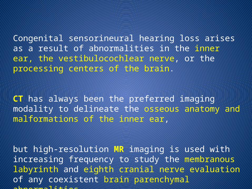
Congenital sensorineural hearing loss arises as a result of abnormalities in the inner ear, the vestibulocochlear nerve, or the processing centers of the brain.
CT has always been the preferred imaging modality to delineate the osseous anatomy and malformations of the inner ear,
but high-resolution MR imaging is used with increasing frequency to study the membranous labyrinth and eighth cranial nerve evaluation of any coexistent brain parenchymal abnormalities.
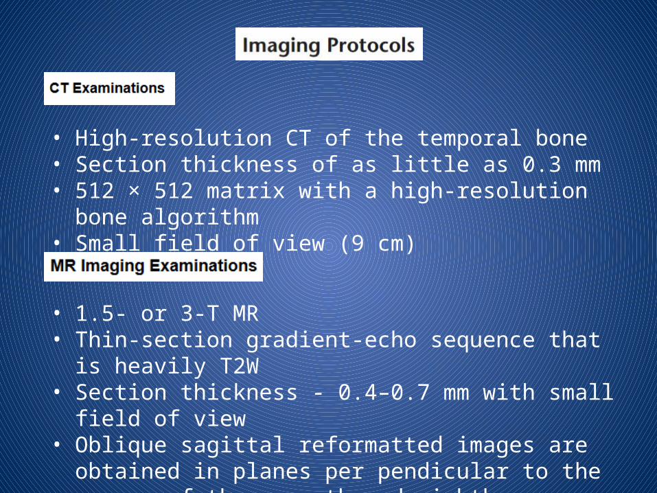
• High-resolution CT of the temporal bone • Section thickness of as little as 0.3 mm• 512 × 512 matrix with a high-resolution bone algorithm • Small field of view (9 cm)
• 1.5- or 3-T MR • Thin-section gradient-echo sequence that is heavily T2W• Section thickness - 0.4–0.7 mm with small field of view • Oblique sagittal reformatted images are obtained in planes per
pendicular to the course of the seventh and eighth nerves • Routine axial T2-weighted imaging of the brain


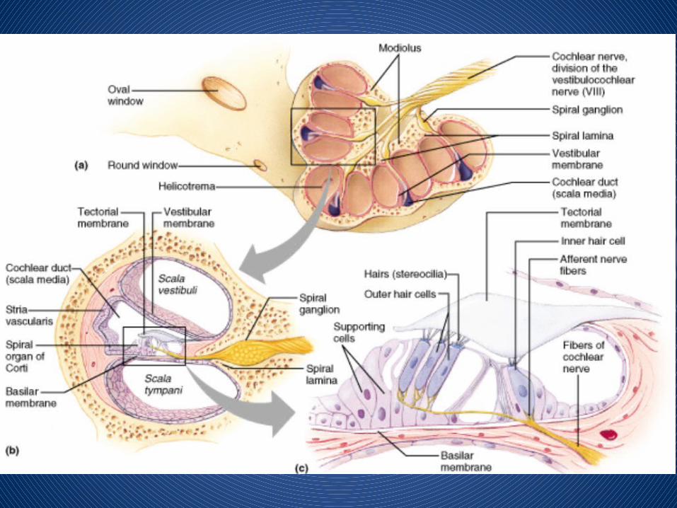

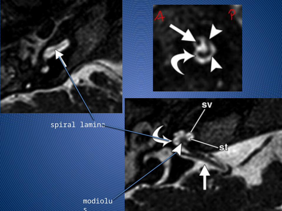
spiral lamina
modiolus

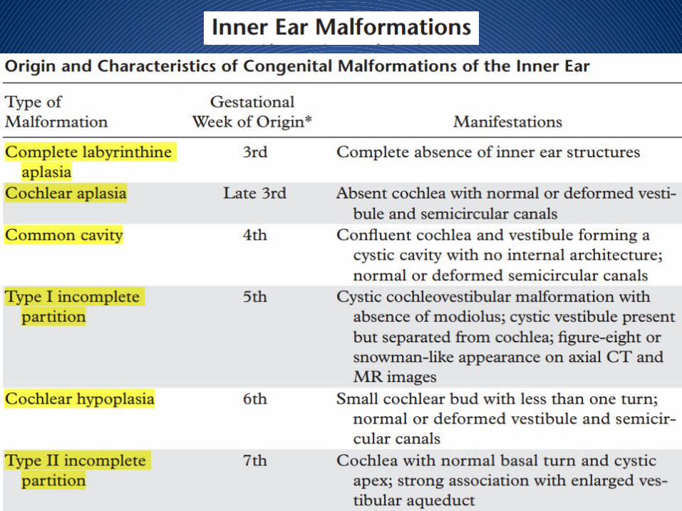

Also known as Michel aplasia
It is the most severe form of inner ear deformity
Caused by developmental arrest of the otic placode during the 3rd gestational week.
The condition is extremely rare, accounting for only 1% of all inner ear malformations.
There is complete absence of inner ear structures.
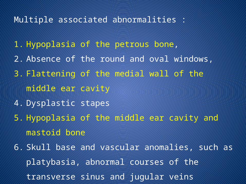
Multiple associated abnormalities :
1. Hypoplasia of the petrous bone,
2. Absence of the round and oval windows,
3. Flattening of the medial wall of the middle ear cavity
4. Dysplastic stapes
5. Hypoplasia of the middle ear cavity and mastoid bone
6. Skull base and vascular anomalies, such as platybasia,
abnormal courses of the transverse sinus and jugular veins
7. Craniocervical junction anomalies
8. Anomalous courses of the facial nerve
9. Posterior fossa abnormalities, such as arachnoid cysts

Oblique sag
COR
AXIAL

Complete absence of the cochlea.
Most likely due to arrested development of the inner ear in the latter part of the 3rd week of gestation
It is a rare anomaly.
In cochlear aplasia, the vestibule and semicircular canals are often malformed
Dense otic bone is present at the site where the cochlea normally would be.

Cochlear aplasia
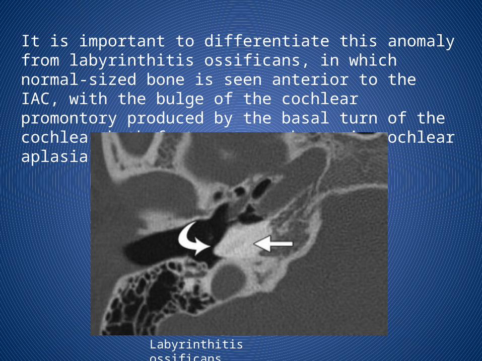
It is important to differentiate this anomaly from labyrinthitis ossificans, in which normal-sized bone is seen anterior to the IAC, with the bulge of the cochlear promontory produced by the basal turn of the cochlea; both features are absent in cochlear aplasia.
Labyrinthitis ossificans
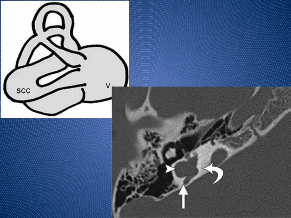
• Results from a developmental arrest in the 4th week of gestation Accounts for about 25% of all cochlear malformations
• Absence of the normal differentiation between the cochlea and vestibule
• • CT and MR images show confluence of the cochlea and vestibule
in a cystic cavity with no internal architecture
• semicircular canals are frequently malformed.
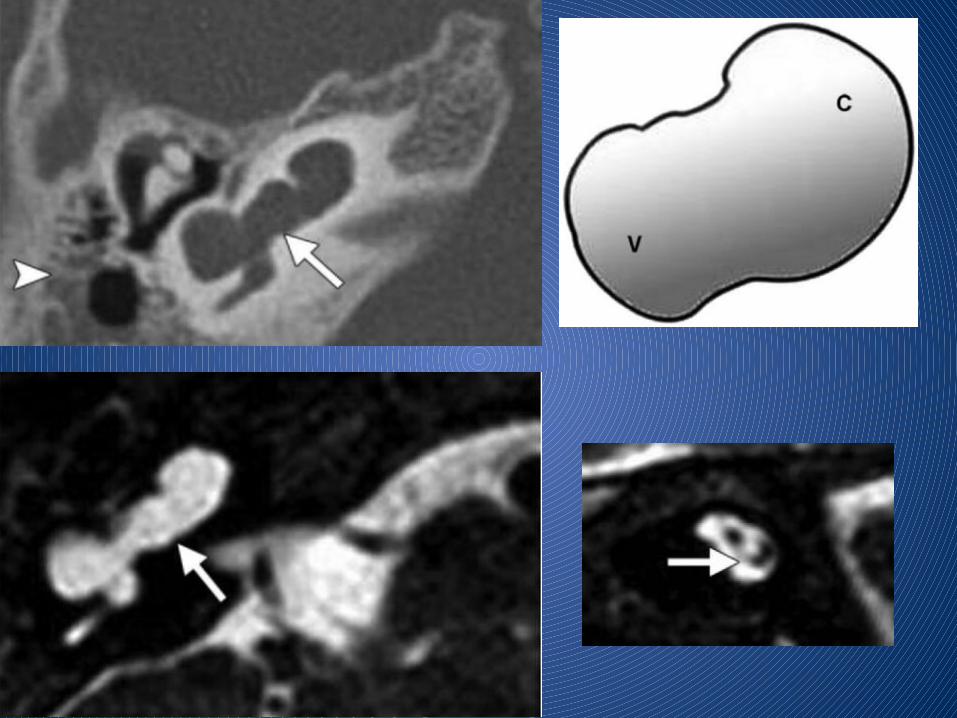
• Results from a developmental arrest in the 5th week of gestation
• The modiolus is entirely absent
• The cochlea has a cystic appearance; and the vestibule is often dilated, forming a figure eight
• The fact that the vestibule is distinguishable from the cochlea makes it possible to differentiate a type I incomplete partition from a common cavity.
• The vestibular aqueduct is normal. • Type I incomplete partition is less well differentiated than
type II incomplete partition.

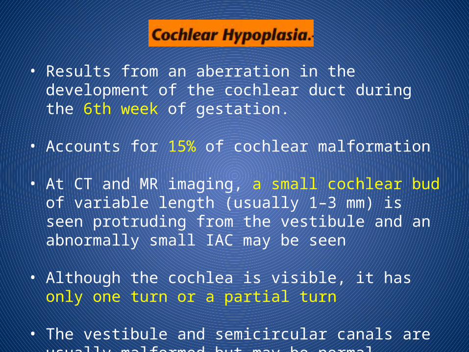
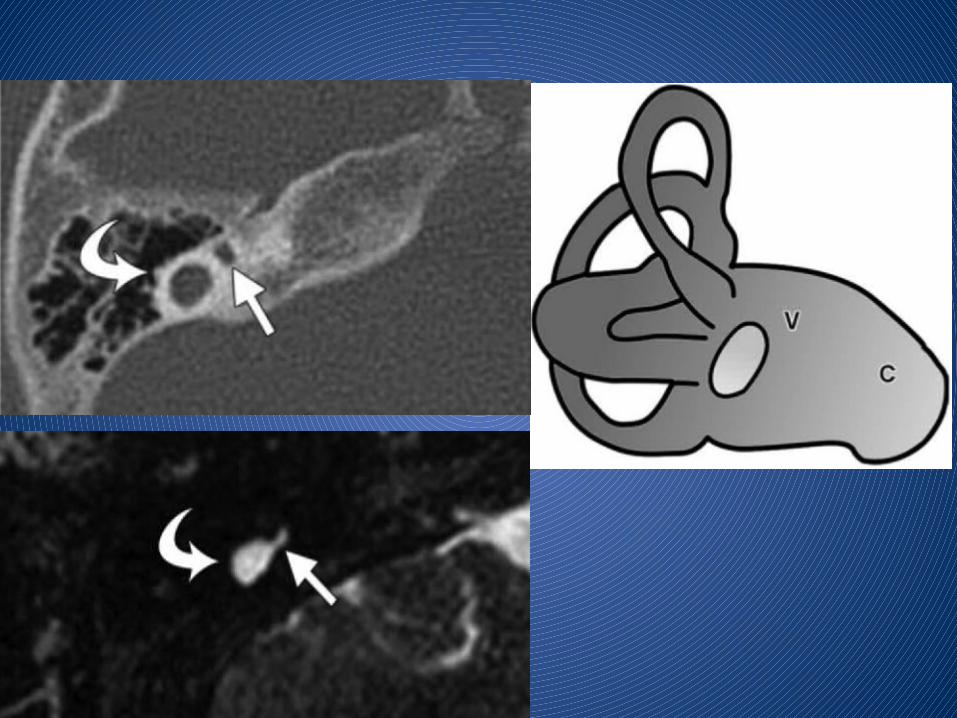
• Results from an aberration in the development of the cochlear duct during the 6th week of gestation.
• Accounts for 15% of cochlear malformation
• At CT and MR imaging, a small cochlear bud of variable length (usually 1–3 mm) is seen protruding from the vestibule and an abnormally small IAC may be seen
• Although the cochlea is visible, it has only one turn or a partial turn
• The vestibule and semicircular canals are usually malformed but may be normal.

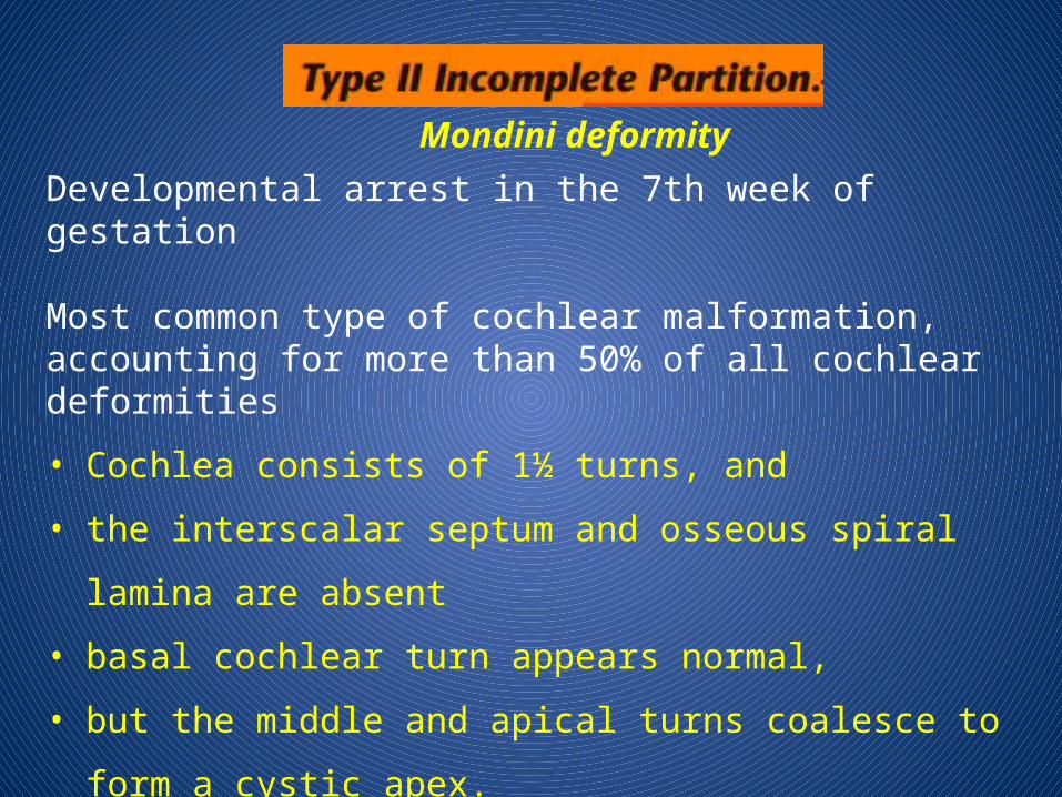
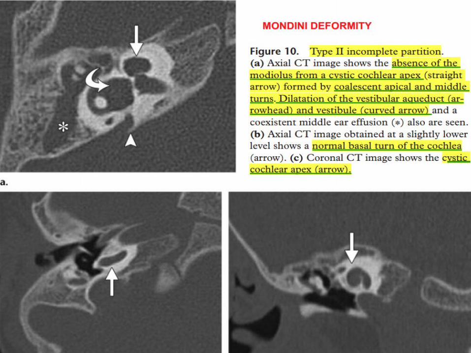
Mondini deformityDevelopmental arrest in the 7th week of gestation
Most common type of cochlear malformation, accounting for more than 50% of all cochlear deformities
• Cochlea consists of 1½ turns, and
• the interscalar septum and osseous spiral lamina are absent
• basal cochlear turn appears normal,
• but the middle and apical turns coalesce to form a cystic apex.
• The modiolus is present only at the level of the basal turn
• associated with a large endolymphatic duct and sac and an
enlarged vestibular aqueduct

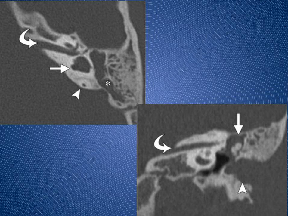
The malformed canals are usually short and wide but may be narrow.
In extensive malformations, the vestibule is dilated and forms a common lumen with the lateral canal. This type of abnormality is described as “lateral semicircular canal–vestibule dysplasia.
Aplasia of the semicircular canals is far less common than dysplasia.
An abnormal course of the facial nerve, atresia of the oval window, and abnormal ossicles are frequently seen in children with aplasia of the canals

It is essential to perform HRCT to confirm the diagnosis of semicircular canal aplasia.
Aplasia and fibrous or calcified obliteration of the canals have the same appearance at MR imaging. However, at CT, the semicircular canal is absent in cases of aplasia, whereas a normal canal is seen in cases of fibrous obliteration (since fibrous tissue cannot be seen at CT) and calcifications within the canal are seen in cases of labyrinthitis ossificans.
Vestibular malformations rarely occur in isolation. Commonly encountered anomalies include mild or globose dilatation of the vestibule with partial or complete assimilation of the semicircular canals into the vestibule

The abnormality is bilateral in as many as 90% of cases, may be asymmetric.
The endolymphatic duct and sac may be considered enlarged when their diameters exceed that of the adjacent ascending portion of the posterior semicircular canal.
In 84% of cases, an enlarged vestibular aqueduct is accompanied by other inner ear anomalies.


The normal diameter of the IAC ranges from 2 to 8 mm, with an average of 4 mm.
An IAC with a diameter of less than 2 mm is described as stenotic
The IAC may also be atretic or may have a bony septum that partitions it into two or more separate canals.
Types of cochlear nerve anomalies:
1. type 1 cochlear nerve anomaly, a stenotic IAC is seen with an absent eighth nerve
2. type 2 anomaly, a common vestibulocochlear nerve is found, with hypoplasia or aplasia of its cochlear branch.
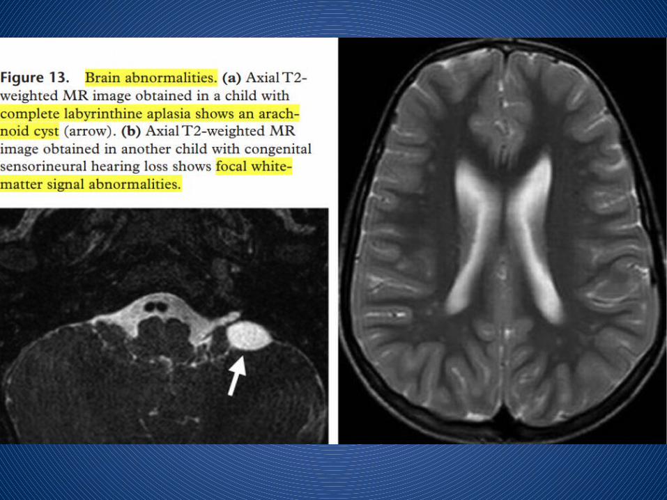
An increased incidence of posterior fossa abnormalities such as asymmetric dilatation of the fourth ventricle and arachnoid cysts has been observed among patients with complete labyrinthine aplasia.
Neuronal migration abnormalities in children with congenital sensorineural hearing loss also have been described.
Congenital cytomegalovirus infection contributes substantially to sensorineural hearing loss in many infant populations. The discrete foci of white-matter signal abnormality that are frequently seen on MR images obtained in children with sensorineural hearing loss have been attributed to cytomegalovirus infection at birth.
Congenital inner ear malformations have been classified by Ramos et al into three groups with respect to their implications for the feasibility of cochlear implantation surgery:
(a) gross malformations constituting surgical contraindications,- complete labyrinthine aplasia, cochlear aplasia, and cochlear nerve deficiency are contraindications for cochlear implantation surgery.
(b) major malformations contributing to increased risks for complications,- common cavity or severe hypoplasia

(c) minor malformations – hypoplasia, abnormalities of the aqueduct, and abnormalities of the vestibule.
If bilateral malformations are found, the surgeon will want to know which inner ear has the more normal structure and the larger cochlear nerve.

HRCT and MR imaging play an important role in the evaluation of pediatric hearing loss by providing crucial information about the inner ear, vestibulocochlear nerve, and brain.
Both modalities precisely and accurately delineate the inner ear anatomy and malformations.
They are often complementary and are used together in the preoperative evaluation of pediatric candidates for cochlear implantation.
Preoperative high-resolution CT offers the advantage of visualizing any coexistent middle or external ear anomalies and important anatomic variants, and MR imaging provides definitive information about the integrity of the cochlear nerve and the fluid-filled spaces of the inner ear.

THANK YOU