state-of-the-art ct imaging techniques for congenital ...the ct imaging techniques for congenital...
TRANSCRIPT

4 Korean J Radiol 11(1), Jan/Feb 2010
State-of-the-Art CT Imaging Techniquesfor Congenital Heart Disease
CT is increasingly being used for evaluating the cardiovascular structures andairways in the patients with congenital heart disease. Multi-slice CT has traditional-ly been used for the evaluation of the extracardiac vascular and airway abnormali-ties because of its inherent high spatial resolution and excellent air-tissue contrast.Recent developments in CT technology primarily by reducing the cardiac motionand the radiation dose usage in congenital heart disease evaluation have helpedexpand the indications for CT usage. Tracheobronchomalacia associated withcongenital heart disease can be evaluated with cine CT. Intravenous contrastinjection should be tailored to unequivocally demonstrate cardiovascular abnor-malities. Knowledge of the state-of-the-art CT imaging techniques that are used forevaluating congenital heart disease is helpful not only for planning and performingCT examinations, but also for interpreting and presenting the CT image findingsthat consequently guide the proper medical and surgical management.
he recent developments in CT techniques are characterized by fasterspeed, longer anatomic coverage, a more flexible ECG-synchronized scanand a lower radiation dose, and these advances have noticeably increased
the cardiac applications of CT. This increasing role of CT also includes the evaluationof congenital heart disease (1--3). Minimization of the radiation exposure delivered byCT is an important issue particularly for children (4, 5). Various dose reductiontechniques are currently available for cardiac CT as a result of the efforts to reduce theCT dose (6, 7). Thus, cardiac radiologists should be familiar with the CT techniquesthat are associated with a cardiac protocol and dose reduction. The CT imagingtechniques for congenital heart disease are not the same as those for acquired heartdisease: they are different according to the imaged anatomic structures, the purposesof the study and the study population evaluated with CT (e.g. children and adults withcongenital heart disease). The state-of-the-art CT imaging techniques for acquiredheart disease have been extensively appraised and frequently updated, while those forcongenital heart disease have not been thoroughly reviewed in the literature. In thisarticle, I review the current CT imaging techniques for congenital heart disease. Theseinclude the CT scan techniques, the dose reduction techniques and the methods forintravenous injection of contrast agent. The current clinical applications of CT and thefuture developments in CT technology are also discussed.
ECG-SYNCHRONIZATION
Among the various CT parameters, temporal resolution is the most important onefor evaluating congenital heart disease because the study population for this malady
Hyun Woo Goo, MD
Index terms:Computed tomography (CT)
techniquesMulti-slice CTCongenital heart disease
DOI:10.3348/kjr.2010.11.1.4
Korean J Radiol 2010;11:4-18Received July 19, 2009; accepted after revision August 19, 2009.
Department of Radiology and theResearch Institute of Radiology, AsanMedical Center, University of UlsanCollege of Medicine, Seoul 138-736,Korea
Address reprint requests to:Hyun Woo Goo, MD, Department ofRadiology and the Research Institute ofRadiology, Asan Medical Center,University of Ulsan College of MedicineAsanbyeongwon-gil 86, Songpa-gu,Seoul 138-736, Korea. Tel. (822) 3010-4388Fax. (822) 476-0090e-mail: [email protected]
T

commonly includes children with high heart rates andlimited ability to cooperate. Therefore, the fastest gantryrotation speed is preferred, and 270-280 ms is currentlythe fastest gantry rotation time. In contrast to the conven-tional interpolation algorithms for non-ECG-synchronizedspiral scans, a dedicated image reconstruction algorithmusing a half scan is employed for the ECG-synchronizedscan. Thus, the temporal resolution of the dedicatedalgorithm corresponds to half of the gantry rotation time,for example, 135 ms for a gantry rotation time of 270 ms.The improved temporal resolution by a half scan itselfsignificantly reduces cardiac pulsation artifacts on the CTimages (8). Although non-ECG-synchronized spiral CT hasbeen mainly used for assessing the extracardiac vascularabnormalities of congenital heart disease (1, 2, 9), theintroduction of the ECG-synchronized scan with eitherretrospective gating or, more recently, prospective trigger-ing is truly a step forward for the CT imaging of congenitalheart disease (2, 3, 10, 11). ECG-synchronization enablesus to accurately evaluate the coronary arteries (10, 11), theconotruncal and other intracardiac structures (3) and theventricle function and volumetry (12). The highesttemporal resolution, 75-83 ms, of the ECG-synchronizedscan is currently achieved with dual-source CT (13, 14).
Non-ECG-Synchronized Spiral CT ScanNon-ECG-synchronized spiral scanning is a simple, easy
technique to perform CT in patients with congenital heartdisease. Cardiac CT with this scan technique has beenwidely used since the introduction of multi-slice CT. Thecapability of multi-slice CT to produce submillimeterisotropic volume data greatly improves the image qualityof the multiplanar reformatted and three dimensional CTimages, which are vital for evaluating congenital heartdisease (Fig. 1). A thinner collimation/slice thickness and areconstruction interval corresponding to approximatelyhalf of the slice thickness (e.g. 50% overlapping) arerecommended to achieve isotropic voxels. A pitch of 1usually provides a good balance between image qualityand the radiation dose. Pitches lower than 1 add a little interms of image quality at the expense of an increasedradiation dose. Conversely, high pitches (> 1) tend todegrade the image quality. It should be noted that highpitches actually result in little dose saving in a CT systemthat employs an effective mA instead of an mA. Based onCT physics, a thin slice thickness increases the image noiseat the same radiation dose. Of note, a slight increase inslice thickness (for example, 0.75 mm rather than 0.6 mmat a collimation of 0.6 mm) offers a substantial benefit forimage quality at the expense of an unnoticeable decreaseof the longitudinal spatial resolution (Fig. 2). The z-flying
focal spot technology increases the longitudinal spatialresolution, by a factor of approximately 1.4, by doublesampling in the z-direction (15). The technique alsoreduces windmill artifacts that are caused by the conebeam geometry of multi-slice CT (15).
The scan technique may be acquired during eitherbreath-holding or free-breathing. Thanks to the short scantime, the respiratory motion artifacts on the multi-slice CTimages of free-breathing young children are surprisinglyless remarkable than those on the single-slice CT images.On top of that, it is interesting to know that the origins andproximal segments of the coronary arteries are frequentlyobserved, in 82% of the patients with congenital heartdisease, on the non-ECG-synchronized spiral CT images(16). Thicker collimation is useful to shorten the CT scantime, which enables young children around 3-6 years ofage to hold their breath during CT scan. In order to meetthe need for a shorter breath-holding time for a child,higher pitches in addition to thicker collimation may behelpful to further reduce the scan time. These strategiesallow reducing the frequency of sedation for a CTexamination. On the other hand, thicker collimation isbetter than a thinner one for older children or adultsbecause thicker collimation reduces the CT radiation doseand it improves the image quality of the CT images.Regarding the CT dose, we should know that unnecessaryradiation exposure outside of a scan range, the so-called ‘z-overranging’ or ‘z-overscanning’, is inevitable with thisscan technique and conventional collimation technology.The contribution of z-overranging to the total CT dose isnot negligible, but it is actually enormous (up to 70%)particularly for pediatric CT with a short scan range, as isused for heart CT (17). Fortunately, adaptive collimationtechnology that can protect from z-overranging is currentlybeing made available by some of CT manufacturers (18).When appropriate CT parameters are used, the radiationdose of the scan technique is usually less than 1.0 mSv inyoung children (9).
Retrospective ECG-Gated Spiral CT ScanThe relatively high radiation dose of retrospective ECG-
gated spiral scanning is primarily attributed to the lowpitch that is necessary to obtain gapless ECG-gated spiraldata (Fig. 3). When a mode of a heart rate-dependent pitchis available, the radiation dose is reduced with a higherpitch that is automatically adapted to higher heart rates(19). However, it should be noted that the mode maydegrade the image quality when the heart rate is unstableduring the actual CT scan. If this is the case during abreath-holding test, then the use of manual selection of thetarget heart rates is better. A multi-segment reconstruction
CT Imaging Technique of Congenital Heart Disease
Korean J Radiol 11(1), Jan/Feb 2010 5

algorithm may be used to increase the temporal resolution.However, a single-segment reconstruction algorithm isusually preferred to a multi-segment one because the latterprolongs the scan time and thus it increases the variabilityof the heart rate. In addition, the temporal resolution ofthe multi-segment reconstruction algorithm is variabledepending on the heart rates. Consequently, there is a
higher chance to obtain better image quality with using asingle-segment reconstruction algorithm. An optimalcardiac phase, which is important in applying ECG-controlled tube current modulation, depends on the heartrate. The mid-diastolic phase, the so-called ‘diastasis’, ischosen at lower heart rates, while the end-systolic phase isselected at higher heart rates (> 75-80 bpm). Then, a widthwith the full tube current should be determined around theselected cardiac phase. If the width is too broad, then therewill be a dose penalty. In contrast, there will be anincreased chance to lose the right cardiac phase if the widthis too narrow. When the reason for performing cardiac CTis only for the morphologic evaluation, a 10% width of thecardiac cycle may be a good balance between the twofactors (for example, 35-45% for the end-systolic phaseand 65-75% for the mid-diastolic phase). The widthshould be broader than 10% when the heart rate is irregu-lar or functional evaluation is indicated. The best cardiacphase with minimal motion may be selected eithermanually by previewing the multi-phase images at one ormore slices or automatically by using a motion map (20).The scan time is relatively short, usually less than 10 s.Breath-holding is usually recommended because this scanmode is relatively vulnerable to respiratory motionartifacts (Fig. 4). Nevertheless, some investigators haveattempted performing a retrospective ECG-gated spiral CTscan in free-breathing neonates (11). The effective doseestimates of this scan mode with ECG-controlled tubecurrent modulation are 5-7 mSv for the patients with
Goo
6 Korean J Radiol 11(1), Jan/Feb 2010
Fig. 1. Volume-rendered CT image with non-ECG-synchronizedspiral scan shows excellent anatomic details of pulmonaryarteries in 12-year-old girl with repaired coarctation of aorta.
Fig. 2. Effect of slice thickness on image quality of non-ECG-synchronized spiral CT in 12-year-old boy with pulmonary atresia andventricular septal defect after Rastelli operation. A. Volume-rendered CT image reconstructed from thin, overlapped axial images with 0.6-mm slice thickness at collimation of 0.6 mmappears quite grainy. That is because slice thickness is too thin at employed CT dose and this thin slice consequently increases imagenoise enough to degrade image quality. There are two ways to improve image quality in this situation: one is to slightly increase slicethickness and the other is to increase radiation dose a lot.B. Slight increase in slice thickness to 0.75 mm substantially improves image quality of volume-rendered CT image. This strategy ishighly recommended because its dose saving effect is great.
A B

acquired heart disease (21) and 2-6 mSv for the patientswith congenital heart disease (10) for dual-source CT.
Prospective ECG-Triggered Sequential CT ScanProspective ECG-triggered sequential scanning is a low
dose ECG-synchronization technique that can reduce theCT dose to 1-3 mSv in adults (22) and to 0.2-0.7 mSv innewborns and infants (10). In contrast to spiral scanning, itdoes not employ continuous table movement or a z-overranging effect (Fig. 5A). The degraded ECG-synchro-nization, seen as stair-step artifacts, may occur at irregularheart rates with this technique. The artifacts are lesspronounced in the images acquired at the end-systolicphase because the phase is relatively constant irrespective
of the heart rates (Fig. 5B). For the same reason, absolutetrigger delay (ms) is preferred to relative trigger delay(percentage) for targeting the end-systolic phase. Ascompared with retrospective ECG-gated spiral scanning,the lack of capability for multi-phase functional evaluationhas been regarded as a limitation of this technique.However, this limitation is partly overcome with a recentlyavailable option of longer data acquisition (Fig. 6). Thislonger data acquisition allows us to perform fine adjust-ment for the best cardiac phase at lower heart rates andeven to evaluate cardiac motion at high heart rates if theacquisition time is long enough to cover the second half ofthe cardiac cycle (Fig. 6; Electronic supplementarymaterial, animation 1).
CT Imaging Technique of Congenital Heart Disease
Korean J Radiol 11(1), Jan/Feb 2010 7
Fig. 3. Diagrams showing retrospective ECG-gated spiral CT scan. A. As demonstrated on schema, spiral scan is acquired with low enough pitch to avoid gap in CT data. This spiral CT data is retrospec-tively reconstructed at specific cardiac phase by synchronizing with simultaneously acquired ECG data.B. For ECG-controlled tube current modulation, period with 100% tube current should be appropriately determined depending on heartrates. End-systolic phase (red rectangle) is used at high heart rates. Tube current can be reduced to 20% (yellow rectangle) or 4% (lightgreen rectangle) during rest of cardiac cycle.
A B
Fig. 4. 6-year-old boy with repaired tetralogy of Fallot. Sagittal (A) and short-axis (B) retrospective ECG-gated spiral CT images, whichare affected by severe respiratory motion artifacts, are shown at midportion of scan range.
A B

In contrast to retrospective ECG-gated spiral scanning,the respiratory motion artifacts tend to be less pronouncedin a beam collimation or one slab scan. Therefore, this scantechnique frequently provides motion-free CT images evenin free-breathing young children (Fig. 7). However, variousdegrees of stair-step artifacts may occur between theadjacent slabs (Fig. 8A). Overlap (minimum value, 2.2-5.0mm) between adjacent slabs, which is achieved by a tablefeed smaller than a beam collimation, makes some of thestair-step artifacts less conspicuous. A difference in cardio-vascular enhancement is frequently noted between adjacentslabs because more than two heart beats are necessary tomove the CT table for the next ECG-triggered scan, the so-called ‘step and shoot’ scan mode (Fig. 8B). Thus, a specialintravenous injection method that prolongs the peakvascular enhancement may ameliorate this difference ofcardiovascular enhancement. The total examination time isprolonged for the same reason, but it becomes shorter withthe increased longitudinal coverage of a recently introducedmulti-slice CT system, for instance, single gantry rotationtime for 320-slice CT (23). As in spiral scanning, the use ofa variable slice thickness other than a multiple of a detectorcollimation has recently become available for prospectiveECG-triggered sequential scanning
Combo CT ScanFor free breathing young children, there is no single scan
mode that can satisfactorily evaluate all the cardiovascular
structures and the airway. A non-ECG-synchronized spiralscan is subject to cardiac and respiratory motion artifacts.In particular, cardiac motion artifacts on the scan modecommonly make the anatomic evaluation of the coronaryarteries, proximal great arteries and intracardiac structuresdifficult. On the other hand, the prolonged examinationtime of a prospective ECG-triggered sequential scan usingthe currently available first-generation dual-source CTsystem may cause motion artifacts other than cardiacpulsation, and there is a difficulty for maintaining peakvascular enhancement. A temporary solution to this mattermay be a combination of both scan modes, the so-called‘Combo’ scan. In this scan mode, two or three slabs of aprospective ECG-triggered sequential scan confined to theconotruncal area of the heart follow a non-ECG-synchro-nized spiral scan at the expense of a small additionalradiation dose (Fig. 9). As a result, all the cardiovascularand airway structures related to various congenital heartdiseases can be much more satisfactorily evaluated withthis ‘Combo’ scan than with using either a non-ECG-synchronized spiral scan or a prospective ECG-triggeredsequential scan alone. The average CT dose of this scanmode is approximately 1.4 mSv. It is speculated that this‘Combo’ scan for free-breathing young children withcongenital heart disease will be replaced by an advancedprospective ECG-triggered sequential scan with second-generation dual-source CT or with a combined (ECG andrespiration) triggering method (Fig. 8B).
Goo
8 Korean J Radiol 11(1), Jan/Feb 2010
Fig. 5. Diagrams showing prospective ECG-triggered sequential CT scan.A. Sequential scan is acquired without table movement at predefined cardiac phase, which is end-systolic phase in this diagram. Then,time period is necessary to move CT table to next scan. Thus, scan mode is commonly called ‘step and shoot’ mode.B. ECG shows variable heart rates ranging from 54 bpm to 111 bpm. This variability in heart rates is commonly considered to bedisadvantageous for performing prospective ECG-triggered sequential scan for mid-diastolic phase. However, this is not case when end-systolic phase is target. As demonstrated on this diagram, end-systolic phases can be consistently acquired at irregular heart rate withprospective ECG-triggered sequential scan. It should be noted that absolute delay, for instance, 240 ms in this case, must be used tohave this benefit.
A B

CT Imaging Technique of Congenital Heart Disease
Korean J Radiol 11(1), Jan/Feb 2010 9
A B
C D
Fig. 6. Multi-phase prospective ECG-triggered sequential CT scan.A. Extended scan mode (0.38 s), in which period longer than necessary for single phase (0.2 s) is obtained, offers multi-phase study.Cardiac function can be evaluated with this scan mode at high heart rates (134 bpm in this case) because acquisition window coversboth end-systole and end-diastole. B-F. After scan, five cardiac phases are retrospectively reconstructed at approximately 40-ms intervals (B, 160 ms; C, 200 ms; D, 240ms; E, 280 ms; F 340 ms).
E F

CINE CT FOR AIRWAYS
Vascular or non-vascular airway narrowing is a frequentcomplication in patients with congenital heart disease, andthis may result in respiratory difficulty or failure to beweaned from a ventilator (24). The airway compressionmay be a fixed form of narrowing, a dynamic form ofnarrowing or a combination of the two. Dynamic airwayobstruction, termed tracheobronchomalacia, may developas a result of longstanding vascular airway compression,
recurrent airway infection/inflammation or a congenitalanomaly. The other CT scan techniques previouslymentioned in this article allow us to make the diagnosis offixed airway narrowing, but only the cine CT techniqueenables us to identify tracheobronchomalacia bydemonstrating the excessive expiratory collapsibility ofairways throughout the respiratory cycle (Fig. 10;Electronic supplementary material, animation 2). Cine CTis a non-spiral, sequential scan that is acquired withouttable movement. In order to minimize the radiation dose
Goo
10 Korean J Radiol 11(1), Jan/Feb 2010
Fig. 7. Prospective ECG-triggered sequential CT scan in free-breathing young children is relatively less susceptible to respiratory motionartifacts than is retrospectively ECG-gated spiral CT scan. With this scan mode, even side-branches (arrowheads) of coronary arteries(A) and stenosis (arrow) involving left bronchi (B) are clearly delineated without cardiac and respiratory motion artifacts in young children.Therefore, more invasive or sophisticated preparation procedures such as general anesthesia or controlled ventilation are not currentlyneeded for only diagnostic purposes.
A B
Fig. 8. Artifacts on prospective ECG-triggered sequential CT scan.A. Various degrees of stair-step artifacts derived from respiration motion are seen on coronal CT image. Artifacts are typically muchmore pronounced around diaphragm. Aneurysmal dilatations involving ascending aorta (AA) and pulmonary trunk (PT) are noted in 3-year-old girl with Loeys-Dietz syndrome.B. Stair-step artifacts on prospective ECG-triggered sequential coronal CT image are remarkably decreased by applying combined ECGand respiratory triggering for same patient who underwent valve sparing aortic root replacement surgery. In contrast, difference in cardio-vascular enhancement is frequently observed between adjacent slabs (arrows) due to long examination time that is further lengthenedby combined triggering.
A B

related to cine CT, the scan time should be limited to theminimum that is slightly longer than one respiratory cycle.The multi-segment CT images are retrospectivelyreconstructed by means of a partial reconstructionalgorithm (180 of gantry rotation + the fan beam angle)(25). The estimated dose of cine CT is low, for example, inthe range of 0.2-0.3 mSv when the images are obtained atsix slice levels (25). In addition to the evaluation oftracheobronchomalacia, air trapping in the lung can also bedetected with cine CT (25).
STRATEGIES FOR CT DOSE REDUCTION
The importance of tailoring the CT scan techniques tominimize the radiation dose, while the diagnostic imagequality is preserved, cannot be overemphasized and partic-ularly in children. It goes without saying that clear andsound clinical indications are the premise. Various usefulstrategies to reduce the cardiac CT dose are described inthis section of the paper. Another important thing for thecardiac radiologists who are involved in CT imaging of
congenital heart disease is having a concrete knowledgeabout how to estimate the CT dose in both children andadults. Among the several dose estimation methods, thedose-length product-based method is the most simple andpractical. The dose estimate is easily calculated bymultiplying the dose-length product on the CT dose reportby an age, gender and scanner-specific conversion factor(5). The only thing to be careful about is that a dose-lengthproduct for children must be derived from a 16-cm CTdose index phantom.
Body-Size Adaptive Low Dose CT ProtocolsEstablishing a body-size adaptive CT protocol is a first
step for CT dose minimization because body-size adapta-tion is critical for children because they have greaterradiosensitivity, a longer life expectancy and a smallerbody size (4, 5). Various indices that reflect body size,including the body weight, the body height, the body massindex, the cross-sectional dimensions and the attenuation,can be used for the adaptive CT protocol. The weight-based protocol is easy to use and practical, and so body
CT Imaging Technique of Congenital Heart Disease
Korean J Radiol 11(1), Jan/Feb 2010 11
A B
Fig. 9. Combo CT scan comprised of non-ECG-synchronized spiral scan with usual scan range (A, B) and prospective ECG-triggeredsequential scan with narrow scan range confined to conotruncal area of heart (C, D) in 9-months-old boy with Williams syndrome.Supravalvular aortic stenosis (arrows on C) and combined valvar (arrow on D) and subvalvar (arrowheads on D) pulmonary stenoses areclearly shown on prospective ECG-triggered sequential CT images (C, D). Dose estimates are 1.6 mSv for non-ECG-synchronized spiralscan and 0.2 mSv for prospective ECG-triggered sequential scan.
C D

weight is the most commonly used body-size parameter.Because the benefits of low kV for thin adults and childrenare well established, a weight-based heart CT protocolusing variable kV and variable mA should be used (5). Onthe other hand, the cross-sectional dimension-basedprotocol is better for adapting the CT dose to an individualbody habitus, as compared with the weight-based protocol(26). Nevertheless, the cross-sectional dimension-based CTprotocol has not been commonly used because a practicalmethod for this is currently not available.
Minimization of Z-Axis Coverage and Z-OverrangingMinimization of the scan range along the z-axis accord-
ing to the individual clinical indications is a simple,effective way to minimize the CT dose. This is attributedto the fact that the CT dose is directly proportional to thescan range when the other CT parameters are the same. Alonger scan range may be necessary for evaluating theinfracardiac type of total anomalous pulmonary venousreturn or the major aortopulmonary collateral arteries.Otherwise, the longitudinal coverage usually ranges fromthe aortic arch to the most caudal margin of the heart.Because the safety margins at both ends are very narrow ata minimal z-axis, the scout image should be updatedwhenever there is any doubt as to whether or not a patientmoved during a preparation period after the scout imaging.In fact, reexamination following a failed study is the worstscenario for minimizing the CT dose. It should be notedthat contribution of ‘z-overranging’ to the total CT dose isgreater for a shorter z-axis range. This relationship is
derived from the physical features of ‘z-overranging’: theeffect is proportional to the beam collimation, thereconstructed slice width and the pitch, while it is irrespec-tive of the planned scan length (27). For the maximal dosesaving effect of minimal z-axis coverage, it is highlyrecommended to use the adaptive collimation technologyfor eliminating ‘z-overranging’.
Tube Current ModulationTube current modulation or automatic exposure control
is a useful dose-saving technique that reduces the CT doseaccording to the body size and shape, and the attenuation.In order to correctly use the technique, we should clearlyunderstand the various methods of tube current modula-tion (28): it may be applied along the x-y plane (angularmode), the z-axis or a combination of both; a reference formodulation is based on the standard deviation, the noiseindex, a reference mA or a reference image. The degree ofdose reduction is variable (it is 9-26% in pediatric heartCT [29]), depending on the anatomic region and theindividual body features. In addition, it should be kept inmind that dose modulation is influenced by many factorssuch as the distance from the gantry isocenter, the kVlevel, scan direction and so on (30). Yet the technique islimited by the fact that there is no guideline for how todetermine a suitable reference value for each anatomicregion, for individual clinical tasks and for a different levelof kV, and so the best possible decision remains vague anddifficult.
Goo
12 Korean J Radiol 11(1), Jan/Feb 2010
Fig. 10. Cine CT (A, inspiratory phase; B, expiratory phase) for diagnosis of tracheomalacia in 16-day-old female newborn withtracheoesophageal fistula, esophageal atresia and tetralogy of Fallot. Excessive expiratory collapsibility of trachea (arrows) is displayedon cine CT images. Distended proximal esophagus is also seen posterior to trachea.
A B
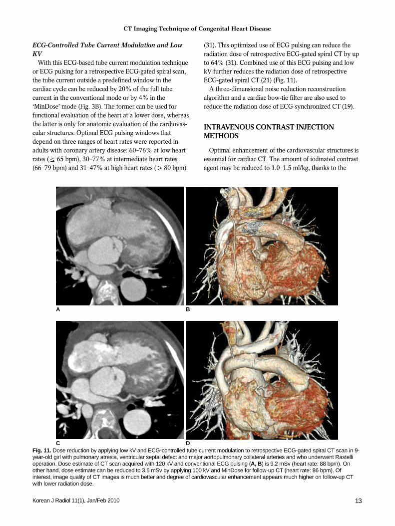
ECG-Controlled Tube Current Modulation and LowKV
With this ECG-based tube current modulation techniqueor ECG pulsing for a retrospective ECG-gated spiral scan,the tube current outside a predefined window in thecardiac cycle can be reduced by 20% of the full tubecurrent in the conventional mode or by 4% in the‘MinDose’ mode (Fig. 3B). The former can be used forfunctional evaluation of the heart at a lower dose, whereasthe latter is only for anatomic evaluation of the cardiovas-cular structures. Optimal ECG pulsing windows thatdepend on three ranges of heart rates were reported inadults with coronary artery disease: 60-76% at low heartrates ( 65 bpm), 30-77% at intermediate heart rates(66-79 bpm) and 31-47% at high heart rates ( 80 bpm)
(31). This optimized use of ECG pulsing can reduce theradiation dose of retrospective ECG-gated spiral CT by upto 64% (31). Combined use of this ECG pulsing and lowkV further reduces the radiation dose of retrospectiveECG-gated spiral CT (21) (Fig. 11).
A three-dimensional noise reduction reconstructionalgorithm and a cardiac bow-tie filter are also used toreduce the radiation dose of ECG-synchronized CT (19).
INTRAVENOUS CONTRAST INJECTIONMETHODS
Optimal enhancement of the cardiovascular structures isessential for cardiac CT. The amount of iodinated contrastagent may be reduced to 1.0-1.5 ml/kg, thanks to the
CT Imaging Technique of Congenital Heart Disease
Korean J Radiol 11(1), Jan/Feb 2010 13
A B
Fig. 11. Dose reduction by applying low kV and ECG-controlled tube current modulation to retrospective ECG-gated spiral CT scan in 9-year-old girl with pulmonary atresia, ventricular septal defect and major aortopulmonary collateral arteries and who underwent Rastellioperation. Dose estimate of CT scan acquired with 120 kV and conventional ECG pulsing (A, B) is 9.2 mSv (heart rate: 88 bpm). Onother hand, dose estimate can be reduced to 3.5 mSv by applying 100 kV and MinDose for follow-up CT (heart rate: 86 bpm). Ofinterest, image quality of CT images is much better and degree of cardiovascular enhancement appears much higher on follow-up CTwith lower radiation dose.
C D

shorter scan time of the current CT systems, in the usualcases of congenital heart disease. A larger amount up to2.0 ml/kg is necessary for the cases with a large intracar-diac or extracardiac shunt, substantial valvular regurgita-tion and/or a severely enlarged heart. A high iodinedelivery rate (mgI/ml/sec), a unit comprising both theiodine concentration and the injection rate, is moreimportant than an high iodine concentration of the contrastagent itself to achieve higher cardiovascular enhancement.The injection rate has to be appropriately adjusted depend-ing on the size of the placed angiocatheter and the bodysize. In my practice, the amount of contrast agent is firstdetermined and the injection rate is then calculated tomeet an approximately 15-second injection time of thecontrast agent in most of cases. For instance, the injectionrate would be 1.0 ml/sec when 1.5 ml/kg of contrast agentis used for a 10-kg child. Other investigators havesuggested a simplified method (32), the so-called ‘contrast-covering time’, which is basically similar to the injectionmethods advocated by others. The intravenous injection
site is important and it should be properly selected inadvance: the leg vein is preferred for the evaluation of theaortic arch and the presence of the left superior vena cava;simultaneous injection of 50% diluted contrast agentthrough the arm and leg veins is preferred for the evalua-tion of Fontan pathway (Fig. 12); the leg vein should notbe used in patients who have undergone bidirectionalcavopulmonary connection (1, 2).
Three methods are used to determine the appropriatescan delay. They include the empirical method, the testinjection method and the bolus tracking method. A bolustracking method is the most commonly used methodamong them (1, 2). In order to minimize the radiationdose, a monitoring delay should be slightly earlier than theactual peak enhancement of a target cardiovascularstructure: 13 seconds for visualizing the right heart and thepulmonary circulation, and 15 seconds for visualizing theleft heart and the systemic circulation. In contrast, otherresearchers have recommended beginning the bolustracking series before the contrast injection starts (33).
Goo
14 Korean J Radiol 11(1), Jan/Feb 2010
A B
Fig. 12. Non-ECG-synchronized multiplanar reformatted (A-C) and volume-rendered (D) CT images clearly show patent Fontan pathwayand pulmonary vessels in 5-year-old boy with functional single ventricle. Simultaneous intravenous injection of 50% diluted contrastagent through arm and leg veins was used to obtain homogeneously high enhancement of cardiovascular structures.
C D

However, a substantial radiation dose might be accumu-lated simply by the bolus tracking series rather than theactual scan. In fact, the dose from the bolus tracking seriesturned out to be obviously too much, that is, 2.7 mSv onthe average (34). The threshold of the CT density to triggera scan is usually between 100 HU and 150 HU. A delaybetween this trigger and the actual scan ranges from aminimal value (2-5 seconds depending on a CT model) to7 seconds, and the latter is necessary for breath-holdinstructions. A region of interest for bolus tracking shouldbe appropriately placed depending on the clinical indica-tions in the cardiac chambers (the right ventricle, the leftatrium and the left ventricle) or vessels (the internaljugular vein in the patients with a Fontan operation totarget the second circulation of contrast agent). It should benoted that the right atrium is not used because this locationis subject to artifacts from the undiluted contrast agent. Asa rule of thumb, the scan delay for bolus tracking istargeted at the peak aortic enhancement, which occursapproximately 5 seconds after the completion of contrast
injection (35). A test injection method is more accuratethan a bolus tracking method for achieving peak vascularenhancement. In general, the scan delay is increased by 3-5 seconds from the calculated transit time of the contrastagent because a larger volume is used at the main CT scan.Therefore, I use the method usually for the evaluation ofpulmonary thromboembolism for which it is important tohave peak enhancement of the pulmonary circulationprecisely during the CT scan. I use the peak enhancementtime of the pulmonary vein for the scan delay for theevaluation of pulmonary thromboembolism. Powerinjection is preferred to manual injection because theresults of the former are more consistent and reproducible.
As has happened for the CT techniques, the contrastinjection protocol has also evolved over time. As for CTimaging of congenital heart disease, its goal is to simultane-ously achieve adequate vascular enhancement and minimalperivenous artifacts. In addition, the injection protocolshould delineate all the cardiovascular structures on CT. Aprotocol employing a saline chaser with using a single- or
CT Imaging Technique of Congenital Heart Disease
Korean J Radiol 11(1), Jan/Feb 2010 15
A B
Fig. 13. Right ventricle CT volumetry in 8-year-old boy with repairedtetralogy of Fallot. Retrospective ECG-gated spiral CT was performedbecause of inconsistent right ventricle volumes on serial cardiac MRexaminations (end-systolic volume of right ventricle was normalized tobody surface area, 86 128 ml/m2). A. Crisp margin of right ventricle is shown with high spatial resolution onvolume-rendered CT image. B, C. Consequently, right ventricular cavity can be accuratelysegmented with three-dimensional region growing method. End-systolicvolume of right ventricle that’s normalized to body surface areacalculated with CT is 86 ml/m2, which indicates that volume at secondcardiac MR examination is inaccurate.
C

dual-head power injector allows us to reduce the amountof contrast agent and the perivenous artifacts for thoracicCT (36). We do not have to worry about the amount of thesaline flush because the intravenous injection would bestopped immediately after the end of the main scan. Atriphasic intravenous injection protocol, in which undilutedcontrast agent is followed by 50-60% diluted contrastagent and then by a saline chaser, was recently developedto improve the visualization of the right heart, to circum-vent poor enhancement of the pulmonary artery and toreduce the perivenous artifacts (37). In my practice, 5-10% diluted contrast agent, instead of a saline chaser, isused at the third phase of the injection protocol whensimultaneous visualization of the systemic veins isrequired.
CLINICAL APPLICATIONS
The clinical applications of CT for patients with congeni-tal heart disease are rapidly changing and growing inkeeping with the technical developments of CT.Classically, the extracardiac great vessels, including theaortic arch and the pulmonary vessels, have beenevaluated with CT (1-3, 9). Evaluation of the patency ofvascular shunts, conduits or stents is another traditionalindication of CT (38). A dedicated reconstructionalgorithm for vascular stents has recently been introducedfor better evaluation of in-stent stenosis. Evaluation ofairways is an exclusive merit of CT over the other imagingmodalities. Vascular or non-vascular airway narrowing,airway anomalies and dynamic airway obstruction can allbe accurately evaluated with CT (24, 39, 40). The
introduction of the ECG-synchronized CT scan improvesthe visibility of those cardiovascular structures that aregreatly affected by cardiac pulsation. They include theascending aorta, the pulmonary trunk, the coronary arteryand the heart. Thus, various congenital heart diseases thatinvolve the above mentioned anatomic structures, such asWilliams syndrome and Marfan syndrome, can beaccurately evaluated with ECG-synchronized CT scanning(Figs. 8, 9) (2, 3, 10, 11). ECG-synchronized CT scanninghas become a reality in our clinical practice thanks to theincreased flexibility and the reduced radiation dose of thescan technique. In place of cardiac MRI, CT may bealternatively used for the evaluation of cardiac functionand ventricle volumetry, for instance, in a patient withrepaired tetralogy of Fallot and with or without apermanent cardiac pacemaker (Fig. 13).
FUTURE DEVELOPMENTS
CT techniques are continuously evolving. For cardiacapplications, second-generation dual-source CT and large-coverage volume CT (256-320 slices) have come into thespotlight (14, 23). On top of the higher spatial resolutionand longer coverage, other innovative technical develop-ments are waiting for the initial clinical results. Theyinclude 1) a high-pitch (up to 3.4) dual-source spiral scan,2) a dual-energy lung perfusion and ventilation scan (Fig.14), 3) a new type of tube current modulation to reducethe radiation dose to superficial radiation-sensitive tissuesuch as the breast, the thyroid gland or the eyeball, 4)automatic suggestions for the CT dose parameters accord-ing to the individual clinical task and the anatomic region,
Goo
16 Korean J Radiol 11(1), Jan/Feb 2010
Fig. 14. Dual-energy lung perfusion CT. In addition to pulmonary CT angiography (A), lung perfusion status (B) can be obtained withoutadditional radiation dose by means of dual-energy CT technology when pulmonary thromboembolism is suspected in patients withcongenital heart disease.
A B

and (5) combined triggering of the ECG and respiration(Fig. 8).
CONCLUSION
The state-of-the-art CT imaging techniques for congenitalheart disease are useful to evaluate diverse cardiovascularand airway abnormalities with improved accuracy andpatient- and user-friendliness. Therefore, CT is steadilybecoming an invaluable imaging modality to fill the gapamong echocardiography, cardiac catheterization andcardiac MRI. Moreover, cardiac CT is not a radiation-intensive study any more when the appropriate dose-saving strategies are used. Last but not least, generalanesthesia or controlled ventilation is virtually unnecessaryfor the uncooperative children to undergo CT examina-tion. To get all the benefits of the currently availablecardiac CT, we as cardiac radiologists, should aware of thedifferent types of CT scans in detail and we should keep aneye open for future technical developments.
Electronic Supplementary MaterialBelow is the link to the electronic supplementary
material.http://kjronline.org/src/SM/kjr-11-1-sm001.gifhttp://kjronline.org/src/SM/kjr-11-1-sm002.gif
References1. Goo HW, Park IS, Ko JK, Kim YH, Seo DM, Yun TJ, et al. CT
of congenital heart disease: normal anatomy and typicalpathologic conditions. Radiographics 2003;23:S147-S165
2. Goo HW, Park IS, Ko JK, Kim YH, Seo DM, Park JJ. Computedtomography for the diagnosis of congenital heart disease inpediatric and adult patients. Int J Cardiovasc Imaging2005;21:347-365
3. Leschka S, Oechslin E, Husmann L, Desbiolles L, Marincek B,Genoni M, et al. Pre- and postoperative evaluation of congenitalheart disease in children and adults with 64-section CT.Radiographics 2007;27:829-846
4. Goo HW. Pediatric CT: understanding of radiation dose andoptimization of imaging techniques. J Korean Radiol Soc2005;52:1-5 [Korean]
5. Yang DH, Goo HW. Pediatric 16-slice CT protocol: radiationdose and image quality. J Korean Radiol Soc 2008;59:333-347
6. Alkadhi H. Radiation dose of cardiac CT--what is the evidence?Eur Radiol 2009;19:1311-1315
7. Mayo JR, Leipsic JA. Radiation dose in cardiac CT. AJR Am JRoentgenol 2009;192:646-653
8. Ha HI, Goo HW, Seo JB, Song JW, Lee JS. Effects of high-resolution CT of the lung using partial versus full reconstructionon motion artifacts and image noise. AJR Am J Roentgenol2006;187:618-622
9. Yang DH, Goo HW, Seo DM, Yun TJ, Park JJ, Park IS, et al.Multi-slice CT angiography of interrupted aortic arch. PediatrRadiol 2008;38:89-100
10. Goo HW, Seo DM, Yun TJ, Park JJ, Park IS, Ko JK, et al.
Coronary artery anomalies and clinically important anatomy inpatients with congenital heart disease: multislice CT findings.Pediatr Radiol 2009;39:265-273
11. Tsai IC, Lee T, Chen MC, Fu YC, Jan SL, Wang CC, et al.Visualization of neonatal coronary arteries on multidetector rowCT: ECG-gated versus non-ECG-gated technique. PediatrRadiol 2007;37:818-825
12. Busch S, Johnson TR, Wintersperger BJ, Minaifar N, BhargavaA, Rist C, et al. Quantitative assessment of left ventricularfunction with dual-source CT in comparison to cardiac magneticresonance imaging: initial findings. Eur Radiol 2008;18:570-575
13. Johnson TR, Nikolaou K, Wintersperger BJ, Leber AW, vonZiegler F, Rist C, et al. Dual-source CT cardiac imaging: initialexperience. Eur Radiol 2006;16:1409-1415
14. Petersilka M, Bruder H, Krauss B, Stierstorfer K, Flohr TG.Technical principles of dual source CT. Eur J Radiol2008;68:362-368
15. Kyriakou Y, Kachelriess M, Knaup M, Krause JU, KalenderWA. Impact of the z-flying focal spot on resolution and artifactbehavior for a 64-slice spiral CT scanner. Eur Radiol2006;16:1206-1215
16. Goo HW, Park IS, Ko JK, Kim YH, Seo DM, Yun TJ, et al.Visibility of the origin and proximal course of coronary arterieson non-ECG-gated heart CT in patients with congenital heartdisease. Pediatr Radiol 2005;35:792-798
17. Tzedakis A, Damilakis J, Perisinakis K, Karantanas A,Karabekios S, Gourtsoyiannis N. Influence of z overscanning onnormalized effective doses calculated for pediatric patientsundergoing multidetector CT examinations. Med Phys2007;34:1163-1175
18. Deak PD, Langner O, Lell M, Kalender WA. Effects of adaptivesection collimation on patient radiation dose in multisectionspiral CT. Radiology 2009;252:140-147
19. McCollough CH, Primak AN, Saba O, Bruder H, Stierstorfer K,Raupach R, et al. Dose performance of a 64-channel dual-sourceCT scanner. Radiology 2007;243:775-784
20. Ruzsics B, Gebregziabher M, Lee H, Brothers RL, AllmendingerT, Vogt S, et al. Coronary CT angiography: automatic cardiac-phase selection for image reconstruction. Eur Radiol2009;19:1906-1913
21. Leschka S, Stolzmann P, Schmid FT, Scheffel H, Stinn B,Marincek B, et al. Low kilovoltage cardiac dual-source CT:attenuation, noise, and radiation dose. Eur Radiol2008;18:1809-1817
22. Stolzmann P, Leschka S, Scheffel H, Krauss T, Desbiolles L,Plass A, et al. Dual-source CT in step-and-shoot mode: noninva-sive coronary angiography with low radiation dose. Radiology2008;249:71-80
23. Rybicki FJ, Otero HJ, Steigner ML, Vorobiof G, Nallamshetty L,Mitsouras D, et al. Initial evaluation of coronary images from320-detector row computed tomography. Int J CardiovascImaging 2008;24:535-546
24. Goo HW. Evaluation of the airways in patients with congenitalheart disease using multislice CT. J Korean Pediatr Cardiol Soc2004;8:37-43
25. Goo HW, Kim HJ. Detection of air trapping on inspiratory andexpiratory phase images obtained by 0.3-second cine CT in thelungs of free-breathing young children. AJR Am J Roentgenol2006;187:1019-1023
26. Jung YY, Goo HW. The optimal parameter for radiation dose inpediatric low dose abdominal CT: cross-sectional dimensions
CT Imaging Technique of Congenital Heart Disease
Korean J Radiol 11(1), Jan/Feb 2010 17

Goo
18 Korean J Radiol 11(1), Jan/Feb 2010
versus body weight. J Korean Radiol Soc 2008;58:169-17527. van der Molen AJ, Geleijns J. Overranging in multisection CT:
quantification and relative contribution to dose--comparison offour 16-section CT scanners. Radiology 2007;242:208-216
28. Lee CH, Goo JM, Ye HJ, Ye SJ, Park CM, Chun EJ, et al.Radiation dose modulation techniques in the multidetector CTera: from basics to practice. Radiographics 2008;28:1451-1459
29. Goo HW, Suh DS. Tube current reduction in pediatric non-ECG-gated heart CT by combined tube current modulation.Pediatr Radiol 2006;36:344-351
30. Goo HW, Suh DS. The influences of tube voltage and scandirection on combined tube current modulation: a phantomstudy. Pediatr Radiol 2006;36:833-840
31. Weustink AC, Mollet NR, Pugliese F, Meijboom WB, NiemanK, Heijenbrok-Kal MH, et al. Optimal electrocardiographicpulsing windows and heart rate: effect on image quality andradiation exposure at dual-source coronary CT angiography.Radiology 2008;248:792-798
32. Lee T, Tsai IC, Fu YC, Jan SL, Wang CC, Chang Y, et al. MDCTevaluation after closure of atrial septal defect with an Amplatzerseptal occluder. AJR Am J Roentgenol 2007;188:W431-W439
33. Frush DP. Technique of pediatric thoracic CT angiography.Radiol Clin North Am 2005;43:419-433
34. Hollingsworth CL, Yoshizumi TT, Frush DP, Chan FP,
Toncheva G, Nguyen G, et al. Pediatric cardiac-gated CTangiography: assessment of radiation dose. AJR Am JRoentgenol 2007;189:12-18
35. Bae KT. Peak contrast enhancement in CT and MR angiogra-phy: when does it occur and why? Pharmacokinetic study in aporcine model. Radiology 2003;227:809-816
36. Hopper KD, Mosher TJ, Kasales CJ, TenHave TR, Tully DA,Weaver JS. Thoracic spiral CT: delivery of contrast materialpushed with injectable saline solution in a power injector.Radiology 1997;205:269-271
37. Litmanovitch D, Zamboni GA, Hauser TH, Lin PJ, Clouse ME,Raptopoulos V. ECG-gated chest CT angiography with 64-MDCT and tri-phasic IV contrast administration regimen inpatients with acute non-specific chest pain. Eur Radiol2008;18:308-317
38. Eichhorn JG, Jourdan C, Hill SL, Raman SV, Cheatham JP,Long FR. CT of pediatric vascular stents used to treat congenitalheart disease. AJR Am J Roentgenol 2008;190:1241-1246
39. Jhang WK, Park JJ, Seo DM, Goo HW, Gwak M. Perioperativeevaluation of airways in patients with arch obstruction andintracardiac defects. Ann Thorac Surg 2008;85:1753-1758
40. Goo HW, Jhang WK, Kim YH, Ko JK, Park IS, Park JJ, et al.CT findings of plastic bronchitis in children after Fontanoperation. Pediatr Radiol 2008;38:989-993