connection of the lower limb bones...linea intertrochanterica of femur, dorsally is attached to the...
TRANSCRIPT
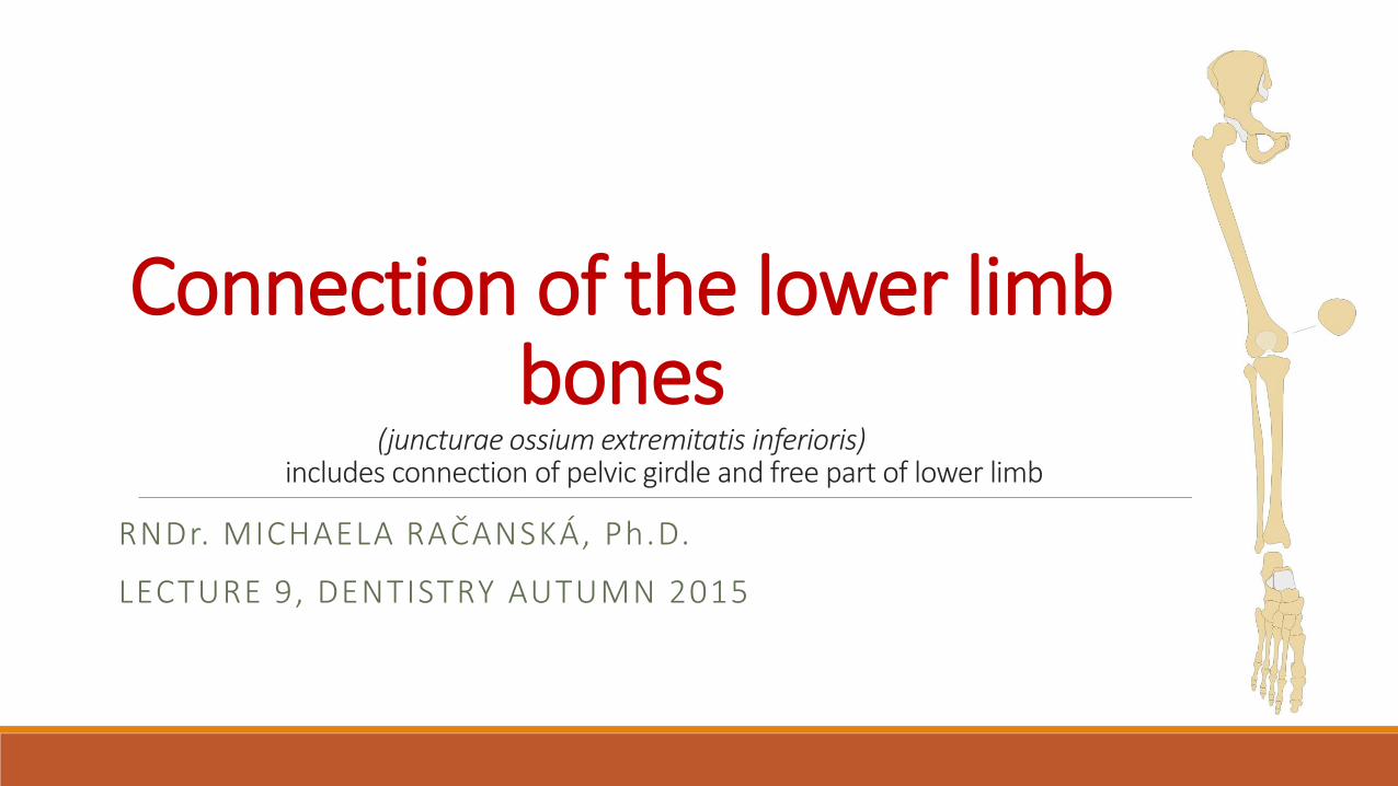
Connection of the lower limb bones
(juncturae ossium extremitatis inferioris)includes connection of pelvic girdle and free part of lower limb
RNDr. MICHAELA RAČANSKÁ, Ph.D.
LECTURE 9, DENTISTRY AUTUMN 2015

Connection of pelvic girdle (juncturae ossium cinguli extremitatis
inferioris)
1. Sacroiliac joint (Articulatio sacroiliaca)Articular surfaces: facies auriculares ossis sacri et illii
Articular capsule: tight and is attached to margins of AS
Auxiliary facilities: capsule is strengthened by ligg. sacroiliaca ventralia and
dorsalia (ventral and dorsal sacroiliac ligaments), ligg.sacroiliaca interossea
(interosseal sacroiliac ligaments)
Type of joint: amphiartrosis
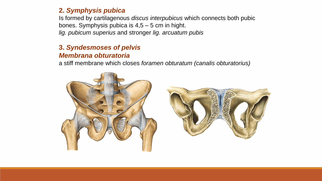
2. Symphysis pubicaIs formed by cartilagenous discus interpubicus which connects both pubic
bones. Symphysis pubica is 4,5 – 5 cm in hight.
lig. pubicum superius and stronger lig. arcuatum pubis
3. Syndesmoses of pelvis
Membrana obturatoriaa stiff membrane which closes foramen obturatum (canalis obturatorius)
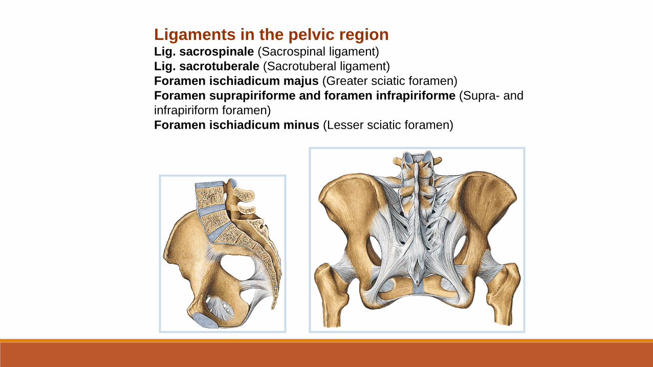
Ligaments in the pelvic regionLig. sacrospinale (Sacrospinal ligament)
Lig. sacrotuberale (Sacrotuberal ligament)
Foramen ischiadicum majus (Greater sciatic foramen)
Foramen suprapiriforme and foramen infrapiriforme (Supra- and
infrapiriform foramen)
Foramen ischiadicum minus (Lesser sciatic foramen)
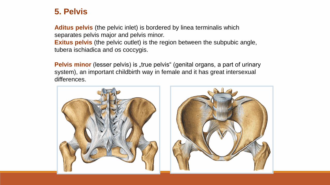
5. Pelvis
Aditus pelvis (the pelvic inlet) is bordered by linea terminalis which
separates pelvis major and pelvis minor.
Exitus pelvis (the pelvic outlet) is the region between the subpubic angle,
tubera ischiadica and os coccygis.
Pelvis minor (lesser pelvis) is „true pelvis“ (genital organs, a part of urinary
system), an important childbirth way in female and it has great intersexual
differences.
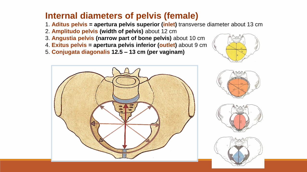
Internal diameters of pelvis (female)1. Aditus pelvis = apertura pelvis superior (inlet) transverse diameter about 13 cm
2. Amplitudo pelvis (width of pelvis) about 12 cm
3. Angustia pelvis (narrow part of bone pelvis) about 10 cm
4. Exitus pelvis = apertura pelvis inferior (outlet) about 9 cm
5. Conjugata diagonalis 12.5 – 13 cm (per vaginam)
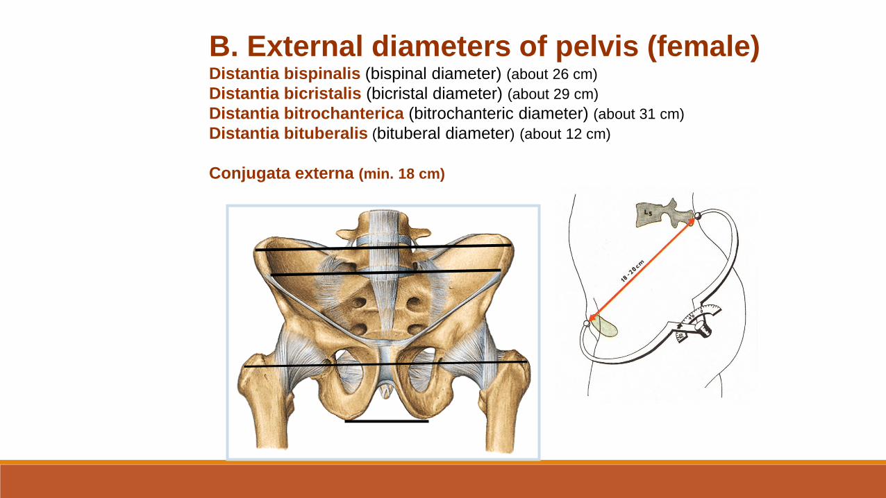
B. External diameters of pelvis (female)Distantia bispinalis (bispinal diameter) (about 26 cm)
Distantia bicristalis (bicristal diameter) (about 29 cm)
Distantia bitrochanterica (bitrochanteric diameter) (about 31 cm)
Distantia bituberalis (bituberal diameter) (about 12 cm)
Conjugata externa (min. 18 cm)
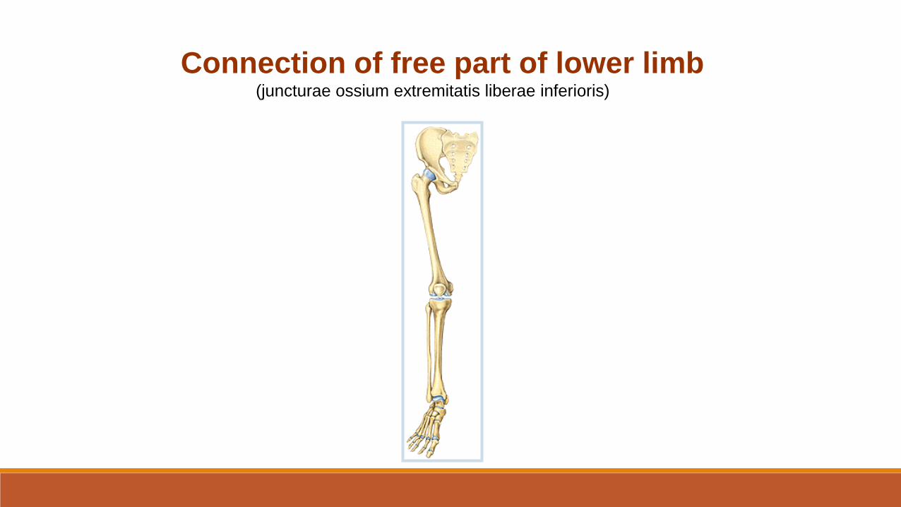
Connection of free part of lower limb(juncturae ossium extremitatis liberae inferioris)
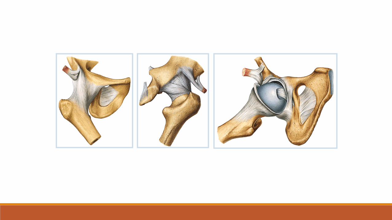
1. Articulatio coxae (hip joint)
Articular surfaces: facies lunata of an acetabulum !!!!! + head of the femur
Articular capsule: is attached to the margins of acetabulum. It reaches ventrally
linea intertrochanterica of femur, dorsally is attached to the collum femoris (neck of
femur) medially away from fossa trochanterica.
Auxiliary facilities :
a) Labrum acetabulare formed by cartilage.
b) Lig. transversum acetabuli runs through incisura acetabuli.c) Lig. iliofemorale d) Lig. pubofemorale e) Lig. ischiofemorale f) Zona orbicularisg) Lig. capitis femorisType of joint: typical spheroid joint (ball-and-socket) with limited movements
(enarthrosis).Movements – flexion, extension, abduction, adduction and internal and external
rotation.
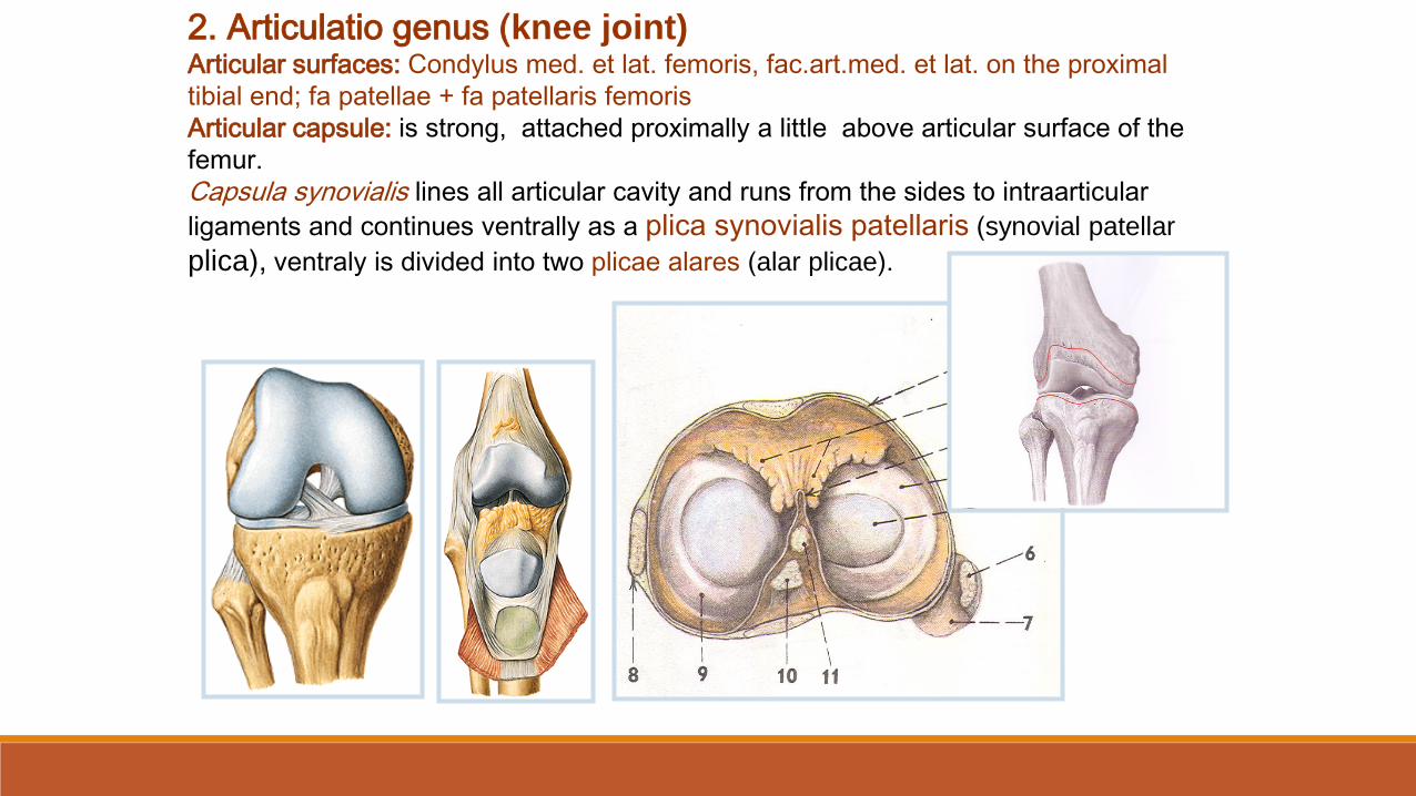
2. Articulatio genus (knee joint)Articular surfaces: Condylus med. et lat. femoris, fac.art.med. et lat. on the proximal
tibial end; fa patellae + fa patellaris femoris
Articular capsule: is strong, attached proximally a little above articular surface of the
femur.
Capsula synovialis lines all articular cavity and runs from the sides to intraarticular
ligaments and continues ventrally as a plica synovialis patellaris (synovial patellar
plica), ventraly is divided into two plicae alares (alar plicae).

Intraarticular auxiliary facilities of an articulatio genus (knee joint):
1. Meniscus – Medial © and lateral (circular)
2. Ligamenta cruciata genus – anterius - limits extension and medial rotation
posterius - limits extension and keeps stability of the joint
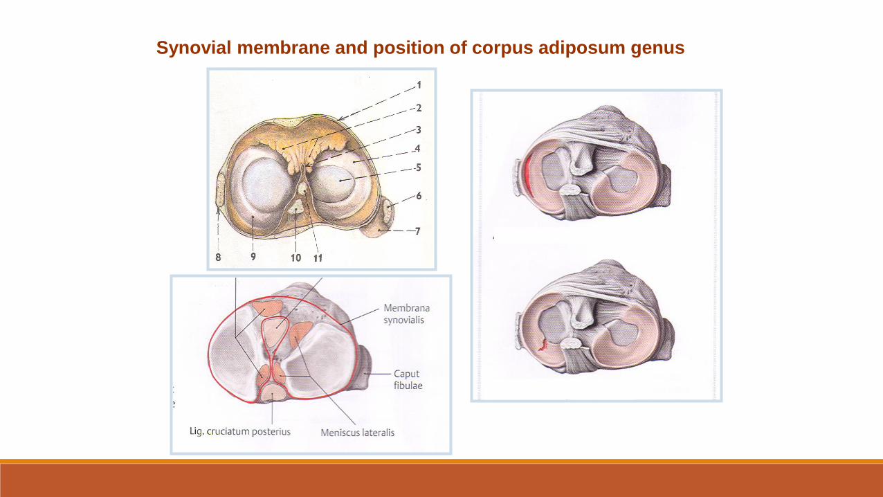
Synovial membrane and position of corpus adiposum genus
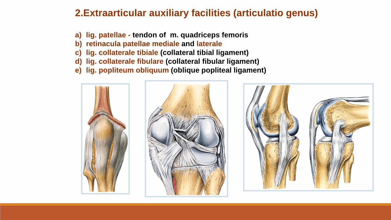
2.Extraarticular auxiliary facilities (articulatio genus)
a) lig. patellae - tendon of m. quadriceps femoris
b) retinacula patellae mediale and laterale
c) lig. collaterale tibiale (collateral tibial ligament)
d) lig. collaterale fibulare (collateral fibular ligament)
e) lig. popliteum obliquum (oblique popliteal ligament)

Type of joint: hinge joint (trochlear)
Movements: flexion and extension.
During a mild flexion is possible slight external and internal rotation.
Middle position of the joint – mild flexion
Bursae synovialesSuprapatellaris, praepatellaris (subcutanea)
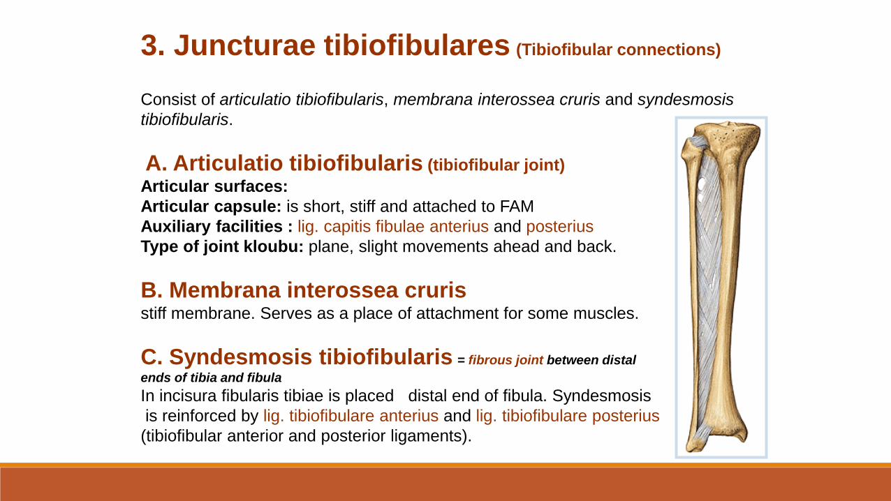
3. Juncturae tibiofibulares (Tibiofibular connections)
Consist of articulatio tibiofibularis, membrana interossea cruris and syndesmosis
tibiofibularis.
A. Articulatio tibiofibularis (tibiofibular joint)
Articular surfaces:
Articular capsule: is short, stiff and attached to FAM
Auxiliary facilities : lig. capitis fibulae anterius and posterius
Type of joint kloubu: plane, slight movements ahead and back.
B. Membrana interossea crurisstiff membrane. Serves as a place of attachment for some muscles.
C. Syndesmosis tibiofibularis = fibrous joint between distal
ends of tibia and fibula
In incisura fibularis tibiae is placed distal end of fibula. Syndesmosis
is reinforced by lig. tibiofibulare anterius and lig. tibiofibulare posterius
(tibiofibular anterior and posterior ligaments).
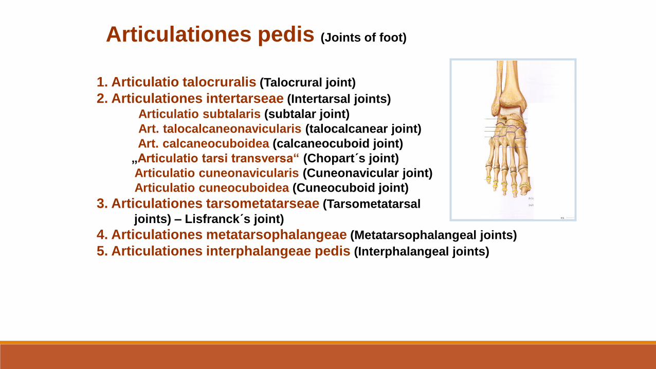
Articulationes pedis (Joints of foot)
1. Articulatio talocruralis (Talocrural joint)
2. Articulationes intertarseae (Intertarsal joints)
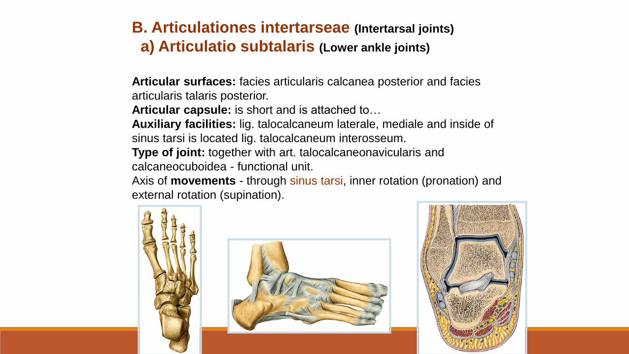
Articulatio subtalaris (subtalar joint)
Art. talocalcaneonavicularis (talocalcanear joint)
Art. calcaneocuboidea (calcaneocuboid joint)
„Articulatio tarsi transversa“ (Chopart´s joint)
Articulatio cuneonavicularis (Cuneonavicular joint)
Articulatio cuneocuboidea (Cuneocuboid joint)
3. Articulationes tarsometatarseae (Tarsometatarsal
joints) – Lisfranck´s joint)
4. Articulationes metatarsophalangeae (Metatarsophalangeal joints)
5. Articulationes interphalangeae pedis (Interphalangeal joints)
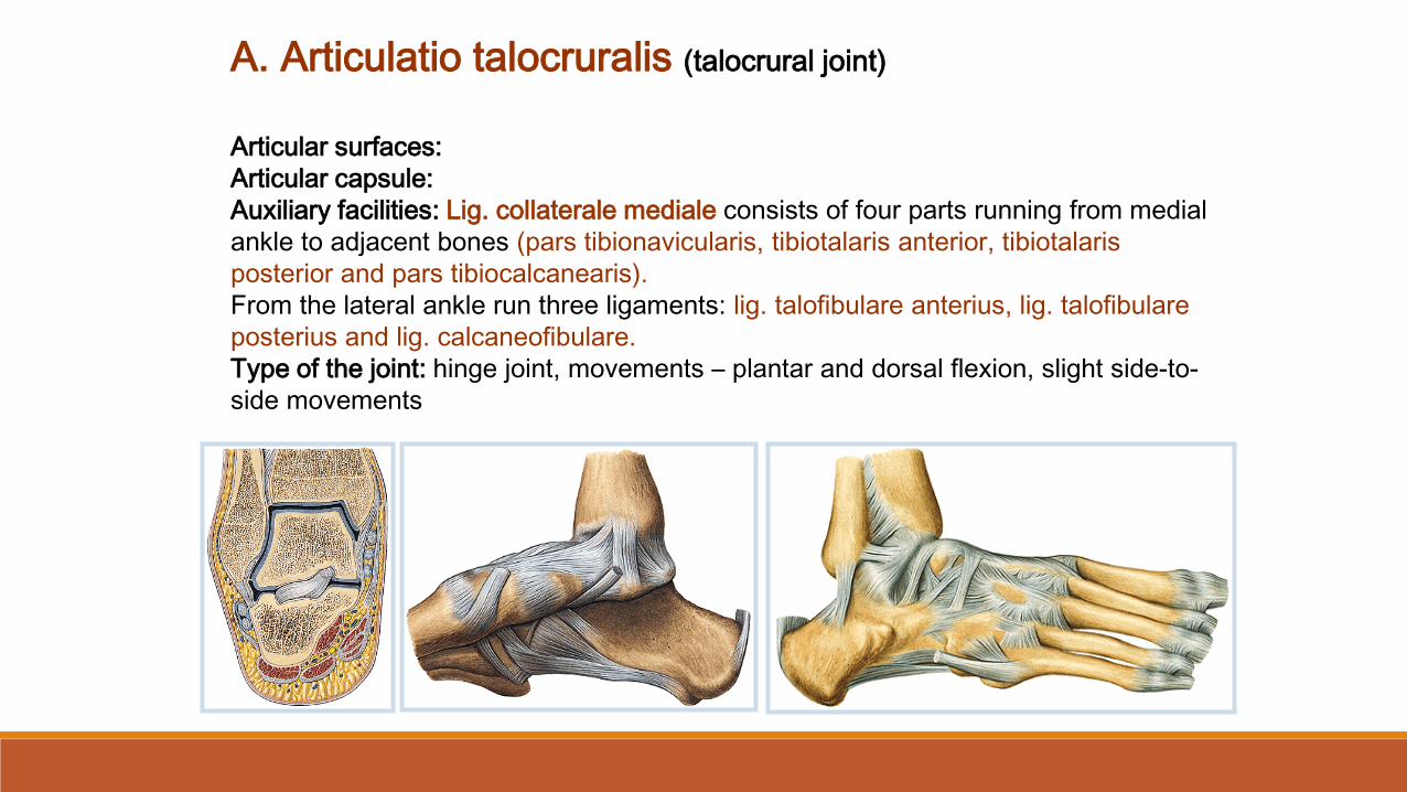
A. Articulatio talocruralis (talocrural joint)
Articular surfaces:
Articular capsule:
Auxiliary facilities: Lig. collaterale mediale consists of four parts running from medial
ankle to adjacent bones (pars tibionavicularis, tibiotalaris anterior, tibiotalaris
posterior and pars tibiocalcanearis).
From the lateral ankle run three ligaments: lig. talofibulare anterius, lig. talofibulare
posterius and lig. calcaneofibulare.
Type of the joint: hinge joint, movements – plantar and dorsal flexion, slight side-to-
side movements.
B. Articulationes intertarseae (Intertarsal joints)
a) Articulatio subtalaris (Lower ankle joints)
Articular surfaces: facies articularis calcanea posterior and facies
articularis talaris posterior.
Articular capsule: is short and is attached to…
Auxiliary facilities: lig. talocalcaneum laterale, mediale and inside of
sinus tarsi is located lig. talocalcaneum interosseum.
Type of joint: together with art. talocalcaneonavicularis and
calcaneocuboidea - functional unit.
Axis of movements - through sinus tarsi, inner rotation (pronation) and
external rotation (supination).
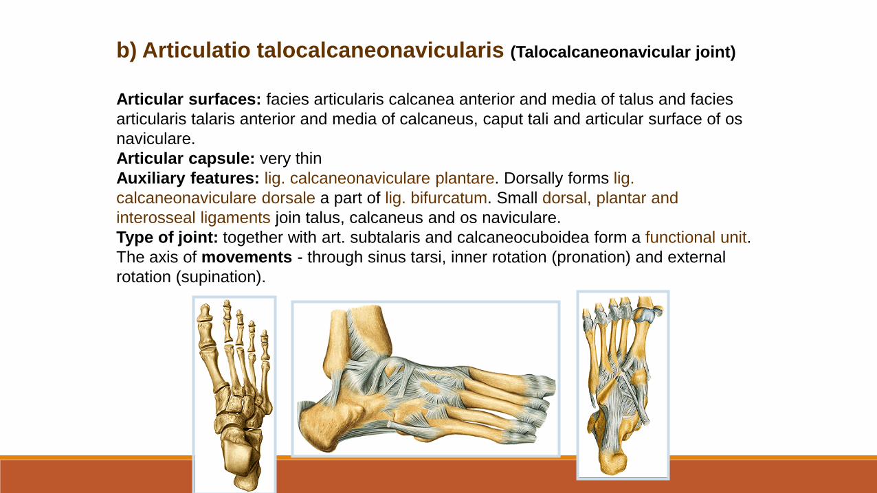
b) Articulatio talocalcaneonavicularis (Talocalcaneonavicular joint)
Articular surfaces: facies articularis calcanea anterior and media of talus and facies
articularis talaris anterior and media of calcaneus, caput tali and articular surface of os
naviculare.
Articular capsule: very thin
Auxiliary features: lig. calcaneonaviculare plantare. Dorsally forms lig.
calcaneonaviculare dorsale a part of lig. bifurcatum. Small dorsal, plantar and
interosseal ligaments join talus, calcaneus and os naviculare.
Type of joint: together with art. subtalaris and calcaneocuboidea form a functional unit.
The axis of movements - through sinus tarsi, inner rotation (pronation) and external
rotation (supination).
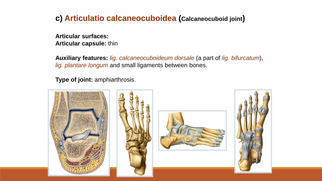
c) Articulatio calcaneocuboidea (Calcaneocuboid joint)
Articular surfaces:
Articular capsule: thin
Auxiliary features: lig. calcaneocuboideum dorsale (a part of lig. bifurcatum),
lig. plantare longum and small ligaments between bones.
Type of joint: amphiarthrosis.
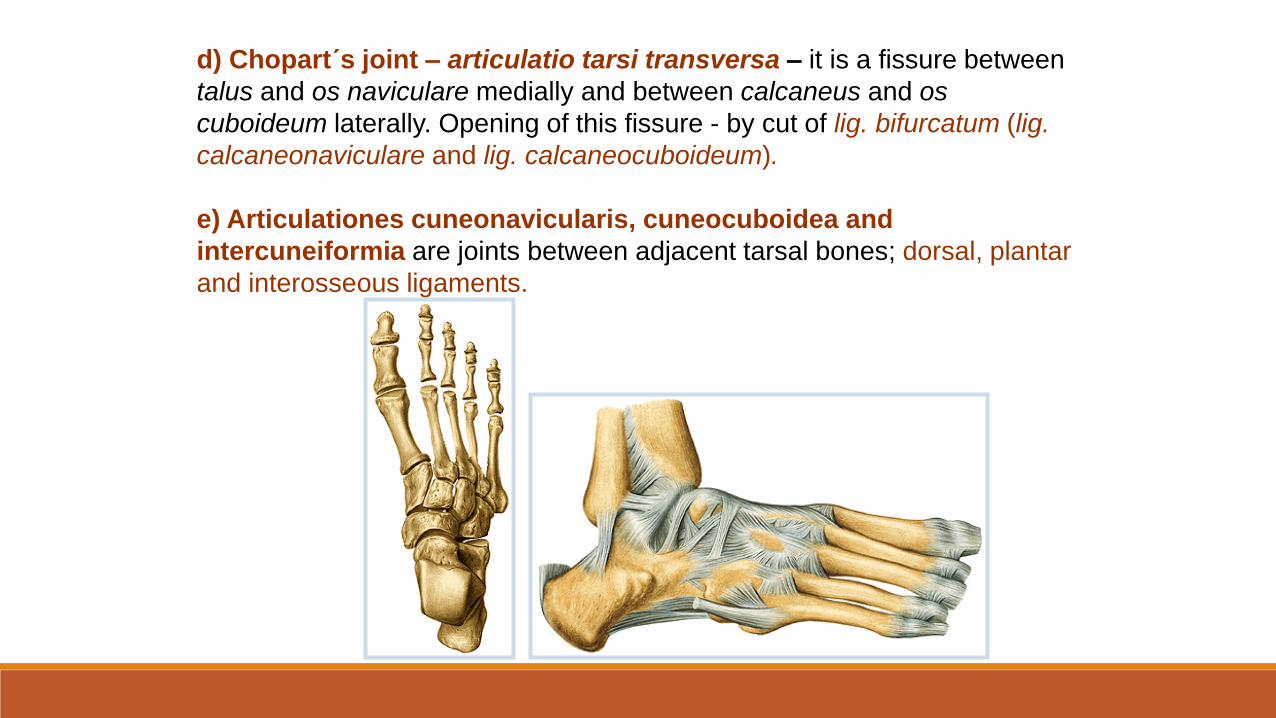
d) Chopart´s joint – articulatio tarsi transversa – it is a fissure between
talus and os naviculare medially and between calcaneus and os
cuboideum laterally. Opening of this fissure - by cut of lig. bifurcatum (lig.
calcaneonaviculare and lig. calcaneocuboideum).
e) Articulationes cuneonavicularis, cuneocuboidea and
intercuneiformia are joints between adjacent tarsal bones; dorsal, plantar
and interosseous ligaments.
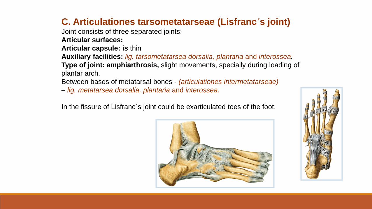
C. Articulationes tarsometatarseae (Lisfranc´s joint)Joint consists of three separated joints:
Articular surfaces:
Articular capsule: is thin
Auxiliary facilities: lig. tarsometatarsea dorsalia, plantaria and interossea.
Type of joint: amphiarthrosis, slight movements, specially during loading of
plantar arch.
Between bases of metatarsal bones - (articulationes intermetatarseae)
– lig. metatarsea dorsalia, plantaria and interossea.
In the fissure of Lisfranc´s joint could be exarticulated toes of the foot.
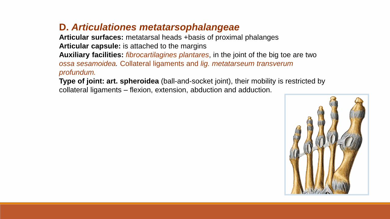
D. Articulationes metatarsophalangeaeArticular surfaces: metatarsal heads +basis of proximal phalanges
Articular capsule: is attached to the margins
Auxiliary facilities: fibrocartilagines plantares, in the joint of the big toe are two
ossa sesamoidea. Collateral ligaments and lig. metatarseum transverum
profundum.
Type of joint: art. spheroidea (ball-and-socket joint), their mobility is restricted by
collateral ligaments – flexion, extension, abduction and adduction.

E. Articulationes interphalangeae pedisArticular surfaces:
Articular capsule: are attached to …. dorsally fuse with
tendons of extensor muscles.
Auxiliary facilities: collateral ligaments, fibrocartilagines
plantares
Type of joint: art. trochlearis (hinge joint) - flexion and
extension of phalanges.

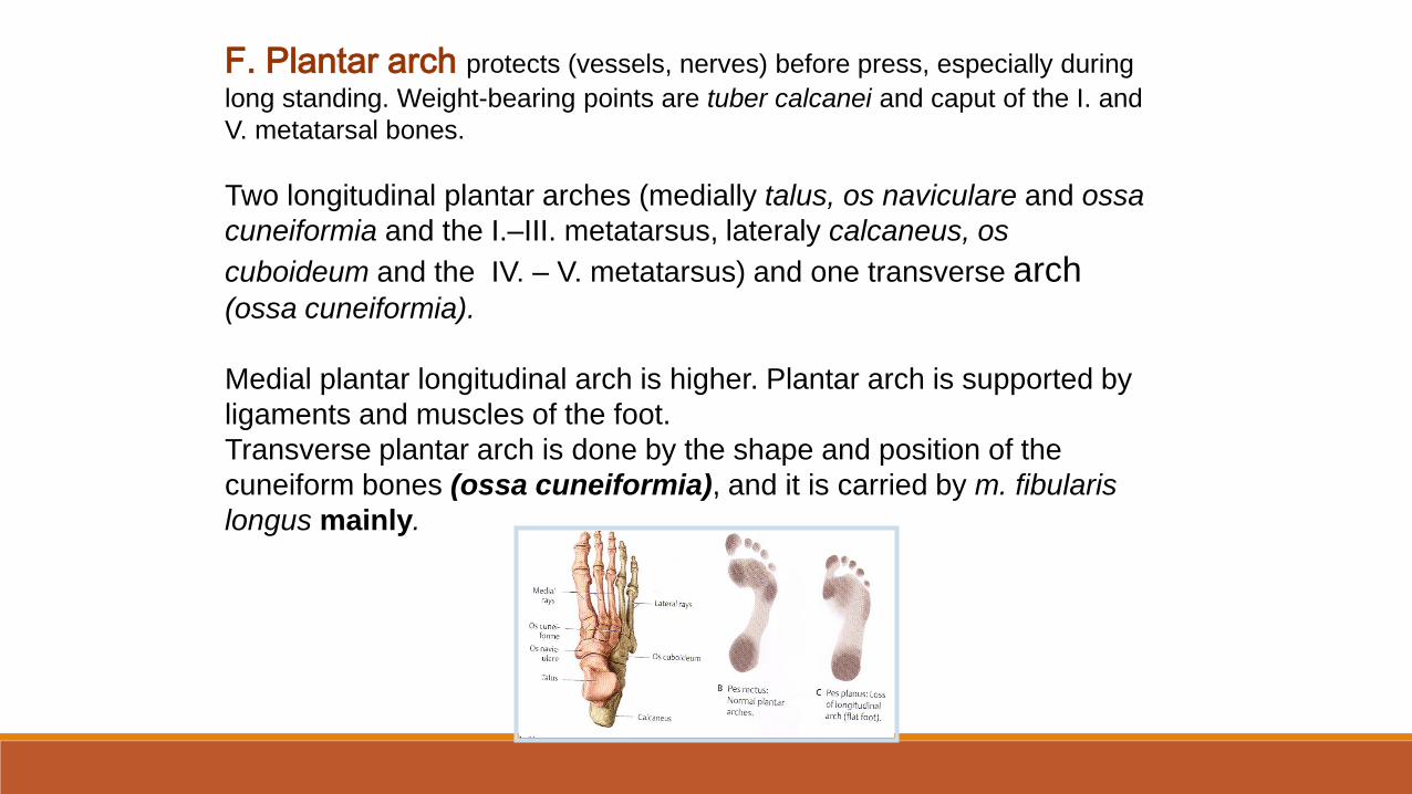
F. Plantar arch protects (vessels, nerves) before press, especially during
long standing. Weight-bearing points are tuber calcanei and caput of the I. and
V. metatarsal bones.
Two longitudinal plantar arches (medially talus, os naviculare and ossa
cuneiformia and the I.–III. metatarsus, lateraly calcaneus, os
cuboideum and the IV. – V. metatarsus) and one transverse arch(ossa cuneiformia).
Medial plantar longitudinal arch is higher. Plantar arch is supported by
ligaments and muscles of the foot.
Transverse plantar arch is done by the shape and position of the
cuneiform bones (ossa cuneiformia), and it is carried by m. fibularis
longus mainly.
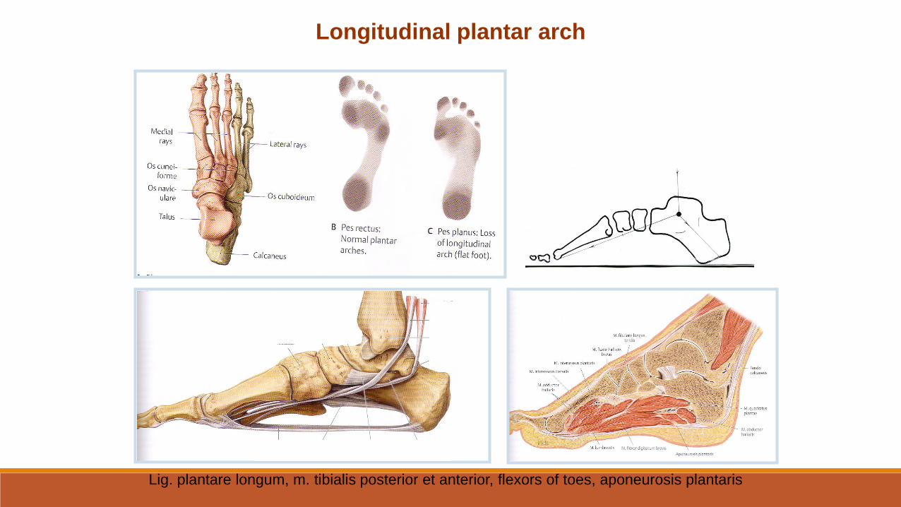
Longitudinal plantar arch
Lig. plantare longum, m. tibialis posterior et anterior, flexors of toes, aponeurosis plantaris
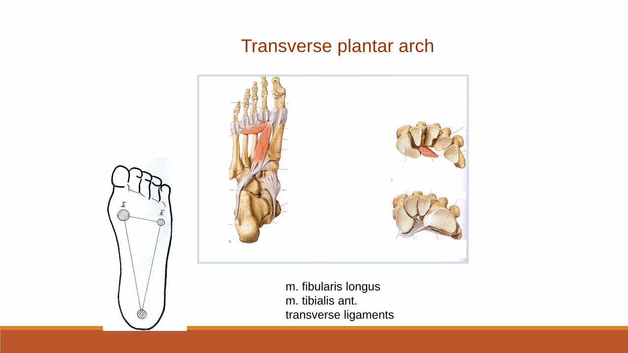
Transverse plantar arch
m. fibularis longus
m. tibialis ant.
transverse ligaments
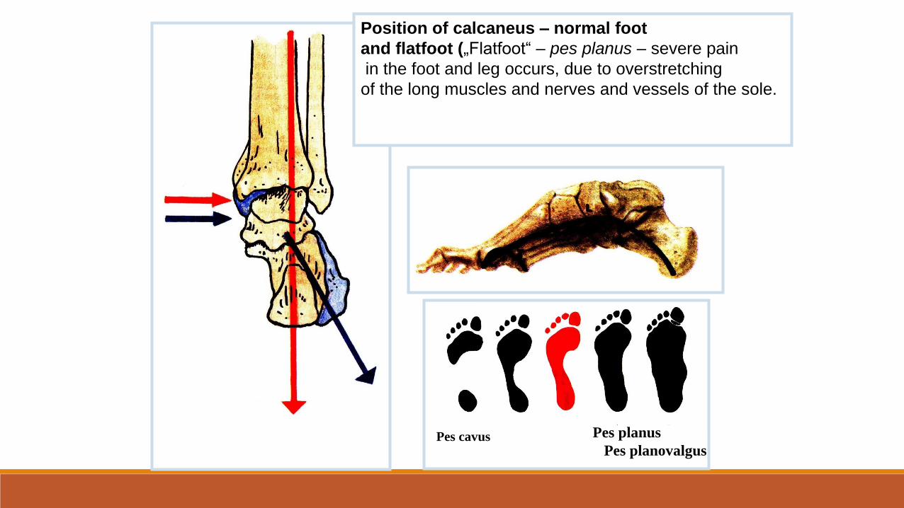
Position of calcaneus – normal foot
and flatfoot („Flatfoot“ – pes planus – severe pain
in the foot and leg occurs, due to overstretching
of the long muscles and nerves and vessels of the sole.
Pes planus
Pes planovalgusPes cavus
Lecture 10. MYOLOGY
GENERAL MYOLOGY
Structure of skeletal muscle (origo, venter musculi, insertio)
Auxiliary muscular equipment (fascie, bursae synoviales, vaginae tendinum, trochleae musculares)
Vascularization, innervation
Classification of muscles according to:
number of heads (one-headed muscle, multi-headed muscle)
number of bellies (one-bellied muscle, multi-bellied muscle)
function (flexors, extensors, abductors, adductors, levators, sfincters...)
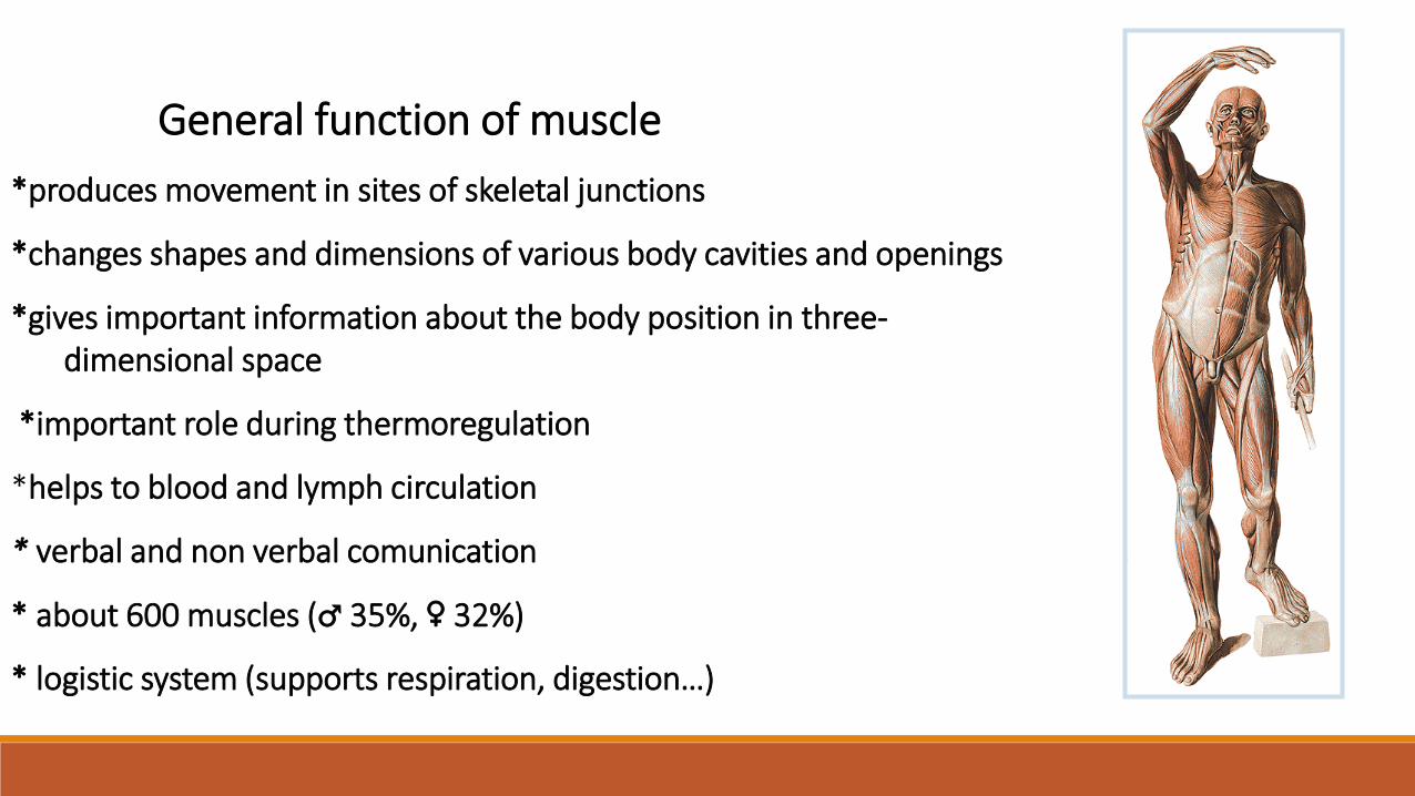
General function of muscle
*produces movement in sites of skeletal junctions
*changes shapes and dimensions of various body cavities and openings
*gives important information about the body position in three-dimensional space
*important role during thermoregulation
*helps to blood and lymph circulation
* verbal and non verbal comunication
* about 600 muscles (♂ 35%, ♀ 32%)
* logistic system (supports respiration, digestion…)
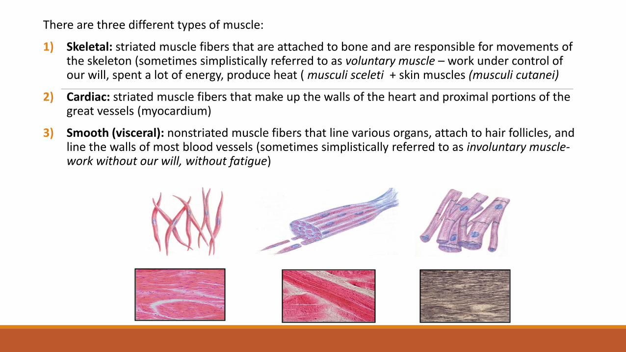
There are three different types of muscle:
1) Skeletal: striated muscle fibers that are attached to bone and are responsible for movements of the skeleton (sometimes simplistically referred to as voluntary muscle – work under control ofour will, spent a lot of energy, produce heat ( musculi sceleti + skin muscles (musculi cutanei)
2) Cardiac: striated muscle fibers that make up the walls of the heart and proximal portions of the great vessels (myocardium)
3) Smooth (visceral): nonstriated muscle fibers that line various organs, attach to hair follicles, and line the walls of most blood vessels (sometimes simplistically referred to as involuntary muscle-work without our will, without fatigue)
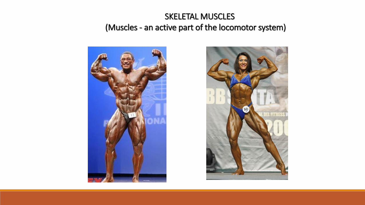
SKELETAL MUSCLES(Muscles - an active part of the locomotor system)
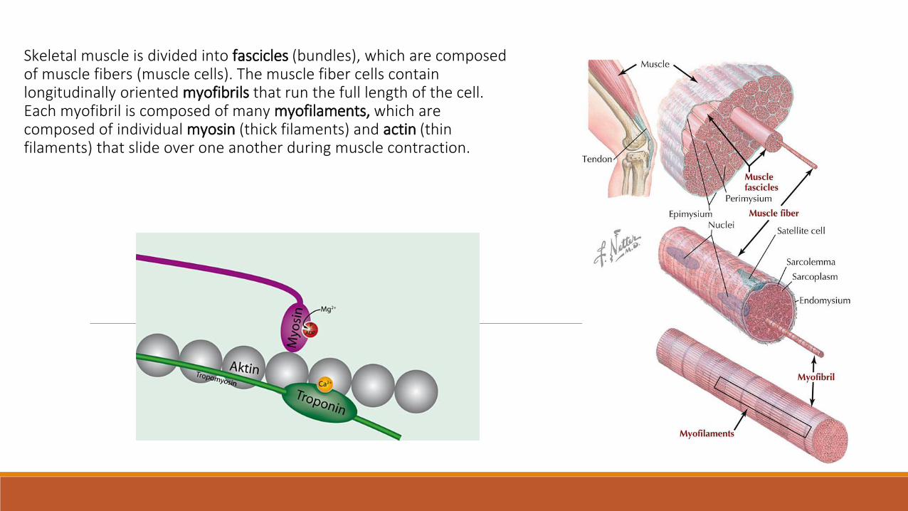
Skeletal muscle is divided into fascicles (bundles), which are composed of muscle fibers (muscle cells). The muscle fiber cells contain longitudinally oriented myofibrils that run the full length of the cell. Each myofibril is composed of many myofilaments, which are composed of individual myosin (thick filaments) and actin (thin filaments) that slide over one another during muscle contraction.
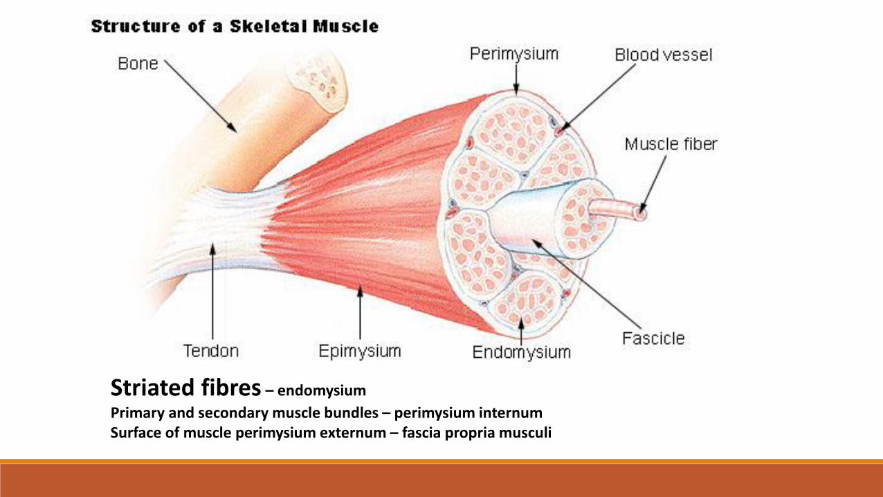
Striated fibres – endomysium
Primary and secondary muscle bundles – perimysium internumSurface of muscle perimysium externum – fascia propria musculi
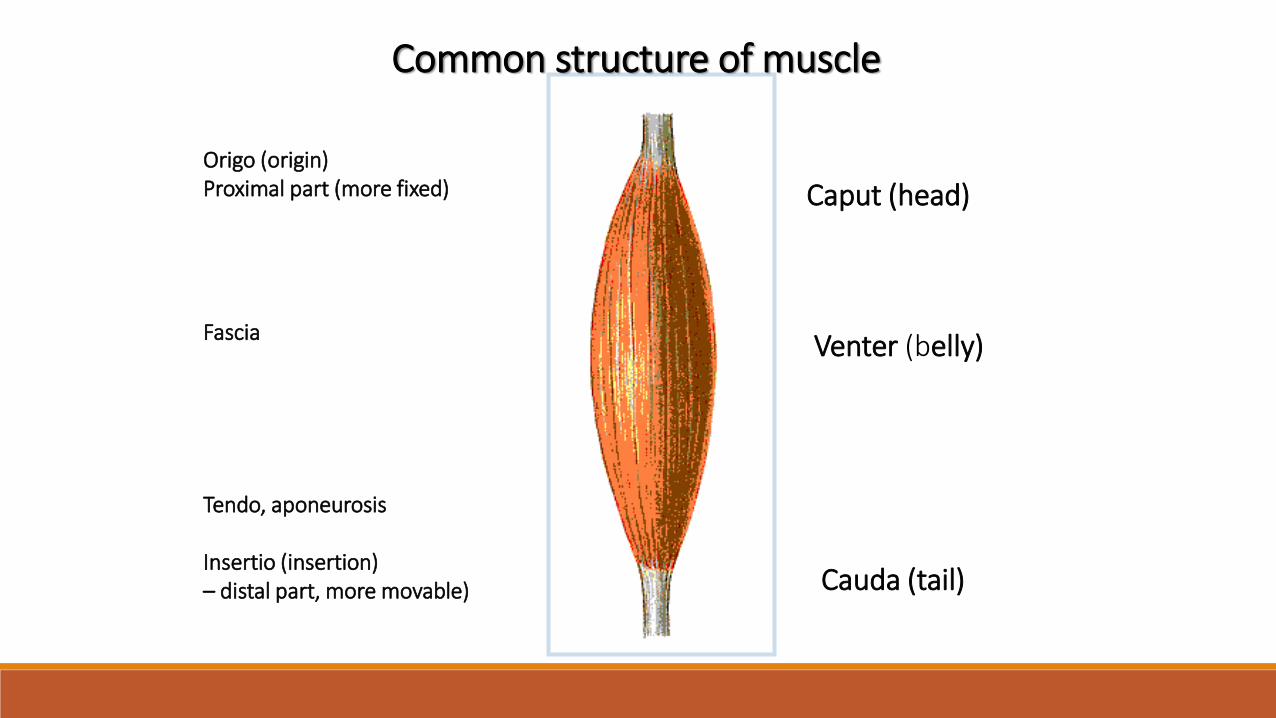
Caput (head)
Venter (belly)
Cauda (tail)
Common structure of muscle
Origo (origin)Proximal part (more fixed)
Fascia
Tendo, aponeurosis
Insertio (insertion) – distal part, more movable)
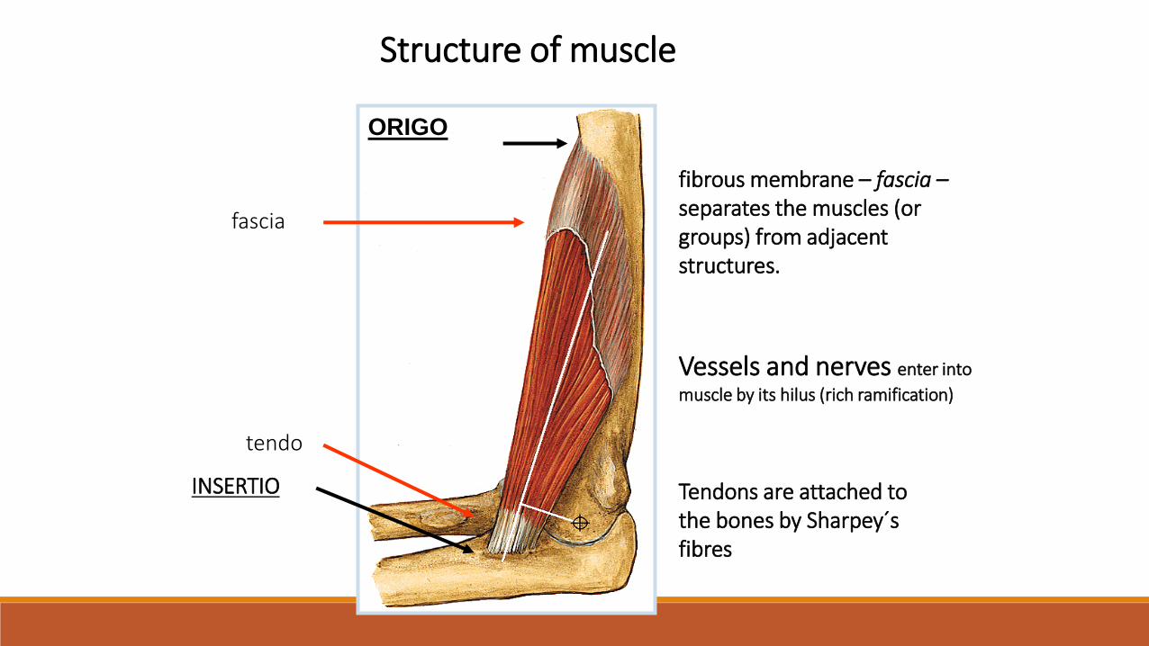
INSERTIO
ORIGO
tendo
fascia
Structure of muscle
fibrous membrane – fascia –separates the muscles (or groups) from adjacent structures.
Vessels and nerves enter into
muscle by its hilus (rich ramification)
Tendons are attached to the bones by Sharpey´sfibres
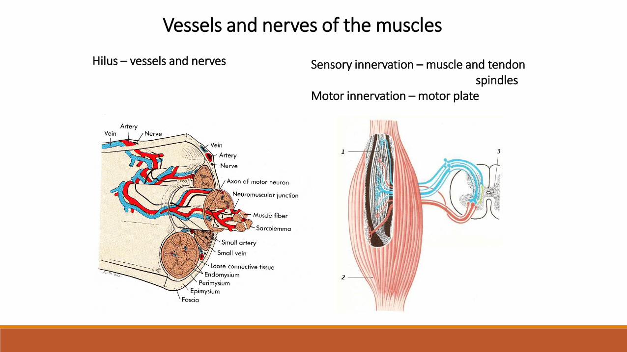
Vessels and nerves of the muscles
Hilus – vessels and nerves Sensory innervation – muscle and tendonspindles
Motor innervation – motor plate
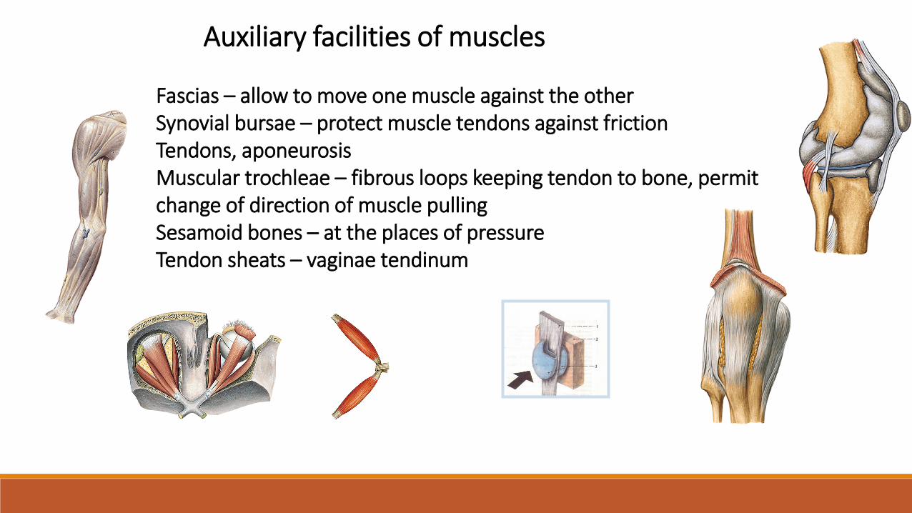
Auxiliary facilities of muscles
Fascias – allow to move one muscle against the otherSynovial bursae – protect muscle tendons against frictionTendons, aponeurosisMuscular trochleae – fibrous loops keeping tendon to bone, permitchange of direction of muscle pullingSesamoid bones – at the places of pressureTendon sheats – vaginae tendinum
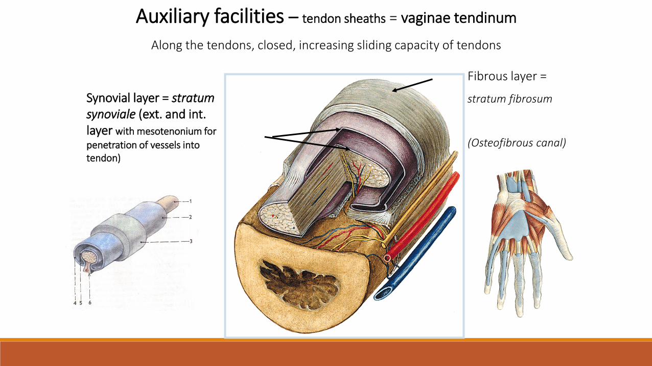
Auxiliary facilities – tendon sheaths = vaginae tendinum
Along the tendons, closed, increasing sliding capacity of tendons
Fibrous layer =
stratum fibrosum
(Osteofibrous canal)
Synovial layer = stratumsynoviale (ext. and int. layer with mesotenonium for
penetration of vessels intotendon)
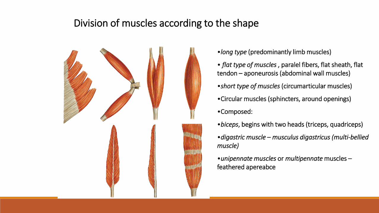
Division of muscles according to the shape
•long type (predominantly limb muscles)
• flat type of muscles , paralel fibers, flat sheath, flat tendon – aponeurosis (abdominal wall muscles)
•short type of muscles (circumarticular muscles)
•Circular muscles (sphincters, around openings)
•Composed:
•biceps, begins with two heads (triceps, quadriceps)
•digastric muscle – musculus digastricus (multi-bellied muscle)
•unipennate muscles or multipennate muscles –feathered apereabce
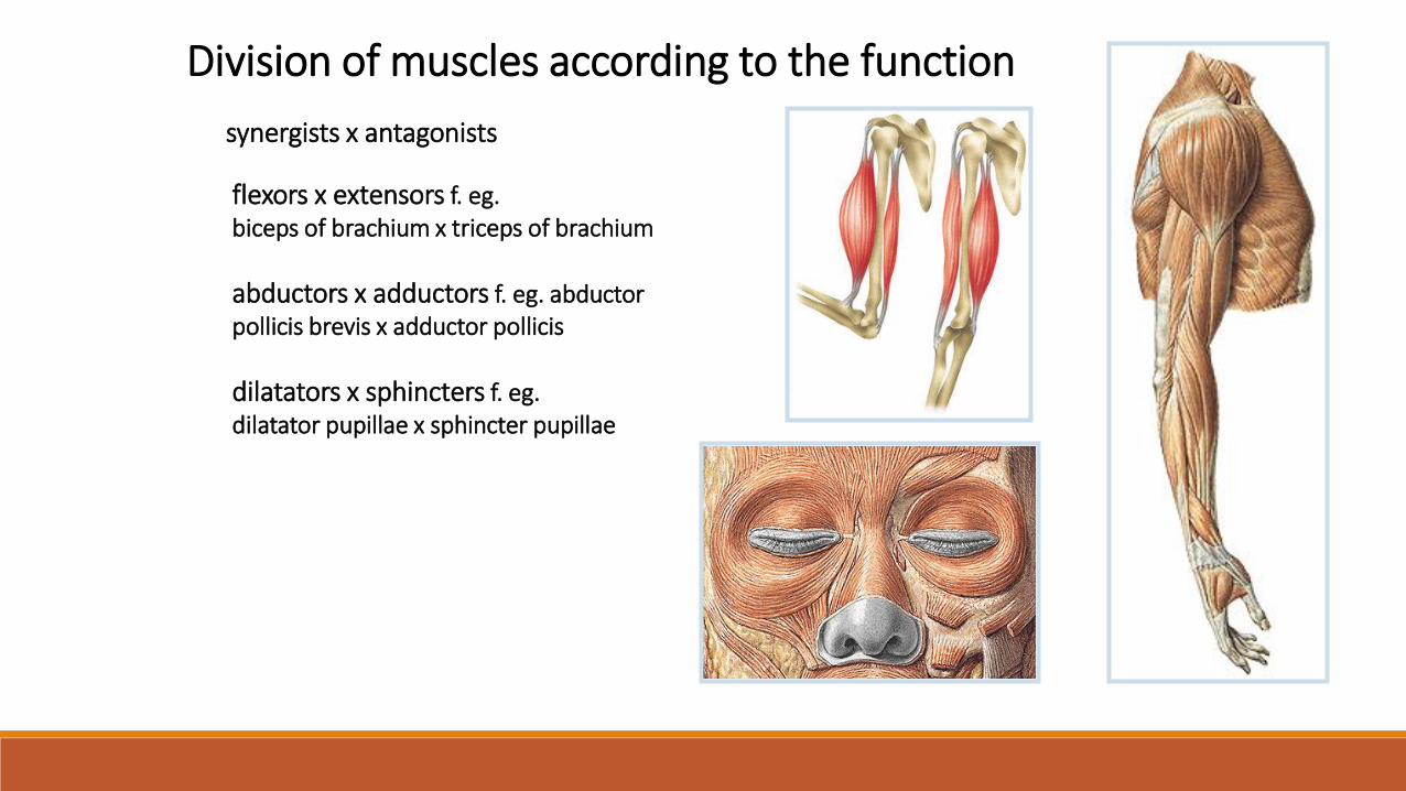
Division of muscles according to the function
synergists x antagonists
flexors x extensors f. eg. biceps of brachium x triceps of brachium
abductors x adductors f. eg. abductor pollicis brevis x adductor pollicis
dilatators x sphincters f. eg. dilatator pupillae x sphincter pupillae
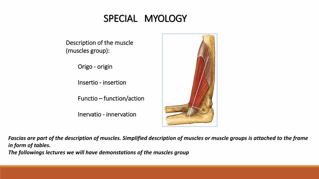
SPECIAL MYOLOGY
Description of the muscle(muscles group):
Origo - origin
Insertio - insertion
Functio – function/action
Inervatio - innervation
Fascias are part of the description of muscles. Simplified description of muscles or muscle groups is attached to the frame in form of tables. The followings lectures we will have demonstations of the muscles group
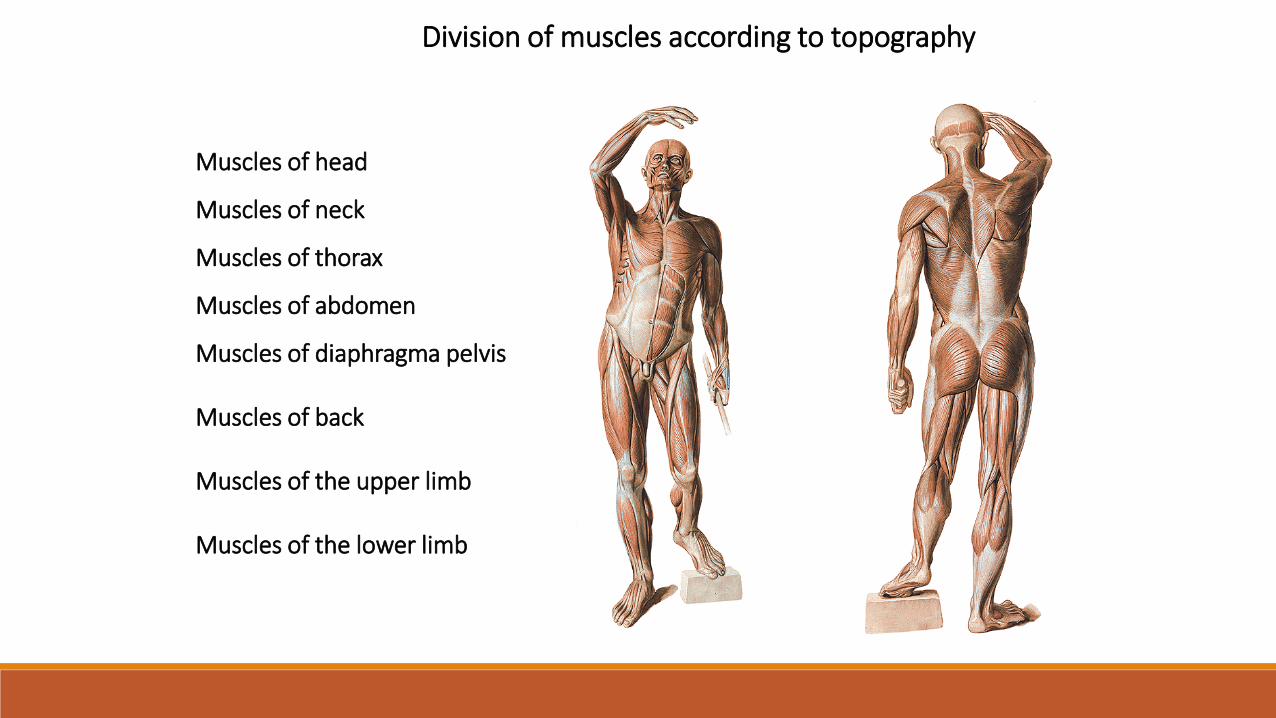
Muscles of head
Muscles of neck
Muscles of thorax
Muscles of abdomen
Muscles of diaphragma pelvis
Muscles of back
Muscles of the upper limb
Muscles of the lower limb
Division of muscles according to topography

Used pictures come from:
Moore, K. L. (1992): Clinical oriented anatomy. Third edition.
Williams&Wilkins, A Waverly Company.
Gilroy, A. M. et all. (2009): Atlas of Anatomy. Thieme New York, Stuttgart.
Putz, R. (2008):
Atlas of Human Anatomy Sobotta. Elsevier Books.
Platzer, W., Kahle, W., Leonhardt H. (1992):
Locomotor system. Georg Thieme Verlag, Stuttgart,
New York, 4th edition.
Čihák, R. (1987): Anatomie 1. Avicenum, Zdravotnické nakladatelství.