contrast-enhanced mr imaging of the facial nerve in 11
TRANSCRIPT

Robert Tien 1 · 2
William P. Dillon1
Robert K. Jackler
Received May 22, 1989; revision requested July 25 , 1989; revision received January 23, 1990; accepted January 30, 1990.
1 Department of Radiology, Division of Neuroradiology, University of California, San Francisco, UCSF L-371 , Box 0628, San Francisco, CA 94143. Address reprint requests toW. P. Dillon.
2 Present address: Department of Radiology, University of California Medical Center, San Diego, CA 92103.
3 Department of Otolaryngology, University of California, San Francisco, CA 94043 .
0195-6108/90/1104-0735 © American Society of Neuroradiology
735
Contrast-Enhanced MR Imaging of the Facial Nerve in 11 Patients with Bell's Palsy
Contrast-enhanced MR images (at 1.5 T) were obtained in 11 patients with facial palsy. The group included five people with acute idiopathic facial (Bell's) palsy, three with chronic idiopathic facial palsy, and one each with acute facial palsy after local radiation therapy, acute facial palsy resulting from herpes zoster virus infection, and facial palsy caused by facial neuroma. Eight of the 11 patients demonstrated marked enhancement of the affected facial nerve from the labyrinthine portion through the descending canal. Three patients also demonstrated mild enhancement of the distal canalicular portion of the facial nerve, simulating small distal acoustic neuromas. No difference in the pattern of enhancement between the acute or chronic Bell's palsy patients was seen. Radiographic resolution appeared to lag behind clinical resolution. The facial neuroma appeared distinct from the other lesions as a focally enhancing mass. The enhancement pattern in the Bell's group correlated with the histopathologic features of Bell 's palsy and is consistent with the viral hypothesis of the syndrome.
Thin-section contrast-enhanced MR scans are recommended for individuals with atypical presentation of facial paralysis. In the proper clinical setting, contrast-enhanced MR imaging may permit a positive radiographic diagnosis of Bell's palsy, which has previously been a diagnosis of exclusion.
AJNR 11:735-741, July/August 1990; AJR 155: September 1990
Idiopathic facial, or Bell's, palsy is the most common cause of lower motor neuron facial paralysis [1, 2] . The clinical syndrome may occur at any age and is typified by an onset with resolution within 6 to 8 weeks. Idiopathic facial palsy is a diagnosis of exclusion , which should be arrived at only if there is a compatible clinical presentation with no historical or physical findings suggestive of disease involving the CNS, temporal bone, or parotid gland. Typical Bell 's palsy rarely requires radiographic analysis . Imaging studies are indicated in patients whose facial palsy persists for longer than 2 months or in whom associated clinical features are atypical for Bell's palsy . Examples would include slowly progressive palsy, facial hyperfunction (spasm) preceding the palsy, recurrent palsies, unusual degrees of pain , and multiple cranial neuropathies. While these atypical presentations may be seen in up to 20% of patients with Bell's palsy, neoplastic (e.g., facial or acoustic neuroma, parotid malignancies, brainstem tumor), infectious (e.g., mastoiditis, cholesteatoma), or degenerative (e.g., multiple sclerosis) diseases affecting the nerve must be excluded before the diagnosis of Bell's palsy can be established. In this setting , thorough radiographic analysis of the entire course of the facial nerveincluding the brain stem, cerebellopontine angle, temporal bone, and parotid glandis required . MR imaging detects the majority of anatomic lesions affecting the facial nerve; however, multiplanar high-resolution CT scanning is often necessary if subtle lesions within the temporal bone are to be detected.
Until recently , the radiographic diagnosis of idiopathic facial palsy has also been one of exclusion, arrived at only when imaging studies are normal. Preliminary reports [3, 4] demonstrating enhancement of the intratemporal portion of the facial

736 TIEN ET AL. AJNR:11 , July/August 1990
nerve on gadopentetate-dimeglumine (Magnevist, Berlex Laboratories, Cedar Knolls, NJ) enhanced MR images in a patient with Bell's palsy suggest that the radiographic diagnosis of this entity may be positively made, at least in some cases, rather than being inferred by an absence of abnormal findings . Enhancement of the facial nerve is nonspecific and is seen not only in Bell's palsy but also in traumatic (postoperative) facial palsy [3 , 5]. However, in the proper setting, this capability may be of substantial benefit to clinicians and patients who would otherwise be faced with therapeutic and prognostic uncertainties that are inherent to a diagnosis made by exclusion. In the present study we expand on these initial
reports by presenting a series of patients with idiopathic facial palsy studied with and without gadopentetate dimeglumine.
Materials and Methods
This study is based on findings in 11 patients with facial paralysis. No other cranial nerves were clinically affected in this series of patients . Eight patients had Bell 's palsy, five acute and three chronic, (one recurrent, two persistent). One patient with ipsilateral recurrent Bell's palsy was imaged during an acute palsy episode (case 3). Three other patients with facial palsy were also included: one with acute Ramsey-Hunt syndrome (facial paralysis, pain, and periauricular
TABLE 1: MR Enhancement Characteristics in 11 Patients with Facial Palsy
Interval from Enhanced Segment of Facial Nerve Case Age
Sex Origin/Site of
Onset of Palsy No. (years) Facial Palsy lAC Labyrinthine Geniculate Tympanic Mastoid to MR Scan Segment Segment Ganglion Segment Segment
36 M Acute left (1) 12 days + + + + Bell 's
(2) 40 days* + * +* +* +* (3) 80 days* + * +* +* +*
2 12 M Acute left 4 days + + + + Bell 's
3 12 F Chronic re- 3 days after + + + + + current most recent Bell 's for 1 episode year
4 38 M Acute left 8 days + + + + Bell's
5 71 F Acute left 5 days after + + + + Bell 's; most recent right Bell's episode resolved 15 months earlier
6 58 M Acute right 40 days + + + + + Bell 's
7 44 M Chronic per- 2 years + + + + sistent left Bell's with 50% hear-ing loss and ver-tigo
8 79 M Chronic left 30 years + + + + facial dys-kinesis and mild palsy
9 48 M Right facial 7 days + + + + palsy after radiation therapy for acoustic neuroma
10 11 M Acute left 5 days + + + + + Ramsey-Hunt syn-drome
11 44 F Right facial 6 months + neuroma (nodular) for 6 months
Note.-IAC = internal auditory canal. · Enhanced, but to a lesser degree than prior MR scan.

AJNR:11 , July/August 1990 MR OF FACIAL NERVE 737
vesicles) due to herpes zoster virus (case 1 0); one patient with radiation-induced facial paralysis following heavy ion radiation therapy for an acoustic neuroma (case 9); and one patient with a neuroma of the descending portion of the facial nerve (case 11 ). Table 1 describes the timing of the MR scans in relation to the onset of facial palsy. In seven patients , the MR studies were performed within 2 weeks of the onset of an acute facial paralysis. In one case, two MR studies were performed after resolution of facial palsy (case 1 ). In addition, one of the patients with acute Bell's had fully recovered from a contralateral Bell 's palsy 15 months prior to her examination (case 5).
All patients were studied on a 1 .5-T superconducting magnet (Signa, GE). The MR studies were performed using axial , coronal , and, in one case, oblique sagittal T1-weighted images, 600/20 (TR/ TE), of the temporal bone region . Ten patients were studied with a routine head coil while one individual was imaged with a 3-in . coil. T2-weighted (long TR/TE) studies were also obtained in the majority of patients. All patients had axial and coronal views before and after administration of gadopentetate dimeglumine (0 .1 mmolfkg). The study parameters included 3-mm slice thickness , 0-1-mm interslice gap, 256 x 256 or 192 matrix , 20-cm field of view, two to four excitations , and an imaging time ranging from approximately 7 to 10 min. Postcontrast T1-weighted images were acquired immediately after administration of the contrast medium. No delayed scans in the same plane were obtained; however, the second postcontrast sequence (usually the coronal view) was generally completed 20-30 min after contrast administration.
Enhancement was evaluated on the basis of visual inspection of the pre- and postcontrast images photographed at the identical window and level settings. The intensity of the affected facial nerve was compared on both pre- and postcontrast images, as well as in relation to the contralateral facial nerve in each case. This provided an internal standard for comparing the presence and intensity of the enhancement. While bilateral Bell's palsy is possible, none of our patients manifested simultaneous bilateral symptoms; thus , we felt that the comparison to the opposite facial nerve was a reliable internal standard. Abnormal enhancement of the facial nerve was assumed if marked asymmetric enhancement of the facial nerve was present as compared with the opposite nerve. In all cases, the facial nerve was easily located on both unenhanced and enhanced sequences by virtue of its typical location in relation to the membranous labyrinth.
Results
Normal Facial Nerves
In two patients, MR imaging demonstrated mild but definite enhancement of the unaffected facial nerve. One patient (case 1) had intense enhancement of the symptomatic and asymptomatic facial nerves. This patient had follow-up MR examinations that demonstrated enhancement of the affected nerve with mild but decreased enhancement of the unaffected side. Enhancement appeared most frequently in the horizontal portion of the facial nerve and the greater superficial petrosal nerve (Fig . 1). The descending portion of the nerve appeared to enhance much less frequently . As a large control group of normal patients was not studied, we cannot assess the frequency of enhancement of the facial nerve in the population.
Abnormal Facial Nerves All 11 patients demonstrated some degree of enhancement
of the symptomatic facial nerve compared with precontrast
Fig. 1.-Case 1: 36-year-old man with left Bell ' s palsy. MR scan was obtained 73 days after clinical recovery. Axial contrast-enhanced T1-weighted image (600/20) shows intense enhancement of affected facial nerve (closed arrows). Mild proximal enhancement of facial nerve within distal internal auditory canal is also visualized on left side. The intensity of this enhancement had decreased from the initial study obtained during the acute phase (not shown). In addition, mild enhancement is present on asymmetric side in region of right horizontal facial nerve and greater superficial petrosal nerve (open arrows).
studies of the ipsilateral nerve and postcontrast studies of the opposite unaffected facial nerve. Table 1 shows the various segments of the facial nerve that enhanced after administration of gadopentetate dimeglumine. The intraparotid portion of the nerve was not evaluated.
The eight patients with Bell 's palsy (five acute, three chronic), the patient with Ramsey-Hunt syndrome, and the patient with radiation-induced palsy all demonstrated diffuse homogeneous enhancement of the facial nerve, including the fallopian canal segment, the geniculate ganglion , and the horizontal and descending segments (Figs. 1 and 2). In two patients with Bell 's palsy (and the patient with Ramsey-Hunt syndrome), mild enhancement of the distal canalicular portion of the facial nerve was also observed in addition to the enhancement of the intratemporal facial nerve (Figs. 1 and 3). In the remaining patients, enhancement of the facial nerve did not occur proximal to the fallopian canal. The neuroma of the facial nerve intensely enhanced but was restricted to a focal region within the descending facial canal (Fig . 4). A CT scan confirmed focal bone erosion at the site of the neuroma.
In one individual (case 1) studied with contrast-enhanced MR 5 weeks and 1 0 weeks after complete recovery of facial function , there was persistent but decreased enhancement (Fig . 1 ). No enhancement of the facial nerve was present in one patient (case 5) studied 15 months after resolution of Bell 's palsy.
The axial , coronal , and sagittal planes were complementary, with no one plane obviously superior to the others. The horizontal , labyrinthine, and canalicular portions of the facial nerve were best visualized in the coronal plane (Figs. 2C, 3E, 2F). Precontrast MR studies were useful in several instances in distinguishing high-intensity fatty marrow, surrounding the facial nerve, from enhancement on postcontrast images. The descending portion of the facial nerve was best identified in

738 TIEN ET AL. AJNR:11, July/August 1990
A 8 c Fig. 2.-Case 2: 12-year-old boy with acute left Bell's palsy. A, Axial contrast-enhanced T1-weighted image (600/20) shows intense enhancement of horizontal portion of facial nerve (arrows). No enhancement of
opposite, unaffected nerve was visualized (compare with 8). B, Axial contrast-enhanced T1-weighted image, 3 mm below A. Enhancement of affected descending portion of left facial nerve is demonstrated (closed
arrow). Normal horizontal portion of right facial nerve does not enhance (open arrows). C, Coronal contrast-enhanced T1-weighted image through temporal bone shows intense enhancement of descending portion of left facial nerve
(arrows).
serial cross section in the axial plane (Figs. 2B and 4). However, the oblique sagittal views oriented to the plane of the descending facial nerve were optimal in demonstrating the majority of the horizontal and vertical portions of the nerve in one section (Figs. 3A-3D).
Discussion
An abundance of evidence exists to support the hypothesis that Bell's palsy results from viral infection of the facial nerve (1 , 5- 8] . Immunological, histological, and virological tests suggest that herpes simplex I and varicella-zoster are the most common causative agents. Neurotropic viruses tend to infect the cell bodies of sensory neurons, where they may reside in dormancy for many years. In Bell 's palsy, reactivation of a dormant virus in the geniculate ganglion, the location of the cell bodies for taste, is a pathogenic mechanism that is consistent with numerous observations regarding this disease (9]. As Bell 's palsy is a self-limited disease, there is a paucity of pathologic material available. Liston and Kleid (1 0] recently reviewed 17 cases in the world literature through 1989. Although considerable variability in histopathologic findings is evident, most cases demonstrated a cellular inflammatory infiltrate of the nerve, some degree of neural degeneration, and hypervascularity. Diffuse involvement of the facial nerve including the fallopian canal segment is the most typical finding . On the basis of a substantial body of histological and electro physiological data it is apparent that the most proximal extent of involvement is the distal end of the internal auditory canal , just medial to the geniculate ganglion [6]. Neural dysfunction appears to be a consequence of an entrapment neuropathy resulting from neural edema within the tight confines of the osseous fallopian canal. Compression may disrupt
nerve function either by vascular congestion with ischemia or by impairment of axoplasmic flow. The fallopian canal is most narrow in its labyrinthine segment and constriction appears to be most severe at this site (6]. Our observation of diffuse enhancement of the facial nerve with gadopentetate dimeglumine from the distal internal auditory canal to the stylomastoid foramen, is entirely compatible with the above outlined etiopathologic hypothesis.
To relieve neural entrapment, Fisch [11] advocated surgical decompression of the facial nerve in the fallopian canal. Although there was formerly much enthusiasm for this therapeutic technique in patients with severe neural degeneration, little evidence exists that it alters the natural course of the disease (12, 13]. Although simple decompression of the mastoid segment of the nerve has been advocated by some, this is illogical, as it does not relieve the critical labyrinthine segment of the nerve (14]. Medical therapy of Bell's palsy is directed toward combating viral infection (acyclovir) and reducing neural edema (corticosteroids) (15]. Fortunately, the natural course of untreated Bell's palsy is quite favorable. In one study of 1000 patients with Bell 's palsy followed over a 15-year period, 50% of the cases entered remission within 3 weeks, and 71% had complete remission [12]. Complete and permanent facial palsy is extremely rare, as some remission of symptoms almost always occurs (16].
The mechanism of enhancement of the facial nerve in Bell's palsy after administration of gadopentetate dimeglumine is uncertain but may relate to either hypervascularity of the perineural structures of the nerve or actual disruption of the blood-nerve barrier. The perineural and epineural tissues lack this barrier and normally enhance to a variable degree (3]. Hyperemia of the nerve and perineural structures may result in enhancement on the basis of an increased vascular pool of contrast material. The endoneural blood vessels of the cranial

AJNR:11 , July/August 1990 MR OF FACIAL NERVE 739
A B c
D E F
Fig. 3.-Case 10: 11-year-old boy with acute left facial palsy resulting from Ramsey-Hunt syndrome. MR images were obtained 5 days after onset of symptoms.
A and 8, Normal (asymptomatic) facial nerve. Oblique parasagittal T1-weighted images (600/20), before (A) and after (8) contrast administration, through horizontal and descending portion of normal right facial nerve (arrows in A). On precontrast image, the facial nerve can be seen throughout its horizontal and descending course. The normal nerve enhances after administration of contrast material (arrows in 8).
C and D, Symptomatic facial nerve. Parasagittal T1-weighted images, before (C) and after (D) administration of contrast material, through symptomatic left facial nerve. Precontrast view shows an appearance similar to the affected facial nerve compared with the opposite, normal side (A). After contrast administration, there is intense enhancement of horizontal and descending portions of facial nerve (D). This enhancement is more marked than the normal baseline enhancement on the unaffected side (8). (Images were photographed at same window and level settings.)
E and F, Contiguous coronal contrast-enhanced T1-weighted images through cochlea (E) and vestibule (F) on symptomatic side. Intense enhancement in anterior genu region of facial nerve (open arrow in E) is noted superior and lateral to cochlea. The fallopian canal portion of the facial nerve enhances to a mild degree (double solid arrows in E). Horizontal portion of facial nerve intensely enhances (solid arrow in F). It is located inferior to lateral semicircular canal (open arrow in F). The vestibule is located medial to these two structures.
(Figure 3 was provided by Dr. Leon Kasel!, San Mateo, CA.)
and peripheral nerves have tight endothelial junctions that create a blood-nerve barrier that prohibits dyes such as Trypan blue or Evans blue from entering the extravascular space [17, 18]. Enhancement may occur if these junctions are disrupted by acute inflammation. A combination of these two mechanisms is also plausible. It is interesting to note that despite intense enhancement of the facial nerve within the temporal bone, enhancement did not usually occur along the cisternal portion within the internal auditory canal (Fig. 2A). Sparing of this segment may reflect its lack of perineurium or restriction of the viral process to the facial nerve distal to the geniculate ganglion. Enhancement of the distal preganglionic
segment of the facial nerve may be confused with a small acoustic neuroma if the distal facial nerve enhancement is not recognized.
Neural enhancement with gadopentetate dimeglumine has also been noted in our institution in other forms of viral neuritis. A patient with viral trigeminal herpes neuritis, recently encountered at our institution, demonstrated marked enhancement of the preganglionic segment of the fifth nerve, which resolved with clinical resolution [19]. MR studies in another patient with acute vertigo resulting from vestibular neuritis showed enhancement of the canalicular portion of the eighth cranial nerve.
740 TIEN ET AL. AJNR:11 , July/August 1990
A B
The normal intratemporal facial nerve may enhance to a mild degree. This is apparent from examination of the contralateral , asymptomatic facial nerves in our patient population. However, intense enhancement is not normal. Enhancement of the intracanalicular portion of the facial nerve should also be considered abnormal. The latter occurred in two of our patients with Bell's palsy but in none of the normal nerves. Comparison with the unaffected opposite side is always useful in this respect, and would therefore support the examination of both temporal bones in these patients. We also advise obtaining both the precontrast and postcontrast images of the same plane, and photographing at identical window and level settings in order to ensure proper comparison of enhancement. Fat-suppression techniques, currently under clinical evaluation, may prove useful for excluding high-signalintensity fat as a potential pitfall on enhanced images. One must be careful not to mistake the enhancing mucosa of the medial tympanic cavity for the horizontal facial nerve. This can be avoided by employing a coronal sequence that will nicely differentiate the two regions.
The causes of acute facial palsy are innumerable [2] . Lesions anywhere along the course of the facial nerve, from its nucleus in the pons to its distal arborization within the parotid gland, may give rise to a unilateral palsy. Neoplasms of the brainstem, cerebellopontine angle, internal auditory canal, middle ear, mastoid, or parotid gland may give rise to facial nerve dysfunction [20, 21 ] . Primary facial nerve tumors may also occur, although they are quite rare [22-25] . In addition, . facial paralysis may occur in the setting of acute or chronic otitis media, temporal bone trauma, inflammatory disease such as sarcoidosis, with infections such as Lyme disease, and after radiation therapy. Enhancement of the facial nerve with gadopentetate dimeglumine may be anticipated in some of these pathologic processes. Indeed, Daniels et al. [3] have demonstrated enhancement of the facial nerve associated with internal auditory canal lesions, particularly in those patients with neurofibromatosis . It is unclear from their report whether the nerve enhanced because of plexiform neurofibromatosis or whether internal auditory canal lesions caused engorgement of the vasculature along the nerve. In a recent
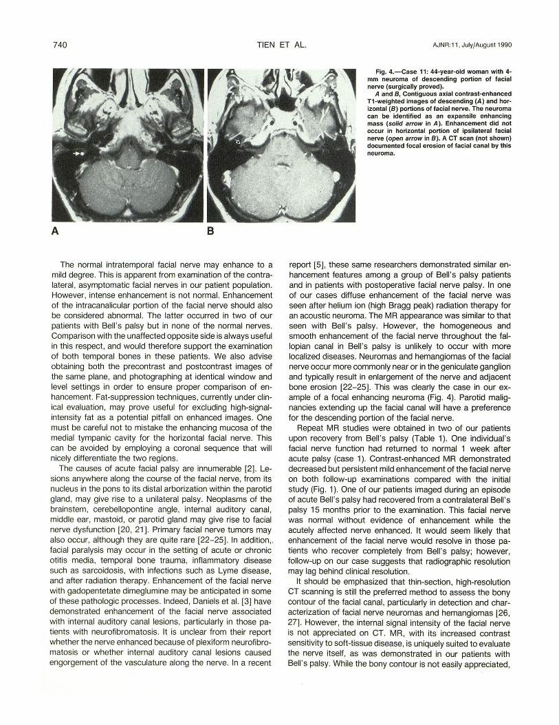
Fig. 4.-Case 11: 44-year-old woman with 4-mm neuroma of descending portion of facial nerve (surgically proved).
A and 8, Contiguous axial contrast-enhanced T1-weighted images of descending (A) and horizontal (8) portions of facial nerve. The neuroma can be identified as an expansile enhancing mass (solid arrow in A). Enhancement did not occur in horizontal portion of ipsilateral facial nerve (open arrow in 8). ACT scan (not shown) documented focal erosion of facial canal by this neuroma.
report [5], these same researchers demonstrated similar enhancement features among a group of Bell's palsy patients and in patients with postoperative facial nerve palsy. In one of our cases diffuse enhancement of the facial nerve was seen after helium ion (high Bragg peak) radiation therapy for an acoustic neuroma. The MR appearance was similar to that seen with Bell's palsy. However, the homogeneous and smooth enhancement of the facial nerve throughout the fallopian canal in Bell 's palsy is unlikely to occur with more localized diseases. Neuromas and hemangiomas of the facial nerve occur more commonly near or in the geniculate ganglion and typically result in enlargement of the nerve and adjacent bone erosion [22-25]. This was clearly the case in our example of a focal enhancing neuroma (Fig. 4). Parotid malignancies extending up the facial canal will have a preference for the descending portion of the facial nerve.
Repeat MR studies were obtained in two of our patients upon recovery from Bell 's palsy (Table 1). One individual 's facial nerve function had returned to normal 1 week after acute palsy (case 1 ). Contrast-enhanced MR demonstrated decreased but persistent mild enhancement of the facial nerve on both follow-up examinations compared with the initial study (Fig. 1 ). One of our patients imaged during an episode of acute Bell's palsy had recovered from a contralateral Bell's palsy 15 months prior to the examination. This facial nerve was normal without evidence of enhancement while the acutely affected nerve enhanced. It would seem likely that enhancement of the facial nerve would resolve in those patients who recover completely from Bell 's palsy; however, follow-up on our case suggests that radiographic resolution may lag behind clinical resolution.
It should be emphasized that thin-section, high-resolution CT scanning is still the preferred method to assess the bony contour of the facial canal , particularly in detection and characterization of facial nerve neuromas and hemangiomas [26, 27]. However, the internal signal intensity of the facial nerve is not appreciated on CT. MR, with its increased contrast sensitivity to soft-tissue disease, is uniquely suited to evaluate the nerve itself, as was demonstrated in our patients with Bell 's palsy. While the bony contour is not easily appreciated,

AJNR:11, July/August 1990 MR OF FACIAL NERVE 741
the membranous labyrinth, cochlea, and soft-tissue component of the facial nerve can be visualized on routine studies [28] . With the addition of gadopentetate dimeglumine, further contrast can be gained on T1-weighted sequences. Axial, coronal, and oblique sagittal views oriented to the course of the descending and horizontal facial nerve canal are complementary.
We agree with Daniels et al. [5] that MR imaging is not a screening examination for patients with Bell's palsy. We recommend a pre- and postcontrast thin-section MR study for individuals with peripheral facial palsy that has not resolved within 2 months or that is associated with features atypical of Bell's palsy. Diffuse enhancement of the entire facial nerve suggests "atypical Bell 's palsy." If enhancement is more focal in appearance, alternative diagnoses, such as perineural spread of malignancy or primary facial nerve tumors, would be suggested . A high-resolution CT scan of the temporal bone is recommended if the MR study is normal in the presence of facial palsy.
Conclusions
A series of patients with facial palsy verifies earlier reports that the facial nerve in Bell 's palsy diffusely enhances with gadopentetate dimeglumine from the fallopian canal to the stylomastoid foramen . This anatomic pattern of facial nerve enhancement is consistent with the hypothesis that Bell 's palsy results from viral geniculate ganglionitis with neural entrapment due to swelling within the tight confines of the fallopian canal. The diagnosis of Bell 's palsy no longer need be one of exclusion . The capability of establishing a positive diagnosis permits the clinician to allay the patient's fears concerning tumors or CNS disease and to confidently institute appropriate therapy for viral neuritis. Contrast-enhanced MR imaging is recommended in the evaluation of patients with atypical or complicated facial palsy and may also prove useful for other cranial neuropathies.
REFERENCES
1. May M, Podvinic M, Ulrich J, Peiterson E, Klein S. Idiopathic (Bell's) palsy, herpes zoster cephalicus, and other facial nerve disorders of viral origin. In: May M, ed. The facial nerve. New York: Thieme, 1986 :365-400
2. May M. Evaluation of facial nerve function: differential diagnosis by history, physical findings , and laboratory results . In: May M, ed. The facial nerve. New York: Thieme, 1986 :181-21 6
3. Daniels DL, Czervionke LF , Pojunas KW, et al. Facial nerve enhancement in MR imaging. AJNR 1987;8 :605-607
4. Millen SJ , Daniels DL, Meyer GA. Gadolinium-enhanced magnetic resonance imaging in temporal bone lesions. Laryngoscope 1989;99 :257-260
5. Daniels DL, Czervionke LF, Millen SJ, Haberkaup TJ , et al. MR imaging of facial nerve enhancement in Bell 's palsy or after temporal bone surgery. Radiology 1989;171 :807-809
6. Yanagihara N. Etiology and pathophysiology of Bell's palsy. Ann Otol Rhinal Laryngo/1988 ;137(Suppl): 1-27
7. Fisch U, Feli x H. On the pathogenesis of Bell's palsy. Acta Otolaryngol 1983;95: 532-538
8. Mulkens PSJZ, Bleeker JD, Schroder FP. Acute facial paralysis: a virological study. Clin Oto/aryngo/1 980;5: 305-310
9. Gussen R. Pathogenesis of Bel l' s palsy. Retrograde epineural edema and postedematous fibrous compression neuropathy of the facial nerve. Ann Otol Rhinal Laryngo/1977;86:549-558
10. Liston SL, Kleid MS. Histopathology of Bell' s palsy. Laryngoscope 1989;99: 23-26
11 . Fisch U. Surgery for Bell 's palsy. Arch Otolaryngo/1 981 ;1 07 :1-11 12. Adour KK, Byl FM, Hilsinger RL, et al. The true nature of Bell's palsy:
analysis of 1000 consecutive patients. Laryngoscope 1978;88:787-811 13. May M, Klein SR, Taylor FH . Idiopathic (Bell' s) facial palsy: natural history
defies steroid or surgical treatment . Laryngoscope 1985;95:406-409 14. Hughes GB. "Simple" facial nerve decompression for Bell's palsy. Am J
Oto/1988;9: 157 15. Adour KK, Hetzler DG. Current medical treatment for facial palsy. Am J
Oto/1984;5:499-502 16. Peitersen E. The natural history of Bell 's palsy. Am J Otol 1982;4 :
107-111 17. Bradbury MWB. The concept of the blood-brain barrier . New York: Wiley,
1979:127-136 18. Rappaport Sl. Blood brain barrier in physiology and medicine . New York:
Raven, 1976 :74-76 19. Tien R, Dillon WP. Herpes trigeminal neuritis and rhomboencephalitis on
Gd-DTPA-enhanced MR imaging. AJNR 1990;11 :413-414 20. Jackson CG, Glasscock ME, Hughes GB, et al. Facial paralysis of neo
plastic origin: diagnosis and management. Laryngoscope 1980;90: 1581-1595
21 . Parisier SC, Som PM, Shugar JMA. Metastatic disease causing peripheral facial paralysis. In: Graham M, House W, eds. Disorders of the facial nerve: anatomy, diagnosis and management. New York: Raven, 1982:197-206
22. Janecka IP, Conley J. Primary neoplasms of the facial nerve. Plast Reconstr
Surg 1987;79: 177-185 23. Inoue Y, Tabuchi T, Hakuba A, et al. Facial nerve neuromas: CT findings .
J Comput Assist Tomogr 1987;11 :942 24. May M. Tumors involving the facial nerve. In: May M, ed. The facial nerve .
New York: Thieme, 1986 :445-468 25. Lo W, Horn KL, Carberry JN, et al. lntratemporal vascular tumors: evalu
ation with CT. Radiology 1986;159 :181 26. Harnsberger HR, Davis RK, Osborn AG, et al. The tailored CT evaluation
of persistent facial nerve paralysis. Laryngoscope 1986;96:347-352 27 . Disbro MA, Harnsberger HR, Osborn AG. Peripheral facial nerve dysfunc
tion: CT evaluation. Radiology 1985;155:659-663 28. Jackler RK , Dillon WP. Computed tomography and magnetic resonance
imaging of the inner ear. Otolaryngol Head Neck 1988;99:494-504