contributed posters monday, july 23 - elettramo001-mo116).pdf · in this work we calculate the...
TRANSCRIPT

CONTRIBUTED POSTERS MONDAY, JULY 23
Mo001Mo001Mo001Mo001Mo001

Mo002Mo002Mo002Mo002Mo002

ATOMIC
AND MOLECULAR RESEARCH
Mo003

Mo004Mo002Mo004Mo004Mo004

SPECTRAL PROPERTIES OF CONFINED ATOMS
J P Connerade1, V K Dolmatov2, A P Lakshmi3, S T Manson4
1 The Blackett Laboratory, Imperial College, London SW7 2BW, UK 2 Starodubtsev Physical-Technical Institute, Tashkent 700084, Uzbekistan
3 The School of Physics, University of Hederabad, Hederabad 500046, India 4 Department of Physics and Astronomy, Georgia State University, Atlanta, 30303, GA, USA
The energy spectra of ground-state, ionized and excited multielectron atoms and ions of the 3d and 4d periods of the Periodic Table centered in impenetrable spherical confinement are detailed using Hartree-Fock configuration average calculations. It is shown that, owing to modifications in 3d and 4d orbital collapse, the filling of shells for the transition sequence becomes more regular than for free atoms with increasing confinement pressure, that s-d competition ultimately disappears, and that, for d-excited states, the crossing between inner-shell excited states and the double-ionization threshold are altered. In general, the Periodic Table for confined atoms differs from that for free atoms.
The properties of hydrogen confined endohedrally at the geometrical center of a spherical,
attractive short-range potential shell are explored. The evolution of the energy spectrum, as a function of the depth of the shell, is found to exhibit avoided crossings and unusual degeneracies. In addition, a new level ordering, principally by the number of nodes in the radial wavefunction, develops. The results apply generally to endohedrally confined atoms.
With the use of the above model, the origin and nature of confinement resonances in
photoionization spectra of endohedrally confined atoms is established. Also found is that near-threshold resonances demonstrate significant sensitivity to the size and thickness of the shell and develop modulations in their intensities as a function of the confinement parameters.
A novel effect - the effect of selective orbital compression in endohedrally confined atoms
is demonstrated. It turns out that even an attractive shell can exert positive pressure on an atomic orbital, making its size even smaller than the radius of the confinement itself.
It is shown that confinement can produce a significant redistribution of oscillator strengths
in endohedral multielectron atoms, making the dominant transitions no longer superior but inferior in strength, and also making electron correlations in such atoms act in the opposite way to free atoms. This is exemplified by calculated results for endohedral Ca.
It is found that non-dipole effects in low energy photoionization of atoms surrounded by a
repulsive semi-transparent potential can be increased by many orders of magnitude due to virtual levels occuring in the spectra of photoelectrons as a result of confinement. The strengths and widths of such resonances in non-dipole channels can be controlled by altering the characteristics of the confining potential, and under certain circumstances can be so large that treating quadrupole transitions as a perturbation breaks down, even for photon energies as low as tens of eV.
This work was financed by the Royal Society, INTAS, CRDF, NATO and NSF.
Mo005Mo001Mo005Mo005Mo005

CORE-CORE AND CORE-AUGER ELECTRON CORRELATIONS INDOUBLE AUGER PROCESSES IN NE
A.G.Kochur1, B.Kanngießer, P.Zimmermann2, V.L.Sukhorukov1
1 Rostov State University of Transport Communication. Rostov-na-Donu 344038 Russia2 Institute of Atomic and Analytical Physics, Berlin Technical University, Hardenberg str.36, 10623 Berlin
Using the photoelectron-photoion coincidence technique with energy dispersed electronswe measured the yields of the photoions upon the decay of the Ne1s-1 state [1]. The yield of theNe3+ ions which is roughly the probability of double Auger processes (DAP) was found to be5.97±0.16%. Our configuration-interaction calculation of only the correlations of core electrons(core-core) gave the total DAP probability of 3%. Earlier many body perturbation theorycalculation [2] gave 4%.
In ref. [3] the contribution to DAP in Ne due to the correlations of core and Augerelectrons (core-Auger), i.e. inelastic scattering of the Auger electron by core electrons, iscalculated with inclusion of only radial correlations, i.e. those where core and Auger electronsare excited into the states with the same orbital quantum numbers. The calculation [3] gave about2% probability of DAP for each final state of the KLL transitions.
In this work we calculate the contribution to DAP from correlations of core and Augerelectrons with inclusion of both radial and angular correlations (the excitations to all possiblechannels with l up to 3 are considered). Calculated DAP probabilities are presented in Table 1.Total calculated DAP probability is close to the experiment. Therefore, core–core and core–Auger electron correlations are the principal mechanisms of double Auger processes in Ne
Table 1. Probabilities (in %) of double Auger processes in Ne
Type of correlationFinal statecore–core [1] core–Auger
1s22s02p6εs 8.16 3.941s22s12p5εp 4.87 2.451s22s22p4ε{s,d} 1.12 2.05Weighted total probability of the K-LLL DAP 5.37
References
[1] B. Kanngiesser, M.Jainz, S.Bruenken et al. Phys. Rev. A. 62 (2000) 014702[2] M.Ya. Amusia, I.S. Lee and V.A. KLilin, Phys. Rev.A. 45(1994) 4576.[3] V.F. Demekhin, N.V. Demekhina. Studied in Russia (electronic journal) 91(2000) 1258-
1270, http://zhurnal.ape.relarn.ru/articles/2000/091.pdf
Mo006Mo001Mo006Mo006Mo006
A VAPOUR PHASE STUDY OF PHOTOIONISATION OF TIN
M. Huttula, H. Aksela, M. Jurvansuu, E. Kukk, and S. Aksela
Department of Physical Sciences, P.O.Box 3000, FIN-90014 University of Oulu, Finland
The photoelectron study of electronic structure of metals as free atoms has beenactively studied since the introduction of synchrotron radiation. Recently, the researchin this field has became more active as the light sources and also experiments havedeveloped allowing more accurate studies.
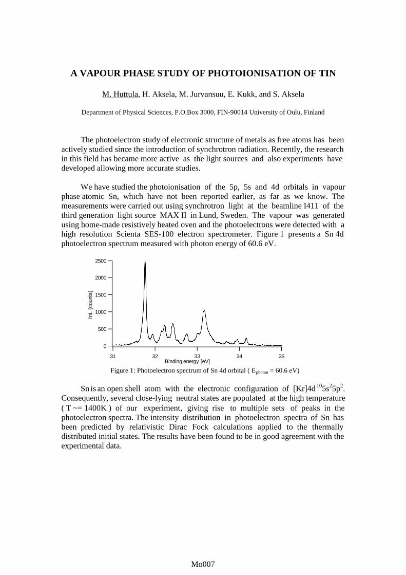
We have studied the photoionisation of the 5p, 5s and 4d orbitals in vapourphase atomic Sn, which have not been reported earlier, as far as we know. Themeasurements were carried out using synchrotron light at the beamline I411 of thethird generation light source MAX II in Lund, Sweden. The vapour was generatedusing home-made resistively heated oven and the photoelectrons were detected with ahigh resolution Scienta SES-100 electron spectrometer. Figure 1 presents a Sn 4dphotoelectron spectrum measured with photon energy of 60.6 eV.
Figure 1: Photoelectron spectrum of Sn 4d orbital ( Ephoton = 60.6 eV)
Sn is an open shell atom with the electronic configuration of [Kr]4d105s25p2.Consequently, several close-lying neutral states are populated at the high temperature( T ~= 1400K ) of our experiment, giving rise to multiple sets of peaks in thephotoelectron spectra. The intensity distribution in photoelectron spectra of Sn hasbeen predicted by relativistic Dirac Fock calculations applied to the thermallydistributed initial states. The results have been found to be in good agreement with theexperimental data.
2500
2000
1500
1000
500
0
Int.
[co
unts
]
3534333231Binding energy [eV]
Mo007Mo001Mo007Mo007Mo007

ELECTRONIC AND ENERGY TRANSFER IN H2O@Rg CLUSTERS(Rg=He, Ne, Ar)
A.V.Kanaev1#, L.Museur2, T.Laarman3, T.Möller3
1LIMHP CNRS, Université Paris-Nord, avenue J.B.Clément, 93430 Vill etaneuse, France2LPL CNRS, Université Paris-Nord, avenue J.B.Clément, 93430 Vill etaneuse, France
3HASYLAB at DESY, Notkestrasse 85, 22603 Hamburg, Germany
We have studied the electron and energy transfer in large RgN-clusters doped with H2O moleculeunder photoexcitation in the 9-30 eV range (140-40 nm). The mean cluster size is varied fromN=400 (argon) to 5⋅103 (neon) and ≥104 (helium). The reaction channel has been characterizedby fluorescence of neutral OH* and H* and ionic H2O
+* fragments. The measurements have beenperformed at the CLULU experimental station at HASYLAB. Rg-clusters have been prepared ina continuous free-jet expansion of a pure rare gas through an orifice-type nozzle of 40 µmdiameter or through a conical-shaped nozzle (D=200 µm, °= 42θ ). The nozzle is mounted on aliquid He cryostat and it can be cooled down until temperatures below 10K. RgN@H2O-clustershave been prepared by a crossbeam technique. Tunable SR (∆λ=0.05 nm) is focused on thedoped cluster. UV-visible fluorescence spectra are collected with a liquid nitrogen cooled CCDcamera installed after a monochromator (f=275 mm, 150 or 1500 l/mm gratings). Thebackground pressure is kept below 10-3 mbar during experiments by continuous pumping of theinteraction volume.
As it is well known, a free water molecule predissociates under the VUV excitation above 9.137eV yielding the OH(A-X) fragment emission. At higher excitation energy the fluorescence bandsbelonging to neutrals OH
**, H
* and ionized H2O
+* excited products appear. We have observed an
eff icient energy transfer from Rg-matrix to the doped H2O molecule. A strong difference in thereaction yield has been observed when going from free water molecule to that one embedded indifferent rare-gas clusters. The preddissociation channel is found to be unaffected by the heliumcluster environment. On the other hand, the ionization exit channel (H2O
+*) seems to be
suppressed inside both helium and neon clusters in favor of the fragmentation into neutralexcited products. Additionally, the neon cluster strongly affects the neutral reaction channel. At17.7 eV excitation instead of strong Balmer series H
*(β,γ,δ,ε), which is characteristic of a free
water molecule and of H2O@HeN clusters, only the intense OH(A-X) emission has beenobserved. The same result has been observed in experiments with H2O@ArN clusters. It has beenfound that higher energy excitation induces higher vibrational excitation of the OH
*(A)
fragment; the rotational temperature in the same time is lower. Our calculations show thatOH
*(A) is rotationally thermalyzed inside Ne-clusters with the temperature as low as 10K. A
bimodal J-distribution of the OH*(A) fragment has been found in Ar-clusters. This effect is
attributed to a different geometric position of the H2O molecule in/on the Rg-cluster. Thisdifference is apparently related to the Rg-Rg and Rg-H2O pair potentials. In contrast to the caseof He or Ne clusters, water molecule does not entry the Ar-cluster and takes the surface site.
The financial support of the project II-98-026EC by the EU program is kindly acknowledged.
# E-mail : [email protected]
Mo008Mo001Mo008Mo008Mo008

CAGE EFFECT IN PHOTODISSOCIATION OF H2O MOLECULE INAr-CLUSTERS
A.V.Kanaev1#, L.Museur2, T.Laarman3, T.Möller3
1LIMHP CNRS, Université Paris-Nord, avenue J.B.Clément, 93430 Vill etaneuse, France2LPL CNRS, Université Paris-Nord, avenue J.B.Clément, 93430 Vill etaneuse, France
3HASYLAB at DESY, Notkestrasse 85, 22603 Hamburg, Germany
We study a caging of an electronically excited fragment OH*(A) after VUV excitation of rare-gasclusters (Rg=He,Ne,Ar) doped with H2O molecule. The measurements have been performed atthe CLULU experimental station at HASYLAB. Our results indicate that by using the standardcrossbeam technique we are unable to achieve the bulk site doping of the Ar-cluster by a watermolecule. This is apparently related to the difference in Ar-Ar and Ar-H2O interaction potentials.In order to overcome this limitation we have employed a three-crossbeam technique. The conicalnozzle (D=200 µm, °= 42θ ) was a source of large host clusters made of light atoms (Ne in ourcase). The first crossbeam dopes the NeN-clusters (N~5000) with water molecules, which take thesites in the interior of the cluster; the second crossbeam dopes the H2O@NeN cluster by Ar-atoms.
Since the temperature of Ne-clusters is very low (~10K, the temperature of Ar-clusters is ~35K),Ar-atoms freeze around H2O molecule inside the Ne-cluster and the low temperature prohibitsthe structural rearrangement in the embedded H2O@Arm cluster. Because of its small size and alow concentration of the doped H2O@Arm@NeN clusters, we used to excite them in the firstexcitonic band of the large host Ne-cluster at ~70 nm. Additional experiments performed withthe Ar@NeN and H2O@ArN clusters show that an eff icient energy transfer exists between theexcited host cluster matrix and the doped center in each case. These experiments give evidencethat after Ne-cluster matrix excitation the energy can be eff iciently transferred onto the watermolecule surrounded by Ar-atoms in the composed multishell cluster.
We have measured the OH(A-X) fluorescence spectra as a function of the Ar-crossbeamintensity, which has been done by a variation of the stagnation pressure (PAr). The size of the Ar-cluster formed around a water molecule is proportional to PAr. We have observed a strongdecrease of the OH*(A) emission intensity at the argon crossbeam pressure above 20 mbar. Asecond decrease of the intensity seems to exist at the pressure higher than 70 mbar. Except forthese points the fluorescence band intensity exhibits only a small variation. We attribute thisfeature to the cage effect of Ar-atoms on the H2O photodissociation fragments: being caged, OH*
and H have high probabilit y to be quenched through the H2O(X) potential. We believe that at thecrossbeam pressure of 20 mbar the first shell of Ar-atoms (m1=12) is almost formed around thewater molecule. The subsequent decrease of the emission intensity above 70 mbar may indicatethe formation of the second atomic shell (m2=54) of the Arm cluster. These studies can be helpfulin understanding of the solid structure formation in small rare gas clusters.
The financial support of the project II-98-026EC by the EU program is kindly acknowledged.
# E-mail : [email protected]
Mo009Mo001Mo009Mo009Mo009

Measurements of double photoionization (γ,2e) in atomic Sr
J.B. West1, K.J. Ross2, H.J. Beyer3, A. De Fanis2,5 and H. Hamdy4
1 Daresbury Laboratory, Daresbury, UK2 Southampton University, Southampton, UK
3 Stirling University, Stirling, UK4 Cairo University, Beni-Suef, Egypt
5 present address: IMRAM, Tohoku University, Sendai, Japan
We present measurements of the differential cross section for double photoionization(DPI) of Sr vapour, collected at the Daresbury Synchrotron Radiation Source. Electrons fromthe 5s2 shell are analyzed in energies by electrostatic hemispherical analyzers and detected incoincidence; the radiation energy is tuned at an energy resonant with the 4p→4d excitation(25.25 eV).
Measurements are reported for equal (4.26 eV) and unequal (6.76 and 1.76 eV) energies ofthe two electrons; both electrons are observed in the plane perpendicular to the photon beam.One electron is detected either parallel (θ1=0) or perpendicular (θ1=90) to the E field of thepartly linearly polarized radiation.
In figure 1 the coincidence pattern for equal electron energy is displayed as a function ofthe relative angle of emission. It is clear that the present data show evidence for antiparallelemission of the two electrons. This is against the selection rules imposed by the dipoleapproximation and 1S0
e symmetries of the initial and doubly charged final states, that predict anode for antiparallel emission of the two electrons. We tentatively explain this anomalous resulteither by the presence of two unresolved maxima close to and either side of θ12= 180°, or by a 3Pcomponent in the 4p →4d resonance.
90 120 150 180 210 240 2700
θ1=90
o
Relative Angle θ12
0
θ1=0ohν=25.25 eV (Sr 4p 4d)
E1=E2=4.26 eV
Figure 1: Coincidence pattern of electrons emitted with thesame kinetic energy in the plane perpendicular to the photonbeam. One electron is emitted either parallel (θ1=0) orperpendicular (θ1=90) to the polarization of the radiation.
Mo010Mo001Mo010Mo010Mo010

PHOTON YIELD FROM SOLID KRYPTON AND XENON AT THE EDGEOF EXCITON ABSORPTION
A.N. Ogurtsov1,2, E.V. Savchenko1, E. Gminder3, S. Vielhauer3, G.Zimmerer3
1 Verkin Institute for Low Temperature Physics & Engineering of the National Academy of Sciences of Ukraine,Lenin Avenue 47, Kharkov 61164, Ukraine
2 Kharkov State Academy of Railway Transport, Majdan Feuerbacha 7, Kharkov 61050, Ukraine3 II. Institut für Experimental Physik der Universität Hamburg, Luruper Chaussee 149, Hamburg 22761, Germany
Spatial and temporal fluctuation of the lattice potential caused by phonons anddeformability of the lattice induce the broadening and existence of low-energy tail of theexcitonic absorption spectra and dressing of excitons by phonons, which leads to self-trapping ofexcitons [1]. Relaxation of electronic excitation in rare gas solids results in formation of varietyof trapped centers [2]. Recently the effect of molecular trapped centers associated with latticeimperfections on the shape of excitation spectra of excitons has been reported [3]. In the presentstudy the time-resolved fluorescence excitation spectroscopy in the VUV has been used to studythe effects of crystal lattice perturbations on relaxation of excitons in solid Kr and Xe. Theexperiments were performed at the SUPERLUMI experimental station at HASYLAB, DESY,Hamburg. The excitation spectra of solid Xe and Kr were measured in the energy range of n=1exciton absorption. Figure 1 shows the decay curve of free-exciton luminescence and selectedexcitation spectra of solid Xe, which were measured in three time windows ∆t with delay δtrelative to excitation synchrotron pulse: 1) window W1 (∆t=1.5 ns, δt=0 ns); 2) window W2(∆t=2 ns, δt=1 ns); and 3) window W3 (∆t=32 ns, δt=5 ns). The effects of interaction of freeexcitons with neutral and charged intrinsic trapped centers are discussed.
Figure 1: Decay curve of free-exciton luminescence (excitation with hν=8.86 eV) and excitation spectrameasured at photon energies (1) 8.05 eV, (2) 8.36 eV, (3) 8.67 eV within time windows W1, W2, W3indicated above the decay curve by rectangles.
References
[1] M. Ueta, H. Kanzaki, K. Kobayashi, Y. Toyozawa, and E. Hanamura, Excitonic Processesin Solids, Springer-Verlag Ser. in Solid-State Sciences vol.60, Berlin 1986.
[2] A.N. Ogurtsov, E.V. Savchenko, M. Kirm, B. Steeg, G. Zimmerer, J. Electron Spectrosc.Relat. Phenom. 101-103, 479 (1999).
[3] A.N. Ogurtsov, E.V. Savchenko, J. Low Temp. Phys. (2001) in press.
7,5 8,0 8,5 9,010-3
10-2
10-1
100
321
W1
7,5 8,0 8,5 9,00,00
0,02
0,04
0,06
Photon energy of excitation, eV
321
W2
7,5 8,0 8,5 9,00,000
0,005
0,010
321
W3
0 10 20 30100
102
104
Inte
nsity
, a.u
.
Time, ns
W3W2W1
Mo011Mo001Mo011Mo011Mo011

First observation of resonant Auger decay of Xe 3d-16p to Xe+4d-2nl
A. De Fanis1, K. Okada2, Y. Shimizu3, M. Okamoto4, K. Kubozuka5,N. Saito6, I. Koyano5, and K Ueda1
1 IMRAM, Tohoku University, Aoba-ku, Sendai 980-8577, Japan2 Department of Chemistry, Hiroshima University, Higashi-Hiroshima 739-8526, Japan
3 Institute for Molecular Science, Okazaki 444-8585, Japan4 Department of Physics, Sophia University, Tokyo 102-8554, Japan
5 Department of Material Science, Himeji Institute of Technology, Kamigori, Hyogo 678-1297, Japan6 Electrotechnical Laboratory, Tsukuba 305-8568, Japan
The resonant Auger (RA) process, where a system with an inner-shell electron promoted toan unoccupied orbital decays by electron emission, has received considerable attention duringthe last decade [1]. Recent improvements on optical resolution of soft X-ray monochromatorsinstalled in the undulator beamlines of synchrotron radiation facilities made it possible toobserve RA emission spectra (RAES) with overall widths narrower than the core-hole lifetimewidths.
We have recorded, for the first time, angle-resolved RAES arising from the transitions Xe3d5/2
-16p3/2 → Xe+ 4d-26p3/2 and 4d-27p3/2 in the kinetic energy region of 520-530 eV. Theexperiments were carried out on beamline 27SU at SPring-8 using an ultrahigh-resolutionhemispherical electron spectrometer (Gammadata-Scienta SES2002). The overall widths wereless than 100 meV, far narrower than the core-hole lifetime width of 500 meV. At this resolutionthe splitting due to the coupling between the Rydberg electron (6p3/2 or 7p3/2) and the doubly-charged ionic core (4d-2) were partly resolved.
The energies of RAES are ~ 4.7 eV lower than those of the corresponding normal Augeremission spectra. With the help of the spectator model [2], where the wave function of theexcited electron is assumed not to change during the de-excitation process, the asymmetryparameters β and branching ratios for each transition in RAES can be related to those for thecorresponding normal Auger transitions. The corresponding M5N4,5N4,5 normal Auger transitionshave been studied both experimentally [3] and theoretically [4]. Comparing the asymmetryparameters β and branching ratios measured for the RA lines with those predicted from thenormal Auger data, we could assign most of the observed RA lines.
This experiment was carried out with the approval of the SPring-8 program advisorycommittee and supported in part by Grants-in-Aids for Scientific Research from the JapanSociety for the Promotion of Science and by the Matsuo Foundation. We are grateful to the staffof SPring-8 for their help.
References
[1] G.B. Armen, H Aksela, T. Åberg and S. Aksela, J. Phys. B 33, R49 (2000).[2] U. Hergenhahn, B Lohmann, N.M. Kabachnik and U. Becker, J. Phys. B 26, L117 (1993).[3] J. Karvonen et al.; Phys. Rev. A 59, 315 (1999).[4] J. Tulkki, N.M. Kabachnik and H. Aksela; Phys. Rev. A 48, 1277 (1993).
Mo012Mo001Mo012Mo012Mo012

HIGH-RESOLUTION C 1s and F 1s RESONANT AUGER EMISSIONIN THE TETRAFLUOROMETHAN MOLECULE
A. De Fanis1, N. Saito2, K. Okada3, M. Okamoto4, K. Hoshino4, M. Kitajima4,K. Kubozuka5, M. Machida5, I. Koyano5 and K. Ueda1
1 IMRAM, Tohoku University, Sendai 980-8577, Japan2 Electrotechnical Laboratory, Tsukuba 305-8568, Japan
3 Department of Chemistry, Hiroshima University, Higashi-Hiroshima 739-8526, Japan4 Department of Physics, Sophia University, Tokyo 102-8544, Japan
5 Department of Material Science, Himeji Institute of Technology, Kamigori, Hyogo 678-1297, Japan
We recorded angle-resolved resonant photoemission spectra of CF4 in the excitationregions of C 1s and F 1s to the lowest antibonding molecular orbitals σ*. The measurementshave been carried out on beamline 27SU at SPring-8, using a high-resolution electron energyanalyser (Gammadata SCIENTA SES2002). In figure 1, the valence photoemission spectrum ofCF4 recorded at the photon energy resonant with the C 1s −> σ* excitation is compared with thenon-resonant spectrum; both collected in the direction parallel to the polarisation vector. Theoverall energy resolution was <50 meV. The tail of the C 2T2 band towards high binding energyin the resonant spectrum is evidence of enhanced nuclear motions in the core-excited states; nosuch enhancements are observed for the D 2A1 bands. Vibrational structure is not resolved in thehigh binding energy part of the C 2T2 band, due to overlap of non-totally symmetric vibrationalmodes with the totally symmetric breathing mode; the non-totally symmetric vibrations arehighly excited through vibronic couplings in the core-excited states. This effect is present alsofor F 1s excitation but is less dramatic, reflecting that the nuclear motion is less significant in theF 1s-1σ* state, due to shorter lifetime, than in the C 1s-1σ* state.
2 5 2 4 2 3 2 20
1
2
3
4
5
Inte
ns
ity (
arb
itra
ry u
nits
)
C2T 2
D2A
1 C F4 photoem iss ion
B ind ing en erg y (e V)
hν =298.21 eV (C 1s t2σ *)
hν = 295.10 eV (non resonant)
Figure 1: Photoemission spectrum of CF4 recorded at photonenergy resonant with the C 1s→t2σ∗ excitation, compared with anon-resonant spectrum.
Mo013Mo001Mo013Mo013Mo013

NEAR-THRESHOLD HIGH-RESOLUTION O 1s PHOTOELECTRONSPECTROSCOPY OF CO2
A. De Fanis1, K Ueda1, M. Kitajima2, M. Okamoto2, M. Hoshino2, H. Tanaka2, K. Okada3,N. Saito4, I. Koyano5, A. Pavlychev6, and D. Yu. Ladonin6
1 IMRAM, Tohoku University, Sendai 980-8577, Japan2 Department of Physics, Sophia University, Tokyo 102-8554, Japan
3 Department of Chemistry, Hiroshima University, Higashi-Hiroshima 739-8526, Japan4 Electrotechnical Laboratory, Tsukuba 305-8568, Japan
5 Department of Material Science, Himeji Institute of Technology, Kamigori, Hyogo 678-1297, Japan6 Institute of Physics, St. Petersburg State University, 198904, St. Petersburg, Russia
The O 1s ionization spectra of CO2 are dominated by the 5σg* and 4σu* shape resonances,centered at ~2 and ~20 eV above the O 1s ionization threshold [1]. If one adopts one-particledescription, valence electrons can be regarded as independent spectators in the excitation of theO 1s electron to the 5σg* and 4σu* molecular orbitals. Double excitations and changes in nuclearmotion, however, often accompany the inner-shell excitations and open questions to the validityof the one-particle description [2]. In the present study we focus on O 1s photoemission at the5σg* shape resonance just above the threshold.
Coupling between the slow photoelectron and the nuclear motion is an intriguingtheoretical problem because in molecular species, in addition to the post-collision interaction [3](PCI) among photoelectrons, Auger electrons and ions, the coupling between the photoelectronand vibrations in the residual singly charged ion are also present. The calculations are based onthe quasi-atomic and optical potential concepts [2] that allow us to predict essential contributionof the quasi-elastic component to the photoelectron band, such as population of high vibrationalcomponents and non-Franck-Condon distribution of the vibrational branching ratios, as well asasymmetry parameters β of these vibrational components.
Experimentally, we collected for the first time angle-resolved O 1s photoelectron spectraof CO2 in the region of the 5σg* shape resonance. Experiments were carried out on beamline27SU at SPring-8 using a high-resolution electron spectrometer (Gammadata-Scienta SES2002).The present high-resolution (∆E ~ 140 meV) allows us to resolve the vibrational structure, whichis dominated by the progression of the antisymmetric stretching mode (313 meV separation), thatbecomes allowed due to symmetry breaking [1]. Vibrational branching ratios and asymmetryparameters β are extracted from the present data by fitting the spectra with PCI-distortedlineshape [3]. The present results show significant non-Franck-Condon behaviour of thebranching ratios and essential changes in asymmetry parameters β across the 5σg* shaperesonance. Semi-quantitative agreement between the measurements and calculations are found.
References
[1] K. Maier et al., Phys. Rev. A 58, 3654 (1998).[2] A. A. Pavlychev, J. Phys. B 32, 2077 (1999).[3] M. Yu. Kuchiev and S.A. Sheinarman, Sov. Phys. JETP 63, 986 (1986).
Mo014Mo001Mo014Mo014Mo014

90 120 150 180
Inte
nsity
(ar
b. u
nits
)
N-O angle (degree)
O1s-13π*
O1s-13sσ
Figure 1. The distributions for the angle between the momenta of the terminal N+ and the O+ ions for the triple fragmentation through the O1s
3π*
resonance (solid circles) and the O1s
3sσ* resonance (open circles).
DEFORMATION OF O1s EXCITED N2O STUDIED BY MOMENTUM MEASUREMENTS OF FRAGMENT IONS
N. Saito1, K. Kubozuka2, M. Machida2, H. Chiba3, A. De Fanis3, Y. Muramatsu3, K. Okada4,
M. Lavollée5, M. Hattass6, A. Czasch6, H. Schmidt-Böcking6, K. Ueda3 and I. Koyano2
1 National Institute of Advanced Industrial Science and Technology, Tsukuba 305-8568, Japan
2 Department of Material Science, Himeji Institute of Technology, Kamigori, Hyogo 678-1297, Japan 3 IMRAM, Tohoku University, Sendai 980-8577, Japan
4 Department of Chemistry, Hiroshima University, Higashi-Hiroshima 739-8526, Japan 5 LURE, Bat. 209d, Centre Universitaire Paris-Sud, F-91898, Orsay Cedex, France
6 Institut für Kernphysik, Universität Frankfurt, D-60486 Frankfurt, Germany
The equivalent-core model predicts that N2O in the O1s-13π* state is bent. The equilibrium bond angle of N2O in the O1s-13π* state is calculated to be 111.7 degrees. The angular distributions of fragment ions from N2O in the O1s-13π* state also suggest that the molecule is bent [1,2]. We measured the momenta of three fragment ions (N+, N+, and O+) from N2O3+ following O1s
3π* photoexcitation in N2O, in order to clarify how the molecule deforms
at the π* resonance. The experiments were performed on the c-branch of BL27SU at SPring-8. The momenta of three fragment ions were measured in coincidence using a time-of-flight mass spectrometer fitted with a position sensitive detector and a supersonic jet of N2O. Figure 1 shows the distributions for the angle between the momenta of the terminal N+ and the O+ ions for the triple fragmentation (Nt-O angle) through the O1s
3π* resonance (solid circles) and the
O1s
3sσ* resonance (open circles). Ions are collected for emission over 4π steradian. Because only the simultaneous three-body break-up contains information about the geometry of the parent ions, in the off-line analysis we selected only these events, rejecting those where the fragmentation proceeds via sequential steps. The Nt-O angle distribution at the O1s-13sσ* state is peaked at about 165 degrees. This angle corresponds to the angle due to the ground-state zero-point vibration. The Nt-O angle distribution through the O1s
3π* excitation
shows a broad peak centered at about 162 degrees, with a long tail towards lower angles. The comparison between the two angle distributions clearly shows that the molecule is more bent at the O1s-13π* state than at the O1s-13sσ* state.
References [1] J.D.Bozek, N.Saito and I.H.Suzuki, J.
Chem. Phys. 98, 4652 (1993). [2] J.Adachi, N.Kosugi, E.Shigemasa and
A.Yagishita, J. Chem. Phys. 102, 7369 (1995).
Mo015Mo001Mo015Mo015Mo015

THE PHOTOABSORPTION CROSS SECTION OF Kr IN THE SUB keV ENERGY REGION
Norio Saito and Isao H. Suzuki
Electrotechnical Laboratory, AIST, Umezono, Tsukuba Ibaraki 305-8568, Japan
It is important to obtain reliable data on photoabsorption cross sections of rare gas atoms in detail for clarification of fundamental interaction between photon and material. However, there are limited number of data reported about those cross sections in the soft X-ray region [1-3]. We report here the precise photoabsorption cross section for Kr in the soft X-ray region using a multi-electrode ion chamber [4]. The spectral purity of monochromatized synchrotron radiation was improved through the low energy operation of the storage ring (TERAS) and through inserting a thin metal foil into the incident photon beam. The ion chamber includes 4 electrodes for collecting photoions. The length of the two electrodes mounted near the entrance window is 100mm and that of the other two is 500 mm. The measurement using the electrodes with different lengths enables us to remove the contribution of the stray light and higher orders in the photoion-currents, as follows. The photoion-current (i) of the electrode from the 1st order light is given by the following equation.
i = enI exp(−dpσ) (1 − exp(−Lpσ) ). (1)
In this equation, I, e, n, d, L, p, and σ denotes the incident photon intensity, the elementary charge, the number of electrons totally produced from a photon in the chamber, the length of insensitive region in front of the electrode, the electrode length, the gas density, and photoabsorption cross section, respectively. The photocurrents from the stray and higher orders are also given by Eq.(1) with different I, n, and σ. The photoabsorption cross section is obtained by fitting Eq.(1) to the photocurrents of the 4 electrodes measured with several gas densities, which include contributions from the first order light, stray and higher orders. Figure 1 shows the photoabsorption cross section of Kr in the region of 80 eV to 1200 eV, together with literature data [1,3]. Although the present results are close to the reported values, there are seen some discrepancies between the previous data and the present ones.
References [1] B.L.Henke et al, Atom. Data Nucl.
Data Tables, 54, 181 (1993). [2] G.V.Marr & J.B.West, Atom. Data
Nucl. Data Tables, 18, 497 (1976). [3] J.B.West & J. Morton, Atom. Data
Nucl. Data Tables, 22, 103 (1978). [4] N.Satio and I.H.Suzuki, J. Electron
Spectrosc. Relat. Phenom. 101-103, 33 (1999)
100 10000.1
1
10 3p 3s3d
present Henke [1] West [3]P
hoto
abso
rptio
n (M
b)
Photon Energy (eV)
Figure 1 Photoabsorption cross section of Kr.
Mo016Mo001Mo016Mo016Mo016

).()( ppn δγ +=
W-VALUES OF RARE GAS ATOMS IN THE SUB -keV X-RAY REGION
Isao H. Suzuki and Norio Saito
Electrotechnical Laboratory, AIST, Umezono, Tsukuba, Ibaraki 305-8568 Japan
The W-value is defined as average energy required to produce an electron-ion pair in a gas system by ionizing radiation. This value is a fundamental constant regarding interaction between the radiation and an atom and/or a molecule. Above several keV, the W-value is insensitive to quality and energy of the radiation. On the other hand, this value for low-energy radiation varies around the inner-shell ionization threshold of the atom [1]. In the present study, photon W-values for rare gas atoms have been obtained in an absolute scale in the region of 90-1000 eV using a multiple electrode ion chamber. Synchrotron radiation from the TERAS electron storage ring was dispersed using a Grasshopper monochromator. Thin filters and low energy operation of the ring were used for improving spectral purity [2]. Monochromatic soft X-rays entered the ion chamber, and then ions produced in the chamber were collected with electrodes, and these photoion currents were measured as a function of the gas density. The photon W-value, Wp, is given with
In this equation, N denotes the number of electrons totally produced in the sufficiently voluminous gas system and Ep is the photon energy. The number of electrons, n, totally produced under a gas density of p, is the summation of the initial ionization and the secondary ionization effects in the chamber.
In eq.(2), γ denotes the number of electrons ejected from the atom absorbing a photon, and δ is the number of electrons secondarily produced through the collision between ambient atoms and emitted electrons. Photon W-values have been determined from ion currents at sufficiently low and high gas densities in the ion chamber. Figure 1 shows photon W-values of Kr in the soft X-rays region (solid circles). The photon W-value shows a peak just below the Kr 3d ionization threshold, increases steeply just above this threshold and becomes nearly constant in higher energies. The open squares indicate data of the W-value for electrons. The solid curve denotes the result derived from a model here proposed, which takes into account atomic shell effects in the initial photo-ionization step. [1] N.Saito and I.H.Suzuki, Chem.
Phys. 108, 327(1986). [2] N.Saito and I.H.Suzuki, J.
Electron Spectrosc. Relat. Phenom., 101-103 , 33 (1999).
(2)
(1) NEW pp =
100 1000
25
30
35
3s3p3d
W-v
alue
Photon Energy (eV)
Fig.1: Photon W-value of Kr.
Mo017Mo001Mo017Mo017Mo017

FINE-STRUCTURE SELECTIVITY OF NEUTRAL DISSOCIATIONOBSERVED IN O2
H. Liebel, R. Müller-Albrecht, S. Lauer, F. Vollweiler, A. Ehresmann, H. Schmoranzer
Fachbereich Physik, Universität Kaiserslautern, D-67653 Kaiserslautern, Germany
The knowledge about the decay mechanisms of superexcited states - states whose internalenergy exceeds the lowest ionization energy - is presently far away from being complete [1].Superexcited states have several possible decay paths such as formation of electrons andmolecular ions through autoionization and of neutral atoms through dissociation where theirinternal energy is distributed among the arising fragments. If a neutral fragment emerges fromthe dissociation process in an excited state which can decay further by fluorescence, photon-induced fluorescence spectroscopy (PIFS, see, e.g., [2]) - measuring the exciting-photon energydependence of the fluorescence spectrum - can be applied.
Synchrotron radiation from BESSY I, Berlin, was monochromatized by a 3m-normal-incidence monochromator (3m-NIM-2) equipped with a 2400 lines/mm grating and focused intoa liquid nitrogen cooled target gas cell. The target cell contained molecular oxygen at a pressureof 40 µbar. For detecting the atomic fragment fluorescence a VUV-photomultiplier with a CsIcoated photocathode and a MgF2 entrance window was used. The exciting-photon fluxtransmitted through the gas cell was monitored and enabled to determine the absorption crosssection simultaneously with the fluorescence spectrum of the dissociation fragment as a functionof the exciting-photon energy. For slit widths of 20 µm a bandwidth of the exciting radiation of0.9 meV was achieved. The exciting-photon energy was varied from 14.6 eV to 16.3 eV in stepsof typically 0.2 meV.
The absolute O2 absorption cross section and the absolute emission cross section for
OI (4So)3s 3So1 →2p4 3PJ were investigated using PIFS in the vicinity of the threshold for neutral
dissociation leading to the OI (4So)3s 3So1 state with a very narrow bandwidth (0.9 meV) of the
exciting radiation. Both cross sections are found to be structured by the vibrational progressions
of the O2 Rydberg states I, I' and I" which converge to the a 4Πu state of O+2. In the present
investigation the fine-structure components of the vibrational progressions of the I, I‘ and I‘‘states were resolved for the first time in a fluorescence excitation spectrum. A certain degree ofselectivity in the population of fine-structure components was observed in the fluorescenceexcitation spectra.
References
[1] Y. Hatano, Phys. Rep. 313, 109 (1999).
[2] H. Liebel, S. Lauer, F. Vollweiler, R. Müller-Albrecht, A. Ehresmann, H. Schmoranzer,G. Mentzel, K.-H. Schartner, O. Wilhelmi,Phys. Lett. A 267, 357 (2000) and further references therein.
Mo018Mo001Mo018Mo018Mo018

AUGER ELECTRON SPECTRA OF Kr2p HOLESUSING MONOCHROMATIC SOFT X-RAYS
K. Kamimori1, I.H. Suzuki2, K. Okada1, J. Sasaki1, S. Nagaoka3, Y. Shimizu3,H. Yoshida1, A. Hiraya1, H. Ohashi4, Y. Tamenori4, and T. Ibuki5
1 Hiroshima Univ., 2 Electrotechnical Lab., AIST, Tsukuba, Japan,
3 Inst. Molecular Sci., 4 JASRI, 5 Kyoto Univ. Education
Radiationless transitions are of great interest because of the dominant mode for de-excitationof atomic inner-shell vacancies and of the main factor for determining hole-state lifetimes [1-3].Rates and energies of Auger and Coster-Krönig transitions provide stringent tests of theoreticalmodels on electronic coupling, correlation and relativistic effects. Auger electron spectra fromKr2p hole states were studied using techniques of electron beam excitation and of ion beamexcitation [1-2], and then they were compared with results calculated by several theoreticalmethods. Since experimental data were obtained at a moderate resolution, however, there hasbeen obtained no clear conclusion on branching ratios for the multiplet final states populateddensely. In the present study normal Auger spectra of 2p holes have been measured usingmonochromatized synchrotron radiation and a high resolution electron spectrometer.
Measurements were performed in the c branch of the undulator beamline 27SU at SPring-8.Ion yield spectra were measured in the region of the ionization thresholds of Kr2p electrons forconfirming the energy of the incident photon beam. Electrons emitted from the sample gas wereobserved at the direction parallel to the photonpolarization with the hemispherical electron energyanalyzer (SES 2002). Auger spectra of L2M45M45
and L3M45M45 are shown at the upper part and thelower in Fig. 1, respectively. Thin dotted curvesdenote peaks calculated for individual final statesusing Gaussian and Lorentzian shapes forinstruments and lifetime widths. Thick curves arethe summation of all thin curves. It is seen that thepresent result has been obtained with the resolutionhigher than the reported data [1-2]. Energies for thespectral peaks here obtained agree with the previousresults at most final states. However relativeintensities for several peaks are appreciably differentfrom the reported ones.
[1] J.C. Levin et al., Phys. Rev. A 33, 968(1986).[2] H. Aksela et al., Phys. Rev. A 22, 1116(1980).[3] S. Nagaoka et al., J. Phys. B 33, L605(2000).
Fig.1: Photoexcited Auger electron spectra of Kr.
a)L2M45M45, b)L3M45M45
1445 1450 1455 1460 1465 1470 14752
4
6
8
hν = 1720.5 eV
1S0
1G43P0,1+1D2
3P2
3F2
3F3
3F4
b ) L3M45M45
Rela
tive
Inte
nsit
y (
arb.
uni
ts )
Kinetic Energy ( eV )
1500 1505 1510 1515 1520 1525
3
4
5 hν = 1779.7 eV
1S0
1G43P0,1+1D2
3P2 3F2 3F3 3F4
a ) L2M45M45
Mo019Mo001Mo019Mo019Mo019

EXPERIMENTAL STUDIES AND AB INITIO CALCULATIONS ONCHARACTERISTICS OF THE C STATE OF SF2 RADICAL
Xiaoguo Zhou, Zhenyu Sheng, Shuqin Yu
Department of Chemical Physics, University of Science and Technology of China, Hefei, 230026, P.R.Chinaand
Abdus Salam International Centre for Theoretical Physics, 34014 Trieste, Italy
SF2 radicals were generated by a pulsed dc discharge in the mixture gas beam of SF2 and Ar.The (2+1) resonance-enhanced multiphoton ionization (REMPI) excitation spectroscopy of SF2
radical was obtained between 325 and 365 nm. The SF+ ion signals were also observed in thesame wavelength range. These results are shown in Fig.1.The excitation spectrum of SF2 can beassigned a two-photon resonant transition from the ground state to 1
1BB (4s) Rydberg state andC state.
3 3 0 3 3 5 3 4 0 3 4 5 3 5 0 3 5 5 3 6 0 3 6 5
( b )
m/z
=7
0 S
ign
al
m/z
=5
1 S
ign
al
3 3 0 3 3 5 3 4 0 3 4 5 3 5 0 3 5 5 3 6 0 3 6 5
( a )
4 s ( 1 05 )
4 s ( 1 02 )4 s ( 1 0
3 )4 s ( 1 0
4 )4 s ( 1
0
1 )
C ( 1 02 ) C ( 1 0
1 ) C ( 0 00 )
L a s e r W a v e l e n g t h ( n m )
Fig. 1 Composite (2+1) REMPI excitation spectrum between 330 and 365 nm,which were carried by a) SF2
+ (m/z 70) and b) SF+ (m/z 51)
Ab initio calculations were carried out using GAUSSIAN98 programs. The optimizedgeometries, harmonic vibrational frequencies, excited energies and the PESs of the ground stateand several excite states of SF2 calculated at CIS/Aug-cc-PVTZ. It is reasonable to assign thesecond 1
1B state as the 11BB (4s) Rydberg state, and the 1
1 A state as the C state in experiment. Italso shows that the 1
1BB states of SF2 radical are a bonding state and the 11AC state is a valence
state with predissociative characteristics, which are in agreement with experiment,.As shown in Fig.1, SF2
+ and SF+ signals can be observed in the same wavelength. The traceof SF+ began at 350 nm, close to the band origin of the C state. According to our experimentsand calculations, we think that the m/z 51 signal originates from the REMPDI processes. That is,the 1
1AC state derives from the ground state by two-photon resonant excitation, then dissociatesto the neutral SF radical, which can ionize after absorbing one laser photon:
21
22
21
22 3581 bbaaL SF2 ( 1
1AX ) → hv2 11
11
22
21
22 43581 bbbaaL SF2 ( 1
1AC ) � SF →hv SF+ .
References
[1] R. D. Johnson III, J. W. Hudgens, J. Phys. Chem. 94 (1990) 3273[2] Q. X. Li, J. N. Shu, Q. Zhang, S. Q. Yu, et. al. J. Phys. Chem. 102 (1998) 7233[3] X. Zhou, Q. Li, Q. Zhang, S. Yu, et. al. J. of Electron Spectro. Relat. Pheono. 108 (2000) 135
Mo020Mo001Mo020Mo020Mo020

FRAGMENT-ION DESORPTION FROM SULFUR-CONTAINING AMINO ACIDS BY LOCALIZED CORE-LEVEL EXCITATION
Yuji Baba, Tetsuhiro Sekiguchi and Iwao Shimoyama
Synchrotron Radiation Research Center, Japan Atomic Energy Research Institute
Tokai-mura, Naka-gun, Ibaraki-ken, 319-1195, Japan
Irradiation of VUV or X-ray on solid surface induces decomposition and desorption triggered by the core-level excitation. Due to the localized nature of core levels, many examples for site-specific and element-specific reactions have been reported. Generally, such specific reaction is clearly observed in thin films of adsorbed molecules. In condensed phase or bulk materials, the specificity is weakened due to the secondary electrons which induce non-specific reaction. Here we present clear examples where the localized core-level excitation in bulk biological molecules induces desorption of specific fragment ions.
The molecules investigated are sulfur-containing amino acids, such as cystine, cysteine and methionine. The fragmentation pattern in these molecules by the core-level excitation is of great importance in elucidating the effect of radiation on living things [1]. The samples were pressed into pellets, and irradiated in ultra-high vacuum by synchrotron beam around the sulfur K-edge. The desorbed ions were detected in-situ by a quadrupole mass spectrometer. For comparison, the desorption of fragment-ions by valence excitation was measured using low-energy electron gun. The photoelectron and Auger electron spectra were also measured by a hemispherical electron energy analyzer.
For valence excitations, various fragment ions containing carbon, nitrogen and oxygen were desorbed, but the core-to-valence resonant excitations at the sulfur K-edge induced only S+ ion desorption. Figure 1 shows the photon-energy dependencies of the total electron yield TEY and S+ ion yield for cystine which has a disulfide bond. When we compare the jump ratio (Ion/Ioff) defined as the peak intensity ratio between off-resonance and on-resonance energies, the Ion/Ioff ratio for the TEY curve is 1.5, but the value for the S+ yield curve exceeds 20. The results clearly show that the S+ desorption is caused not by the secondary electrons but the direct core-to-valence resonance localized at the sulfur atom. As to the photon-energy dependencies, the TEY curve exhibits the double structure of the S 1s * resonance peak, corresponding to the excitations into the * state localized at the S-S bond (peak A) and that localized at the S-C bond (peak B) [2]. While the S+ yield curve has single structure around the peak A. The results also suggest that the direct core-to-valence resonant excitation localized at the S-S bond has high efficiency for the S+ desorption. The mechanism of desorption is discussed on the basis of the photoelectron and Auger electron spectra. References [1] A. Yokoya, K. Kobayashi, N. Usami and S. Ishizaka, J. Rad. Res. 32, 215 (1991). [2] Y. Baba, K. Yoshii and T.A. Sasaki, J. Chem. Phys. 105, 8858 (1996).
���� ���� ���� ���� ����
�
��
��
��
��
���
���
���
���
���
%[UVKPG
+QP
+QHH
$���5��U �5�%�
#���5��U �5�5�
6';
5
�
�
+PVGPUKV[�CTD�WPKVU�
2JQVQP�GPGTI[�G8�
Figure 1: Photon-energy dependencies of the TEY and S+ yield around S K-edge for cystine.
Mo021Mo001Mo021Mo021Mo021

MOLECULAR SIZE EFFECT ON THE SITE SPECIFICFRAGMENTATION OF THE N AND O K-SHELL EXCITED
CH3CO(CH2)nCN (n=0, 1, 3) MOLECULES
T. Ibuki 1, K. Okada 2, S. Tanimoto 2, T. Gejo 3, and K. Saito 2
1 Kyoto University of Education, Kyoto 612-8522, Japan
2 Hiroshima University, Higashi-Hiroshima 739-8526, Japan3 Institute for Molecular Science, Okazaki 444-8585, Japan
Site specific photofragmentation following the innermost 1s electron has been investigated inexpectation of a possibility that a chemical bond fission will be localized around the atomic siteof excitation of a molecule [1]. We anticipate that the site specific photofragmentation maydepend on the molecular size. In the present work we employed a series of CH3CO(CH2)nCN (n= 0,1,3) molecules since they have the CO and CN groups.
The experiments were performed on thebeamline BL8B1 at the UVSOR of IMS. Areflectron-type TOF mass spectrometer wasinstalled in the main chamber that wasrotatable from –20 to 110 degrees with respectto the linearly polarized electric vector ofsynchrotron radiation. In this work it was fixedat the magic angle. Figure 1 shows the TOFmass spectra of CH3COCN andCH3CO(CH2)3CN excited at the π*CN ← N(1s)and π*CO ← O(1s) resonance transitions. Wesee that the site dependent fragmentation issmall or negligible in the small CH3COCN andCH3COCH2CN (not shown) molecules. Thesite specific photofragmentation was clearlyobserved in the long-chained CH3CO(CH2)3CNmolecule: At the terminal N(1s) excitationmany small fragment ions were produced andat the O(1s) excitation the main product wasCH3CO+ fragment. The observed site specificfragmentation seems to be related to the shapeof the valence orbital of bonding electrons.
Reference
0
400
800
12000
200
400
0
400
800
1200
0 10 20 30 40 50 60 70 80
0
400
800
1200
COCN+
CH3COCNππππ*CN 399.5 eV
Rel
ativ
e In
tens
ities
Mass Number (m/e)
CN+
CH3COCNππππ*CO 531.5 eV
CH3CO+
CH3CO(CH2)3CNππππ*CN 401.9 eV
CH3+
CH3CO(CH2)3CNππππ*CO 531.5 eV
Figure 1. Reflectron-type TOF mass spectra ofCH3COCN and CH3CO(CH2)3CN moleculesexcited at the O and N K-shells.
[1] W. Eberhardt et al., Phys. Rev. Lett. 50 (1983) 1038.
Mo022Mo001Mo022Mo022Mo022

RESONANT AUGER SPECTRA OF Kr NEAR THE L3 THRESHOLD
T. Ibuki 1, K. Kamimori 2, K. Okada 2, J. Sasaki 2, A. Hiraya 2, H. Yoshida 2, S. Nagaoka 3,Y. Shimizu 3, H. Ohashi 4, Y. Tamenori 4, N. Saito 5, and I. H. Suzuki 5
1 Kyoto University of Education, Kyoto 612-8522, Japan
2 Hiroshima University, Higashi-Hiroshima 739-8526, Japan
3 Institute for Molecular Science, Okazaki 444-8585, Japan
4 JASRI/SPring-8, Mikazuki 679-5198, Japan
5 Electrotechnical Lab., Tsukuba 305-8568, Japan
The 2p electron of krypton can be excited into the vacant 5s orbital [1]. The resonant Augerelectron spectrum following photoexcitation to the 5s level was first observed by use of acylindrical-mirror-type electron analyzer [2]. However, assignment of the final states is open forquestion, because a low resolution was employed. In the present work we measured the Auger
electron spectra in the energy region crossing the Kr(2p) → 5s resonance excitation by using ahigh resolution analyzer (SES 2002). Measurements were performed on the undulator beamlineBL27SU of SPring-8.
Figure 1 shows the Auger spectrum of
L3-M45M455s transition at hν = 1677.3 eV.The final states were assigned from Ref. 3.The dots are the experimental data, the thincurves denote the peaks for individual finalstates, and the thick one is the sum of thethin curves. The Auger peaks shiftedlinearly with the photon energies as areexpected. The intensity of 1G4 peak increa-sed with photon energy and that of the3P0,1+
1D2 peak was maximum at the Kr(2p)
→ 5s resonance excitation.
References
1460 1465 1470 14752000
3000
4000
5000
6000
7000
8000
3F2,3,43P2
3P0,1+1D2
1G4
3p3/2
Rel
ativ
e In
tens
ity
Kinetic Energy (eV)
[1] F. Wuilleumier, J. Phys. (Paris) 32 (C4) 88 (1971).[2] S. Nagaoka et al., J. Phys. B At. Mol. Opt. Phys. 33 L605 (2000).[3] U. Kleiman et al., J. Phys. B At. Mol. Opt. Phys. 32 4781 (1999).
Figure 1. Resonant Auger spectrum ofL3M45M455s of Kr.
Mo023Mo001Mo023Mo023Mo023

ANGLE-, ENERGY-, AND MASS-RESOLVED PHOTOFRAGMENTATION OF THE C AND N K-SHELL EXCITED CF3CN MOLECULE
K. Okada1, S. Tanimoto1, T. Ibuki2, K. Saito1, and T. Gejo3
1 Department of Chemistry, Hiroshima University, Higashi-Hiroshima 739-8526, Japan
2 Kyoto University of Education, Kyoto 612-8522, Japan 3 Institute for Molecular Science, Okazaki 444-8585, Japan
Inner-shell photoexcitation dynamics of trifluoroacetonitrile (CF3CN) is of great interest
because the C–C≡N skeleton is linear in the ground state. In addition, fluorine is the most electronegative atom and induces the largest chemical shift around it in a molecule. Thus, we can selectively excite the K-shell electron of either carbon atom besides the N(1s) of CF3CN.
Measurements of the angle-resolved time-of-flight (TOF) mass spectra were done on the beamline BL8B1 at UVSOR facility. First, photoabsorption spectra were observed at room temperature in the C and N K-shell regions. The TOF mass spectra were then measured at the several prominent resonance peaks observed. The spectra were acquired at 0° and 90° angles with respect to the linearly polarized electric vector of the incident photon. Figure 1 shows the enlarged TOF mass spectra recorded at the π* ← CN(1s) resonance excitation of CF3CN. It is noteworthy that the peaks of CN+ and CF3
+ observed at the 90° angle distinctly split into triplets. The profiles of CN+, CF+, and CF3
+ peaks were reproduced by the fitting method developed by Saito and Suzuki [1] to obtain the angular distributions of the energetic photofragment ions. The CN+ and CF3
+ ions are produced by typical Π–Σ transition, which means that the symmetry basically holds also for the relatively large CF3CN molecule. The anisotropy parameters for CN+ were found to be +0.10, -0.57, and -0.85 for kinetic energies 0.01–0.41, 0.67–3.61, and 4.33–6.86 eV, respectively. More distinct results were obtained for the N(1s) excitation.
26 28 30 32 68 70
0
10
20
30
40
π* CN(1s)
0o
CF3CN
Mass number (m/e)
05
10152025
CF+
CN+ CF3
+90o
Rel
ativ
e in
tens
ities
Figure 1: Experimental and simulated angle-resolved TOF mass spectra recorded at the π* ← CN(1s) resonance excitation of CF3CN. The solid curves are the simulated profiles for the kinetic energy components of 0.01–6.86, 0.01–6.86, and 0.01–3.17 eV for CN+, CF+, and CF3
+ ions, respectively. Reference [1] N. Saito and I. H. Suzuki, Int. J. Mass Spectrom. Ion Processes 82, 61 (1988).
Mo024Mo001Mo024Mo024Mo024

SYMMETRY BREAKING OF SiF4 MOLECULE BY F 1s EXCITATION
K. Okada1, Y. Tamenori2, I. Koyano3, and K. Ueda4
1 Department of Chemistry, Hiroshima University, Higashi-Hiroshima 739-8526, Japan 2 SPring-8/Japan Synchrotron Radiation Research Institute, Mikazuki, Hyogo 679-5198, Japan
3 Department of Material Science, Himeji Institute of Technology, Kamigori, Hyogo 678-1297, Japan 4 Institute of Multidisciplinary Research for Advanced Materials, Tohoku University, Sendai 980-8577, Japan
The lifetime of the core-excited state of a molecule consisting of light atoms (C, N, O, and F) is > 10–15 s and thus nuclear motion in the molecular core-excited state can proceed before the Auger decay. The angular distribution measurement of the fragment ions provides us with information not only on symmetries of the core-excited states but also symmetry breaking due to asymmetric nuclear motion in the core-excited states. In the present study we have investigated the angular distribution of the fragment ions ejected after F 1s excitation of the SiF4 molecule to probe the symmetry breaking by the F 1s excitation.
The experiments were performed on the soft X-ray photochemistry beamline BL27SU at
SPring-8. Using a pair of energetic ion detectors of retarding-potential type mounted in the direction parallel (0°) and perpendicular (90°) to the polarization vector, we measured yield curves of energetic ions (F+ with KE ≥ 6.4 eV) of SiF4 in the F 1s excitation region. We also measured the total ion yield (TIY) curve simultaneously.
Figure 1 shows the total and
energetic ion yield spectra of SiF4 in the F 1s excitation region. The peak at 689.2 eV is clearly separated from other structures in the present spectra, while it appeared as a shoulder structure in ref. [1]. We can clearly see anisotropic angular distributions of the F+ energetic fragment ions, suggesting the symmetry breaking in the F 1s core-excited state. This result can be interpreted as the localization of the valence holes produced by Auger decay of the F 1s core-hole state, as in the case of SF6 [2] and CF4 [3]. References [1] A. S. Vinogradov and T. M. Zimkina,
Opt. Spectrosc. 31, 288 (1971). [2] K. Ueda et al., Phys. Rev. Lett. 79, 3371
(1997). [3] Y. Muramatsu et al., J. Phys. B: At.
Mol. Opt. Phys. 32, L213 (1999).
690 695 700 705
0
1(b)
β
Photon energy (eV)
0
10
20
30
40
50
(a)SiF
4 F1s
Inte
nsity
(ar
b. u
nits
) TIY
I(0o)
I(90o)
Figure 1: The yield spectra and the anisotropy parameterβ of photofragment ions of SiF4 in the F 1s excitationregion.
Mo025Mo001Mo025Mo025Mo025

PHOTODOUBLE IONIZATION OF D2 AND He WITH ASYMMETRIC
KINEMATIC CONDITIONS
S. A. Collins1, A. Huetz
2, T. J. Reddish
1, D. P. Seccombe
1, and K. Soejima
3
1 Physics Department, Newcastle University, Newcastle-upon-Tyne, NE1 7RU, UK.2 Laboratoire de Spectroscopie Atomique et Ionique, Université Paris-Sud,
Bat. 350, 91405, Orsay Cedex, France
3 Graduate School of Science and Technology, Niigata University, Niigata-shi 950-21, Japan.
Following on from the first molecular (γ, 2e) ‘triple’ differential cross section (TDCS)
measurements for D2 [1], with equal energies for the two ejected electrons (E1 = E2), we present
mutual angular distributions for E1 ≠ E2. These photodouble ionization (PDI) studies were
performed at the Super-ACO synchrotron source (Orsay, France) using a dual-toroidal electron-
electron coincidence spectrometer [2] and full details are available in [3]. Measurements taken
using helium under near-identical conditions are also presented for comparison. Both targets
were excited to 25 eV above their PDI thresholds (79.00 and 51.17 eV for He and D2,
respectively) using linearly polarized light (S1 = 0.9 ± 0.05, S3 = 0). Mutual angular distributions
were recorded between electrons with energies of 5 and 20 eV. The results for θ1 = 0º and 90ºare shown in Figure 1, where θ is measured relative to the electric field direction.
The helium TDCSs are in excellent agreement with recent ‘hyperspherical R-matrix with
semiclassical outgoing waves’ (HRM-SOW) calculations [4]. The D2 results have similar
features to those of He, yet there are some significant differences. Of most interest is the
suppression of the back-to-back maximum for θ1 = 0º (Figure 1b) which could be evidence of
interference effects arising from the two center nature of the target.
He D2
θθθθ1 = 0o(a) (b) θθθθ
1 = 90o(c) (d)
He D2
Figure 1. (γ, 2e) triple differential cross sections for He (a,c) and D2 (b,d) on an arbitrary scale. The reference
electron has an energy (E1) of 5 eV and E2 is 20 eV; in all case the data points are plotted in 5° intervals. The scatter
is generally greatest at θ2 ~ 270° due to a minimum in the argon coincidence spectrum used for normalization.
[1] T. J. Reddish et al., Phys. Rev. Lett. 79 2438 (1997).
[2] T. J. Reddish et al., Rev Sci Instrum 68 2685 (1997).
[3] S. A. Collins et al., Phys. Rev. Lett. Submitted (2001).
[4] L. Malegat, P. Selles, and A. K. Kazansky, Phys Rev Lett. 85 4450 (2000).
Mo026Mo001Mo026Mo026Mo026

80 70 60 50 400
1000
2000
3000
4000
5000
3s-1
3p-1
Cou
nts
binding energy [eV]
PHOTOIONIZATION OF ATOMIC TITANIUM
K. Godehusen1, T. Richter1, S. Brünken1, B. Kanngießer1, K. Tiedtke1, P. Zimmermann1, M. Martins2
1 Institut für Atomare und Analytische Physik, Technische Universität Berlin,Hardenbergstr. 36, D-10623 Berlin, Germany
2 Institut für Experimentalphysik, Freie Universität Berlin,Arnimallee 14, D-14195 Berlin ,Germany
The 3p multiplet structure of the photoelectron spectra for the 3d metal atoms is domi-nated by the Coulombic 3p-3d interaction due to the large overlap of the 3p hole state and the(collapsed) 3d wave functions of the valence electrons. Previous experiments on heavier 3dmetal atoms [1,2] have shown that the subsequent Auger decay has a distinct influence on thelinewidth of the different components giving rise to a near suppression of the so-called low-spincomponents.
The 3p-1 photoelectron spectra of the free Titanium atoms has been recorded using a highresolution Scienta electron analyzer and the synchrotron radiation from the high-brilliance lightsource Bessy II. For the understanding of the 3p ionization of Ti not only the term-dependentlinewidth of the different multiplet states, but also shake-up satellites and the influence of thenearby 3s ionization must be considered.
Figure 1: Photoelectron spectrum of atomic Titanium, hν= 110 eV.
References
[1] A. von der Borne, R.L. Johnson, B. Sonntag, M. Talkenberg, A. Verweyen, Ph. Wernet, J.Schulz, K. Tiedtke, Ch. Gerth, B. Obst, P. Zimmermann, J.E. Hansen, Phys. Rev. A 62,052703 (2000)
[2] K. Tiedtke, Ch. Gerth, B. Kanngießer, B. Obst, P. Zimmermann, Phys. Rev. A 60, 3008(1999)
Mo027Mo001Mo027Mo027Mo027

0.0
0.2
0.4
0.6
0.8
1.0
1.2
21.0 21.5 22.0 22.5 23.0 23.5 24.0 24.520
25
30
35
v=1
v=0
4
3
3
544
656548765
3
3
ndσg
nsσg
Pho
toab
sorp
tion
cros
s se
ctio
n [M
b]
Pho
toem
issi
on
cros
s se
ctio
n [M
b]
Exciting-photon energy [eV]
6nsσg
ndσg
CBA
Figure 1: Measured photoemission and photoabsorption
cross sections of O2.
NEUTRAL DISSOCIATION OF MOLECULAR OXYGEN BY VUVRADIATION
H. Liebel1, A. Ehresmann1, H. Schmoranzer1,B.M. Lagutin2, Ph. V. Demekhin2, V.L. Sukhorukov2
1 Fachbereich Physik, Universität Kaiserslautern, D-67653 Kaiserslautern, Germany
2 Rostov State University of Transport Communications, 344038, Rostov-on-Don, Russia
The neutral dissociation of molecular oxygen excited by synchrotron radiation was studiedexperimentally and theoretically. The experimental setup was as described previously [1].
The energy of the exciting photons was varied in the interval 20.8-24.8 eV which
corresponds to excitation of the guu nσσ )c(2 41 −− Σ Rydberg states. The absolute value of the
photoemission cross section for atomic fragmentfluorescence in the detection range from 97 nmto 131 nm was measured for the first time. Themeasured cross section is shown in figure 1 incomparison with the photoabsorption crosssection from [2].
To understand why some featuresobserved in the photoabsorption cross section,e.g., the peaks A, B and C, are suppressed in thephotoemission cross section, the Auger rates for
the guu nσσ )c(2 41 −− Σ Rydberg states were
calculated as well as the dissociation lifetimesτdv. The τdv values were computed using the
potential curves from [3].
The calculations showed that the τdv for the first vibrational level v=0 is equal to3.4⋅10-12
s and significantly larger than the Auger lifetime (3.3⋅10-15, 1.6⋅10-14, 3.3⋅10-14 s for the
first three Rydberg members, respectively). As a consequence, the levels corresponding to v=0decay by emitting an Auger electron rather than by dissociation and are invisible inphotoemission. The dissociation lifetime of the v=1 level is equal to 6.3⋅10-14
s and thus smallenough to explain the features observed in the photoemission cross section.
References
[1] Liebel H., Lauer S., Vollweiler F., Müller-Albrecht R., Ehresmann A., Schmoranzer H.,Mentzel G., Schartner K.-H., Wilhelmi O., Phys. Lett. A 267, 357 (2000)
[2] Holland D.M.P., Show D.A., McSweeney S.M., MacDonald M.A., Hopkirk A.,Hayes M.A., Chem. Phys. 173, 315 (1993)
[3] Beebe N.H.F., Thulstrup E.W., Andersen A., J. Chem. Phys. 64, 2080 (1976)
Mo028Mo001Mo028Mo028Mo028
24.8 25.00
10
20
30
40
Present work
Our previous calculations (1997)
24.8 25.0 25.20
10
20
30
Kr
Flemming et al (1991)Experiment
4p-P
I Cro
ss s
ectio
n [M
b]
Exciting-photon energy [eV]
COMPETITION OF SINGLE- AND DOUBLE-EXCITATION PROCESSESIN THE THRESHOLD PHOTOIONIZATION OF FREE ATOMS.
I.D. Petrov1, B.M. Lagutin1, Ph.V. Demekhin1, V.L. Sukhorukov1, K.-H.Schartner2, H. Liebel3,A. Ehresmann3, H. Schmoranzer3
1 Rostov State University of Transport Communications, 344038, Rostov-on-Don, Russia
2 I. Physikalisches Institut, Justus-Liebig-Universität, D-35392 Giessen, Germany
3 Fachbereich Physik, Universität Kaiserslautern, D-67653 Kaiserslautern, Germany
High-resolution study of the photoionization (PI) of outer shells of noble-gas atoms is thesubject of a long-standing interest starting from the work [1]. The reason lies in a variety ofresonant pathways of the PI which manifest themselves as prominent autoionization profilesappearing on the smooth background of the non-resonant PI cross section (CS).
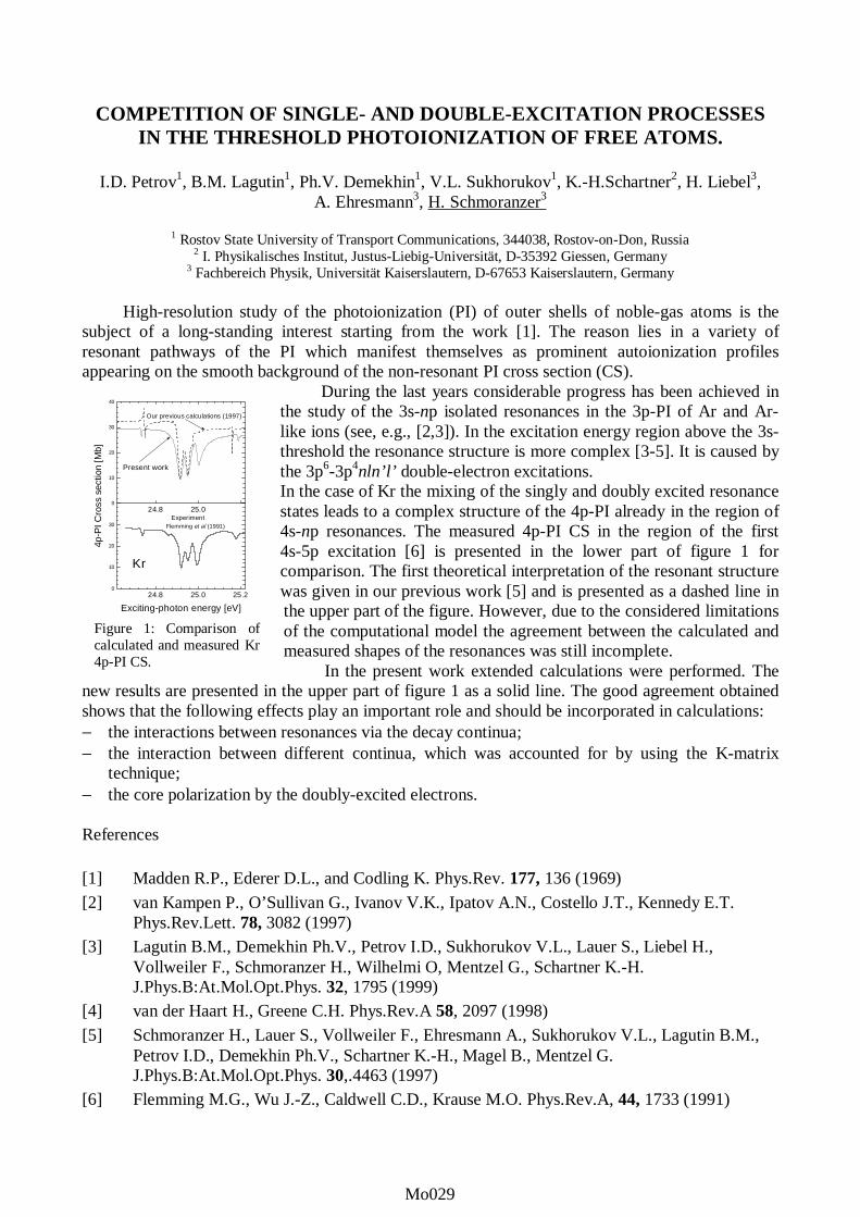
During the last years considerable progress has been achieved inthe study of the 3s-np isolated resonances in the 3p-PI of Ar and Ar-like ions (see, e.g., [2,3]). In the excitation energy region above the 3s-threshold the resonance structure is more complex [3-5]. It is caused bythe 3p6-3p4nln’l’ double-electron excitations.In the case of Kr the mixing of the singly and doubly excited resonancestates leads to a complex structure of the 4p-PI already in the region of4s-np resonances. The measured 4p-PI CS in the region of the first4s-5p excitation [6] is presented in the lower part of figure 1 forcomparison. The first theoretical interpretation of the resonant structurewas given in our previous work [5] and is presented as a dashed line inthe upper part of the figure. However, due to the considered limitationsof the computational model the agreement between the calculated andmeasured shapes of the resonances was still incomplete.
In the present work extended calculations were performed. Thenew results are presented in the upper part of figure 1 as a solid line. The good agreement obtainedshows that the following effects play an important role and should be incorporated in calculations:− the interactions between resonances via the decay continua;− the interaction between different continua, which was accounted for by using the K-matrix
technique;− the core polarization by the doubly-excited electrons.
References
[1] Madden R.P., Ederer D.L., and Codling K. Phys.Rev. 177, 136 (1969)
[2] van Kampen P., O’Sullivan G., Ivanov V.K., Ipatov A.N., Costello J.T., Kennedy E.T.Phys.Rev.Lett. 78, 3082 (1997)
[3] Lagutin B.M., Demekhin Ph.V., Petrov I.D., Sukhorukov V.L., Lauer S., Liebel H.,Vollweiler F., Schmoranzer H., Wilhelmi O, Mentzel G., Schartner K.-H.J.Phys.B:At.Mol.Opt.Phys. 32, 1795 (1999)
[4] van der Haart H., Greene C.H. Phys.Rev.A 58, 2097 (1998)
[5] Schmoranzer H., Lauer S., Vollweiler F., Ehresmann A., Sukhorukov V.L., Lagutin B.M.,Petrov I.D., Demekhin Ph.V., Schartner K.-H., Magel B., Mentzel G.J.Phys.B:At.Mol.Opt.Phys. 30,.4463 (1997)
[6] Flemming M.G., Wu J.-Z., Caldwell C.D., Krause M.O. Phys.Rev.A, 44, 1733 (1991)
Figure 1: Comparison ofcalculated and measured Kr4p-PI CS.
Mo029Mo001Mo029Mo029Mo029

Dissociative single and double photoionization of CF4 and ionic fragmentation of
CF4+ and CF4
2+ in the range from 23 to 120 eV
Toshio Masuoka, Atsuo Okaji, and Ataru Kobayashi
Department of Applied Physics, Faculty of Engineering, Osaka City University,
Sugimoto 3-3-138, Sumiyoshi-ku, Osaka 558-8585, Japan
We have studied dissociative single and double photoionization processes with time-of-flightmass spectrometry and the photoion-photoion-coincidence (PIPICO) method by use of synchrotronradiation in the photon energy range of 23-120 eV. The TOF mass spectra and the PIPICO spectrawere measured at an angle of ~55° with respect to the polarization vector where the second-orderLegendre polynomial is close to zero. Under these conditions, the effects of anisotropic angulardistributions of fragment ions are minimized [1]. To obtain accurate ion branching ratios, the radiofrequency (rf) signal of the storage ring was used as the start signal of a time-to-amplitude converter.
The present study focuses on the determination of the ratio of double to single photoionization(σ2+/σ+) and the partial cross sections for single (σ+) and double (σ2+) photoionization as a function ofphoton energy. Second, the ion branching ratios and the partial cross sections for the individual ionsrespectively produced from the parent CF4
+ and CF42+ ions are separately determined. Third, the
dissociation ratio of the parent CF42+ ions into two ionic fragments is determined.
The ion branching ratios and the absolute partial cross sections for the production of singlycharged CF3
+, CF2+, CF+, F+, and C+ ions, as well as doubly charged CF3
2+ and CF22+ ions have been
previously reported [2]. The ratio of double to single photoionization was obtained first, increasingmonotonically with photon energy. The threshold of double ionization 37.5±0.5 eV is in goodagreement with the value 37.6±0.6 eV reported by Codling et al. [3]. Above 100 eV, the ratio exceeds0.3. Since the total photoabsorption cross section of CF4 in this photon energy range has been reported[4], the σ2+/σ+ ratio is converted to the absolute cross sections for single and double photoionization.
Ion branching ratios for the individual ions respectively produced from the parent CF4+ and CF4
2+
ions are determined separately, thus enabling more detailed study of the dissociation processes of theCF4
+ and CF42+ ions. For the ion branching ratios of CF4
+ , the major ions produced are CF3+ and their
ratio still increases at higher photon energies. The ratio for C+ also increases with photon energy up toabout 85 eV. As for the fragmentation of CF4
2+, two body dissociation F++CF3+ takes place first.
Depending on the number of neutral fluorine atoms in the dissociation, the different channels(F++CF2
++F, F++CF++2F, and F++C++3F) appear one after another.
References[1] T. Masuoka, I. Koyano, and N. Saito, J. Chem. Phys. 97, 2392 (1992).[2] T. Masuoka and A. Kobayashi, J. Chem. Phys. 113, 1559 (2000).[3] K. Codling, L. J. Frasinski, P. A. Hatherly, M. Stankiewicz, and F. P. Larkins, J. Phys. B 24, 951(1991).[4] J. W. Au, G. R. Burton, and C. E. Brion, Chem. Phys. 221, 151 (1997).
Mo030Mo001Mo030Mo030Mo030

Fragmentation of doubly charged CF42+ ion
Toshio Masuoka, Atsuo Okaji, and Ataru Kobayashi
Department of Applied Physics, Faculty of Engineering, Osaka City University,
Sugimoto 3-3-138, Sumiyoshi-ku, Osaka 558-8585, Japan
The doubly charged CF42+ ion has received much attention recently by the advent of
synchrotron radiation. Recently Hall et al. [1] reported the threshold for double ionization to be37.5±0.5 eV using threshold photoelectron(s) coincidence (TPEsCO) spectroscopy.Experimental information on the CF4
2+ dication has also been obtained via Auger spectroscopy,double-charge-transfer (DCT) spectroscopy, PIPICO, and PEPIPICO experiments. Among theseexperiments, Codling et al. [2] determined the thresholds for the ion-pair formation of CF4
2+ intoF++CF3
+ (37.6 eV), F++CF2+ (42.4 eV), F++CF+ (47.5 eV), and C++F+ (62.0 eV) and tentatively
correlated these thresholds with specific two-hole states of CF4 calculated by Lurkins and Tulea[3].
In the present study, we have studied dissociative double photoionization processes withthe photoion-photoion-coincidence (PIPICO) method by use of synchrotron radiation. ThePIPICO spectra were measured at an angle of ~55° with respect to the polarization vector tominimize any effects of anisotropic angular distributions of fragment ions [4]. Al optical filterwas used to eliminate higher order radiation. The PIPICO branching ratios obtained for thesemany-body fragmentation channels increase at different photon energies, indicating theexistence of fragmentation pathways at these different photon energies. In order to correlatethese fragmentation pathways more clearly to the electronic states of CF4
2+, the PIPICObranching ratios for these fragmentation channels were differentiated with respect to the photonenergy.
Larkins and Tulea [3] have calculated the energy of the 107 two-hole states associatedwith the seven outermost orbitals. The electron configuration of the ground electronic state ofCF4 is (1a1
21t26)(2a1
2)(3a122t2
6)(4a123t2
61e44t261t1
6): 1A1. The important bonding orbitals are 4t2,4a1, 2t2, and 3a1 [2]. In the attempt to correlate initial states of the CF4
2+ ion with the abovethresholds for fragmentation, Codling et al. [2] used various simplifying assumptions: the firstone is that no fragmentation occurs where both orbitals are non-bonding or antibonding (1t1, 1e,3t2), and they shifted the calculation of Larkins and Tulea by 4.8 eV. We follow their treatment.The results show that the three-body fragmentation occurs in a relatively narrow energy rangefrom the threshold to about 49 eV, where 14 two-hole states, 1t1, 4t2(2); 3t2, 4t2(7); 1e, 4t2 (2);4a1, 1t1 (2); and 4a1, 4t2 (1) lies. The value in the parentheses represents the number of the statesin this range. That is, only the outer-valence electrons are involved. The four-bodyfragmentation takes place in a rather wide energy range from the threshold to about 73 eV,where both inner-valence and outer-valence electrons are involved.
References[1] R. I. Hall, L. Avaldi, A. G. McConkey, M. A. MacDonald, and G. C. King, Chem. Phys. 187,125 (1994).[2] K. Codling, L. J. Frasinski, P. A. Hatherly, M. Stankiewicz, and F. P. Larkins, J. Phys. B 24,951 (1991).[3] F. P. Larkins and L. C. Tulea, J. Phys. (Paris), Colloq. C9(12) 48, 725 (1987).[4] T. Masuoka, I. Koyano, and N. Saito, J. Chem. Phys. 97, 2392 (1992).
Mo031Mo001Mo031Mo031Mo031

Molecular and dissociative single and double photoionization of CS2
Toshio Masuoka, Atsuo Okaji, and Ataru Kobayashi
Department of Applied Physics, Faculty of Engineering, Osaka City University,
Sugimoto 3-3-138, Sumiyoshi-ku, Osaka 558-8585, Japan
Molecular and dissociative single and double photoionization processes of carbon disulfide havebeen studied with time-of-flight (TOF) mass spectrometry in the 20-120 eV range by the use ofsynchrotron radiation. The experimental details can be found elsewhere [1]. The observed ions areCS2
+, S2+, CS+, S+, C+, and CS2
2+. The ion branching ratios for these ions increase at various photonenergies, indicating the presence of dissociation pathways at these photon energies. In order tocorrelate these dissociation pathways more clearly to the electronic states of CS2
+ and CS22+, the ion
branching ratios for these ions were differentiated with respect to the photon energy. These differentialspectra are similar by nature to those measured by threshold photoelectron-photoion coincidencespectroscopy (TPEPICO) except for a low spectral resolution of the present spectra.
The first peak in the photoion spectrum (dBR/dE) for CS+ indicates that the C state of CS2+
dissociates into CS+ (and also into S+) in agreement with previous observation. The satellite bands dueto configuration interaction have been observed in the 19.1-35 eV range by Carnovale et al. [2]. Thefirst peak covers the lower part of the satellite bands, meaning that the lower part of the satellite bandsdissociates into CS+. The threshold for formation of the metastable CS2
2+ ions lies at 27.05±0.02 eVmeasured by TPEsCO spectroscopy [3]. The second peak locates in the double ionization region,probably indicating that the CS+ ions are formed by the charge separation CS++S+ of the dication. It isinteresting to note that the CS+ ions are formed only in a restricted energy range from about 31 toabout 42 eV.
The results of the differential spectrum for the CS22+ ions show that the dication is formed only in
a narrow energy range from 27.05 to about 35 eV with a peak at about 29 eV. Hochlaf et al. havereported the potential energy curves along the SC-S coordinate for 14 electronic states of CS2
2+ usingcomplete active space self-consistent field (CASSCF) approach and have shown that all low-lyingelectronic states of CS2
2+ are separated by large barriers from their dissociation asymptotes [4]. Theyhave further mentioned that all electronic states up to about 32-33 eV have bound parts on theirpotential curves and are stable with respect to the dissociation. The present observation is essentiallyin agreement with their calculation.
References[1] T. Masuoka and A. Kobayashi, J. Chem. Phys. 113, 1559 (2000).[2] F. Carnovale, M. G. White, and C. E. Brion, J. Electron Spectrosc. Relat. Phenom. 24, 63 (1981).[3] M. Hochlaf, R. I. Hall, F. Penent, J. H. D. Eland, and P. Lablanquie, Chem. Phys. 234, 249 (1998).[4] M. Hochlaf, G. Chambaud, and P. Rosmus, J. Chem. Phys. 108, 4047 (1998).
Mo032Mo001Mo032Mo032Mo032

An angular correlation function for double photoionization of an atom us-ing the density and efficiency matrix approach
Chiranjib Sur† and Dipankar Chattarji
Department of Physics, Visva Bharati, Santiniketan, West Bengal, INDIA, 731235†email : [email protected]
We consider the double photoionization process in a rare gas atom as a two-step process,namely (i) photoionization in an inner shell followed by (ii) the emission of an Auger electronfrom an outer shell. Our treatment makes use of the density matrix approach along with the effi-ciency matrix which takes care of limitations of the detecting equipment. No reference is madeto perturbation theory. Results are obtained for the Xenon atom undergoing photoionization inthe 4D5/2 shell and a subsequentN5 − O23O23
1S0 Auger transition. Comparison is made withexperimental results given by Kammerling and Schmidt [1] for unpolarized light.
References :
1. B. Kammerling and V. Schmidt, J.Phys.B,26(1993), p1141
Mo033Mo001Mo033Mo033Mo033

CASCADE PROCESSES AFTER 3p-SHELL THRESHOLD PHOTOIONIZATION OF Kr
T. Matsui1, I. Higurashi1, E. Murakami2, T. Aoto3, T. Onuma3, Y. Itoh3, Y. Morioka3, H. Yoshii4,
A. Yagishita4 and T. Hayaishi1
1 Institute of Applied Physics, University of Tsukuba, Tsukuba, Ibaraki 305-8573, Japan 2 Department of Physics, Chiba Institute of Technology, Narashino, Chiba 275-0023, Japan
3 Institute of Physics, University of Tsukuba, Tsukuba, Ibaraki 305-8571, Japan 4 Photon Factory, Institute of Materials Structure Science, Tsukuba, Ibaraki 305-0801, Japan
Inner-shell photoionization of atoms induces sequential vacancy transfer, such as cascades of Auger and Coster-Kronig decays. Multiply charged ions are consequently produced according to steps of the cascades. The branching ratio for channels of the decays can be estimated from yields of the multiply charged ions. In threshold ionization, particularly the branching ratio can be well decided by small disturbances from ionization of other shells.
Multiply charged ions were measured in coincidence with threshold electrons near the 3p-shell ionization limits of Kr. Figure 1 shows yield spectra of total ions, threshold electrons and the multiply charged ions (Kr2+, Kr3+, Kr4+ and Kr5+). Peaks at 216 and 223 eV in the yield spectra of the multiply charged ions are due to threshold ionization, in which post-collision interaction effects appear; the profile is asymmetric and broadened [1, 2, 3].
References [1] T. Hayaishi, E. Murakami, Y. Morioka, E. Shigemasa, A. Yagishita and F. Koike, Journal
of Physics B, 27, L115 (1994). [2] T. Hayaishi, E. Murakami, Y. Lu, E. Shigemasa, A. Yagishita, F. Koike and Y. Morioka,
Physical Review A, 54, 4064 (1996). [3] T. Hayaishi, Y. Fujita, M. Izumisawa, T. Tanaka, E. Murakami, E. Shigemasa, A.
Yagishita and Y. Morioka, Journal of Physics B, 33, 37 (2000).
Figure 1: Yields spectra of total ions, threshold electrons and multiply charged ions incoincidence with threshold electrons near the Kr 3p-shell ionization limits.
0
20M
40M
60M
3 p 1/23 p 3/2
CO
UN
TS /
408
sec
Total ion
205 210 215 220 225 230 2350
50k
100k
Threshold electron
0
1000
2000
K r 2+
205 210 215 220 225 230 2350
5000
10000
15000K r
3+
0
2000
4000 K r 4+
205 210 215 220 225 230 2350
500
1000
1500 K r 5+
P H O T O N E N E R G Y (eV)
Mo034Mo001Mo034Mo034Mo034

STUDY ON THE AUGER ELECTRON CASCADES INDUCED BYPHOTO-EXCITATION OF Kr 3dj→→→→5p AND Xe 4dj→→→→6p(j=3/2, 5/2)
M. Okamoto1, M. Kitajima1, M. Hoshino1, H. Tanaka1, Y. Shimizu2, H. Chiba2, T. Hayaishi3,S. Fritzsche4, F. Koike5, N. M. Kabachnik6, and K. Ueda2
1 Department of Physics, Sophia University, Tokyo 102-8554, Japan2 Research Institute for Scientific Measurements, Tohoku University, Sendai 980-8577, Japan
3 Institute of Applied Physics, Tsukuba University, Tsukuba 305-8577, Japan4 Fachbereich Physik, University of Kassel, D-34109 Kassel, Germany
5 School of Medicine, Kitasato University, Sagamihara 228-8555, Japan6 Faculty of Physics, Bielefeld University, 33615 Bielefeld, Germany
We report the comparison between the experimental results of high-resolution angulardistribution measurements and the results of multi-configuration Dirac-Fock (MCDF)calculations for the first-step and the second-step Auger transitions following the photoexcitationin Kr 3dj
-15p and Xe 4dj-16p (j=3/2, 5/2) resonances. The experiment was carried out on the 24-m
spherical grating monochromator installed in the soft X-ray undulator beamline 16B at thePhoton Factory in Japan using home-made angle-resolved electron spectroscopy apparatus.
The measured electron spectra of the first-step Auger transitions following the Kr 3dj→5pexcitation are shown in Figure 1 (j=5/2) and Figure 2 (j=3/2). Arabic numbers given in the figurecorrespond to the line numbers given by Mursu et al.[1]. We have obtained values of the angularanisotropy parameter β for the groups of strong lines indicated by roman numbers in Figures 1and 2. The spectra showed not only the "diagram" lines of the spectator Auger decay but alsomany of the correlation satellite lines and shake-up satellite lines of the diagram lines. Fairlygood agreement between the experimental and the theoretical results for j=5/2 excitation havebeen obtained. Similar results for Xe will also be presented.
25 30 35 40 450
2
4
6
8
0 deg 90 deg
4s1 4
p5 (1 P)6
p
4s0 4
p6 (1 S)6
p 4s0 4
p6 (1 S)5
p
4s2 4
p3 4d
(1 P)5
p
4s1 4
p5 (3 P)6
p
VII
VI V
IV
III
II
4s1 4
p5 (1 P)5
p
147
136,137 62-64
42
20 1718
13-15
2
4
4s1 4
p5 (3 P)5
p
10
6-8
I
Inte
nsi
ty (
arb
. u
nits
)
Kinetic energy (eV)
25 30 35 40 450
2
4
6
8
135-137
100,10164
60,61
16
147
42,43
12-15
2
V'VII
VI
V
IV
III
II
I
4s0 4
p6 (1 S)6
p
4s0 4
p6 (1 S)5
p
4s2 4
p3 4d(
1 P)6
p
4s2 4
p3 4d
5p
4s1 4
p5 (1 P)6
p
4s1 4
p5 (3 P)6
p
4s1 4
p5 (1 P)5
p
4s1 4
p5 (3 P)5
p
0 deg 90 deg
Inte
nsi
ty (
arb
. u
nits
)
Kinetic energy (eV)
Figure 1: The first step Auger electron spectrumfollowing the Kr 3d5/2
-15p excitation.Figure 2: The first step Auger electron spectrumfollowing the Kr 3d3/2
-15p excitation.
References
[1] J. Mursu, J. Jauhiainen, H. Aksela, and S. Aksela, J. Phys. B 31, 1973 (1998).
Mo035Mo001Mo035Mo035Mo035

POST-COLLISION INTERACTION EFFECTS FOLLOWING 4p-SHELL IONIZATION OF Xe
T. Hayaishi1, T. Matsui1, I. Higurashi1, E. Murakami2, H. Yoshii3, A. Yagishita3 , T. Aoto4, T.
Onuma4, Y. Itoh4 and Y. Morioka4
1 Institute of Applied Physics, University of Tsukuba, Tsukuba, Ibaraki 305-8573, Japan 2 Department of Physics, Chiba Institute of Technology, Narashino, Chiba 275-0023, Japan
3 Photon Factory, Institute of Materials Structure Science, Tsukuba, Ibaraki 305-0801, Japan 4 Institute of Physics, University of Tsukuba, Tsukuba, Ibaraki 305-8571, Japan
Multi-step post-collision interaction (PCI) often occurs in Auger cascades following inner-shell threshold ionization [1, 2, 3]. In the cascades, an initially ejected threshold photoelectron is overtaken sequentially by faster Auger electrons ejected in every step of the cascades. Sudden changes of the Coulomb field due to the overtaking cause the multi-step PCI effect. As a result, the PCI energy distribution for the photoelectron obtains a characteristic profile; the profile is asymmetric and broadened, and its maximum is shifted in energy. Measurements of multiply charged ions in coincidence with threshold electrons enable one to acquire PCI effects divided into the cascades, because multiply charged ions are produced from the cascades following the threshold ionization.
Figure 1 shows yield spectra of Xe3+ and Xe4+ ions in coincidence with threshold electrons near the 4p-shell ionization limit of Xe. A peak above the ionization limit exhibits the PCI profile.
Figure 1: Yields spectra of Xe3+ and Xe4+ ions in coincidence with threshold electrons near the Xe 4p-shell ionization limit. Arrows indicate the 4p-shell threshold ionization limit.
References [1] T. Hayaishi, E. Murakami, Y. Morioka, E. Shigemasa, A. Yagishita and F. Koike, Journal
of Physics B, 27, L115 (1994). [2] T. Hayaishi, E. Murakami, Y. Lu, E. Shigemasa, A. Yagishita, F. Koike and Y. Morioka,
Physical Review A, 54, 4064 (1996). [3] T. Hayaishi, Y. Fujita, M. Izumisawa, T. Tanaka, E. Murakami, E. Shigemasa, A.
Yagishita and Y. Morioka, Journal of Physics B, 33, 37 (2000).
138 140 142 144 146 148 1504000
8000
12000
16000
CO
UN
TS /
230
sec
X e3+ - eth
138 140 142 144 146 148 1500
2000
4000
6000
X e4+ - eth
PHOTON ENERGY (eV)
Mo036Mo001Mo036Mo036Mo036

RESONANT AUGER SPECTRA OF GAS PHASE KCl
V. Pennanen, T. Matila, E. Kukk, M. Huttula, H. Aksela and S. Aksela
Department of Physical Sciences, P. O. Box 3000, FIN-90570 University of Oulu
Normal and resonant Auger spectra of gas phase KCl at the K 2p edge have beenmeasured using synchrotron radiation. The measurements were carried out at beamline I411at MAX-II laboratory in Lund, Sweden. The beamline is equipped with a Zeiss SX-700 planegrating monochromator. The electron spectra were measured using a Scienta SES-100analyzer at the magic angle of 54.7°. KCl crystals were evaporated in a resistively heated
oven at the temperature of about 600°C under the base pressure of 1.5-3.3 x 10⁻⁷mbar.
Measurements were made as follows: first a total ion yield spectrum at the K 2p edgewas measured using a Wiley-McLaren type time-of-flight spectrometer (Figure 1). Thespectrum has four distinct maxima that are most likely due to excitations from the K 2p to msand nd (m=4, n=3) Rydberg orbitals [1,2]. Figure 2 displays the Auger electron spectrameasured at the two strong resonances (hν=296.75 and 299.5 eV). Drastic differences seenin the Auger intensity distribution cannot be due to the 2p spin-orbit splitting only, but mustbe assigned to molecular effects instead. The spectra resemble the results from solid KCl [3],where the orientation of crystal-field-split collapsed 3d orbital was thought to play a crucialrole. Similar symmetry considerations could apply in the molecular case.
Figure 1: Total ion yield of gas phase KCl. Figure 2: Auger electron spectra of the KCl molecule.
References
[1] M. Meyer et al., Phys. Rev. A 49 (1994) 3685.[2] A. Kikas et al., J. Electron Spectrosc. Relat. Phen. 68 (1994) 287.[3] M. Elango et al., Phys. Rev. B 47, 11736 (1993).
296 297 298 299 300 301
B
A
Inte
nsit
y [a
rb. u
nit
]
Photon Energy [eV]
10 15 20 25 30 35 40 45 50 55 60 65
B
A
Inte
nsit
y [a
rb. u
nit]
Binding Energy [eV]
Mo037Mo001Mo037Mo037Mo037

DIRECT PROBE OF THE BENT AND LINEAR GEOMETRIES FOR THE CORE-EXCITED RENNER-TELLER PAIR STATES IN CO2
BY TRIPLE-ION-COINCIDENCE MOMENTUM IMAGING
Y. Muramatsu1, K. Ueda1, H. Chiba1, N. Saito2, M. Lavollée3, A. Czasch4, T. Weber4, O. Jagutzki4, H. Schmidt-Böecking4, R. Moshammer5, K. Kubozuka6 and I. Koyano6
1 IMRAM,Tohoku University, Sendai 980-8577, Japan
2 AIST, Tsukuba 305-8568, Japan 3 LURE, Bat. 209d, Centre Universitaire Paris-Sud, F-91898, Orsay Cedex, France
4 Frankfurt University, D-60486 Frankfurt, Germany 5 Freiburg University, D-79104 Freiburg, Germany
6 Himeji Institute of Technology, Kamigori 678-1297, Japan
The core-excited Π state of a linear molecule CO2 splits into Renner-Teller pair states A1 and B1 (in C2v symmetry) due to vibronic coupling with bending motion. In the present study, we directly measure that A1 and B1 states are bent and linear, respectively. The experiment was carried out on the beamline 27SU at SPring-8 in Japan. We employed the triple-ion-coincidence momentum imaging technique, which is based on the time-of-flight method combined with a position-sensitive detector, to extract complete information on the linear momenta of the three ions C+, O+ and O+ produced from the CO2
3+ parent ion. Figure1 shows Newton diagrams for the three-body break-up of CO2
3+ after the C 1s → 2πu excitation. In these diagrams the amplitude of the linear momentum of the first O+ is normalized to unity and its direction is set to negative x, the linear momenta of C+ and the second O+ are plotted in the positive and negative y directions, respectively. We selected the excitation to the A1 state selecting the events that eject the C+ ion in the direction parallel to the E vector and the excitation to the B1 state selecting the events that eject the two O+ ions within the plane perpendicular to the E vector. Diagrams (a) and (b) correspond to the selections for the B1 and A1 states, respectively. The diagram (a) coincides with those recorded at any other excitation energies, illustrating that the B1 state is linear: the molecule looks slightly bent in diagram (a), due to zero-point bending motion. In the diagram (b), however, one can recognize a long tail of each island. This tail reflects how much the molecule is bent when it breaks up and thus is direct evidence that the A1 state is bent.
Figure 1: Newton diagrams for the three-body break-up CO2
3+ → C+ + O+ + O+ for the C 1s → 2πu excitation by 290.7 eV photons, (a) to the B1 state and (b) to A1.
Mo038Mo001Mo038Mo038Mo038

Electron Beam Ion Trap Spectroscopy of Highly
Ionized Tungsten
Roger Hutton1 , C. Beidermann2, R. Radtke 2 and Yaming Zou3.
1Dept. of Physics, Lunds Universitet, Lund, Sweden. 2Max-Planck-Institute for Plasma Physics, Berlin, Germany.
3Applied Physics Department, Shanghai Jiaotong University, China.
.In a 1980 Physical Review Letter by Curtis and Ellis [1] an interesting simplification of the lowest excited levels of 61 electron ions was discussed. 61 electron ions belong to the Pm I iso-electronic sequence and the ground configuration of Pm I is [Xe]4f56s2, which is far from simple. However for higher members of the sequence the 4f orbitals undergo a so-called collapse and the ground state becomes [Pd]4f 145s. The 1st excited levels are then the 4f145p 2P levels and an alkalai-like doublet is expected in the spectra. A number of experiments have been attempted to observes this doublet for highly ionized members of the Pm I sequence, for example beam -foil observations of Au. However, in the beam-foil spectra a multitude of lines were observed and only tentative identifications of the Pm-like lines could be given, see [2]. It was suggested in [1] that these doublet lines should be strong in the spectra of plasmas containing such elements. As Tungsten is of current interest in Tokamak fusion plasmas there is an interest in identifying these two, expected strong, lines for Pm-like W (W13+). The fact that these lines do not appear as strong features in the beam-foil spectra should not be taken is a discouraging way. The population mechanisms for excited states in the beam-foil interaction are not expected to be the same as those in a Tokamak plasma. It is evident however that the excitation mechanisms in an Electron Beam Ion Trap (EBIT) are more similar to those in a Tokamak plasma, disregarding the active diagnostic lines for now. Hence we made a search for the Pm-like doublet in the spectra of W using the Berlin EBIT. This led to rather clean spectra compared to the beam-foil case, as we may expect from the cleaner excitation conditions. References. [1] L.J. Curtis and D.G. Ellis, Phys. Rev. Letts. 45 , 2099 (1980). [2] E. Träbert and P.H. Heckmann, Z. Phys. D1, 381 (1986).
Mo039Mo001Mo039Mo039Mo039

TWO-PHOTON EXCITATION/IONIZATION OF 1S-SHELLOF HIGHLY CHARGED IONS
S.A.Novikov and A.N.Hopersky
Chair of Mathematics, Rostov State University of Transport Communication,Narodnogo Opolcheniya Square 2, Rostov-on-Don, 344038 , Russia
E-mail: [email protected]
The cross section of two-photon absorption by neutral many-electron atoms’ inner shells arecalculated. The case of Cl atom [1] and case of Ne atom [2] are considered. The aim of this work is to calculate the cross section of two-photon absorption by an innershell of highly charged positive ions with inclusion of the relaxation effects and the effect ofvacancy stabilization. A simple system with 1S0 ground state term is chosen as the subject of thestudy, namely, +6Ne and +8Ne ions. The calculations are fulfilled both for linear and circularpolarization of the laser beam. The calculation results of cross section absolute values and shape of the considered process(excitation/ionization of 1s-shell of +6Ne , +8Ne ions and neon neutral atom by two linearlypolarized photons) accounting both the effect of relaxation of the atomic residue in the field ofthe creating vacancies and the effect of 1s-vacancy stabilization are presented in Figure. Dottedline – for the 0Ne atom [2]; solid line – for the +6Ne ion; short dashed line – for the +8Ne ion. The strong dependence of absolute values and shape of the cross section on the ion charge ispredicted.
References:
[1] J.Abdallah Jr.,L.A.Collins,G.Csanak,A.G.Petschek,G.T.Schappert, Z. Phys. D34, 233 (1995).
[2] S.A.Novikov, A.N.Hopersky, J. Phys. B: At. Mol. Opt. Phys. 33 , 2287 (2000).
10 100 100010-17
10-7
103
1013
Ne8+
Ne6+
Ne0
σ1s(2),10-52 cm4sec linearly polarized photons
Photon energy, eV
Mo040Mo001Mo040Mo040Mo040

��������������������� ����������������������������
��������������� ��������������� �������������� ������ ����������
������������������ � ����!"!�#� �������$�������!���%�&'()'�#� �������*���� ��
��
����!�+ ������� �,��!����!+���� ���+������������+-!� � -+�������� ����!� �� � !�������!��� � �.+!+ � � ������ -����� !�!�+ � .�+������� -�+���+�������� .������+���� /��� ���� +-� �+ ����������� !����!� � � !�� �+ !�,!� +-� �!�+�.����� .������ � �� �����!��� ��� 0���� ��� ��!�+.������� /����.�+.��!��������+-!� �� � +0 ��� ���!�������-+������ ��������!�+ �� �!������.����� ��!�������� +0 �!+�����,���.������.+ ��+�����+ �.�+�������� ��� ����!�+�.�������+ � !�+ �����!+���-+��.�+���� �� ����!�+ ������1�,��!��� !�� ��� !� �.������ ����� ���� +-� ����+0� �� ����������� �+������!��������!��.������������.+!+������+-��+��������.������+����
2� + ����,.����� !����..�+������.���� !���!�!����+0�����!+��!����!��.+!+�+ �3�!�+ �+-������1�� ���!��� .+!+-����� !�� � � !�� �����1��!�� �+��!� 45�56� ������� 4�7819:��56� ;<=���+!+�+ �3�!�+ � ��� �������� +�!� �-!��� -+���!�+ � +-� �,��!��� .+!+-����� !�� ��� ��� �� !���1�+�����!���!� ����������1.�+������.������5�51�����!�+ ��2�!���1+-1-���!�������.��!�+��!�����������!+���������.��!�����+ ��������+-�!��.�������.+!+.�+���!�������-� �!�+ �+-�!��5�51.+!+ �� �������������<��+0�������!�.������,��.���!��.+!+�+ �3�!�+ ���+������!�+ �+-��,��!����!+����+,��� � 4#4<>66� � � !�� � ����� ������� ��!0�� � <9� �5� � �� <8��5� ;9=��#4<>6� ��� �� ���!��� ��� ��.�������.+!+-����� !�+-�+3+ ��4#?6����.�������.+!+�������!��79%(� ���@!����-+� ��!�!������!�.+!+�+ �3�!�+ � �+��� +!� +����� � � !�� -���!� 4��� 4�A<<�):��566� �+ !� ���� +-� #�� ��!� ��!�+ �� !� ��!�� ��� .���� !� � � ����� �+ !� ��� 4�>� 4�A<&�'8� �56� � �� ��� 4�A<)�)%��566�� @ !� ������+ � ��������+���� ������+0�!���>1�+ !� ����0�����������!+�BC�1-+������ �!�� ��!�+ ��� !+�
��!+�+ �3� �� �� ���!� �������� �!�!���� �����!��-�+�� ���� !� 0+��� + � !�� .+!+������ +-��!�+�.����� !����� ������ ���� ��� ��+�� ���+ +,���� 4��#6� ��+�� �� ��+,���� 4#��#6�����+�� �� .��+,���� 4��##��6� � �� �!������+����� 4��D#�6� ;?=� ��� .���� !��� 0������!+�+ �3�!�+ � � �� �+ �3�!�+ � !���+������� �� -+�� !�� �����!� �� ��!��!�+ � +-� !��E�� !��� �!�!��� +-� .������� .+!+-����� !���2��+��!�� �!+���� .+!+�+ �3�!�+ � ��+������!�+ �� ���+0� !+� ���� �� ��������� ��� �� ����!�+�� � �� E�� !��� ������� +-� �+�.�!� ��.+!+������.�+�������������������;<=� ��� ������ ��� ��� ������ � ���
�� ������������������� ���� ��������� �� �� ����� @ �!����� ��� <?<'�49(((6��
;9=� ��� ������ ��� ��� ������ � ������ ��� ��� ����� ��� �� ����� � ����� ���� ����� �� �� 2� ��� :9%9?�49(((6��
;?=�� ��� ��� ��� ��� ������ ��� ��������� � � �� ��� ���� ��� ����������2������!!���49((<6��
0.4
0.2
0
Inte
nsity
[ar
b. u
nits
]
18171615141312
Energy [eV]
(2P
o) 3 4 5
ns' 4 5 76ns''
3 5nd''
nd'3 4 56
(2D
o)
4
�����������+!+�+ �������+-�#�� -�+��#4�>6�� � !��5�51��������� /�� ��!�+ �� � !+� ��!+�+ �3� �� �������� �!�!��� � ��!�������.��!� ���+ ���� �������!������� ����!����
Mo041Mo001Mo041Mo041Mo041

������������� ������������ �� ������������ ������ ��� ��� ���� ������
���������������� �������������� ���������� ��������������
����� � ����������������������� ��!�� ������" �� � �����#�$%&'%�!�� ������(��) ����
�*�� +����������,�-�+� ���������������%.%&$�+����������,�-��,��� ������� ��/�����
���/� ����/����������0��� ��/���������� �1��-����-�)��/2�� �� ������3��)/���,� ����,��������/������,��4,����2/�) ��/��/����3���22�����/2�) ������5�-1���/�,��/���� ��1/21��1 ����� )������ �� �-� ����� ���� ��/� -�� ���-�� ��/6� 2/�� ��� 2����� ��)�� �������- ��/��� /�� ���� ��/� ���� ���/����������1������0��� ��/�����2������,�������/�������-�/2���)����)/���,�����������/�������������,����/������ ��/� ���� ���/����� �/��1�/1� ������ �� �����/��� /2� 7�� �7��� ���8�� ��� 9!� �9��� ���8� ���!��� ���8�� ��� ����� ��,�����������0����)����� ���� ������/,�� �� ����$%:�1+(;1�� )����� ����� ��/� -�� ���-� "�++<1==� 6���� � ��� �� �/�� ������� /2� ��/� ���� )/���,���� ��� ��,������ ���)� �,���� ��),�� ��/,����� +) ��� ����-�� ��2��� � �� ��� 2 �� ���/6� ��� � �,� �� ����6����������)�>�� ���� �-�������������������6������/� ����)/���,���� �����,������� �����/���������
+) ��� ������ �� �����2��� ��� ����� �/� ��� ��/� ����)/���,��� � �� ��� /2� ��� /����� /2� � 2�6�)�>� ��6���� ��� �-�����������6����� ��������� ��� ���/������������/��1�/1� �������� �����/���������� �-��� ���2/,����/��������������������/������0�������/��1��-���/�����/����-��/� �����1
�����2��� �� ��/�����-,����� �/6�� �� ������ �� �0 )���� ���7��� � �*� �� �����/��/2� ��,������� �����/� ���� 7��� "/���,����� ��� �)/���������� �� ����� ���-�� �� ��� ������ ��� �-��� ��� �) ��� �,����-��2�� ���� ��-,����*���/6�� ���� ��������6�/2� ��� 2����� �6/����� 1��/� ���� �����/�����?@&���/6��� �� ���� �?@���,����� �� ������������-� � �����2�� /2�'�)�>� 2/�� ��,��������� ������/������/� ����)/���,���� �,����1)/��� ��� �912 ��/���/2� ��� �-������� ��/� �� �� �����/��� ��� ���,���� ���
��,������6������ ���-���� �����������2/�����-��� �-���������� )/���,� ��-�/)����������,�������)/���,����� The results are discussed in terms of size-dependent changes in electronic and geometric structure upon cluster formation as well as dynamic stabilization of core-excited mole-cules that are localized in clusters using the quasiatomic approach.
Rel
. Int
ensi
ty [
arb.
uni
ts]
402401400Energy [eV]
Cluster
Molecule
Rel
. Int
ensi
ty [
arb.
uni
ts]
400.90400.80Energy [eV]
401.15401.05
6 meV
v'=0
v'=1
Molecule Cluster
������ ���� ��� ���/���������/2�)/���,� �� �����,�����������/-��� ��� ���7�������8���-�)������ ��� -����,�������3��A7B��������) �����/������C&���
������ ��� D�� ����� ���6� /�� ��� 2������6/� ���� ��/� �� �� �����/��� /2� ���7�������81� ���� !���� �������� ����/���� ��� �-���E� �0����)��� �� � � ��/����F� �/���� �����E� >/�-�� 2��� /2� � ����0����)��� ��� � ���
Mo042Mo001Mo042Mo042Mo042

AUGER CASCADE PROCESS OF XE FOLLOWING 4d5/2-16p
EXCITATION
S.-M. Huttula1, S. Heinäsmäki1, H. Aksela1, E. Kukk1, M. Jurvansuu1, and S. Aksela1
1 Department of Physical Sciences, P.O.Box 3000, FIN-90014 University of Oulu, Finland
Recent studies [1] on the cascade Auger process following the 2p-14s excitation in Arshowed that angular distributions are very sensitive to relativistic effects. Within themulticonfiguration Dirac-Fock (MCDF) formalism mixing of configurations was also important,signaling the rather strong electron-electron correlation.
In this work, the transition rates and angular distribution of the Xe cascade processXe(1S0) + hv → Xe*(4d95s25p66p, J0=1) (1)
→ Xe*+(4d105s15p56p) + e- (2) → Xe++(4d105s25p4) + e- (3)
have been studied theoretically and compared with experimental spectra.
The transition rates and the angular distribution parameters for the first and second stepAuger transitions have been calculated using the MCDF method. We have also investigatedconfiguration interaction (CI) effects due to the mixing of 4d105s15p54s, 4d105p35d16p1 and4d105p36p16s1 configurations.
The first and second step Auger spectra of Xe were measured with very high photon andelectron energy resolution at the I411 undulator beamline at Max-laboratory in Lund, Sweden. Inorder to determine the angular anisotropies of resonant Auger transitions the rotatable SES-200electron analyser was set at angles 0°, 54.7° and 90° with respect to the electric vector of theincident light.
References
[1] S.-M. Huttula, S. Heinäsmäki, H. Aksela, J. Tulkki, A. Kivimäki, M. Jurvansuu, and S. Aksela, Phys. Rev. A 63 032703 (2001)
Mo043Mo001Mo043Mo043Mo043

CASCADE DECAY OF INNERSHELL HOLE STATES OF ATOMICMAGNESIUM STUDIED BY PHOTOELECTRON-PHOTOION
COINCIDENCE SPECTROSCOPY
B. Kanngießer, S. Brünken, K. Godehusen, W. Malzer, N. Schmidt, P. Zimmermann
Technische Universität Berlin, Institut für Atomare und Analytische Physik, Hardenbergstr. 36, D-10623 Berlin, Germany
The photoelectron-photoion coincidence technique has been shown to be a very versatilemethod for the investigation of the decay channels of atomic innershell hole states. In thistechnique an initial hole state is identified by a signal of the electron energy analyser whichsimultaneously serves as a start signal for a time-of-flight measurement for the coincidentphotoion with respect to its charge state. The ratio of the differently charged photoions thengives information about the decay routes.
For the case of non-cascading transitions the ratio of radiative to non-radiative transitionsor the ratio of single to double Auger transitions can be directly related to the correspondingratios of the differently charged photoions. For cascading transitions, however, additionalcoincidence measurements are needed. As the first element in the periodic table Magnesium(1s22s22p63s2) shows these cascading transitions.
We investigated the whole cascade of Magnesium by using our photoelectron-photoioncoincidence technique on the 1s-, 2s-, and 2p-decay. The new undulator beamline U41-PGM atBESSY II delivered enough flux to realize these measurements on free Magnesium atoms in thewhole energy range needed, i.e. at 1400 eV for the 1s- and at 170 eV for the 2s- and 2p- decay.
References
[1] B. Kanngießer, M. Jainz, S. Brünken, W. Benten, Ch. Gerth, K. Godehusen, K. Tiedtke,P. van Kampen, A. Tutay, and P. Zimmermann, Phys. Rev. A 62, 014702 (2000).
[2] B. Kanngießer, S. Brünken, K. Godehusen, Ch. Gerth, W. Malzer, M. Richter, andP. Zimmermann, Nucl. Instr. and Meth. A (accepted).
Mo044Mo001Mo044Mo044Mo044

PCI EFFECTS IN XENON 4d PHOTOIONIZATION FOLLOWED BYTWO AUGER ELECTRON EJECTION
S.Sheinerman1, P.Lablanquie2, P.Penent3, R.I.Hall3, M.Ahmad3, Y.Hikosaka4, K.Ito4
1 Department of Physics, St.Petersburg State Maritime Technical University, 198262 St.Petersburg, Russia2 LURE, Centre Universitaire Paris-Sud, Bât. 209D, BP 34, 91898 Orsay, France
3 DIAM, Université P. & M. Curie, 75252 Paris Cedex 05, France4 IMSS, Photon Factory, Oho 1-1, Tsukuba 305-0801, Japan
We report an investigation of two Auger electrons, one very slow, one fast, which havebeen detected in coincidence following near threshold 4d-photoionization of the Xe atom. Theenergy distribution or line shape of the fast Auger electron was measured at photon energies inthe threshold region for the inner-shell ionization process where the photoelectron energy varied
from near zero to 30 eV. The measurements were made at Super ACO in France1 and at thePhoton Factory in Japan2. For analysis we selected two line shapes of the fast electron, associatedwith a zero-energy electron, which have energies near 3.4 eV and 5.4 eV. These electrons can bethe result of Auger decay of both the 4d5/2 and 4d3/2 holes of the Xe+ ion to the ground state ofXe3+. All the measured spectra reveal PCI effects that shift and broaden the line shapes, thisdistortion decreasing with increasing excess photon energy.
There are three possibilities whereby the 4d holes decay ejecting two electrons, one ofwhich has zero kinetic energy: double Auger decay (DA) where two electrons are ejectedsimultaneously and cascade Auger decay where the two electrons are emitted sequentially thefast electron being the first (CA1) or the second (CA2) emitted. The theoretical description ofPCI distortion of the Auger lines is based on the eikonal approach for both the cascade Auger
process and the double Auger process3. We have calculated the incoherent contribution of thethree processes to the cross section and estimated their role.
Our calculations show quite reasonable agreement of CA2 curves with the experimentaldata compared to those for DA and CA1. The best agreement with experiment was obtained for awidth of the intermediate state, Γ , in the range: 45 meV < Γ < 70 meV. So comparison of the
experimental data with the prediction from our calculations clearly demonstrated that the
dominant process is CA2 in both 4d5/2 and 4d3/2 decay.
References[1] D.Thomas et al., Synchrotron Radiation News 5, 8 (1992)[2] E.Shigemasa et al., J.Synchrotron Rad. 5, 777 (1998)[3] S.A.Sheinerman, J.Phys. B 27, L571 (1994) S.A.Sheinerman, J.Phys. B 31, L361 (1998)
Mo045Mo001Mo045Mo045Mo045

OBSERVATION OF TRIPLET DOUBLY EXCITED STATES OF HELIUMBELOW SECOND IONIZATION LIMIT.
F. Penent1,*, P. Lablanquie2, R. I. Hall1, M. Zitnik3, K. Bucar3, S. Stranges4, R. Richter5,M. Alagia6, P. Hammond7, and J. Lambourne8
1 DIAM, Université P & M Curie, 75252 Paris Cedex 05, France2 LURE, Centre Universitaire Paris-sud, Bâtiment 209D, BP 34, 91898 Orsay, France
3 J. Stefan Institute, Jamova 39, 1000 Ljubljana, Slovenia4Dipartimento di Chimica, Universitá di Roma La Sapienza e Unitá INFM, P. le A. Moro 5, I- 00185 Rome, Italy
5 SincrotroneTrieste, I-340 12 Trieste, Italy6 INFM-TASC, Padriciano, I-340 12 Trieste, Italy
7 Department of Physics, University of Western Australia, Nedlands, WA6907, Australia8 Department of Physics & Astronomy, University of Manchester, Manchester M13 9PL, United Kingdom
The helium atom is an archetypal three body system ideal for the study of electroncorrelation. Doubly excited states of helium have been studied extensively and, since these states
are imbedded in an ionization continuum, autoionization was, for a long time, considered to bethe only appreciable decay channel. So, the ion yield was considered to be a good measurementof the photon absorption cross-section. However recent experiments [1][2] have shown that someaspects of the formation and decay of doubly excited states had been overlooked: this concernsthe fluorescence decay of doubly excited states and the role of relativistic effects .
An important experimental advance was made by the detection of the fluorescence productsnamely UV photons [1] and also metastable atoms [2]. The availability of third generationsynchrotron sources with high photon flux and resolution now allows new insight in this domain.
We present here results obtained on the gas phase beam line in Elettra. The states studied areHe 2lnl’ states below the second ionization limit of helium. We have detected both UV photons(2p->1s) and metastable atoms resulting from radiative decay of helium doubly excited states.
The very high sensitivity of metastable detection has allowed the first direct observation of LSforbidden doubly excited triplet states [3]-
Two new series are clearly observed: the 2pnd 3D1o and the (sp, 2n-) 3Po . Excitation of these
states is possible due to spin orbit interaction of the ‘inner’ 2p electron which can overcome theCoulomb electron-electron interaction for high n values. A simple first order perturbation modelaccounts for the excitation of these states with essentially triplet character via their mixing with(sp, 2n+) 1Po optically allowed states. They decay essentially to the metastable 23S state of Heand this process is responsible for the intense metastable signal below threshold.
References[1] T.W. Gorczyca et al, Phys. Rev. Lett. 85, 1202 (2000) and references included[2] M..Odling-Smee et al, Phys.Rev.Lett. 84, 2598 (2000)
[3] F.Penent et al, accepted in Phys. Rev. Lett (2001)
Mo046Mo001Mo046Mo046Mo046

HIGH RESOLUTION AUGER SPECTROSCOPY:APPLICATION to the COSTER KRONIG DECAY of the Ar 2s HOLE
Y.Hikosaka1, K.Ito1, P.Lablanquie2, R.I.Hall3, F.Penent3
1 IMSS, Photon Factory, Oho 1-1, Tsukuba 305-0801, Japan2 LURE, Centre Universitaire Paris-Sud, Batiment 209D, BP 34, 91898 Orsay, France
3 DIAM, Université P. & M. Curie, 75252 Paris Cedex 05, France
An Auger spectrum gives clues to both the dynamics of the associated inner shell hole decayand to the spectroscopy of the final doubly charged ionic state. However, this information isblurred by two factors: (1) raw Auger spectra contain the contributions associated with differentinitial hole states, (2) the resolution of the final state is limited by the lifetime of the initial innershell hole. In order to overcome these two limitations, we are developing a technique based onphotoionisation and the use of synchrotron radiation. It consists simply in measuring the Augerspectrum in coincidence with the photoelectron that defines the initial hole. It is clear that onlythe Auger spectrum associated with this well defined hole will be recorded. It can also be shown,by simple energy conservation arguments, that the limitation associated with the hole lifetime isremoved [1]. This situation is referred to as the "sub-lifetime regime" [2].
Our experiment [3] uses a high luminosity threshold electron spectrometer, dedicated to thedetection of the photoelectron, and for the Auger electron, a hemispherical electrostatic analyzer,equipped with a two dimension position sensitive detector. Measurements on Argon were madeon the BL16 beam line at the Photon Factory, Japan, in January 2001.
We will present at the conference new results on Argon 2s Coster-Kronig decay. The 2s holeis known for its extremely short lifetime__, that causes a broadening of 1/__ = 2.25 eV of theAuger spectrum. This obscures the intensities and positions associated with the Ar++ state thuscreated, whether measured in electron [4] or photon [5] impact. In a previous experiment, with a600 meV resolution [1], we revealed structure hidden in the non-coincident Ar L1L2,3M2.3
spectrum. Our new measurement reached a better resolution of 500 meV for these Ar++ 2p-13p-1
states. Furthermore, they located the Ar++ 2p-13s-1 states and revealed the satellite lines,
associated to Ar++ 2p-13p-2nl configurations, that have recently been predicted [5].
References[1] P. Lablanquie et al., Phys.Rev.Lett. 84, 47, (2000)[2] J. Viefhaus et al., Phys.Rev.Lett. 80, 1618, (1998)[3] Y.Hikosaka et al., Meas.Sci.Tech. 11, 1697, (2000)[4] W. Melhorn, Z.Phys. 201, 1, (1968)[5] T.Kylli et al., Phys.Rev.A 59, 4071 (1999)
Mo047Mo001Mo047Mo047Mo047

ANGULAR DISTRIBUTION IN RESONANT AUGER SPECTRA OF XENON EXCITED BELOW THE 3d5/2 IONIZATION THRESHOLD
R. Sankari1, A. Kivimäki1, M. Huttula1, H. Aksela1, S. Aksela1, M. Coreno2, G. Turri2,
R. Camilloni3, M. de Simone4, K.C. Prince5
1 Department of Physical Sciences, P.O. BOX 3000, 90014 University of Oulu, Finland 2 INFM-TASC, I-34012 Trieste, Italy
3 CNR-IMAI, Area della Ricerca di Roma, CP-10, I-00016 Rome, Italy 4 Dipartimento di Fisica, Università di Roma III, I-00146 Rome, Italy
5 Sincrotrone Trieste, I-34012 Trieste, Italy
So far the study of the Xe 3d5/2-1np (n = 6,7) core hole resonant states (Figure 1) has been
modest due to very small excitation cross-sections and relatively high binding energies. The strong development of both the synchrotron radiation sources and the analyzers has, however, made it possible to study such weak resonant states. The angle-resolved Xe 3d5/2
-1np → 4d-2np resonant Auger spectra (Figure 2) [1] were measured at the Gas Phase Photoemission beamline of Elettra storage ring in Trieste, Italy, using an end station equipped with an array of hemispherical electron energy analyzers. Comparison with corresponding normal Auger measurements [2] reveals the role of the resonantly excited electron and its effects on energy, intensity and angular distributions. Furthermore, the angle-resolved measurements confirmed experimentally the theoretical gross spectator model [3,4].
Figure 1. M5 pre-edge Rydberg excitations in Xe.
Figure 2. The electron spectrum of Xe at the 3d5/2→6p excitation.
References [1] R. Sankari, A. Kivimäki, M. Huttula, H. Aksela, S. Aksela, M. Coreno, G. Turri,
R. Camilloni, M. de Simone, and K.C. Prince, Phys. Rev. A 63, 032715 (2001). [2] J. Karvonen, A. Kivimäki, H. Aksela, S.Aksela, R. Camilloni, L. Avaldi, M. Coreno,
M. de Simone, K.C. Prince, Phys. Rev. A 59, 315 (1999). [3] B. Kämmerling, B. Krässig, and V. Schmidt, J. Phys. B 23, 4487 (1990). [4] J. Tulkki, H. Aksela, and N.M. Kabachnik, Phys. Rev. A 48, 1277 (1993).
672 673 674 675 676 677
7p 8p 9p
6p3d5/2M5
Inte
nsi
ty
Photon energy (eV)158 156 154 152 150 148 146 144 142
0
500
1000
1500
2000
2500
3000
3500
Binding enrgy (eV)
Co
un
ts
Mo048Mo001Mo048Mo048Mo048

High resolution Auger spectroscopy of free barium atoms
M. Martins1, G. Snell2,3, E. Kukk2,3, W.T. Cheng2,4, and N. Berrah2
1 Freie Universitat Berlin, Institut fur Experimentalphysik, Arnimallee 14, 14195 Berlin, Germany,2 Dept. of Physics, Western Michigan University, Kalamazoo, MI 49008-5151,3 Lawrence Berkeley National Laboratory, University of California, Berkeley,
4 Dept. of Physics, National Central University, Chung-Li, Taiwan, 32054, R.O.C.A 94720,
Atomic barium is an excellent system to study electron-electron correlations, due to thestrong correlations of the valence electrons in the 4d excited state. This is caused by thecollape of the 5d wavefunction resulting in a near degeneracy of the 6s, 6p, 5d and 4f electrons.High resolution experimental Auger electron spectra subsequent to a 4d photoionizationhave been measured at the AMO undulator beamline 10.0.1 of the Advanced Light Source(ALS) synchrotron radiation facility at Lawrence Berkeley National Laboratory using aScienta SES200 high resolution electron spectrometer [1]. The present measurements wereall performed at an emission angle of 54.7◦ with respect to the electric field vector, in a planeperpendicular to the propagation direction of the beam of linearly polarized photons. Theanalyzer was operated at the constant pass energy of 40 eV with an electron energy resolutionof 40-50 meV. The Auger spectra have been measured at a photon energy of 131 eV to avoidany overlap between Auger and photo lines.In recent studies of the 5p and 4d photoionization of atomic barium, the experimentalphotoelectron spectra have been described theoretically in great detail by a configurationinteraction (CI) approach using Hartree-Fock wavefunctions as a zero order approximation[2,3]. Since the agreement with the measurements were excellent, we decided to performsimilar calculations to analyze the NOO Auger electron spectra.The overall agreement between the measured and calculated Auger spectra is very good.However, an assignment is possible only for a very limited number of lines, because theAuger spectra consist of thousands of lines. For this reason we have calculated the termdependent Auger spectra, i.e. the spectra which originate from the different initial holestates corresponding to the different lines of the satellite-rich 4d photoelectron spectrum.We found striking differences between the Auger decay of the final states of the photoion-ization process, which can be attributed to the strong electron-electron correlations. Theseresults enable us to assign several more Auger lines to Ba2+ states.
References
1. N. Berrah et al., J. Electron Spectrosc. Relat. Phenom. 101-103, 1 (1999).
2. G. Snell et al., Phys. Rev. A, in press (2001).
3. T. Matila and H. Aksela, J. Phys. B 33, 653 (2000).
Mo049Mo001Mo049Mo049Mo049

INNER-SHELL EXCITATION AS A PROBE OF INTERMOLECULAR INTERACTIONS
A.A. Pavlychev(1), L.V. Gatsenko(1), D. A. Mistrov(1), R. Flesch(2), W. Tappe(2), E. Rühl(2)
(1) Institute of Physics, St. Petersburg University, St. Petersburg, 198904, Russia
(2) Fachbereich Physik, Universität Osnabrück, Barbarastr. 7, D-49069 Osnabrück, Germany Perspectives on inner-shell excitation as a probe of intermolecular interactions are presented. These are provided by the recent experimental progress in experimental high resolution studies on Van der Waals clusters [1,2] and the development of the quasi-atomic approach [3,4] that allows to describe core-to-valence transitions, such as 1s→1πg
* (π*) and 1s→σu* (σ*), in nitrogen
and carbon monoxide. The concept of dynamic core-hole localization allows us to examine site-selectively changes in electronic and atomic structures of Van der Waals molecules, which includes a fast probe of intramolecular and intermolecular dynamics. Photoprocesses, such as
(N2) y+ + (N2)n-x-y + x N2 + e-
(N2)n + ħω → [N2+(N2)n-1 + e-] → [N2
+(N2)n-2 N2-*]
(N2) y+ + (N2)*
n-x-y + x N2 + e-
are considered which influence the 1s photoabsorption and photoionization cross sections at intermolecular distances that are close to the intermolecular Van der Waals distance in weakly bound species. Essential changes in electron-optical properties and intermolecular dynamics of molecular nitrogen are revealed at the σu
*-shape resonance and near the 1s-ionization threshold. Recent high resolution studies indicate that there are distinct changes in vibrational fine structure of the low-lying 1πg
* (π*)-resonance upon the transition from the isolated molecule to clusters [1,2]. These changes can be due to: (i) freezing of molecular rotations, (ii) the occurrence of intermolecular vibrations (soft modes), and (iii) dynamic stabilization of the core-excited molecule, resulting in a slight redshift of the vibrationally resolved core-to-valence transitions, which is of the order of a few meV [1]. It is shown that the intermolecular vibrations lead to phonon-like broadening and dynamic stabilization converts to a polaron-like shift, when the number of molecules in a cluster reaches infinity. Dynamic stabilization is primarily due to changes in intermolecular interactions that are induced by the inner-shell excitation process. Model studies on N2 that is confined within an endohedral environment (e. g. within a fullerene system) are also presented in order to illustrate general importance of dynamic stabilization. References [1] R. Flesch, A.A. Pavlychev, J.J. Neville, J. Blumberg, M. Kuhlmann, W. Tappe, F. Senf,
O. Schwarzkopf, A.P. Hitchcock, E. Rühl, Phys. Rev. Lett, submitted (2000) [2] R. Flesch, W. Tappe, A.A. Pavlychev, E. Rühl, J. Synchroton Radiat. 2001 (in press) [3] A.A. Pavlychev, A.S. Vinogradov, A. Stepanov, A. Shulakov, Opt. Spect, 75 (1993) 553 [4] A. A. Pavlychev, E. Rühl, J. El. Spect. Relat. Phenom. 106 (2000) 207; 107 (2000) 203
Mo050Mo001Mo050Mo050Mo050

LARGE NONDIPOLE EFFECTS IN THE CORE-LEVEL THRESHOLDREGIONS OF SMALL MOLECULES
O. Hemmers1, M. Lotrakul1, G.Öhrwall1, S. W. Yu1, D. Lukic2, I. A. Sellin2, and D. W. Lindle1
1Department of Chemistry, University of Nevada, Las Vegas, Nevada, USA2Department of Physics, University of Tennessee, Knoxville, Tennessee, USA
The electric-dipole or uniform-electric-field approximation has long served as a basis forunderstanding many aspects of the interactions between radiation and matter. Deviations fromthe dipole approximation (DA) for atomic targets can be attributed to the variation in phase ofthe incident radiation over the spatial dimensions of the absorbing charge distributions, whichincorporates higher (electric quadrupole, magnetic dipole,...) terms into the radiation-matterinteraction. Accordingly, significant departures from DA are commonly thought to occur only atwavelengths comparable to or smaller than the spatial dimensions of the absorbing electronicorbitals, an expectation born out by recent experimental photoionization studies on rare gasatoms performed at sufficiently high photon energies [1]. Somewhat surprisingly, however, otherrecent experiments and theoretical calculations have demonstrated atoms also may exhibitsignificant nondipole effects which are somewhat beyond the usual dipole expectations also atlonger incident photon wavelengths [2]. Recent experimental results on N2 [3] with a detailedtheoretical analysis of the contributions of first-order nondipole terms to the interactions betweenradiation and matter attributes the observed behaviors about 60 eV above threshold to thepresence of strongly anisotropic bond-length-dependent photoionization amplitudes, andsuggests that those results may be indicative of a potentially universal behavior in molecularphotoionization more generally.
Here we report experimental observations of significant deviations from the DA in theangular distributions of K-shell electrons photoemitted from gas-phase CO and N2. In contrast tothe situation for atomic targets [1,2] and the previous observations in N2 [3], large deviationsfrom the DA are found at suprisingly low incident photon energies just a few eV above the core-level ionization thresholds in CO and N2. The measurements were performed at the AdvancedLight Source (ALS) at the Lawrence Berkeley National Laboratory on undulator beamline 8.0.The ALS, operated in two-bunch mode, providing a photon pulse every 328 ns, allowingphotoelectron detection using the time-of-flight technique which is described elsewhere [4].
References
[1] B. Krässig, M. Jung, D. S. Gemmell, E. P. Kanter, T. LeBrun, S. H. Southworth, and L.Young, Phys. Rev. Lett. 75, 4736 (1995).
[2] A. Derevianko, O. Hemmers, S. Oblad, H. Wang, S.B. Whitfield, R. Wehlitz, I.A. Sellin,W.R. Johnson, and D.W. Lindle, Phys. Rev. Lett. 84, 2116 (2000).
[3] O. Hemmers, H. Wang, P. Focke, I. A. Sellin, D. W. Lindle, J. C. Arce, J. A. Sheehy, andP.W. Langhoff, Phys. Rev. Lett. (submitted).
[4] O. Hemmers, S. B. Whitfield, P. Glans, H. Wang, D. W. Lindle, R. Wehlitz, and I. A.Sellin, Rev. Sci. Instrum. 69, 3809 (1998).
Mo051Mo001Mo051Mo051Mo051

PHOTOABSORPTION CROSS SECTIONS OF PLANETARYMOLECULES IN THE VUV RANGE
Bing-Ming Cheng,1 Eh Piew Chew1
Mohammed Bahou,2 Chao-Yu Chung,2 and Yuan-Pern Lee2
1 SRRC, No.1 R&D Road VI, Science-Based Industrial Park, Hsinchu 300, Taiwan 2 Deptartment of Chemistry, National Tsing Hua University, 101, Sec. 2, Kuang Fu Road, Hsinchu 300, Taiwan
Absolute photoabsorption cross sections of several planetary molecules and theirisotopomers have been measured in the VUV range. For this purpose we used a dual beamabsorption set-up to normalize the intensity fluctuation of the light source, as shown in Figure 1.The light of VUV was the synchrotron radiation dispersed with beam line located at theSynchrotron Radiation Research Center in Taiwan. Absorption spectra of samples were recordedat various pressures. At each wavelength absorbance (=ln (I0/I)) determined at 5 to 20 pressureswas plotted against number densities and fitted with least-squares method to a line to yieldabsorption cross section according to the Beer’s law.
Photoabsorption cross sections of H2O, HDO, D2O, CH4, CH3D, C2H6, C2H5D, HCl, DCl,CH3OH, CH3OD, CD3OH, and CD3OD have been measured. Results combined with theinformation of isotopic fractionation of planetary molecules can be applied to the understandingof atmosphere and evolution of planets. For example, the major implication of H2O and HDOdata is on the evolution of Martian water, which must have lost at least a 50-m global layer ofwater.
Figure 1: Schematics of the dual-beam photoabsorption experiment.
PMT
PMT
VUV
pump
CaF2
CaF2
CaF2
sample input
pump
CaF2
pump
Photon counter
SR
Stepping motor
Micro computer
Sodium salicylate
Sodium salicylate
pressure meter
pump
Mo052Mo001Mo052Mo052Mo052
SYMMETRY-RESOLVED VIBRATIONAL SPECTROSCOPYFOR THE C1s-1 EXCITED RENNER-PAIR STATES IN CO2
H. Yoshida1, K. Okada2, S. Tanimoto2, A. De Fanis3, N. Saito4, and K. Ueda3
1 Department of Physical Science, Hiroshima University, Higashi-Hiroshima 739-8526, Japan2 Department of Chemistry, Hiroshima University, Higashi-Hiroshima 739-8526, Japan
3 IMRAM, Tohoku University, Sendai 980-8577, Japan4 National Institute of Advanced Industrial Science and Technology, Tsukuba 305-8568, Japan
Carbon dioxide (CO2) is a linear molecule in the ground state. When the Carbon 1s coreelectron is excited to the lowest unoccupied molecular orbital 2πu, the doubly degenerate C 1s-1
2πu states split into Renner-Teller pair states. The lower-energy state has a bent stable geometrywhile the other has a linear stable geometry. The bent state has a π electron whose orbital lies inthe bending plane of the molecule (in-plane A1 in C2v) while the linear state has the out-of-planeπ electron (out-of-plane B1 in C2v). We present the vibrationally-resolved spectra for symmetry-resolved A1 and B2 excitations.
Angle-resolved ion-yield spectra for the C 1s −> 2πu resonance were first obtained byAdachi et al. [1] at the Photon Factory. They suggested that the shift of the peak positions for theenergetic-ion-yield spectra recorded in the direction parallel and perpendicular to the polarizationvector of the incident light is ascribed to the Renner-Teller splitting but they could not resolveany vibrational structures. Total ion yield spectrum was measured by Kukk et al. [2] at the ALSat higher resolution. They observed a vibrational progression with spacing of 151 meV andassigned it to the symmetric stretch mode (ν1) of the linear B1 state.
We have carried out angle-resolved ion-yield spectroscopy on the c branch of the beamline27SU at SPring-8 in Japan. This beamline provides linearly polarized monochromatic soft X-raywith the photon band pass of < 30 meV in the C 1s excitation region. Energetic ion yield spectraI(0) and I(90) were recorded using two identical high-pass (> 6 eV) ion detectors mounted in thedirection parallel and perpendicular to the polarization vector of the incident light. The total ionyield (TIY) spectrum was also measured at the same time. In the TIY spectrum, we couldclearly observe two vibrational progressions with the mean vibrational spacing of 140(10) meV.Note that the energetic ion yield spectrum I(0) directly reflects the excitation spectrum to the A1
state. Combining the present yield spectra I(0) and I(90) with the excitation ratios to the A1 andB1 states measured by means of the triple-ion-coincidence momentum imaging method [3], wehave obtained symmetry-resolved absorption spectra for the A1 and B1 excitations. Onevibrational progression with the mean vbrational spacing of 140(10) meV is recognized in eachspectrum. These progressions are assigned to the symmetric stretching modes in the core-excitedA1 and B1 states.
References[1] J. Adachi, N. Kosugi, E. Shigemasa, and A. Yagishita, J. Chem. Phys., 107, 4919 (1997).[2] E. Kukk, J. D. Bozek, and N. Berrah, Phys. Rev. A62, 032708 (2000).[3] Y. Muramatsu, K. Ueda, N. Saito et al., to be published.
Mo053Mo001Mo053Mo053Mo053

MIRRORING DOUBLY EXCITED RESONANCES IN RARE GASES
S. E. Canton-Rogan1, A. A. Wills1, T. W. Gorczyca1, E. Sokell2, J. D. Bozek3, M. Wiedenhoeft1,O. Nayandin1, Chien-Nan Liu4, and N. Berrah1
1 Department of Physics, Western Michigan University, Kalamazoo, MI 49008, USA2 Department of Experimental Physics, University College Dublin, Ireland
3 Advanced Light Source, Lawrence Berkeley Laboratory, University of California, Berkeley, CA 94720, USA4 Max-Planck-Institut für Physik komplexer Systeme, Nöthnitzer Strasse 38, D-01187 Dresden, Germany
The electronic structure of atoms is still a generally unsolved problem due to many-bodyinteractions. One manifestation of such interactions is the resonant excitation of two electrons topreviously unfilled orbitals, following absorption of a single photon. While this process hasreceived much successful theoretical attention for atoms such as helium and lithium, the resultsfor more complex atoms have not been so successful, due in part to the increasing importance ofrelativistic effects. The significance of these and other effects is difficult to assess in general.However, with the aid of experimental results from third generation light sources, indications canbe found to guide theoretical efforts.
One example is the study of the autoionization of doubly excited resonances in argon. Thehighest resolution data for resonant processes has commonly been obtained using total cross-section measurements. However, there is a class of resonances, whose contribution to the totalcross-section can be zero and may only be observed in measurements of partial cross-sections.They are characterised by equal and opposite resonant contributions to the individual partialcross sections and are termed mirroring resonances[1].
Recent measurements[2,3] below the 3s ionization threshold in argon represent an elegantexample of the mirroring phenomenon. Previously, the photoionization spectrum of argon[4] inthis region showed the 3s-1np(1P1) Rydberg series, and two low-lying, doubly-excited resonances,3s23p44s(2P1/2,3/2)4p(1P1). However, partial cross-section measurements have revealed tworesonant states between the n = 7 and 8 singly excited states. These resonances have profileswhich mirror each other in each of the two final channels and so when added, cancel such thatthere is no discernible presence in the summed spectrum. Consequently, even higher resolutiontotal cross-section spectra may not be able to see such spectral features where present.
The requirements for such perfect mirroring give, in the current case, an indication of thelevel of spin-orbit effects in this particular spectral region. The new resonances have beendetermined to be LS-forbidden triplet doubly-excited states[2]. Therefore, if spin-orbit effects arenot accurately modeled theoretically, the resulting cancellation will not be so complete. Thesetypes of measurements then provide a sensitive testing ground for future calculations whichattempt to charaterise spin-orbit effects.
References
[1] C. N. Liu and A. F. Starace, Phys. Rev A 59 R1731 (1999) [2] S. E. Canton et al, Phys. Rev. Lett. 85 3113 (2000).[3] C. D. Caldwell et al. Mol. Phys. 98, 1075 (2000).[4] R. P Madden, D. L. Ederer and K. Codling, Phys. Rev. 177 136 (1969).
Mo054Mo001Mo054Mo054Mo054
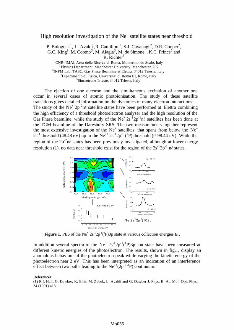
High resolution investigation of the Ne+ satellite states near threshold
P. Bolognesi1, L. Avaldi1,R. Camilloni1, S.J. Cavanagh2, D.R. Cooper2,G.C. King2, M. Coreno3, M. Alagia3, M. de Simone4, K.C. Prince5 and
R. Richter51 CNR- IMAI, Area della Ricerca di Roma, Monterotondo Scalo, Italy
2 Physics Department, Manchester University, Manchester, UK3INFM Lab. TASC, Gas Phase Beamline at Elettra, 34012 Trieste, Italy
4Dipartimento di Fisica, Universita’ di Roma III, Rome, Italy5Sincrotrone Trieste, 34012 Trieste, Italy
The ejection of one electron and the simultaneous excitation of another oneoccur in several cases of atomic photoionisation. The study of these satellitetransitions gives detailed information on the dynamics of many-electron interactions.The study of the Ne+ 2p-2nl satellite states have been performed at Elettra combiningthe high efficiency of a threshold photoelectron analyser and the high resolution of theGas Phase beamline, while the study of the Ne+ 2s-12p-1nl satellites has been done atthe TGM beamline of the Daresbury SRS. The two measurements together representthe most extensive investigation of the Ne+ satellites, that spans from below the Ne+
2s-1 threshold (48.48 eV) up to the Ne2+ 2s-12p-1 (1P) threshold (≈ 98.44 eV). While theregion of the 2p-2nl states has been previously investigated, although at lower energyresolution (1), no data near threshold exist for the region of the 2s-12p-1 nl states.
Figure 1. PES of the Ne+ 2s-12p-1(3P)3p state at various collection energies Ek.
In addition several spectra of the Ne+ 2s-12p-1(1P)3p ion state have been measured atdifferent kinetic energies of the photoelectron. The results, shown in fig.1, display ananomalous behaviour of the photoelectron peak while varying the kinetic energy of thephotoelectron near 2 eV. This has been interpreted as an indication of an interferenceeffect between two paths leading to the Ne2+(2p-2 3P) continuum.
References(1) R.I. Hall, G. Dawber, K. Ellis, M. Zubek, L. Avaldi and G. Dawber J. Phys. B: At. Mol. Opt. Phys.24 (1991) 413
87,5 88,0 88,5 89,0 89,51,5
1,6
1,7
1,8
1,9
2,0
2,1
2,2
binding energy (eV)
colle
ctio
n en
ergy
(eV
)
87 88 89 900,2
0,3
0,4
0,5
Yie
ld (a
.u.)
binding energy (eV)
87 88 89 900,2
0,3
0,4
0,5
Ek = 2.0 eV Y
ield
(a.u
.)
binding energy (eV)
87 88 89 900,2
0,3
0,4
0,5
Ne 2s-12p -1( 1P)3p
Ek = 1.5 eV
Ek = 2.2 eV
binding energy (eV)
Yie
ld (a
.u.)
1,4 1,6 1,8 2,0 2,2 2,4 2,6 2,8 3,0 3,2
0,2
0,4
0,6
0,8 b.e. = 88.50 eV
collection energy (eV)
Yie
ld (
a.u.
)
Mo055Mo001Mo055Mo055Mo055

ELECTRONIC STRUCTURE AND ENERGY RELAXATION OF DOPEDRARE GAS CLUSTERS WITH A SHELL-LIKE STRUCTURE
T. Laarmann, K. von Haeften, H. Wabnitz and T. MöllerHASYLAB at DESY, Notkestrasse 85, 22603 Hamburg, Germany
The electronic structure and dynamics of clusters has attracted considerably interest for many
years since it gives new insight in the properties of condensed matter. Investigations on doped
species offer the opportunity to get information about the host cluster and the impurity, because
the excitation is usually localised on the impurity atom. In this contribution, we report on energy
dissipation processes in ArM clusters (M<80) covered with a shell of Kr atoms (up to 30 atoms)
and embedded in large NeN clusters (N=3500). The ArMKrKNeN complex is studied with energy-
resolved fluorescence excitation spectroscopy.
Doped clusters are prepared in a supersonic expansion and subsequent pick up of atoms from
two cross-jets. After excitation with monochromatic synchrotron radiation (11-22.5 eV) the
emitted fluorescence light is detected by two photomultipliers (sensitivities: 2-6 and 4-11 eV).
For spectrally resolved measurements in the visible and near-infrared spectral range a
monochromator with a CCD camera is used. The absorption of clusters is monitored by
fluorescence excitation. In the energy range of the Ar cluster absorption (11-13 eV) the intensity
ratios of bands differ considerably from those of free clusters due to the influence of the shell
atoms. The surface states disappear and a new absorption band occurs, which we interpret as Ar-
Ne-interface excitations. The dependence of the transition energies on the size of the embedded
Ar clusters can be explained using the Frenkel-exciton model. Dipole transition moments could
be derived from the energy shifts. They are in good agreement with those of the corresponding
molecular Ar2 transitions.
After excitation into the Ne 1l’-exciton the fluorescence spectrum of Ar clusters embedded
inside Ne clusters consists of sharp lines which are due to free atomic Ar transitions (4p→4s).
Electronically excited Ar atoms move through the Ne cluster, desorb and emit in the vacuum. By
coating the Ar cluster with Kr atoms, the Ar lines disappear and 5p→5s transitions of Kr become
dominant. Additionally, new bands occur, which we interpret as Ar transitions of perturbed
atomic 4p-states inside Ne clusters. We suggest a simple kinematic model to describe the
movement of excited Ar atoms inside Ne clusters.
Mo056Mo001Mo056Mo056Mo056

INHERENT LIFETIME WIDTHS OF AR 2p-1, KR 3d-1, XE 3d-1 AND XE 4d-1
STATES
M. Jurvansuu, A. Kivimäki, and S. Aksela
Department of Physical Sciences, P.O. Box 3000, FIN-90014 University of Oulu, Finland
The lifetime is one of the most fundamental factors characterizing a core-ionized or a core-excited state. Yet core-hole lifetimes of even rare-gas atoms are rather poorly known. Perhaps
the most straightforward method to determine the lifetime of a singly ionized species is
photoelectron spectroscopy. The lineshape of a core photoelectron line is given by the
convolution of the Lorentzian lifetime broadening, Doppler broadening and the instrumental broadening. The latter is further composed of the photon energy and kinetic energy
contributions, and is often approximated to have a Gaussian shape. Recent development in
synchrotron radiation sources and electron analyzers has greatly diminished the instrumental broadenings, making it possible to determine the lifetime widths more accurately.
The measurements were performed on the I411 [1] undulator beamline at the MAX II storage ring at Lund, Sweden. Synchrotron radiation was monochromatized with a modified SX-700 plane grating monochomator. The photon energy resolution of the beamline is competitive
in the photon energy range 55–300 eV and reasonably good up to 900 eV. The ejected electrons
were energy analyzed with a high-resolution Scienta SES-200 electron spectrometer. The kinetic
energy resolution of the analyzer was determined by recording the Kr 4p photoelectron lines that have essentially zero lifetime broadening. The Ar 2p, Kr 3d, Xe 3d, and Xe 4d photoelectron
spectra were measured at several photon energies, keeping as many experimental parameters the
same as possible as in the Kr 4p calibration measurements.
The inherent lifetime widths of the Ar 2p-1, Kr 3d-1, Xe 3d-1, and Xe 4d-1 states were
determined with higher experimental resolution than before. The results are compared with the
previous determinations and also with the lifetime broadenings obtained for the corresponding
core-excited states using photoabsorption or electron energy loss spectroscopies.
References
[1] Bässler, M., J-.O. Forsell, O. Björnehol, R. Feifel, M. Jurvansuu, S. Aksela, S. Sundin, S. L. Sorensen, R. Nyholm, A. Ausmees, and S. Svensson, J. Electron Spectrosc. 101-103, 953 (1999).
Mo057Mo001Mo057Mo057Mo057

ION-PAIR FORMATION FROM DOUBLY EXCITED RYDBERG STATESIN NO, O2, AND CO
S W J Scully, R A Mackie, R Browning, C J Latimer, and K F Dunn
Department of Pure and Applied Physics, The Queen’s University of Belfast, Belfast BT7 1NN, UK
Ion-pair production is an important method for the investigation of highly excited statesof neutral molecules. In particular doubly excited Rydberg states, lying between the single anddouble ionization thresholds provide fundamental information on electron correlation [1].
We have measured negative ion yield curves using synchrotron radiation in the energyrange 18-55eV in O2, CO and NO gases. A large number of states are observed as resonancesover the entire energy range. Figure 1 displays the O- yield in NO in the region of the lowestthermochemical threshold and shows that pair production proceeds via direct and predissociatingRydberg states. The results in figure 2 illustrate for the first time the existence of a number ofquasibound excited electronic states of NO around the double ionization threshold and thebinding energy of the 1σ molecular orbitals. Further work is needed to uniquely identify theseresonances and their decay channels.
Reference
[1] A. Dadouch, G. Dujardin, L.Hellner, and M.J. Besnard-Ramage, Phys. Rev. A 43, 6057(1991).
Figure 2: O- formation from NO in the region of
known NO++ states (solid lines) and the bindingenergies of the 1σ molecular orbitals (dottedlines)
Figure 1:O- formation from NO in the region of thethermochemical threshold. Lines indicate N+(3P),N+(1D) and N+(1S) ions.
Photon Energy (eV)
36 38 40 42 44 46 48 50
0.20
0.25
0.30
0.35
0.40
0.45
Photon Energy (eV)
18 20 22 24 26 28
Sig
nal (
arb.
uni
ts)
0
2
4
6
8
10
Mo058Mo001Mo058Mo058Mo058

PHOTOIONISATION OF ETHANE IN THE VUV
R A Mackie, A M Sands, R Browning, K F Dunn, and C J Latimer
Department of Pure and Applied Physics, The Queen’s University of Belfast, Belfast BT7 1NN, UK.
The study of the ionization and fragmentation processes in ethane C2H6 is particularlyimportant since this molecule has been prominently and quite successfully treated in theoreticalcalculations based on the statistical quasi-equilibrium theory of mass spectra QET [1,2].
We have obtained, using synchrotron radiation within the energy range 11-35eV, bothpositive and negative ion yield curves in C2H6 with better resolution and counting statistics thanin all previous work. As can be seen in the ion breakdown diagram (figure 1), the generalfeatures are indeed in accord with QET, each successive fragment ion rising from threshold to along flat plateau. However in several cases, see for example C2H4
+, we also see significantstructure indicating that autoionization and predissociation compete effectively with thestatistical energy randomization involving fast coupling between the vibrational modes. Figure 2shows the negative photoion yield curve of H− formation in C2H6 along with ionizationthresholds obtained from photoelectron spectra. Many of the main features correlate with andappear just below these thresholds clearly indicating that superexcited Rydberg states areinvolved. However additional features, probably due to previously unknown states, are alsoapparent.
References[1] W A Chupka and J Berkowitz, J Chem Phys 47, 2921 (1967).[2] R Stockbauer, J Chem Phys 58, 3800 (1973).
Figure 2: H− formation in C2H6. The lines
indicate known ionization thresholds.Figure 1: Positive ion breakdown curves from thephotoionization of ethane
Mo059Mo001Mo059Mo059Mo059

PROBING ELECTRON ESCAPE DEPTH FOR FREE CLUSTERS
M.Tchaplyguine1, O.Björneholm1, R.Feifel1, M.Gisselbrecht2, R. R. T. Marinho3, S.L.Sorensen4, S.Svensson1
1 Uppsala University, Department of Physics, Uppsala, Sweden
2 MAX-LAB, Lund University, Lund, Sweden 3 Institute of Physics, Brasilia University, Brasilia, Brazil
4 Department of Synchrotron Radiation, Lund University, Lund, Sweden
The variety of processes taking place after X-ray irradiation in solid noble gases has been studied extensively by photoemission, absorption and Auger spectroscopy in bulk and adsorbed multilayers by many authors. Free clusters were always much more difficult for investigation - primarily since the “sample” density was insufficient for most of the X-ray sources [1]. Free clusters however are almost an ideal object for getting the information about the interaction of atoms at the scale of still countable but already quite large amount of participants. For comparison the study of multilayers always has to deal with the influence of the substrate. Thus one of the most puzzling and fascinating things at this nanoscale - the interplay of surface and bulk properties – is obscured by the interface-determined phenomena.
The experiments reported here were performed using the high resolution soft X-ray beam
line I411 at the 3rd generation synchrotron radiation facility MAX-II. It allowed obtaining sufficient electron signal from argon and xenon clusters in photoemission studies of the core-level ionisation processes. In the argon 2p and xenon 4d core-level photoemission spectra the resolution was enough to unambiguously separate the contribution of the atoms located on the cluster surface and in the bulk of the clusters. These measurements were performed at different excitation energies covering the range of 200 eV above the ionisation threshold for argon and 100 eV for xenon. It was demonstrated that bulk or surface aspects of the cluster electronic and geometric structure can be emphasized by using different photon energies. From the ratio between the “surface” atom signal to the “bulk” atom signal the efficient electron escape depth was estimated for different electron kinetic energies. These results as well as absorption, normal and resonant Auger studies will be presented in detail.
Figure 1: Xe 4d Photoemission spectra for 108eV(solid) and 130eV (dashed). Atomic, cluster bulk and cluster surface signals are indicated.
References [1] O.Björneholm, F.Federmann, F.Fössing, and T.Möller. Phys.Rev.Lett.,v.74, p.3017, 1995.
70,5 70,0 69,5 69,0 68,5 68,0 67,5 67,0 66,5 66,0 65,5
bulk
bulksurface
surface
130eV
108eV
cluster
cluster
atom 4d3/2
Inte
nsity
atom 4d5/2
Bind ing Energy, eV
Mo060Mo001Mo060Mo060Mo060

Selective fragmentation of valence and core electron excited CD4 and SF6 molecules
M. Stankiewicza, J. Rius i Riub, E. Kukkc, P. Ermanb, P. Hatherlyd, M. Huttulac, A.
Karawajczykb, E. Rachlewb and P. Winiarczyka.
aInstytut Fizyki im. Mariana Smoluchowskiego, Uniwersytet Jagiellonski, ul. Reymonta 4, 30-059 Krakow, Poland bSection of Atomic and Molecular Physics, Department of Physics I, Royal Institute of Technology, KTH,
SE-10044 Stockholm, Sweden cElectron Spectroscopy Group, Department of Physical Sciences, University of Oulu, FIN-90014 Oulu, Finland dJ.J.Thomson Physical Lab. The University of Reading, Whiteknights, P.O.Box 220. Reading, RG6 6AF, U.K.
Electron-ion coincidence measurements with energy resolved electrons are a powerful
tool in studies of molecular fragmentation processes. By analyzing electron kinetic energy only fragmentations from a specific doorway state are monitored while the remaining reactions are discriminated [1]. Presently, we have applied this technique in measurements of coincidence spectra of the CD4 and SF6 molecules following valence and core electron excitation. Our experiment has been implemented using an electron-ion coincidence spectrometer. It comprises a 125 mm electron spectrometer and 110 mm time-of-flight mounted collinearly. An inbuilt gas cell provides a target pressure 10-100 times above the chamber pressure. The measurements are performed at the magic angle with respect the synchrotron light polarization’s direction.
Our results show that the CD4+ molecule in the 1t2
-1 state is stable or fragments into CD3+
+ D only. None of these reactions occur from the 2a1-1 state for which the D+, CD+ and CD2
+ fragments were observed only. CI computations reveal that the CD4
+ fragments into CD3+ + D in
a process in which the initially excited 2B1 state of the C2v geometry undergoes the transition to the state in the C3v geometry which instantaneously fragments to CD3
+ + D. Dissociations from the 2a1
-1 state are governed by the 2 2A1 states in C2v and C3v geometries. Molecular fragmentation is also studied following core excitations of the C 1s electrons in CD4. Autoionization of the excited state to the 1t2
-1 state significantly alters the CD4+/CD3
+ fragment ratio, which is the first demonstration of a correlation between nuclear motion and molecular dissociation in the CD4 molecule. In addition, strong spectator Auger transitions that create double-hole ionic states result in a drastically different fragmentation pattern. Molecular fragments D+, C+, CD+ and CD2
+ are detected in coincidence with the Auger electrons, indicating a more complete breakdown of the molecule. Our results of the coincidence measurements on SF6 show that the SF6
+ molecule in the 1t1g-1 state is unstable and fully dissociates into the SF5+
+ F channel. Also the SF6+ ions in the 4t1u
-1 state are unstable and fragments to SF3+ + 3F in full
agreement with predictions of Hitchcock et al [2]. Also the mass spectra acquired in coincidence with the 5t1u, 3eg, 1t2u, 1t2g, and 5a1g electrons reveal strong selectivity in dissociation from these states. This selectivity reflects the bonding properties of the potential surfaces involved in the studied processes. References [1] K. Ueda, M. Simon, C. Miron, N. Leclercq, R. Guillemin, P. Morin, S. Tanaka Phys.
Rev. Lett. 83, 3800 (1999). [2] A. P. Hitchcock, M. J. van der Wiel J. Phys.B 12, 2153 (1979).
Mo061Mo001Mo061Mo061Mo061

”Novel decay processes in core-excited diatomic molecules”
R. Feifela, M. N. Piancastellia,b, P. Salekc , R. F. Finka, M. Bässlera, O. Björneholma, F. Burmeistera, S. L. Sorensend, C. Mirona, H. Wanga, K. Wiesnera, I. Hjeltea, A. Naves de
Britoe, F. Kh. Gel’mukhanovc,f, H. Ågrenc, L. Karlssona and S. Svenssona
aDepartment of Physics, Uppsala University, Uppsala, Sweden bDepartment of Chemical Sciences and Technologies, University “Tor Vergata”, Rome, Italy
cTheoretical Chemistry, Royal Institute of Technology, Stockholm, Sweden dDepartment of Synchrotron Radiation Research, Institute of Physics, Lund University, Lund, Sweden
eInstitute of Physics, University of Brasilia, Brasilia DF, Brazil fInstitute of Automation and Electrometry, Novosibrisk, Russia
With the availability of third-generation synchrotron light sources in the soft X-ray regime in combination with high-resolution photoelectron spectrometers, the investigation of very weak processes, often hitherto not possible at all to study, has become feasible. The first example is the N1s → π* resonant Auger decay to the B final state in N2
+ [Pia00] which demonstrates that a classification of decaying core-hole processes into ”spectators” (2h-1e final state) and ”participators” (1h final state) is too rough in some cases. Other examples are the observation of ”atomic holes” in ultrafast dissociation of core-excited molecules, as recently shown for the decay of the 2p → 6σ* core-excited state to the 4σ-1 final state in HCl [Fei00], and the spin selectivity in the vibrational progression of the X-participator decay following the same resonant excitation which revealed a novel type of propensity mechanism [Fin01]. Furthermore, the resonant Auger decay following C1s → π* excitation in CO was reinvestigated at high resolution, demonstrating that one can investigate the decay properties of higher vibrational levels than only v´ = 0, 1, 2, even if these higher vibrational states are not ”visible” in a total ion or total electron yield absorption spectrum [Fei01].
The experimental spectra have been recorded at the undulator beam line I411 [Bäss00], MAX-II, Lund, Sweden, equipped with a modified Zeiss SX 700 monochromator and with a high resolution Scienta SES 200 electron spectrometer. High photon flux combined with state-of-the-art electron energy resolution allowed us to investigate these remarkable decay processes in detail for the first time. References: [Pia00] M. N. Piancastelli et al., J. Phys. B: At. Mol. Opt. Phys. 33, 1819, 2000 [Fei00] R. Feifel et al., Phys. Rev. Lett. 85 (15), 3133, 2000 [Fin01] R. F. Fink et al., submitted to Phys. Rev. Lett. [Fei01] R. Feifel et al., in manuscript [Bäss00] M. Bässler et al., Nucl. Instr. Methods (accepted 2000) and references therein
Mo062Mo001Mo062Mo062Mo062

Observation of quantum oscillations in the C
70
photoemissionpartial cross sections up to 200 eV
B. Langer
1,2
, A. Wills
2
, G. Prümper
2
, R. Hentges
2
, and U. Becker
2
1
Max-Born-Institut, Max-Born-Str.2A, 12489 Berlin, Germany
2
Fritz-Haber-Institut der MPG, Faradayweg 4-6, 14195 Berlin, Germany
Fullerenes, in particular C
60
and C
70
, are a very specific form of matter causing unexpectedbehavior even in such matured fields as photoelectron emission. In recent years it has been shownthat the partial photoemission cross sections of the two outermost valence shells of C
60
showstrong oscillations [1-3] due to the potential in which the corresponding electrons are bound. Thefrequency of the oscillation is related to the avarage radius of the fullerene molecule, which is forthe case of C
60
the bucky ball radius. In the case of C
70
which has a more egg like shape the averageradius of the rotating molecule should cause the oscillating behavior. Former measurements of C
70
[4] were not performed at sufficiently close energy intervals in order to prove this assumption.Therefore, a new photoemission experiment of C
70
was performed at the beamline BW III of HA-SYLAB and the new MBI-Beamline at BESSY II.
Figure 1 shows ratio of the cross sections of theoutermost C
70
photolines between 40 and 190 eV, Thesolid line is the result of a fit of the data by an exponen-tially decaying sine function. The result of this fit givesthe mean radius R of the rotating C
70
molecule. The val-ue is in good agreement with what one would expectfrom averaging over all three axis of the ellipsoidellyshaped fullerene. This corrobotates the simple quantumoscillation model used to explain the partial cross sec-tion oscillation in fullerenes. The next step of a more so-phisticated model would be to look for quantum beats inthe oscillations as a signature of the thickness of the real shell like potential in which the electrons arebound [5,6]. Our data are still not taken in fine enough steps as well as limited to too low photon en-ergies in order to draw a conclusion in this direction. This new measurement, however, is a very solidverification of the basic oscillation predicted by the quantum oscillation model for the case of C
70
.
References
[1]. P. Benning, D. Poirier, N. Troullier, J. Martins, J. Weaver, R. Haufler, L. Chibante, andR. Smalley, Phys. Rev. B
44
, 1962 (1991).[2]. T. Liebsch, O. Plotzke, F. Heiser, U. Hergenhahn, O. Hemmers, R. Wehlitz, J. Viefhaus,
B. Langer, S. B. Whitfield, and U. Becker, Phys. Rev. A
52
, 457 (1995) .[3]. T. Liebsch, R. Hentges, A. Rüdel, J. Viefhaus, U. Becker, and R. Schlögl, Chem. Phys. Lett.
279
, 197 (1997).[4]. [4] A. Rüdel, A. Hempelmann, U. Hergenhahn, G. Prümper, J. Viefhaus, T. Liebsch, and
U. Becker, HASYLAB at DESY Annual report p. 231 (1996).[5]. Y. B. Xu, M. Q. Tan, and U. Becker, Phys. Rev. Lett.
76
, 3538 (1996) .[6]. O. Frank and J. Rost, Chem. Phys. Lett.
271
, 367 (1997).
R-1HO
MO
HO
MO
HO
MO
HO
MO
-1-1
Figure 1: Oscillation in the ratio of the crosssections of the outermost C70 photolines.
Mo063Mo001Mo063Mo063Mo063

Figure 1: Circular dichroism in the angulardistribution (CDAD) of photoelectronsemitted by free NO molecules.
-60
-40
-20
0
20
40
60
-180-150-120 -90 -60 -30 0 30 60 90 120 150 180
theta(ez)
CIRCULAR DICHROISM IN THE VALENCE-PHOTOIONIZATION OFFREE NO MOLECULES
O. Gessner1, B. Zimmermann1, A. Hempelmann1, P.-M. Guyon2, U. Becker1
1 Fritz-Haber-Institut der Max-Planck-Gesellschaft, Faradayweg 4-6, D-14195 Berlin, GERMANY
2 Laboratoire des Collisions Atomiques et Moléculaires, Bât. 351, Université Paris Sud, 91405 Orsay, FRANCE
We have measured NO 4σ inner valence photoelectron angular distributions in themolecular frame. Using circularly polarized light of two different helicities we were able toderive the circular dichroism in the angular distribution (CDAD) of the photoelectrons [1]. The
CDAD describes the difference between theangular distributions for the two different lighthelicities. While the angular distributions haveto be described by a rather big number of matrixelements and phase shifts, the CDAD is afunction of only a few terms containinginformation on the relative phase shift betweenthe outgoing σ and π continuum waves and thepartial wave composition. We observed aremarkable f-wave contribution in the outgoingphotoelectron wave which can be explained bythe influence of the shape resonance beingobserved previously in the relevant photonenergy region [2,3].
The CDAD has previously beenmeasured for molecules adsorbed on surfaces[4]. To our knowledge this is the first time that ithas been observed on free fixed-in-spacemolecules. The target molecules were notstatically oriented before the photoionizationtook place, rather the molecular orientation wasderived at the instant of photoionization bymeasuring the vector correlation between thephotoelectrons and the molecular ionicfragments.
References
[1] N. A. Cherepkov, Chem. Phys. Lett. 87, 344 (1982).[2] T. Gustafsson and H.J. Levinson, Chem. Phys. Letters 78 (1981) 28
[3] H.-P. Steinrück, C. Schneider, P.A. Heimann, T. Pache, E. Umbach and D. Menzel,
Surface Sci. 208 (1989) 136
[4] C. Westphal, J. Bansmann, M. Getzlaff, and G. Schönhense, Phys. Rev. Lett. 63, 151
(1989).
Mo064Mo001Mo064Mo064Mo064

C2+
C+ O+
CO22+
C2+
CO+
Figure 1: Auger electron-ion coincidence spectra forC 1s ionised CO2 (photon energy = 430 eV)
AUGER ELECTRON - ION COINCIDENCE SPECTROSCOPY OF COREIONISED CARBON DIOXIDE
B.O. Fisher1, P.A. Hatherly1, D. Collins1, M. Stankiewicz2, M.D. Roper3
1 J.J. Thomson Physical Laboratory, University of Reading, Whiteknights, Reading RG6 6AF , United Kingdom.2 Institute of Physics, Jagiellonian University, ul Reymonta 4, 30 059 Kraków, Poland3 CLRC Daresbury Laboratory, Daresbury, Warrington WA4 4AD, United Kingdom.
The Auger electron - ion double coincidence technique was pioneered by Eberhardt et al[1] and extended to a triple coincidence technique correlating Auger electrons with ion pairs byHanson et al [2] and Alkemper et al [3]. We will present recent data exploiting both the doubleand triple Auger electron coincidence techniques with carbon dioxide exploring ion percentageyields and fragmentation dynamics.
As an example, Figure 1 shows the variation of the fragmentation pattern of C 1s ionisedCO2 molecules as a function of Auger electron energy. It is firstly noticeable that the degree offragmentation decreases with increased Auger energy, as observed by Alkemper et al [3] in thecase of S 2p ionised CS2. Such a result may be understood by noting that a high energy Auger
electron will leave the final doubly chargedmolecule in a low lying state, and vice-versafor low energy Auger electrons.
Other data will be presented for the C 1sand O 1s edges and the C 1s→π* resonance andthe results compared with an earlier study onnear threshold phenomena in core excited CO2
[4]. In particular further details offragmentation dynamics will be discussed inthe light of triple coincidence experimentsrevealing ion pair correlations.
This work was carried out on beamline5U.1 of the SRS, Daresbury Laboratory, UnitedKingdom and has been supported by theEngineering and Physical Science ResearchCouncil.
References
[1] Eberhardt W, Plummer E W, Lyo I-W, Carr R and Ford W K. 1987 Phys. Rev. Letts. 58207
[2] Hanson D M, Ma C I, Lee K, Lapiano-Smith D and Kim D Y. 1990 J. Chem. Phys. 939200
[3] Alkemper U and von Busch F. 1998 J. Electron Spect. and Relat. Phenom. 93 115[4] Hatherly P A, Codling K, Stankiewicz M and Roper M. 1995 J. Phys. B At. Mol. Opt.
Phys. 28 3249
Mo065Mo001Mo065Mo065Mo065

Interchannel interaction vs. relativistic effects:the Xe 5p photoionization case
B. Zimmermann
1
, G. Snell
2
, B. Schmidtke
2
, J. Viefhaus
1
, N. A. Cherepkov
1,3
, B. Langer
4
,M. Drescher
2
, N. Müller
2
, U. Heinzmann
2
, and U. Becker
1
1
Fritz-Haber-Institut der Max-Planck-Gesellschaft, Faradayweg 4-6, D-14195 Berlin, Germany
2
Fakultät für Physik, Universität Bielefeld, Universitätsstraße 25, D-33615 Bielefeld, Germany
3
State University of Aerospace Instrumentation, 190000 St. Petersburg, Russia
4
Max-Born-Institut, Rudower Chaussee 6, D-12489 Berlin, Germany
Xenon 5p subshell photoionization has been studied in the vicinity of the Xe 4d shape reso-nance by angle and spin resolved photoelectron spectroscopy. Both kinds of spin polarization,transferred and dynamical, were determined between 40 eV and 150 eV. Dipole matrix elementsand relative phase shifts derived from these experimental data are compared with calculations withregard to the strength of interchannel and relativistic interactions. The comparison shows stronginfluence of interchannel interactions on the transition matrix elements, particularly on the phaseshift in the vicinity of the 4d shape resonance, but gives little evidence for relativistic interactionsof comparable strength This shows the persistence of LS coupling for continuum electrons in thepresence of a shape resonance for a heavy atom like xenon.
References
[1]. G. Snell, B. Langer, M. Drescher, N. Müller, B. Zimmermann, U. Hergenhahn, J. Viefhaus,U. Heinzmann, and U. Becker, Phys. Rev. Lett.
82
, 2480 (1999).
[2]. U. Heinzmann, J. Phys. B
13
, 4353, 4367 (1980).
[3]. C. Heckenkamp, F. Schäfers, G. Schönhense, and U. Heinzmann, Z. Phys. D
2
, 257 (1986).
[4]. K. N. Huang, W. R. Johnson, and K. T. Cheng, At. Dat. Nucl. Dat.
26
, 33 (1981).
[5]. M. Ya. Amusia and L. V. Chernysheva,
Computation of Atomic Processes - A Handbook forthe ATOM Programs
(Institute of Physics Publishing, Bristol, Philadelphia, 1997).
Figure 1: a) Transferred and b) dynamical spin polarization (see Ref [1]) of the Xe 5p photoionization linesshown as function of the incident photon energy.}!Ref[2,3], (5p1/2: ^, 5p3/2: *) - present measurements;dashed lines: relativistic RRPA Ref.[4]; solid and dotted lines: our nonrelativistic RPAE and HF calcula-tions, respectively, using the code from Ref[5]).
Xe+
Mo066Mo001Mo066Mo066Mo066

Absolute photoionization cross section measurements on atomic ions
J B West1, T Andersen2, H Kjeldsen2, F Folkmann2, R Brooks2,3 and H Knudsen2
1 Daresbury Laboratory, Warrington WA4 4AD, UK2 Institute of Physics and Astronomy, University of Aarhus, DK-8000 Aarhus C, Denmark
3 Permanent address: Guelph-Waterloo Physics Institute, University of Guelph, Ontario, Canada N1G 2W1
A merged ion-photon beam experiment is now in operation at the ASTRID storage ring inDenmark, installed on the undulator beamline and providing high intensity well collimated radiationover the photon energy range 25 — 180 eV. By measurement of the overlap between ion andphoton beams, and calibration of ion and photon detectors, cross sections with a precision of–10% have been measured for a range of ions of astrophysical interest. For the lighter ions, eg C+,the results have been compared with calculations from the IRON [1] and OPACITY projects [2].
We present here our measurements on the ions of Mg and Al, which we have used both torevise spectral assignments made earlier in the literature and also to compare with RPAE [3] andour own MCHF calculations. The ability to provide values of the oscillator strength has provedvery useful in making these assignments, as well as making direct comparisons along theisoelectronic sequence Na, Mg+ and Al++. The identification of the numbered resonances shown inthe figure below will be presented in detail on the poster presentation.
Figure 1: The absolute photoionization cross sections of Mg+ (lower) and Al++ (upper). Thenumbers refer to the corresponding 2p —ns, nd transitions.
References[1] The Opacity Project, Vol 1 (Institute of Physics, Bristol, UK) 1995[2] S N Nahar and A K Pradhan, Phys. Rev. A 49 181 (1994)[3] V K Ivanov, J B West, G F Gribakin and A A Gribakina, Z. Phys. D 29 109 (1994)
Mo067Mo001Mo067Mo067Mo067

EFFECTS OF LIGHT POLARIZATIONS ON 2σ g PHOTOELECTRON ANGULAR DISTRIBUTIONS FROM ORIENTED N2 MOLECULES
A. Yagishita1,2, S. Motoki2, J. Adachi1,2, K. Ito1, K. Ishii3, K. Soejima4,
S. K. Semenov5, N. A. Cherepkov5
1 Photon Factory, Institute of Materials Structure Science, KEK, Tsukuba-shi, Ibaraki 305-0801 Japan 2 Graduate School of Science, The University of Tokyo, Bunkyo-ku, Tokyo 113-0033 Japan
3 Department of Physics, Tokyo Metropolitan University, Hachioji-shi, Tokyo 192-0397 Japan 4 Graduate School of Science and Technology, Niigata University, Niigata-shi, Niigata 950-2181 Japan
5 State University of Aerospace Instrumentation, 190000 St. Petersburg, Russia
To perform complete experiments of molecular photoionization, we have measured the angular distributions of 2σg photoelectrons from oriented N2 molecules using three different polarization states of incoming photons with 58.2eV, i.e., linear, left-elliptical and right-elliptical polarization.
Experimental results on the photoelectron angular distributions are shown in Figure 1. The
molecular axis is aligned to the vertical line, and the polarization ellipse is indicated in the figure. These photoelectron angular distribution patterns can be expressed by the dipole matrix elements, phases, and Stokes parameters describing the polarization state of incoming light. Because we have determined the Stokes parameters before or after the measurements of the photoelectron angular distributions from fixed- in-space N2 molecules, the unknown parameters are the dipole matrix elements and phases. Taking p σ, p π , f σ, and f π partial waves into account, we have analyzed our photoelectron angular distribution patterns. Fitting curves are shown in the figure. To determine the unique solution of the matrix elements and phases, we have examined the solutions obtained for three different data sets; each data set gives more than two solutions. In this procedure, we have succeeded in picking up the unique solution, i.e., the common solution for the different data sets. The dipole matrix elements and phases determined uniquely by the complete experiments will be compared with the results by RPA calculations.
(a) (b) (c)
Figure 1: 2σg photoelectron angular distributions from fix-in-space N2 ; (a) linear polarization, S1=0.97 (b) left-elliptical polarization, S1=0.42 (b) and S3=-0.91 (c) right-elliptical polarization S1=0.50 and S3=0.87
Mo068Mo001Mo068Mo068Mo068

NONDIPOLE EFFECTS IN CORE-ELECTRON PHOTOEMISSIONANGULAR DISTRIBUTIONS OF SMALL MOLECULES
G. Öhrwall 1, O. Hemmers1, S. W. Yu1,2, M. Lotrakul1, D. Lukic3, I. A. Selli n3, and D. W. Lindle1
1 Department of Chemistry, University of Nevada, Las Vegas, NV 89154-4003, USA2 Center for X-Ray Optics, Ernest Orlando Lawrence Berkeley National Laboratory,
University of California, Berkeley, California 94720, USA3 Department of Physics and Astronomy, University of Tennessee, Knoxvill e, TN 37996-1200, USA
Much of our understanding of the photoionization of matter is based on the electric-dipoleapproximation. It is generally known this approximation breaks down for photon energies abovea few keV, as has been experimentally found [1], but it has been assumed the nondipolecontributions to the photoionization matrix elements can be neglected for wavelengths in the softx-ray and ultraviolet regions of the electromagnetic spectrum. However, recent experiments andtheoretical developments have shown that significant nondipole effects occur for atomic [2] andmolecular [3] photoemission at much larger wavelengths than expected. In the soft-x-ray regime,the first-order effects are predicted to be the major deviations for photoionization. First-orderdeviations from the dipole approximation for photoemission only affect the angular distributionof photoelectrons, not the cross section, and manifest themselves in a forward/backwardasymmetry relative to the photon direction.
Here, we report observations of significant deviations from the dipole approximation forthe angular distribution of core-shell photoemission of CO2, CF4 and SF6. For all three molecules,strong effects are found in the near-edge region. Particularly SF6 has a complex behavior, withrapid changes in the nondipole deviations over the t2g and eg continuum resonances above the SL2,3 edges. The measurements were performed at the Advanced Light Source synchrotronradiation facilit y at the Lawrence Berkeley National Laboratory. Four time-of-f lightspectrometers, positioned both in and out of the plane perpendicular to the photon beam to makethe nondipole-effect measurements possible, were used to record the photoelectron spectra [4].
References
[1] B. Krässig, M. Jung, D. S. Gemmell , E. P. Kanter, T. LeBrun, S. H. Southworth, and L.Young, Phys. Rev. Lett. 75, 4736 (1995).
[2] A. Derevianko, O. Hemmers, S. Oblad, H. Wang, S. B. Whitfield, R. Wehlitz, I. A. Selli n,W. R. Johnson, and D. W. Lindle, Phys. Rev. Lett. 84, 2116 (2000).
[3] O. Hemmers, H. Wang, P. Focke, I. A. Selli n, W. R. Johnson, D. W. Lindle, J. C. Arce, J.A. Sheehy, and P. W. Langhoff , Phys. Rev. Lett. (submitted).
[4] O. Hemmers, S. B. Whitfield, P. Glans, H. Wang, D. W. Lindle, R. Wehlitz, and I. A.Selli n, Rev. Sci. Instrum. 69, 3809 (1998).
Mo069Mo001Mo069Mo069Mo069

Ni 2p Photoabsorption and Resonant Photoelectron Spectroscopy of
Molecular High-spin Ni complex, Ni(N,N’-dimethylethylenediamine)2Cl2)
Hiroshi Oji, Yasutaka Takata, Takaki Hatsui, and Nobuhiro Kosugi
Institute for Molecular Science, Myodaiji, Okazaki 444-8585, JAPAN
We found that some planar molecular Ni complexes with a 3d8 low-spin ground state showdifferent resonant behaviors from Ni metal and Ni oxide [1]. The kinetic energy of Ni 3p satellitepeaks decrease as the photon energy increases, indicating the one electron, or excitonic, featureof the excited states in these systems. In the present study, we have measured soft X-ray Ni 2pabsorption and resonant Ni 3s and 3p photoelectron spectra of a molecular Ni complex with a 3d8
high-spin, Ni(N,N'-dimethylethylenediamine)2Cl2 (Ni(DED)2Cl2) to clarify the effect of the spinstate on the core-excited states of the system.
Measurements of X-ray absorption andphotoelectron spectra were performed atBL1A soft X-ray beamline of the UVSORfacility in the Institute for Molecular Science.Figure 1 shows the kinetic energy for the Ni3p and 3s primary ion states and the satellitepeaks observed in the resonant photoelectronspectra as a function of the photon energy.This shows nearly linear relationship betweenthe kinetic energy of these satellite peaks andthe photon energy with the slope (∆KE/∆hν)of +1. This dependence of satellite bands onthe photon energy is different from that of thelow-spin complexes where the slope becomesnegative (e.g. ∆KE/∆hν�= �0.55±0.05 forK2[Ni(CN)4] [1]), but is similar to that of NiOwith important electron correlation andmultiplet interaction. This indicates that theexcited states in this high-spin Ni complexcannot be described within the one-electronpicture. A series of our studies on the Nicomplexes with various electronic statesreveals that the resonant behavior ofphotoelectron spectra reflects the electronconfiguration of core-excited metal atomwhich depends on the chemical bonding statebetween the metal and the ligand molecules.
Fig. 1. Photon energy dependence of the kinetic energyfor Ni 3p, 3s primary and satellite photoelectron peaks.
Reference[1] Y. Takata, T. Hatsui, and N. Kosugi, J.Electron Spectrosc. Relat. Phenom. 88, 235(1998)
Mo070Mo001Mo070Mo070Mo070

PHOTOABSORPTION CROSS SECTIONS OF MOLECULAR CHLORINE
Youngmin Chung1 and Yang-Soo Chung2
1 Pohang Accelerator Laboratory, San31, Hyoja-dong, Pohang, 790-784, Korea 2 Department of Physice, Chungnam University, Yooseong-ku, Taejon, 305-764, Korea
The absolute total photoabsorption cross sections of molecular chlorine has been measured from ionization threshold to 30 eV using double electrode ion chamber and synchrotron radiation. The photoabsorption cross section shows three broad peaks at around 97, 92.5 and 85 nm superimposed to a continuous background. Also, small increase of photoabsorption cross sections were found at around 74, 60 and 53 nm. In general, the agreement between the present data and Samson’ s[1] one is excellent with maximum deviation of about 5% up to 83 nm. The earlier measurement by W.J. van der Meer et al.[2] was about 30% higher than the present one at 58.4 nm. It seems neither length nor velocity approximation in the Hartree-Fock calculation[3] provides satisfactory results for the photoionization of molecular chlorine. It is interesting that velocity approximations fits better in the lower energy side and length approximations in the higher energy side.
0.0
20.0
40.0
60.0
80.0
100.0
120.0
40 45 50 55 60 65 70 75 80 85 90 95 100 105 110 115
Wavelength (nm)
Abs
orpt
ion
Cro
ss S
ectio
n (M
b)
Figure 1. The total photoabsorption cross sections of molecular chlorine: Filled circle, present data. Open circle, the total photoabsorption cross sections measured by Samson et al.[1].
References [1] J.A.R. Samson and G.C. Angel, J. Chem. Phys. 86, 1814 (1987). [2] W.J. van der Meer, R.J. Butselaar and C.A. de Lange, Aust. J. Phys. 39, 779 (1986). [3] M. Braunstein and V. McKoy, J. Chem. Phys. 92, 4887 (1990).
Mo071Mo001Mo071Mo071Mo071

ABSORPTION CROSS SECTIONS OF O3 IN VUV
S. J. Tu1 and J. B. Nee1,2 1Department of Physics and 2Department of Chemistry
National Central University Chung-Li, Taiwan 32054
ROC
The absorption cross sections of O3 in the 105-200 nm wavelength region were measured. Ozone was prepared by the silent discharge of oxygen and trapped in silica gel. Absorption cross sections were measured by flowing the purified ozone through the experimental system. Minor oxygen absorption bands were subtracted to derive the ozone spectrum.
The absorption spectrum of ozone in the VUV wavelengths consists of several regions.
Between 155 and 200 nm, a low absorption spectrum was found. A region strong bands consists of several excited states was found at 105-140 nm. Two progressions of vibrational states were found between 113 and 105 nm. The structures in 105-108 nm, which are previously unknown, are similar to the other one in 108-113 nm. Our cross sections in λ> 110 nm are comparable to the previous measurements [1-2].
105 110 115 120 125 130 135 1400
5
10
15
20
25
30
Cro
ss s
ectio
n (M
b)
Wavelength(nm)
References [1] N. J. Mason, J. M. Gingell, J. A. Davies, H. Zhao, I. C. Walker and M. R. F. Siggel,
J. Phys. B:At. Mol. Opt. Phys. 29 (1996) 3075. [2] Y. Tanaka, E. C. Y. Inn and K. Watanabe, J. Chem. Phys. 21 (1953) 1651.
Mo072Mo001Mo072Mo072Mo072

SPIN-RESOLVED ELECTRON SPECTROSCOPYOF THE OCS MOLECULE
G. Snell1, B. Langer 2, M. Martins3, E. Kukk4, and N. Berrah1
1 Western Michigan University, Department of Physics, Kalamazoo, MI 49008, USA2 Fritz-Haber-Institut der Max-Planck-Gesellschaft, 14195 Berlin, Germany
3 Freie Universität Berlin, Institut für Experimentalphysik, 14195 Berlin, Germany4 Dept. of Physics, Oulu University, 90570 Oulu, Finland
Spin-resolved electron spectroscopy of atoms and molecules can yield valuableinformation about the dynamics of the photoionization and Auger decay processes as well asabout the electronic structure of the samples. Quantum mechanically this information isexpressed in terms of the wavefunctions for the initial and final states and the dipole andCoulomb transition matrix elements. A complete set of matrix elements, amplitudes and phaseshifts, enables the prediction of all observable parameters of the emission process, such as theangular distribution and spin polarization of the electrons. For this reason, big advancementswere made in complete photoionization experiments of atoms in recent years [1].
In contrast, there have been only very few studies of inner-shell photoionization ofmolecules beyond intensities and angular distributions [2]. A molecular ‘complete experiment’ ismuch more difficult than the determination of matrix elements for atoms, because of the largenumber of possible outgoing partial waves. Additionally, the molecular environment caninfluence the core orbitals of the atoms in the molecule. This can lead to the splitting of energylevels due to vibrations, lower than spherical symmetry of the system, etc.
Recently, Kukk et al. [3] investigated the sulphur 2p photoionization of OCS molecules byhigh-resolution, angle-resolved electron spectroscopy. They found that the angular distributionparameter β of the two molecular-field-split components of the sulphur 2p3/2 line differssignificantly at a broad range of photon energies above the 2p threshold. Since the origin of thisdifference cannot be traced solely by angle-resolved spectroscopy, we measured the spinpolarization of the S 2p lines. We used circularly and linearly polarized light of 185-220 eVphoton energy from the new elliptical polarization undulator (EPU) beamline of the AdvancedLight Source storage ring to carry out this experiment. The spin polarization componentmeasured with linearly polarized light is almost zero for all lines of the spectra. In contrast, in themeasurements with circularly polarized radiation the 2p1/2 line is almost completely polarizedand the molecular field split components of the 2p3/2 line show clearly different degree ofelectron polarization.
References
[1] G. Snell, B. Langer, M. Drescher, N. Müller, U. Hergenhahn, J. Viefhaus, U. Heinzmann,and U. Becker, Phys. Rev. Lett. 82, 2480 (1999).
[2] E. Shigemasa, J. Adachi, K. Soejima, N. Watanabe, A. Yagishita, and N. A. Cherepkov,Phys. Rev. Lett. 80, 1622 (1998).
[3] E. Kukk, J. D. Bozek, J. A. Sheehy, P. W. Langhoff, and N. Berrah, J. Phys. B, At. Mol.Opt. Phys. 33, L51 (2000).
Mo073Mo001Mo073Mo073Mo073

ANGULAR DISTRIBUTION OF 1S PHOTOELECTRONS FROM FIXED-IN-SPACE OCS MOLECULES
S Motoki1, J Adachi1, 2, Y Takahashi3, K Ito2 and A Yagishita1, 2
1 Graduate School of Science, The University of Tokyo, Bunkyo-ku, Tokyo 113-0033, Japan
2 Photon Factory, Institute of Materials Structure Science, Tsukuba-shi, Ibaraki-ken 305-0801, Japan 3 Research Institute of Electrical Communication, Tohoku University, Aoba-ku, Sendai 980-8577, Japan
To understand the mechanism of the shape resonance, we have measured the angular distributions of photoelectrons from C and O K-shells of the fixed- in-space CO molecules and determined dipole matrix elements and phase shift differences describing the photoionization process (i.e. perfect experiment) [1], [2]. However this kind of experiments has been done only for the diatomic molecules. There still has been a great distance to clarify the origin of shape resonance for polyatomic molecules. As a prototype of polyatomic molecules, we have selected OCS molecules and measured S-, C- and O-1s photoelectron angular distribution from them.
Figure 1 shows the angular distributions of 1s photoelectrons from fixed- in-space OCS
molecules on the top of the shape resonances above the S, C and O K-edge. The angular distribution patterns for S, C and O K-shell are very different each other, as can be seen from the figure. It implies that the shape resonances in the S, C and O K-shell ionization continua are caused by different mechanisms, reflecting the different molecular potentials. As these angular distribution patterns are expressed by the dipole matrix elements and phase shift differences, we can determine them analyzing the experimental data. At the conference, we will present dipole matrix elements and phase shift differences, which are obtained by the same procedure of Ref. [2], and try to make the origin of the three shape resonances clear.
hν=2494eV hν=310eV hν=550.2eV ε=15.3eV ( S-1s ) ε=14.5eV ( C-1s ) ε=10.3eV ( O-1s ) (a) (b) (c)
Figure 1: Polar plots of photoelectrons angular distributions (full circles) for the S (a), C (b) and O K-shell (c) of OCS. The electric vector of linearly polarized light is parallel to the molecular axis. Photoelectron kinetic energies (ε) are shown in the figure.
References [1] S. Motoki et.al., J. Phys. B 33 (2000), 4193. [2] N. A. Cherepkov et.al., J. Phys. B 33 (2000), 4213.
S C O E
Mo074Mo001Mo074Mo074Mo074

Inner shell Photoabsorption in the chlorine iso-electronic sequence
M. Martins1, A.S. Schlachter2, A.M. Covington3, A. Aguilar3, I. Alvarez4, C. Cisneros4,M.F. Garaibeh3, G. Hinojosa3, M.M. Sant’Anna5, R. A. Phaneuf3, and B. McLaughlin6
1Freie Universitat Berlin, Institut fur Experimentalphysik, Germany2 Advanced Light Source, Lawrence Berkeley National Laboratory, Berkeley, California 94720, USA
3Department of Physics, University of Nevada-Reno, Reno NV 89557, USA4Centro de Ciencias Fisicas, UNAM, Apartado Postal 6-96, Cuernavaca 62131, Mexico
5Departamento de Fisica, Pontificia Universidade Catolica, Rio de Janeiro, Brazil6Queens University Belfast, N. Ireland
Photoionization is one of the fundamental processes in the interaction of photons and matter.Unfortunately detailed studies of the inner-shell photoionization exist only for a limitednumber of free atoms and ions. However, especially for ions these data is of great interest,due to their importance in astrophysics, as space is filled primarily with ionized matter. Wepresent a study on the resonant 2p photoabsorption process of atomic chlorine, single ionizedargon, doubly ionized potassium and triple ionized calcium. First experimental data on freeclorine atoms have been presented by Caldwell et al. [1]. A first theoretical study of thephotoionization process for chlorine has been presented in [2]. For single ionized argon firstexperiments have been performed using an ion source installed at beamline 10.0.1.2 at theAdvanced Light Source (ALS). Experimental data for higher ionized atoms are not availableup to now.The electronic structure in the chlorine sequence is comparable to argon, but with an unfilled3p shell, so an absorption similar to the Ar 2p case, with relatively broad resonances (Γ ≈100 meV) and a pronounced Rydberg structure is expected. However, the absorption ofthe open shell atom and ions is different. Here a large number of narrow resonances withlinewidths around 10 meV are observed in the experiment. Above the first 2p threshold astrong broadening is observed due to a spin flip decay, which is not observed in the case ofargon.To analyze the photoabsorption process, we are using a theoretical approach based on theHartree-Fock (HF) method and the extended Fano formalism. For the case of chlorine noclear Rydberg structure is visible in the spectrum, due to the strong mixing of the states.Nevertheless a good description of the experimental data is achieved. For Ar+ also a gooddescription of the experimental data is found. Here the Rydberg structure is much more pro-nounced compared to chlorine, but the states are still mixed. This mixing has nearly vanishedfor K2+ and Ca3+ and an unpertubed Rydberg series of narrow resonances (∆E ≈ 10 meV)is observed.
References
1. C. Caldwell et al., Phys. Rev. A 59, R926 (1999).
2. M. Martins, J. Phys. B, in print (2001).
Mo075Mo001Mo075Mo075Mo075

High-resolution symmetry-resolved ion yield spectra of N2 : double excitations near the K-shell threshold
E. Shigemasa, T. Gejo, H. Oji, M. Nagasono, T. Hatsui, and N. Kosugi
Institute for Molecular Science, Myodaiji, Okazaki 444-8585, Japan
Recent work [1, 2] has revealed resonances below and just at the 1s threshold of N2, which
do not originate from singly excited states. These resonances were assigned to core hole double excitations. Although the double excitations in the continuum are easily observed in the normal absorption spectrum of N2, the other doubly excited states may lie in the same photon energy region. It is highly desirable to decompose degenerate features in the absorption spectrum and to determine their excitation energy, intensity, line width, and symmetry.
The experiments were performed on the newly constructed beamline BL4B at the UVSOR,
IMS. The fragment-ion yield spectra of N2 were measured with two identical ion detectors having retarding grids, which were set at 0° and 90° relative to the electric vector of the light, by scanning the photon energy. The monochromator bandwidth was set to about 80 meV for a high-resolution mode.
The symmetry-resolved ion yield spectra of N2 measured with a resolving power of about
5000 are shown in Figure 1. The I90 spectrum reveals previously unresolved double excitations just above the K-shell ionization threshold and even on the σ* shape resonance position. The I0 spectrum also exhibits double excitations around 415 eV. Their excitation energies and nature will be discussed with the help of theoretical calculations for the potential energy curves of the doubly excited states.
Figure 1: High-resolution symmetry-resolved ion yield spectra of N2 in
the K-shell ionization region. References [1] M. Neeb, A. Kivimäki, B. Kempgens, H.M. Köppe, A.M. Bradshaw, Phys. Rev. Lett. 76,
2250 (1996). [2] M. Neeb, A. Kivimäki, B. Kempgens, H.M. Köppe, K. Maier, A.M. Bradshaw, and N.
Kosugi, Chem. Phys. Lett. 320, 217 (2000).
Mo076Mo001Mo076Mo076Mo076

Fragmentation of state-selected SF5CF3+ probed by threshold-photoelectron
photoion coincidence (TPEPICO) spectroscopy :the bond dissociation energy of SF5−−CF3, and its atmospheric implications
R. Y. L. Chim1, R. A. Kennedy1, R. P. Tuckett1, W. Zhou1, G. K. Jarvis2, D. J. Collins3 and P. A.Hatherly3.
1 School of Chemistry, University of Birmingham, Edgbaston, Birmingham, UK. B15 2TT2 School of Physics and Astronomy, University of Birmingham, Edgbaston, Birmingham, UK. B15 2TT
3 Department of Physics, University of Reading, Whiteknights, Reading, UK RG6 2AF
A recent paper (Science 2000, 289, 611) has suggested that a new anthropogenic greenhouse gas,
SF5CF3, recently detected in the Antarctic, has the highest radiative forcing per molecule of any
gas in the earth’s atmosphere. Using tunable vacuum-UV radiation from a synchrotron in the
range 12-26 eV, we have measured the threshold photoelectron – photoion coincidence
(TPEPICO) spectrum of this molecule. The ground state of SF5CF3+ is repulsive in the Franck-
Condon region, the parent ion is not observed, and the onset of ionisation can only give an upper
limit to the energy of the first dissociative ionisation pathway of SF5CF3 (to CF3+ + SF5 + e-).
Using a variation of TPEPICO spectroscopy at high time-of-flight resolution to determine the
kinetic energy (KE) released into the two fragments over a range of energies, however, we have
extrapolated the data to zero KE to obtain a value for the first dissociative ionisation energy
(DIE) for SF5CF3 of 12.9 ± 0.4 eV. A similar experiment for CF4 (to CF3+ + F + e-) and SF6 (to
SF5+ + F + e-) yields values for their DIEs of 14.45 ± 0.20 and 13.6 ± 0.1 eV, respectively. We
have therefore determined new values for the adiabatic ionisation energy of the CF3 (8.8 ± 0.2
eV) and SF5 (9.6 ± 0.2 eV) free radicals, for the enthalpy of formation at 0 K of SF5CF3 (−1753
± 46 kJ mol-1), and for the dissociation energy of the SF5−CF3 bond at 0 K (392 ± 48 kJ mol-1 or
4.1 ± 0.5 eV). The implications of the bond strength for the lifetime of SF5CF3 in the earth’s
atmosphere are discussed. In addition, over the complete energy range of 12-26 eV, coincidence
ion yields of SF5CF3 have been determined. CF3+ and SF3
+ are the most intense fragment ions,
with SF5+, SF4
+ and CF2+ observed very weakly. At threshold, SF3
+, SF4+ and CF2
+ can only
form in their lowest-energy dissociation channel, i.e. with CF4+F, CF4 and SF6, respectively.
Thus these fragment ions can only form following migration of a fluorine atom across the S−C
bond. The translational KE release into CF3+ + SF5 are also measured at photon energies
between 14 and 19 eV, and the results discussed in terms of different dissociation mechanisms of
the ground and excited states of SF5CF3+.
Mo077Mo001Mo077Mo077Mo077

POTASSIUM 2p PHOTOELECTRON SPECTRA OF GAS PHASE KCl
V. Pennanen, T. Matila, E. Kukk, M. Huttula, H. Aksela and S. Aksela
Department of Physical Sciences, P. O. Box 3000, FIN-90014 University of Oulu, Finland
The K 2p photoelectron spectra have been measured from molecular KCl sample, usingsynchrotron radiation at beamline I411 at MAX-II laboratory in Lund (Sweden), equippedwith a modified Zeiss SX-700 plane grating monochromator. The spectra were recorded witha Scienta SES-100 analyzer at the “magic” emission angle of 54.7° and the sample wasevaporated in a resistively heated oven at the temperature of about 600°C.
Figure 1 shows the K 2p photoelectron lines excited at 346 eV photon energy under thetotal instrumental broadening of about 630 meV. The main 2p1/2 and 2p3/2 spin-orbit splitcomponents are accompanied by an expanse of satellite transitions at the high binding energyside of the main structure. In order to clarify the origin and assignment of the observedtransitions, multiple open-shell average-of-configurations Dirac-Hartree-Fock calculationcombined with a small general active space configuration interaction (GASCI) calculationwas carried out. The calculations have been performed using fully relativistic Dirac [1]program package. The resulting theoretical spectrum, convoluted with a 0.9 eV Gaussian andshifted by –0.85 eV, is also shown in Figure 1.
Figure 1. K 2p photoelectron spectrum of molecular KCl.
REFERENCES
[1] Dirac, a relativistic ab initio electronic structure program, Release 3.1 (1998), written by T. Saue,T. Enevoldsen, T. Helgaker, H. J. Aa. Jensen, J. K. Laerdahl, K.Ruud, J. Thyssen, and L. Vissche(http://dirac.chem.sdu.dk).
4000
3000
2000
1000
Cou
nts
315310305300Binding energy (eV)
experiment theory
Mo078Mo001Mo078Mo078Mo078

FLUORESCENCE AND QUENCHING STUDIES FOR THE EXCITED STATES OF NO IN 160-220 NM REGION
C. Y.Juan1, J. C. Yang1, J. B. Nee1, C.Y. Tseng2, and C. T. Kuo2
1 National central University, Chung-Li, Taiwan 320 2 National Sun Yat-Sen University, Kaohsiung, Taiwan 804
Production of the fluorescence from the excited states A2Σ+ (v’=0-7), B2Π (v’=5 -8), B’ 2∆(v’=0-2), C2Π(v’=0), and D2Σ+ (v’=0) of NO in 160 -220 nm wavelength region were investigated by using the VUV synchrotron radiation at SRRC as the excitation light source. The fluorescence in the VUV to infrared wavelengths was measured by using a combination of filters and photomultiplier tubes. Quenching rates were measured for gases He, Ar, N2, O2, CO2, and CF4. Some bands above the dissociation level was observe to emit at the increased buffer gases pressure conditions. These included bands of B(v’=9), C(v’=1) among others. Collision related processes were employed to explain these data.
Mo079Mo001Mo079Mo079Mo079

AUGER-FINAL-STATE-DEPENDENT DISSOCIATION PROCESSES OF CORE-EXCITED ACETONITRILE
Y. Senba1, H. Yoshida1, K. Kato1, Y. Mishima1, M. Morita1, T. Gejo2, K. Mase3, and A. Hiraya1
1 Department of Physical Science, Hiroshima University, Japan
2 UVSOR, Institute for Molecular Science, Japan 3 Photon Factory, Institute of Materials Structure Science, Japan
Photochemical reactions of complex organic molecules induced by core excitation have
been studied extensively in recent years [1]. Since core-excited organic molecules dissociate via various Auger-decay processes, dissociation processes are expected to depend on Auger-final state. A coincidence measurement of energy-analyzed Auger-electrons and fragment ions is, thus, a powerful tool to investigate such dissociation processes. Acetonitrile is a quasi-linear polyatomic molecule which has two different bonds of a C−C and a C≡N. We reported angle-resolved time-of-flight mass spectra of acetonitrile following N1s→π* excitation, previously [2]. In the present study, we have measured Auger electron-photoion(-photoion) coincidence (AEPI(PI)CO) spectra of acetonitrile-d3 (CD3CN) for N1s→π* excitation to investigate the Auger-final states dependence of dissociation processes.
Measurements were carried out at the soft X-ray beamline BL8B1 of UVSOR using a cylindrical mirror type electron energy analyzer and a time-of-flight mass spectrometer. Electrons and ions were extracted by an electrostatic field of 40 V/cm.
Doubly-charged fragment CD2CN2+ is observed in AEPICO spectra. It would be produced by autoionization from resonant-Auger-final states with single charge and D atom elimination. When the energy of resonant-Auger-final state is higher than the threshold of double ionization, initial momenta of fragment ions become larger because of coulomb repulsion between two fragment ions. To elucidate dissociation mechanism of doubly-charged ions, we have measured AEPIPICO spectra. A part of the AEPIPICO 2-D maps for the final-state-energy of 50 eV are shown in Fig.1. Coincidence peaks with a slope of –1 are observed not only for (D+/C2D2N+), (N+/C2D3
+) and (CD3+/CN+) but also for
(CD2+/DCN+) which produced with
rearrangement. These ion pairs are produced by two-body dissociation. On the other hand, the slopes of (C+/CD3
+) and (D+/CD2N+) peaks are about –2 and –0.7, respectively. These values are in good agreement with the calculated values for the sequential three-body dissociation processes as following. CD3CN2+→ CD3
+ + CN+ → CD3+ + C+ + N
CD3CN2+→ D+ + C2D2N+ → D+ + C + CD2N+
[1] A.P. Hitchcock and J.J. Neville, Photoionization Dynamics from Inner shell Mass Spectrometry; Chemical Applications of Synchrotron Radiation, T.K. Sham ed. (World Scientific 2000)
[2] Y. Senba, H. Yoshida, T. Ogata, D. Sakata, A. Hiraya, and K. Tanaka, J. Electron Spectrosc. Relat. Phenom., 101-103, 131 (1999)
3 4 5 612
13
14
15
16
C2D+ / CN+
C2D2+ / CDN+
CD2N+
D2+
D+
C2N+
CDCN+
CD2CN+
TOF of 1st ion / µsec
TO
F o
f 2nd
ion
/ µs
ec
8 9 10 11 128
9
10
11
12
13
14
C2D2+ / CDN+
C2D3+ / CD2N
+
C2D+ / CN+
CD3+
CD2+
CD+ / N+
C+
Figure 1: AEPIPICO 2-D maps of acetonitrile (CD3CN)
Mo080Mo001Mo080Mo080Mo080

Detailed Analysis of the 2p−1 → 3pπ−2 Normal Auger Spectra in HCl and DCl
R. Püttner1, V. Pennanen2, T. Matila2, A. Kivimäki2, M. Jurvansuu2, S. Alitalo2, E. Nõmmiste2,3, H. Aksela2, and S. Aksela2
1 Freie Universität Berlin, Institut für Experimentalphysik, Arnimallee 14, 14195 Berlin, Germany
2 Department of Physical Sciences, University of Oulu, P. O. Box 3000, FIN-90014 University of Oulu, Finland 3 Institute of Physics, University of Tartu, Riia 142, EE-51014 Tartu, Estonia
Molecular normal-Auger spectra due to the decay of core-hole states to quasistable two-hole final states exhibit a rich vibrational fine structure. For core-hole states with angular momenta l > 0 (i.e. p and d holes) the spectra can be influenced by spin-orbit and ligand-field splitting. These splittings as well as vibrational splittings of the core-hole state lead to lifetime interference contributions and result in highly complicated spectra.
Recently, we performed a detailed analysis of the 3d−1 → 4pπ−2 normal-Auger spectra of HBr and DBr, where spin-orbit and ligand-field splitting as well as vibrational lifetime interference were properly taken into account for the first time, leading to a much deeper insight into the potential-energy surfaces of the states involved.
In the present study, we carried out a similar evaluation of the 2p−1 → 3pπ−2 normal-Auger spectra of HCl and DCl. For that purpose high-resolution Auger spectra of both HCl and DCl were measured at three photon energies using the undulator beamline I411 at MAX II storage ring in Lund, Sweden. The Auger spectra were fitted simultaneously with the 2p photoelectron spectra taken at the same photon energy; this allows to take the electronic lifetime interference into account semi-empirically. The equilibrium distances and vibrational energies of the final states 3Σ−, 1∆, and 1Σ+ are derived and compared with calculations, revealing good agreement. From the fits we also derived the intensity ratios of the different 2p−1 → 3pπ−2 normal-Auger transitions and the energetic splitting between the states 3Σ−, 1∆, and 1Σ+.
In addition we observed some spectral features in the Auger spectra that vary with the excitation energy. These spectral features are explained with the Auger decay of 2p−13pσ−1σ*, 2p−13pπ−1σ*, 2p−13pσ−1Ryd states that are resonantly excited at different photon energies.
Mo081Mo001Mo081Mo081Mo081

ANGULAR CORRELATION BETWEEN ELECTRONS EMITTED INDIRECT DOUBLE AUGER PROCESS
B. Rouvellou1, S. Rioual1, P. Bolognesi3, K. Soejima2, P. Selles2, S.A. Sheinerman4 and A. Huetz2
1 L.C.E.A., Université de Bretagne Occidentale, 6 av. le Gorgeu, 29285 Brest Cedex, France2 L.S.A.I., Université Paris Sud, Bat. 350, 91405 Orsay Cedex, France
3 CNR-IMAI, Area della Ricerca di Roma, CP 10, 00016 Monterotondo scalo, Italy4 Department of Physics, St Petersburg Agricultural University, 189620 St Petersburg/Pushkin, Russia
Double photoionization may occur via a direct (D) or an indirect (I) process. The latter (I)is characterized by the formation of an excited state of the singly charged ion that may decay intothe double continuum via Auger electron emission. The emission of the photoelectron and theAuger electron can be treated within a two-step model, made up by two uncorrelated successiveprocesses. Except particular cases [1], the two-step model correctly describes the measurements.The direct transition (D) related to the dipole operator would not be possible without correlationin the initial and final state. It has been shown that electron-electron repulsion and the symmetryof the final state explain the main features of the angular spectra [2,3].
We here present results obtained in an experiment in which relaxation is now responsiblefor a simultaneous emission of two electrons. We have chosen the relaxation of a 3d 5/2 or 3/2 holeof Kr+ leading to Kr+++ ion. This double Auger processes can occur via direct or indirectprocesses:
h��+ Kr� Kr+ (3d5/2, 3/2) + ep � Kr+++(4p3) 4S,2D,2P + ep + eA1 + eA2 (DDA)
h����Kr� Kr+ (3d5/2, 3/2) + ep � Kr++* + ep + eA1 � Kr+++(4p3) 4S,2D,2P + ep + eA1 + eA2 (DAS)
Angular correlations between the two Auger electrons A1 and A2 were studied bymeasuring both electrons in coincidence for a photon energy of 120 eV using synchrotronfacilities of LURE. We tried to isolate the direct double Auger (DDA) from the sequential one(DAS). In a DDA process, one can assume that coulomb repulsion and symmetry effectsdetermine the correlation like for (D), except that relaxation is, here, involved. For the selectedfinal states, the symmetries of the Auger electron pair do not allow anti-parallel emission. Likefor the direct double photoionization (D) of helium an angular node was expected in the back-to-back emission [2], but we did not clearly observed it. Contamination by others processes, likedirect triple ionization, which in our case does not seem to be negligible, may explain thisbehavior. Theoretical calculations and interpretations are in progress.
References
[1] Selles et al., 1998, J. Phys B 31, L353[2] Lablanquie et al., 1995 Phys Rev. Letts 74, 2192[3] Mazeau et al., 1998, J. Phys B., 30, L293
Mo082Mo001Mo082Mo082Mo082

BEHAVIOUR OF LINEWIDTHS IN RESONANT AUGER
AND CASCADE AUGER PROCESSES
S. Hein�asm�aki, S.{M. Huttula, H. Aksela and E. Kukk
Department of Physical Sciences, P.O.Box 3000, FIN{90014 University of Oulu, Finland
Resonant Auger spectra are well known to exhibit the linewidth{narrowing e�ect. Tuning
the photon band width the resulting resonant Auger spectra can in principle be made to
exhibit widths from zero (convoluted with the detector eÆciency) up to the natural lifetime
width. When the remaining ion permits a further Auger decay the second{step widths are in
most cases determined by the natural width of the �rst{step �nal state. This happens when
the resonant Auger electron has suÆciently large energy so that the second{step decay is
decoupled from the resonant Auger emission [1].
In cases where the �rst{step and second{step spectra are at the same energy region
it may be diÆcult to identify the peaks corresponding to the resonant Auger transitions
from the second{step ones. One example of such a case is the cascade process following the
4d! 6p excitation in Xe. The resonant Auger electrons corresponding to the [Xe]5s15p56p1
ionic states overlap energetically with the second{step Auger electrons corresponding to the
[Xe]5p4 states. It is thus an interesting possibility to use the photon band widths to tune the
width of the �rst{step peaks, while the second{step peaks are left untouched. This opens up
the possibility to extract detailed information about the cascade process, which is known to
be extremely sensitive to relativistic and correlation e�ects [2]. We present simulated and
experimental spectra illustrating the process.
[1] S.{M. Huttula, S. Hein�asm�aki, H. Aksela, M. Jurvansuu and S. Aksela, submitted to
J. Phys. B: At. Mol. Opt. Phys.
[2] S.{M. Huttula, S. Hein�asm�aki, H. Aksela, J. Tulkki, A. Kivim�aki, M. Jurvansuu and
S. Aksela, Phys. Rev. A 63 032703 (2001)
Mo083Mo001Mo083Mo083Mo083

The structure of water adsorbed on Cu(110)
A.Gómez1, C.Glover2, D.Nordlund1,2, H.Ogasawara1, A.Nilsson1
1. Physics Department, Uppsala University, Box 530, S-751 21 Uppsala, Sweden
2. Max-Lab, Lund University, Box 118, 221 00 Lund, Sweden
The interaction of water with surfaces is a key area of research, given the manyassociated chemical and biological processes that occur in nature. Water interactingwith copper surfaces is complex system [1,2], since both the type of bonding with thesurface and the probability of water dissociation, is not clear. Some previous worknfer a dissociative adsorption process [1], while others claim to observe dissociationonly under an oxygen precovered surface [2]. In isolating a monolayer coverage(ML) of water on the Cu(110) surface we hope to clarify the understanding of thewater-Cu(110) interface.
We have used X-ray Photoelectron Spectroscopy (XPS) to study the uptake ofwater on Cu(110) surface using undulator beamline, I511, at Max-Lab synchrotronradiation facility in Lund,Sweden [3]. In order to prepare a well defined monolayer(ML), approximately 2ML of water were first adsorbed at 90 K and subsequentlyannealed to 160 K for 5 min. The O1s XPS spectra of the ML show two differentchemical shifted peaks at 532.5 eV and 530.7 eV binding energies, indicating a clearevidence that two different species exist on the surface. (See fig. 1). The low bindingenergy species is interpreted as an OH species bonded both through the oxygen to thesurface but also hydrogen bonded to other water molecules.
NEXAFS measurements have also been performed on different surfacepreparations, also a function surface orientation and excitation E vector, in order toprobe in-plane and out-of-plane bonding contributions. The interpretation of theNEXAFS data is supported by preliminary ab initio calculations.
INTE
NSITY
(arbi
trary
units
)
536 534 532 530 528BINDING ENERGY (eV)
XPS O1S D2O/Cu(110)
2 MONOLAYERS MONOLAYER
a) b)Figure 1: a) Adsorbed structure of water Cu(110) surface and, b) corresponding XPS spectra.
References
[1] A. Spitzer, H.Lüth, Surface Sci. 38-64, 160, (1985).[2] K.Bange, D.E.Grider, T.Madey, J.K. Sass, Surface Sci. 353-361, 136, (1984).[3] R. Denecke, P. Vaterlein, M. Bassler, N. Wassdahl, S. Butorin, A. Nilsson, J.-
E. Rubensson, J. Nordgren, N. Martensson and R. Nyholm, J. ElectronSpectrosc. Relat. Phenom.,101-103, 971, (1999).
Mo084Mo001Mo084Mo084Mo084

INTERFERENCE EFFECTS IN ANGLE RESOLVED AUGER-PHOTOELECTRON COINCIDENCE EXPERIMENTS
S. Rioual1, B. Rouvellou1, L. Avaldi2, R. Camilloni2, P. Bolognesi2, G. Battera2, G. Turri3 and G.
Stefani4
1 LCEA, Université de Brest, UFR Sciences et Techniques; 6 av. V. Le Gorgeu, 29285 Brest Cedex, France2 CNR-IMAI, Area della Ricerca di Roma, CP 10, 00016 Monterotondo scalo, Italy
3 INFM TASC, Padriciano 99, 34102 Trieste Italy and Dipartimento di Fisica, Politecnico di Milano, Milan, Italy4 Unita’ INFM and Dipartimento di Fisica, Universita’ di Roma Tre, via della Vasca Navale 84, 00146 Rome, Italy
The study of coherence is one among the most debated topics in quantum mechanics.Observations of such effects can be performed in atomic physics when the interaction betweenan atom and a photon or an electron proceeds through many undistinguishable channels leadingto the same final state. Then, the experimental challenge is to achieve in both the exciting sourceand the analyzers a resolving power comparable with the width ��of the involved intermediateatomic states. Such a resolution is mandatory to observe the interference effects typical ofcoherence.
Double photoionization of rare gases offers unique opportunities to investigate suchinterference effects. This process may, indeed, proceed either directly with the simultaneousemission of two photoelectrons, or indirectly with the sequential emission of a photoelectron andan Auger electron. As we will show in this poster, the latter process, characterized by theformation of an intermediate excited state of the singly charged ion that decays by Auger decayinto the double continuum, is of particular relevance to the investigation of interference effects.In particular, two different cases have been investigated experimentally with themulticoincidence apparatus located at the Gas Phase beamline of the Elettra storage ring. In thefirst case, the photon energy has been tuned in order to obtain a photoelectron kinetic energyclose to the Auger electron one [1,2] . In the second one , we have studied a process in which thesame doubly charged final state can be reached via different paths which involve the populationof two different intermediate states [3]. This latter case is analogous to the Young’s classicaltwo-slit experiments.
References
[1] L. Vegh and J.H. Macek, Phys. Rev. A 50, 4031 (1994).[2] S. Rioual, B. Rouvellou, L. Avaldi, G. Battera, R. Camilloni, G. Stefani, et G. Turri, Phys.
Rev. Lett. 86 1470 (2001).[3] J.A. de Gouw, J. van Eck, A.Q. Wollrabe, J. van der Weg, and H.G.M. Heideman, Phys.
Rev. A 50, 4013 (1994).
Mo085Mo001Mo085Mo085Mo085

High resolution resonant 2p–3d photoelectron and photoion spectroscopy ofatomic Scandium
B. Obst1∗, T. Richter1, M. Martins2, P. Zimmermann1
1 Technische Universitat Berlin, Institut fur Atomare und Analytische Physik,
Hardenbergstraße 36, D-10623 Berlin
2 Freie Universitat Berlin, Institut fur Experimentalphysik,
Arminallee 14, D-14195 Berlin
The study of the resonant np–md photoionization especially of the transition metalelements and its neighbors in the periodic table has attracted very much interest for the lastdecades. The 3p–3d-excitation has been studied in great detail [1, and references therein].In recent years the focus has switched to the study of the 2p–md excitation. Studies of theelement Calcium (Ca) have shown that the resonant photoionization is very sensitive to theelectron-electron-correlations of the outer electrons [2,3]. This contribution presents data onthe neighboring element Scandium.
The experiments present were carried out at the BESSY II beamline U49-1/SGM inJanuary 2000. Scandium (Sc) is evaporated in a Molybdenum crucible heated by elec-tron bombardment. the resulting atomic beam is collimated and crossed with the linearlypolarized undulator radiation. The photoions can be detected with a time of flight massspectrometer. Alternatively the kinetic energy of photoelectrons emitted under the magicangle with respect to the main axis of the polarization ellipse of the synchrotron radiationcan be analyzed in a hemispherical electron analyzer (SCIENTA SES200).
The experimental data are compared to theoretical calculations using a Hartree-Fockapproach. The calculations treats the resonant photoionization as a two-step process calcu-lating the excitation and the decay separate. The photoion yield spectrum as well as thephotoelectron spectra show an excellent agreement with the calculated ones. The detailedanalysis reveals that almost all oscillator strength is concentrated in the 2p–3d transition.Only one structure in the photoion spectrum can be assigned to Sc∗ 2p3d4s24d states. Thephotoelectron spectra are dominated by lines assigned to highly excited states of the Sc II. Itcan be shown that the decay is dominated by a spectator process which leaves the originallyexcited electron unaffected.
References
[1] B. Sonntag and P. Zimmermann Rep. Prog. Phys., 911–987 (1992)[2] B. Obst, W. Benten, A. von dem Borne, J. Costello, L. Dardis, Ch. Gerth, P. Glatzel, A.
Gray, J. E. Hansen, O. Meighan, E. Kennedy, C. McGuiness, B. Sonntag, A. Verweyen,Ph. Wernet, and P. Zimmermann J. Electron Spectrosc. Relat. Phenom. 101-103, 39-42(1999)
[3] M. Meyer, E. von Raven, B. Sonntag, and J.E. Hansen Phys. Rev. A 49, 3685-3703(1994)
∗present address: Max-Planck-Institut fur Plasmaphysik, c/o Fritz-Haber-Institut, Faradayweg 4-6,D-14195 Berlin
Mo086Mo001Mo086Mo086Mo086

High Resolution Resonant Auger Raman Spectra of HBr
Y.F. Hu1,2, R. Püttner3,4, G.M. Bancroft1, A. Kivimäki3, M. Jurvansuu3, S. Aksela3
1 Department of Chemistry, University of Western Ontario, London Ontario Canada N6A 5B72 Synchrotron Radiation Center, University of Wisconsin-Madison, Stoughton WI USA 53589
3 University of Oulu, Department of Physical Sciences, PL 3000, 90401 Oulu, Finland4. Freie Universität Berlin, Institute für Experimentalphysik, Arnimallee 14, D-14195 Berlin, Germany
Resonant Auger Raman spectra of atoms and molecules have been studied extensively dueto the advances in the synchrotron light sources and high resolution electron energy analyzers[1,2]. The major advantage of the “resonant Auger” process over the normal Auger process isthat the lifetime of the core excited state does not contribute to the experimental linewidth. Thebroadening and the complication due to the ligand-field effect commonly observed in d and fcore level spectra can be eliminated in the resonant Auger spectra [3].
High resolution resonant Auger spectra of HBr are presented at the Br 3d to 5s, 5p, 6p and7p resonances. The resonant Auger spectra subsequent to the 3d→5s and 3d→5p excitations canbe described by the splitting of the 4pπ -2 two-hole final states derived from normal Augerspectrum of HBr [4] and a coupling of the Rydberg electron to the final states, with littlevibrational fine structure. However, the spectra due to the 3d→6p and 3d→7p excitationsexhibit complex features. Surprisingly, these features can be described with a combination ofthe splitting of the 4pπ -2 configuration into the final states 3Σ−, 1∆, and 1Σ+ and the vibrationalprofile obtained from the 3d−1→4pπ -2 normal Auger process [4]. We conclude that theequilibrium distance of the 4pπ -2 5s, 5p final states is close to that of the ground state and the 3dcore-hole state, while the equilibrium distance of the 4pπ -2 np (when n≥6) Rydberg states is closeto that of the 4pπ -2 two-hole states, i.e., the lower Rydberg states have a bonding character andthe higher Rydberg states a non-bonding character.
References
[1] S. Svensson, A. Ausmees, Appl. Phys. A, 65, 107, 1997.[2] M.N. Picncastelli, J. Electron Spect. Relat. Phenom., 108, 1, 2000.[3] Z.F. Liu, G.M. Bancroft, K.H. Tan, M. Schachter, Phys. Rev. Lett., 72, 62, 1994.[4] R. Püttner, Y.F. Hu, G.M. Bancroft, H. Aksela, E. Nõmmiste, J. Karvonen, A. Kivimäki, S.
Aksela, Phys. Rev. A, 59, 4438, 1999.
Mo087Mo001Mo087Mo087Mo087

Alignment of Ar+ ions produced after resonant Auger decayof Ar* 2p5 3d resonances
M. Meyer(a,b), R. Flesch(c), A. Marquette(a,b), E. Rühl(c), and A. Grum-Grzhimailo(a,d)
(a) LU.R.E, Centre Universitaire Paris-Sud, Bâtiment 209D, F-91898 Orsay cedex, France(b) CEA/DRECAM/SPAM, CEN Saclay, F-91105 Gif-sur-Yvette, France
(c) Fachbereich Physik, Universität Osnabrück, Barbarastr. 7, D-49069 Osnabrück, Germany(d) Institute of Nuclear Physics, Moscow State University, Moscow 119899, Russia
The relaxation of the Ar* 2p5 (2P3/2, 2P1/2) 3d resonances is governed by the resonant Auger
decay leading predominantly to singly charged ions with electron configurations Ar+ 3p4 3d, 4d
and 5d [1, 2]. In the present work dispersed fluorescence spectroscopy has been used to study the
subsequent radiative decay of these excited ions. Similar to experiments on 4d excited Xe [3],
the degree of linear polarization of the fluorescence was determined allowing to deduce the
alignment of Ar+ ions produced after the resonant Auger decay. The experiments have been
performed at the U49/1-SGM beamline of BESSY II using monochromatized synchrotron
radiation with high degree of linear polarization. UV-visible fluorescence was investigated in the
wavelength regime 320nm≤λ(fluo)≤530nm with a spectral resolution of about ∆λ(fluo)=0.5nm.
A part of the fluorescence spectra is displayed in Figure 1 showing the variation of line
intensities for different relative orientations between the polarization vectors of the fluorescence
and the synchrotron radiation. The spectra are recorded after excitation of the Ar* 2p5 (2P3/2) 3d
resonance at hν(SR) = 246.9 eV and show mainly transitions of the type Ar+ 3p4 4d --> 3p4 4p
and 3p4 4p --> 3p4 4s. The negligibly small differences for lines arising from initial states with
total angular momentum J = 1/2 demonstrate qualitatively the consistency of our results. A
detailed analysis of the data, taking into account also contributions from possible radiative
cascades, is under progress and will be presented at the conference.
inte
nsity
(arb
.uni
ts)
495490485480475470465
fluorescence wavelength (nm)
Ar* 2p5 (2P
3/2) 3d
parallel
perpendicular
difference
1/2-1/25/2-5/2
1/2-1/2 3/2-3/23/2-5/2
3/2-1/2
5/2-5/2 7/2-5/25/2-3/2
3/2-3/2
Figure 1: Part of two dispersed fluorescence spectra
recorded after resonant Ar* 2p5 (2P3/2)3d excitation for
parallel and perpendicular orientation between the
polarization vector of the fluorescence and the
synchrotron radiation. The differences between the
intensities of the individual lines are given as a histogram
on top of the figure.
[1] M. Meyer, E. v. Raven, B. Sonntag, and J.E. Hansen, Phys. Rev. A 43, 177 (1991)
[2] J. Mursu, H. Aksela, O.P. Sairanen, A. Kivimäki et al., J. Phys. B29, 4387 (1996)
[3] M.Meyer, A.Marquette, A.Grum-Grzhimailo, U.Kleimann, B.Lohmann, Phys.Rev.A, subm.
Mo088Mo001Mo088Mo088Mo088
Coupling between the vibrational modes of core-excited and valence ionizedstates in CO2 and N2O
M . M e y e r ( 1 , 2 ) , M. M a r q u e t t e ( 1 , 2 ) , S . A l o i s e ( 1 , 2 ) , P. van Kampen( 3 ) ,
S. Al-Moussalami( 4 ) , C. McGinley( 4 ) , Th. Möller( 4 ) , R. Flesch( 5 ) , and E. Rühl( 5 )
( 1 ) L . U . R . E . , B â t i m e n t 2 0 9 D , U n i v e r s i t é P a r i s - S u d , F - 9 1 4 0 5 O r s a y C é d e x a n d
( 2 ) C E A / D R E C A M / S P A M , C E N S a c l a y , F - 9 11 9 1 G i f - s u r -Y v e t t e C é d e x
(3) Dublin City University, Glasnevin 9, Dublin, Ireland
(4) HASYLAB / DESY, Notkestr. 85, 22603 Hamburg, Germany
(5) Fachbereich Physik, Universität Osnabrück, Barbarastr. 7, D-49069 Osnabrück, Germany
Fluorescence spectroscopy in the wavelength region 200nm ≤ λ(fluo) ≤ 1000nm has been
used to probe the vibrational structure of CO2+ and N2O
+ outer valence states, which are
produced after electronic relaxation of core-excited resonances. The experiments have been
performed using monochromatized synchrotron radiation from the high-resolution beamlines
U49-1-SGM (BESSY II) and BW3 (HASYLAB). The small bandwidth of the exciting photons
allows to select different members of the strongly overlapping vibrational states of 1s-π*
resonances, which are not completely resolved due to lifetime broadening. The spectral analysis
of the fluorescence has been performed with a high-resolution spectrograph (Jobin-Yvon HR460,
aperture f / 5.3) providing a maximum spectral resolution of ∆λFL = 0.03nm.
Similar to earlier studies on vibrational resolved fluorescence of N2+ after 1s-π* excitation
[1], in the present work the coupling between the vibrational modes of core-excited and valence-
ionized states of CO2 and N2O has been investigated. The high spectral resolution enables us to
resolve the very close lying vibrational states of the tri-atomic molecules [2]. In the case of N2O,
the radiative relaxation N2O+ A 2Σ (n'1,n'2,n'3) --> X 2Π (n1,n2,n3) formed upon N-1s and O-1s
excitation has been observed showing a strong enhancement of the bending modes of the N2O+ A
2Σ state upon excitation at the low energy side of the 1s-π* resonances. For CO2, the complex
structure of the C2O+ A 2Π (n'1,n'2,n'3) --> X 2Π (n1,n2,n3) displays also pronounced variations of
the relative intensities of the lines, when the photon energy of the exciting synchrotron radiation
is tuned across the C 1s-π* resonance. For both molecules the importance of bending modes,
excited due to the Renner-Teller effect, has been emphasized upon 1s-π* excitation [3, 4]. The
analysis of fluorescence spectra allows to obtain detailed complementary information on the
symmetry of the excited inner-shell resonances and its influence on the valence shell ionization.
[1] A. Marquette, M. Meyer, F. Sirotti, R. F. Fink, J. Phys. B 32, L325 (1999)
[2] M. Meyer, A. Marquette, M. Gisselbrecht, J. Electr. Spectr. 101-103, 81 (1999)
[3] J. Adachi et al., J. Chem. Phys. 102, 7369 (1995) and J. Chem. Phys. 107, 4919 (1997)
[4] P. Morin et al., Phys. Rev. A61, 050701R (2000)
Mo089Mo001Mo089Mo089Mo089

STATE- AND SITE- SELECTIVE DISSOCIATION PROCESSES OF CORE-EXCITED METHANOL
T. Tokushima, Y. Mishima, M. Morita, M. Yamashita, H. Yoshida, and A. Hiraya
Hiroshima university, Higashi-hiroshima 739-8526, Japan
Pronounced state-selective H2
+ formation from H2O molecule following the O 1s to the 2b2 resonant excitation has been reported by Piancastelli et al.[1] They also observed H3
+ formation from core-excited methanol (CH3OH) [2], though state selectivity in H3
+ formation was not concluded. Also reaction pathway, whether H3 formed by elimination from methyl group or by head and tail (H3C and OH) reaction, was still not clear. To elucidate details of the dissociation mechanism of the core-excited methanol molecule, especially for the H3
+ formation, high-resolution ion-yield spectra of photofragment ions by using photoelectron-photoion coincidence (PEPICO) and ion-pair correlation spectra by using photolectron-photoion-photoion coincidence (PEPIPICO) were measured for CH3OH and CD3OH. PEPIPICO and PEPICO measurements were carried out at the BL8B1 beamline of UVSOR and the BL27SU of SPring-8, respectively.
As only D3+ is observed for core-excited CD3OH, the reaction pathway to form H3
+ (D3+)
is concluded as the molecular elimination from the methyl group. This result was further supported by the existence of the D3
+/COH+ ion-correlation peak in the PEPIPICO spectra. Figure 1 shows the high-resolution partial ion-yield spectra of D3
+, COH+, CD3+, OH+ and
total ion-yield spectra of CD3OH in the C 1s (3sa’, 3pa”) region. Partial ion-yields of D3
+ and COH+, formed by C-D bond scissions, are lower at the 3sa’ resonance than those at the 3pa” resonance. On the contrary, CD3
+ and OH+ formed by the C-O bond scission are stronger at the 3sa’. Another interesting state- and site- selectivity for O-H bond scission was found in the O 1s region. In the O 1s excitation, suppression of OH+ and COH+ while enhancement of O+ and DCO+ relative to the total ion-yield are observed at the 3sa’. This excited state selectivity for the O-H bond scission is not observed in the C 1s excitation at all. Therefore, the O-H bond scission is not only state-selective but also the site- (excited atom-) selective reaction. References [1] M.N. Piancastelli, A. Hempelmann, F. Heiser, O. Gessner, A. Rüdel, and U. Becker, Phys.
Rev. A 59 (1999) 300 [2] A Hempelmann, M N Piancastelli, F Heiser, O Gessner, A Rüdel and U Becker, J. Phys. B:
At. Mol. Opt. Phys. 32 (1999) 2677
287.5 288.0 288.5 289.0 289.5 290.00
1
23pa"
3sa'
COH+
D3
+
Rel
ativ
e in
tens
ity
Photon energy / eV
TIY
0
1
2 3pa"
3sa'
OH+
CD3
+
TIY
Figure 1. H3+, COH+, CD3
+, OH+ and total ion-yield spectra of CD3OH in the C 1s (3sa’, 3pa”) region.
Mo090Mo001Mo090Mo090Mo090

460 480 500 5200
20
40
60
80
100
120
140460 480 500 520
0
1
2
3
AE
PIC
O In
tens
ity /
arb.
uni
ts
Kinetic Energy /eV (Exp.)
AEPICO yield
Auger electron
Bond dissociation factor
Calculated Intensity /arb. units
Kinetic Energy /eV (Calc.)
Auger transition probability
FIG. 1. Calculated and Experimental Spectra of core excited H2O. Symbols: broken line; Auger transition probability, thick line; bond dissociation factor for OH, dotted line; normal Auger spectrum, and square; AEPICO yield spectrum for H+ ion.
Study of Auger Decay Process and Subsequent Ion Desorption Reaction of Some Simple Molecules Using Molecular Orbital Theory
Shin-ichi Wada1, Erika O. Sako1, Yuki Kanameda1, Masaki Mitani2,
Osamu Takahashi2, Ko Saito2, Suehiro Iwata2, Tetsuji Sekitani1 and Kenichiro Tanaka1
1Department of Physical Science, Hiroshima University, Higashi-Hiroshima 739-8526, Japan 2Department of Chemistry, Hiroshima University, Higashi-Hiroshima 739-8526, Japan
Recent progress in synchrotron radiation
techniques in a soft x-ray region has developed various newly experimental approaches. Especially the Auger electron - photoion coincidence (AEPICO) spectroscopy is one of powerful technique to investigate the Auger stimulated ion desorption (ASID) mechanism of core excited molecules, as it clarifies the Auger decay process (i.e. Auger final state) and the ion desorption mechanism related to the Auger process [1,2]. In this study, in order to elucidate the experimental results of AEPICO spectroscopy in detail, the Auger transition probabilities and dissociation factors for bonds were calculated using molecular orbital (MO) theory.
Because of the difficulty in exact calculation of the Auger transition probability and the applicability for various molecules, the calculation was based on the single configuration state function (CSF) and limited configuration interaction (CI) methods. The probability was estimated by the overlap between core and valence MOs using electron densities of valence MOs on the excited atom [3]. The bond dissociation factor was calculated as the product of the change of electron densities on the bond and the Auger transition probability in order to elucidate the result of the AEPICO experiment.
Figure 1 shows the calculated Auger transition probability and bond dissociation factor for H2O molecule with the experimental normal Auger and AEPICO yield spectra as an example. The calculated probability well-reproduces the Auger electron spectrum. The correspondence between the bond dissociation factor and AEPICO yield spectrum indicates that the H+ ion desorbs efficiently from the Auger final states where two holes are produced in the bonding MOs. The suitability of this calculation including spectator Auger transition is also confirmed for H2O, NH3 and CH3CN molecules and the ASID mechanisms are discussed for each molecule. [1] K. Mase, S. Tanaka, S. Nagaoka and T. Urisu, Surf. Sci. 451 (2000) 143. [2] E. Ikenaga, K. Isari, K. Kudara, Y. Yasui, S. A. Sardar, S. Wada, T. Sekitani, K. Tanaka, K.
Mase and S. Tanaka J. Chem. Phys. 114 (2001) 2751.; E. Ikenaga, K. Kudara, K. Kusaba, K. Isari, S. A. Sardar, S. Wada, K. Mase, T. Sekitani and K. Tanaka J. Electron Spectrosc. Relat. Phenom. in press.
[3] E. O. Sako, Y. Kanameda, E. Ikenaga, M. Mitani, O. Takahashi, K. Saito, S. Iwata, S. Wada, T. Sekitani and K. Tanaka J. Electron Spectrosc. Relat. Phenom. in press.
Mo091Mo001Mo091Mo091Mo091

K-SHELL PHOTOIONIZATION OF O2 AND N2 STUDIED BY
FIXED-MOLECULES PHOTOELECTRON ANGULAR DISTRIBUTIONS
J. Adachi1,2, K. Ito1, K. Soejima3, E. Shigemasa4, S. Motoki2, and A. Yagishita1,2
1) Photon Factory, Institute of Materials Structure Science, Tsukuba 305-0801, Japan 2) Graduate School of Science, Univ. of Tokyo, Bunkyo-ku 113-0033, Japan
3) Graduate School of Science and Technology, Niigata Univ., Niigata 950-2181, Japan 4) UVSOR, Institute for Molecular Science, Okazaki 444-0805, Japan
In order to understand the 1s photoionization dynamics of molecules, the angle-resolved photoelectron-photoion coincidence (ARPEPICO) technique [1] has been applied to the 1s photoionization of O2, in which there is no shape resonance enhancement in contrast to the 1s photoionization of N2. The previous measurement of the angular distribution patterns (ADP) of 1s photoelectron of N2 reveal that the 1sσg → fσu channel has a primary role in the σ* shape resonance [1,2]. On the other hand, there is no prominent feature above the O1s ionized threshold for O2, so few attention to O1s photoionization dynamics of O2 have been paid. The residual O2
+ following the 1s photoionization has two multiplet states, the doublet and quartet states. In the present measurement, these states are not resolved. The ADP obtained with ARPEPICO is fitted with the Legendre polynomial (PK) expansion to determine the relative values for those coefficients AK. Assuming that the higher l (l ≥ 4) partial wave are neglected, the square of the dipole matrix element for the 1s → fσu, |Dfσ|2, is proportional to the coefficient A6. Roughly speaking, the value of |Dfσ|2 very gently decreases as the incident photon energy becomes higher. However, the ADPs of the 1s photoelectrons remarkably change as a function of the incident photon energy. These results mean that the ADP is strongly affected by the phase shift and the relative intensities of the lower l (l ≤ 2) component to the |Dfσ|. References 1. E. Shigemasa, J. Adachi, M. Oura, and A. Yagishita, Phys. Rev. Lett. 74, 359 (1995). 2. N. A. Cherepkov, S. K. Semenov, Y. Hikosaka, K. Ito, S. Motoki, and A. Yagishita, Phys. Rev. Lett. 84, 250 (2000).
Figure Partial cross sections for the K-shell excitation and ionization of O2. Open circles show the photoion yield observed in 0° direction respect to the electric vector of the incident light. Triangles and filled circles show the partial cross section for the 1s → εfσu transition and εsσg + εpσu + εdσu
transitions. The inserted figures show the ADPs for O1s photoelectrons measured at hν = 546.7, 555.7, and 568.7 eV.
OO OO
OO OO
535 540 545 550 555 560 565 570535 540 545 550 555 560 565 570
|Dfσ|2
|Dsσ|2 + |Dpσ|2 + |Ddσ|2
O K-shellO2
Par
tial
Cro
ss S
ecti
ons
(arb
. uni
ts)
Photon Energy (eV)
OO OO
Mo092Mo001Mo092Mo092Mo092
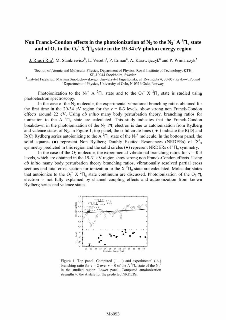
Non Franck-Condon effects in the photoionization of N2 to the N2+ A 2ΠΠΠΠu state
and of O2 to the O2+ X 2ΠΠΠΠg state in the 19-34 eV photon energy region
J. Rius i Riua, M. Stankiewiczb, L. Vesethc, P. Ermana, A. Karawajczyka and P. Winiarczykb
aSection of Atomic and Molecular Physics, Department of Physics, Royal Institute of Technology, KTH,
SE-10044 Stockholm, Sweden bInstytut Fizyki im. Mariana Smoluchowskiego, Uniwersytet Jagiellonski, ul. Reymonta 4, 30-059 Krakow, Poland
cDepartment of Physics, University of Oslo, N-0316 Oslo, Norway
Photoionization to the N2+ A 2Πu state and to the O2
+ X 2Πg state is studied using photoelectron spectroscopy.
In the case of the N2 molecule, the experimental vibrational branching ratios obtained for the first time in the 20-34 eV region for the v = 0-3 levels, show strong non Franck-Condon effects around 22 eV. Using ab initio many body perturbation theory, branching ratios for ionization to the A 2Πu state are calculated. This study indicates that the Franck-Condon breakdown in the photoionization of the N2 1πu electron is due to autoionization from Rydberg and valence states of N2. In Figure 1, top panel, the solid circle-lines (-●-) indicate the R(D) and R(C) Rydberg series autoionizing to the A 2Πu state of the N2
+ molecule. In the bottom panel, the solid squares (■) represent Non Rydberg Doubly Excited Resonances (NRDERs) of 1Σ+
u symmetry predicted in this region and the solid circles (●) represent NRDERs of 1Πu symmetry.
In the case of the O2 molecule, the experimental vibrational branching ratios for v = 0-3 levels, which are obtained in the 19-31 eV region show strong non Franck-Condon effects. Using ab initio many body perturbation theory branching ratios, vibrationally resolved partial cross sections and total cross section for ionization to the X 2Πg state are calculated. Molecular states that autoionize to the O2
+ X 2Πg state continuum are discussed. Photoionization of the O2 πg electron is not fully explained by channel coupling effects and autoionization from known Rydberg series and valence states.
0,5
0,6
0,7
0,8
0,9
1,0
1,1
0 2 43 5 7
5 8
5 10R(D)
n = 5
n = 4v' =
v' =
ξ A s
tate
(a.u
.)
n = 4
n = 5R(C)v' =
v' =
B
ranc
hing
Rat
io
21 22 23 24 25 26 27 28 29 30 31 32 3310-4
10-3
10-2
10-11 ΠΠΠΠ
u
1 ΣΣΣΣ +u
Excitation Energy (eV)
Figure 1. Top panel. Computed ( ― ) and experimental (-o-) branching ratio for v = 2 over v = 0 of the A 2Πu state of the N2
+ in the studied region. Lower panel. Computed autoionization strengths to the A state for the predicted NRDERs.
Mo093Mo001Mo093Mo093Mo093

High-resolution inner-shell studies of free radicals and transient species.
M. Alagia1, R. Richter2, and S. Stranges3
1 TASC-INFM, Area Science Park, 34012 Basovizza, Trieste, Italy.2Sincrotrone Trieste, Area Science Park, 34012 Basovizza, Trieste, Italy.3Department of Chemistry, University of Rome La Sapienza and INFM Unit, P.le A. Moro 5, 00185 Rome, Italy.
Third generation SR undulator beam lines allow low density and highly reactive species tobe studied in the gas phase using high photon energy resolution. Core-excited states in the OHand OD free radicals and the CS transient molecule have been investigated for the first time. Inthe case of the lowest O 1s excited state (2Σ+) of OH and OD, vibronic components have beenclearly observed in the total-ion-yield spectra using time-of-flight mass spectrometry. Relativetransition probabilities, excitation energies, and core-hole lifetimes have been measuredaccurately for the vibronic components of this state. Excited states at higher energies have beenalso observed. The free radicals have been produced in situ by the fast atom-molecule reactionH (D) + NO 2 → OH (OD) + NO using a microwave technique to generate the H atom. In thecase of CS, C 1s and S 2p excited states have been observed. Some of them display a resolvedvibrational progression. As for the S 2p excited states, the extent of the vibrational progression,and therefore the change in molecular geometry, varies largely depending on the specificresonant state. Spin-orbit and molecular field splitting effects in S 2p excitation processes areobserved for the first time in a diatomic molecule. The CS transient species has been producedin situ using the CS2 precursor and the same microwave technique.
Core-excited states of transient and radical species are often observed as “intermediatefragments” in studies of ultra-fast dissociation processes of core-excited molecules. Ourexperiments provide, for the first time, a direct information on the inner-shell spectroscopy ofthose fragments.
Mo094Mo001Mo094Mo094Mo094

STATISTICAL PROPERTIES OF INTER-SERIES MIXING IN
HELIUM: FROM INTEGRABILITY TO CHAOS
R. P�uttner1, B. Gr�emaud2, D. Delande2, M. Domke1, M. Martins1,
A. S. Schlachter3, and G. Kaindl1
1Institut f�ur Experimentalphysik, Freie Universit�at Berlin,
Arnimallee 14, D-14195 Berlin-Dahlem, Germany2Laboratoire Kastler-Brossel T12 E1, Universit�e Pierre et Marie Curie,
4 Place Jussieu, 75005 Paris, France3Lawrence Berkeley National Laboratory, 1 Cyclotron Road, Berkeley, CA 94720, USA
Due to the non-integrability of the classical three-body problem, no exact quantum num-
bers can be found to describe the helium atom. Nevertheless, for not too high excitation
energies, the approximate classi�cation scheme N;Kn (with N (n) being the quantum num-
ber of the inner (outer) electron and K the angular correlation quantum number) can be
used to describe the 1Po doubly excited states in helium. However, in the semiclassical limit,
i.e. close to the double-ionization threshold, the in uence of the underlying classical phase
space is expected to become more and more important for the quantum mechanical solution.
We present high-resolution photoabsorption spectra below the N=9 ionization threshold
measured at beamline 9.0.1 at the Advanced Light Source in Berkeley/California, and cal-
culated photoionzation cross sections in the same energy region using the complex-rotation
method. Based on the excellent agreement between experiment and theory, we performed a
statistical analysis of the nearest neighbor spacing (NNS) using the theoretical results.
The NNS distribution was determined by two di�erent procedures: (i) globally by consid-
ering all resonances regardless of the series to which they belong; (ii) individually for each
series associated with a given value of N �K, i.e. constant number of bending quanta with
respect to a collinear eZe con�guration. In the �rst case the NNS distribution reveals a Pois-
son distribution that is typical for a regular system, while in the second case a development
of the NNS distribution towards a Wigner distribution � which is typical for chaotic systems
� was found. This corresponds to the loss of the radial quantum number N , whereas N �K
remains approximately a good quantum number, and it is directly related to the instability
of the eZe orbits along the radial direction and their stability with respect to bending.
In addition, 1 dimensional (1D) stretched helium was studied theoretically below the N=9,
13, and 17 ionization threshold revealing that the NNS distribution develops clearly towards
a Wigner distribution with increasing N . This study provides an estimate for the observation
of a fully chaotic regime in 3D helium (for N � 17).
In conclusion, we show clear evidence of chaotic e�ects in both the 3D and 1D helium
spectra on the basis of statistical analysis, emphasizing the role played by mixing between
di�erent Rydberg series.
The work in Berlin was supported by the Deutsche Forschungsgemeinschaft, project DO
561/1-3, and the Bundesministerium f�ur Bildung und Forschung, project 05-SR8KE1-1.
Mo095Mo001Mo095Mo095Mo095

Ultra-fast dissociation processes in poly-atomic molecules.
I. Hjeltea, M. N. Piancastellia,b, R. F. Fink a, c, O. Björneholma, M. Bässlera, R. Feifela, K.Wiesner a, S. L. Sorensene and S. Svenssona
aDept. of Physics, Uppsala University, S- 751 21 Uppsala, SwedenbDept. of Chemical Sciences and Technologies, University “Tor Vergata”, 00133 Rome, Italycalso at: Dept. of Theoretical Chemistry, Chemical Centre, Box 124, S-22100 Lund, Sweden
eDept. of Synchrotron Radiation Research, Inst. of Physics, Lund University, Box 118, S-221 00 Lund, Sweden
Core-excitation processes can be induced in a molecule if radiation of a suitable wavelength isused to promote one electron from a core level to an empty molecular or Rydberg orbital.Core-excited species are unstable and decay processes follow rapidly; the most long-livedcore holes have a lifetime in the order of 10 fs. Core excitations lead frequently to dissociativeor predissociative states. In some cases, the nuclear dynamics is so rapid that dissociationtakes place on the same time scale as Auger electron emission. Ultra-fast dissociation is aterm used to describe dissociation taking place on a time scale comparable to that of theAuger decay placing it in the low femtosecond regime. This time scale corresponds to theshortest molecular dissociation times i.e. to the fastest existing chemical reactions. It haspreviously been shown to occur in a number of molecules such as HBr [1], HCl [2], H2S [3],O2 [4] and N2 [5]. Since the beginning of the nineties, there has been a long-standingdiscussion on the possible occurrence of ultra-fast dissociation of core-excited water into O*Hand H (* indicates core excitation) [6] which has been indicated in ion yield spectroscopy [7,8]. However, for ammonia no conclusive evidence of this phenomenon has been presentedpreviously.
We present direct evidence for ultra-fast dissociation of molecular water [9] and ammonia inconnection to photo-excitation of the O1s→4a1 and N1s→4a1 resonance's respectively. Thecore-excited molecules are shown to dissociate into a core-excited fragment and a neutralfragment on a time scale comparable to the core hole lifetime, i.e. a few femtoseconds. Thisconclusion is based on a resonant Auger study and qualitative arguments concerning thedispersive behavior of the fragment versus the molecular lines while tuning the frequency ofthe exciting light around the resonance as well as ab initio calculations in the case of water.
Figure 1. The Auger decay spectra of molecular water and ammonia after core excitation.
[1] P. Morin, I. Nenner, Phys. Rev. Lett. 56 (1986) 1913.[2] H. Aksela, et. al., Phys. Rev. A41 (1990) 6000.[3] A. Naves de Brito,et. al. , J. Mol. Structure (Theochem) 394 (1997) 135.[4] S. J. Schaphorst, et. al., Chem. Phys. Lett. 213 (1993) 315.[5] M. Neeb, et. al., Phys. Rev. Lett. 76 (1996) 2250.[6] D. Coulman, et. al., J. Chem. Phys. 93 (1990) 58.[7] M.N. Piancastelli, et. al., Phys. Rev. A 59 (1999) 300.[8] A. Naves de Brito, et. al., Chem. Phys. Lett. 309 (1999) 377.[9] I. Hjelte, et. al., Chem. Phys. Lett. 334 (2001) 151.
390388386384382380378
3a11e
NH3N1s → 4a1
Fragment peaks
520518516514512510508
H2OO1s → 4a1
1b13a11b2
Mo096Mo001Mo096Mo096Mo096

Photoexcitation Processes of Methanol Isotopmers in VUV Region
Bing-Ming Cheng,1 Mohammed Bahou,2 Yuan-Pern Lee,2 and L. C. Lee3
1 SRRC, No. 1, R&D Road VI, Hsinchu Science- Based Industrial Park, Hsinchu 300, Taiwan
2 Department of Chemistry, National Tsing Hua University, No. 101, Sec. 2, Kuang Fu Road, Hsinchu 300, Taiwan
3 Department of Electrical and Computer Engineering, San Diego State University, San Diego, California 92182
The photoabsorption cross sections of CH3OH, CH3OD, CD3OH, and CD3OD are measuredin the 110-220 nm region using the synchrotron radiation light source in Taiwan. In the 160-220nm region, the absorption maximum and band shape of CH3OH are same as CD3OH, and thoseof CH3OD are same as CD3OD; but the absorption maximum of CH3OD is blue-shifted fromCH3OH. This result shows that the excitation is clearly related to the O-H bond. On the otherhand, in the 140-160 nm region the absorption spectrum of CH3OH is similar to CH3OD, and thatof CD3OH is similar to CD3OD; but the band origin of CD3OH is blue-shifted from CH3OH. Thisresult shows that the excitation in this region is related to the C-O bond. Based on the absorptioncross sections combined with theoretical calculations, the quasi-diatomic potential curves for theC-O and O-H bonds of methanol are derived.
The absorption in the 160-220 nm is a smooth continuous band that results a repulsivecurve for the O-H bond. This curve is produced from perturbation between the 3s Rydberg stateand the valence state that dissociates into CH3O + H products. A vibrational progression with anirregular spacing appears in the 150-160 nm region. The irregularity is caused by perturbationbetween the 3p Rydberg state and the valence state that dissociates into CH3 + OH products. Avibrational progression with a regular spacing appears in the 140-150 nm region. The upper stateof this band is assigned to the 3p' Rydberg state of which the energy is higher than the repulsivevalence state. The regular spacing indicates that this 3p' state is a bound state, and its potentialcurve is not perturbed by a valence state.
The deuteration technique is a powerful tool to study photoexcitation processes ofmolecules in the infrared region. The current results demonstrate that this technique can also beextended to study the photoexcitation processes in the VUV region.
Mo097Mo001Mo097Mo097Mo097

The vacuum-UV photofragmentation of a range of hydrofluorocarbon (HFC) cations, CxHyFz
+, studied using coincidence techniques
W.D. Zhou, D.P. Seccombe, R.Y.L. Chim and R. P. Tuckett
School of Chemistry, University of Birmingham, Edgbaston, Birmingham B15 2TT, UK
HFCs are being increasingly used as substitutes for the banned chlorofluorocarbons in a range of domestic and industrial applications. The lifetime of these species in the atmosphere can be very long (ca. years) due to their slow reaction with the important tropospheric free radicals such as OH and O (1D). The removal of these species from the atmosphere can therefore be governed by processes occurring in the mesosphere. We are investigating these possible uni- and bi-molecular processes in the laboratory. The bi-molecular processes include reactions of HFCs with small positive ions and with electrons. The principal uni-molecular process is vacuum-UV photodissociation, especially with wavelengths in the range 50-150 nm, and this poster presents a progress report on our work.
Using vacuum ultraviolet radiation from the Daresbury synchrotron source, we have used threshold photoelectron-photoion coincidence (TPEPICO) spectroscopy to study the decay dynamics of the valence electronic states of the parent cation of several HFCs (CHF3, CF3−CHF2, CF3−CH2F, CHF2−CHF2, CF3−CH3) in the energy range 10 to 25 eV. This project represents a follow-on to a successful study of both saturated and unsaturated perfluorocarbon cations by the TPEPICO technique [1]. Electrons and ions are detected by threshold electron analysis and time-of-flight mass spectrometry respectively, allowing breakdown diagrams showing the formation probability of fragment ions as a function of the internal energy of the parent ion to be obtained. Higher resolution, fixed-energy TPEPICO spectra were also performed on many of the heavier fragment ions, and the translational kinetic energy released in fragmentation determined [2]. By comparing the integrated threshold photoelectron signal with the total ion yield as a function of photon energy, the importance of autoionisation in these molecules can be gauged.
For many of these saturated HFCs, the parent ions are not observed, indicating that the ground states of the ions are repulsive in the Franck-Condon region. Both from the analysis of the breakdown diagrams and the high kinetic energy releases, we conclude that non-statistical effects are occurring for states of these cations below ca. 18 eV. This suggests that decay from these states takes place rapidly before internal conversion to the ground state of the parent cation can occur. The substituted ethanes therefore seem to be mimicing the behaviour of C2F6
+ (CF3−CF3+) where non-statistical effects are well
known [1], and not that of C2H6+ where statistical dissociation is observed [3]. F--anion migration
across the C−C bond must occur to explain the observation of some of the daughter ions. References [1] G.K. Jarvis, K.J. Boyle, C.A. Mayhew and R.P. Tuckett, J. Phys. Chem. A., (1998) 102, 3219 and 3230 [2] G.K. Jarvis, D.P. Seccombe and R.P. Tuckett, Chem. Phys. Letts., (1999) 315, 287 [3] F. Guthe and K.M. Weitzel, Ber. Buns. Ges. Phys. Chem., (1997) 101, 484
Mo098Mo001Mo098Mo098Mo098
Resonant Auger Raman spectroscopy at the Kr 3p edge
K.C. Prince1, M. Coreno2 and M. de Simone3,
Sincrotrone Trieste, Strada Statale 14, km. 163.5, in Area Science Park,I-34012 Basovizza (Trieste) Italy,
INFM- TASC, Laboratorio ELETTRA, I-34012 Trieste, Italy,Università di Roma III ed Unità INFM, Via della Vasca Navale 84, I-00146 Rome, Italy.
The x-ray absorption spectra of the sharp core levels of noble gases, Ne 1s, Ar 2p, Kr 3d, etc,have been extensively studied and are well understood [1, 2,] but less attention has been given to theirbroader, more difficult core levels [3, 4]. At the Kr 3p edge, the Rydberg sates are about 1.4 eV wide,overlap one another and are superimposed on a strong 3d continuum [4]. Thus the peaks are difficult toidentify and controversy exists about their assignment [5].
The 3p-1nl (n>4, l=0; n>3, l=2) Rydberg states decay rapidly via Coster-Kronig (CK) processesto 3d-14p-1nl, 3d-14s-1nl and related shake states, and via super Coster-Kronig processes. We haveidentified the CK states, fig. 1, and they lie close to the 3d satellite states 3d-14p-1np. On the first main3p-1nl resonance, there is a strong increase in emission below and above the 3d satellites. The peakshave widths determined by the resolution of the photons and the energy analysers, about 200 meV,fulfilling Resonant Auger Raman conditions. The new peaks disperse with photon energy as expected.
Constant Initial State spectra were measured by setting the analysers to energies correspondingto each group of peaks, fig. 2. The CIS spectra are shown in fig. 2, and clear differences in the resonantbehaviour are seen. Peaks A, B and C show similar resonant behaviour on the absorption peak that hasbeen attributed to the 3p-15s, both for 3p3/2 and 3p1/2 at 209.8 and 218 eV photon energy respectively. Incontrast the E and F multiplets show completely different resonances, and are assigned to ionic statesderived from different intermediate excited states.
We have successfully applied the Resonant Auger Raman technique and CIS to the problem ofthe absorption spectrum of Kr at the 3p edge.
[1] M. Coreno, et al, Phys. Rev. A 59 (1999) 2494.[2] O.P. Sairanen et al, Phys. Rev. A 54 (1992) 2834.[3] T. Kylli et al, Phys. Rev. A. 59 (1999) 4071.[4] I.T. Steinberger et al, Phys. Rev. B 60 (1999) 3995.[5] M. Ohno, Phys. Rev. A 51 (1995) 1042.
Fig. 1. Photoemission spectra below resonance(lower curves), and on resonance below the Kr 3pthreshold.
Fig. 2. CIS spectra, Kr 3p threshold.
800
600
400
200
0
Counts
969492908886Kinetic energy (eV)
1009896949290
hv=209.8 eV
hv=205.0 eV
AB
C
D
E
F
30x103
20
10
0Inte
nsity
(co
unts
)
220215210205Photon energy (eV)
C C
E (offset)
F (offset)
Mo099Mo001Mo099Mo099Mo099

1
High resolution photoabsorption studies at the C and O K edges
K.C. Prince1, M. de Simone2, R. Richter1, M. Coreno3 and M. Alagia3,1Sincrotrone Trieste, Strada Statale 14, km 163.5, Area Science Park,
I-34012 Basovizza (Trieste), Italy.2Università di Roma III ed Unità INFM, Via della Vasca Navale 84, I-00146 Rome, Italy,
3INFM- TASC, Laboratorio ELETTRA, I-34012 Trieste, Italy,
We report the carbon and oxygen K edge x-ray absorption spectra of fourtetrahedral molecules CX4 (X = H, Cl, F) and a series of oxygenated organicmolecules (CH2O, HCOOH, CH3CH2O, CH3COCH3, CH3OH and CH3OCH3),with the cross-section detemined in absolute units.
The antibonding valence peaks of the halides do not show any vibrationalstructure [1-3] but sharp Rydberg states are observed above these states at the C 1sedge. We identify a possible Fermi resonance in the Rydberg states of core excitedCF4, and we report the CCl4 Rydberg states for the first time. The C 1s intrinsicline widths were compared to theoretical predictions of ion state line widths andreasonable values are found for CH4 and CCl4 but not for CF4. The discrepancy isassigned to valence band - Rydberg mixing, which is especially strong in CF4,and/or to multi-centre Auger processes [4].
At the carbon K edge of the oxygenated species, most gases showvibrational structure; the spectrum of CH2O is in good agreement with that of [5],whereas few high resolution spectra of the other gases have been reported. At theoxygen edge, only CH2O shows vibrational structure. This is important as itindicates that for the very similar molecules CH3COCH3 and CH3CH2O, thestructure is not absent due to lifetime broadening, but is obscured by manyoverlapping vibrational bands. This implies that the excited states of all aldehdydesand ketones are most likely bound. On the othe hand the situation is unclear for thesaturated molecules CH3OH and CH3OCH3.
Fig. 1. Oxygen K edge spectrum of
formaldehyde. The first 5 vibrational states
(CO stretch) are marked .
References.1. K. Ueda et al, Chem. Phys. Lett. 236 (1995) 311.2. W. Zhang, T. Ibuki and C.E. Brion, Chem. Phys. 160 (1992) 435.3. B.S. Itchkawitz et al, Rev. Sci. Instrum. 66 (1995) 1531.4. T. X. Carroll, et al, Phys. Rev. A 61 (2000) 42503.5. G. Remmers et al, Phys. Rev. A 46 (1992) 3935.
80x103
60
40
20
0Inte
nsity
(ar
b. u
nits
)
532.0531.0
Photon energy (eV)
0
1
23 4
Mo100Mo001Mo100Mo100Mo100

MEASUREMENTS OF THE ABSOLUTE PHOTOIONIZATION CROSSSECTION OF THE DOUBLY-CHARGED O2+ ION
J.-P. Champeaux,1,2 J.-M. Bizau,2 C. Blancard,1 D. Cubaynes, 2 A. Compant La Fontaine,1
C. Couillaud,1 R. Marmoret,1 D. Hitz,3 M. Delaunay,3 S. Nahar,4 and F. J. Wuilleumier1
1Centre d’Etudes de Bruyères-le-Châtel, BP12, 91680 Bruyères-le-Châtel, France2Université Paris-Sud, LSAI, UMR 8624 CNRS, B. 350, 91405 Orsay3DRF, SI2A, CEA-Grenoble, rue des Martyrs, 38054 Grenoble, France
4S. Nahar, The Ohio State University, Columbus, Ohio 43210 , USA
Photoionization of O2+ ion over the energy range 50-70 eV has been investigated experi-mentally using the end-station for studies of photon-ion interactions at the Super ACO synchro-tron radiation source in Orsay. The experiments were performed at the SU6 undulator beamline,which provides monochromatic photon beams in the energy range 30-160 eV. The photon beamwas merged with a collimated 500 nA-beam of O2+ ions produced in an ECR ion source. The spa-tial overlap of the beams was determined by three sets of translating-wire beam-profile monitors.After an interaction path of 20 cm, the parent O2+ beam and the O3+ product ions were demergedand counted separately. In the interaction region, the O2+ ions are mainly in the 2s22p2 3P0,1,2ground state, but some of them are also in the 2s22p2, 1S or 1D, and 2s2p3 5S metastable states.Photo-ion yield measurements were carried out at a resolving power of 225. Previous R-Matrix[1] and new MCDF calculations were used to interpret the data.
In Fig. 1, we show the variation of the photoionization cross section as a function of photonenergy between 47 eV and 65 eV. Continuum and resonant structures are seen in the measuredcross section. The onset of the cross section at the 2p ionization threshold of the 3P ground stateis clearly seen at 54.90 eV. The lines are produced mainly by autoionization of 2s2p2np(n ≥ 2)2S+1LJ excited states of O2+ ion into the continua of the 2s22p O3+ ion. The results of both R-matrix and MCDF calculations agree satisfactorily with experiment and allow to identify the lines.Lines 1 and 2, below the 3P0,1,2 thresholds, are due to 2p-excitation of O2+ ions in the 2s2p3 5S
and 2s22p2 1S metastable states, respectively.Above the 3P thresholds, several lines (line 3 at58.27 eV as the first one) originate from excita-tion of O2+ ions in the 3P0,1,2 ground state. Otherlines are due to similar excitations of O2+ ionseither in the 1D (line 4 at 58.90 eV) or 1S (line 5at 61.07 eV) metastable states. We determinedalso the absolute value of the cross section forphotoionization into the continuum. Expe-rimental and theoretical values of energies andcross sections will be presented and discussed atthe conference.
1. S. Nahar, Phys. Rev. A 58, 3677 (1998).
Fig. 1 - Measured photoionization cross section of O2+
between 47 and 65 eV photon energy. The cross sectionscale is in arbitrary units.
Mo101Mo001Mo101Mo101Mo101

HIGH-RESOLUTION STUDIES OF CORRELATION SATELLITES INPHOTOIONIZATION OF SODIUM ATOMS IN THE 2p-SUBSHELL
D. Cubaynes,1 S. Diehl,2 J.-M. Bizau,1 F. J. Wuilleumier,1 J. D. Bozek,3 B. Rude,3 S. Canton,4
and N. Berrah4
1LSAI, UMR CNRS 8624, Université Paris-Sud, B. 350, Orsay 91405, France2Laboratoire DIAM, UMR CNRS, Université Paris VI, place Jussieu, 75231 Paris, France
3Lawrence Berkeley Laboratory, ALS, University of California, Berkeley, CA 94720, USA4Western Michigan University, Department of Physics, Kalamazoo, MI 43309, USA
In earlier studies of photoionization of sodium using electron spectroscopy, [1, 2] the manycomponents of the various groups of satellites could not be resolved because the spectral and elec-tron spectrometer resolutions were not good enough. In the work presented here, we have used thehigh-resolution Scienta electron spectrometer with an ultimate spectral resolution of 25 meV, andthe photon beam available at the 10.0 1 beamline of the Advanced Light Source in Berkeley bet-ween 50 and 110 eV photon energy. The total instrumental resolution, including contributionsfrom both excitation and detection channels as well as the Doppler effect, was at best 35 meVFWHM. With such a high-resolution, we were able to study the dynamics of 2p-photoionizationtransitions to correlation satellite states created without transfer of angular momentum: ∆l = 0(electronic configuration 2p54s, shake-up transitions) and with transfer of angular momentum: ∆l = 1 (electronic configuration 2p53p) and ∆l = 2 (electronic configuration 2p53d).
In the photoelectron spectrum following photoionization in the 2p-subshell, three groups oflines are clearly indentified: the main lines due to single ionization of a 2p-electron, ∆l = 0, (2p53sfinal ionic states), the satellites produced with ∆l = 1 (states with 2p53p electronic configuration),and the high-energy satellites produced with ∆l = 0 (states with electronic configuration 2p54s),and ∆l = 2 (states with electronic configuration 2p53d). Resolving the satellites within the latergroup requires as high a resolution as possible. In Figure 1, we show this later group of satellitesmeasured at 54 eV and 90 eV photon energy. One clearly sees three components: peak numbered1 corresponds to a mixture of satellites states with both 2p54s and 2p53d electronic configurations,peak 2 is mainly due to 2p54s satellite states, and peak 3 is assigned to only 2p53d satellite states.Peaks 2 and 3 are good references to detect any difference in the energy dependence of the rela-tive intensity of the ∆l = 0 and ∆l = 2 satellites, respectively. At 54 eV, the three componentshave roughly the same intensity, at 90 eV, the relative intensity of peak 3 has clearly decreased,revealing a significant energy dependence of the 2p53d state intensity. The 2p53p correlation satel-lites were all resolved. Detailed results will be presented at the conference and compared to exis-ting photoionization calculations.
[1]. S. Krummacher et al., J. Phys. B 15, 4363 (1982).[2]. D. Cubaynes et al., Phys. Rev. A 57, 4432 (1998).
Fig. 1 - Electron spectra showing theintensity of the correlation satellites pro-duced with ∆l = 0 (peak 2, mainly 2p54sfinal ionic states) and with ∆l = 2 (peak3, 2p53d final ionic states) at 54 eV (leftpanel) and 90 eV (right panel) photonenergy, respectively.
Mo102Mo001Mo102Mo102Mo102

Beat structure in the doubly excited Rydberg states converging
to the N=2 threshold of helium
R. Follath, G. Reichardt, O. Schwarzkopf and W. Gudat
BESSY, Albert-Einstein-Stra�e 15, 12489 Berlin, Germany
Doubly excited helium is the prototype system for studying autoionisation phenoma, i.e. the
e�ect of electron correlation. The presence of only two electrons makes it readily accessible
for ab initio calculations of the parameters describing the autoionisation resonances.
In the present work we investigated the helium Rydberg series converging to the N=2 au-
toionisation threshold (IP2) by means of total photoionisation yield measurements. Three
Rydberg series are converging to this ionisation threshold, namely the principle (2; 0n) and
the secondary series (2; 1n) and (2;�1n). With a resolving power of 90 000 a hitherto unre-
solved beat structure in the series was observed at very high excitation levels.
Figure 1 gives an enlarged view of the region just below the N=2 ionization threshold. The
amplitudes of the autoionisation pro�les decrease continuously up to a minimum value at
n = 26 as already observed in experiments with lower energy resolution [1]. But with the
highest currently available energy resolution, an increase of the amplitudes above this pro�le
with a maximum value at the (2; 029) line is observed. For n > 29, the amplitude is again
decreasing. The beat can be explained by a mixing of two series converging to di�erent ion-
isation thresholds and having an energy spacing corresponding to the energy split between
the 2p1=2 and 2p3=2 level of singly ionised helium. This indicates that the electron interac-
tion at high excitation levels can be described within the jj-coupling scheme of two electrons.
65,37 65,38 65,39 65,40 65,41
0,95
1,00
1,05
1,10 IP250
2,026
2,0n
3022n=21 23 24 25 26
phot
oion
izat
ion
yiel
d [a
rb.
units
]
energy [eV]
Figure 1: Near thresh-
old region of the N=2
Rydberg series. Between
the pro�les (2; 026) and
(2; 034) a beat structure is
observed. The arrow in-
dicates the 26th pro�le of
the (2; 0n) series, where
the minimum in the am-
plitudes occurs. IP2 de-
notes the ionization limit.
[1] K. Schulz, G. Kaindl, J. D. Bozek, P. A. Heimann and A. S. Schlachter, J. Electr. Spectr.
Rel. Phenom. 79, 253 (1996).
Mo103Mo001Mo103Mo103Mo103

NUCLEAR MOTION EFFECTS OBSERVED IN PHOTOELECTRONSPECTRA OF HCL AND DCL MOLECULES
F. Burmeister1, L. M. Andersson2, H. O. Karlsson2, S. L. Sorensen3, O.
Björneholm1, A.Naves de Brito4, R. F. Fink1, R. Feifel1, I. Hjelte1, K.
Wiesner1, A. Giertz1, M. Bässler1, C. Miron1, H. Wang1, M. N.
Piancastelli5, L. Karlsson1, S. Svensson1 and O. Goscinski2
1 Dept of Physics, Box 530, S-75121 Uppsala, SWEDEN
2 Dept. of Quantum Chemistry, BoxBox 518, S-751 20 Uppsala SWEDEN
3 Dept. of Synchrotron Radiation Research, Institute of Physics, Box 118, S-221 00 Lund, Sweden
4 Laboratorio Nacional de Luz Sincrotron, Box 6992, CEP 13083 Campinas SP, BRAZIL
5 Dept. Of Chemical Sciences and Technologies, University "Tor Vergata", Rome, Italy
The HCl inner-valence photoelectron band at 26 eV binding energy has been recorded with
a photon energy of 64 eV at high resolution. Discrete peaks arising from at least two separate
vibrational progressions are superimposed on the broad continuum. Fano profiles are visible in
one of the progressions. This indicates interference between superimposed electronic states. In
the isotopic DCl molecular spectrum the discrete lines are much less pronounced. The difference
between HCl and DCl is due to dissociation dynamics[1], where non-adiabatic, non-avoided
crossing behavior is more pronounced for the lighter HCl molecule. A theoretical treatment of
the systems, with simulated spectra based upon the potential curves [2] is also presented.
References
[1] H. Eyring, J. E. Walter, and G. E. Kimball, Quantum Chemistry, 326-330 (Wiley, New
York, 1944).
[2] M. Hiyama, S. Iwata, Chem. Phys. Lett, 210, 187 (1993).
Mo104Mo001Mo104Mo104Mo104

Tot
al Io
n Y
ield
Photon Energy / eV
285 286
CH3
N
Figure 1. C 1s÷B* total ion yield spectra ofpyridine (top), toluene (middle) and benzene(bottom).
Core excitation and ionic fragmentation of aromatic molecules
John J. Neville
Department of Chemistry, University of New Brunswick, Box 45222, Fredericton, NB E3B 6E2, Canada
C 1s ÷ B* resonant excitation of benzene yields a core-excited state in which the core hole islocalised on a single carbon atom and the aromaticity is lost [1]. This symmetry lowering isaccompanied by a rehybridisation of the bonding orbitals of the excited carbon atom, loss ofplanarity and the excitation of both symmetric and asymmetric vibrational modes. In the case ofaromatic molecules of lower symmetry, such as pyridine and toluene, resonant core-excitation canin principal be performed selectively at each of the chemically inequivalent carbon centres, givensufficiently large chemical shifts in the C 1s energy levels between inequivalent sites and asufficiently narrow photon bandwidth. The resulting core-excited states might be expected to havediffering vibrational structure depending upon the location of the core-excited atom with respect tothat of the heteroatom or the substitution site. In all cases, the core-excited state is short lived;electronic relaxation typically results in multiple ionisation and molecular fragmentation.
Results will be presented of studies of the coreex c i t a t ion spec t roscopy and subsequentphotofragmentation of a number of aromatic molecules,performed with high resolution using synchrotronradiation and ion time-of-flight mass spectrometry. Inparticular, the vibrational structure of the C 1s ÷ B*
transitions of benzene, toluene and pyridine, shown inFigure 1, will be discussed and contrasted. The ionicfragmentation processes occurring in these moleculesfollowing selective core excitation will be examined asa function both of core-hole position and of vibrationalexcitation.
Reference
[1] Y. Ma, F. Sette, G. Meigs, S. Modesti and C. T.Chen, Phys. Rev. Lett. 63 (1989) 2044--2047.
Mo105Mo001Mo105Mo105Mo105

NUCLEAR MOTION EFFECTS AS OBSERVED IN THE RESONANT AUGER DECAY TO THE X2Π ELECTRONIC GROUND STATE OF N2O
+
C. Miron1-3, M. Simon1,2, P. Morin1,2, S. Nanbu4, N. Kosugi4, S. L. Sorensen5, A. Naves de
Brito6, M. N. Piancastelli7, O. Björneholm3, R. Feifel3, M. Bässler3 and S. Svensson3
1 LURE, Bât. 209d, BP. 34, Université Paris-Sud, 91898 ORSAY Cedex, France
2 Laboratoire “Francis Perrin” and CEA/DRECAM/SPAM, Bât. 522, CE Saclay, 91191 Gif/Yvette Cedex, France 3 Department of Physics, Uppsala University, Box 530, S-751 21 Uppsala, Sweden
4 Institute for Molecular Science, Myodaiji, JP 444-8585 Okazaki, Japan 5 Department of Synchrotron Radiation Research, University of Lund, Box 118, S-221 00 Lund, Sweden
6 Laboratorio Nacional de Luz Sincrotron, BR 13083-970 Campinas, Brazil 7 Department of Chemical Sciences and Technologies, University “Tor Vergata”, 00133 Rome, Italy
High-resolution Auger spectroscopy applied under resonant Auger Raman (RAR) conditions is shown to be a powerful tool for characterizing complex potential energy surfaces in core-excited systems. Using the example of Nt1s-1 → π* resonant Auger transition in nitrous oxide we emphasize the interplay between the nuclear motion and the electronic decay. We show how the choice of the excitation energy allows selection of core-excited species of different geometries [1,2]. The nuclear dynamics of these species are mapped by measuring the resonant Auger decay spectra. In addition to the changes in vibrational structure observed for the resonant Auger decay spectra, a strong influence of the nuclear motion on the electronic decay is revealed inducing the so-called “dynamical Auger emission”. The experiments have been carried-out at the undulator beamline I411 at the Swedish synchrotron radiation facility Max-Lab in Lund. The experimental results are supported by ab-initio quantum chemical calculations restricted to a linear geometry of the core excited state (Figure 1).
Inte
nsity
[Arb
. uni
ts]
16.015.014.013.0Binding Energy [eV]
NNO - RAS (X2Π)
Figure 1: Comparison between ab-initio calculations (bars and continuous line) and the experimental resonant Auger decay spectrum (empty circles) of the X2Π electronic state of N2O measured on the top of the π* resonance: hν =401.3 eV.
References [1] J. Adachi, N. Kosugi, E. Shigemasa and A. Yagishita, J. Chem. Phys. 102, 7369 (1995). [2] J. Adachi, N. Kosugi, E. Shigemasa and A. Yagishita, J. Chem. Phys. 107, 4919 (1997).
Mo106Mo001Mo106Mo106Mo106

Hot-electron dynamics in mass-selected transition metal clusters probed byfemtosecond pump-probe photoelectron spectroscopy
N. Pontius, G. Lüttgens, P.S. Bechthold, M. Neeb, and W. EberhardtInstitut für Festkörperforschung, Forschungszentrum Jülich GmbH, 52425 Jülich, Germany
Clusters in the gas phase are confined systems ideally suited to follow the internal energydissipation from a non-thermally excited state into the thermal equilibrium. Of emerging interestis the dynamics of excited electrons in a non-equilibrium state, which is created when an electron
is photoexcited by an ultrashort light pulse. Here we show by time-resolved photoelectronspectroscopy that electron relaxation processes are efficient energy dissipation channels not onlyin bulk metals but also in extremely small transition metal clusters [1]. An analysis of the time-resolved two-photon photoemission spectra of optically excited small Ni, Pd,, Pt- and metal-carbonyl clusters reveal effective electron relaxation times of less than 100 fs for electronsexcited below the vacuum threshold (hνpump=1.5 eV). A comparative series of pump-probe
spectra of different Pd-clusters demonstrates that the relaxation times vary with the cluster size.This is attributed to the partial density of states of small clusters as deduced from the resonanttwo-photon photoemission spectra of Pd3-7. In comparison to simple metal clusters, e.g. Agn, andNan, the bulk-like inelastic scattering processes in open d-shell transition metal clusters areattributed to the larger electronic level density caused by both the small d-bandwidth at the Fermilevel and the larger number of valence electrons. Furthermore, statistical evaporation of CO-ligands has been observed as alternative energy relaxation channel in metal-carbonyl clusters.While Pt2(CO)5
- reveals thermal desorption of a single CO ligand reaching up to the nanosecondregime, Au2(CO)- desorbs the CO-ligand within some hundreds of femtoseconds. The reason forthe high desorption rate in Au2(CO)- is attributed to the much smaller CO-desorption barrier andthe smaller number of vibrational degrees of freedom..
References
[1] N. Pontius, P.S. Bechthold, M. Neeb, W. Eberhardt, Phys. Rev. Lett. 84, 1132 (2000).
Mo107Mo001Mo107Mo107Mo107

The electronic structure of supported endohedrally doped fullerenes
R. Klingeler, I. Wirth, G. Kann, S. Eisebitt, P. S. Bechthold, M. Neeb, and W. Eberhardt
Institut für Festkörperforschung, Forschungszentrum Jülich GmbH, 52425 Jülich, Germany
We have investigated the electronic structure of endohedrally doped fullerenes which havebeen deposited onto a substrate from a mass-selected ion beam [1]. The valence orbitals ofendohedrally doped C60 can be explained by those of pristine C60, taking a charge transfer and aJahn-Teller effect into account. The band gap depends on the kind of encapsulated atom, asshown by scanning tunneling spectroscopy. Semiconductor-like and metal-like densities of stateshave been observed for Ce@C60 and La@C60, respectively. The core level region exhibits a shiftto higher binding energies with respect to the bulk due to metal-to-cage charge transferprocesses, as demonstrated by X-ray photoelectron spectroscopy in the case of Ce 3d in Ce@C60.
References
[1] R. Klingeler, P.S. Bechthold, M. Neeb, W. Eberhardt, J. Chem. Phys. 113, 1420 (2000); M.Neeb, R. Klingeler, P.S. Bechthold, G. Kann, I. Wirth, S. Eisebitt, W. Eberhardt, Appl.Phys. A 72, 289 (2001)
Mo108Mo001Mo108Mo108Mo108

EVIDENCES OF LOCALIZATION EFFECT AFTER S 1s EXCITATION AND AUGER DECAY IN THE SO2 AND CS2 MOLECULES
E. S. Cardoso1,2, F. Burmeister3, O. Björneholm3, and A. Naves de Brito2,4
1 Campinas State University-UNICAMP/IFGW - Campinas-SP - Brazil 2 Laboratório Nacional de Luz Síncrotron-Box 6192 CEP 13083-970-Campinas-SP-Brazil
3 Uppsala University - Department of Physics - Uppsala - Sweden 4 Brasília University - Institute of Physics -Brasília-DF - Brazil
We present the measured electron-multi-ion coincidence spectra after photoexcitation of the SO2 and CS2 molecules around the S1s edge. A procedure for complete determination of all set of ions formed is described. The dissociation channels and its behavior with the photon energy of these molecules are presented in comparison with the Total Ion Yield (TIY). We observed a charge distribution preferentially of asymmetric form with higher charge in the excitation atom for SO2 molecule, see Fig.1. In CS2, similar asymmetric distribution was observed. Furthermore, above S 1s first resonance, the channel with an even charge distribution between the two oxygen in SO2 in the most abundant. In CS2 it is not, in it, one of the Sulfur atoms is core ionized. Auger cascade after the 1s core hole creation leading to 2p double hole states need to be taken in to account to explain the observed memory effects. The time scale of the nuclear motion and decay is taken into account to explain the competition between intra-atomic cascade Auger leading to asymmetric charge distribution and Coulomb explosion giving rise to symmetric charge distribution in CS2. Recombination forming an O2 ion was found for SO2
TIY
35
30
25
20
15
2475247024652420.80 2520.80
2.5
0.0
S+C
+S
2+
S+C
2+S
+
CS2+
S2+
Charge 4
CS2
photon energy (ev)
rel.
abu
nd
ance
(%
)
40
30
20
10
0
248024762472 2523.332423.33
0.50.0
O+S
++O
+
O++
S+O
+
S+++
O 2+
& S+ O 2
+++
rel.
abu
nd
ance
(%
)
photon energy (eV)
Charge 4
SO2
TIY
Figure 1: Relative abundance of selected dissociation channels with total ionic charge 4. The total ion yield (TIY) is also shown.
Reference [1] E. S. Cardoso, F. Burmeister, O. Björneholm, and A. Naves de Brito, Submitted for
publication in J. Chem. Phys. (2001
Mo109Mo001Mo109Mo109Mo109

EVIDENCE AGAINST ATOMIC-LIKE RESONANT AUGER DECAY IN N2 DOUBLY- EXCITED CORE STATES BY HIGH-RESOLUTION
EXPERIMENTS
A. Naves de Britoa,1, I. Hjelteb, K. Wiesnerb, R. Feifelb, M. Bässlerb, S. L. Sorensenc, O.
Björneholmb, M.N. Piancastellid, L. Karlssonb and S. Svenssonb
a Laboratório Nacional de Luz Síncrotron (LNLS), Box 6192 CEP: 13083-970, Campinas-Brazil, BRAZIL b Dept. of Physics, Uppsala University, Box 530, S-751 21 Uppsala, SWEDEN
cDept of Synchrotron Radiation Research, Univ. of Lund, Box 118, S-221 00 Lund, SWEDEN d Dept. of Chemical Sciences and Technologies, University "Tor Vergata", I-00133, Rome, ITALY
Resonant Auger spectra following the decay of doubly-excited core states in N2 in the range 409-411 eV have previously been assigned to “atomic” lines indicating ultrafast dissociation. Using high-resolution synchrotron radiation electron spectroscopy from the MAX II facility in Sweden we have remeasured the resonant Auger spectrum of N2 in the vicinity of the N 1s threshold. Contrary to earlier studies, we find vibrational progressions that can be associated to the final C 2Σu
+ and 2 2Πg states in N2+ . We conclude that the decay is entirely
leading to molecular final states.
References [1] A. Naves de Brito , I. Hjelte, K. Wiesner, R. Feifel, M. Bässler, S. L. Sorensen, O.
Björneholm, M.N. Piancastelli, L. Karlsson and S. Svensson, Submitted for publication in Phys. Rev. Lett. (2001)
1 [email protected]; on leave from Inst. of Physics, Brasilia University, Brasilia Brazil
Mo110Mo001Mo110Mo110Mo110
Dissociative photoionization of ethylene molecules and clusters
in a supersonic molecular beam
D. Ascenzi1, D. Bassi1, M. Coreno2, M. de Simone3, P. Franceschi1, P. Tosi1
1 INFM and Dipartimento di Fisica, Università degli Studi di Trento I-3850 Povo, Italy2 INFM - Laboratorio Nazionale TASC, I-34012 Trieste, Italy
3INFM and Dipartimento di Fisica, Università di Roma Tre, Roma, Italy
The photoionization and photodissociation cross-sections of ethylene monomers and
clusters have been measured in the 20-70 eV photon energy range at the Gas Phase
Photoemission Beamline, ELETTRA. Our experimental apparatus is a supersonic beam source
coupled to a quadrupole mass spectrometer, that we have specifically developed for the Gas
Phase Photoemission Beamline for photoionization studies on atomic and molecular clusters.
In the case of the ethylene monomer the measurement of the branching ratios for the
different dissociation channels has allowed us to extend previous synchrotron radiation studies
[1], whereas the data on ethylene clusters complete and extend previous investigations carried
out using a H2 lamp and TOF mass spectroscopy [2]. The main fragmentation channels
characterizing our spectra correspond to C2nH4n+ and C2n-1H4n-3
+ ions, the latter obtained from the
cluster ion by loss of a neutral CH3 fragment.
Further data on possible correlation between patterns of photofragmentation of the cluster
ions and the cross-section for reaction of the ethylene ion with its neutral parent will also be
presented.
References
[1] D.M.P. Holland, D.A. Shaw, M.A. Hayes, L.G. Shpinkova, E.E. Rennie, L. Karlsson, P.Balzers and B. Wannberg, Chem. Phys. 219, 91 (1997).
[2] J.A. Booze, T.N. Feinberg, J. W. Keister and T. Baer, J. Chem. Phys. 100, 4294 (1994).
Mo111Mo001Mo111Mo111Mo111

THIOPHENE PHOTOELECTRON SPECTRA IN THE GAS PHASE
M. Bäßler1*, A. Giertz1, K. J. Børve2, L. J. Sæthre2, and S. Svensson1
1 Department of Physics, Uppsala University, Box 530, SE-751 21 Uppsala, Sweden2 Department of Chemistry, University of Bergen, Allégaten 41, NO-5007 Bergen, Norway
* Present address: Carl-Zeiss Oberkochen, TechnologieZentrum Messtechnik, Carl-Zeiss-Strasse, DE-73447
Oberkochen
The C 1s and S 2p photoelectron spectra of thiophene were investigated with a total
instrumental resolution of around 60 meV and 30 meV, respectively. The experiments were
performed at the third generation synchrotron radiation facility MAX II in Sweden, at BL I411. The
spectra were also theoretically modelled with the Frank-Condon factors calculated using molecular
geometries, vibrational frequencies and normal modes for neutral and excited states obtained
analogously as described in reference [1].
In the C 1s XP spectrum, two chemically shifted carbon lines (split by 300 meV) are observed,
each with a vibrational progression. The S 2p XPS is more complicated due to the molecular-field
splitting (MFS) of the S 2p3/2 line. Analysis of the spectra results in a value of 99 meV for the MFS,
i.e. of the same size as the main vibrational spacing. The MFS has also been calculated using
second-order Møller-Plesset perturbation theory and large atomic basis sets. The computed splitting,
93 meV, confirms the value obtained from fitting the experimental spectrum.
References
[1] Karlsen Tor, Børve Knut J., J. Chem. Phys. 112, 7979 (2000)
Mo112Mo001Mo112Mo112Mo112

PHOTOIONIZATION OF ATOMIC SCANDIUM
Zikri Altun* and Steven T. Manson$
*Department of Physics, Marmara University, Istanbul, TURKEY $Department of Physics and Astronomy, Georgia State University, Atlanta, Georgia
30303, USA We are in the process of recalculating the partial cross sections corresponding to the photoionization of 3p, 3d and 4s electrons of the scandium atom in the [Ne]3s23p63d4s2 ground state using our MCHF-enhanced MBPT methodology. In our previous calculations1 we did not explicitly include photoionization with excitation channels; two-electron transitions that leave the Sc+ ion in a [Ne]3s23p63d2 state. The threshold energies of these photoionization with excitation channels are nearly degenerate with the 3d and 4s single photoionization thresholds. The calculated threshold energies for the 4s and 3d single photoionization channels are listed in Table I and for the photoionization with excitation channels in Table II.
Table I: Threshold energies for single-electron excitation channels
Ionic Core [Ar]3d4s(1D) [Ar]3d4s(3D) [Ar]3d4s(1S) Energy (au) 0.2443245 0.2244494 0.26785456
Table II: Threshold energies for two-electron excitation channels
Ionic Core [Ar]3d2(1S) [Ar]3d2(1D) [Ar]3d2(1G) [Ar]3d2(3P) [Ar]3d2(3F) Energy (au) 0.3717778 0.2993129 0.2985242 0.2863842 0.2713485
We expected markedly different behaviour from our previous calculation,1 particularly for the 3d partial cross section around its threshold, and our preliminary results indicate that this is so. This region is now dominated by the two-electron resonances corresponding to the {3p63d2(1S,1D,1G,3F)}np(nf)(2P,2D,2F) autoionizing doubly excited configurations. We are in the processes of calculating the region where the autoionization of the 3p53d24s2(2P,2D,2F) singly excited resonance configurations play a dominant role. We have obtained partial results for channels with 2F final coupling, and the results indicate an increase in the widths of these resonances because the various 3d24s2 excitations now have other channels to decay into that were omitted in the previous calculation. This work was supported by NASA and NSF. Computing grant from Auburn University is gratefully acknowledged. 1Z. Altun and S. T. Manson, Phys. Rev. A 59, 3576 (1999).
Mo113Mo001Mo113Mo113Mo113

Vacuum−UV Absorption Spectrum of Laser Produced Rubidium and StrontiumPlasmas
A. Neogi1 , E. T. Kennedy1, J−P. Mosnier1, P. van Kampen1, C. McGuinness1,2 , G. O’Sullivan2 and J.T. Costello1
1National Centre for Plasma Science and Technology, School of Physical Sciences, DCU, Glasnevin, Dublin 9, Ireland2Department of Physics, University College Dublin, Dublin 4, Ireland
The Dual Laser Plasma (DLP) photoabsorption technique has been used to measure the timeand space resolved VUV photoabsorption spectra of laser produced rubidium and strontiumplasmas. In this technique a high power laser is focussed onto the material under investigation (hererubidium and strontium) and an absorbing plasma is formed. After an adjustable time delay asecond laser pulse if fired onto a high atomic number target (which is tungsten here) which givesrise to a VUV continuum emitting plasma source used to back−light the absorbing plasma [1].
Data on inner shell transitions are relevant not only for research in atomic structure anddynamics but also for applications in astrophysical, fusion, analytical and materials research. Thepresent work on inner−shell transitions in ions of the Kr isoelectronic sequence follows earlier workon the Ar sequence [2]. We are specifically interested here in excitation of the 3d and 4s subshellsof Rb+ and Sr2+. The 3d−photoabsorption spectra of the Sr isonuclear sequence has already beenstudied [3] while the corresponding data on the Rb II spectrum are new. We compare theseparticular data with Hartree−Fock calculations. We will also present the 4s−photoabsorption spectrawhich can be compared with recent many−body calculations.
References
[1] E T Kennedy, J T Costello, J−P Mosnier, A A Caffola, M Collins, L Kiernan, U Köble, MH Sayyad & M Shaw, Opt. Eng 33, 3964 (1994)
[2] P. van Kampen, G. O’Sullivan, V.K. Ivanov, A.N. Ipatov, J.T. Costello and G. O’Sullivan,Phys. Rev. Lett. 78 3082 (1997).
[3] C. McGuinness, G. O’Sullivan, P. K. Carroll, D. Audley and M. W. D. Mansfield,Phys.Rev.A 51, 2053 (1995)
Mo114Mo001Mo114Mo114Mo114

K-Shell Photodetachment of Li- : Experiment and theory
N. Berrah1, J. D. Bozek2, A. A. Wills1, G. Turri1,2, H. –L. Zhou3, S. T. Manson3, G.Akerman2, B. Rude2, N. D. Gibson4, C. W. Walter4, L. VoKy5, A. Hibbert6 and
S. Fergusson1
1Western Michigan University, Physics Department, Kalamazoo, MI 490082 Lawrence Berkeley National Laboratory, Advanced Light Source, Berkeley CA 947203 Department of Physics and Astronomy, Georgia State University, Atlanta, GA 30309
4Department of Physics and Astronomy, Denison University, Granville OH 430235 DAMAP, UMR 8588 du CNRS, Observatoire de Paris 92195, Meudon Cedex, France
6 Queen’s University of Belfast, Belfast, BT7 1NN, United Kingdom
An experimental K-shell photodetachment/photoionization study of Li- giving rise to doublyphotoionized Li+ ions has been carried out at the Advanced Light Source, using a collinearphoton-ion beam apparatus. The experiment reveals dramatic structure, differing substantiallyboth qualitatively and quantitatively from the corresponding processes in neutral atoms andpositive ions, as predicted by our enhanced R-matrix calculation. Theexperimental/theoretical comparison shows good agreement over some of the photon energyrange, and also reveals some puzzling discrepancies.
Mo115Mo001Mo115Mo115Mo115

SYNCHROTRON RADIATION PHOTOEMISSION STUDY OF SOLIDPHASE TRANSITION METAL COORDINATION COMPOUNDS
Corrado Crotti1, Erica Farnetti2, Teresa Celestino2, Maddalena Pedio3
1 CNR – IMAI, Sez. di Trieste - S.S.14, Km163.5, 34012 Basovizza (Trieste) - ITALY2 Dip. Scienze Chimiche, Univ. di Trieste - V. Giorgieri 1, 34127 Trieste - ITALY
3 CNR – ISM, Sez. di Trieste - S.S.14, Km163.5, 34012 Basovizza (Trieste) - ITALY
Core-levels photoemission spectroscopy has been intensively used to investigate TransitionMetal Coordination Compounds (TMCC) [1]. Also in this field, in the last 15 years SynchrotronRadiation has allowed much more resolved spectra than traditional X-Rays sources. However itsapplication has been limited to gas phase TMCC samples [2], due to the insulating nature of thiskind of molecules, which causes dramatic broadening and shifting of the peaks in solid phasesamples. Unfortunately, only a very small number of TMCC can be evaporated without thermaldecomposition, thus preventing such application to important fields like bioinorganic chemistryand homogeneous catalysis. High resolution XPS of solid phase TMCC would provide apowerful tool to correlate chemical behaviuor with bonding abilities and electron densities.
In order to fully exploit the advantages of such technique, we have searched for an optimalsample preparation, suitable to minimize the differential charging effects. The results obtainedwith the model compound W(CO)4(dppe) (dppe=1,2-bis(diphenylphosphino)ethane) show thatthe best method is by spin-coating a solution of the compound on a conducting support. Then wehave compared the spectra obtained by that way with those acquired on samplesprepared by more traditional techniquesor by evaporating the compound invacuum on a clean Gold foil. Figure 1shows the spectra taken at VUV beam-line at Elettra: spectra from traditionaltechniques (brushing or dipping) areabsolutely unacceptable, whereas byoptimizing the solvent and the spin-coating parameters the core-levelspeaks become narrow enough(FWHM~0.65 eV) to be comparablewith the peaks obtained by sampleevaporation (FWHM~0.42 eV) andwith no significant difference in thechemical shift.
10x103
8
6
4
2
Inte
nsity (
a.u
.)
170165160155Kinetic Energy (eV)
brushing dipping spin coating in CH3COCH3 spin coating in CHCl3 spin coating in CH2Cl2 evaporation (P.E.=5 eV)
hν= 201.31 eVP.E.=10 eV
Figure 1: W 4f XPS spectra of W(CO)4(dppe).
References
[1] C.D.Cook, K.Siegbahn et al., J.Amer.Chem.Soc., 1971, 93, 1904; L.Wang, P.G.Gassman etal., Organometallics, 1996, 15, 4240 and references cited therein.
[2] J.Wu, G.M.Bancroft et al., Inorg.Chem., 1999, 38, 4688 and references cited therein.
Mo116Mo116