contributions to our knowledge ofisœtes sampathkumarani rao, l. n
TRANSCRIPT

C O N T R I B U T I O N S T O O U R K N O W L E D G E O F I S d E T E S S A M P A T H K U M A R A N I R A O , L. N.
Part I. Vegetative Parts
BY MISS USHA SHARMA, M.Sc. (Research Scholar, Botany Department, Lucknow University, Lucknow)
Received October 31, 1957 [Communicated by Prof. Dr. L. Narayana Rao, M.SC., Ph.n. (London), r.n.s.]
CONTENTS PAGE
I. INTRODUCTION . . . . . . . . . . . . 210 2. MATERIAL AND METHODS . . . . . . . . . . 211 3. RHIZOMORPH--
(a) Morphology . . . . . . . . . . 212 (b) Anatomy . . . . . . . . . . . . 212
4. ROOT- (a) Morphology . . . . . . . . . . 214 (b) Anatomy . . . . . . . . . . . . 214
5. LEAr- (a) Morphology . . . . . . . . . . 216 (b) Anatomy . . . . . . . . . . . . 218
6. SUMMARY .. ~. . . . . . . . . 220 7. REFERENCES . . . . . . . . . . . . 221 8. EXPLANATION OF TEXT-FIGURES . . . . . . . . 222 9. EXPLANATION OF PHOTOGRAPHS . . . . . . 222
INTRODUCTION
THE interesting genus ls~tes is represented in India by four species. Iscetes coromandelina (amphibious) which occurs abundantly on the east coast of peninsular India (Ekambaram and Venkatanathan, 1935, p. 194), the Upper Gangetic Plain (Bharadwaja, 1935, p. 300), Bombay Presidency (McCann, 1934), and Bengal (Griffith, 1849, p. 572; Prain, 1905, p. 327). The sporo- genesis of this species was worked out by Ekambaram and Venkatanathan 210
Iscetes Sampathkumarani Rao, L . N . - - I 211
(lot. cit.) and they have also given a good summary of the previous work done o n this genus.
Mahabale (1938, p. 62) described briefly another species of [sautes under the name L Sahyadriensis from the Sahyadri Hills in South India. Shende (1942, p. 50) described another amphibious species of Isaetes, I. Dixiti from Panchagani in the Bombay Presidency.
L. N. Rao (1944, p. 286) gave a brief description of yet another species of Is~etes, L Sampathkumarani from Bangalore, South India.
Gaekwad and Deshmukh (1954, p. 457)have recorded the occurrence of an unidentified species of Is~etes in Guj arat.
Some material of Isaetes Sampathkumarani collected by Professor L. N. Rao was very kindly made over to the author for detailed investigation. Some more fresh material of this species was also collected for me again by Professor L. N. Rao and subsequently by Dr. Razi and recently by Dr. A. R. Rao. To all of them I wish to express my grateful thanks.
In the present paper the vegetative parts of the plant are described. The reproductive parts and sporogenesis and embryology will be described in another part.
MATERIAL AND METHODS
The material was fixed in 90~o Formal Acetic Alcohol. In the inves- tigation of the material acetocarmine, light green, fast green, orange G, hmmotoxylin and safranin were used for staining. The rhizomorph did not offer any difficulty in sectioning because it is soft and particularly so since it was already preserved long enough in formal acetic alcohol. Serial sections in paraffin were cut 12tz to 25/z in thickness. Schiilzes macerating fluid was also used for the study of cuticular features.
The plants are submerged or immersed according to Prof. L. N. Rao, and are on the average 5.8 cm. long, slender and herbaceous (Photo 1). But some of the plants recently collected are nearly 10 cm. long. They are on the whole shorter than lsetes coromandelina. It was also noticed that the plants are not strictly aquatic. Many of them grow on the margin of the water but away from it. The species may therefore be considered as amphibious. The corms are generally deeply 2-1obed and the leaves are about 9.5cm. long and about 1.29 to 2.5cm. in thickness. Prof. Rao (1944, p. 287) has already given a brief description of the general morphological characters of the species and also its habitat. According to him the plant grows in shallow depressions in granite rocks associated

212 Miss UsnA Saa.g~tA
with Eriocaulon, Myriophyllum and Ilysanthes. The only locality from which the species has so far been reported is from Lalbagh garden in Bangalore.
As stated by Prof. L. N. Rao only megasporangia have been found in this species of Isoetes. No microsporangia or loose microspores have been found in spite of intensive search. It is likely that the microsporangia are produced at an entirely different season and for a very brief period so that it has escaped notice. This happens in some species of Isoetes (Pfeiffer, 1922, p. 88).
RHIZOMORPH Morphology
A distinctive feature of this species of Isoetes is its two-lobed rhizomorph (Photo 2) as reported by Prof. L. N. Rao (1944, p. 287). I have however found an adult specimen with 3 lobes (Photo 3) and another with 4 lobes (Photo 4). In four or five plants, one lobe of the rhizomorph is suppressed and the other is 3-lobed (Text-Fig. 1 and Photos 5 & 6). I think this is an accidental feature and of no special significance. The rhizomorph develops the 2-lobed condition in very young plants at even one- or two-leaved stage.
The rhizomorph is short .8 cm. thick by -35 cm. high. L Sampath- kumarani is the only Indian species that has 2-lobed rhizomorphs. The 2- or 3-lobed condition can be derived from a 4-lobed condition by the suppression of one or two dichotomies of the rhizomorph. A 2-lobed rhizomorph is normally found in L triquetrum, L lacustris, L echinospora and others (see Baker, 1,887, pp. 124-25). Some of these species like L echinospora show both 2- and 3-lobed rhizomorphs as in L Sampathkumarani. As in other species the roots are borne mostly between the 2 lobes of the rhizomorph (Text-Figs. 2--4).
Anatomy Vertical longitudinal sections.--Vertical longitudinal sections in the plane
of the lobes (Text-Figs. 2-5) show the sloughing outer layers (sLL) of the rhizomorph. They also show some of the root traces (r.t.) passing through the lobes of the rhizomorph and the leaf traces (Lt.) produced from the axis stele (ax.). Text-Fig. 2 shows a tangential section of the rhizomorph in the plane of the lobes. It shows the roots transversely cut between the lobes and the root traces cut in the lobes themselves. Photos 7 and 8 are medium longitudinal sections of the rhizomorph showing the stele and also the apical regions. The leaves and their young ligules (l. & lig.) are dearly seen in the photograph. The stele is cylindrical, but dilated at the basal part. An interesting feature is that the stele is somewhat compact, proto- stelic in the apical region (x.), while it is loose towards the base (Photos 7

Iscetes Sampathkumarani Rao, L . N . - - I 213
& 8). Since this feature is found in almost all the slides that I have, I think it is a normal feature of the species. It does not however seem to have been noticed in other species by other workers. The leaf traces (l.t.) are broad and come off almost at fight angles (Photo 7) from the denser part of the stele and curve very broadly upward. The root traces (r.t.) are narrow and come out in numbers from the basal part (Photo 7). There is a short clear region where there are neither root traces nor leaf traces. This may be inter- preted perhaps as a short hypocotyledonary part. Photos 9 and 10 also show longitudinal sections of the rhizomorph. In Photo 9 and Text-Fig. 6 the peripheral part of the stele is seen, the cortex with its large cells (cortex) is also dear. Between the above tissues occurs a layer or two of Cambium (Camb.) and a tissue which may be phloem (ph.) or what is generally regarded as a part of the prismatic (pr.t.) tissue. This tissue is more clearly seen highly magnified in Photo 10.
Photo 11 and Text-Figs. 7-9 illustrate vertical sections of the rhizo- morph at fight angles to the plane of the lobes, and parallel to the furrow in a 2-lobed specimen. The stele here (Photo 11 and Text-Fig. 7) appears short with its basal part drawn out into a double anchor-like structure, as recorded in various other species (Eames, 1936, p. 54; Smith, 1938, p. 209; McLean & Ivimey-Cook, 1951, p. 595). This shows further that the basal part of the rhizomorph is drawn up into the double anchor in a plane at fight angles to the plane of the two lobes. Text-Figs. 8 and 9 show a lobe cut beyond the stele. The root traces (r.t.), leaf traces (l.t.) and the descend- ing roots (r.) are also seen dearly.
Transverse sections. Photo 12 is a transverse section of the upper part of the rhizomorph where the xylem (xy.) is more compact. There is not much of parenchyma and the xylem occupies the entire stelar region although in a very disordered way. The leaf traces (l.t.) can be seen in sections to be made up of at the most, 4 to 5 tracheids. Photo 13 and Text-Fig. 10 show transverse sections through the middle of the axis where the xylem (xy.) becomes lacunar With air-spaces (a.sp.), scattered xylem and parenchyma (pal'.). The stele here can perhaps be described as a kind of mixed protostele. The cambium is 2 or 3 cells deep (Photo 14). Photo 15 and Text-Fig. 11 show transverse sections at the base of the rhizomorph passing obliquely along the line a-a indicated in Photo 11 cutting the double anchor at the tip on the right side and in the middle on the left side. The central part of the stele shows only parenchyma.
In the longitudinal as well as transverse sections of the rhizomorph (see Photos 7 to 15) there is not dearly visible any tissue that could be
214 Miss US~.A SnARUA
designated as phloem. There is no evidence of clearly recognizable sieve tubes or sieve plates. Between the xylem and cambium there is a tissue which is generally regarded as the prismatic tissue and even interpreted by some as phloem. I do not know how far it is justifiable to call this tissue as the phloem particularly in the absence of sieve tubes. In my preparations a part of this tissue--that part adjoining the xylem--shows slightly horizon- tally elongated cells which take up a parenchyma stain and contain large nuclei and dense protoplasmic contents (? ph. in Text-Fig. 6 and Photos 10 and 14). This tissue is in continuation with a similar tissue in the leaf trace as in Isoetes lacustris (Mclean & Ivimey-Cook, 1951, p. 593). Perhaps this tissue may be regarded as a kind of phloem and the remaining part of the tissue may be considered as the prismatic tissue. After this tissue, on the outer side, there are regular radially arranged parenchymatous cells (Photo 10) some of which are definitely the cambium (Camb., Photo 10). It might be pointed out here that in L echinospora and Isoetes Engelmanni (Smith, R. W., 1900, p. 227) and also in L lacustris (Farmer, 1890, p. 42) no cleady recognizable phloem has been found.
The cortex is wide and made up of isodiametric cells and consists of an inner zone of smaller cells heavily loaded with starch and an outer layer of larger empty cells (Text-Fig. 13). In the cortical tissue clear intercellular spaces are found.
ROOT
Morphology The roots are borne, as already stated, on the region between the lobes
of the rhizomorph as in other species of Istetes] (Photos 5 & 6 and Text- Fig. 2). No roots are seen coming out of the underside of the rkizomorph lobe. The roots generally measure about 4 cm. long and • 643 mm. broad, are covered by dense unicellular hairs and are generally unbranched except at the tips where they are frequently dichotomously branched (Photos 5 and 6). Sometimes as in Text-Fig. 12, they may be repeatedly dichotomising.
Anatomy The root traces come off in large numbers or in bundles from the basal
part of the short axis (Photos 7 and 8). These root traces are narrower than the leaf traces and dichotomise only in the cortex (Photo 7). The root trace is made up of spiral and annular dements as seen in longitudinal sections (Photo 16). In transverse sections (Photo 17) the root shows a narrow stele, eccentrically placed and connected with the cortex by means of a short parenchymatous bridge. The cortex is formed of three or four

~" ,,~ .., ":
I
//I.t.
sl.l.
t o
• , • o
. ~ , V . . ~ " ~ : ~ ........ ~ ,5.. ' . : , : - "" " ' ~ " , , . ~ a x .
__. ~ . - . : . , . •
,k l.t. -"-l ,
9
Cortex
Canlb.
r ~ I " . 9, o I : / . ~ f ~-~" .j
.,-".:!:;:..: ,
7 \r . t ,
:" • . . . ? ' - " r . ~ / r .
1 I
M.L
sl.l.
• l . t .
- - - . , , ' , ' , . * ~ : - - - - - ~ - - . _ _ L , ~ x .
--_.. >-':>'>- ~ : 7 1
~~.~" . ~S~
T l ~ r - h G s . 1-12

216 Miss UsHA SHARmA
layers of cells and the cells are isodiametric and without any starch. The stele is separated from the cortex by an air-space which varies in breadth in different roots. The stele is surrounded by some parenchyma which is evidently cortical in nature. The endodermis (end., Photo 18) is made up of large cells and is clearly recognized by its casparian strips. The phloem surrounds the xylem all round except at the protoxylem point. In some roots the phloem is concentrated on the side nearest to the air-space (Photo 18) as is the case in L hystrix (Scott & Hill, 1900, p. 438). The phloem is poorly developed and no clear sieve plates are seen. The pericycle too is not very clear or complete. The xylem as noticed in longitudinal sections shows spiral and annular thickenings (Photo 16).
LEAF Morphology
The leaves are upto 9.5 cm. long and 1.25 mm. broad, cylindrical, with an acute tip which in older leaves turns brown in colour. The basal part of the leaf is comparatively colourless being free from chlorophyll as in I. hystrix (Scott & Hill, 1900, p. 434). The phyllotaxis is roughly spiral (Text-Fig. 14). The basal-winged part of the leaf partly overlaps the next younger leaf and is itself overlapped by that of an older leaf. The cuticle shows epidermal cells with straight walls (Photo 19) and stomata appear to be of the haplocheil type. The surrounding epidermal cells form the subsidiary cells of the stoma. Stomata are absent on the lower half of the leaf and are present only in the upper half. The number and frequency of the stomata increase from the base of the upper half towards the apex of the leaf. The stomata occur in four strips. Each strip is opposite an air canal and contains roughly two rows of stomata, alternating with each other but with all stomata directed in the same way. The stoma reaches a maximum dimension of 75.46/~ x 43.48/~. The guard cells measure 14.6tL broad and the aperture 40.58/z x 14.5/~.
Each leaf bears as usual a ligule sunk into the ligular pit just above the sporangium (see Photo 20). The velum covers the basal part of the ligule and is known as labium. It also covers the sporangium partially (Photo 21). The ligule (Photo 22) is flat and thin. It is several cells thick in the middle and thins out towards the periphery. The cells of the ligule are of three kings (Text-Fig. 15 and Photo 23), peripheral cells (h.c.) with dense protoplasmic contents from which the multicellular and unicellular hairs (h.) arise; irregu- larly-shaped empty cells (e.c.) underlying these; and more or less isodiametric angular cells in the centre with some protoplasmic contents. The shape of the ligule varies; it may be triangular (Text-Fig. 16), cordate (Text-Fig. 17),

13 x~so
:<~;,'-,,:
i ) ; /
sp. \
19 x4,S
x ~
P
~..:::-::.-.~.~.~':. :..>~..'.~,::~:-- , ~,,:'.~:,?.:;~...:fq.:.:.:..~:!::~.q"
TEXT-FIGs. 13-22
~20 x45
v.b,
SL
22

218 Miss USg_A S~IARMA
irregularly lobed (Text-Fig. 18) or with a bifid apex (Text-Fig. 19) or broadly elliptic (Text-Fig. 20) and roughly 3-lobed (Text-Fig. 21). The margin of the ligule is irregularly serrate and is drawn into multicellular hairs (Text:Fig. 15 and Photos 22 and 23) which arise from irregularly-shaped ceils with dense protoplasmic contents.
Anatomy The anatomy of the leaf in general is similar to that of other species.
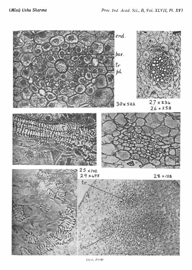
At the very base, the leaf in transverse sections shows wing-like expansions (W., Photo 24) and large air-spaces with narrow parenchymatous strands separating them. The leaf traces come off from the compact portion of the stele (Photos 7 and 11). It is the peripheral tracheids of the stele that seem to pass off into the leaf trace (Photo 12). In the cortex they pass almost horizontally and consist of nearly twenty or more tracheidsmmostly with annular and spiral thickenings, but often with reticulate thickenings also. No clear phloem is visible although a few small elongated cells with dense protoplasmic contents are seen occurring on the two sides of the xylem. This may be phloem (ph., Photo 25). In transverse sections also two patches of small cells may be seen on the outer side of the xylem (ph., Photo 26). These two patches may be the phloem.
Almost all the leaves are potentially sporophyllous but some sterile leaves without sporangia were also observed. A transverse section of a young leaf at the base, below the sporangium (even when it exists) shows (Photo 27) a collateral vascular bundle with a number of xylem elements forming two or three disconnected strands. In these strands some narrow elements occur in the middle with larger elements on both sides, a seemingly mesarch condition as is the case in L lacustris (McLean & Ivimey-Cook, 1951, p. 599).
A transverse section of the leaf taken between the top of the sporangium and the base of the ligular pit shows (Photo 28) that the vascular strand is now reduced to 3 or 4 tracheids (tr.). The phloem is not very clear. In the section can be seen the tracheidal cells (tr.c.) with scalariform thicken- ingsmregarded by some as " transfusion-tissue". These cells further mag- nified in Photo 29 are not connected in any way with the leaf trace. This condition also obtains in I. lacustris (Mclean & Ivimey-Cook, 1951, p. 600) and I. hystrix (Scott & Hill, 1900, p. 434) and also in I. echinospora and I. Engelmanni (Wilson Smith, 1900, p. 242). A little higher up, a transverse section of the leaf shows the vascular elements still further reduced to al- most a single centrally placed tracheid (tr.) sorrounded by a ring of paren- ehymatous cells (Photo 30) as in Isoetes echinospora (Smith, R. W., 1900,

Iscetes Sampathkumaranl Rao, L . N . - - I 219
p. 232). No phloem is clearly seen, but the smaller cells (ph.) with proto- plasmic contents seen on either side of the vascular strand may perhaps be the phloem. Photo 31 is a transverse section of a young leaf towards the tip where the lacunar tissue closes up, the parenchyma increases and the stomata are also present. Photo 32 shows a similar transverse section at the extreme tip where the lacunar tissue is replaced by parenchymatous cells. A few stomata may occasionally be seen here too.
In older leaves (Photos 33 and 34) the leaf trace consists of nearly twenty tracheids, sometimes arranged in a plate (Photo 33, better seen in Photo 34), probably because of compression against the growing sporangium. Beyond the sporangium the tracheids may increase in number and assume a ring- like form. Still higher up the tracheids gradually decrease in number until towards the tip, the leaf supply consists of only 2 or 3 tracheids (Photo 35). The lacunar tissue also develops clearly only beyond the level of the glossopodium of the lignle. The phloem of the leaf trace too becomes clear only beyond the glossopodium and is represented by only two patches of small-sized cells (see Photo 26) without any clear sieve plates, as in L lacus- tris and L velata (Farmer, 1890, p. 48). In L Engelmanni and L echinospora also (Smith, R. W., 1900, p. 232) two patches of phloem are found, one on either side of the leaf trace. Photo 35 shows a transverse section of the leaf near the apical re#on showing the four well-defined Mr-spaces, which have decreased in width. The rows of stomata opposite to them and the centrally placed stele with 2 or 3 tracheids (tr.) can be seen. The phloem at this level is poor in quantity and arranged in two patches of 2 or 3 cells each, one on either side of the xylem. It might be noticed that the sept~e separating the air-spaces from each other are at least 4 or 5 cells wide while in the basal part (Photo 24) they are only 1 or 2 cells wide. The leaf is flat in transverse section at the base, broadly elliptic in the middle and somewhat circular (Photo 31) towards the tip.
In longitudinal sections (Photo 20) the vascular supply arising from the stem stele remains undivided and gradually reduces itself in quantity as it ascends into the leaf. The air-spaces, as already stated, become well defined beyond the glossopodium level. The glossopodium (gL, Photo 21 and Text-Fig. 14) or the basal part of the ligule sunk into the ligular pit is a massive tissue which appears as though it is made up of two parts or lobes, when a transverse section is cut at its apical region (Photo 36). In the basal part (Text-Fig. 14) the glossopodium appears as a single tissue. It is made up of large empty cells surrounded by a single endodermal layer with clear casparian strips (Csp., Photo 37). The ligular pit is bordered on the one
220 MISS USHA SHARMA
side (Photo 20) by the leaf and on the other by the labium, the lower part of which extends over the sporangium as the velum. The tissue of this velum and labium as also its basal part is made up of large empty cells. The ligule which is itself several cells thick at the base, gradually thins out and at the extreme tip it is only one cell thick. The diaphragms in the leaf occur at regular intervals but may be lacking on one side (Photo 1).
It was found in some leaves that amongst the layer of cells lining the air-space in the leaf, there occur a few cells generally in a fixed position (see c in Text-Fig. 22 and Photo 35). They are sometimes absent but generally present in one or more of the air-spaces. They number 2 or 3 and have clear hyaline cell wails, often with dense protoplasmic contents. Their exact nature could not be ascertained. They are, however, very different from certain cells, which occur adjacent to the air cavity in Ixoetes lacustre L. (Johanna Liebig, 1931, p. 336, Fig. 4), where they are called the dia- phragm cells. So far as noticed by me no peripheral strands were found in the leaves.
SUMMARY
A detailed morphological and anatomical study of the vegetative parts of Is~etes Sampathkumarani Rao, L. N. is made. This species was described in 1944 by Prof. L. N. Rao from the Lalbagh Garden in Bangalore--the only locality where it has been so far observed to grow. The rhizomorph in this amphibious species is normally 2-lobed although 3 or 4 lobes occasionally occur. The axis stele is very short and, as in other species, consists mostly of a disorderly mass of xylem and parenchyma. The upper part of the stele is comparatively densely packed with xylem while the lower is lacunar and mixed with parenchyma. The stele in the rhizomorph is in the form of a double anchor which occupies a plane at right angle to the plane of the lobes. There are no clear sieve tubes or sieve plates and consequently no clearly recognizable phloem--although a tissue which is closely approximated to the xylem in the leaf and rhizomorph is interpreted as a possible phloem tissue.
The roots as in other species arise from the sides of the furrow on the rhizomorph and contain a single undivided root-trace eccentrically placed and attached to the cortex by a parenchymatous bridge only on one side. The leaves which are hardly 10 cm. long and about a couple of mm. thick contain a single vascular bundle which at the very base is a thin plate of fifteen to twenty tracheids. These become gradually reduced in quantity at the higher levels of the leaf, until at the tip there are only two or three tracheids. A few tracheidal cells unconnected to the leaf trace occur below the glossopodium of the ligule. Stomata are of the haplocheil type and

Iscetes Sampathkumarani Rao, L.N.--I 22i
occur in bands facing the air canals. No peripheral strands or slime canals are seen in the leaf. The ligules are of varied forms and are generally several cells thick in the middle and become thin towards the margin. Unicellular and multicellular hairs come out of marginal cells with dense protoplasmic contents. The velum covers the sporangium partially. But for a few sterile leaves found rather rarely, all the leaves are megasporophylls. No microsporophylls have so far been found, pobably because the two kinds of sporophylls are produced at different seasons.
My grateful thanks are due to Dr. A. R. Rao, under whose guidance this work was undertaken and completed. I am also indebted to Prof. S. N. Das Gupta for kindly providing me with all laboratory facilities and evincing interest in my work.
This work has been carried out during the tenure of a Government of India Scholarship and my thanks are also due to them for this help. I am also grateful to Mr. S. K. Nath who took the photomicrographs. I am also very grateful to Prof. L. N. Rao, for very kindly communicating this paper and also correcting the proofs.
REFERENCES
Baker, J. G.
Bharadwaja, Y.
Fames, A. G.
Ekambaram, T. and Venkatanathan, T. N.
Farmer, J. B. Gaekwad, L. K. and
Deshmukh, Y. S.
Griflith, W. Leibig, J.
Mahabale, T. S.
McCann, C.
McLean, R. C. and Ivimey-Cook, W. R.
Pfeiffer, N. E.
133
. . Handbook o f the Fern-Allies, 1887, London, George Bell and Sons, York Street, Covent Garden.
.. " T h e Occurrence of Istetes in India," Curr. Sci•, 1934-35, 5, 300.
. . Morphologjl o f Vascular Plants. Lower Groups, 1936, McGraw-Hill Book Company, Inc., New York and London.
" Studies on Isoetes coromandelina, L., 1. Speregenesis, J. Indian bet. See., 1933, 12, 191-225.
. . " On Isoetes lacustris, L.," Ann. of Bat., 1890, 5, 37-62. "Occurrence of Iso~tes at Baroda from Bombay State," Sci.
and Cult., 1954, 19, 457.
. . Notuke ad Plantas Asiaticus, 1849, pt 2. 572. •. "Erg/inzungen zur Entwicklungsgeschichte Von Iso~tes
laeustre, L. ," Flora, 1931, 25, 321-58•
•. " O n a new species of Isaetes in India," Curr. Sci., 1938, 7, 62-63.
. . "Occurrence of Isaetes coromandelina L. in Bcmbay Fre- sidency," Jour. Bomb. Nat. Hist. Soc., 1934, 37, 501-502•
. . Text-Book of Theoretical Botany, 1951, 1, Longmans, Green and Co., London, New Ycrk, Toror'to.
. . Monograph o f the Isoetacea,, Ann. o f ~[iss. Bet. Gard., 1922, 9, 79-232.
222 Miss USHA SI-IARMA
Praiv., D. . . Rec. Ind. Bet. Sure., 1905, 3, 327. Kao, L .N . . . " A new species of lswtes from Bangalore, Mysore State,"
Curr. Sci., 1944, 13, 286--87. Scott, D. H. and Hill, T .G. . . "The structure of Is~tes hystrix," Ann. o f Bet., 1900, 14,
413-54. Shende, D.V. . . " A new species of Is~tes from the Bombay Presidency,"
Your. Univ. Bomb., 1942, 14, 50-52. Smith, G . M . . . Cryptogamic Botany, 2, Bryophytes and Pteridophytes, 1938,
McGraw-Hill Book Co., Inc., New York & London. Smith, R .W. . . "The structure and development of the sporophylls and
sporangia of lsostes," Bet. Gaz., 1900, 29, Pt. I, 225-58; Pt. II, 323-46.
EXPLANATION OF TEXT-FIGURES
Tax-r-FIGs. 1-12. Fig. 1. Transverse section of an abnormal rhizomorph having one lob suppressed and the other developed into three smaller lobes. Figs. 2-4. Vertical longitudinal s~ctions cut tangentially. Section in Fig. 2 cuts across the furrow, the others pass tangentially in the lobes. Fig. 5. Median vertical section through rhizomorph lobes. Fig. 6. Part of a vortical loagitudiaal section m~gnified to show the stelar tissues like the cambium, phloem and xylem Figs. 7-9. Vertical longitudinal sections at right angles to the plane of the rhizomorph lobes. Figure 7 is a m,~dian vertical section passing through the furrow while Figs. 8 and 9 are slightly taagential sections cut beyond the stele. Figs. 10-11. Transverse sections of rhizomorph passingin the middle (Fig. 10), and in the lower part (Fig. l l ) . Fig. 12. A root showing repeated dichotomy at the tip.
ax., axis stele; camb., cambium; f., furrow; i.e., inner cortex; /., leaf; lt.,leaftraco;o.c., outer cortex; ph., phloem; r., root; r.t., root trace; sl.l., sloughing layer; xy., xylem.
All figures are camera lucida sketches and are magnified 7.5 times except Fig. 6 which is magni- fied 390 times.
TExT-FIos. 13-22. Fig. 13. Transverse section ofrhizomorph showing clearly the two layers of the cortex; the inner one richly loaded with starch, × 180. Fig. 14. Transverse sections of the central part of an old plant. The overlapping of the leaves, their vascular bundles, sporangia, glosso- podium, tracheidal tissue and ligule are shown, × 45. Fig. 15. Peripheral part of the ligule showing the uuicellular and multicellular hairs, coming out of the cells with dense protoplasmic contents and the underlying empty cells. Figs. 16-21. Ligules of varied shapes; triangular. Fig. 16. cordate. Fig. 17. irregularly lobed. Figs. 18-19. bifid apex. Fig. 20. broadly elliptic, and three-lobed. Fig. 21. Note the irregularly dentate margin of the ligule, all x45. Fig. 22. Transverse section of an old leaf nearer the apex~ The two cells marked c. are the peculiar cells referred to in the text, x87-5.
a.sp., air-space; c., pecular cells; e.c., empty cells; gL, glossopodium, h., hair; b.c., hair colts; lig., ligule; sp., sporangium; st., stoma; tr.t., tracheal tissue; v.b., vascular bundle.
PHOTO
EXPLANATION OF PHOTOGRAPHS
PLATE XII
Is~tes Sampathkumarani
1. Entire plant showing the 2-lobed rhizomorph and cylindrical leaves in which diaphragms can he seen.

(Miss) Usha Sharma Proc. Ind. Acad. Sci., B, Vol. XLVll, PI. X,
~!i"~ t I xl.q
¢" / i
III~ ;
2.x~
~' x 70
FIGS. 1-9

(Miss) Usha Sharma Proc. Ind. Acad. Sci., B, Vol. XLVII, Pl. XI
Co~ IO~ 7~2
I~ ~ 210
~ ~ I ~ i
FIGS. IO-14

(Miss) Usha Sharma Proc. Ind. Acad. Sci., B, Vol. XLVII, P1. XIV
Iqx l~t2
16 x .79
p~ end
FIGS. 15-19
• -eJ ~
Ig xk6~"

(Miss) Usha Sharma Proc. Ind. Acad. Sci., B, Vol. XLVII, PI, XV
2ox3g ~lx ~ .~'
a~k. 2Z x/+4,
2.4 x 56
. . .
t~
23 ×u+#
F i t s 21t-24
(Miss) Usha Sharma Proc. lnd~ Acad. Sci., B, Vol. XLVII, PI. XVI
er~d.
])O.T.
j,/,
3ox 52-2, 27 ×z3a 2 6 x 25"8
5 x7oL 2ct ~ . q s
. . . . . . . . . . . : ! , ~ • ii ~ ~ i i i i ~ ~ ~ ; ¢ ~ i ~ . i i ~ ' ~
Fl(;~. 25-30

(~iss) Usha Sharma Proc. Ind. Acad. Sci., B, Vol. XLVII, PI. XVII
!
s ~ i ~'"" L'J
3 3 x3s
35 xS"o
:~.~. trf~
D
FIGS. 3 1 - 3 7

Iscetes Sampathkumarani Rao, L. N.- -I 223
PHOTO 2. The lower side of the 2-lobed rhizomorph with some roots still attached, the furrow separating the rhizomorph lobes is in the middle.
PHOTO 3. A 3-lobed rhizomorph as seen from below.
PHOTO 4. A 4-lobed rhizomorph as seen from below.
PHOTOS 5-6. Abnormal specimens showing rhizomorphs (rh.) with one lobe (left) suppressed and the other one (righ0 3-lobed, in both, the bifurcation of the roots at the tip can be seen.
PHOTO 7. Median vertical longitudinal section passing at right angles to the lobes of the rhizomorph; camb., cambium; L, leaf; lig., lignle; Lt., leaf trace; r.t., root trace; st. stele. At the region indicated by x the xylem is compact.
PHOTO 8. The stele in the above photograph magnified; camb., cambium; l.t., leaf trace; x., compact stele.
PHOTO 9. A part of the above stele magnified to show the camb., cambium ; pr.t., prismatic tissue; and xy., xylem.
PHOTO 10.
PHOTO 1 t.
PHOTO 12.
PHOTO 13.
PHOTO 14.
PLATE XIII
Part of a longitudinal section of the stele enlarged to show the cambium, camb,, ph., phloem, pr.t., prismatic tissue and xy., xylem.
Vertical section of the rhizomorph passing in the plane of the furrow. The stele can be seen in the form of a double anchor in the plane of the furrow itself. A section passing along the line a-a in the photograph would present an appearance as seen in Photo 15. Lt., leaf trace; r.t., root trace; st., stele.
Transverse section of the upper part of the rhizomorph showing only the stelar part where the xylem is compact and gives off leaf traces l.t.
Transverse section through the middle of the rhizomorph showing the lacunar stele; a.sp., air-space; cx., cortex; camb., cambium; Lt., leaf trace; par., parenchyma; pr.t., prismatic tissue; xy. , xylem.
Part of the section seen in Photo 13 photographed on a large scale to show the different tissues, a-sp., air-space; camb., camb~m; l.t., leaf trace; par., paren- chyma; ph.. phloem; pr.t., prismatic tissue; xy., xylem.
PuoTo 15.
PHOTO 16.
PHOTO 17.
PHOTO 18,
PHOTO 19.
PLATE XIV
Transverse section of the rhizomorph in the basal part passing obliquely along the line a-a indicated in Photo 11. ex., cortex; par., parenchyma; r.t., root trace; xy., xylem.
Part of a vertical section of the rhizomorph showing a root trace cut longitudinally; the thickenings of the tracheids and the phloem (ph.) which is generally more on one side (see Photo 18) are seen.
Transverse section of a root showing the large air-space and the eccentric stele attached to the cortex on one side.
The stele of the above root magnified; end., endodermis; ph., phloem.
Epidermal peeling of leaf showing epidermal cells and two stomata,
224 Mms USHA SahaMA
PHOTO 20.
PHOTO 21.
PHOTO 22. PHOTO 23.
PHOTO 24.
PHOTO 25.
PHOTO 26.
PHOTO 27.
PHOTO 28.
PHOTO 29. PHOTO 30.
PLATE XV
Longitudinal section of a sporophyU, a., air-space; gl., glossopodium; lab., labium; lig., ligule; sp., sporangium; spr., spore; tr., trabecula; tr.t., tracheal tissue; ~'eL, velum; v.b., vascular bundle.
Basal part of a sporophyll, gl., glossopodium; lig., ligule; sp., sporangium; Tel., velum; v.b., vascular bundle.
An entire ligule. Peripheral part of another ligule enlarged to show the different kinds of cells ,~¢cn;
the hairs, the peripheral cells from which they arise, irregularly-shaped empty cells below these, and the underlying isodlametric cells (see Text-Fig. 15).
Transverse section of an old leaf near the basal part above the level of the sporangium; a.sp., air-space; epi., epidermis; p.str., parenchymatous strand; st. , stele; w., wing.
PLATE XVI
Leaf trace magnified to show the spiral, annular and scalariform thickenings of the tracheids and the tissue that might be phloom, ph.
Transverse section of an old leaf near the basal part above the level of the sporangium showing the stele, ph., phloem.
Transverse section of a young leaf at the base, the stele showing a large number of tracheids forming three disconnected strands.
Transverse section of leaf taken between the top of the sporangium and the base of the ligular pit. tr., tracheids of the leaf trace reduced to just four; tr.c., tracheidal cells.
Tracheidal cells seen in the above section enlarged. Transverse section a little higher than the section photographed in Photo 28,
showing a single centrally placed tracheid, tr., and phloem, ph.
PLATE XVII
PHOTO 31.
PHOTO 32.
PHOTO 33.
PHOTO 34.
PHOTO 35.
PHOTO 36.
PHOTO 37.
Transverse section of a young leaf towards the top, showing the stele, a..~p., air-space and st., stoma.
Transverse section cf a young leaf at the extreme tip showingne air-spaces nor stele, a single stoma, st., can be seen.
Transverse section of a sporophyll passing through the sporangium; sp., m.spr . megaspore; trb., trabecula; eel., velum and v.b., vascular bundle.
The vascular bundle seen in the above section magnified to show the plate of trachekh v.b., vascular bundle.
Transverse section of an old leaf nearer the-apex, a.sp., air space;c., peculiar cells; st., stoma; v.b., vascular bundle.
Transverse section of the central part of an old plant showing the mutually overlapping leaves, the sporangium; sp. and gl., the gloss-podium of the ligule; v,b., vascular bundle.
Part of the glossopodium tissue enlarged to show the endodermis, end., with its casparian strips, csp.