copyright ©2006 by the mcgraw-hill companies,...
TRANSCRIPT


The Developing Brain 2
Copyright ©2006 by The McGraw-Hill Companies, Inc. All rights reserved. No part of this publication may be reproduced or distributed in any form or by any means, or stored in a database or retrieval system, without the prior written consent of The McGraw-Hill Companies, Inc., including, but not limited to, network storage or transmission, or broadcast for distance learning.
Send all inquiries to:Glencoe/McGraw-Hill21600 Oxnard St., Suite 500Woodland Hills, CA 91367
ISBN 0-07-868974-1
Printed in the United States of America2 3 4 5 6 7 8 9 10 009 12 11 10 09 08

The Developing Brain 3Copyright © Glencoe/McGraw-Hill, a division of The McGraw-Hill Companies, Inc.
The Human Brain ..........................................................................7
• Brain Anatomy Brain Hemispheres The Forebrain The Hindbrain Brain Ventricles Different Views of the Brain• Neurons• Neurotransmitters
Early Brain Development ............................................................17
• Prenatal Brain Development• Windows of Opportunity
Brain Imaging Technology ......................................................... 19
• Electroencephalogram (EEG)• Computed Tomography (CT)• Magnetic Resonance Imaging (MRI) Functional Magnetic Resonance Imaging (fMRI) Magnetoencephalography (MEG) Magnetic Resonance Spectroscopy (MRS)• Positron Emission Tomography (PET)• Single-Photon Emission Computed Tomography (SPECT)
Contents

Copyright © Glencoe/McGraw-Hill, a division of The McGraw-Hill Companies, Inc.
The Developing Brain 4
The Learning Brain .................................................................... 28
• Brain Development• The Role of Memory• Activity Areas of the Brain• The Impact of Media• The Role of School• Mature Reasoning Develops• The Adult Brain
Adolescent Brain Development ................................................ 34
• Executive Functions• Adrenaline & Company
The Waking & Sleeping Brain ....................................................37
• The Pineal Gland• Stages of Sleep
Feeding the Brain ........................................................................39
• Carbohydrates• Proteins• Fats• Micronutrients
Drugs & Alcohol Effects on the Brain ..................................... 43
• Nicotine• Marijuana• Cocaine• Alcohol• Alcohol Withdrawal

The Developing Brain 5Copyright © Glencoe/McGraw-Hill, a division of The McGraw-Hill Companies, Inc.
Brain-Related Diseases & Disorders ....................................... 49
• Holoprosencephaly• Sturge-Weber Syndrome• Encephalitis• Meningitis• Creutzfeldt-Jakob Disease• Epileptic Seizures• Schizophrenia• Brain Hemorrhage• Glioma (Tumor) & Brain Cancer• Cerebral Atrophy• Stroke• Parkinson’s Disease• Alzheimer’s Disease
Brain-Related Glossary ..............................................................67
Brain-Related Internet Resources ............................................93
Brain-Related Print Resources ............................................... 103
Brain-Related Activities ............................................................ 111

Content Contributors: Marilyn Swierk, M.S., CFCS, CFLEKimberly Moore, Ph.D.Patricia K. O. Rambo
Cover Photo: Simon Fraser/Photo Researchers, Inc.
Transparency Photo and Illustration Credits: Dr. Sterling Clarren, 29Photo Researchers, Inc.: Airelle-Joubert, 34; AJPhoto, 14; Anatomical Travelogue, 9; Tim Beddow, 19; Biophoto Associates, 8; John Bavosi, 5, 10, 37; Scott Camazine, 1, 4, 15, 40, 41; Gary Carlson, 7; Charing Cross Hospital, 20; Michael Donne, 15; S. Faser, 43; Simon Fraser, 11, 35, 36; Dr Robert Friedland, 47; GJLP, 45; Pascal Goetgheluck, 26, 27, 28, 30; Mehau Kulyk, 31, 32, 33; Dr. J. Mazziotta et al./Neurology, 22; MediVisuals (Amygdala and Hippocampus added by Publisher), 2; Alfred Pasieka, 24; Phanie, 16, 17, 20, 46; Dr. M. Phelps & Dr. J. Mazziotta et al./Neurology, 21; Will & Deni McIntyre, 19; Hank Morgan, 25, 39; Hans-Ulrich Osterwalder, 48; Erich Schrempp, 18; Dr. Neal Scolding, 6; Science Source, 12, 13, 28; WDCN/ Univ. College London, 23; Zephyr, 3, 42, 44
Brand Name Disclaimer
Publisher does not necessarily recommend or endorse any particular company or brand name product that may be discussed or pictured in The Developing Brain. Brand name products are used because they are readily available, likely to be known to the reader, and their use may aid in the understanding of the text. Publisher recognizes other brand name or generic products may be substituted and work as well or better than those featured in The Developing Brain.

The Developing Brain 7Copyright © Glencoe/McGraw-Hill, a division of The McGraw-Hill Companies, Inc.
The brain is the hardest working, most vital organ in your body. It represents less than 2 percent of your body weight, yet uses 25 percent of the oxygen you breathe and 70 percent of your glucose supply. It is oblong in shape, weighs almost 3 pounds, and takes up about half of the volume of your head. It has the appearance of a pinkish-gray wrinkled walnut, and feels soft and slimy like gelatin. The brain is the commander-in-chief of everything your body does 24 hours a day.
Brain researchers have learned more about the workings of the human brain in the past two decades than they had in the previous century. As you read this booklet, you will gain a basic understanding of how the human brain works, the imaging technology used to study the brain, the implications of brain development, and how drugs and disease affect the brain. The images shown throughout these pages will encourage you to research the latest findings in neuroscience and brain development over the lifespan. It is never too late to expand your learning and use it to enhance your life and the lives of those around you.
Brain Anatomy .....................................................................................8
Neurons ................................................................................................13
Neurotransmitters ............................................................................ 15
The Human Brain

Copyright © Glencoe/McGraw-Hill, a division of The McGraw-Hill Companies, Inc.
The Developing Brain 8
The cerebrum makes up about 70 percent of the brain. The cerebral cortex is the covering of the outer layer
of the cerebrum. It is a ¼ inch-thick blanket of cells folded into bumps and grooves that look like a long, curled up rope. The convoluted shape increases the amount of cortex that can fit in the skull. If smoothed out, the cortex would be about the size of a full page of news-paper, approximately 324 square inches.
The cerebral cortex is where you perceive the world through your senses. It is divided into four lobes, each spanning both hemispheres. The illustration below identifies the frontal lobe, parietal lobe, temporal lobe and occipital lobe of the brain. The cerebellum is also labeled as a reference point.• The frontal lobes process higher level think-
ing and movement. The prefrontal cortex is located just behind the forehead and handles working memory, critical thinking, reasoning and problem solving. It is known as the chief executive officer of the brain. The motor cortex, located where a headband would sit, involves planning, coordinating and carry-ing out movements. The basal ganglia help
the prefrontal cortex prioritize information related to motor control, cognition, emotions and learning.
• The parietal lobes interpret and integrate information from the senses and are on the sides toward the back of the brain. The sen-sory cortex is right behind the motor cortex and processes information received when the skin is touched, including pressure, tempera-ture, and pain.
• The temporal lobes (on the side near the ears) processes stimuli, which affect speech, hearing and language development.
• The occipital lobes (at the back of the brain) processes vision and how you interpret what you see. It is also referred to as the visual cortex. The brain is divided into two hemispheres.
The left and right hemispheres of the cerebral cortex are specialized to process information in distinct ways. The analytical left hemisphere is the fact-finding side of the brain that processes information before it takes action. It controls the right side of the body. This hemisphere is responsible for verbal skills, reasoning, reading, writing, and skills.
Brain Anatomy
Brain Research Activity—Research
the function of each lobe within the human brain.

Copyright © Glencoe/McGraw-Hill, a division of The McGraw-Hill Companies, Inc.
bundle of nerve fibers that works like a coor-dinator. It lets one hemisphere know what the other is doing so they can work together. For example, the left side reads a word while the right side “pictures” it.
Since infants learn with their senses first, the right hemisphere is dominant. As language develops the left side becomes more active, however, daily functioning makes use of both hemispheres of the brain and allows people to become flexible learners eventually developing a dominance preference. Here are some ways to use both hemispheres.• Memorize multiplication tables to music.• Cook a meal following several recipes.• Build a model using the directions.• Watch a movie and then explain the story to
a friend.
Brain HemispheresThe brain is divided into two hemispheres.
The left and right hemispheres of the cerebral cortex are specialized to process information in distinct ways. The analytical left hemisphere is the fact-finding side of the brain that processes information before it takes action. It controls the right side of the body. This hemisphere is responsible for verbal skills, reasoning, reading, writing, and math skills.
The intuitive right hemisphere of the brain is focused on perceptions and often wants to act before it thinks. This hemisphere controls the left side of the body. It sees the “whole picture” and is responsible for imagination, creativity, insight, and awareness of three-dimensional forms.
The hemispheres interact to help us interpret our environment. The corpus callosum is a
The Developing Brain 9
(continued)
Brain Anatomy (continued)
Left Hemisphere Right Hemisphere
Sequential/Analytical Intuitive/Random
Part to whole Whole to part
Abstract/Symbolic Visual/Spatial
Language—vocabulary skills Interpreting voice tone and inflection
Music—lyrics, structure Music—melody
Math and Science facts Math and Science concepts
Decoding and Reading skills Reading Comprehension
Writing Skills Creative Expression
Organization—lists, order Organization—graphic organizers
Practical and serious Imaginative and playful

Copyright © Glencoe/McGraw-Hill, a division of The McGraw-Hill Companies, Inc.
The Developing Brain 10
The ForebrainThe human brain is made up of the forebrain,
midbrain, and hindbrain. The largest por-tion of the forebrain consists of the cerebrum which includes the cerebral cortex. Beneath the cerebrum lies the remainder of the forebrain’s structures: the thalamus, basal ganglia, hypo-thalamus, amygdala, and hippocampus.
From its position at the top of the brain stem, the thalamus acts as a two-way relay station that sorts, processes, and directs signals from the spinal cord and mid-brain structures to the cere-bral cortex and from the cerebral cortex down the spinal cord. The thalamus consists of two egg-shaped masses of nerve tissue, each about the size of a walnut, deep within the brain. It is the key relay station for sensory information flowing to the brain. The thalamus filters only the information of particular importance from the mass of signals entering the brain.
The basal ganglia are a large collection of nerve cells that form a ring around the thalamus.
The hypothalamus is a small structure located at the base of the brain where signals from the brain and the body’s hormonal system interact. It controls hunger, thirst, sleep, sexual-ity, and emotions.
The amygdala is an almond-shaped struc-ture that plays a central role in producing and responding to nonverbal signs of anger, fear, and defensiveness. It also influences the “fight or flight” reaction to stress.
The hippocampus is a seahorse-shaped struc-ture located deep within the brain that encodes information into short- or long-term memory and helps with the recall of spatial relationships.
The hypothalamus, amygdala, and hippocam-pus are part of the limbic system. These and others channel the full range of emotions and are involved in the formation of memory
The MidbrainThe midbrain serves as the nerve pathway to
the cerebral hemispheres. It also contains the auditory and visual reflex centers.
Brain Research Activity—Research
the function of the cerebrum, thalamus, hypothalamus, amygdala, hippocampus, cerebellum, pons, and medullaoblongata.
Brain Anatomy (continued)

The Developing Brain 11Copyright © Glencoe/McGraw-Hill, a division of The McGraw-Hill Companies, Inc.
The HindbrainThe hindbrain is composed of the cerebel-
lum, pons, and medulla oblongata which work together to support vital bodily processes. The cerebellum is located behind the top of the brain stem. It coordinates the brain’s instruc-tions for skilled, repetitive movements, and helps maintain balance and posture. The cer-ebellum is also involved in coordinating think-ing. The more complicated the activity, the more the cerebellum is called upon to help the higher brain centers process information.
The pons and medulla oblongata make up the stick-like structure at the base of the brain called the brain stem. The pons is a bridge-like structure that connects to the cerebellum and is involved in movement and posture. The pons controls the respiratory center. It is involved in motor control and sensory analysis, level of consciousness, and sleep.
The medulla oblongata is joined to the spi-nal cord, a cable of nerves that descends from the brain stem to the lower back. The location allows the medulla to act as a relay station
between the brain and the nervous system. It controls functions basic to survival, such as heart rate, breathing, digestive processes, and sleeping. It also controls reflexes such as swal-lowing, coughing and vomiting.
Brain Ventricles The human brain also contains four
ventricles, or communicating cavities that provide a pathway for cerebrospinal fluid. The ventricles all flow into the central canal of the spinal cord. There are two lateral ventricles, a median ventricle, and an inferior ventricle as shown in the brain scan below.
Both of the lateral ventricles are located deep within the top section of the brain. Each communicates with the third or median ven-tricle that sits between the thalamus and hypothalamus. The median ventricle is called the aqueduct of the midbrain (or the aqueduct of Sylvius). The inferior ventricle extends from the median ventricle to the upper end of the spinal cord.
Brain Research Activity—Research
the function of the brain’s four ventricles and the pituitary gland.
(continued)
Brain Anatomy (continued)

Copyright © Glencoe/McGraw-Hill, a division of The McGraw-Hill Companies, Inc.
The Developing Brain 12
Different Views of the BrainThe brain can be viewed many different
ways. The illustration on this page shows a coronal section of the human brain. Coronal sections are easy to visualize because it is like looking directly through a person who is fac-ing you. Notice the fairly symmetrical left and right hemispheres. The anterior view, or front section of the brain, is closest to the face, nose, and mouth. The posterior view, or rear section of the brain, is closest to the back of the head.
The limbic system is a complex network of nerve pathways that govern the expression of fear, rage, and pleasure and is involved in the formation of memory. It is also known as the brain’s emotional thermostat. The illustration below highlights the various parts of the limbic system. The cerebellum is labeled as a reference point.
Grey Matter & White MatterGrey matter is a major component of the
central nervous system. It consists of neurons and glial cells. This micrograph shows assorted brain cells in the grey matter. White matter forms the bulk of the deep parts of the brain. It is one of the solid components of the central nervous system.
Brain Research Activity—Research
the limbic system and the function of each part within it.
Brain Anatomy (continued)

The Developing Brain 13Copyright © Glencoe/McGraw-Hill, a division of The McGraw-Hill Companies, Inc.
(continued)
Unlike any other cell in the body, neu-rons consist of a central cell body that is characterized by long fibrous projec-
tions called axons and shorter, branch-like pro-jections called dendrites. They are responsible for the transmission of nerve impulses.
Motor neurons send electrical signals from the central nervous system to muscle and gland neurons. Sensory neurons receive electrical signals to the central nervous system from sen-sory cells. Interneurons are only found in the brain and spinal cord. They send information between sensory and motor nerons or between interneurons. Scientists estimate there are more than 100 billion neurons in the brain sending signals to thousands of other cells at a rate of about 200 miles per hour.
Packed tightly around and between the neurons are one trillion glial cells that pro-
vide a supporting framework that resembles a honeycomb. Glial cells nourish and protect the neurons. There are approximately three times as many glial cells as there are neurons. The illustration on this page shows the parts of a neuron.• The cell body controls the cell and directs its
activities.• Each axon is a long, single nerve fiber that
transmits chemical and electrical impulses from the body of the neuron to dendrites of other neurons, or directly to body tissues such as muscles. These signals allow you to move, taste, feel, and remember.
• Myelin is a sheath of fatty insulating material that covers the axon to help the speed of the signal and to prevent loss of the signal.
• Dendrites are fine nerve fibers that branch out from the nerve cell body like trees. They receive signals in the form of chemical mes-sages from the axons of other neurons and relay them to the cell’s nucleus. Dendrites stimulate activity in the receiving neuron.
Neurons
DENDRITES
CELL BODY
NUCLEUS
AXON MYELIN
SHEATH
AXON
TERMINALS
Direction
of Impulse

Copyright © Glencoe/McGraw-Hill, a division of The McGraw-Hill Companies, Inc.
The Developing Brain 14
When a neuron fires a signal (or impulse) it travels down the axon to the dendrite of another neuron. When the signal reaches the synapse, or junction where neuron communica-tion occurs, neurotransmitters are released into the synaptic cleft, a tiny gap (one-millionth of an inch) that is the site of information transfer, to reach the next neuron. An axon may send a signal to a neighboring neuron or to one on the other side of the brain. However, it is important
to note that synapses are markedly susceptible to the effects of oxygen deficiency, anesthetics, therapeutic drugs, and toxic chemicals.
A stimulus does not have to be obvious. It could be something as simple as a mother rub-bing her baby’s cheek. A stimulus could also be a song that reminds you of a happy occasion. This stimulus causes neurons to fire and con-nect, bringing up memories of whom you were with, what was said, what you wore, and so on.
Brain Research Activity—Research
“Action potential” to learn more about how neurons transmit messages.

The Developing Brain 15Copyright © Glencoe/McGraw-Hill, a division of The McGraw-Hill Companies, Inc.
Neurotransmitters
The brain experiences about 100,000 chemical reactions each second. The key to these chemical reactions are
neurotransmitters, the chemicals that act as messengers between neurons. Neurotransmit-ters are released into the synaptic cleft when a nerve impulse reaches the end of an axon so the signal can pass to the next neuron. They are responsible for the maintenance, activity, and longevity of synapses and neurons.
Receptors are molecules whose structures precisely match those of the neurotransmitters released during synaptic transmission. Recep-tors and neurotransmitters attach in order to activate the receiving dendrite or cell body. The illustration below highlights the synaptic junc-tion (knob) between two neurons. When neu-rotransmitters reach the receptor cells they bind and trigger an electrical impulse in the receptor cell. The cigar-shaped mitochondria are the sites of energy production taking place within the cell.
Several dozen neurotransmitters in the brain have been identified so far, each with specific and often complex roles in brain function and human behavior. There are two types of neu-rotransmitters: excitatory (similar to an “on” signal), which pass messages to the next neu-ron; and inhibitory (similar to an “off” signal), which prevent signals from being produced in the next neuron. Some common neurotransmit-ters include:• Acetylcholine• Endorphins• Serotonin• Dopamine• Norepinephrine
Acetylcholine is the most abundant neu-rotransmitter in the body and the primary neu-rotransmitter between neurons and muscles. The stomach, spleen, bladder, liver, and heart are just some of the organs that this neurotrans-
Brain Research Activity—Research additional neurotrans-
mitters such as melatonin.
(continued)

Copyright © Glencoe/McGraw-Hill, a division of The McGraw-Hill Companies, Inc.
The Developing Brain 16
NERVE IMPULSE
AXON
VESICLE
SYNAPSETRANSPORTER
MOLECULE
NEUROTRANSMITTERS
RECEPTOR
MOLECULES
DENDRITE
OF RECEIVING
NEURON
mitter controls. Acetylcholine helps control muscle tone, learning, primal drives, emotions, and the release of the pituitary hormone vaso-pressin, which is involved in learning and in the regulation of urine output. Low levels of acetylcholine can contribute to lack of concen-tration and forgetfulness and may cause light sleep. The body synthesizes acetylcholine from the nutrients choline, lecithin, and vitamins C, B1, B5, and B6, along with the minerals zinc and calcium.
Norepinephrine (also known as noradrena-line) is both a neurotransmitter and a hormone. As a neurotransmitter, it is involved in arousal, and regulation of sleep, mood, and blood pres-sure. As a hormone, it works with adrenaline to give the body sudden energy in times of stress.
Endorphins produced in the brain gener-ate cellular and behavioral effects like those of morphine to mediate pain at receptor sites. In an injury, receptors in skin make electrical signals that go up the spinal cord to the brain. The brain then evaluates pain by releasing endorphins which mediate pain. Endorphins affect the dopamine pathway that feeds into the frontal lobe. These pathways inhibit the flow of
dopamine. When vast quantities of endorphins are released and nerves are shut off more dopa-mine flows to the frontal lobe to replace pain with pleasure.
Serotonin is a hormone found in the pineal gland, blood platelets, the digestive tract, and the brain. It is a neurotransmitter in the regu-lation of mood. Low levels of serotonin may result in anxiety, depression, aggression, or other mood disorders. Serotonin helps regulate pain. It causes blood vessels to narrow and plays an important role in stimulating a strong heart beat and reducing the potential for blood clotting. It also serves as a precursor for the pineal hormone melatonin, which regulates the body’s internal clock
Dopamine regulates emotional responses and helps execute smooth and controlled move-ments. It is also thought to produce feelings of bliss (the pleasure chemical). More dopamine in the frontal lobe lessens pain and increases plea-sure. A lack of dopamine may cause disrupted or incoherent thought. Too much dopamine in the limbic system and not enough in the cere-bral cortex may produce bouts of paranoia or may inhibit social interaction.
Neurotransmitters (continued)

The Developing Brain 17Copyright © Glencoe/McGraw-Hill, a division of The McGraw-Hill Companies, Inc.
Early Brain Development
People used to think that providing a child with food, clothing, and shelter in a loving, healthy, and safe environment would ensure the optimum development of the child. However, research has shown that early experiences can help determine the physical structure, or “architecture,” of the brain and the extent to which a person reaches his or her potential. At birth, a baby’s brain is about 25 percent of its approximate adult weight. By age three, a child’s brain has reached 90 percent of its full potential and is twice as active as the brain of an adult.
Prenatal Brain Development ............................................................ 18
Windows of Opportunity ................................................................... 19

Copyright © Glencoe/McGraw-Hill, a division of The McGraw-Hill Companies, Inc.
The Developing Brain 18
Prenatal Brain Development
Even before most women realize they are pregnant, the brain is forming, along with the rest of the central
nervous system. • Approximately three weeks after conception,
the brain has a definite forebrain, midbrain, and hindbrain.
• By six weeks, the cerebral hemispheres are growing faster than other sections of the brain.
• At eight weeks, the brain constitutes almost half of an unborn child’s body weight and continues to grow at an extraordinary rate.
• During the third trimester, the brain weight increases between 400-500 percent and consumes more than 50 percent of the energy used by the fetus.The development of the brain and spinal
cord continues throughout pregnancy. Maternal exposure to substances such as alcohol during pregnancy can destroy dendrites in the hip-pocampus, where a large part of learning and memory take place.
Fetal alcohol syndrome (FAS) develops in an unborn child when the mother drinks too much alcohol, particularly during the earliest stages of pregnancy. FAS is the leading cause of learn-ing disabilities, neurological impairment, and mental retardation. This occurs because alcohol prevents the fetus from receiving sufficient oxy-gen and nourishment. In turn, this prevents the brain and other vital organs from developing as they should. FAS babies are often seriously disabled for the rest of their lives.
Brain Research Activity—Research
the impact of FAS on brain development. Find out how the brain can set up alternative pathways to the damaged areas of the brain if intervention occurs early enough in life.
The Developing Brain 19Copyright © Glencoe/McGraw-Hill, a division of The McGraw-Hill Companies, Inc.
(continued)
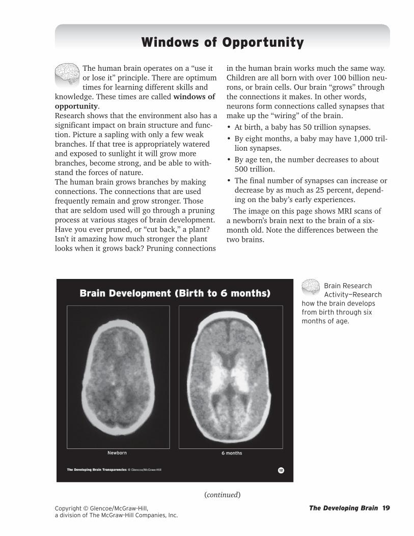
Windows of Opportunity
The human brain operates on a “use it or lose it” principle. There are optimum times for learning different skills and
knowledge. These times are called windows of opportunity. Research shows that the environment also has a significant impact on brain structure and func-tion. Picture a sapling with only a few weak branches. If that tree is appropriately watered and exposed to sunlight it will grow more branches, become strong, and be able to with-stand the forces of nature.The human brain grows branches by making connections. The connections that are used frequently remain and grow stronger. Those that are seldom used will go through a pruning process at various stages of brain development. Have you ever pruned, or “cut back,” a plant? Isn’t it amazing how much stronger the plant looks when it grows back? Pruning connections
Brain Research Activity—Research
how the brain develops from birth through six months of age.
in the human brain works much the same way. Children are all born with over 100 billion neu-rons, or brain cells. Our brain “grows” through the connections it makes. In other words, neurons form connections called synapses that make up the “wiring” of the brain. • At birth, a baby has 50 trillion synapses.• By eight months, a baby may have 1,000 tril-
lion synapses. • By age ten, the number decreases to about
500 trillion.• The final number of synapses can increase or
decrease by as much as 25 percent, depend-ing on the baby’s early experiences.The image on this page shows MRI scans of
a newborn’s brain next to the brain of a six-month old. Note the differences between the two brains.

Copyright © Glencoe/McGraw-Hill, a division of The McGraw-Hill Companies, Inc.
The Developing Brain 20
Genes are responsible to the basic format of the brain, but experiences actually fine-tune the connections and help the child adapt to his or her environment—the people, culture, and geography. The human brain is continually reshaping itself to meet the demands of every-day life. The MRI scans below show how differ-ent the brain of a 20-month-old looks from the brain of a 5-year-old.
The brain’s plasticity, or the ability of the brain to develop and change in response to the demands of the environment, is what aids learning. While there are “windows of opportu-nity” that are ideal, learning can take place at any time. However, some abilities can be more challenging to learn at later stages in life. For example, people who choose to learn a new
Brain Research Activity—Research
how the brain develops through 5 years of age.
language after puberty will be able to do so, but they will often speak that new language with a foreign accent. This is because the “window of opportunity” for language-learning ends around the age of puberty.
While neuroscientists do not agree on the basis for these optimal learning periods, there is evidence that they exist. Brain development is activity-dependent. Like computer circuits, neurons process information through the flow of electrical signals. Unlike computer circuits, the neural networks in the human brain are not fixed structures. As neural connections are made stronger through pruning, the brain adjusts to fit its environment. Even older adults can and do learn new things. Remember, learn-ing never ends!
Windows of Opportunity (continued)

The Developing Brain 21Copyright © Glencoe/McGraw-Hill, a division of The McGraw-Hill Companies, Inc.
Brain Imaging Technology
Technological breakthroughs in the field of neuroscience are enabling brain researchers to study how the brain actually functions. Through the use of noninvasive imaging technologies, scientists are now able to study the brains of living people and diagnose a variety of brain disorders and diseases. EEG, CT, MRI, fMRI, MEG, MRS, PET, and SPECT scans are routinely used to diagnose a variety of conditions. The most recent advances in brain imaging technology are helping researchers unlock the secrets of the human brain.
Electroencephalograph (EEG) ......................................................... 22
Computed Tomography (CT) .............................................................23
Magnetic Resonance Imaging (MRI) .............................................. 24
Functional Magnetic Resonance Imaging (fMRI) ........................24
Magnetoencephalography (MEG) ...................................................25
Magnetic Resonance Spectroscopy (MRS) ..................................25
Positron Emission Tomography (PET) ........................................... 26
Single-Photon Emission Computed Tomography (SPECT) ...........27

Copyright © Glencoe/McGraw-Hill, a division of The McGraw-Hill Companies, Inc.
The Developing Brain 22
An electroencephalograph (EEG) is a study of the electrical current within the brain. An EEG allows doctors and
scientists to see the impact of certain envi-ronmental factors or conditions on the brain’s electrical activity. Electrodes are attached to the patient’s scalp and wires attach these elec-trodes to a machine that records the electrical impulses. The pattern of the impulses can indi-
cate problems within the brain. EEGs generally catch gross abnormalities because they see only a moment in time within the brain. There are 24- and 48-hour EEGs, typically used to detect epileptic seizure activity.
In the image below a 4-year-old girl is under-going an EEG. The results on the computer screen show her brain has normal activity.
Brain Research Activity—Research
the electroencephalogram (EEG) process and identify what abnormal results will tell a physician.
Electroencephalograph (EEG)

The Developing Brain 23Copyright © Glencoe/McGraw-Hill, a division of The McGraw-Hill Companies, Inc.
Computed Tomography (CT)
An older imaging technology called computed tomography (CT) involves passing short bursts of low dose X-rays
through the body at different angles. A donut-shaped X-ray machine takes images at many different angles. These images are then pro-cessed by a into a cross-sectional images. CT scans provide more detail than normal X-rays and produce good contrast between tissues and bone. CT scans use less radiation than standard X-rays and can often detect acute bleeding on the brain.
CT scans provide accurate images of body structures such as internal organs. A CT scan can be performed with or without an iodine-based dye. The dye may enhance the detection of tumors and blood clots.
A new technique called CT perfusion allows doctors to pinpoint which areas of a patient’s brain are dead and which are dying so they can decide the best course of treatment.
The CT scan shown below is an axial (hori-zontal) section. The skull (white) circles the two hemispheres of the brain (grey) divided by the longitudinal cerebrum. The ventricular cavities are filled with cerebrospinal fluid (dark grey).
Brain Research Activity—Research
the various ways a brain can be sectioned: axial, coronal, and sagittal.

Copyright © Glencoe/McGraw-Hill, a division of The McGraw-Hill Companies, Inc.
The Developing Brain 24
Magnetic Resonance Imaging (MRI)
Magnetic resonance imaging (MRI) is a noninvasive technique that uses mag-netic energy to generate 3-D images of
organs and structures inside the body without the use of radiation. Essentially, MRI scans are pictures of the brain’s water. An MRI exposes the body to a powerful magnetic field and then measures the energy that resonates off the atoms within the body. A computer then trans-lates the information into detailed images. MRI images can be constructed in any plane and are often used to determine the extent of tumors and early evidence of potential stroke damage. MRIs often reveal minute changes that occur over time and can show whether a disease has progressed.
Note: Patients with pacemakers or metal implants, chips, or clips cannot be scanned with MRI technology because of the effect of the magnet. Some patients may become claus-trophobic during MRI scanning. Open MRI machines are becoming more available so that claustrophobia is no longer an issue.
The MRI scan on this page shows eight sec-tions of a healthy 16-year-old male’s head. As you can see, the MRI can reveal brain structure. However, the functional magnetic resonance imaging (fMRI) is more promising in that it allows researchers to compare brain activity under resting and activated conditions. The fMRI measures changes in a patient’s blood
Brain Research Activity—Research
the magnetic resonance imaging process and deter-mine what an abnormal MRI could tell a physician.

The Developing Brain 25Copyright © Glencoe/McGraw-Hill, a division of The McGraw-Hill Companies, Inc.
as he or she performs various tasks. The fMRI provides more detailed maps of the areas of the brain involved in mental activities and disease.
The combined use of fMRI and the new MEG is providing researchers with more understand-ing of how the brain actually works when it is healthy or diseased. Magnetoencephalography (MEG) reveals the source of weak magnetic fields emitted by neurons by showing “movies” of brain circuitry in motion.
A fairly new technology called magnetic resonance spectroscopy (MRS) measures the concentration of specific chemicals (e.g., neu-
rotransmitters) in different parts of the brain. MRS holds great promise as researchers con-tinue to study the aging process, Alzheimer’s disease, schizophrenia, autism, and stroke.
A new type of MRI called diffusion ten-sor imaging (DTI) is now providing surgeons with an incredible view inside the brain during surgery. DTI technology allows the surgeons to limit the destruction of nerves, blood vessels, and tissue when removing tumors and obstruc-tions in the brain.
Magnetic Resonace Imaging (MRI) (continued)
Brain Research Activity—Research how the slicing nature of MRI scans can help physicians focus on specific injuries
or conditions.

Copyright © Glencoe/McGraw-Hill, a division of The McGraw-Hill Companies, Inc.
The Developing Brain 26
Positron Emission Tomography (PET)
Positron emission tomography (PET) allows researchers to measure blood flow or energy (glucose) consumption
in the brain. PET scans are used to study the level of function in tissues as opposed to the structural information given by CT scans.
A PET scan shows how the brain uses its energy source (glucose). A radioactive tracer chemical resembling glucose is injected into the patient’s body. He or she is then asked to per-form routine activities such as writing a letter, speaking, looking at pictures, or reading a book. With each heartbeat, 25 percent of the blood goes straight to the brain. The tracer chemical shows up in different colors to enable doctors or scientists to see which areas of the brain are used to perform the activities. They can actu-
ally see a picture of what is going on inside the brain and measure the release and binding of neurotransmitters.
PET studies have helped researchers under-stand how drugs affect the brain as well as what happens during a stroke. PET scans are useful in pinpointing specific areas which are metabolically affected by brain-related diseases and disorders such as Alzheimer’s, epilepsy and schizophrenia.
In the PET scan shown below, the cerebral layer shows brain activity from low (blue) to high (yellow). Normal brain metabolic activ-ity produces a roughly symmetrical pattern in the yellow areas of the left and right cerebral hemispheres.
Brain Research Activity—Research
positron emission tomog-raphy and determine what an abnormal PET scan could tell a physician.

The Developing Brain 27Copyright © Glencoe/McGraw-Hill, a division of The McGraw-Hill Companies, Inc.
Single-Photon Emission Computed Tomography (SPECT)
Single-photon emission computed tomography (SPECT) involves the injection of a radioactive tracer into
the bloodstream to reveal metabolic activity in the brain. The radioactive substances used in SPECT have longer decay times than those used in PET and emit single gamma rays, instead of double. Most importantly, SPECT can provide information about brain blood flow and metabolism.
Like a PET scan, a SPECT scan can also show areas of the brain that are affected by disease or dysfunction. SPECT scans are less expensive, but also less detailed than PET scans.
Unlike CT and MRI, patients do not have to worry about prolonged procedures, radia-tion from the machine, or claustrophobia. The camera rotates around the patient. The SPECT scan below shows the axial (horizontal) section with areas of high activity (red, yellow) and low activity (grey, blue).
Brain Research Activity—Research
single-photon emission computed tomography and determine what an abnormal SPECT could tell a physician.

Copyright © Glencoe/McGraw-Hill, a division of The McGraw-Hill Companies, Inc.
The Developing Brain 28
The brain is the most amazing organ in the human body. Everything that we do revolves around neurons in the brain that specialize in particular functions. Research suggests that the brain devel-ops in cycles and that these cycles are repeated throughout the life span.
Brain Development ........................................................................... 29
The Role of Memory ......................................................................... 30
Activity Areas of the Brain ...............................................................31
The Impact of Media ..........................................................................32
The Role of School .............................................................................32
Mature Reasoning Develops .............................................................33
The Adult Brain ..................................................................................33
The Learning Brain

The Developing Brain 29Copyright © Glencoe/McGraw-Hill, a division of The McGraw-Hill Companies, Inc.
(continued)
Brain Development By the beginning of the fourth month of
prenatal development, brain cells are forming and connecting like a high-speed transportation system. This growth spurt in the brain will last through the infant’s first year of life. If no one talks to the infant or responds to his or her coos and babbles, the connections for language will not form accurately. If the coos and babbles are responded to in Spanish as opposed to English, a different set of neural connections will be made. If the infant hears more than one lan-guage, the neural connections will be support all of those languages when the child begins to talk around the age of two.
Likewise, every experiences help direct the brain’s architecture. The environment in which a child learns has a great impact on his or her abilities. For example, the recent discovery of
mirror neurons, which fire when one person performs the same action that is performed by another person, proves that infants are learn-ing when imitate what they see, such as playing peek-a-boo.
By age 5, a child’s vocabulary is usually over 15,000 words. These words were added by daily experiences, exploration, and play. Play allows the brain to process information, make sense of things, and store the information for later use.
Another important part of play is repetition and rehearsal. Repetition, the act of doing the same task over and over, strengthens syn-aptic connections, which improves memory. Rehearsal is also a form of repetition, but it includes a developmental component. For example, each time dramatic play is staged, it is an opportunity to learn a new role.
AT BIRTH
1 MONTH
3 MONTHS
6 MONTHS
15 MONTHS
2 YEARS
The Learning Brain (continued)

Copyright © Glencoe/McGraw-Hill, a division of The McGraw-Hill Companies, Inc.
The Developing Brain 30
The Role of MemoryMemories come in many forms. Working
memory, sometimes called short term memory, is information you recall to perform a specific task, such as spelling a word. Working memory is a function of the prefrontal cortex.
Information you use on a frequent basis can convert from working memory to long-term memory. Long-term memory is intended for storage of information over an extended period of time. Long-term memory involves the con-scious retrieval of things that have happened in the past that help you deal with the present and the future. The hippocampus, in the brain’s emotional limbic system, is responsible for con-verting experiences into long-term memory. The information is then stored in different areas of the brain.
Five different types of memory have been identified. They include semantic, episodic, procedural, automatic, and emotional memory.
Semantic memory involves remembering names, numbers, words and phrases. Chunk-ing is a technique used to assist the semantic memory. For example, glance at the contents of Box #1 quickly and then look away. Now try to repeat the letters without looking back at the page.
Rehearsal performs two functions:
1. It maintains information in short-term memory.
2. It enables us to transfer information to long-term memory.
There are two types of rehearsal:
Rote Rehearsal—deliberate, continuous repetition of material in the same form in which it entered short-term memory.
Elaborative Rehearsal—elaborating or integrating information, giving it some kind of meanin; creating chunks of reminders.
Episodic memory is location driven. Ques-tions like, “Where were you when 9-11 hap-pened?” will trigger memories about specific events. Remembering what you were doing at that time will help you remember other things that happened about the same time.
Procedural memory refers to “how” to do something like riding a bicycle, operating a software program. Automatic memory is trig-gered by repetitive processes, music, or devices like flash cards. Multiplication tables and lyrics to songs on the radio are examples. Both proce-dural and automatic memory are functions of the cerebellum, so you can perform these types of tasks without consciously thinking about them.
Emotional memory is all the experiences that involve emotion. These memories are cata-loged by the amygdala and can be easily trig-gered by a familiar odor, location, or person.
Rehearsing new social, emotional, intel-lectual, and physical skills improves your memory. There are two types of rehearsal. Rote rehearsal is the deliberate repetition of information in the same form so that it may be stored in working memory. Elaborative rehearsal involves the integration of informa-tion, giving it some meaning, and creating chunks to remind yourself of this information.
MR ITVF BIJ FKU SA
MRI TV FBI JFK USA
Box #2
Now glance at the contents of Box #2 quickly and then look away. Try to repeat the letters without looking back at the page.
Box #1
The Learning Brain (continued)

The Developing Brain 31Copyright © Glencoe/McGraw-Hill, a division of The McGraw-Hill Companies, Inc.
(continued)
Activity Areas of the BrainThe brain responds to the senses in different
ways. The sense of sight is a response to light. The senses of hearing and touch are responses to mechanical stimulation. The senses of taste and smell respond to chemical stimulation.
The PET scans shown below highlight areas of metabolic activity in the brain during the performance of different tasks.• Sight activates the visual area in the occipital
cortex. Seeing colors and reading use two dif-ferent types of visual pathways of the brain.
• Hearing activates the auditory area in the temporal cortex. Your ears sense sounds, but it’s your brain that does the “hearing.” Your brain has to be selective about what it senses, or hears; otherwise it would be overwhelmed by sound.
• Touching Braille script activates the brain’s tactile parietal area as well as an area of cognition.
• Thoughts in the frontal cortex are used to generate words and language.
Brain Research Activity—Research the functions of the occipital cortex, temporal cortex, tactile parietal area, and the frontal cortex within the human brain.
The Learning Brain (continued)

Copyright © Glencoe/McGraw-Hill, a division of The McGraw-Hill Companies, Inc.
The Developing Brain 32
The Impact of MediaRepeated exposure to any stimulus in a
child’s environment may impact both mental and emotional growth. This repeated exposure either sets up a “habit of mind” or deprives the brain of other experiences. Television, movies, music videos, computer games, and the Internet all involve a repeated stimulus that can become addictive. These types of media often become a substitute for reading, which can limit language development.
While fast-paced media fascinates, it also has drawbacks. This is because a “two-minute attention span” makes people impatient with any material that requires a longer processing time. Limits on the amount of time spent with these forms of media, coupled with time set aside for reading will result in better learning performance.
The Role of SchoolBetween the ages of 6 and 10 the brain is
undergoing a dynamic state of change. At this time, the white matter that insulates axons for nerve-signal transmissions thickens. This pro-cess, called myelination, allows the signals to move faster and more efficiently. Before myelin-ation, messages travel erratically. Myelin for-mation occurs in cycles that coincide with the child’s mastery of increasingly complex learn-ing. The areas that mature in early childhood are those that control sensory functions such as motor, vision, hearing, touch, and spatial pro-cessing, followed by the areas that coordinate those functions. Then the brain begins to prac-tice combining sensory patterns from more than one area, and motor coordination and visual perceptions begin to merge to allow hitting a baseball or reading music while playing an instrument. This window of opportunity is also when children fine tune the skills of reading and writing. The illustration below shows the areas of the brain where reading is managed.
PHONOLOGICAL PROCESSING
(’a-p`l)WORD MEANING
LETTER
IDENTIFICATION
(a–p–p–l–e)
ASSOCIATION OF VISUAL
SYMBOL WITH LETTER SOUND
The Learning Brain (continued)

The Developing Brain 33Copyright © Glencoe/McGraw-Hill, a division of The McGraw-Hill Companies, Inc.
(continued)
Mature Reasoning DevelopsAt the beginning of adolescence, pruning
begins at a rapid pace. However, it does not proceed at the same rate in all parts of the brain. Very little pruning takes place in the parts of the brain that govern basic survival. The cerebral cortex is the most actively pruned area.
About the age of puberty, mature reasoning begins to develop as the frontal lobe takes con-trol. However, the time it takes for this process can be as long as from age 11 to age 20. During this time of growth teens begin to think in the realm of limitless possibilities and from dif-ferent points of view. While this type of think-ing is exciting, teens lack the skill to examine possibilities with rational thought. This lack of judgment can also cause the teen brain a lot of stress. Add to the situation the hormonal changes that come with puberty and you have a tormented mind. Refer to the section on Adoles-cent Brain Development for more information.
The Adult BrainThe brain never stops growing. Neurons con-
tinue to fire impulses and synapses connect and store that information for immediate or future use. Unlike teens, the adult brain has a great capacity for mature reasoning. While it may be easier to learn some things earlier in life, new skills can be learned at any time across the life span. Research shows that the brain’s capacity for learning is limitless. The key component is interest and need. Like children, adults have to want to learn new things.
Despite the myth that older people cannot learn new things, brain research has found no evidence to support this in healthy older people. Nor does aging mean that one will lose his or her memory. In fact, research sug-gests that the more active you keep your brain as you age; the more mentally agile you will remain. It takes older people longer to learn, but they retain what they have learned as well as younger people.
The Learning Brain (continued)

Copyright © Glencoe/McGraw-Hill, a division of The McGraw-Hill Companies, Inc.
The Developing Brain 34
Adolescent Brain Development
The brain undergoes two major devel-opmental spurts, one in the womb and the second from late childhood through
adolescence (the teen years). It is important to understand the anatomical changes taking place in the adolescent brain and to consider how these changes affect cognitive and behav-ioral functions.
The second wave of proliferation and prun-ing that occurs during adolescence affects the highest mental functions. The cerebral cortex is the most actively pruned area. Unlike the pre-natal changes in the brain, this pruning alters the number of synapse (connections) between nerve cells.
When a child is between the ages of 6 and 12, neurons make dozens of connections to other neurons and create new pathways for nerve signals. This thickening of the gray matter peaks during puberty and is completed by the early 20s. At the same time, the brain’s white matter thickens. The white matter is composed of axons covered by fatty myelin sheaths that insulate the nerve-signal transmissions that are moving faster and more efficiently.
Even though the brain becomes much more efficient during adolescence, scientists believe that some of the behavioral and emotional changes that occur during the teen years are related to the developing brain. The biochemi-cal burst of hormones during puberty is another important part of the teen-brain story. At about the same time that puberty kicks in the brain begins the pruning process. However, the two events are not closely linked.
Researchers have found that brain develop-ment proceeds on schedule even when a child experiences late puberty. The areas of the brain that mature earliest are those that control sen-sory functions such as vision, hearing, touch, and spatial processing. Next to develop are the areas that coordinate those functions. These areas include the parts of the brain that help you find your way to the door when the elec-tricity goes out.
The last part of the brain to be pruned to itsadult dimensions is the prefrontal cortex, home of the executive functions—planning, setting priorities, organizing thoughts, suppress-ing impulses, and weighing the consequences of one’s actions. In other words, the final part of the brain to grow up is the part capable of mak-ing complex decisions.
(continued)
DENDRITE
SPINES
CELL
BODY
AXON
SYNAPSESSYNAPSES
MYELIN
SHEATH

The Developing Brain 35Copyright © Glencoe/McGraw-Hill, a division of The McGraw-Hill Companies, Inc.
MRI technology has helped researchers discover that the teen’s frontal lobes are not yet fully developed. Teen brains show greater activity in the amygdala when they are process-ing emotions. Adolescents seek experiences that create intense feelings. Unfortunately, this thrill seeking behavior often is paired with participa-tion in drugs, gangs, and dangerous liaisons that put teens at risk. For example, the illegal consumption of alcohol paired with a drive in a fast car may result in the death of one or more persons.
In contrast, the adult brain shows more activity in the frontal lobes—particularly the prefrontal cortex. Since teens do not have fully developed frontal lobes, they are more prone to react on instinct rather than rational analysis of a given situation.
Misreading CuesThe adolescent brain also struggles because it
lacks the experience and judgment to read intri-cate facial cues. Misreading these cues often results in inappropriate reactions that produce stress. The heightened stress level in turn inter-feres with learning.
Three chemicals are released when a person is threatened or stressed: adrenaline, cortisol, and vasopressin. The short-term effects of these chemicals include diminished memory, reduced ability to set priorities, and repetition of behaviors. This combination of effects is not conducive to positive experiences. Stress is also heightened by unexpressed emotions, especially in adolescents.
OCCIPITAL LOBE—
processes vision
PARIETAL LOBE—
controls the reception
and processing of sensory
information from the bodyFRONTAL LOBE—
handles decision making,
problem solving, and
planning
TEMPORAL LOBE—
related to memory,
emotion, hearing,
and language
CEREBRUM
CORPUS CALLOSUM
CEREBRAL CORTE
Adolescent Brain Development (continued)
(continued)

Copyright © Glencoe/McGraw-Hill, a division of The McGraw-Hill Companies, Inc.
The Developing Brain 36
Psycho-Social DevelopmentAdolescents are trying to establish their own
identities. They crave independence and are therefore rebellious. Teens seek intimacy and many equate that with sex. They also desire to become comfortable with their own sexuality. Teens desire to achieve; success is very impor-tant to them as it gives them self-worth.
Teens may be clumsy because of growth spurts, especially in arm and leg length. They also sleep longer because their bodies are undergoing rapid growth.
Adolescent girls become very sensitive about their weight. Teens also compare themselves to their peers and want to be accepted. Adolescent boys strive to impress others by living on the edge. Both sexes believe that no one else has experienced what they are feeling.
Adolescents become cause-oriented and participate in community campaigns. They also have difficulty seeing any “gray” areas and are quick to point out any inconsistencies between adult’s actions and words.
Adolescent LearnersIn order for adolescents to learn during this
time of brain development and raging hor-mones, it is important to remember how they learn best. By supplying the correct situations, teens will learn without as much frustration. Adolescent learners need to:• feel a need to learn.• be in a safe, supportive learning environ-
ment.• be challenged and accept the challenge.• be able to interact with others while they are
learning.• have some degree of choice and control.• use what they already know to construct new
meaning.• receive constructive feedback.• feel that their efforts are supported.
How Can You Respond?
• Don’t compare teens to others.
• Encourage teens to get enough sleep.
• Encourage healthy eating habits and physical activity.
• Provide honest answers about sex.
• Understand their need for physical space.
• Don’t take it personally when they discount your past experiences.
• Involve teens in decisions about rules and consequences.
• Talk to teens about their viewpoints.
Adolescent Brain Development (continued)

The Developing Brain 37Copyright © Glencoe/McGraw-Hill, a division of The McGraw-Hill Companies, Inc.
(continued)
The Waking & Sleeping Brain
The Pineal GlandThe need for sleep varies from person to
person. Some people may feel refreshed after having only four to five hours of sleep while other people may need more than the average eight hours.
The brain plays an important part in our abil-ity to sleep. The pineal gland is an endocrine gland that is located in the brain between the two hemispheres. The pineal gland is sensitive to varying levels of light. It produces a hormone called melatonin during periods when it is dark, which helps induce sleep. The production of melatonin is stopped when light hits the retina of the eye. The pineal gland produces melato-nin in a person beginning at age three months and production steadily decreases from puberty through adulthood. The production of melato-nin is critical to the functioning of a person’s “biological clock.” The image below shows a side view of the brain and the pineal gland.
The Stages of SleepNormal sleep progression goes through five
stages. These stages are associated with the electrical patterns in the brain. The first four stages of sleep are commonly referred to as nonrapid eye movement (nonREM) sleep. The first stage begins with slow eye movements in the beginning of sleep. A person in this stage will often believe that they are still awake. In the second stage no eye movements occur and dreaming is very rare. A person is unconscious but can be easily awakened. In the third stage, delta waves or large, slow brain waves begin to take place. This stage is associated with the beginning of deep sleep.
The fourth stage of nonREM sleep is com-monly referred to as the deepest stage of sleep. Dreaming is more common in this stage than in any other stage of nonREM sleep. However, the content of dreams tends to be disjointed and not as vivid as in the fifth stage of sleep.
Brain Research Activity—Research
the pineal gland and the function of melatonin.

Copyright © Glencoe/McGraw-Hill, a division of The McGraw-Hill Companies, Inc.
The Developing Brain 38
The Waking & Sleeping Brain (continued)
A parasomnia—any sleep disorder such as sleep walking, teeth grinding, night terrors, or restless leg syndrome—is most likely to occur during this stage. Parasomnias may be attrib-uted to depression, stress, or possibly biologi-cal factors and are typically more common in children than in adults.
The fifth stage of sleep is referred to as rapid eye movement (REM). This occurs when eye movement is active. The REM stage of sleep is similar to when the body is awake, yet your eyes are closed. Neurons in the brain are actu-ally sending signals during REM sleep. How-ever, the heart and pulse rates, as well as body temperature, are often irregular. This is the stage of sleep when vivid dreams will often occur that you may remember upon waking.
Sleep deprivation is a shortage of quality, undisturbed sleep that can result in detrimental effects in a person’s physical and mental well-being. A common condition of sleep deprivation is insomnia, which is when a person experi-ences a feeling of inadequate sleep even though they have an adequate opportunity for sleep. Scientists use an EEG to detect sleep patterns and monitor individuals to help determine any sleep disorders. The image below shows a PET scan of brain activity in a person who gets normal sleep beside a PET scan showing brain activity in a person who suffers from sleep deprivation.
Brain Research Activity—Research
the stages of sleep and the health implications of sleep deprivation.

The Developing Brain 39Copyright © Glencoe/McGraw-Hill, a division of The McGraw-Hill Companies, Inc.
(continued)
Feeding the Brain
Research indicates that food not only affects your waistline, but it also influ-ences your brain. Each food that is
eaten has an effect on how the brain functions. It is a complicated concept, but we can take a closer look at a simple breakfast food for start-ers. Oatmeal provides fiber, carbohydrates, and a potent antioxidant called ferulic acid that protects brain cells. When oatmeal it eaten it also provides the brain with energy from the
carbohyrdrates as they are broken down into glucose (sugar). Glucose gets your brain going and helps you concentrate. Advertisements also claim that oatmeal reduces cholesterol. Some scientists also think that it may help reverse the cognitive decline that comes with aging.
Below are some additional examples of foods and the effect they have on your brain. Think about what you eat!
Food Effect on the Brain
Apples Protects brain function; promotes lung function by aiding oxygen supply; possibly prevents cancer.
Blueberries Boosts short-term memory, spatial learning, and motor skills; possibly reverses some of the cognitive decline of aging.
Eggs Rich in choline, which the body uses to produce acetylcholine; boosts learning; increases memory; may possibly slow age-related memory loss.
Kidney beans Promotes a steady energy supply to the brain; improves alertness; increases attention and memory.
Oatmeal Protects brain cells; lowers cholesterol; possibly reverses some of the cognitive decline of aging.
Oranges Protects brain function; reduces cholesterol; prevents vision loss; acts as an anti-inflammatory.
Red grapes Protects brain function; preserves nerve cells; possibly prevents cancer.
Salmon Protects nerve-cell function; enhances learning; relieves depression; acts an as anti-inflammatory.
Spinach Boosts short-term memory and motor skills; possibly reverses some of the cognitive decline of aging.
Walnuts Prevents and relieves blood clots; increases energy; acts as an anti-inflammatory.
Feeding the most complex creation in the universe is no simple task. Food is the brain’s link to its environment and its development. Your brain needs carbohydrates, proteins, fats, and micronu-trients to function optimally. Changing what you eat will affect how well your brain functions!

Copyright © Glencoe/McGraw-Hill, a division of The McGraw-Hill Companies, Inc.
The Developing Brain 40
Feeding the Brain (continued)
CarbohydratesYour brain uses carbohydrates to produce
energy. In fact, your brain needs twice the energy as any other group of cells in the human body. Even when you’re sleeping, neurons are constantly repairing worn parts and manufac-turing neurotransmitters that must travel to the ends of dendrites far, far away. Neurons con-tinue to create new connections with thousands of other neurons—and this takes lots of energy!
Glucose, or blood sugar, is the only fuel used by brain cells. Neurons cannot store glucose, so they depend on the bloodstream to deliver a constant supply. Glucose is obtained from car-bohydrates—starches and sugars that you eat in the form of fruits, vegetables, grains, legumes, and some dairy products.
Complex carbohydrates such as potatoes are harder to digest than simple carbohydrates such as honey. Therefore, complex carbohy-drates provide a steady supply of glucose to the bloodstream—and to the brain. Soft drinks and candy are simple carbohydrates that provide a quick surge of glucose to the brain. In fact, a few hours later you may feel spaced-out or con-fused and weak. Your ability to focus and think clearly will also be affected. This is not good for the brain.
ProteinsYour brain uses amino acids from proteins to
communicate among neurons. These chemical communicators that connect neurons are neu-rotransmitters. They can shift your mood and alter your mind from focused to scattered.
Neurotransmitters are made from amino acids, the building blocks of protein. Amino acids are important to all parts of the body. The body pulls the amino acids from the blood-stream before the brain cells do. Eating foods that contain complete proteins that contain the essential amino acids is important. Fish, meat, poultry, eggs, yogurt, and cheese are complete proteins. Incomplete proteins that are found in nuts, seeds, legumes, and grains, also provide amino acids. Many meals contain complementary proteins, such as beans and rice, combine to make a complete protein.
One way to avoid blood sugar spikes is to combine protein foods with a carbohydrate. Below are some suggested combinations.• Salmon and a baked potato• Almonds and apple slices• Bean dip and broccoli florets• An egg and a slice of toast• Walnuts and pear slices
Brain Alert—Craving Carbs
When the brain receives a steady supply of carbohydrates for fuel it moves at a steady pace. However, when blood sugar levels fluc-tuate, the brain reacts and both learning and behavior become erratic. Complex carbs have a calming effect.
Brain Alert—Caffeine
Foods that contain caffeine give the brain a buzz. This can be helpful when you need to stay awake to study, but it can also be a detri-ment when you need to stay relaxed under pressure.
(continued)

The Developing Brain 41Copyright © Glencoe/McGraw-Hill, a division of The McGraw-Hill Companies, Inc.
Fats About two-thirds of your brain is composed of
fat! When you digest food that contains fat it is broken down into fatty acid molecules of vari-ous lengths. Your brain uses these fatty acids to assemble special types of fat that it uses in its neuron membranes. These assembled fats are called lipids. They make the neuron membrane flexible—but strong. It is important to know that nerve impulses travel along these neuron membranes. Your brain uses these impulses to communicate throughout the nervous system with your entire body.
Your brain needs large quantities of omega-3 (linoleic) and omega-6 (alpha linolenic) fatty acids for proper brain development, as well as the development of your circulatory and reproductive systems. The myelin sheath, the insulated coating on nerve cells, is composed of these fatty acids and must constantly be replen-ished. From these fatty acids the body can make others.
• Food sources of omega-3 fatty acid include walnuts, green leafy vegetables, flax seeds, chia seeds, sea vegetables, and cold water fish such as salmon, sardines, trout, and mackerel.
• Food sources of omega-6 fatty acid include corn, sesame, safflower, and sunflower oils, as well as meat, eggs, and dairy products.Most people consume too many omega-6 fatty
acids and not enough omega-3 fatty acids. This imbalance can be corrected by eating more fish and by avoiding trans fats which are found in hydrogenated oils, margarine, shortening, salad dressings, French fries, cookies, and doughnuts. Trans fats are a problem for the brain because they are shaped differently from other fatty acids. Their molecular shape is altered due to the way food is processed or cooked. These straighter molecules pack more tightly together, making the cell membrane less flexible.
Feeding the Brain (continued)
Brain Alert—Chocolate
Chocolate can be a good source of iron, calcium, potassium, magnesium, and zinc. Cocoa butter, the fat that gives chocolate that appealing taste, is metabolized by the body like a monosaturated fat.
(continued)

Copyright © Glencoe/McGraw-Hill, a division of The McGraw-Hill Companies, Inc.
The Developing Brain 42
MicronutrientsYour brain uses micronutrients, or vitamins
and minerals, to safeguard its cells. Micronu-trients are most abundant in vegetables and fruits. Antioxidants, such as vitamin C, protect brain cells by preventing or slowing the damage caused by oxygen.
Vitamin C is very concentrated in the fluid around neurons. When your brain needs more vitamin C, it takes it from other body tissues. Vitamin C is used to synthesize two key neu-rotransmitters (dopamine and norepinephrine) and to protect them from oxidation. Since it is water soluble, vitamin C needs to be consumed frequently. Good sources include citrus fruits, tomatoes, artichokes, strawberries, broccoli, cabbage, cranberries, peppers, and Swiss chard.
Vitamin B1, or thiamine, is necessary for proper nerve functioning. It helps convert glu-cose into brain energy to keep myelin sheaths intact around nerve fibers. Even a mild vitamin B1 deficiency can cause you to be irritable, apa-thetic, and forgetful. The B-vitamins are water-soluble and must be replenished through diet. Good food sources include eggs, beans, leafy green vegetables, whole grains, wheat germ, and yeast.
Folic acid is a B-vitamin that helps prevent birth defects of the brain and spinal cord. It must be taken before and during the first few weeks of pregnancy to be effective. It is recom-mended that all women of childbearing age take folic acid, since half of all pregnancies are unplanned.
Feeding the Brain (continued)
Vitamin B12 aids folic acid in the formation of red blood cells and the use of iron. It is also required to synthesize protein and metabolize fats and carbohydrates. Vitamin B12 promotes normal cell growth and development by main-taining the myelin sheaths that protect nerve endings. In addition, B12 may help in the production of acetylcholine, a neurotransmitter that assists learning and memory.
Vitamin E is the primary fat-soluble brain protector. It is found in the fatty membrane of the neuron and in the nucleus where DNA is housed. Vitamin E helps make more oxygen available to the brain. However, your body does not store it as readily as other fat-soluble vitamins. A deficiency of vitamin E can lead to neuromuscular impairment, insomnia, fatigue, poor skin conditions, or cold toes and fingers.
Selenium is a trace mineral that detoxifies heavy metals that can damage the brain and other organs. Selenium binds to mercury, lead, arsenic, and cadmium. It then removes these metals from brain cells.
Magnesium is a mineral found in the cere-brospinal fluid that surrounds the brain and spinal cord. It controls the balance of sodium and potassium that is essential to the electrical activity of neurons. Your brain also uses mag-nesium to build myelin sheaths that insulate nerve fibers. It also is used in the production of energy from glucose. Proper brain function depends on a constant supply of magnesium.
Brain Alert—Foods that Drain
Foods that drain the brain include: alcohol, artificial food colorings and sweeteners, soft drinks, corn syrup, hydrogenated fats, nico-tine, and white bread.
Brain Alert—Foods that Build
Some foods that help the brain work bet-ter include: avocados, bananas, broccoli, brown rice, cheese, chicken, eggs, legumes, milk, oatmeal, oranges, peanut butter, pota-toes, salmon, soybeans, spinach, tuna, turkey, wheat germ, and yogurt.

The Developing Brain 43Copyright © Glencoe/McGraw-Hill, a division of The McGraw-Hill Companies, Inc.
Drug & Alcohol Effects on the Brain
One of the nation’s most serious health problems is drug abuse, with alcohol and alcohol addic-tion being the nation’s major drug problem. The misuse of drugs and alcohol involves taking a substance for an unintended use or non-medical purpose. Drugs act on the body’s nervous system and alter the ways that neurotransmitters carry messages from neuron to neuron. Drug abuse can eventually alter the structure of the brain and may possibly cause a brain disorder or disease.
There are many drugs that may cause serious harm to the human body. Some of these drugs include nicotine, marijuana, cocaine, heroin, amphetamines, ecstasy, and rohypnol (the “date rape” drug). Even the abuse of over-the-counter or prescription medications can cause serious damage to the brain. This section covers several of these harmful substances.
Nicotine .............................................................................................. 44
Marijuana ........................................................................................... 45
Cocaine ............................................................................................... 46
Alcohol ................................................................................................47
Alcohol Withdrawal ........................................................................... 48

Copyright © Glencoe/McGraw-Hill, a division of The McGraw-Hill Companies, Inc.
The Developing Brain 44
Nicotine
Nicotine is one of the most commonly used addictive drugs It is found in cigarettes, cigars, and chewing tobacco.
When consumed, nicotine can affect the brain, heart, lungs, and stomach. When inhaled, nicotine can reach the brain in about seven seconds. It can remain in the body for approxi-mately two hours. The amount of nicotine that is absorbed by the body depends on the type of tobacco, whether it is inhaled, and whether a filter is used. Chewing tobacco, which is held in the mouth between the lip and gum, releases a much greater amount of nicotine into the body.
Nicotine can act as a stimulant or a depres-sant depending on the dosage and history of use. It also increases dopamine levels in the brain. Dopamine is naturally produced in the brain and functions as a neurotransmitter, or chemical that transmits signals between nerve cells. Dopamine helps regulate movement, emo-tion, motivation, and feelings of pleasure.
The smoking of tobacco products appears to inhibit monoamine oxidase B (MAO B), an enzyme responsible for breaking down neu-
rotransmitters such as dopamine. It is doubtful that nicotine does this by itself.
The PETs below show the levels of MAO B found in the brain of a smoker. The top set of PET scans show the MAO B levels found in a non-smoker. Note the higher levels of dopamine present in the smoker’s brain shown in the bot-tom set of PET scans.
The use of nicotine has the ability to increase blood pressure and heart rate, constrict arteries, and stimulate the central nervous system. Long term exposure to tobacco and nicotine may increase the risk of developing cancer.
Nicotine also results in dependence and addiction. Due to the high level of addiction, when someone tries to quit smoking, they may experience withdrawal symptoms such as anxiety, headaches, fatigue, and depression. Research is being performed to determine the effects that nicotine has on the body when used by itself. The nicotine patch is currently avail-able for people who are trying to quit smoking.
Brain Research Activity—Research
the role of MAO B and dopamine in the brain and how smoking affects the production of dopamine.

The Developing Brain 45Copyright © Glencoe/McGraw-Hill, a division of The McGraw-Hill Companies, Inc.
Marijuana
Marijuana is the most commonly abused illegal drug. It comes from the hemp plant, Cannabis sativa, and is a
green or brown mix of flowers, stems, seeds, and leaves. Marijuana is usually smoked as a cigarette. It can also be mixed in food or brewed as a tea. As a more concentrated form marijuana is called hashish. Marijuana smoke has a very distinctive odor. There are numer-ous slang terms for marijuana, some of which include pot and weed.
The active chemical in marijuana is called THC (delta-9-tetrahydrocannabinol). When someone smokes marijuana, the THC passes through the lungs and into the bloodstream to organs of the body, including the brain. THC attaches to the membranes of some nerve cells in the brain that contain protein receptors. When the THC attaches to these nerve cell membranes, it leads to the “high” that mari-juana user’s experience. THC also decreases the immune system’s ability to fight off diseases.
Effects of marijuana may include loss of short-term memory, difficulty in thinking or
problem solving, loss of coordination, and increased heart rate. Usage of marijuana can also cause coughing and burning and stinging of the mouth and throat. People who smoke marijuana tend to inhale more deeply than a cigarette smoker does, which increases the lungs’ exposure to the THC. It may even lead to the development of lung cancer.
Long-term usage of marijuana often leads to addiction. This addiction can interfere with daily activities involving family, school, work, and recreational activities. The top row of PET scans below show the differing levels of activ-ity in the brain of a non-marijuana user. The bottom row of PET scans are from the brain of a marijuana user.
Withdrawal symptoms such as irritability, sleeplessness, anxiety, and increased aggression can make it difficult for people who try to quit. There are no medications available for the treat-ment of marijuana abuse, but people can ben-efit from the help of drug treatment programs.
Brain Research Activity—Research
the effects of THC on the brain.

Copyright © Glencoe/McGraw-Hill, a division of The McGraw-Hill Companies, Inc.
The Developing Brain 46
Cocaine
Cocaine is a stimulant drug that is obtained from the leaves of the coca plant. Cocaine is distributed on the
streets illegally for recreational use in two main forms. Cocaine hydrochloride is a white crys-tallized powder that can be snorted or dissolved in water and injected into the bloodstream. Crack cocaine hydrochloride is a white crys-tallized powder that can be mixed with ammo-nia or baking soda and water then is smoked or injected into the bloodstream. Cocaine may also be used legally by physicians as a topical anes-thetic for eye, nose, and throat surgeries.
Cocaine can be highly addictive. Its effects can last from 20 minutes in duration to sev-eral hours, depending on the amount taken. Initial effects of cocaine include hyperactivity, increased blood pressure, increased heart rate, and euphoria. The feeling of euphoria occurs because cocaine enters the brain and dramati-cally increases the effects of the neurotrans-mitter dopamine, a pleasure chemical. As the drug wears off, the euphoria can sometimes be
followed by a feeling of depression and a crav-ing to experience more of the drug. The PET scans below show the effect of cocaine use on the brain.
Side effects of cocaine may include hallucina-tions, paranoid delusions, itching, and tachy-cardia, or an abnormally high heart rate. An overdose of cocaine can cause an elevation in blood pressure and tachyarrhythmia, a distur-bance in the regular rhythm of the heartbeat. These can prove to be life-threatening, espe-cially if existing cardiac problems are present.
Long-term cocaine abuse requires more and more amounts of cocaine to achieve the same effect. When a cocaine user tries to quit, the withdrawal from cocaine can possibly cause a depressed mood, fatigue, unpleasant dreams, insomnia, increase in appetite, anxiety, and psychomotor slowness. Treatment for long-term cocaine abuse typically involves participation in a drug treatment program. Research is being conducted for cocaine vaccines that hope to stop the effects from the drug.
Brain Research Activity—Research
the effects of cocaine on the brain in general and the impact of cocaine use on fetal development.

The Developing Brain 47Copyright © Glencoe/McGraw-Hill, a division of The McGraw-Hill Companies, Inc.
Alcohol
Alcohol is a depressant, or a drug that slows down the brain. It is a drug that is abused by teens and adults alike.
Alcohol changes the way a person thinks and feels. It also affects coordination and a person may have difficulty walking or trying to drive a car while under the influence of alcohol. Therefore, alcohol laws with driving restrictions and age limitations have been established for people’s protection.
Alcoholism is a chronic disease that lasts a lifetime. It is also known as alcohol depen-dence. A person suffering from alcoholism is referred to as an alcoholic. An alcoholic experi-ences a strong need, or urge, to drink. Once an alcoholic gives in to the urge to drink, they are usually unable to control the amount of alcohol they consume.
Research has shown that the risk for develop-ing alcoholism can be inherited. A person’s life-style also contributes to the cause of alcoholism.
Drinking in moderation can cause few problems for an individual. However, there are certain people who should not drink at all. These people include individuals who are taking prescription medication, people who plan to drive or engage in other activities that require alertness, people with medical conditions that can be made worse by drinking, and recovering alcoholics.
Women who are pregnant or trying to become pregnant should not drink. If a pregnant woman drinks, she can risk hurting her child. Fetal alcohol syndrome (FAS) develops in an unborn child when the mother drinks too much alcohol, particularly during the earliest stages of pregnancy. There is no known safe amount of alcohol when pregnant. FAS is a leading cause of learning disabilities, neurological impair-ment, and mental retardation. For more infor-mation on FAS refer to the section on Early Brain Development.
Brain Research Activity—Research
how alcohol impacts the developing brain and the potential consequences of underage drinking.

Copyright © Glencoe/McGraw-Hill, a division of The McGraw-Hill Companies, Inc.
The Developing Brain 48
Alcohol Withdrawal
Alcohol withdrawal refers to a group of symptoms that may occur from sud-denly stopping the use of alcohol after
chronic or prolonged ingestion. Not all people who stop drinking experience withdrawal symptoms, but most people who have been drinking frequently for a long period of time will experience some form of withdrawal.
These withdrawal symptoms can range in severity from mild to life-threatening. The severity of these symptoms is usually based on how much alcohol has been consumed over a long period of time. Unfortunately, cravings also occur, making it difficult for an individual to stop drinking without additional support.
Mild symptoms of alcohol withdrawal may include shakiness, sweating, nausea, headaches, anxiety, a rapid heart beat, and increased blood pressure. More moderate withdrawal symptoms often include general headaches, vomiting, loss of appetite, insomnia, paleness, rapid heart rate, enlarged or dilated pupils, clammy skin, and hand tremors.
Severe symptoms of alcohol withdrawal may include hallucinations, which can also involve sounds and smells. These hallucinations can
last for a few hours up to weeks. Convulsions or seizures can also occur. Medical treatment should be obtained if this happens. The seizures can progress to delirium tremens (DTs) after three to five days without alcohol. DT symp-toms include profound confusion, disorienta-tion, hallucinations, hyperactivity, and extreme cardiovascular disturbances. Unfortunately, once the DTs begin, there is no known medical treatment that can be used to stop them. Grand mal seizures, heart attacks, and strokes can occur during the DTs.
The PET scan below shows an alcoholic’s brain during withdrawal. The top two images show brain activity levels after 10 days of with-drawal and the bottom two images show brain activity after 30 days. Notice how the activity levels increase as withdrawal time increases.
Fortunately, for people who are mildly dependent on alcohol, doses of vitamins and a proper diet can prevent most of the mild with-drawal symptoms. For people who are severely dependent on alcohol, medication can be administered by a physician. Alcohol treatment programs also offer support for an alcoholic.
Brain Research Activity—Research
the effects of alcohol with-drawal on brain activity.

The Developing Brain 49Copyright © Glencoe/McGraw-Hill, a division of The McGraw-Hill Companies, Inc.
Holoprosencephaly ............................................................................ 51
Sturge-Weber Syndrome .................................................................. 52
Multiple Sclerosis ..............................................................................53
Encephalitis ....................................................................................... 54
Meningitis .......................................................................................... 55
Creutzfeldt-Jakob Disease .............................................................. 56
Epileptic Seizures ..............................................................................57
Brain-Related Diseases & Disorders
Despite enormous advances in research, brain and central nervous system diseases and disorders remain the nation’s leading cause of disability. They also account for more hospitalizations and prolonged care than almost all other diseases combined. Here are just a few brain-related diseases and disorders. Think about how many people you know are affected by one of the following.
• Alcohol Abuse• Alzheimer’s Disease• ALS (Lou Gehrig’s)• Anxiety Disorder• Ataxia• Autism• Birth Defects• Blindness• Cerebral Palsy
• Deafness• Depression• Drug Abuse• Eating Disorders• Epilepsy• Huntington’s Disease• Learning Disabilities• Mental Retardation• Multiple Sclerosis
• Muscular Dystrophy• Paralysis• Parkinson’s Disease• Schizophrenia• Sleep Disorders• Spina Bifida• Spinal Cord Injury• Stroke• Tourette Syndrome
On the following pages you will learn more about some of these brain-related diseases and disorders. Perhaps you will even be inspired to research some of the other listings.
(continued)

Copyright © Glencoe/McGraw-Hill, a division of The McGraw-Hill Companies, Inc.
The Developing Brain 50
Schizophrenia .....................................................................................59
Brain Hemorrhage ............................................................................ 60
Glioma (Tumor) & Brain Cancer ........................................................61
Cerebral Atrophy .............................................................................. 62
Stroke ................................................................................................. 63
Parkinson’s Disease ......................................................................... 64
Alzheimer’s Disease ......................................................................... 65
Brain-Related Dieseases & Disorders (continued)

The Developing Brain 51Copyright © Glencoe/McGraw-Hill, a division of The McGraw-Hill Companies, Inc.
During the fifth and sixth weeks of a normal pregnancy, the embryo’s fore-brain begins to develop and divides to
form the bilateral cerebral hemispheres, or left and right halves of the brain. Facial features such as the eyes, nose, and mouth also begin to develop at this stage.
Holoprosencephaly is a birth defect caused by the failure of the embryo’s forebrain to fully develop and divide. This can cause death before birth or mild to severe brain and facial deformi-ties. Seizures and mental retardation may also occur. An MRI or CT scan is used to diagnose this disorder.
There are three classifications of holoprosen-cephaly. The first is alobar holoprosencephaly. This is the most serious form in which the fore-brain does not divide at all. A baby will often die before birth due to the severity of brain
and facial deformities. The most severe facial deformity that can occur is cyclopia, which is characterized by one eye located where the root of the nose usually is, and a missing nose or proboscis (tubular appendage) located above the eye.
The second form of the disorder is semilobar holoprosencephaly, in which the brain’s hemi-spheres divide slightly. Intermediate brain and facial deformities such as closely spaced eyes or small head size might be noted. The least severe form of the disorder is commonly referred to as lobar holoprosencephaly. Despite the brain being nearly normal, consider-able division of the brain hemispheres has occurred. Minor facial deformities such as a median cleft lip are the outward signs of this form of the disorder.
Holoprosencephaly
Brain Research Activity—Research
specific genes that have been identified to cause holoprosencephaly.

Copyright © Glencoe/McGraw-Hill, a division of The McGraw-Hill Companies, Inc.
The Developing Brain 52
Sturge-Weber Syndrome is a rare neu-rological disorder present at birth. The cause remains unknown. This disorder
is typically non-life threatening and is charac-terized by seizures and a large facial birthmark known as a port-wine stain. The birthmark is typically located on an upper eyelid and the forehead on one side of the face. The color of the birthmark varies from light pink to deep purple and is caused by an overabundance of capillaries underneath the surface of the affected area. Treatment to lighten or remove the birthmark with laser surgery can be per-formed on children as early as one month of age.
Seizures associated with this disorder may begin in a child as early as one year of age and the risk of developmental delay is significantly greater at this time if the seizures are resistant
to treatment. As a child becomes older, the severity of the seizures may worsen. Seizures usually occur on the opposite side of the body from where the birthmark is located. Muscle weakness on this side of the body can also become a concern. Treatment for seizures is attempted through oral medications.
Another common symptom of Sturge-Weber Syndrome is glaucoma, or increased pressure within the eye. It may be present at birth or will develop later. The affected eye is usually on the same side of the face as the birthmark. The increased eye pressure may cause buphthal-mos, a condition in which the eyeball enlarges and bulges out of its socket. Treatment for glaucoma includes eye drops and oral medica-tions. If this treatment does not work, surgery becomes the next option. This disorder rarely affects any other body organs.
Sturge-Weber Syndrome
Brain Research Activity—Research
the frequency of occur-rence of Sturge-Weber Syndrome and additional treatment options.

The Developing Brain 53Copyright © Glencoe/McGraw-Hill, a division of The McGraw-Hill Companies, Inc.
Multiple Sclerosis, or (MS), is a debili-tating disease of the central nervous system that mostly afflicts adults from
the ages of 20 to 40. Although the cause of MS is unknown, it is believed to be an autoim-mune disease, or a disease in which the body’s own immune system attacks itself. In the case of MS, the body attacks and damages its own myelin (the substance that insulates the body’s nerve fibers and speeds up message conduc-tion). This deterioration of myelin impairs the communication of the brain and spinal cord with the rest of the body. As the myelin becomes more damaged, underlying fibers may also suffer damage or become severed.
An immediate relative (i.e. sibling, parent, or child) of a person diagnosed with MS is more likely to also be afflicted with the disease. MS is more prevalent in temperate climates such as than it is in the tropics. Therefore, both genetic and environmental factors may be involved in the cause of MS.
Some of the more common symptoms of MS include blurred vision, fatigue, numbness, and poor balance and coordination. Additional symptoms such as slurred speech, uncontrolla-ble tremors, loss of bladder control, and paraly-sis may also occur. Any of these symptoms can occur individually or in combinations and will vary in range of intensity from mild to severe. Complete or partial remission of symptoms occurs early in approximately seventy percent of patients diagnosed with MS.
Testing to diagnose MS is completed with an MRI or MRS, evoked potentials (an electro-physiological test), and an examination of the cerebro-spinal fluid that surrounds the spinal cord.
Unfortunately, there is no cure for MS. A wide range of medications and interferons—protein messengers which cells of the immune system manufacture and use to communicate with one another—are used to manage the symptoms.
Multiple Sclerosis
Brain Research Activ-ity—Research new
treatments that are being developed for multiple scle-rosis.

Copyright © Glencoe/McGraw-Hill, a division of The McGraw-Hill Companies, Inc.
The Developing Brain 54
Encephalitis is an inflammation of the brain typically caused by viral or bac-terial infections. There are two main
types of encephalitis. Primary encephalitis, or viral encephalitis, affects the brain and spinal cord due to a viral infection. Secondary encepha-litis, or post-infective encephalitis, results from complications attributed to a prior viral infec-tion. Secondary encephalitis usually occurs two to three weeks after the first viral infection. Many cases encephalitis cases are caused by herpes simplex types 1 and 2, the rabies virus, lyme disease, and arboviruses, which are bites obtained from infected insects such as ticks and mosquitoes.
Symptoms of encephalitis are often flu-like, consisting of sudden fever, vomiting, headache, neck and back stiffness, confusion, drowsiness,
clumsiness, and irritability. More severe symp-toms that would require immediate emergency medical treatment include seizures, muscle weakness, memory loss, poor responsiveness or loss of consciousness. Symptoms materialize suddenly and cases of encephalitis can range from very mild to severe. Anyone can con-tract encephalitis, but people with a weakened immune system are of the highest risk.
Early diagnosis of encephalitis is imperative. If it is not treated quickly enough, encephalitis can lead to brain damage or even death. Physi-cians are able to diagnose encephalitis through a neurological examination, laboratory test-ing of blood, urine, and body secretions, spi-nal taps, and a CT scan or MRI. Treatment of encephalitis typically requires hospitalization with the exception of extremely mild cases.
Encephalitis
Brain Research Activity—Research
common forms of mosquito-transmitted viral encephalitis.

The Developing Brain 55Copyright © Glencoe/McGraw-Hill, a division of The McGraw-Hill Companies, Inc.
Meningitis is an inflammation of the meninges, or the membranes that sur-round the brain and spinal cord. Menin-
gitis can be contracted through many different viruses and bacteria, by a fungal infection, poor reactions to some medications or medical pro-cedures, inflammatory diseases such as lupus, or a traumatic injury to the spine or head.
Several types of bacterial meningitis include:• Pneumococcal meningitis is the most com-
mon form in children under age 2 and adults with a weakened immune system.
• Meningococcal meningitis is common in children between the ages of 2-18.
• Haemophilus meningitis used to be the most common form of bacterial meningitis in the United States. However, the development of the haemophilus influenza b vaccine has decreased the amount of reported incidents.
Meningitis appears with flu-like symptoms over a period of 1-2 days. Symptoms such as severe headaches, high fevers, nausea, vomit-ing, and stiffness of the neck are often reported. Rashes that are distinctive in appearance may also be seen.
Early diagnosis of this disease is critical since symptoms appear suddenly and if left untreated can cause brain damage, hearing and speech loss, blindness, and possibly death. Neurologi-cal exams, blood and urine screenings, a spinal tap, or CT and MRI scans can be performed to help diagnose this disease. Treatment typically includes strong doses of intravenous antibiot-ics in severe cases. Preventative measures are offered for this disease in the form of vaccines.
Meningitis
Brain Research Activ-ity—Investigate cur-
rent research efforts for different types of meningitis.

Copyright © Glencoe/McGraw-Hill, a division of The McGraw-Hill Companies, Inc.
The Developing Brain 56
Creutzfeldt-Jakob Disease (CJD) is a rare, degenerative brain disorder that is invariably fatal. It is the most
commonly known human transmissible spon-giform encephalopathies disease (TSEs), or prion disease. A prion is a special protein that be transmitted to cause degenerative diseases of the nervous system. The term spongiform refers to the infliction of holes in the brain until it appears “sponge-like.” This disorder affects approximately one person in every one million people per year worldwide. The majority of people diagnosed with CJD die within one year.
The three major categories of Creutzfeldt-Jakob Disease are sporadic, hereditary, and acquired. CJD can only be diagnosed through a brain biopsy or an autopsy.• Sporadic CJD is the most common variety and
it occurs even when a person has no known risk factors.
• Hereditary CJD is a family history of the dis-order.
• Acquired CJD is extremely rare. However, it could potentially be transmitted through surgical procedures involving exposure to the brain or nervous system tissue. The onset of CJD symptoms typically occur
at about age 60. CJD symptoms may include memory failure, behavioral and personality changes, impaired thinking and judgment, lack of muscular coordination, and impaired vision. Some people may also experience depression, insomnia, or unusual sensations.
As CJD progresses, mental deterioration, or dementia, becomes severe. Involuntary mus-cle movements also may occur. Weakness of extremities, blindness, and coma are also symp-toms of later-stage CJD. Unfortunately, there is no treatment for this disease.
Creutzfeldt-Jakob Disease
Brain Research Activity—Research
the variants of Creutzfeldt-Jacob Disease and their causes and symptoms.

The Developing Brain 57Copyright © Glencoe/McGraw-Hill, a division of The McGraw-Hill Companies, Inc.
Epilepsy is a brain disorder that causes chronic seizures. These seizures occur when brain cells fire electrical impulses
at a rate that is much higher than normal. Epileptic seizures can cause temporary loss of consciousness, uncontrollable body movements, staring spells, and confusion. Symptoms vary depending on the type of seizure. Individuals diagnosed with epilepsy typically experience the same symptoms during each seizure.
There are two classifications of seizures. Partial seizures affect a small area in the brain and do not result in a loss of consciousness. Generalized seizures, which include petit mal seizures, myoclonic seizures, atonic seizures,
and grand mal seizures, vary in intensity. The petit mal seizure is the briefest and does not result in a loss of consciousness. The grand mal seizure is the most intense and causes full loss of consciousness and spastic body movements lasting for at least five minutes.
The illustration on this page shows an epi-leptic seizure’s impact on the brain based on a patient’s description of what an epileptic sei-zure feels like. The large shaft of lightning runs along the end of a nerve cell and across the syn-apse to another cell. The spheres crossing the synaptic junction are neurotransmitters (chemi-cal messengers) that carry an impulse from one nerve cell to the next.
Epileptic Seizures
(continued)

Copyright © Glencoe/McGraw-Hill, a division of The McGraw-Hill Companies, Inc.
The Developing Brain 58
Epilepsy is commonly diagnosed by a blood test, computerized tomography (CT) scan, electroencephalogram (EEG), or magnetic reso-nance imaging (MRI). The newest development in diagnosing epilepsy is the use of positron emission tomography (PET) and magnetoen-
cephalography (MEG). This new imaging tech-nology allows doctors to be able to determine the exact location of the seizures. The three PET scans featured show the brain during the vari-ous stages of an epileptic seizure.
Epileptic Seizures (continued)
Brain Research Activity—Research
epilepsy, its potential causes, the frequency of occurrence, and treatment options.

The Developing Brain 59Copyright © Glencoe/McGraw-Hill, a division of The McGraw-Hill Companies, Inc.
Schizophrenia is a chronic and dis-abling brain disorder that affects approximately one percent of the popu-
lation. Schizophrenia affects men and women equally, but men are prone to develop symp-toms in their late teens to early twenties while women are typically affected in their twenties to early thirties. It has been determined that people with a family history of schizophrenia are more likely to develop the disorder.
Symptoms of schizophrenia often include hallucinations and delusions such as hearing voices that can not be heard by others, believ-ing that people can read their minds, control their thoughts, or are intending to harm them. Behavior and speech can also be unintelligible or frightening to others. Approximately one-third of the people suffering from schizophre-nia will experience delusions of persecution or grandeur. Schizophrenia also affects a person’s
ability to concentrate or focus attention. The person may also have extreme difficulty deal-ing with emotions and be prone to thoughts of suicide.
A common treatment for schizophrenia is the prescription of antipsychotic medications such as clozapine and risperidone. However, due to the potential severe side effects from these medications, blood tests are frequently per-formed. The image below shows a PET scan of a patient’s brain with schizophrenia as it looks off of all medication.
There is no single known cause for schizo-phrenia. Research is being conducted to exam-ine genetic and behavioral factors as well as brain development. There have been significant advances in neuroimaging technology that are allowing researchers to uncover more knowl-edge about schizophrenia and its causes.
Schizophrenia
Brain Research Activity—Research
the effects of schizophre-nia on the human brain and potential treatment options.

Copyright © Glencoe/McGraw-Hill, a division of The McGraw-Hill Companies, Inc.
The Developing Brain 60
A brain hemorrhage occurs when bleeding within the head kills brain cells. It can be caused by a variety of
factors. The most common cause of a brain hemorrhage is a head injury, especially for peo-ple under the age of 50. Other causes include high blood pressure occurring over a long period of time, abnormalities in blood vessels found in and around the brain, an aneurysm or swelling from a weakening in a blood ves-sel wall, and amyloid angiopathy which is an abnormality in blood vessel walls.
Symptoms of a brain hemorrhage develop rapidly and may include sudden headaches,
weakness, numbness, confusion, loss of speech or vision, nausea, vomiting, seizures, and loss of consciousness. Doctors can confirm a brain hemorrhage diagnosis through the use of a com-puted tomography (CT) scan, magnetic reso-nance imaging (MRI), or possibly a spinal tap.
Treatment for a brain hemorrhage will vary depending on the cause, where the bleeding is, and how large of an area is affected. Treatments may include diagnostic radiology, interventional radiology, and surgery. If immediate treatment is not obtained, a brain hemorrhage can often lead to severe disabilities or even death.
Brain Hemorrhage
Brain Research Activity—Research
implications of the location of a brain hemorrhage, fre-quency of occurrence, and treatment options.

The Developing Brain 61Copyright © Glencoe/McGraw-Hill, a division of The McGraw-Hill Companies, Inc.
When cells within the body are unable to control their growth, cells will divide haphazardly and frequently. The extra
formation of cells may develop into a mass of tissue called a tumor. A glioma is a tumor that develops from glial cells, which are the cells that nourish and protect neurons.
Tumors are classified as either benign or malignant. Benign tumors do not contain any cancer cells. These tumors do not spread to other areas of the body, can usually be removed, and are not likely to recur. Malignant tumors contain cancerous cells. These tumors are life-threatening, typically grow very quickly, and will spread to surrounding tissues.
Brain cancer develops from a malignant tumor that has metastasized, or spread to other healthy cells. The MRI scan of brain cancer shown on this page indicates a tumor in an axial section of a patient’s brain. The tumor has metastasized from a malignant melanoma, which is a form of skin cancer.
The cause of brain tumors is not known. Brain tumors can develop at any age, but they are more common in children ages 3 to 12 and adults ages 40 to 70. Symptoms often include morning headaches, seizures, vomiting, poor coordination, changes in vision, drowsiness,
loss of feeling in upper and lower extremi-ties, and changes in personality, memory, and speech. Treatment varies depending on the severity and location of the tumor. Surgery, radiation, and chemotherapy are common treat-ment options.
A new type of MRI called diffusion tensor imaging (DTI) is now providing surgeons with a better view inside the brain, limiting the destruction of nerves, blood vessels, and tissue that can be caused by surgery.
Glioma (Tumor) & Brain Cancer
Brain Research Activity—Research diffusion tensor
imaging (DTI) technology and how DTI will impact future surgeries.

Copyright © Glencoe/McGraw-Hill, a division of The McGraw-Hill Companies, Inc.
The Developing Brain 62
Atrophy is the shrinkage and wast-ing away of tissue, which means a loss of cells. Cerebral atrophy refers
to a loss of neurons and connections between them. It can affect all areas of the brain, or it may only affect a limited area only and result in a decrease of function in that area of the brain. If atrophy affects the cerebral hemispheres (the two lobes of the brain that form the cere-brum) thought and voluntary processes may be impaired.
Atrophy of parts of the brain can occur in conjunction with encephalitis, multiple sclero-sis, epilepsy, stroke, Parkinson’s, Alzheimer’s,
and AIDS. How quickly atrophy may progress depends on the brain disorder involved.
Dementia is an impairment of memory and intellectual function that interferes with social and work skills. It is commonly associated with many disorders that cause cerebral atro-phy. Dementia can affect memory, orientation, abstraction, learning ability, visual-spatial per-ception, planning, organizing, and sequencing. There is no cure for cerebral atrophy. Research is being conducted to treat brain damage by reviving or replacing neurons that are destroyed by cerebral atrophy.
Cerebral Atrophy
Brain Research Activity—Research
the potential causes of cerebral atrophy, the effects it can have on the human brain, and treat-ment options.

The Developing Brain 63Copyright © Glencoe/McGraw-Hill, a division of The McGraw-Hill Companies, Inc.
A stroke is a brain disorder involving a sudden interruption of the blood sup-ply to the brain or when a blood vessel
bursts and blood spills into the space surround-ing the brain cells. A stroke causes brain cells to die because they no longer receive oxygen and nutrients from the blood. Symptoms of a stroke include numbness and weakness often occurring on one side of the body, the loss of or difficulty understanding speech, trouble with vision, dizziness, poor coordination, and severe headaches.
An ischemic stroke is caused by a blockage of a blood vessel supplying blood to the brain. A hemorrhagic stroke is caused by bleeding around or into the brain.
Prevention, immediate treatment, and post-rehabilitation are all important treatments for a stroke.• Stroke prevention is based on individual risk
factors such as high blood pressure, heart problems, and diabetes.
• Immediate treatment of a stroke is done by removing a blood clot or by stopping the bleeding.
• Post-rehabilitation treatments will vary depending on disabilities caused by the stroke. A stroke not only affects the brain but can cause damage to the entire body.The most common damage caused by a stroke
is hemiplegia, or complete paralysis on one side of the body. A stroke may also cause dif-ficulty with thinking, attention, learning, mem-ory, awareness, and judgment. Many stroke survivors experience depression and difficulty controlling their emotions.
In the illustration below, a magnetic reso-nance angiography (MRA) scan shows internal bleeding in a stroke patient’s brain. In this case the bleeding is in the left hemisphere of the brain, causing paralysis in the right half of the body as well as a speech disorder.
Stroke
Brain Research Activity—Research
the potential effects of a stroke on the brain.

Copyright © Glencoe/McGraw-Hill, a division of The McGraw-Hill Companies, Inc.
The Developing Brain 64
Parkinson’s disease is a neurologic dis-ease believed to be caused by the loss of brain cells that are able to produce
dopamine (a chemical that helps direct muscle activity). Typically Parkinson’s affects people over the age of 50, but over recent years more cases of people under 40 have been diagnosed.
The four primary symptoms of Parkinson’s include tremors in the arms, hands, legs, jaw, and face; stiffness in the limbs and trunk; impaired coordination; and bradykinesia, or slowness of movement.
Parkinson’s is a chronically progressive disor-der in that it persists over a long period of time and the symptoms continually worsen until they interfere with the completion of simple daily tasks. The rate in which Parkinson’s progresses varies depending upon the individual. Addi-tional symptoms such as depression, difficulty in speaking and sleeping, trouble swallowing or
chewing, incontinence, a shifting gait, and skin problems may also begin to occur.
Parkinson’s can be difficult to diagnose accu-rately. Physicians use brain scans and labora-tory tests in addition to neurological exams to try to rule out the possibility of other diseases.
There is no cure for Parkinson’s, but medica-tions are beneficial in relieving the symptoms. A therapy called deep brain stimulation has been approved for individuals who don’t respond to the medications. Deep brain stimulation is an electronic therapy in which electrodes are implanted in the brain to decrease tremors and coordination problems. New developments are continuing to be researched for Parkinson’s disease.
The computed tomography (CT) scan below shows how Parkinson’s disease has caused the lower ventricles to increase in size as the brain loses density due to the death of cerebral tissue.
Parkinson’s Disease
Brain Research Activity—Research
the effects of Parkinson’s disease and treatment options.

The Developing Brain 65Copyright © Glencoe/McGraw-Hill, a division of The McGraw-Hill Companies, Inc.
(continued)
Alzheimer’s disease is the most com-mon form of dementia among older people. It affects the parts of the brain
that control thought, memory, and language. Alzheimer’s usually develops after age 60 and the risk increases with age. However, there have been known cases of Alzheimer’s between the ages of 30-60, but they are very rare.
Alzheimer’s symptoms usually begin slowly with mild forgetfulness. As Alzheimer’s pro-gresses, symptoms become more apparent and serious. For example, the patient may become more agitated and wander away from home. Gradually they are unable to recognize familiar people or places and often experience diffi-culty in understanding, speaking, reading, and writing. Alzheimer’s patients also may forget
how to perform normal daily activities such as brushing their teeth or combing their hair.
Physicians can diagnose Alzheimer’s by per-forming memory tests, blood tests, and brain scans. The illustration below shows a CT scan comparing an axial section of a healthy brain on the left with a brain affected by Alzheimer’s on the right. Notice how Alzheimer’s disease causes the shrinkage of the brain away from the skull.
There is no cure for Alzheimer’s but early diagnosis can help patients and their families make future care plans while the patient is still able to make decisions. Medications are typically used to help control the symptoms of Alzheimer’s and to make the patient more comfortable.
Alzheimer’s Disease

Copyright © Glencoe/McGraw-Hill, a division of The McGraw-Hill Companies, Inc.
The Developing Brain 66
Researchers continue to search for causes and cures of Alzheimer’s. Scientists have found that damage of the hippocampus can sometimes be seen on brain scans before symptoms of the disease occur. The PET scans below compare an axial section of a healthy brain with the brain of an Alzheimer’s patient. The Alzheimer’s scan shows a reduction of function on both sides of the brain.
At autopsy, Alzheimer’s can be verified by the plaques and tangles that form in the brain tis-sue. The last illustration shows the progression of Alzheimer’s disease as it kills the brain cells associated with insoluble protein and decreases the level of the neurotransmitter acetylcholine.
Brain Research Activity— Research
how PET scans are used to help determine treatment options for Alzheimer’s disease.
Brain Research Activity—Research
how the death of brain cells relates to Alzheimer’s disease.

The Developing Brain 67Copyright © Glencoe/McGraw-Hill, a division of The McGraw-Hill Companies, Inc.
(continued)
Brain-Related GlossaryBrain-Related Glossary
A
alobar holoprosencephaly: the most serious form of holoprosencephaly in which the forebrain does not divide at all.
accessory olfactory system: The sensory cells in the vomeronasal organ of the nose and their central connections in the brain that receive social and sexual informa-tion, in the form of pheromones, from other organisms of the same species. It is separate from the main olfactory system, which is involved in the sense of smell.
acetylcholine: The most abundant neurotransmitter in the body and the primary neurotransmitter between neurons and muscles. The stomach, spleen, bladder, liver, and heart are just some of the organs that this neurotransmitter controls.
actin: A protein that is important in maintaining cell shape. It also acts together with myosin to produce cell movement.
action potential: This occurs when a neuron is activated and temporarily reverses the electrical state of its interior membrane from negative to positive. This elec-trical charge travels along the axon to the neuron’s terminal where it triggers or inhibits the release of a neurotransmitter and then disappears.
adolescence: The teen years.
adrenal cortex: An endocrine organ that secretes corticosteroids for metabolic functions: aldosterone for sodium retention in the kidneys, androgens for male sexual development, and estrogens for female sexual development.
adrenal medulla: An endocrine organ that secretes epinephrine and norepineph-rine for the activation of the sympathetic nervous system.
adrenaline: A chemical that is released when a person is threatened.
affective psychosis: A psychiatric disease relating to mood states. It is generally characterized by depression unrelated to events in the life of the patient, which alternates with periods of normal mood or with periods of excessive, inappropri-ate euphoria and mania.

Copyright © Glencoe/McGraw-Hill, a division of The McGraw-Hill Companies, Inc.
Brain-Related Glossary
The Developing Brain 68
agonist: A neurotransmitter, a drug or other molecule that stimulates receptors to produce a desired reaction.
Alzheimer’s disease: The most common form on dementia among older people. It affects the parts of the brain that control thought, memory, and language.
amblyopia: A partial or total loss of vision that is not due to abnormalities in the retina of the eye.
amino acid transmitters: The most prevalent neurotransmitters in the brain, these include glutamate and aspartate, which have excitatory actions, and glycine and gamma-amino butyric acid (GABA), which have inhibitory actions.
amygdala: The emotional center of the brain. It also influences behavior such as eating, sexual interest, and the immediate “fight or flight” reaction to stress.
amyloid angiopathy: An abnormality in blood vessel walls.
amyloid plaques: A protein fragment that is broken down and eliminated in a healthy brain. In Alzheimer’s disease, these plaques become insoluble and accu-mulate between neurons.
androgens: Sex steroid hormones, including testosterone, found in higher levels in males than females. They are responsible for male sexual maturation.
aneurysm: Swelling from a weakening in a blood vessel wall.
antagonist: A drug or other molecule that blocks receptors. Antagonists inhibit the effects of agonists.
anterior view: Front section of the brain, is closest to the face, nose, and mouth.
antioxidants: Micronutrients that protect brain cells by preventing or slowing the damage caused by oxygen.

The Developing Brain 69Copyright © Glencoe/McGraw-Hill, a division of The McGraw-Hill Companies, Inc.
(continued)
Brain-Related Glossary
anxiety: A feeling of excessive apprehension or fear that produces physical symp-toms such as palpitations, sweating, and feelings of extreme stress. Anxiety disor-ders are chronic and can grow progressively worse if they are not treated.
aphasia: Disturbance in language comprehension or production, often as a result of a stroke.
arboviruses: Bites obtained from infected insects such as ticks and mosquitoes.
atrophy: The shrinkage and wasting away of tissue.
auditory nerve: A bundle of nerve fibers extending from the cochlea of the ear to the brain, which contains two branches: the cochlear nerve that transmits sound information and the vestibular nerve that relays information related to balance.
auditory system: The sensory cells in the ear and their central connections in the brain that are involved in the sense of hearing.
autoimmune disease: a disease in which the body’s own immune system attacks itself.
automatic memory: Memory that is triggered by repetitive processes, music, or devices like flash cards.
autonomic nervous system: Part of the peripheral nervous system that supplies neural connection to glands and smooth muscles of internal organs; made of two divisions (sympathetic and parasympathetic) and sometimes is considered to have a third division called the enteric system.
axial: A horizontal section.
axon: A long, single nerve fiber that transmits messages via chemical and elec-trical impulses from the body of the neuron to dendrites of other neurons or directly to body tissues such as muscles.

Copyright © Glencoe/McGraw-Hill, a division of The McGraw-Hill Companies, Inc.
The Developing Brain 70
B
basal ganglia: The part of the forebrain that is involved in movement control. It also helps the prefrontal cortex prioritize information.
basilar membrane: A membrane in the cochlea, an organ of the inner ear, con-taining hair cells that respond to the vibrations produced by sound.
benign tumor: A tumor that does not contain any cancer cells. It does not spread to other areas of the body, can usually be removed, and is not likely to recur.
bilateral cerebral hemispheres: The left and right halves of the brain.
bradykinesia: Slowness of movement.
brain: The part of the central nervous system located within the cranium (skull). The brain is the primary receiver, organizer, and distributor of information for the human body.
brain hemorrhage: When bleeding within the head occurs and kills brain cells; most commonly caused by a head injury.
brain stem: Found at the base of the brain and connected to the spinal cord. The brain stem controls functions basic to survival, such as breathing, circulation, digestive processes, and sleeping.
broca’s area: The brain region located in the frontal lobe of the left hemisphere that is important for the production of speech.
buphthalmos: a condition in which the eyeball enlarges and bulges out of its socket.
C
carbohydrates: Found in foods; used by the brain to produce energy.
cell body: Controls the cell and directs its activities.
Brain-Related Glossary

The Developing Brain 71Copyright © Glencoe/McGraw-Hill, a division of The McGraw-Hill Companies, Inc.
(continued)
Brain-Related Glossary
central nervous system (CNS): The part of the nervous system that consists of the brain and spinal cord.
cerebellum: Located at the top of the brain stem, the cerebellum coordinates the brain’s instructions for skilled, repetitive movements and helps maintain balance and posture.
cerebral atrophy: A loss of neurons and connections between them.
cerebral cortex: The outer covering of the brain. It is involved in functions such as thought, voluntary movement, language, reasoning, planning, and perception. The right and left sides of the cerebral cortex are connected by a thick band of nerve fibers (corpus callosum).
cerebral hemispheres: Right and left halves of the forebrain. The left hemisphere is linked to speech, writing, language and calculation. The right hemisphere is linked to spatial abilities, face recognition in vision, and some aspects of music perception and production.
cerebrospinal fluid: A liquid found within the ventricles of the brain and the cen-tral canal of the spinal cord.
cerebrum: The largest brain structure in humans, accounting for about two-thirds of the brain’s mass and positioned over and around other brain structures. The cerebrum is divided into left and right hemispheres (sides), and consists of four lobes (frontal, parietal, occipital, temporal). The cerebrum is associated with the executive functions of the brain.
cholecystokinin: A hormone released from the lining of the stomach during the early stages of digestion which acts as a powerful suppressant of normal eating. It also is found in the brain.
chunking: A technique used to assist the semantic memory.
cilia: Short, hairlike projections from the surface of certain cells, such as hair cells.

Copyright © Glencoe/McGraw-Hill, a division of The McGraw-Hill Companies, Inc.
Brain-Related Glossary
The Developing Brain 72
classical conditioning: Learning in which a stimulus that naturally produces a specific response (unconditioned stimulus) is repeatedly paired with a neutral stimulus (conditioned stimulus). As a result, the conditioned stimulus can evoke a response similar to that of the unconditioned stimulus.
cognition: The process or processes by which an organism gains knowledge or becomes aware of events or objects in its environment and uses that knowledge for comprehension and problem-solving.
color blindness: In most cases, the inability to distinguish red from green, or to see red and green in the same way as most people do, because of an abnormality in the red or green photoreceptors.
complementary proteins: Foods such as beans and rice that combine to make a complete protein.
complete protein: Foods that contain the essential amino acids.
complex carbohyrdrate: A carbohydrate that is harder to digest than a simple carbohydrate.
computed tomography (CT): An older imaging technology that involves passing short bursts of low dose X-ray through the body at different angles.
cones: Cone-shaped photoreceptor cells located in the retina, responsible for high-acuity vision and color vision in moderate or bright light. The three types of cone cells, loosely called blue, green, and red, are sensitive to different wave-lengths of light; their interaction forms the basis of color vision.
coronal section: Section of the human brain that is easy to visualize because it is like looking directly through a person who is facing you. Left and right hemi-spheres are fairly symmetrical.
cornea: A thin, curved transparent membrane on the surface of the front of the eye. It begins the focusing process for vision.
corpus callosum: A large bundle of nerve fibers connecting the left and right hemispheres of the brain.

The Developing Brain 73Copyright © Glencoe/McGraw-Hill, a division of The McGraw-Hill Companies, Inc.
(continued)
Brain-Related Glossary
cortex: The outermost layer of the cerebral hemispheres of the brain.
cortisol: A chemical that is released when a person is threatened.
Creutzfeldt-Jakob Disease (CJD): A rare, degenerative brain disorder that is invari-ably fatal. It is the most commonly known prion disease.
cyclopia: a facial deformity which is characterized by one eye located where the root of the nose usually is, and a missing nose or proboscis (tubular appendage) located above the eye.
D
deep brain stimulation: An electronic therapy in which electrodes are implanted in the brain to decrease tremors and coordination problems.
delta waves: Large, slow brain waves that begin in the third stage of sleep.
dementia: An impairment of memory and intellectual function that interferes with social and work skills.
dendrite: A branch-like projection of a neuron that receives signals in the form of chemical messages from the axons of other neurons and relays them to the cell’s nucleus.
depression: A mental disorder characterized by depressed mood and abnormali-ties in sleep, appetite, and energy level.
diencephalon: Part of the midbrain; consists of the thalamus and hypothalamus.
diffusion tensor imaging (DTI): A new type of magnetic resonance imaging.
DMT: Dimethyltryptamine.
DNA: Deoxyribonucleic acid.

Copyright © Glencoe/McGraw-Hill, a division of The McGraw-Hill Companies, Inc.
Brain-Related Glossary
The Developing Brain 74
dopamine: A chemical produced by brain cells that helps direct muscle activity. It is also thought to produce feelings of bliss.
dorsal: Anatomical term referring to structures toward the back of the body or top of the brain.
dorsal horn: An area of the spinal cord where many nerve fibers from peripheral pain receptors meet other ascending and descending nerve fibers.
E
elaborate rehearsal: Involves the integration of information, giving it some mean-ing, and creating chunks to remind yourself of this information.
electrode: A thin conductor insulated except at its tip that is placed either near or inside a nerve cell. It can pick up signals generated by the cell’s electrical activity, or can be used to stimulate this activity.
electroencephalograph (EEG): The recording of brain waves by means of elec-trodes attached to the skull.
emotional memory: All the experiences that involve emotion.
encephalitis: An inflammation of the brain typically caused by viral or bacterial infections.
encephalization factor: A measure of brain size relative to body size.
endocrine organ: An organ that secretes a hormone directly into the bloodstream to regulate cellular activity of certain other organs.
endorphins: Neurotransmitters produced in the brain that generate cellular and behavioral effects like those of morphine to mediate pain at receptor sites.
epilepsy: A brain disorder that causes chronic seizures when brain cells fire elec-trical impulses at a rate that is much higher than normal.

The Developing Brain 75Copyright © Glencoe/McGraw-Hill, a division of The McGraw-Hill Companies, Inc.
(continued)
Brain-Related Glossary
epinephrine: A hormone released by specialized sites in the brain that acts with norepinephrine to affect the sympathetic division of the autonomic nervous sys-tem. Sometimes called adrenaline.
episodic memory: Memory that is location driven.
epithelium: A sheet of cells that covers the body surface or lines body cavities.
estrogens: A group of sex hormones found more abundantly in females than males. They are responsible for female sexual maturation and other functions.
evoked potentials: A measure of the brain’s electrical activity in response to sen-sory stimuli. This is obtained by placing electrodes on the surface of the scalp (or more rarely, inside the head), repeatedly administering a stimulus, and then using a computer to average the results.
excitatory: A type of neurotransmitter that passes messages to the next neuron.
executive functions: Functions of the brain regulated by the frontal lobes. These include critical thinking, planning and organizing, setting priorities, suppressing impulses, and weighing consequences.
explicit memory: Memory that requires conscious recollection.
F
fatty acids: The chemical structures that make up fats.
fetal alcohol syndrome (FAS): A disease caused by alcohol abuse during preg-nancy, particularly during the early stages. FAS prevents the fetus from receiving sufficient oxygen and nourishment during critical stages of development.
folic acid: A B-vitamin that helps prevent birth defects of the brain and spinal cord.

Copyright © Glencoe/McGraw-Hill, a division of The McGraw-Hill Companies, Inc.
Brain-Related Glossary
The Developing Brain 76
forebrain: The frontal division of the brain which contains cerebral hemispheres, the thalamus, hypothalamus, cerebral cortex, and basal ganglia. It is credited with the highest intellectual functions.
frontal lobe: The lobe of the brain that handles working memory, critical think-ing, reasoning, and problem solving.
functional magnetic resonance imaging (fMRI): An imaging technology that com-pares brain activity under resting and activated conditions.
G
gamma-amino butyric acid (GABA): An amino acid transmitter in the brain whose primary function is to inhibit the firing of neurons.
glaucoma: increased pressure within the eye.
glial cell: A cell that nourishes and protects neurons.
glioma: A tumor that develops from glial cells, which are the cells that nourish, protect, and surround the neurons.
glucose: Blood sugar.
glutamate: An amino acid neurotransmitter that acts to excite neurons. Glutamate stimulates N-methyl-D-aspartate (NMDA) receptors that have been implicated in activities ranging from learning and memory to development and specification of nerve contacts in a developing animal. Stimulation of NMDA receptors may promote beneficial changes, while overstimulation may be a cause of nerve cell damage or death in neurological trauma and stroke.
gonad: Primary sex gland: testis in the male and ovary in the female.
grand mal seizure: The most intense generalized seizure which causes full loss of consciousness and spastic body movements lasting for at least five minutes.
grey matter: A major component of the central nervous system. It consists of neu-rons and glial cells.

The Developing Brain 77Copyright © Glencoe/McGraw-Hill, a division of The McGraw-Hill Companies, Inc.
(continued)
Brain-Related Glossary
growth cone: A distinctive structure at the growing end of most axons. It is the site where new material is added to the axon.
gyrencephalic: When the cerebral cortex is highly folded and convoluted (due to gyri and sulci).
gyrus: Raised portion of convoluted brain surface.
H
Haemophilus meningitis: Used to be the most common form of bacterial meningi-tis in the United States. The development of the haemophilus influenza b vaccine has decreased the amount of reported incidents.
hair cells: The receptor cells found in the inner ear. Hair cells bear hair-like projections, cilia, which vibrate in response to sound or movement of the head. Movement of the cilia leads to the opening of ion channels in the cells and to the production of brief electrical signals.
hemiplegia: Complete paralysis on one side of the body; often caused by a stroke.
hemispheres: The two nearly symmetrical halves of the cerebrum. In most people, the left hemisphere is specialized for speech, writing, language, and calculation; the right hemisphere is specialized for spatial abilities and pattern recognition.
hemorrhagic stroke: A stroke that is caused by bleeding around or into the brain.
hindbrain: The rear division of the brain, includes the cerebellum, ponds, and medulla (also called the rhombencephalon).
hippocampus: A seahorse-shaped structure located deep within the brain, the hip-pocampus is involved in memory and learning.
holoprosencephaly: a brain birth defect caused by the failure of the forebrain to fully develop and divide.

Copyright © Glencoe/McGraw-Hill, a division of The McGraw-Hill Companies, Inc.
Brain-Related Glossary
The Developing Brain 78
hormones: Chemical messengers secreted by endocrine glands to regulate the activity of target cells. They play a role in sexual development, calcium and bone metabolism, growth, and many other activities.
Huntington’s disease: A movement disorder caused by death of neurons in the basal ganglia and other brain regions. It is characterized by abnormal movements called chorea—sudden, jerky movements without purpose.
hypothalamus: A small, complex structure located at the base of the brain where signals from the brain and the body’s hormonal system interact. It controls hun-ger, thirst, sleep, sexuality, and emotions.
I
immediate memory: A phase of memory that is extremely short-lived, with infor-mation stored only for a few seconds. It also is known as short-term and working memory.
implicit memory: Memory that does not require conscious recollection.
incomplete proteins: Foods such as nuts, seeds, legumes, and grains that do not contain all the essential amino acids.
inferior ventricle: Extends from the median ventricle to the upper end of the spi-nal cord and provides a pathway for cerebrospinal fluid.
inhibitory: A type of neuron that prevents signals from being produced in the next neuron.
insomnia: A common condition of sleep deprivation when a person experiences a feeling of inadequate sleep even though they have an adequate opportunity for sleep.
interferons: protein messengers which cells of the immune system manufacture and use to communicate with one another.
interneuron: A neuron that simply signals another neuron.

The Developing Brain 79Copyright © Glencoe/McGraw-Hill, a division of The McGraw-Hill Companies, Inc.
(continued)
Brain-Related Glossary
ions: Electrically charged atoms or molecules. The movement of ions across cell membranes leads to electrical signaling in the brain.
ion channel: Channel in a cell’s surface membrane that controls the flow of ions into the cell.
iris: A circular diaphragm that contains the muscles which alter the amount of light that enters the eye by dilating or constricting the pupil. It has an opening in its center.
ischemic stroke: A stroke that is caused by a blockage of a blood vessel supplying blood to the brain.
K
Korsakoff’s Syndrome: A disease associated with chronic alcoholism, resulting from a deficiency of vitamin B-1. Patients sustain damage to part of the thalamus and cerebellum. Symptoms include inflammation of nerves, muttering delirium, insomnia, illusions and hallucinations and a lasting amnesia.
L
lateral: Anatomical term meaning toward the side (versus medial).
lateral ventricles: Located deep within the top section of the brain. Each com-municates with the third or median ventricle that sits between the thalamus and hypothalamus by providing a pathway for cerebrospinal fluid.
LGN: Lateral Geniculate Nucleus, a part of the thalamus that relays signals from the eye to the visual cortex. It also receives signals back from the cortex.
limbic system: A complex network of nerve pathways that govern the expression of fear, rage, and pleasure and is involved in the formation of memory. It is also known as the brain’s emotional thermostat.
lipids: Fats that make neuron membranes flexible, but strong.

Copyright © Glencoe/McGraw-Hill, a division of The McGraw-Hill Companies, Inc.
Brain-Related Glossary
The Developing Brain 80
lobar holoprosencephaly: The least severe form of holoprosencephaly denoted by minor facial deformities such as a median cleft lip.
long-term memory: The memory of past events or knowledge that has been encoded and stored for later retrieval.
M
magnesium: A trace mineral found in the cerebrospinal fluid that surrounds the brain and spinal cord. It controls the balance of sodium and potassium that is essential to the electrical activity of nerve cells.
magnetic resonance imaging (MRI): An imaging technology that uses magnetic energy to generate images of the structural details of the brain.
magnetic resonance spectroscopy (MRS): An imaging technology that measures the concentration of neurotransmitters in different parts of the brain.
magnetoencephalography (MEG): An imaging technology that reveals the source of weak magnetic fields emitted by neurons. An MEG shows “movies” of brain circuitry in motion.
malignant tumor: A tumor that contains cancerous cells. It is life-threatening, typically grows very quickly, and will spread to surrounding tissues.
mania: A mental disorder characterized by excessive excitement, exalted feelings, elevated mood, psychomotor over-activity, and overproduction of ideas. It may be associated with psychosis; for example, delusions of grandeur.
median ventricle: The third ventricle that sits between the thalamus and hypo-thalamus. The median ventricle is called the aqueduct of the midbrain.
medulla oblongata: Functions as a relay station between the brain and the spinal cord. It also contains the respiratory, vasomotor, and cardiac centers as well as control of reflexes such as swallowing, coughing, and vomiting.
melatonin: A hormone that is produced by the pineal gland during periods when it is dark, which helps induce sleep. The production of melatonin is stopped when light hits the retina of the eye.

The Developing Brain 81Copyright © Glencoe/McGraw-Hill, a division of The McGraw-Hill Companies, Inc.
(continued)
Brain-Related Glossary
memory: The ability to recover information about past events or knowledge.
memory consolidation: The physical and psychological changes that take place as the brain organizes and restructures information in order to make it a permanent part of memory.
meninges: The membranes that surround the brain and spinal cord.
Meningitis: An inflammation of the meninges that can be contracted through many different viruses and bacteria, by a fungal infection, poor reactions to some medications or medical procedures, inflammatory diseases such as lupus, or a traumatic injury to the spine or head.
Meningococcal meningitis: A form of meningitis that is common in children between the ages of 2-18.
metabolism: The sum of all physical and chemical changes that take place within an organism and all energy transformations that occur within living cells.
metastasized: The spreading of a malignant tumor to other healthy cells.
microelectrode: A very small electrode used to pick up electrical signals, often from a single nerve cell.
micronutrients: Vitamins and minerals that help safeguard brain cells.
midbrain: Serves as the nerve pathway to the cerebral hemispheres. It also con-tains the auditory and visual reflex centers.
mirror neurons: Neurons that fire when one person performs the same action that is performed by another person.
mitochondria: Small cylindrical particles inside cells that provide energy for the cell by converting sugar and oxygen into special energy molecules, called ATP.
monoamine oxidase (MAO): The brain and liver enzyme that normally breaks down norepinephrine, dopamine, epinephrine, and serotonin.

Copyright © Glencoe/McGraw-Hill, a division of The McGraw-Hill Companies, Inc.
Brain-Related Glossary
The Developing Brain 82
motor cortex: A region of the cerebral cortex that sends impulses to motor neu-rons; involved in coordination of movement; found in the frontal lobe.
motor neurons: Nerve cells that send electrical signals to muscle neurons.
multiple sclerosis (MS): A debilitating autoimmune disease of the central nervous system in which myelin is gradually destroyed within the brain, spinal cord, or both. This destruction causes muscle weakness, loss of coordination, and speech and visual disturbances. The cause of MS is unknown.
myelin: The fatty substance that sheaths most nerve cell axons, helping to insu-late and protect the nerve fiber and helping to speed up the transmission of nerve impulses.
N
neocortex: Recently evolved six-layered portions of the cerebral cortex (found in mammals); sometimes referred to as the “isocortex”; the neocortex occupies the bulk of the cerebral hemispheres.
nerve growth factor: A substance whose role is to guide neuronal growth dur-ing embryonic development, especially in the peripheral nervous system. Nerve growth factor also probably helps sustain neurons in the adult.
nervous system: Extends throughout the entire body and connects every organ to the brain; can be divided into the central nervous system (CNS) and the periph-eral nervous system (PNS); the basic building blocks of the nervous system are nerve cells or neurons.
neural connectivity: Estimated from length of dendritic trees of cortical neurons.
neural network: A computational device made of units that resemble neurons. Such networks are often used to simulate brain activity.
neuroanatomy: The structure of the nervous system.
neurofibrillary tangles: Insoluble twisted fibers found inside the brain cells that help transport nutrients and other substances from one part of the neuron to another. In Alzheimer’s disease, these tangles cause the neurons to collapse.

The Developing Brain 83Copyright © Glencoe/McGraw-Hill, a division of The McGraw-Hill Companies, Inc.
(continued)
Brain-Related Glossary
neurons: Nerve cells that consist of a central body characterized by long fibrous projections called axons and shorter, branch-like projections called dendrites. They are responsible for the transmission of nerve impulses.
neuroscience: The study of the brain and nervous system, including structure, function, and disorders.
neurotransmitter: A chemical that is released into the synaptic cleft when a nerve impulse reaches the end of an axon. It serves as a messenger of neurologic infor-mation from one neuron to another.
nociceptors: In animals, nerve endings that signal the sensation of pain. In humans, they are called pain receptors.
nonrapid eye movement (nonREM): The first four stages of sleep in which mini-mal or no eye movements occur.
norepinephrine: A neurotransmitter produced both in the brain and in the periph-eral nervous system. It is involved in arousal and regulation of sleep, mood, and blood pressure.
O
occipital lobe: The lobe of the brain that processes vision and how you interpret what you see. Also referred to as the visual cortex.
ocular dominance: The tendency of clusters of nerve cells in the visual system to respond primarily to one eye rather than to the other.
olfactory bulb: A pea-sized structure on the undersurface of the frontal lobe of the brain that receives inputs from the olfactory neurons and projects to the regions of the brain concerned with the sense of smell.
olfactory cell: A neuron that senses odorant molecules. Olfactory cells lie in the upper part of the nose. Their outer surfaces bear hair-like cilia. From their lower surfaces the cells extend an axon that passes to the olfactory bulb.

Copyright © Glencoe/McGraw-Hill, a division of The McGraw-Hill Companies, Inc.
Brain-Related Glossary
The Developing Brain 84
olfactory system: The sensory cells in the nose and the regions of the brain with which they are connected that collectively are involved in the sense of smell.
omega-3: Linoleic fatty acids needed for proper brain development.
omega-6: Linolenic fatty acids needed for proper brain development.
ophthalmoscope: An instrument used to view the interior of the eye, especially the retina.
optic chiasm: The site on the base of the brain where roughly half the nerve fibers from each eye cross to the opposite side of the brain and half stay on the same side.
oxytocin: A pituitary hormone that stimulates muscle contraction and sensitizes nerves. Dopamine stimulates the production of oxytocin.
P
parasomnia: Any sleep disorder such as sleep walking, teeth grinding, night terrors, or restless leg syndrome. It is most likely to occur in the fourth stage of sleep.
parasympathetic nervous system: Part of the autonomic nervous system concerned with the conservation of the body’s energy and resources during relaxed states. It constricts pupils, stimulates salivation, constricts airways, slows the heartbeat, and stimulates digestion.
parietal lobe: The lobe of the brain that interprets and integrates information from the senses, including pressure, temperature, and pain.
Parkinson’s disease: A neurologic disease believed to be caused by the loss of brain cells that are able to produce dopamine.
pathway: A set of nerve connections through which information can travel from one brain region to another.
The Developing Brain 85Copyright © Glencoe/McGraw-Hill, a division of The McGraw-Hill Companies, Inc.
(continued)
Brain-Related Glossary
peptides: Chains of amino acids that can function as neurotransmitters or hor-mones.
periaqueductal gray area: A cluster of neurons lying in the thalamus and pons. It contains endorphin-producing neurons and opiate receptor sites and thus can affect the sensation of pain.
peripheral nervous system: The part of the nervous system that is outside the brain and spinal cord. It connects the central nervous system to the organs, muscles, blood vessels, and glands.
petit mal seizure: The briefest generalized seizure, which does not result in a loss of consciousness.
phenylethylamine: Chemical which plays a critical role in the limbic system known to give a feeling of bliss. It is a natural ingredient in chocolate.
photon: A unit of light.
photoreceptor: One of the nerve cells in the retina that emist electrical signals when activated by light of a particular wavelength. There are two types of photo-receptor cells: rods are used in low light, and cones respond in brighter light and to color.
pineal gland: An endocrine gland that is located in the brain between the two hemispheres that produces a hormone called melatonin. It is sensitive to varying levels of light.
pituitary gland: An endocrine organ located at the base of the skull. It is closely linked with the hypothalamus and regulates the activity of other endocrine organs in the body.
plasticity: The ability of the brain to develop and change in response to the demands of the environment, is what aids learning.
Pneumococcal meningitis: The most common form of meningitis in children under age two and adults with a weakened immune system.

Copyright © Glencoe/McGraw-Hill, a division of The McGraw-Hill Companies, Inc.
Brain-Related Glossary
The Developing Brain 86
pons: A part of the hindbrain that, with other brain structures, controls respira-tion and regulates heart rhythms. The pons is a major route by which the fore-brain sends information to and receives information from the spinal cord and peripheral nervous system. It is involved in motor control and sensory analysis.
Positron Emission Tomography (PET): A type of scan that measures changes in blood flow associated with brain function by detecting positrons, positively charged particles emitted by radioactively labeled substances that have been injected into the body.
posterior view: Rear section of the brain, is closest to the back of the head.
post-infective encephalitis: Secondary encephalitis that results from complications attributed to a prior viral infection.
prefrontal cortex: The most anterior region of the frontal cortex, located just behind the forehead; involved in problem solving, emotion, and complex thought.
primary motor cortex: Region for initiation of voluntary movement.
primary somatosensory cortex: Region which receives tactile information from the body.
primary visual cortex: The region of the occipital cortex where most visual infor-mation first arrives.
prion: A special protein that causes degenerative diseases of the nervous system.
prion disease: A degenerative disease of the nervous system caused by a special protein. Also known as the most common transmissible spongiform encephalopa-thy.
procedural memory: Refers to “how” to do something like riding a bicycle, operat-ing a software program, or dancing.
protein: The primary source of amino acids.

The Developing Brain 87Copyright © Glencoe/McGraw-Hill, a division of The McGraw-Hill Companies, Inc.
(continued)
Brain-Related Glossary
prozac: Fluoxetine hydrochloride, an anti-depressant that boosts serotonin, a neurotransmitter.
psychosis: A severe symptom of mental disorders characterized by an inability to perceive reality. It can occur in many conditions, including schizophrenia, mania, depression, and drug-induced states.
R
rapid eye movement (REM): The fifth stage of sleep that occurs when eye move-ment is active. This stage of sleep is similar to when the body is awake, yet the eyes are closed.
receptor: The terminal structure of a neuron, specialized to receive stimuli such as light, sound, or odorant molecules and transmit them to the central nervous system.
receptor cell: A specialized sensory cell designed to pick up and transmit sensory information.
rehearsal: A form of repetition, but it includes a developmental component.
repetition: The act of doing the same task over and over, strengthens synaptic connections, which improves memory.
retina: A multilayered sheet of nerve cells at the back of each eye which converts light into electrical signals that are transmitted to the brain through the optic nerves and tracts.
retinal: A derivative of vitamin A which absorbs light. Retinal is a component of visual receptor proteins.
retinitis pigmentosa (RP): A genetic disorder that causes the degeneration of cells in the retina. If severe, it may lead to complete blindness.
reuptake: A process by which released neurotransmitters are absorbed for subse-quent re-use.

Copyright © Glencoe/McGraw-Hill, a division of The McGraw-Hill Companies, Inc.
Brain-Related Glossary
The Developing Brain 88
rhodopsin: The light-sensitive receptor protein in rod cells of the retina. When rhodopsin absorbs a photon of light, its molecular shape is changed and it releases energy, leading ultimately to an electrical signal.
rhombencephalon: The hindbrain; lies caudal to the midbrain (mesencephalon); made of the metencephalon and myelencephalon.
rod: A sensory neuron located in the periphery of the retina. The rod is sensitive to light of low intensity and specialized for nighttime vision.
rod cell: A rod-shaped photoreceptor in the vertebrate retina that is responsible for vision in dim light. Rods, which are far more numerous than cones, have rho-dopsin as their photopigment.
rote rehearsal: The deliberate repetition of information in the same form so that it may be stored in working memory.
S
sagittal: The plane that bisects the body or brain into right and left halves.
schizophrenia: A chronic and disabling brain disorder that affects approximately one percent of the population. Symptoms often include hallucinations and delu-sions as well as the inability to concentrate or focus attention.
second messengers: Substances that trigger communications among different parts of a neuron. These chemicals play a role in the manufacture and release of neurotransmitters, carbohydrate metabolism, and processes of growth and devel-opment.
selenium: This trace mineral is Vitamin E’s ally. It detoxifies heavy metals that can damage the brain and other organs.
semantic memory: Involves remembering names, numbers, words, and phrases.
semilobar holoprosencephaly: A form of holoprosencephaly in which the brain’s hemispheres divide slightly; may be denoted by closely spaced eyes or head size.

The Developing Brain 89Copyright © Glencoe/McGraw-Hill, a division of The McGraw-Hill Companies, Inc.
(continued)
Brain-Related Glossary
senses: The five senses: vision, hearing, smell, touch, and taste. Touch encom-passes all sensations from the body surface, joints, and viscera, including pain, thermal sensitivity, and position sense.
sensitization: A change in behavior or biological response by an organism that is produced by delivering a strong, generally noxious, stimulus.
sensory deprivation: Serotonin system is effected through means of fasting and other related activities. The levels of serotonin in the thalamus are shifted caus-ing the thalamus to lose some of its ability to control the flow of information coming in from the senses.
sensory neurons: A nerve cell that receives electrical signals from sensory cells.
serotonin: A hormone found in the pineal gland, blood platelets, the digestive tract, and the brain. It acts as a neurotransmitter and also causes blood vessels to narrow. Changes in serotonin levels in the brain can alter mood. It also plays an important role in blood clotting, stimulating a strong heart beat, and initiating sleep.
short-term memory: Recent or working memory that is temporarily encoded and stored; a phase of memory in which a limited amount of information may be held for several seconds to minutes.
simple carbohyrdrate: A carbohydrate that is easy to digest and provides a quick surge of glucose.
single-photon emission computed tomography (SPECT): Involves the injection of a radioactive tracer into the bloodstream to reveal metabolic activity in the brain.
sleep deprivation: A shortage of quality, undisturbed sleep that can result in detri-mental effects in a person’s physical and mental well-being.
somatosensory areas: Areas of the brain involved in sensations such as pain, pres-sure, temperature, joint position, muscle sense, and movement.
spinal cord: The “other half” of the central nervous system (with the brain). The spinal cord is a cable that descends from the brain stem to the lower back. It con-sists of an inner core of gray matter surrounded by white matter.

Copyright © Glencoe/McGraw-Hill, a division of The McGraw-Hill Companies, Inc.
Brain-Related Glossary
The Developing Brain 90
spongiform: The infliction of holes in the brain until it appears “sponge-like.”
stimulus: An environmental event capable of being detected by sensory receptors.
strabismus: A visual disorder, also called cross-eye or wall-eye, involving the inability to fuse the images in the two eyes.
stroke: A brain disorder involving a sudden interruption of the blood supply to the brain or when a blood vessel bursts and blood spills into the space surround-ing the brain cells.
Sturge-Weber Syndrome: A rare neurological disorder present at birth; cause unknown. This disorder is not hereditary nor life threatening. It is characterized by seizures and a large facial birthmark known as a port-wine stain, typically located on an upper eyelid and the forehead on one side of the face.
substance P: An 11-amino acid peptide that is believed to be important as a neu-rotransmitter in the pain fiber system. This substance may also be important in eliciting local tissue reactions resembling inflammation.
sulcus: A furrow of convoluted brain surface (opposite of gyrus).
synapse: The junction where an axon approaches another neuron or its extension (a dendrite or axon). Nerve impulses traveling down an axon reach the syn-apse and release neurotransmitters into the synaptic cleft, the tiny gap between neurons that functions as the site of information transfer from one neuron to another.
synaptic cleft: The tiny gap between two neurons where neurotransmitters are released.
synaptic transmission: The process of cell-to-cell communication in which one neuron sends a chemical signal across the synaptic cleft to another neuron.

The Developing Brain 91Copyright © Glencoe/McGraw-Hill, a division of The McGraw-Hill Companies, Inc.
(continued)
Brain-Related Glossary
T
tectum: The dorsal portion of the midbrain.
tegmentum: Ventral part of the midbrain.
telencephalon: The frontal subdivision of the forebrain, includes the cerebral hemispheres and the hippocampus, basal ganglia, and amygdala.
temporal lobe: The lobe of the brain that processes auditory stimuli, which affect speech, hearing, and language development.
thalamus: A structure consisting of two egg-shaped masses of nerve tissue, each about the size of a walnut, deep within the brain. Located at the top of the brain stem, it acts as a two-way relay station, sorting, processing, and directing signals from the spinal cord and mid-brain structures to the cerebrum, and from the cere-brum down the spinal cord.
touch: The sense by which we determine the size, shape, and texture of objects, using receptors in the skin.
transduction: The conversion of environmental stimuli, such as light, heat, or vibration, into electrical signals that can be recognized by the nervous system.
transmissible spongiform encephalopathy: Also known as prion disease.
tumor: A mass of tissue that develops from an extra formation of cells within the body.
V
vasopressin: A hormone that is involved in learning and in the regulation of urine output.
ventricle: A cavity within the brain that is filled with cerebrospinal (CSF) fluid, which is produced within the ventricle wall.

Copyright © Glencoe/McGraw-Hill, a division of The McGraw-Hill Companies, Inc.
Brain-Related Glossary
The Developing Brain 92
ventricles: Of the four ventricles, comparatively large spaces filled with cerebrospi-nal fluid, three are located in the forebrain and one in the brainstem. The lateral ventricles, the two largest, are symmetrically placed above the brainstem, one in each hemisphere.
viral encephalitis: The primary form of encephalitis that affects the brain and spinal cord due to a viral infection.
visual cortex: Located in the occipital lobe; involved in detection of simple visual stimuli.
vitamin B1: Also called thiamine, this vitamin is necessary for proper nerve func-tioning. It helps convert glucose into brain energy and create myelin sheaths around nerve fibers.
vitamin B12: A vitamin that aids folic acid in the formation of red blood cells and the use of iron. It is also required to synthesize protein and metabolize fats and car-bohydrates. B12 promotes normal cell growth and development by maintaining the fatty sheaths that protect nerve endings.
vitamin C: A vitamin used to synthesize two important neurotransmitters—dopa-mine and norepinephrine—and to protect them from oxidation.
vitamin E: The primary fat-soluble vitamin that protects the brain. It is found in the fatty membrane of the neuron and in the nucleus of cells. Vitamin E also helps make more oxygen available to the brain.
W
wernicke’s area: A brain region responsible for the comprehension of language and the production of meaningful speech.
white matter: Forms the bulk of the deep parts of the brain. It is one of the solid components of the central nervous system.
windows of opportunity: Optimum times for learning different skills and knowledge.
working memory: The information that you use on a frequent basis, such as how to drive to school.

The Developing Brain 93Copyright © Glencoe/McGraw-Hill, a division of The McGraw-Hill Companies, Inc.
Brain-Related Internet Resources
AARPwww.aarp.org
ALS Associationwww.alsa.org
Alexander Graham Bell Association for the Deaf and Hard of Hearingwww.agbell.org
All Kinds of Mindswww.allkindsofminds.org
Alliance for Aging Researchwww.agingresearch.org
Alzheimer’s Associationwww.alz.org
Alzheimer’s Disease Education and Referral Centerwww.nia.nih.gov/alzheimers
American Academy of Neurologywww.aan.com
American Academy of Sleep Medicinewww.aasmnet.org
American Autoimmune Related Diseases Associationwww.aarda.org
American Federation of Teacherswww.aft.org
Disclaimer
The following Internet listings are a source of extended information related to our text. We have made every effort to recommend sites that are informative and accurate. However, these sites are not under the control of Glencoe/McGraw-Hill, and therefore Glencoe/McGraw-Hill makes no representation concerning the content of these sites. We strongly encourage teachers to preview Internet sites before students use them. Many sites contain links to other sites, and following such links may eventually lead to exposure to inappropriate material. Internet sites are sometimes “under construction” and may not always be available. Sites may also move or have been discontinued completely by the time you or your students attempt to access them.
(continued)

Copyright © Glencoe/McGraw-Hill, a division of The McGraw-Hill Companies, Inc.
The Developing Brain 94
American Parkinson’s Disease Associationwww.apdaparkinson.org
American Psychological Association: Women & Menwww.apa.org/topics/topicwomenmen.html
American Sleep Apnea Associationwww.aasmnet.org
American Speech-Language Hearing Association (ASHA)www.asha.org
American Stroke Associationwww.strokeassociation.org
Anxiety Disorder Education Program Librarywww.nimh.nih.gov
The Arc of the United Stateswww.thearc.org
Asperger Syndrome Coalition of the U.S.www.asperger.org
Attention Deficit Disorder Associationwww.add.org
Autism Society of Americawww.autism-society.org
Autism Speakswww.autism speaks.org
Brain Connectionwww.brainconnection.com
Brain Injury Association of Americawww.biausa.org
Brain Resources for Seniorswww.dana.org/seniors
Brain Trustwww.braintrust.org
Brainy Kids Onlinewww.dana.org/kids

The Developing Brain 95Copyright © Glencoe/McGraw-Hill, a division of The McGraw-Hill Companies, Inc.
Center for the Improvement of Early Reading Achievement (CIERA)www.ciera.org
Child Development Institutewww.cdipage.com
Children and Adults with Attention Deficit/Hyperactivity Disorder (CH.A.D.D.)www.chadd.org
CogNet: MIT Cognitive and Brain Science Community Onlinehttp://cognet.mit.edu
Content Area Reading Onlinewww.content-reading.org
Continuing Medical Educationwww.cmellc.com
Core Knowledge®
www.coreknowledge.org
The Council for Exceptional Childrenwww.cec.sped.org
Council for Learning Disabilities (CLD)www.cldinternational.org
Crayolawww.crayola.com
Creative Learning Presswww.creativelearningpress.com
Dana Brain Webwww.dana.org/brainweb
Dana-Farber Cancer Institutewww.dana-farber.org
The Dana Foundationwww.dana.org
Depression and Bipolar Support Alliancewww.dbsalliance.org
Depression and Related Affective Disorders Association (DRADA)www.drada.org
(continued)

Copyright © Glencoe/McGraw-Hill, a division of The McGraw-Hill Companies, Inc.
The Developing Brain 96
Digital Anatomisthttp://www9.biostr.washington.edu/da.html
The Dyslexia Parents Resourcewww.dyslexia-parent.com
The Dyslexia Teacher Onlinewww.dyslexia-teacher.com
Dystonia Medical Research Foundationwww.dystonia-foundation.org
Edward deBono’s Official Websitewww.edwdebono.com/
Endowment for Human Developmentwww.ehd.org/science
Epilepsy Foundationwww.efa.org
Eric Chudler’s Neuroscience Resource Sitehttp://faculty.washington.edu/chudler/ehceduc.html
Family Caregiver Alliancewww.caregiver.org
Florida Center for Reading Researchwww.fcrr.org
Future Problem Solving Programwww.fpsp.org
Gallery of Mammalian Brainswww.neurophys.wisc.edu/brain
Helen Keller National Center for Deaf/Blind Youth and Adultswww.hknc.org
HighIQWorldwww.s-2000.com/hi-iq/intelligence/gifted_kids.html
Hoagie’s Gifted Education Pagewww.hoagiesgifted.org
Howard Hughes Medical Institute—Seeing, Hearing, and Smelling the Worldwww.hhmi.org/senses
The Developing Brain 97Copyright © Glencoe/McGraw-Hill, a division of The McGraw-Hill Companies, Inc.
Huntington’s Disease Society of Americawww.hdsa.org
Institute for Brain and Immune Disorderswww.mmrf.org/research
International Baccalaureate Organizationwww.ibo.org
International Dyslexia Associationwww.interdys.org
International Foundation for Research and Education on Depression (iFred)www.ifred.org
International Reading Associationwww.reading.org
John Hopkins Medicinewww.hopkinsmedicine.org
Kid’s Health Web Site—Dyslexiawww.kidshealth.org/parent/medical/learning/dyslexia.html
Learning Disabilities Association of America (LDA)www.ldanatl.org
Literacy and Technologywww.oswego.org/staff/cchamber/literacy/index.cfm
Literature Circles Resource Centerwww.litcircles.org
Lupus Foundation of Americawww.lupus.org
March of Dimes Birth Defects Foundationwww.marchofdimes.com
Massachusetts General Hospital Neurology Departmentwww.massgeneral.org/neurology/index.asp
The Mental Health Research Associationwww.narsad.org
Multiple Sclerosis Association of Americawww.msaa.com
(continued)

Copyright © Glencoe/McGraw-Hill, a division of The McGraw-Hill Companies, Inc.
The Developing Brain 98
Muscular Dystrophy Associationwww.mdausa.org
Myasthenia Gravis Foundation of Americawww.myasthenia.org
Narcolepsy Networkwww.narcolepsynetwork.org
National Alliance for Autism Researchwww.naar.org
National Alliance for Caregivingwww.caregiving.org
National Alliance on Mental Illnesswww.nami.org
National Aphasia Associationwww.aphasia.org
National Association for the Education of Young Childrenwww.naeyc.org
National Association of Anorexia Nervosa and Association Disorderswww.anad.org
National Ataxia Foundationwww.ataxia.org
National Attention Deficit Disorder Associationwww.add.org
National Brain Tumor Foundationwww.braintumor.org
National Center for Learning Disabilitieswww.ncld.org
National Center on Birth Defects and Developmental Disabilities (CDC)www.cdc.gov/ncbddd/bdlist.htm
National Clearinghouse for English Language Acquisition and Language Instruction Educational Programswww.ncela.gwu.edu

The Developing Brain 99Copyright © Glencoe/McGraw-Hill, a division of The McGraw-Hill Companies, Inc.
National Council on Agingwww.ncoa.org
National Down Syndrome Societywww.ndss.org
National Eating Disorders Associationwww.nationaleatingdisorders.org
National Information Center for Children and Youth with Disabilities (NICHCY)www.nichcy.org
National Institute for Literacywww.nifl.gov
National Institute of Mental Healthwww.nimh.nih.gov
National Institute of Neurological Disorders and Strokewww.ninds.nih.gov
National Institute on Agingwww.nia.nih.gov
National Institute on Deafness and Other Communication Disorderswww.nidcd.nih.gov
National Institute on Drug Abusewww.nida.nih.gov
National Mental Health Associationwww.nmha.org
National Organization on Fetal Alcohol Syndromewww.nofas.org
National Research Center on the Gifted and Talented (NRC/GT)www.gifted.uconn.edu/NRCGT.html
National Sleep Foundationwww.sleepfoundation.org
NeurOn—Neurolab Onlinehttp://quest.arc.nasa.gov/neuron
(continued)

Copyright © Glencoe/McGraw-Hill, a division of The McGraw-Hill Companies, Inc.
The Developing Brain 100
Neuroscience for Kidswww.faculty.washington.edu/chudler/neurok.html
Neuroscience on the Internetwww.neuroguide.com
Neuroscience Tutorialhttp://thalamus.wustl.edu/course
New Horizons for Learningwww.newhorizons.org
Odyssey of the Mindwww.odyssey.org
Online Asperger Syndrome Information and Support (O.A.S.I.S.)www.udel.edu/bkirby/asperger
Organization for Human Brain Mappingwww.humanbrainmapping.org
The Partnership for Readingwww.nifl.gov/partnershipforreading
Pennsylvania Training & Technical Assistance Network (PaTTAN)www.pattan.k12.pa.us
Pituitary Network Associationwww.pituitary.org
Probe the Brainwww.pbs.org/wgbh/aso/tryit/brain
Public Broadcasting Service Teacher Sourcewww.pbs.org/teachersource
READ 180www.teacher.scholastic.com/read180
Reading Recovery Council of North Americawww.readingrecovery.org
Reading Rockets®
www.readingrockets.org
Restless Legs Syndrome Foundationwww.rls.org

The Developing Brain 101Copyright © Glencoe/McGraw-Hill, a division of The McGraw-Hill Companies, Inc.
Rethinking Schools Onlinewww.rethinkingschools.org
Robert Packard Center for ALS Research at Johns Hopkinswww.alscenter.org
San Francisco’s Exploratorium Onlinewww.exploratorium.com
Seeing, Hearing, and Smelling the Worldwww.hhmi.org/senses
Sjogren’s Syndrome Foundationwww.sjogrens.org
The Society for Neurosciencewww.sfn.org
The Sturge-Weber Foundationwww.sturge-weber.com
Success for All Foundationwww.successforall.net
United Cerebral Palsy Research and Education Foundationwww.usp.org
United States Department of Educationwww.ed.gov
Well Spouse Associationwww.wellspouse.org
Whole Brain Atlaswww.med.harvard.edu/AANLIB/home.html
The World Wide Web Virtual Library: Neuroscience (Biosciences)http://neuro.med.cornell.edu/VL

The Developing Brain 103Copyright © Glencoe/McGraw-Hill, a division of The McGraw-Hill Companies, Inc.
Brain-Related Print Resources
Andreasen, Nancy C. 2001. Brave New Brain. New York, NY: Oxford University Press.
Andreasen, Nancy C. 2005. The Creating Brain: The Neuroscience of Genius. New York, NY: Dana Press.
Armstrong, Thomas. 2000. Multiple Intelligences in the Classroom, Second Edition. Alexan-dria, VA: Association for Supervision and Curriculum Development (ASCD).
Association for Supervision and Curriculum Development. 2006. Teaching the Adolescent Brain. Alexandria, VA: Association for Supervision and Curriculum Development (ASCD).
Balog, David. 2006. The Dana Sourcebook of Brain Science. New York, NY: Dana Press.
Bear, Mark F. and Barry W. Connors and Michael A. Paradiso. 2007. Neuroscience: Exploring the Brain, Third Edition. Baltimore, MD: Lippincott Williams & Wilkins.
Benesh, Bonnie Roberts. 1999. The Human Brain Inquiry Kit. Alexandria, VA: Association for Supervision and Curriculum Development (ASCD).
Black, Kaye. 2000. KidVid Fun-damentals of Video Instruction. Tucson, AZ: Zephyr Press.
Bloom, Floyd and Charles A. Nelson and Arlyne Lazerson. 2001. Brain, Mind, and Behavior, Third Edition. Upper Saddle River, NJ: Worth Publishers.
Bloom, Floyd E. and M. Flint Beal, and David J. Kupfer. 2002. The Dana Guide to Brain Health. New York, NY: Dana Press.
Caine, Geoffrey and Renate Nummela Caine and Carol McClintic and Karl Klimek. 2005. 12 Brain/Mind Learning Principles in Action. Thousand Oaks, CA: Corwin Press.
Caine, Geoffrey and Renate Nummela Caine and Sam Crowell. 1999. Mind Shifts, Second Edition. Tucson, AZ: Zephyr Press.
Carter, Rita. 1998. Mapping the Mind. Berkeley and Los Angeles, CA: University of California Press.
Costa, Arthur L. and Bena Kallick. 2000. Activating & Engaging Habits of Mind. Alexandria, VA: Association for Supervision and Curriculum Development (ASCD).
Costa, Arthur L. and Bena Kallick. 2000. Discovering & Exploring Habits of Mind. Alexandria, VA: Association for Supervision and Curriculum Development (ASCD).
(continued)

Copyright © Glencoe/McGraw-Hill, a division of The McGraw-Hill Companies, Inc.
The Developing Brain 104
Cram, Henry G. and Vito Germinario. 2000. Leading & Learning in Schools. Lanham, MD: Scarecrow Press.
Damasio, Antonio. 2003. Looking for Spinoza: Joy, Sorrow, and the Feeling Brain. Orlando, FL: Harcourt.
Dana Alliance for Brain Initiatives. 2002. Answering Your Questions About Brain Research. New York, NY: The Dana Alliance for Brain Initiatives.
Dana Alliance for Brain Initiatives. 2003. Brain Connections. New York, NY: The Dana Alli-ance for Brain Initiatives.
Dana Alliance for Brain Initiatives. 2004. Answering Your Questions About Brain Research. New York, NY: The Dana Alliance for Brain Initiatives.
Dana Alliance for Brain Initiatives and NRTA. 2005. Staying Sharp: Depression. New York, NY: The Dana Alliance for Brain Initiatives. Washington, DC: NRTA.
Dana Alliance for Brain Initiatives and NRTA. 2005. Staying Sharp: Learning Throughout Life. New York, NY: The Dana Alliance for Brain Initiatives. Washington, DC: NRTA.
Dana Alliance for Brain Initiatives and NRTA. 2005. Staying Sharp: Memory Loss and Aging. New York, NY: The Dana Alliance for Brain Initiatives. Washington, DC: NRTA.
Dana Alliance for Brain Initiatives and NRTA. 2005. Staying Sharp: Quality of Life. New York, NY: The Dana Alliance for Brain Initiatives. Washington, DC: NRTA.
Dana Alliance for Brain Initiatives. 2006. Brain Connections. New York, NY: The Dana Alli-ance for Brain Initiatives.
Dana Alliance for Brain Initiatives. 2006. The 2006 Progress Report on Brain Research. New York, NY: The Dana Alliance for Brain Initiatives.
Danielson, Charlotte and Leslye Abrutyn. 1997. An Introduction to Using Portfolios in the Classroom. Alexandria, VA: Association for Supervision and Curriculum Development (ASCD).
DePorter, Bobbie and Mark Reardon and Sarah Singer-Nourie. 1999. Quantum Teaching: Orchestrating Student Success. Needham Heights, MA: Allyn and Bacon.
Diamond, M.C. and A.B. Scheibel and L.M. Elson. 1985. The Human Brain Coloring Book. New York, NY: HaperCollins.

The Developing Brain 105Copyright © Glencoe/McGraw-Hill, a division of The McGraw-Hill Companies, Inc.
(continued)
Diamond, Marian and Janet Hopson. 1998. Magic Trees of the Mind: How to Nurture Your Child’s Intelligence, Creativity, and Healthy Emotions from Birth Through Adoles-cence. New York, NY: Dutton.
Dickmann, Michael H. and Nancy Stanford-Blair. 2002. Connecting Leadership to the Brain. Thousand Oaks, CA: Corwin Press.
Educational Leadership, Volume 56, Number 3, November 1998. “How the Brain Learns.” Alexandria, VA: Association for Supervision and Curriculum Development (ASCD).
Educational Leadership, Volume 58, Number 3, November 2000. “The Science of Learn-ing.” Alexandria, VA: Association for Supervision and Curriculum Development (ASCD).
Feinstein, Sheryl. 2004. Secrets of the Teenage Brain. San Diego, CA: The Brain Store.
Fox, Janet and Peter Perret. 2006. A Well-Tempered Mind: Using Music to Help Children Listen and Learn. New York, NY: Dana Press.
Friedenberg, Jay and Gordon Silverman. 2006. Cognitive Science: An Introduction to the Study of Mind. Thousand Oaks, CA: Sage Publications.
Gangwer, Timothy. 2005. Visual Impact, Visual Teaching: Using Images to Strengthen Learning. San Diego, CA: The Brain Store.
Gazzaniga, Michael S. and Megan S. Steven and Laurence Tancredi and Henry T. Greely. 2004. Neuroscience and the Law: Brain, Mind, and the Scales of Justice. New York, NY: Dana Press.
Gazzaniga, Michael S. 2005. The Ethical Brain. New York, NY: Dana Press.
Goldberg, Stephen. 2003. Clinical Neuroanatomy Made Ridiculously Simple, Interactive Edition 3. Miami, FL: MedMaster.
Gopnik, Alison and Patricia K. Kuhl and Andrew N. Meltzoff. 1999. The Scientist in the Crib. New York, NY: HarperCollins.
Greenleaf, Robert K. 2005. Brain Based Teaching: Making Connections for Long-Term Memory and Recall. Newfield, ME and Lake Forest, IL: Greenleaf & Papanek Publications.

Copyright © Glencoe/McGraw-Hill, a division of The McGraw-Hill Companies, Inc.
The Developing Brain 106
Greenleaf, Robert K. 2005. Creating and Changing Mindsets, Movies of the Mind. Newfield, ME and Lake Forest, IL: Greenleaf & Papanek Publications.
Greenleaf, Robert K. and Doris Wells-Papanek. 2005. A Mastery Toolkit for the “Can-do” Student. Newfield, ME and Lake Forest, IL: Greenleaf & Papanek Publications.
Greenleaf, Robert K. and Doris Wells-Papanek. 2005. Memory, Recall, the Brain & Learning. Newfield, ME and Lake Forest, IL: Greenleaf & Papanek Publications.
Greenleaf, Robert K. and Doris Wells-Papanek and Billie Donegan. 2006. Coaching Reluctant Learners: A Practical Framework for Classroom Success. Newfield, ME and Lake Forest, IL: Greenleaf & Papanek Publications.
Grennon Brooks, Jacqueline and Martin G. Brooks. 1999. In Search of Understanding: The Case for Constructivist Classrooms. Alexandria, VA: Association for Supervision and Curriculum Development (ASCD).
Hallowell, Edward M. 1999. Connect. New York, NY: Pocket Books.
Herschkowitz, Norbert and Elinore Chapman Herschkowitz. 2004. A Good Start in Life: Understanding Your Child’s Brain and Behavior from Birth to Age 6. New York, NY: Dana Press.
Jensen, Eric. 1998. Teaching with the Brain in Mind. Alexandria, VA: Association for Supervision and Curriculum Development (ASCD).
Katz, Lawrence C. and Manning Rubin. 1999. Keep Your Brain Alive. New York, NY: Workman Publishing Company.
Kaufeldt, Martha. 1999. Begin with the Brain: Orchestrating the Learner-Centered Class-room. Tucson, AZ: Zephyr Press.
Kingore, Bertie. 1999. Assessment: Time-Saving Procedures for Busy Teachers. Austin, TX: Professional Associates.
Kinsley, Carol W. and Kate McPherson. 1995. Enriching the Curriculum Through Service Learning. Alexandria, VA: Association for Supervision and Curriculum Development (ASCD).
Lazear, David. 1999. Eight Ways of Knowing: Teaching for Multiple Intelligences, Third Edition. Arlington Heights, IL: SkyLight Professional Development.
Lazear, David. 1999. Eight Ways of Teaching: The Artistry of Teaching with Multiple Intelligences, Third Edition. Arlington Heights, IL: SkyLight Professional Development.

The Developing Brain 107Copyright © Glencoe/McGraw-Hill, a division of The McGraw-Hill Companies, Inc.
(continued)
Lazear, David. 1999. Multiple Intelligence Approaches to Assessment. Tucson, AZ: Zephyr Press.
Marcus, Steven J. 2002. Neuroethics: Mapping the Field Proceedings of the Conference. New York, NY: Dana Press.
Margulies, Nancy. 1995. Map It!, Interactive Comics Volume One. Tucson, AZ: Zephyr Press.
Margulies, Nancy. 1997. Inside Brian’s Brain, Interactive Comics Volume Three. Tucson, AZ: Zephyr Press.
McGuinness, Diane. 2004. Growing a Reader from Birth: Your Child’s Path from Language to Literacy. New York, NY: W.W. Norton & Company.
MediaScan. March 2006. “Drug Week.” New York, NY: McGraw-Hill.
Miller, Lew. 2003. Memory Tips for Teachers. Surrey, UK: Vine House Essential Ltd.
Oates, John and Andrew Grayson. 2004. Cognitive and Language Development in Chil-dren. Milton Keynes, UK: The Open University.
Pierce, Howard J. 2006. The Owner’s Manual for the Brain: Everyday Applications from Mind-Brain Research, Third Edition. Austin, TX: Bard Press.
Rolheiser, Carol and Barbara Bower and Laurie Stevahn. The Portfolio Organizer. Alexan-dria, VA: Association for Supervision and Curriculum Development (ASCD).
Safire, William. 2003. Beyond Therapy: Biotechnology and the Pursuit of Happiness. New York, NY: Dana Press.
Scientific American Mind, Volume 17, Number 2. April/May 2006. New York, NY: Scien-tific American, Inc.
Scientific American Mind, Volume 17, Number 3. June/July 2006. New York, NY: Scien-tific American Inc.
Silberg, Jackie. 1999. 125 Brain Games for Babies. New York, NY: MJF Books.
Simon, Seymour. 1997. The Brain: Our Nervous System. New York, NY: William Morrow and Company.
Smilkstein, Rita. 2003. We’re Born to Learn: Using the Brain’s Natural Learning Process to Create Today’s Curriculum. Thousand Oaks, CA: Corwin Press.

Copyright © Glencoe/McGraw-Hill, a division of The McGraw-Hill Companies, Inc.
The Developing Brain 108
Smith, Alistair. 2005. The Brain’s Behind It. Norwalk, CT: Crown House Publishing Company.
Solomon, Mildred Z. 1999. The Diagnostic Teacher. New York, NY: Teachers College Press.
Society for Neuroscience. 2002. Brain Facts: A Primer on the Brain and Nervous System. Washington, D.C.: Society for Neuroscience.
Society for Neuroscience. 2005. Brain Facts: A Primer on the Brain and Nervous System. Washington, D.C.: Society for Neuroscience.
Sousa, David A. 2001. How the Brain Learns, Second Edition. Thousand Oaks, CA: Corwin Press.
Sousa, David A. 2001. How the Special Needs Brain Learns. Thousand Oaks, CA: Corwin Press.
Sousa, David A. 2003. How the Gifted Brain Learns. Thousand Oaks, CA: Corwin Press.
Sousa, David A. 2005. How the Brain Learns to Read. Thousand Oaks, CA: Corwin Press.
Sprenger, Marilee. 2003. Differentiation Through Learning Styles and Memory. Thousand Oaks, CA: Corwin Press.
Steele, Bob. 1998. Draw Me a Story. Winnipeg, Manitoba Canada: Peguis Publishers.
Sternberg, Robert J. and Elena L. Grigorenko. 2000. Teaching for Successful Intelligence. Arlington Heights, IL: SkyLight Professional Development.
Strauch, Barbara. 2003. The Primal Teen: What the New Discoveries About the Teenage Brain Tell Us About Our Kids. New York, NY: Doubleday.
Sylwester, Robert. 2000. A Biological Brain in a Cultural Classroom: Applying Biological Research to Classroom Management. Thousand Oaks, CA: Corwin Press.
Sylwester, Robert. 2005. How to Explain a Brain. Thousand Oaks, CA: Corwin Press.
Tate, Marcia L. 2004. “Sit & Get” Won’t Grow Dendrites. Thousand Oaks, CA: Corwin Press.
Tate, Marcia L. 2005. Reading and Language Arts Worksheets Don’t Grow Dendrites. Thou-sand Oaks, CA: Corwin Press.
Tomlinson, Carol Ann. 1999. The Differentiated Classroom: Responding to the Needs of all Learners. Alexandria, Va: Association for Supervision and Curriculum Development (ASCD).

The Developing Brain 109Copyright © Glencoe/McGraw-Hill, a division of The McGraw-Hill Companies, Inc.
Tracey, Kevin J. 2006. Fatal Sequence: The Killer Within. New York, NY: Dana Press.
von Oech, Roger. 1992. A Whack on the Side of the Head. Menlo Park, CA: Creative Think.
Willis, Judy. 2006. Research-Based Strategies to Ignite Student Learning. Alexandria, VA: Association for Supervision and Curriculum Development (ASCD).
Wolfe, Patricia. 2001. Brain Matters: Translating Research into Classroom Practice. Alex-andria, VA: Association for Supervision and Curriculum Development (ASCD).
Wolfe, Patricia and Pamela Nevills. 2004. Building the Reading Brain, PreK-3. Thousand Oaks, CA: Corwin Press.
Wurtman, Judith J. 1986. Managing Your Mind & Mood Through Food. New York, NY: Harper & Row.

The Developing Brain 111Copyright © Glencoe/McGraw-Hill, a division of The McGraw-Hill Companies, Inc.
Test Your Brain ..................................................................................113
Replicating the Brain ........................................................................115
Making Connections ..........................................................................117
Everyday Experiences .......................................................................119
Connecting Through Music ...............................................................121
Brain Nutrition ..................................................................................123
Brain-Builder Enrichment Box ........................................................125
Become a Brain Expert! ...................................................................127
Brain-Related Activities

The Developing Brain 113Copyright © Glencoe/McGraw-Hill, a division of The McGraw-Hill Companies, Inc.
(continued)
Long-term memories are created in the amygdala and hippocampus, two structures of the brain’s limbic system. The hippocampus converts an experience to a memory, which is then stored in dif-ferent parts of the brain. The amygdala is involved with the fight-or-flight response and helps our brain remember threats. For example, if a dog bit you when you were little and you are still fearful around these animals, it is because of your limbic system.
Directions: Follow the instructions for each question to see your amygdalas at work.
1. Using the list provided, write down the emotions and memory each entry evokes in you. Try to form a picture in your mind as you think of the word.
Kittens ____________________________________________________________________________
Puppies ___________________________________________________________________________
Your favorite song __________________________________________________________________
A day at the beach __________________________________________________________________
Valentines _________________________________________________________________________
Football games _____________________________________________________________________
The prom _________________________________________________________________________
2. Describe an incident that made you very happy. How much detail could you remember?
__________________________________________________________________________________
__________________________________________________________________________________
__________________________________________________________________________________
__________________________________________________________________________________
__________________________________________________________________________________
3. Describe an incident that made you very sad. How much detail could you remember?
__________________________________________________________________________________
__________________________________________________________________________________
__________________________________________________________________________________
__________________________________________________________________________________
__________________________________________________________________________________
Test Your Brain
Name ________________________________________ Date _______________ Class _________________

Copyright © Glencoe/McGraw-Hill, a division of The McGraw-Hill Companies, Inc.
The Developing Brain 114
4. As you read the following words, try to imagine what they smell like. Describe the scents and emotions here:
Chocolate cake _____________________________________________________________________
__________________________________________________________________________________
Apple pie __________________________________________________________________________
__________________________________________________________________________________
Licorice ___________________________________________________________________________
__________________________________________________________________________________
Cinnamon buns ____________________________________________________________________
__________________________________________________________________________________
Thanksgiving turkey ________________________________________________________________
__________________________________________________________________________________
Freshly brewed coffee _______________________________________________________________
__________________________________________________________________________________
5. As you read the following words, try to imagine what they sound like. Describe the sounds and emotions here:
Your alarm in the morning ___________________________________________________________
__________________________________________________________________________________
Rush hour traffi c ___________________________________________________________________
__________________________________________________________________________________
Your favorite song __________________________________________________________________
__________________________________________________________________________________
Roaring applause ___________________________________________________________________
__________________________________________________________________________________
Waves crashing on the beach _________________________________________________________
__________________________________________________________________________________

The Developing Brain 115Copyright © Glencoe/McGraw-Hill, a division of The McGraw-Hill Companies, Inc.
Directions: The best way to understand how the brain looks, feels, and works is to replicate the brain. The following activities give you the opportunity to create a brain model, replicate brain connections, and use the hemispheres of your brain differently.
1. Create a brain model using one of the meth-ods below. Then reproduce brain connections using the information that follows.
• Prepare pink gelatin in a brain-shaped mold. Molds are available from:
The American Chart Company
The American Medical Association
The Brainstore at www.brainstore.com
–or–
• Use this basic playdough recipe tinted pinkish gray:
2 cups water
2 cups flour
4 teaspoons cream of tartar
1/4 cup vegetable oil
Red food coloring with a slight tint of blue food coloring added
4. Transmit Messages Like the
Brain DoesStand in a circle with a ball of yarn.
The first person should hold on to the beginning of the yarn and pass the ball to someone in the circle. Continue pass-ing the ball of yarn until a complex web is formed. Make note of the number of connections you make in five minutes.
This will give you a rough idea of the many connections made
by neurons in a very short time.
2. Visit the Neuroscience for Kids Web site at faculty.washington.edu/chudler/neurok.html for lots of information, activities, brain trivia, notepads, letterhead, and resource information.
Replicating the Brain
Name ________________________________________ Date _______________ Class _________________
3. Challenge YourHemispheresThis drawing
is upside down. Try to copy it.This activity forces youto use thehemispheresin the brain differently. You may be surprised at how well your copy turns out.

Name ________________________________________ Date _______________ Class _________________
The Developing Brain 117Copyright © Glencoe/McGraw-Hill, a division of The McGraw-Hill Companies, Inc.
A positive environment for the child is important even from the earliest beginnings. Caregivers and/or parents are responsible for providing a nurturing environment and helping the child make connections by using a variety of methods.
Directions: Observe a caregiver working with a child. Describe in your own words connections you see in the following areas. Be sure to record the child’s response.
1. Stroking, holding, talking, or singing __________________________________________________
__________________________________________________________________________________
__________________________________________________________________________________
__________________________________________________________________________________
2. Use of eye contact __________________________________________________________________
__________________________________________________________________________________
__________________________________________________________________________________
__________________________________________________________________________________
3. Use of music or squeak toys __________________________________________________________
__________________________________________________________________________________
__________________________________________________________________________________
__________________________________________________________________________________
4. Actions during feeding ______________________________________________________________
__________________________________________________________________________________
__________________________________________________________________________________
__________________________________________________________________________________
5. Use of various textures or shapes for the child to manipulate _____________________________
__________________________________________________________________________________
__________________________________________________________________________________
__________________________________________________________________________________
Making Connections

Name ________________________________________ Date _______________ Class _________________
Copyright © Glencoe/McGraw-Hill, a division of The McGraw-Hill Companies, Inc.
The Developing Brain 119
Everyday Experiences
Directions: Describe what a 3-5 year old might “learn” as part of the following everyday experiences.
1. A trip to the grocery store ___________________________________________________________
__________________________________________________________________________________
__________________________________________________________________________________
__________________________________________________________________________________
2. A visit to the zoo ___________________________________________________________________
__________________________________________________________________________________
__________________________________________________________________________________
__________________________________________________________________________________
3. A nature walk collecting various types of leaves ________________________________________
__________________________________________________________________________________
__________________________________________________________________________________
__________________________________________________________________________________
4. Helping parents prepare dinner ______________________________________________________
__________________________________________________________________________________
__________________________________________________________________________________
__________________________________________________________________________________
5. Helping wash the family pet _________________________________________________________
__________________________________________________________________________________
__________________________________________________________________________________
__________________________________________________________________________________

The Developing Brain 121Copyright © Glencoe/McGraw-Hill, a division of The McGraw-Hill Companies, Inc.
Name ________________________________________ Date _______________ Class _________________
Connecting Through Music
Many parts of the brain control the processing of sensory information, processing vision, con-trolling emotions, memory, hearing, and language. Through the use of music activities, children may increase their chances of making connections for higher learning and the control of these processes.
Directions: Listen to a children’s song and answer the following questions related to making con-nections through music. Here are a few suggested songs:
Lollipop tree On Top of Spaghetti Little Children Put Your Finger in the Air Hush Little Baby Magic Penny
Chosen Song: __________________________________________________________________________
Part 1: Predict how the song makes a “connection” to the following parts of the child’s brain.
1. Frontal Lobe ______________________________________________________________________
__________________________________________________________________________________
2. Occipital Lobe _____________________________________________________________________
__________________________________________________________________________________
3. Temporal Lobe ____________________________________________________________________
__________________________________________________________________________________
4. Prefrontal Lobe ____________________________________________________________________
__________________________________________________________________________________
5. Parietal Lobe ______________________________________________________________________
__________________________________________________________________________________
Part 2: Plan an activity for a preschooler using this song. Describe the activity below.
______________________________________________________________________________________
______________________________________________________________________________________
______________________________________________________________________________________
______________________________________________________________________________________

Name ________________________________________ Date _______________ Class _________________
The Developing Brain 123Copyright © Glencoe/McGraw-Hill, a division of The McGraw-Hill Companies, Inc.
Brain Nutrition
Directions: Build a model brain using beans, caulifl ower, broccoli, celery, plastic wrap, play dough, and string. Identify the parts of the brain, and predict how each component impacts a baby’s devel-opment. Write your predictions on the lines below.
1. Cerebrum _________________________________________________________________________
2. Cerebral cortex ____________________________________________________________________
3. Cerebellum _______________________________________________________________________
4. Brain stem ________________________________________________________________________
5. Spinal cord _______________________________________________________________________
6. Thalamus _________________________________________________________________________
7. Temporal lobe _____________________________________________________________________
8. Frontal lobe _______________________________________________________________________
9. Prefrontal lobe ____________________________________________________________________
10. Parietal lobe ______________________________________________________________________
11. Occipital lobe _____________________________________________________________________
12. Corpus callosum ___________________________________________________________________
13. Hypothalamus _____________________________________________________________________
14. Hippocampus _____________________________________________________________________
15. Amygdala _________________________________________________________________________

The Developing Brain 125Copyright © Glencoe/McGraw-Hill, a division of The McGraw-Hill Companies, Inc.
Name ________________________________________ Date _______________ Class _________________
Brain-Builder Enrichment Box
A brain-builder enrichment box is a theme-based kit that can be used to help a child stretch his imagination and enrich the brain. It can be created using a shoebox and adding inexpensive items based on the chosen theme.
Directions: Select a theme, such as bugs, and create an enrichment box plan in the space below. Then gather free or inexpensive materials to place in the box. You could use your box to share ideas on how to enrich a child’s brain. Or, you could choose to donate your box to a child you know.
Theme ________________________________________________________________________________
Materials _____________________________________________________________________________
______________________________________________________________________________________
______________________________________________________________________________________
______________________________________________________________________________________
Describe how the box will enhance the areas of development below:
Physical ______________________________________________________________________________
Emotional ____________________________________________________________________________
Social ________________________________________________________________________________
Brain ________________________________________________________________________________
Art __________________________________________________________________________________
Music ________________________________________________________________________________
Dramatic play _________________________________________________________________________
Other ________________________________________________________________________________
______________________________________________________________________________________
______________________________________________________________________________________
______________________________________________________________________________________
______________________________________________________________________________________
______________________________________________________________________________________

Name ________________________________________ Date _______________ Class _________________
Copyright © Glencoe/McGraw-Hill, a division of The McGraw-Hill Companies, Inc.
The Developing Brain 127
Become A Brain Expert
Directions: Imagine you are presenting a topic on early brain development. As a member of an expert panel, you will be allowed to share only ten items “in a nutshell” that you want the audience to remember. Use Internet and print resources to help you identify ten items of signifi cance about one of the topics below.
Suggested Topics
• Anatomy of the Human Brain• Prenatal Brain Development• Brain Imaging Technology• The Learning Brain• Adolescent Brain Development
• The Waking & Sleeping Brain• Feeding the Brain• Drugs & Alcohol Effects on the Brain• Brain-Related Diseases & Disorders
The title of my presentation is: ____________________________________________________
1. ____________________________________________________________________________
2. ____________________________________________________________________________
3. ____________________________________________________________________________
4. ____________________________________________________________________________
5. ____________________________________________________________________________
6. ____________________________________________________________________________
7. ____________________________________________________________________________
8. ____________________________________________________________________________
9. ____________________________________________________________________________
10. ____________________________________________________________________________