copyright © 2014 american college of sports medicine chapter 6 interpretation of clinical exercise...
TRANSCRIPT

Copyright © 2014 American College of Sports Medicine
Chapter 6Interpretation of
Clinical Exercise Test Results
Chapter 6Interpretation of
Clinical Exercise Test Results

Copyright © 2014 American College of Sports Medicine
Exercise Testing as a Screening Tool for Coronary Artery Disease
Exercise Testing as a Screening Tool for Coronary Artery Disease
• Bayes’ theorem
– Bayes’ theorem states that the posttest probability of having a disease is determined by the disease probability before the test and the probability that the test will provide a true result.
– The probability of a patient having a disease before the test is most importantly related to the presence of symptoms (particularly chest pain characteristics), in addition to the patient’s age, sex, and the presence of major CVD risk factors (see Table 2.2).

Copyright © 2014 American College of Sports Medicine
Exercise Testing as a Screening Tool for Coronary Artery Disease (cont.)
Exercise Testing as a Screening Tool for Coronary Artery Disease (cont.)
• Typical or definite angina
– Substernal chest discomfort that may radiate to the back, jaw, or arms
– Symptoms provoked by exertion or emotional stress and relieved by rest and/or nitroglycerin

Copyright © 2014 American College of Sports Medicine
Exercise Testing as a Screening Tool for Coronary Artery Disease (cont.)
Exercise Testing as a Screening Tool for Coronary Artery Disease (cont.)
• Atypical angina
– Chest discomfort that lacks one of the mentioned characteristics of typical angina

Copyright © 2014 American College of Sports Medicine
Exercise Testing as a Screening Tool for Coronary Artery Disease (cont.)
Exercise Testing as a Screening Tool for Coronary Artery Disease (cont.)
• The use of exercise testing in asymptomatic individuals may be useful to health/fitness and clinical exercise professionals given its ability to
– reflect general health,
– identify normal and abnormal physiologic responses to physical exertion,
– provide information to more precisely design the exercise prescription (Ex Rx), and
– provide prognostic insight, especially among those with multiple CVD risk factors.
Copyright © 2014 American College of Sports Medicine
Interpretation of Responses to Graded Exercise Testing
Interpretation of Responses to Graded Exercise Testing
• Assessing the diagnostic, prognostic, and therapeutic applications of the test
– Hemodynamics: assessed by the heart rate and systolic and diastolic blood pressure responses
– ECG waveforms: particularly ST-segment displacement and supraventricular and ventricular dysrhythmias
– Limiting clinical signs or symptoms
– Ventilatory gas exchange responses

Copyright © 2014 American College of Sports Medicine
Box 6.1 Electrocardiographic, Cardiorespiratory, and Hemodynamic Responses to Exercise Testing and Their Clinical Significance(Variables and Their Clinical Significance)
Box 6.1 Electrocardiographic, Cardiorespiratory, and Hemodynamic Responses to Exercise Testing and Their Clinical Significance(Variables and Their Clinical Significance)
ST-segment depression (ST ↓): An abnormal ECG response is defined as ≥1 mm of horizontal or downsloping ST ↓ 60–80 ms beyond the J point, suggesting myocardial ischemia.
ST-segment elevation (ST ↑): ST ↑ in leads displaying a previous Q wave MI almost always reflects an aneurysm or wall motion abnormality. In the absence of significant Q waves, exercise-induced ST ↑ often is associated with a fixed high-grade coronary stenosis.
Supraventricular dysrhythmias: Isolated atrial ectopic beats or short runs of SVT commonly occur during exercise testing and do not appear to have any diagnostic or prognostic significance for CVD.
Copyright © 2014 American College of Sports Medicine
Box 6.1 Electrocardiographic, Cardiorespiratory, and Hemodynamic Responses to Exercise Testing and Their Clinical Significance (cont.)(Variables and Their Clinical Significance)
Box 6.1 Electrocardiographic, Cardiorespiratory, and Hemodynamic Responses to Exercise Testing and Their Clinical Significance (cont.)(Variables and Their Clinical Significance)
Ventricular dysrhythmias: The suppression of resting ventricular dysrhythmias during exercise does not exclude the presence of underlying CVD; conversely, PVCs that increase in frequency, complexity, or both do not necessarily signify underlying ischemic heart disease. Complex ventricular ectopy, including paired or multiform PVCs, and runs of ventricular tachycardia (≥3 successive beats) are likely to be associated with significant CVD and/or a poor prognosis if they occur in conjunction with signs and/or symptoms of myocardial ischemia in patients with a history of sudden cardiac death, cardiomyopathy, or valvular heart disease. Frequent ventricular ectopy during recovery has been found to be a better predictor of mortality than ventricular ectopy that occurs only during exercise.
Copyright © 2014 American College of Sports Medicine
Box 6.1 Electrocardiographic, Cardiorespiratory, and Hemodynamic Responses to Exercise Testing and Their Clinical Significance (cont.)(Variables and Their Clinical Significance)
Box 6.1 Electrocardiographic, Cardiorespiratory, and Hemodynamic Responses to Exercise Testing and Their Clinical Significance (cont.)(Variables and Their Clinical Significance)
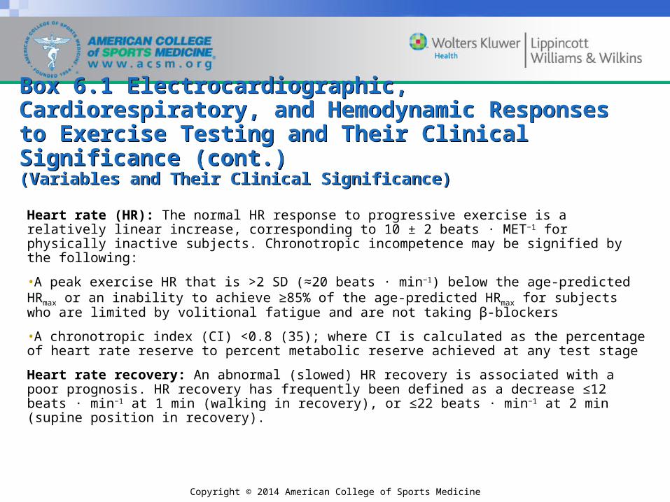
Heart rate (HR): The normal HR response to progressive exercise is a relatively linear increase, corresponding to 10 ± 2 beats ∙ MET−1 for physically inactive subjects. Chronotropic incompetence may be signified by the following:
•A peak exercise HR that is >2 SD (≈20 beats · min−1) below the age-predicted HRmax or an inability to achieve ≥85% of the age-predicted HRmax for subjects who are limited by volitional fatigue and are not taking β-blockers
•A chronotropic index (CI) <0.8 (35); where CI is calculated as the percentage of heart rate reserve to percent metabolic reserve achieved at any test stage
Heart rate recovery: An abnormal (slowed) HR recovery is associated with a poor prognosis. HR recovery has frequently been defined as a decrease ≤12 beats ∙ min−1 at 1 min (walking in recovery), or ≤22 beats ∙ min−1 at 2 min (supine position in recovery).

Copyright © 2014 American College of Sports Medicine
Box 6.1 Electrocardiographic, Cardiorespiratory, and Hemodynamic Responses to Exercise Testing and Their Clinical Significance (cont.)(Variables and Their Clinical Significance)
Box 6.1 Electrocardiographic, Cardiorespiratory, and Hemodynamic Responses to Exercise Testing and Their Clinical Significance (cont.)(Variables and Their Clinical Significance)
Systolic blood pressure (SBP): The normal response to exercise is a progressive increase in SBP, typically 10 ± 2 mm Hg ∙ MET−1 with a possible plateau at peak exercise. Exercise testing should be discontinued with SBP values of >250 mm Hg. Exertional hypotension (SBP that fails to rise or falls [>10 mm Hg]) may signify myocardial ischemia and/or LV dysfunction. Maximal exercise SBP of <140 mm Hg suggests a poor prognosis.
Diastolic blood pressure (DBP): The normal response to exercise is no change or a decrease in DBP. A DBP of >115 mm Hg is considered an endpoint for exercise testing.
Anginal symptoms: Can be graded on a scale of 1–4, corresponding to perceptible but mild, moderate, moderately severe, and severe, respectively. A rating of 3 (moderately severe) generally should be used as an endpoint for exercise testing.

Copyright © 2014 American College of Sports Medicine
Box 6.1 Electrocardiographic, Cardiorespiratory, and Hemodynamic Responses to Exercise Testing and Their Clinical Significance (cont.)(Variables and Their Clinical Significance)
Box 6.1 Electrocardiographic, Cardiorespiratory, and Hemodynamic Responses to Exercise Testing and Their Clinical Significance (cont.)(Variables and Their Clinical Significance)
Cardiorespiratory fitness: Average values of VO2max /VO2peak expressed as METs, expected in healthy sedentary men and women, can be predicted from one of several regression equations (28).
Also, see Table 4.9 for age-specific VO2max norms. Recent meta-analysis suggests each 1 MET increase in aerobic capacity equates to 13% and 15% decrease in all-cause mortality and cardiovascular events, respectively (32).
Ventilatory efficiency: Normal VE/VCO2 slope value <30. Elevated value is strongly prognostic in patients with heart failure and potentially patients with pulmonary hypertension. Values of ~45 or more are indicative of particularly poor prognosis in patients with heart failure. Elevated values are clearly indicative of worsening ventilation perfusion abnormalities in heart failure and pulmonary hypertension populations and thus provide an accurate depiction of disease severity (8).
. .
.
. .
Copyright © 2014 American College of Sports Medicine
Box 6.1 Electrocardiographic, Cardiorespiratory, and Hemodynamic Responses to Exercise Testing and Their Clinical Significance (cont.)(Variables and Their Clinical Significance)
Box 6.1 Electrocardiographic, Cardiorespiratory, and Hemodynamic Responses to Exercise Testing and Their Clinical Significance (cont.)(Variables and Their Clinical Significance)
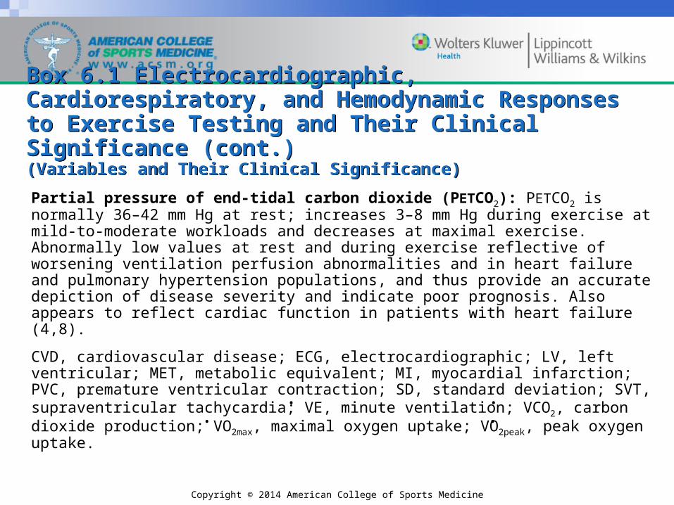
Partial pressure of end-tidal carbon dioxide (PETCO2): PETCO2 is normally 36–42 mm Hg at rest; increases 3–8 mm Hg during exercise at mild-to-moderate workloads and decreases at maximal exercise. Abnormally low values at rest and during exercise reflective of worsening ventilation perfusion abnormalities and in heart failure and pulmonary hypertension populations, and thus provide an accurate depiction of disease severity and indicate poor prognosis. Also appears to reflect cardiac function in patients with heart failure (4,8).
CVD, cardiovascular disease; ECG, electrocardiographic; LV, left ventricular; MET, metabolic equivalent; MI, myocardial infarction; PVC, premature ventricular contraction; SD, standard deviation; SVT, supraventricular tachycardia; VE, minute ventilation; VCO2, carbon dioxide production; VO2max, maximal oxygen uptake; VO2peak, peak oxygen uptake.
. .. .

Copyright © 2014 American College of Sports Medicine
Heart Rate ResponseHeart Rate Response
• Maximal heart rate (HRmax) may be predicted from age using any of several published equations (see Chapter 7).
• The relationship between age and HRmax for a large sample of subjects is well established; however, interindividual variability is high (±12 beats · min−1).

Copyright © 2014 American College of Sports Medicine
Heart Rate Response (cont.)Heart Rate Response (cont.)
• There is potential for considerable error in the use of methods that extrapolate submaximal test data to an age-predicted Hrmax.
• Using the 220 – age equation, failure to achieve an age-predicted HRmax ≥85% in the presence of maximal effort (chronotropic incompetence) is an ominous prognostic marker.
Copyright © 2014 American College of Sports Medicine
Heart Rate Response (cont.)Heart Rate Response (cont.)
• Failure to achieve an age-predicted HRmax >80% (chronotropic incompetence), using the equation {[HRpeak – HRrest]/[(220 – age) – HRrest]},is also an indicator of increased risk for adverse events.
• A delayed decrease in HR early in recovery after a symptom-limited maximal exercise test (≤12 beats · min−1 decrease after the first minute in recovery) is also a powerful independent predictor of overall mortality and should therefore be included in the exercise test assessment.
• Achievement of age-predicted HRmax should not be used as an absolute test endpoint or as an indication that effort has been maximal because of its high intersubject variability.

Copyright © 2014 American College of Sports Medicine
Blood Pressure ResponseBlood Pressure Response
The normal BP response to dynamic upright exercise consists of:
•A progressive increase in SBP
•No change or a slight decrease in DBP
•A widening of the pulse pressure
Copyright © 2014 American College of Sports Medicine
Blood Pressure Response (cont.)Blood Pressure Response (cont.)
• A drop in SBP (≥10 mm Hg decrease in SBP with an increase in workload), or failure of SBP to increase with increased workload, is considered an abnormal test response.
• The normal postexercise response is a progressive decline in SBP.
• In patients on vasodilators, calcium channel blockers, angiotensin-converting enzyme inhibitors, and α- and β-adrenergic blockers, the BP response to exercise is variably attenuated and cannot be accurately predicted in the absence of clinical test data (see Appendix A).

Copyright © 2014 American College of Sports Medicine
Blood Pressure Response (cont.)Blood Pressure Response (cont.)
• Although HRmax is comparable for men and women, men generally have higher SBPs (~20 ± 5 mm Hg) during maximal treadmill testing.
• The sex difference is no longer apparent after 70 yr.
• The rate pressure product, or double product (SBP HR), is an indicator of myocardial oxygen demand.
• Maximal double product values during exercise testing are typically between 25,000 (10th percentile) and 40,000 (90th percentile).

Copyright © 2014 American College of Sports Medicine
Electrocardiograph WaveformsElectrocardiograph Waveforms
• The normal ECG response to exercise includes the following:
– Minor and insignificant changes in P wave morphology
– Superimposition of the P and T waves of successive beats
– Increases in septal Q wave amplitude

Copyright © 2014 American College of Sports Medicine
Electrocardiograph Waveforms (cont.)Electrocardiograph Waveforms (cont.)
– Slight decreases in R wave amplitude
– Increases in T wave amplitude (although wide variability exists among clients/patients)
– Minimal shortening of the QRS duration
– Depression of the J point
– Rate-related shortening of the QT interval

Copyright © 2014 American College of Sports Medicine
ST-Segment ElevationST-Segment Elevation• ST-segment elevation (early repolarization) may be seen in the normal
resting ECG in various patterns.
• Data suggest that an early repolarization pattern in the inferior leads may indicate an increased risk of cardiac mortality in middle-aged individuals.
• Benign early repolarization can be common in the ECG of athletes and is typically localized to the chest leads V2–V5.
• Early repolarization exclusively observed in the anterolateral left precordial leads is not thought to be associated with increased risk for sustained ventricular arrhythmias, whereas global early repolarization (limb and precordial leads) appears to indicate a higher risk.

Copyright © 2014 American College of Sports Medicine
ST-Segment Elevation (cont.)ST-Segment Elevation (cont.)
• Exercise-induced ST-segment elevation in leads with Q waves consistent with a prior myocardial infarction may be indicative of wall motion abnormalities, ischemia, or both.
• Exercise-induced ST-segment elevation on an otherwise normal ECG (except in augmented voltage right [aVR] or chest leads V1 and V2) generally indicates significant myocardial ischemia and localizes the ischemia to a specific area of myocardium.
• This response may also be associated with ventricular arrhythmias and myocardial injury.

Copyright © 2014 American College of Sports Medicine
ST-Segment DepressionST-Segment Depression
• ST-segment depression (depression of the J point and the slope at 80 ms past the J point) is the most common manifestation of exercise-induced myocardial ischemia.
• Horizontal or downsloping ST-segment depression is more indicative of myocardial ischemia than is upsloping depression.
• The standard criterion for a positive test is ≥1.0 mm (0.1 mV) of horizontal or downsloping ST-segment at the J point extending for 60–80 ms.
Copyright © 2014 American College of Sports Medicine
ST-Segment Depression (cont.)ST-Segment Depression (cont.)
• Slowly upsloping ST-segment depression should be considered a borderline response, and added emphasis should be placed on other clinical and exercise variables.
• ST-segment depression does not localize ischemia to a specific area of myocardium.
• The more leads with (apparent) ischemic ST-segment shifts, the more severe the disease.
• Significant ST-segment depression occurring only in recovery likely represents a true positive response and should be considered an important diagnostic finding.

Copyright © 2014 American College of Sports Medicine
ST-Segment Depression (cont.)ST-Segment Depression (cont.)
• Adjustment of the ST-segment relative to the HR may provide additional diagnostic information.
– The ST/HR index is the ratio of the maximal ST-segment change (µV) to the maximal change in HR from rest to peak exercise (beats · min−1).
– An ST/HR index of >1.6 µV ∙ beats−1 ∙ min−1 is defined as abnormal.
– The ST/HR slope reflects the maximal slope relating the amount of the ST-segment depression (µV) to HR (beats ∙ min-1) during exercise.
– An ST/HR slope of ≥2.4 µV ∙ beats−1 ∙ min−1 is defined as abnormal.

Copyright © 2014 American College of Sports Medicine
ST-Segment Normalization orAbsence of Change
ST-Segment Normalization orAbsence of Change
• Ischemia may be manifested by normalization of resting ST-segments.
• ECG abnormalities at rest including T-wave inversion and ST-segment depression may return to normal during anginal symptoms and during exercise in some patients.

Copyright © 2014 American College of Sports Medicine
DysrhythmiasDysrhythmias
• Exercise-associated dysrhythmias occur in healthy people as well as patients with CVD. Increased sympathetic drive and changes in extracellular and intracellular electrolytes, pH, and oxygen tension contribute to disturbances in myocardial and conducting tissue automaticity and reentry, which are major mechanisms of dysrhythmias.

Copyright © 2014 American College of Sports Medicine
Dysrhythmias (cont.)Dysrhythmias (cont.)
• Supraventricular dysrhythmias
– Atrial flutter or atrial fibrillation may occur in organic heart disease or may reflect endocrine, metabolic, or medication effects.
– Sustained supraventricular tachycardia (SVT) occasionally is induced by exercise and may require pharmacologic treatment or electroconversion if discontinuation of exercise fails to abolish the rhythm.
Copyright © 2014 American College of Sports Medicine
Dysrhythmias (cont.)Dysrhythmias (cont.)• Ventricular dysrhythmias
– The suppression of PVCs that are present at rest with exercise testing does not exclude the presence of CVD, and PVCs that increase in frequency, complexity, or both do not necessarily signify underlying ischemic heart disease.
– Serious forms of ventricular ectopy include paired or multiform PVCs or runs of ventricular tachycardia (≥3 PVCs in succession).
– These dysrhythmias are likely to be associated with significant CVD, a poor prognosis, or both, if they occur in conjunction with signs or symptoms of myocardial ischemia, or in patients with a history of resuscitated sudden cardiac death, cardiomyopathy, or valvular heart disease.

Copyright © 2014 American College of Sports Medicine
Dysrhythmias (cont.)Dysrhythmias (cont.)
• Criteria for terminating exercise tests based on ventricular ectopy include sustained ventricular tachycardia, multifocal PVCs, and short runs of ventricular tachycardia.
• The decision to terminate an exercise test should also be influenced by simultaneous evidence of myocardial ischemia and/or adverse signs or symptoms (see Box 5.2).

Copyright © 2014 American College of Sports Medicine
Limiting Signs and SymptomsLimiting Signs and Symptoms
• All of the following criteria for maximal effort can be subjective and therefore possess limitations to varying degrees:
– Failure of HR to increase with further increases in exercise intensity.
– A plateau in oxygen uptake (or failure to increase oxygen uptake by 150 mL · min−1) with increased workload (this criterion has fallen into disfavor because a plateau is inconsistently seen during continuous graded exercise tests and is confused by various definitions and how data are sampled during exercise).
Copyright © 2014 American College of Sports Medicine
Limiting Signs and Symptoms (cont.)Limiting Signs and Symptoms (cont.)– A respiratory exchange ratio (RER) ≥1.10 is a
minimal threshold that may be obtained in most individuals putting forth a maximal effort, although there may be considerable interindividual variability with an RER ≥1.10.
– Various postexercise venous lactic acid concentrations (e.g., 8–10 mmol · L−1) have been used; however, there is also significant interindividual variability in this response.
– A rating of perceived exertion >17 on the 6–20 scale or >9 on the 0–10 scale.

Copyright © 2014 American College of Sports Medicine
Ventilatory Expired Gas Responses to Exercise
Ventilatory Expired Gas Responses to Exercise
• Direct measurement of ventilatory expired gas during exercise provides a more precise assessment of exercise capacity and prognosis and helps to distinguish causes of exercise intolerance.
• The combination of this technology with standard GXT procedures is typically referred to as cardiopulmonary exercise testing (CPX).
• Maximal volume of oxygen consumed per unit time (VO2max) or peak oxygen uptake (VO2peak) provides important information about cardiorespiratory fitness and is a powerful marker of prognosis.
..

Copyright © 2014 American College of Sports Medicine
Ventilatory Expired Gas Responses toExercise (cont.)
Ventilatory Expired Gas Responses toExercise (cont.)
• The assessment of ventilatory efficiency (i.e., VE/VCO2 slope and partial pressure of end-tidal carbon dioxide [PETCO2]) provides robust prognostic and/or diagnostic information in patients with congestive heart failure (CHF) and pulmonary hypertension.
• Ventilatory expired gas responses often are used in clinical settings as an estimation of the point at which lactate accumulation in the blood occurs, sometimes referred to as the lactate or anaerobic threshold. Assessment of this physiologic phenomenon through ventilatory expired gas is typically referred to as ventilatory threshold (VT).
• It should be remembered that VT provides only an estimation, and the concept of anaerobic threshold during exercise is controversial.
. .

Copyright © 2014 American College of Sports Medicine
Ventilatory Expired Gas Responses to Exercise (cont.)
Ventilatory Expired Gas Responses to Exercise (cont.)
• In addition to estimating when blood lactate values begin to increase, maximal minute ventilation (VEmax) can be used in conjunction with the maximal voluntary ventilation (MVV) to assist in determining if there is a ventilatory limitation to maximal exercise.
• The relationship between VEmax and MVV, typically referred to as the ventilatory reserve, traditionally is defined as the percentage of the MVV achieved at maximal exercise (i.e., the VEmax/MVV ratio).
• In most normal healthy people, the VEmax/MVV ratio is ≤0.80. Values surpassing this threshold are indicative of a reduced ventilatory reserve and a possible pulmonary limitation to exercise.
.
.
.
.

Copyright © 2014 American College of Sports Medicine
Ventilatory Expired Gas Responses to Exercise (cont.)
Ventilatory Expired Gas Responses to Exercise (cont.)
• Pulse oximetry should also be assessed when CPX is used to assess possible pulmonary limitations to exercise. A decrease in pulse oximeter saturation >5% during exercise also indicates a pulmonary limitation.
• Most currently available ventilatory expired gas systems also possess capabilities for pulmonary function testing. Obstructive or restrictive patterns on baseline pulmonary function testing provide insight into the mechanism of limitations to exercise.
• A >15% decrease in FEV1.0 and/or peak expiratory flow following CPX compared to baseline values is indicative of exercise-induced bronchospasm.

Copyright © 2014 American College of Sports Medicine
Diagnostic Value of Exercise TestingDiagnostic Value of Exercise Testing
• Sensitivity
– The percentage of patients tested with known CVD who demonstrate significant ST-segment (i.e., positive) changes.
• Specificity
– The percentage of patients without CVD who demonstrate nonsignificant (i.e., negative) ST-segment changes.

Copyright © 2014 American College of Sports Medicine
Diagnostic Value of Exercise Testing (cont.)Diagnostic Value of Exercise Testing (cont.)
• Sensitivity
– A true positive exercise test reveals horizontal or downsloping ST-segment depression of ≥1.0 mm or more and correctly identifies a patient with CVD.
– False negative test results show no or nondiagnostic ECG changes and fail to identify patients with underlying CVD.

Copyright © 2014 American College of Sports Medicine
Box 6.2 Sensitivity, Specificity, and Predictive Value of DiagnosticGraded Exercise Testing
Box 6.2 Sensitivity, Specificity, and Predictive Value of DiagnosticGraded Exercise Testingsensitivity = TP/(TP + FN) = the percentage of patients with CVD who have a positive test
specificity = TN/(TN + FP) = the percentage of patients without CVD who have a negative test
predictive value (positive test) = TP/(TP + FP) = the percentage of patients with a positive test result who have CVD
predictive value (negative test) = TN/(TN + FN) = the percentage of patients with a negative test who do not have CVD
CVD, cardiovascular disease; FN, false negative (negative exercise test and CVD); FP, false positive (positive exercise test and no CVD); TN, true negative (negative exercise test and no CVD); TP, true positive (positive exercise test and CVD).

Copyright © 2014 American College of Sports Medicine
Box 6.3 Causes of False Negative Test ResultsBox 6.3 Causes of False Negative Test Results• Failure to reach an ischemic threshold
• Monitoring an insufficient number of leads to detect ECG changes
• Failure to recognize non-ECG signs and symptoms that may be associated with underlying CVD (e.g., exertional hypotension)
• Angiographically significant CVD compensated by collateral circulation
• Musculoskeletal limitations to exercise preceding cardiac abnormalities
• Technical or observer error
CVD, cardiovascular disease; ECG, electrocardiographic.
Copyright © 2014 American College of Sports Medicine
Box 6.4 Causes of Abnormal ST-Segment Changes in the Absence ofObstructive Cardiovascular Diseasea
Box 6.4 Causes of Abnormal ST-Segment Changes in the Absence ofObstructive Cardiovascular Diseasea
• Resting repolarization abnormalities (e.g., left bundle-branch block)
• Cardiac hypertrophy
• Accelerated conduction defects (e.g., Wolff-Parkinson-White syndrome)
• Digitalis
• Nonischemic cardiomyopathy
• Hypokalemia
• Vasoregulatory abnormalities

Copyright © 2014 American College of Sports Medicine
Box 6.4 Causes of Abnormal ST-Segment Changes in the Absence ofObstructive Cardiovascular Diseasea (cont.)
Box 6.4 Causes of Abnormal ST-Segment Changes in the Absence ofObstructive Cardiovascular Diseasea (cont.)
• Mitral valve prolapsed
• Pericardial disorders
• Technical or observer error
• Coronary spasm in the absence of significant coronary artery disease
• Anemia
• Being a woman
aSelected variables simply may be associated with rather than be causes of abnormal test results.
Copyright © 2014 American College of Sports Medicine
Diagnostic Value of Exercise Testing (cont.)Diagnostic Value of Exercise Testing (cont.)
• Predictive value
– A measure of how accurately a test result (positive or negative) correctly identifies the presence or absence of CVD in tested patients
– Cannot be estimated directly from a test’s specificity or sensitivity because it depends on the prevalence of disease in the population being tested
Copyright © 2014 American College of Sports Medicine
Prognostic Applications of theExercise Test
Prognostic Applications of theExercise Test
• Several clinical factors contribute to patient outcome including
– severity and stability of symptoms,
– left ventricular function,
– angiographic extent and severity of CVD,
– electrical stability of the myocardium, and
– the presence of other comorbid conditions.

Copyright © 2014 American College of Sports Medicine
FIGURE 6.2. Duke nomogram uses five steps to estimate prognosis for a given individual from the parameters of the Duke score. First, the observed amount of ST depression is marked on the ST-segment deviation line. Second, the observed degree of angina is marked on the line for angina, and these two points are connected. Third, the point where this line intersects the ischemia reading line is noted. Fourth, the observed exercise tolerance is marked on the line for exercise capacity. Finally, the mark on the ischemia reading line is connected to the mark on the exercise capacity line, and the estimated 5-yr survival or average annual mortality rate is read from the point at which this line intersects the prognosis scale (40).

Copyright © 2014 American College of Sports Medicine
The Bottom LineThe Bottom Line• Interpreting the results of a clinical exercise test requires a multivariable approach.
• The HR, hemodynamic, and ECG response to exercise are key objective parameters that require intricate assessment from an experienced clinician. In addition, the subjective symptoms including RPE, angina, and dyspnea are important components of exercise test interpretation.
• When ventilatory expired gas is assessed during the clinical exercise test, a highly accurate determination of aerobic capacity is possible in addition to a potentially more accurate quatification of exercise effort (i.e., peak RER) and assessment of submaximal exercise performance and ventilatory efficiency.
• Clinical exercise testing assists in the diagnosis of CVD as well as the physiologic mechanisms for abnormal functional limitations such as unexplained exertional dyspnea.
• The diagnostic accuracy of clinical exercise testing depends on the characteristics of the patient who is undergoing the assessment and the quality of the test.
• Clinical exercise testing data, and in particular aerobic capacity, provide valuable prognostic information in virtually all individuals undergoing this procedure.