correlated mutations contain information about protein-protein
TRANSCRIPT

Correlated Mutations Contain Information AboutProtein±protein Interaction
Florencio Pazos1, Manuela Helmer-Citterich2, Gabriele Ausiello2
and Alfonso Valencia1*
1Protein Design GroupCNB-CSIC, Campus U.AutoÂnoma, CantoblancoMadrid 28049, Spain2Dipartimento di BiologiaU. di Roma ``Tor Vergata''Rome, Italy
Many proteins have evolved to form speci®c molecular complexes andthe speci®city of this interaction is essential for their function. The net-work of the necessary inter-residue contacts must consequently constrainthe protein sequences to some extent. In other words, the sequence of aninteracting protein must re¯ect the consequence of this process of adap-tation. It is reasonable to assume that the sequence changes accumulatedduring the evolution of one of the interacting proteins must be compen-sated by changes in the other.
Here we apply a method for detecting correlated changes in multiplesequence alignments to a set of interacting protein domains and showthat positions where changes occur in a correlated fashion in the twointeracting molecules tend to be close to the protein±protein interfaces.This leads to the possibility of developing a method for predicting con-tacting pairs of residues from the sequence alone. Such a method wouldnot need the knowledge of the structure of the interacting proteins, andhence would be both radically different and more widely applicable thantraditional docking methods.
We indeed demonstrate here that the information about correlatedsequence changes is suf®cient to single out the right inter-domain dock-ing solution amongst many wrong alternatives of two-domain proteins.The same approach is also used here in one case (haemoglobin) wherewe attempt to predict the interface of two different proteins rather thantwo protein domains. Finally, we report here a prediction about theinter-domain contact regions of the heat- shock protein Hsc70 based onlyon sequence information.
# 1997 Academic Press Limited
Keywords: correlated mutations; protein contacts; docking; co-adaptation;Hsc70*Corresponding author
Introduction
The protein±protein interaction problem
Molecular recognition is a key process in biologi-cal systems. The order and control of protein±pro-tein interactions in signalling pathways andmetabolic networks are important aspects of mol-ecular biology and biochemistry. DNA replicationand transcription, RNA splicing, protein sorting,cell adhesion, signalling cascades and metaboliccycles are just some examples of the many complex
processes dominated by protein±protein recog-nition.
The unravelling of this complex process requirestwo major steps. First, it is necessary to ®nd theinteracting proteins in the cell soup (e.g. the longquest for the downstream effectors in the ras-p21signalling cascade); and second, to describe at themolecular level how the interaction takes place,e.g. how and where ras-p21 interacts with the raf-kinase and how conformational changes relatedwith GTP hydrolysis control the interactionbetween the proteins.
The ®rst issue of searching for the interactingcomponents in a functional complex is a daily pro-blem for experimental biology but has remained sofar untouched from the theoretical point of view.The problem of describing and predicting the mol-
E-mail: [email protected] used: RMS, root-mean-square deviation;
Xd, harmonic difference between binned populations;Nt, N-terminal; Ct, C-terminal.
J. Mol. Biol. (1997) 271, 511±523
0022±2836/97/340511±13 $25.00/0/mb971198 # 1997 Academic Press Limited
ecular complexes in detail, also known as the``docking problem'' has instead attracted a greatdeal of attention and has led to the development ofseveral different theoretical methods.
Current physical approaches to thedocking problem
Docking has attracted much attention (for recentreviews, see Lengauer & Rarey, 1996; Strynadkaet al., 1996). Undoubtedly, progress has been made,and some methods are ready for challenges suchas the prediction of the interaction between lacta-mase and one of its inhibitors. It was accomplishedquite successfully by six different groups(Strynadka et al., 1996), or the more recent CASP-2meeting (WWW: http://iris4.carb.nist.gov/casp2).All current docking methods require the three-dimensional structures of the interacting proteinsto be known. In all methods, the interactingsurfaces are described by different physical proper-ties (Connolly surfaces, grids, protein slices,property vectors, etc.) to allow the identi®cation ofgeometrically complementary regions between thetwo proteins (Cher®ls et al., 1991; Fischer et al.,1995; Helmer-Citterich & Tramontano, 1994;Jackson & Sternberg, 1995; Jiang & Kim, 1991;Shoichet & Kuntz, 1991; Stoddard & Koshland,1992; Walls & Sternberg, 1992; Katchalski-Katziret al., 1992).
Most algorithms treat proteins as rigid bodiesand in only a few cases is protein ¯exibilitytaken into account (Totrov & Abagyan, 1994;O'Donoghue, S. & Nilges, M., personal communi-cation). Flexibility is an inherent dif®culty in thedocking problem, since most inter-protein com-plexes undergo induced-®t movements upon bind-ing, and hence a rigid-body description of theindividual components may not be accurate en-ough to predict the structure of the ®nal complex.
Docking methods and the characteristics ofprotein interfaces
It is generally accepted that the physical prin-ciples underlying protein folding and protein±pro-tein association are similar. This belief is supportedby detailed studies of similarities in the packing ofprotein interfaces and protein interiors (Walls &Sternberg, 1992), and similarities in the overallresemblance of the hydrophobicity patterns (Younget al., 1994). However, our understanding of thepeculiar characteristics of protein-protein inter-action is still very limited. Earlier attempts to studycomplementary surfaces between proteins (Argos,1988; Janin & Chothia, 1990; Janin et al., 1988) werehampered by the lack of experimental data. Morerecent studies (Jones & Thornton, 1996; Tsai et al.,1996) are for the ®rst time providing tools for asystematic approach to the characterisation of pro-tein±protein interfaces by rigorous scanning ofdata bases of protein complexes.
The study of the evolution of oligomerisationhas also become an important issue and the ®rstideas about the origin of the adaptation in proteincomplexes from the initial components are emer-ging (Fletterick & Bazan, 1995; Bennett et al., 1995).
A new approach to predict protein±proteincontact regions based on sequence information
We propose here a new and completely differentapproach to the study and prediction of protein±protein interaction. Instead of considering thestructural nature of the interactions, we try to de-tect the sequence traces that evolution may haveleft on the interacting sequences during the processof preserving the protein±protein interaction sites.Therefore, our approach is not restricted to thecases in which the structures of the proteins to bedocked are known and is applicable to any familyof interacting proteins for which a large enough se-quence family is available.
Sequence information and the process ofprotein±protein co-adaptation
There is a common agreement among research-ers that interacting proteins undergo a process ofco-evolution. ``Over time, amino acid substitutionmay stabilise an interface that does not exist in theclosed monomer . . . stabilising mutations in theseinterfaces would be favoured in natural selection''(Bennett et al., 1995); however, no explicit strategyhas been proposed for detecting the traces of thisprocess from protein sequences. We propose that itis possible to detect this signal by studying com-pensatory mutations. In order to do so, we haveappropriately modi®ed our previously publishedmethod for the calculation of correlated mutationsin multiple-sequence alignments (GoÈbel et al., 1994;Pazos et al., 1997).
Defining correlated mutations
Several groups have studied correlatedmutations: technical differences between differentapproaches have led to con¯icting conclusionsabout the nature and intensity of this phenomena(Altschuh et al., 1987, 1988; GoÈbel et al., 1994;Neher, 1994; Shindyalov et al., 1994; Taylor &Hatrick, 1994). Thus, it is important to de®neexactly our notion of correlated mutations. In thisand previous work, we have used the term corre-lated mutations to indicate a tendency of positionsin proteins to mutate co-ordinately. We measurethis tendency by analysing the correlation betweenchanges in pairs of positions in multiple sequencealignments, with an unambiguous de®nition ofcorrelation (see Methods).
Biological meaning of compensatory mutations
There is clear experimental support for the roleof compensating mutations in protein stability
512 Protein Docking and Correlated Mutations

(Serrano et al., 1990) and function (Gregoret &Sauer, 1993). Vernet et al. (1992) directly tested thein¯uence in protein stability of some pairs of corre-lated mutations. We believe that the signaldetected by our method corresponds mainly tonetworks of positions that have undergonecompensating mutations during evolution. If inter-actions between proteins are of the same physicalnature as intra-protein interactions, then their con-sequences at the sequence level are most likely alsosimilar. Therefore, we apply our method, whichwe have previously used to predict contacts inglobular proteins (GoÈbel et al., 1994; Pazos et al.,1997), to the problem of predicting interactionsbetween proteins. As we will show here, the signalat the sequence level for inter-protein contactsturns out to be even more speci®c than that forintra-protein contacts, possibly because it is subjectto a stronger selective pressure.
Testing the method
The purpose of this work is to test the feasibilityof a sequence-based approach to the prediction ofinteracting regions in protein complexes. To do so,we ®rst show that correlated pairs between twodifferent proteins are signi®cantly closer to eachother than other pairs of positions in the same pro-teins, and second that they can be used to discrimi-nate the correct docking solutions among manyalternative wrong ones in proteins of known struc-ture. We then carry out a bona ®de prediction of theyet unknown interaction site between the twodomains of the Hsc70 heat shock protein
Results
The results are presented in the following order:®rst we demonstrate that correlated mutations docontain information about inter-domain contacts.We tested our method mainly for inter-domaininteractions to take advantage of the larger set ofexamples of proteins of known structure for whichmany homologous sequences are available. Asdescribed in detail later, we demonstrate that, onaverage, pairs of residues detected as ``correlated''by our method are closer to each other than theaverage pairs of residues in the same protein.
Next we show how the correlated mutation anal-ysis can be used to identify docking solutions veryclose to the native solution from many wrong sol-utions. The aim of this experiment is to empiricallyevaluate how much information about inter-do-main contacts is contained in correlated mutations.It is important to remember that we are notattempting to replace existing docking methodsbased on structural information; we only want toestablish that correlated mutations are good indi-cators of contacting residues. Our results shouldnot be compared to any current docking method.
The docking algorithm here is used only as a rapidtool to generate many alternative ``reasonable'' sol-utions. In a ®rst set of experiments, we generatedthousands of alternative solutions that fully coverthe space of possible solutions without any attemptto increase the number of solutions close to the realdocking position. The second set of experimentscorresponds to a more demanding test, since inthis case the set of solutions among which wewant to discriminate consists of hundreds of physi-cally realistic solutions, corresponding to the bestscoring complementary surfaces calculated with astandard docking program.
The third section contains the results obtainedfor the complex between a and b haemoglobin, atest selected to prove that our method has similarperformances when applied to inter-protein as wellas inter-domain contacts. A more complete test ofprotein±protein complexes is prevented by thevery limited availability of data on protein±proteincomplexes where both the structure and a suf®-cient number of aligned sequences for the samespecies (see later) are known.
Finally, a bona ®de prediction is reported.Sequence information is used to predict the con-tacting residues between the two domains of theheat-shock protein Hsc70. This prediction is usedto illustrate the novel feature of our approach: thatprediction can be made in the absence of structuralinformation. The example is also biologically rel-evant: the function of Hsc70 is based on the inter-action between its two domains. The N-terminal(Nt) domain contains the ATP-binding site, whilethe C-terminal (Ct) domain is mainly responsiblefor peptide binding (Chappel et al., 1987; Gragerovet al., 1994; Montgomery et al., 1993). The inter-action between the two domains generates the bio-logical functions of peptide binding and release(McCarty et al., 1995). The structure of both iso-lated domains (Nt domain of hsc70; Flaherty et al.,1990; Ct domain of its related protein DnaK, Zhuet al., 1996) has been solved, although the Ctdomain structure is not yet publicly available. Thisis an appropriate moment for a bona ®de prediction,since the structure of the complex has not yet beensolved and ``classical'' docking methods cannot beused until both structures are available.
Prediction of domain±domain contacts fordifferent protein families
We have previously shown that in single-do-main proteins correlated residues tend to be closerthan other residues (GoÈbel et al., 1994). This generalresult is illustrated in Figure 1(a) for papain (9pap,Kamphuis et al., 1984). In the Figure we comparethe distribution of the distances between pairs ofcorrelated residues with that of the distancesbetween all pairs of residues in each of thedomains of papain. The distribution shows a clearshift of the population of correlated positions towardcloser distances. This example supports our earlierconclusion that, in globular proteins, correlated
Protein Docking and Correlated Mutations 513

positions are statistically closer than non-correlatedpositions.
The spatial proximity of correlated positionsinside globular proteins can be extended to theproximity of correlated positions belonging to twodifferent domains. The actual values of inter-do-
main distances for all residues and for correlatedpositions belonging to two different domains forpapain are compared in Figure 1(b). Once again aclear shift of the correlated pairs toward smallerdistances is observed. This shift indicates that cor-related positions have a tendency to be closer to
Figure 1. Bar diagrams comparing the proportions of pairs of residues at different distances. Distributions are rep-resented for all residues (®lled bars) and for correlated pairs of residues (hatched bars) in papain (9pap). (a) Distancesbetween pairs in the two independent domains, and (b) distances between the two domains. Correlated positions areshifted toward smaller distances.
Table 1. The 21 monomeric protein families with two domains
Xd
PDB Domain HSSPI/2
codea Sizeb Sizec Domain de®nitiond NALIGNe I-If II-IIg I-IIh Referencei
A. Disjoin domains4mt2 61 29/32 1±29/30±61 72 Nep 2,36 ÿ2,22 (2)3dfr 162 80/68 2±32, 112±160/38±105 18 11,68 7,92 ÿ1,63 (3)4tnc 162 88/72 3±90/91±162 139 4,39 10,16 0,45 (2)3cln 148 74/66 5±78/82±147 167 2,57 8,61 1,63 (3)1clm 148 85/59 4±88/89±147 175 1,58 10,51 2,19 (1)
B Conjoin domains1rnd 124 73/51 1±49,80±103/50±79, 104±124 62 7,57 3,56 ÿ0,77 (1)4tms 316 223/93 1±52,146±316/53±145 20 4,39 Ne 1,50 (1)3pgk 416 199/216 1±188,405±415/189±404 42 5,07 1,95 2,36 (1)
C. Interacting domains2gcr 173 82/92 1±82/83±174 45 5,22 16,71 0,81 (3)1alc 123 67/55 38±104/1±37,105±122 67 2,40 4,26 2,53 (2)3blm 257 156/86 1±67,168±256/69±154 33 2,94 9,66 3,72 (3)2pf2 156 62/83 1±62/63±145 20 2,39 5,91 4,05 (1)2bbm 148 74/66 5±78/82±147 177 4,42 9,34 4,31 (3)1ppl 323 212/111 1±192,304±323/193±303 30 4,39 ÿ1,14 4,53 (1)3est 240 126/103 16±29,122±233/30±120,234±245 211 18,35 18,87 5,27 (1)3adk 195 144/50 1±37,88±194/38±87 34 13,70 ÿ6,87 6,72 (1)3rp2 224 109/108 16±21,128±230/28±122,231±243 181 12,30 13,37 7,10 (3)9pap 212 112/100 1±16,113±208/17±112,209±212 68 18,32 7,98 7,12 (1)2c2c 112 50/62 1±33,96±112/34±95 117 8,60 8,44 8,85 (2)3trx 105 72/35 1±72/74±108 45 3,91 8,46 9,53 (3)1sgt 223 139/80 16±28,69±80,121±234/29±68,81±120 158 19,37 15,00 12,70 (1)
Nep, Not enough pairs for Xd calculation.a PDB and chain identi®ers.b Chain length (in amino acid residues).c Length of each domain (in amino acid residues).d Domain de®nitions (DI/DII).e Number of sequences in the multiple sequence alignment. From the HSSP Data Base (Sander & Schneider, 1993).f Xd, the harmonic weighted difference between the binned distributions of distances between all residues and correlated pairs of
positions in the ®rst domain (see Methods).g As f but for residue pairs in the second domain.h As f but for pairs of residues belonging to different domains.i The domain de®nition was taken from: (1) Holm & Sander (1994); (2) Siddiqui & Barton (1995); (3) Sowdhamini & Blundell
(1994); (4) Swindells (1994).
514 Protein Docking and Correlated Mutations
the inter-domain interface. In order to quantify thedifference between the population of distancesbetween correlated pairs and that of all other pairs,we de®ne the parameter Xd, as the harmonicdifference between the two binned populations(see Methods).
Table 1 shows the results for a set of 21 proteinswith two domains. In almost all cases, the popu-lation of correlated pairs inside the individualdomains (domain I or domain II) is shifted towardsmaller distances as indicated by large positive Xdvalues. There are four exceptions with negative orvery small Xd values. We suspect that this may bedue to imprecise de®nitions of domain boundariesleading to non-perfectly globular proteins, withdistorted residue distance distributions.
Table 1 shows that correlated positions betweenthe two protein domains are, on average, closerthan non-correlated positions, with positive Xdvalues for 17 out of the 21 cases. In this Table,proteins are divided into three categories accordingto their degree of interaction, from weakly tostrongly interacting domains (disjoin, conjoin andinteracting, following Sowdhamini & Blundell(1994)). Correlated positions are closer only whenthere is a real association between domains, asdemonstrated by higher Xd values for interactingdomains than for any of the other two categories.
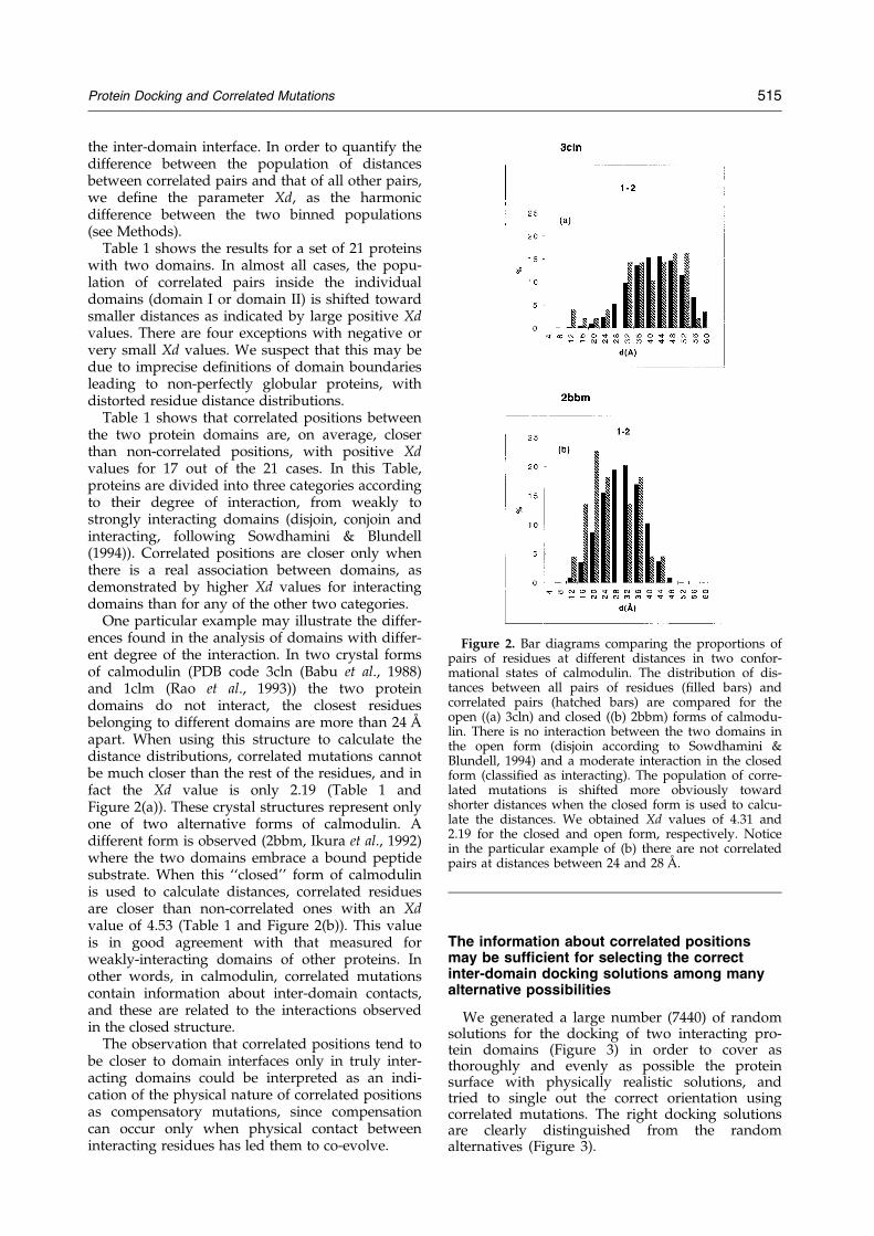
One particular example may illustrate the differ-ences found in the analysis of domains with differ-ent degree of the interaction. In two crystal formsof calmodulin (PDB code 3cln (Babu et al., 1988)and 1clm (Rao et al., 1993)) the two proteindomains do not interact, the closest residuesbelonging to different domains are more than 24 AÊ
apart. When using this structure to calculate thedistance distributions, correlated mutations cannotbe much closer than the rest of the residues, and infact the Xd value is only 2.19 (Table 1 andFigure 2(a)). These crystal structures represent onlyone of two alternative forms of calmodulin. Adifferent form is observed (2bbm, Ikura et al., 1992)where the two domains embrace a bound peptidesubstrate. When this ``closed'' form of calmodulinis used to calculate distances, correlated residuesare closer than non-correlated ones with an Xdvalue of 4.53 (Table 1 and Figure 2(b)). This valueis in good agreement with that measured forweakly-interacting domains of other proteins. Inother words, in calmodulin, correlated mutationscontain information about inter-domain contacts,and these are related to the interactions observedin the closed structure.
The observation that correlated positions tend tobe closer to domain interfaces only in truly inter-acting domains could be interpreted as an indi-cation of the physical nature of correlated positionsas compensatory mutations, since compensationcan occur only when physical contact betweeninteracting residues has led them to co-evolve.
The information about correlated positionsmay be sufficient for selecting the correctinter-domain docking solutions among manyalternative possibilities
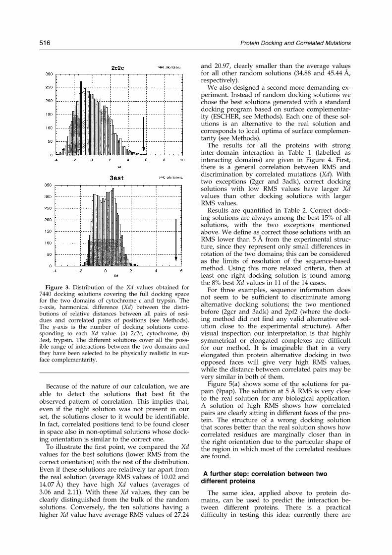
We generated a large number (7440) of randomsolutions for the docking of two interacting pro-tein domains (Figure 3) in order to cover asthoroughly and evenly as possible the proteinsurface with physically realistic solutions, andtried to single out the correct orientation usingcorrelated mutations. The right docking solutionsare clearly distinguished from the randomalternatives (Figure 3).
Figure 2. Bar diagrams comparing the proportions ofpairs of residues at different distances in two confor-mational states of calmodulin. The distribution of dis-tances between all pairs of residues (®lled bars) andcorrelated pairs (hatched bars) are compared for theopen ((a) 3cln) and closed ((b) 2bbm) forms of calmodu-lin. There is no interaction between the two domains inthe open form (disjoin according to Sowdhamini &Blundell, 1994) and a moderate interaction in the closedform (classi®ed as interacting). The population of corre-lated mutations is shifted more obviously towardshorter distances when the closed form is used to calcu-late the distances. We obtained Xd values of 4.31 and2.19 for the closed and open form, respectively. Noticein the particular example of (b) there are not correlatedpairs at distances between 24 and 28 AÊ .
Protein Docking and Correlated Mutations 515
Because of the nature of our calculation, we areable to detect the solutions that best ®t theobserved pattern of correlation. This implies that,even if the right solution was not present in ourset, the solutions closer to it would be identi®able.In fact, correlated positions tend to be found closerin space also in non-optimal solutions whose dock-ing orientation is similar to the correct one.
To illustrate the ®rst point, we compared the Xdvalues for the best solutions (lower RMS from thecorrect orientation) with the rest of the distribution.Even if these solutions are relatively far apart fromthe real solution (average RMS values of 10.02 and14.07 AÊ ) they have high Xd values (averages of3.06 and 2.11). With these Xd values, they can beclearly distinguished from the bulk of the randomsolutions. Conversely, the ten solutions having ahigher Xd value have average RMS values of 27.24
and 20.97, clearly smaller than the average valuesfor all other random solutions (34.88 and 45.44 AÊ ,respectively).
We also designed a second more demanding ex-periment. Instead of random docking solutions wechose the best solutions generated with a standarddocking program based on surface complementar-ity (ESCHER, see Methods). Each one of these sol-utions is an alternative to the real solution andcorresponds to local optima of surface complemen-tarity (see Methods).
The results for all the proteins with stronginter-domain interaction in Table 1 (labelled asinteracting domains) are given in Figure 4. First,there is a general correlation between RMS anddiscrimination by correlated mutations (Xd). Withtwo exceptions (2gcr and 3adk), correct dockingsolutions with low RMS values have larger Xdvalues than other docking solutions with largerRMS values.
Results are quanti®ed in Table 2. Correct dock-ing solutions are always among the best 15% of allsolutions, with the two exceptions mentionedabove. We de®ne as correct those solutions with anRMS lower than 5 AÊ from the experimental struc-ture, since they represent only small differences inrotation of the two domains; this can be consideredas the limits of resolution of the sequence-basedmethod. Using this more relaxed criteria, then atleast one right docking solution is found amongthe 8% best Xd values in 11 of the 14 cases.
For three examples, sequence information doesnot seem to be suf®cient to discriminate amongalternative docking solutions; the two mentionedbefore (2gcr and 3adk) and 2pf2 (where the dock-ing method did not ®nd any valid alternative sol-ution close to the experimental structure). Aftervisual inspection our interpretation is that highlysymmetrical or elongated complexes are dif®cultfor our method. It is imaginable that in a veryelongated thin protein alternative docking in twoopposed faces will give very high RMS values,while the distance between correlated pairs may bevery similar in both of them.
Figure 5(a) shows some of the solutions for pa-pain (9pap). The solution at 5 AÊ RMS is very closeto the real solution for any biological application.A solution of high RMS shows how correlatedpairs are clearly sitting in different faces of the pro-tein. The structure of a wrong docking solutionthat scores better than the real solution shows howcorrelated residues are marginally closer than inthe right orientation due to the particular shape ofthe region in which most of the correlated residuesare found.
A further step: correlation between twodifferent proteins
The same idea, applied above to protein do-mains, can be used to predict the interaction be-tween different proteins. There is a practicaldif®culty in testing this idea: currently there are
Figure 3. Distribution of the Xd values obtained for7440 docking solutions covering the full docking spacefor the two domains of cytochrome c and trypsin. Thex-axis, harmonical difference (Xd) between the distri-butions of relative distances between all pairs of resi-dues and correlated pairs of positions (see Methods).The y-axis is the number of docking solutions corre-sponding to each Xd value. (a) 2c2c, cytochrome, (b)3est, trypsin. The different solutions cover all the poss-ible range of interactions between the two domains andthey have been selected to be physically realistic in sur-face complementarity.
516 Protein Docking and Correlated Mutations

few cases where the three-dimensional structure ofthe protein complex and many corresponding se-quences from different species are available.
We have chosen to test the concept using haemo-globin, since the wealth of sequences for this pro-tein made it possible to select an appropriatesubset of sequences. We selected the a1-b2 mono-mers of the tetramer because they contain the func-tional interface that undergoes structural changesbetween the oxy and deoxy forms (Jayaraman et al.,1995). We generated multiple docking solutions forthe a1-b2 dimer and used, as in previous cases,the information about correlated mutations todiscriminate among them. As can be seen inFigure 5(b), there is a clear difference betweengood solutions and wrong ones. Xd values can beused to discriminate between them: the right sol-ution always scores among the ®rst 6% and the Xdvalue is 3.36, in the range of the examples of inter-acting domains shown in Table 1. This result is sat-isfactory, especially in view of the fact that this is adif®cult case with a very small interacting surface(Lesk & Chothia, 1980; Perutz, 1978).
Prediction of contact between domains in theabsence of three-dimensional structures
To illustrate the predictive value of our methodwe present a prediction of the domain interaction
Figure 4. Scatter plot of the values of Xd against RMS.The y-axis, harmonic difference (Xd) between the distri-butions of relative distances between all residues andcorrelated pairs of positions (see Methods). The x-axis isthe RMS between the real structure and the differentalternative docking positions. (a) 1sgt, trypsin; (b) 2c2c,cytochrome; (c) 9pap, papain; (d) 3rp2, rat mast cell pro-
tease; (e) 3est, elastase; (f) 2bbm, calmodulin bound tosubstrate; (g) 2pf2, prothrombin; (h) 1alc, a-lactoalbu-min; (i) 2gcr, g-crystallin; (j) 3blm, b-lactamase; (k) 1ppl,penicillopepsin; (l) 3adk, adenylate kinase; (m) 3trx,thioredoxin. The different examples cover all the rangeof interacting domains. The right solution is alwaysselected among the 8% best docking solutions, exceptfor 2gcr, 3blm, 2pf2 and 3adk (see Table 2). Solutionswith RMS smaller than 5 AÊ can be considered as validsolutions for the level of resolutions expected from amethod based only on sequences (vertical thick line onthe plots).
Table 2. Percentage of docking solutions that score bet-ter than the correct one for 11 proteins with interactingdomains
PDBld %a %(5 AÊ )b
2gcr 53.49 53.491alc 15.07 7.953blm 13.90 13.902pf2 13.76 13.762bbm 6.25 3.081ppl 2.90 2.903est 9.76 1.223adk 28.73 28.483rp2 2.53 0.009pap 3.08 3.082c2c 7.69 0.003trx 7.75 2.461sgt 0.00 0.00
a Percentage of solutions scored better than the real one.b The same considering all the solutions in the range 0 to 5 AÊ
RMS as correct.
Protein Docking and Correlated Mutations 517

Figure 5. Scatter plot of the values of Xd against RMS for different docking solutions of (a) 9pap and (b) a1-b2 of hae-moglobin (1hbb, Kavanaugh et al., 1992). The x-axis, RMS between the real structure and the different alternativedocking positions. The y-axis, harmonical difference (Xd) between the distributions of relative distances of all residuesand correlated pairs. The right docking solution is among the 5% best scored ones. In the case of haemoglobin it isdif®cult to generate alternative docking solutions close to the real one, since the surface of interaction of the mono-mers is sharp and small. A ribbon representation of some docking solutions including the real one is shown. The resi-dues participating in the pairs with higher correlation value are highlighted.
518 Protein Docking and Correlated Mutations

in the heat-shock protein Hsc70. The interactionbetween the Nt and Ct domains is essential for thefunction of the protein. The three-dimensionalstructure is known only for the Nt domain(Flaherty et al., 1990: ribbon plot in Figure 6). Thestructure of the Ct domain has been solvedrecently for DnaK, a related protein (Zhu et al.,1996), but it is not yet publicly available. A cartoondepicting the secondary and super-secondarystructure of this protein as assigned by Morshauseret al. (1995) is shown in Figure 6.
The correlated mutations that we identi®ed forthis case clearly predict that two de®ned regionsshould be part of the interacting surface. Bothregions map in the front face of the Nt domain(with respect to the standard view of Figure 6).This information could be directly tested bymutagenesis experiments and will be ultimatelyvalidated by the experimental determination of thecomplex between the Nt and Ct domains.
Discussion
The co-evolution of a protein±protein complexin different organisms must leave visible traces atthe sequence level. Part of this information can becaptured as correlated positions in multiplesequence alignments. We have previously shownthat there is indeed a trend for correlated pairs ofresidues to be closer in space than non-correlatedpairs of residues in single-domain proteins (GoÈbelet al., 1994).
Here we verify that this behaviour is character-istic for residues in single domains (intra-proteincontacts) and for residues sitting in two differentprotein domains (inter-protein). The informationcontained in our de®nition of correlated mutationsis able to discriminate between the real dockingsolution and many other realistic but wrongalternatives in a signi®cant number of cases. Wehave tested the method for two-domain proteins,for which it was possible to obtain a collection ofexamples. We anticipate that the same results holdfor interacting proteins. Indeed, in the case of hae-moglobin, correlated positions between the a and bchains are suf®cient to single out the right orien-tation of the two domains among many alterna-tives.
We evaluated the performances of the methodon known cases, and then used it to carry out areal prediction for the inter-domain interaction ofthe heat-shock protein Hsc70. This example illus-trates the potential of the method to generatespeci®c predictions about contacting residues andregions even when the protein structure isunknown.
Limitations of the method
The ability of correlated positions to discriminatebetween correct and incorrect relative positions oftwo domains is clearly related to the degree ofphysical proximity between the domains. Ourinterpretation is that only the co-evolution ofclosely interacting residues leaves detectablesignatures at the sequence level.
The case of calmodulin is instructive, since corre-lated positions properly describe the domain con-tacts in the closed form of the protein (2bbm)without being biased by the existence of an openform. Therefore, predictions should be carried outonly for interacting proteins, as is the case forHsc70.
Figure 6. Predicted contacts between the Nt and Ctdomains of Hsc70. In the upper part the three-dimen-sional structure of the Nt domain (3hsc, Flaherty et al.,1990) is shown as a ribbon plot. A schematic view ofthe NMR secondary structure assignment of the Ctdomain (Morshauser et al., 1995) is shown in the lowerpanel. Strands are represented by arrows, helices byboxes. The residues undergoing correlated mutationsbetween domains are shown as sticks in the ribbonplot. The ten best correlations are shaded and con-nected by lines. Their residue number and code arealso given. Residues participating in the ten next bestcorrelations are also shown as light sticks on the rib-bon plot. Additionally, correlations between residues inthe Ct domain are represented as broken lines. TheFigure points to a clear docking solution between thetwo b sheets of the Ct domain and between the ®rst bsheet of the Ct domain and the back of the Nt domains.
Protein Docking and Correlated Mutations 519

It is important to note that other factors in¯u-ence the quality of our predictions: the quality ofthe alignment, the distribution of sequences in thefamily and the family size. As a rule of thumb, pre-dictions are reliable only in families with morethan 15 sequences. These sequences have to bewell distributed, with both distant and close hom-ologues. Since correlation is based on the co-adap-tation of proteins, the analysis requires thealignment of co-evolved proteins. Although thesedata are not yet available for many proteinfamilies, the current pace of the different sequen-cing projects suggests that this limitation will beovercome very soon.
A further limitation of the method is that it isunreliable when applied to homo-multimersbecause it is impossible to distinguish between sig-nals coming from inter or intramonomer contacts.As with NMR studies on homo-multimers, thisproblem can in principle be solved (O'Donoghueet al., 1996).
Future prospects
Methods that use the three-dimensional struc-tures of the proteins to be docked (Cher®ls et al.,1991; Fischer et al., 1995; Helmer-Citterich &Tramontano, 1994; Jackson & Sternberg, 1995; Jiang& Kim, 1991; Shoichet & Kuntz, 1991; Stoddard &Koshland, 1992; Walls & Sternberg, 1992;Katchalski-Katzir et al., 1992) are probably more ac-curate in the structural detail than that proposedhere. However, our method has the clearadvantage that it can be applied in the absence ofany structural information, as we have shown herefor Hsc70, and the prediction of contacts betweendomains could be a useful guide for experimentalapproaches even when structural information isnot available.
It remains a major challenge to develop methodsfor detecting molecular partners using onlysequence information. Correlated mutations maybe used in this context, scanning data bases of mul-tiple-sequence alignments for cases of compatiblesignals, presumably found in interacting proteins.For example, should we have a data base whereeach protein is represented by the same number ofhomologous sequences all from the same species,then we could in principle inspect the data basewith a similar multiple alignment of the protein ofinterest and single out those proteins where thehigher number of positions show a similar patternof variation, i.e. those that have a higher numberof correlated positions with respect to the querysequence.
It remains to be seen whether the developmentof such a method is feasible, but its existencewould be extremely valuable for the various gen-ome analysis projects, where complete cellular sys-tems are described only by the sequence of theircomponents and any procedure able to predicttheir network of interactions could be of enormoushelp.
Methods
Selection of a test set of two domain proteins
We have taken the two-domain proteinsdescribed by different authors (Holm & Sander,1994; Siddiqui & Barton, 1995; Sowdhamini &Blundell, 1994; Swindells, 1994). From the initial setof 80 protein families, we left out those with lessthan 15 sequences in the HSSP data base (Sander &Schneider, 1993). Also discarded were those withmany positions with gaps (positions with morethan 10% of gaps are not included in the calcu-lation of correlated mutations). Homodimers werealso excluded, since it is impossible to distinguishbetween intra and inter-protein contacts. Our ®nallist has 21 examples of two-domain proteins (givenin Table 1 by their PDB identi®ers, Bernstein et al.,1977). We deliberately avoided manipulating theinput data: multiple sequence alignments anddomain de®nitions were taken directly from publicsources. In the case of the haemoglobin a and bchains we have treated them as if they were asingle protein with two domains by appending thesequences of the b chains to their corresponding achains. Those species for which only one of thechains (a or b) is known were not included in thealignment. The ®nal grand alignment contains 151sequences coming from 147 species.
Calculation of correlated mutations anddefinition of correlation thresholds
Correlated mutations were calculated as de-scribed (GoÈbel et al., 1994). Each position in thealignment is coded by a distance matrix. This pos-ition-speci®c matrix contains the distances betweenall pairs of sequences at that position. Distancesare de®ned by the scoring matrix of McLachlan(1971). The association between each pair of pos-itions is calculated as the average of the correlationfor each corresponding bin of the position-speci®cmatrices. Positions with more than 10% gaps orcompletely conserved were not included in the cal-culation.
The exact formula used in our calculation of thecorrelation coef®cient (rij) for each pair of positionsi and j of a protein with N proteins in its alignmentis:
rij � 1
N2
Xkl
Wkl�Sikl ÿ<Si>��Sjkl ÿ<Sj>�sisj
For each position in the alignment we have anN � N matrix where each element (k and l runningfrom 1 to N) is the similarity (Sikl) between the tworesidues (k and l) in this position (i) according tothe given homology matrix. hSii is the mean of Sikl,si is the standard deviation of Sikl.
Given that the accuracy of the predictions ofcontacts directly depends on the correlation values(GoÈbel et al., 1994), the pairs of positions are sortedby their correlation value and the top M residues
520 Protein Docking and Correlated Mutations

are de®ned as predicted contacts, with M pro-portional to the protein size. For this study, thenumber is set to half of the sequence length L, acompromise between accuracy of the predictionand the possibility of using a statistically signi®-cant number of correlated pairs. In practice the L/2most correlated pairs of residues are split in threeclasses, domain I±domain I, domain II±domain IIand domain I±domain II. The values given inTable 1 refer to these classes. In two cases novalues are given in the Table because there werenot enough pairs of residues among the L/2 bestcorrelations.
Distance calculation and definition of theharmonic average (Xd)
We have previously used ACCURACY (numberof correctly predicted contacts over total number ofpredicted contacts) to assess the reliability of pre-dictions of contacts. ACCURACY is not the bestmeasure in the case of domain±domain proximity,since we are looking for relative proximity betweenresidues rather than for direct physical contact andin this case it is more reasonable to use a continu-ous measure of proximity. Distances between pairsof residues are grouped in bins of 4 AÊ and the dis-tribution represented as relative proportions ofpairs of contacts. Two different distributions ofbinned data are obtained for correlated pairs andfor all pairs of positions. The difference betweenthe two distributions is calculated bin by bin andweighted by a factor inversely proportional to thenormalised distance (in AÊ ) of the correspondingbin to increase the weight of closer distances.
Distances between residues correspond to Cb-Cb
distances, Ca for glycine:
Xd �Xi�n
i�1
Pic ÿ Pia
di � nwhere, n is the number of distance bins (there are15 equally distributed bins from 4 to 60 AÊ ); di is theupper limit for each bin, e.g. 8 for the 4 to 8 bin(normalised to 60). Pic is the percentage of corre-lated pairs with distance between di and di ÿ 1. Pia
is the same percentage for all pairs of positions.De®ned in this way Xd � 0 indicates no separationbetween the two distance populations, Xd > 0 indi-cates positive cases where the population of corre-lated pairs is shifted to smaller distances withrespect to the population of all pairs.
Generation of alternative docking solutions
To test if correlated positions contain infor-mation about protein-protein docking we com-pared the distance between correlated pairs ofresidues in the real structure with the distance inalternative docking solutions.
In the ®rst experiment the full docking spacewas searched and a set of 7440 docking solutionswere generated by rotating the second domain
with respect to the ®rst in 30� steps; for each orien-tation, ten random translations were generated tobring the two domains into contact (744 non-redundant domain I±domain II relative orien-tations and ten random translations for each one ofthem). For the ®ne-grain search around the realdocking solution, a large number of alternativedocking solutions were generated with a dockingprogram called ESCHER (Ausiello et al., 1997).Each protein is cut in 1.5 AÊ thick slices and the ac-cessible surface of each slice is described as a poly-gon with 1.5 AÊ sides. The polygons representingthe ®rst protein are orderly superimposed to thepolygons representing the second protein and thecomplementarity between them is evaluated. Theevaluation of the geometric ®t between the twosurfaces depends on the number of sides that canbe superimposed maintaining the correspondingvertices at a distance lower than a ®xed threshold.
Complementarity is translated into a scoringscheme. A complete search in the rotation spaceis exerted by rotating the smaller protein in allpossible orientations around to the bigger one.The cylindrical symmetry inherent to this kind ofapproach is very convenient in order to transforma three-dimensional surface matching probleminto a simpli®ed two-dimensional polygon com-parison, but offers a very poor description of thetarget domain poles. In the solutions analysedhere the target has been described only oncewith the interaction site parallel with the axiscrossing the domain poles. In two cases (c2c andest) different sets of solutions were generated ro-tating the target protein 90� around the verticalaxis. The ef®ciency of our method was similarconsidering one or more sets of solutions (notshown). For the purpose of this study the dis-tance between the correct solution and alternativedocking solutions is evaluated as the RMS devi-ation of the position of the second protein aftersuperimposing the ®rst one.
Acknowledgements
We are indebted to G. Cesareni, G. Casari, C. Ouzou-nis, U. GoÈbel and B. Rost for critical reading of the ®rstmanuscript draft. We also appreciate interesting discus-sions with Chris Sander. The help of Anna Tramontanoand Sean O'Donoghue in the preparation of the ®nal ver-sion and their scienti®c suggestions have been invaluableto us. The work of the Protein Design group CNB-CSICin this area is ®nanced by CICYT project BIO94-1067.ESCHER development has been supported by the Super-computing Resource for Molecular Biology, HumanCapital and Mobility Programme, Access to Large ScaleFacilities grant, contract ERBCHGECT940062 and Tele-thon contract number 902.
References
Altschuh, D., Lesk, A. M., Bloomer, A. C. & Klug, A.(1987). Correlation of co-ordinated amino acid sub-
Protein Docking and Correlated Mutations 521
stitutions with function in viruses related to tobaccomosaic virus. J. Mol. Biol. 193, 693±707.
Altschuh, D., Vernet, T., Berti, P., Moras, D. & Nagai, K.(1988). Coordinated amino acid changes inhomologous protein families. Protein Eng. 2, 193±199.
Argos, P. (1988). An investigation of protein subunitand domain interfaces. Protein Eng. 2, 101±113.
Ausiello, G., Cesareni, G. & Helmer-Citterich, M. (1997).ESCHER: a new docking procedure applied to thereconstruction of protein tertiary structure. Proteins:Struct. Funct. Genet, In the press.
Babu, Y. S., Bugg, C. E. & Cook, W. J. (1988). Structureof calmodulin re®ned at 2.2 AÊ resolution. J. Mol.Biol. 204, 191±204.
Bennett, M. J., Schlunegger, M. P. & Eisenberg, D.(1995). 3D domain swapping: a mechanism for oli-gomer assembly. Protein Sci. 4, 2455±2468.
Bernstein, F. C., Koetzle, T. F., Williams, G. J. B., Meyer,E. F., Brice, M. D., Rodgers, J. R., Kennard, O.,Shimanouchi, T. & Tasumi, M. (1977). The ProteinData Bank: a computer-based archival ®le formacromolecular structures. J. Mol. Biol. 112, 535±542.
Chappel, T. G., Konforti, B. B., Schmid, S. L. &Rothman, J. E. (1987). The ATPase core of a clathrinuncoating protein. J. Biol. Chem. 262, 746±751.
Cher®ls, J., Duquerroy, S. & Janin, J. (1991). Protein-pro-tein recognition analyzed by docking simulation.Proteins: Struct. Funct. Genet. 11, 271±280.
Fischer, D., Lin, S. L., Wolfson, H. L. & Nussinov, R.(1995). A geometry-based suite of molecular dock-ing processes. J. Mol. Biol. 248, 459±477.
Flaherty, K. M., DeLuca-Flaherty, C. & McKay, D. B.(1990). Three-dimensional structure of the ATPasefragment of a 70 K heat shock cognate protein.Nature, 346, 623±628.
Fletterick, R. F. & Bazan, J. F. (1995). When one and oneare not two. Nature Struct. Biol. 2, 721±723.
GoÈbel, U., Sander, C., Schneider, R. & Valencia, A.(1994). Correlated mutations and residue contactsin proteins. Proteins: Struct. Funct. Genet. 18, 309±317.
Gragerov, A., Zeng, L., Zhao, X., Burkholder, W. &Gottesman, M. E. (1994). Speci®city of DnaK-pep-tide binding. J. Mol. Biol. 235, 848±854.
Gregoret, L. M. & Sauer, R. T. (1993). Additivity ofmutant effects assessed by binomial mutagenesis.Proc. Natl Acad. Sci. USA, 90, 4246±4250.
Helmer-Citterich, M. & Tramontano, A. (1994). PUZZLE:a new method for automated protein docking basedon surface shape complementarity. J. Mol. Biol. 235,1021±1031.
Holm, L. & Sander, C. (1994). Parser for protein foldingunits. Proteins: Struct. Funct. Genet. 19, 256±268.
Ikura, M., Clore, G. M., Gronenborn, A. M., Zhu, G. &Klee, C. B. (1992). Solution structure of a calmodu-lin-target peptide complex by multidimensionalNMR. Science, 256, 632±638.
Jackson, R. M. & Sternberg, M. J. E. (1995). A conti-nuum model for protein-protein interactions: appli-cation to the docking problem. J. Mol. Biol. 250,258±275.
Janin, J. & Chothia, C. (1990). The structure of protein-protein recognition sites. J. Biol. Chem. 265, 16027±16030.
Janin, J., Miller, S. & Chothia, C. (1988). Surface, subunitinterfaces and interior of oligomeric proteins. J. Mol.Biol. 204, 155±164.
Jayaraman, V., Rodgers, K. R., Mukerji, I. & Spiro, T. G.(1995). Hemoglobin allostery: resonance ramanspectroscopy of kinetic intermediates. Science, 269,1843±1848.
Jiang, F. & Kim, S. H. (1991). ``Soft docking'': matchingof molecular surface cubes. J. Mol. Biol. 219, 79±102.
Jones, S. & Thornton, J. M. (1996). Principles of protein-protein interactions. Proc. Natl Acad. Sci. USA, 93,13±20.
Kavanaugh, J. S., Rogers, P. H., Case, D. A. & Arnone,A. (1992). High-resolution X-Ray study of deoxyhe-moglobin Rothschild 37 beta TRP! ARG : a mu-tation that creates an intersubunit chloride-bindingsite. Biochemistry, 31, 4111±4121.
Kamphuis, I. G., Kalk, K. H., Swarte, M. B. A. &Drenth, J. (1984). Structure of papain re®ned at1.65 AÊ resolution. J. Mol. Biol. 179, 233±256.
Katchalski-Katzir, E., Shariv, I., Eisenstein, M., Friesen,A., A¯alo, C. & Vakser, I. (1992). Molecular surfacerecognition: determination of geometric ®t betweenproteins and their ligands by correlation techniques.Proc. Natl Acad. Sci. USA, 89, 2195±2199.
Lengauer, T. & Rarey, M. (1996). Methods for predictingmolecular complexes involving proteins. Curr. Opin.Struct. Biol. 5, 402±406.
Lesk, A. M. & Chothia, C. (1980). How different aminoacid sequences determine similar protein structures:the structure and evolutionary dynamics of theglobins. J. Mol. Biol. 136, 225±270.
McCarty, J. S., Buchberger, A., Reinstein, J. & Bukau, B.(1995). The role of ATP in the functional cycle ofthe DnaK chaperone system. J. Mol. Biol. 249, 126±137.
McLachlan, A. D. (1971). Test for comparing relatedamino acid sequences. J. Mol. Biol. 61, 409±424.
Montgomery, D., Jordan, R., McMacken, R. & Freire, E.(1993). Thermodinamic and structural analysis ofthe folding/unfolding transitions of the Escherichiacoli molecular chaperone DnaK. J. Mol. Biol. 232,680±692.
Morshauser, R. C., Wang, H., Flynn, G. C. &Zuiderweg, E. R. P. (1995). The peptide-bindingdomain of the chaperone protein Hsc70 has an unu-sual secondary structure topology. Biochemistry, 34,6261±6266.
Neher, E. (1994). How frequent are correlated changesin families of protein sequences?. Proc. Natl Acad.Sci. USA, 91, 98±102.
O'Donoghue, S., King, G. & Nilges, M. (1996). Calcu-lation of symmetric multimer structures from NMRdata using a priori knowledge of the monomerstructure, co-monomer restraints, and interfacemapping: the case of leucine zippers. J. Biomol.NMR, 8, 193±206.
Pazos, F., Olmea, O. & Valencia, A. (1997). A graphicalinterface for correlated mutations and other struc-ture prediction methods. CABIOS, 13, 319±321.
Perutz, M. F. (1978). Hemoglobin structure and respirat-ory transport. Sci. Am. 239(6), 92±125.
Rao, S. T., Wu, S., Satyshur, K. A., Ling, K. Y., Kung,C. & Sundaralingam, M. (1993). Structure of Parame-cium tetraurelia calmodulin at 1.8 AÊ resolution. Pro-tein Sci. 2, 436±447.
Sander, C. & Schneider, R. (1993). The HSSP data baseof protein structure-sequence alignments. Nucl.Acids Res. 21, 3105±3109.
Serrano, L., Horovitz, A., Avron, B., Bycroft, M. &Fersht, A. R. (1990). Estimating the contribution ofengineered surface electrostatic interactions to pro-
522 Protein Docking and Correlated Mutations

tein stability using double mutant cycles. Biochemis-try, 29, 9343±9352.
Shindyalov, I. N., Kolchanov, N. A. & Sander, C. (1994).Can three-dimensional contacts in protein structuresbe predicted by analysis of correlated mutations.Protein Eng. 7, 349±358.
Shoichet, B. K. & Kuntz, J. I. D. (1991). Protein dockingand complementarity. Mol. Biol. 221, 327±346.
Siddiqui, A. & Barton, J. (1995). Continuous and discon-tinuous domains: an algorithm for the automaticgeneration of reliable protein domain de®nitions.Protein Sci. 4, 872±884.
Sowdhamini, R. & Blundell, T. (1994). An automaticmethod involving cluster analysis of secondarystructures for the identi®cation of domains inproteins. Protein Sci. 4, 506±520.
Stoddard, B. L. & Koshland, D. E. (1992). Prediction ofthe structure of a receptor-protein complex usong abinary docking method. Nature, 358, 774±776.
Strynadka, N. C., Eisenstein, M., Katchalski-Katzir, E.,Shoichet, B. K., Kuntz, I. D., Abagyan, R., Totrov,M., Janin, J., et al. (1996). Molecular docking pro-grams succesfully predict the binding of a b-lacta-mase inhibitor protein to TEM-1 b-lactamase. NatureStruct. Biol. 3, 233±239.
Swindells, M. B. (1994). A procedure for detecting struc-tural domains in proteins. Protein Sci. 4, 103±112.
Taylor, W. R. & Hatrick, K. (1994). Compensatingchanges in protein multiple sequence alignments.Protein Eng. 7, 341±348.
Totrov, M. M. & Abagyan, R. A. (1994). Detailed abinitio prediction of lysozyme-antibody complexwith 1.6 AÊ accuracy. Nature Struct. Biol. 1, 259±263.
Tsai, C. J., Lin, S. L., Wolfson, H. J. & Nussinov, R.(1996). Protein-protein interfaces: architectures andinteractions in protein-protein interfaces and in pro-tein cores. Their similarities and differences. Crit.Rev. Biochem. Mol. Biol. 31, 127±152.
Vernet, T., Tessier, D. C. & Khouri, H. E. (1992). Corre-lation of co-ordinated amino acid changes at thetwo-domain interface of cysteine proteases withprotein stability. J. Mol. Biol. 224, 501±509.
Walls, P. H. & Sternberg, M. J. (1992). New algorithm tomodel protein-protein recognition based on surfacecomplementarity. Applications to antibody-antigendocking. J. Mol. Biol. 228, 277±297.
Young, L., Jernigan, R. L. & Covell, D. G. (1994). A rolefor surface hydrophobicity in protein-proteinrecognition. Protein Sci. 3, 717±729.
Zhu, X., Zhao, X., Burkholder, W. F., Gragerov, A.,Ogata, C. M., Gottesman, M. E. & Hendrickson,W. A. (1996). Structural analysis of sustrate bindingby the molecular chaperone DnaK. Science, 272,1606±1614.
Edited by A. R. Fersht
(Received 1 April 1997; received in revised form 6 June 1997; accepted 6 June 1997)
Protein Docking and Correlated Mutations 523