cortex and memory: emergence of a new paradigm (fuster)
TRANSCRIPT

Cortex and Memory: Emergence of a New Paradigm
Joaquín M. Fuster
Abstract
■ Converging evidence from humans and nonhuman pri-mates is obliging us to abandon conventional models in favorof a radically different, distributed-network paradigm of corti-cal memory. Central to the new paradigm is the concept ofmemory network or cognit—that is, a memory or an item ofknowledge defined by a pattern of connections between neu-ron populations associated by experience. Cognits are hi-
erarchically organized in terms of semantic abstraction andcomplexity. Complex cognits link neurons in noncontiguouscortical areas of prefrontal and posterior association cortex.Cognits overlap and interconnect profusely, even across hier-archical levels (heterarchically), whereby a neuron can be partof many memory networks and thus many memories or itemsof knowledge. ■
INTRODUCTION
The history of cognitive neuroscience began in the 19thcentury with the controversy between phrenologists andexperimentalists about the cerebral localization of func-tions and with Brocaʼs (1861) publication of the disorderof language from frontal injury that bears his name(Young, 1990). Since that time, the field has been dividedinto two camps or schools of thought. In one are thosewho advocate that each complex cognitive function is lo-calized in a separate part of the cerebral cortex, as Brocaadvocated with respect to articulated speech. In theother are those who maintain that complex cognitivefunctions are widely distributed in the cortex, as is theinformation they use. Until now, however, this secondposition has remained in the shadows for lack of empiri-cal support, whereas modular views of cognition havethrived, largely inspired by the successes of reductionismin most all other fields of neuroscience. The cognitiveneuroscience of memory has evolved on both sides ofthat conceptual divide.Two lines of evidence traditionally support the localiza-
tion of memory in the cortex, that is, the allocation of agiven cortical area or an anatomical module to a givenmemory content: (1) discrete cortical lesions cause dis-crete memory deficits, and (2) electrical stimulation atcertain locations, especially in cortex of association, canelicit vivid memories. Further, modular views of memoryhave been inferred from cortical sensory physiology. Sen-sory qualities are represented in discrete module-likeareas of sensory cortex. From this evidence derives theunproven assumption that, beyond those sensory areas,perceptual memory is represented in modules of associa-tion cortex. At most, however, those lines of evidence or
extrapolation indicate that some cortical areas are morerelated to one kind of memory than to others.
Nonetheless, modular concepts are ubiquitous in cog-nitive neuroscience. A functional module, as generallyunderstood, is a continuous and circumscribed portionof cortex dedicated to one particular function and notothers. In cortical physiology, certain anatomical config-urations of neural elements (e.g., microscopic columns)have been identified as functional modules inasmuch asthey contain geometrical arrangements of neurons spe-cialized in a particular sensory or motor function. Argu-ably, even beyond primary sensory and motor cortices,certain circumscribed areas are functionally modular, inthat they specialize in discrete physiological functionssuch as the detection of visual movement (area MT) orocular motility (FEFs). A serious problem arises, how-ever, when a cognitive function such as perception, mem-ory, attention, language, or intelligence is ascribed to adiscrete module of cortex as defined above. Modular mod-els are all based on that definition of a module, which atleast with regard to memory is theoretically and empiri-cally inconsistent with the recent literature.
Network memory models, on the other hand, are notincompatible with the presence of physiological modulesat the interface of the associative—cognitive—cortex withthe environment. In fact, the present model assumes sen-sory and motor modules at the foundation of memorynetworks. However, with those modules at the base,the architecture of the present network model takesthe form of a massive scaffolding of hierarchically orga-nized memory networks in a continuum of increasingnetwork size, from the primary cortex to the highest lev-els of association cortex.
That the cortex in its totality is a network is a truism.What is far from a truism is the parceling of that giganticnetwork into the multiplicity of overlapping, interactive,University of California, Los Angeles
© 2009 Massachusetts Institute of Technology Journal of Cognitive Neuroscience 21:11, pp. 2047–2072

and specialized memory networks that emerges from therecent studies compiled in this review. The emergingmodel postulates that the neocortex harbors an immensearray of distinct neuronal networks dedicated to therepresentation and retrieval of individual memory andknowledge. Still largely unknown, however, are the struc-ture and the dynamic properties of those networks, in-cluding their mechanisms, their resilience in the face ofneural damage, their deterioration in disease and old age,and their potential for rehabilitation.
As noted, the present network model is built upon amodular model. Sensory modules, conceivably with sim-ple netlike structure, represent simple sensory stimuli as-sociated in evolution with others with similar features(see later, phyletic memory). Complex sensory stimuliof the same or different modality associate those simplesensory networks into larger networks of association cor-tex. Those, in turn, form even larger networks to rep-resent yet more complex perceptual information. Thus,memory networks of increasing amplitude come to rep-resent progressively more complex perceptual memoriesin progressively higher levels of posterior cortex. In sum,a hierarchy of increasingly wider networks develops thereto represent a hierarchy of progressively higher and morecomplex memories and knowledge, from sensory qualiaat the bottom to semantic and conceptual memories atthe top. Arguably, a comparable hierarchy develops infrontal cortex to represent motor, “procedural,” and ex-ecutive memories and knowledge. However, at somestage in the hierarchies from sensory and motor corticesinto association cortex, the present network model de-parts radically from other modular or network modelsof memory in four fundamental ways:
1. In the present model, a memory or an item of knowl-edge consists of a widespread cortical network of con-nections, formed by experience, that joins dispersed cellpopulations. These cell populations represent the asso-ciated percepts and actions that, together, constitute thatmemory or cognitive item. Thus, the memory code is fun-damentally a relational code, sparse and distributed,etched in cortical space by connections between distrib-utedneurons—unlike the information in a theoretical “mod-ule of memory.”2. A complex memory network, such as an autobio-
graphical memory, is largely interregional, linking neuronassemblies and smaller networks in separate and non-contiguous areas of the cortex; in turn, those assembliesor networks represent other, more concrete aspects ofmemory or knowledge.3. As a result of the practically infinite combinational
power of billions of cortical neurons, memory networksdiffer widely in content, complexity, source, temporal ori-gin, and level of abstraction—from concrete sensation oraction to semantic or conceptual knowledge and plans ofaction. Accordingly, the individuality of memory derivesfrom that combinational power.
4. Memory networks overlap and interlink profuselywith one another by common nodes (i.e., smaller net-works), whereby a cortical neuron or neuronal assembly,practically anywhere in the cortex, can be part of multi-ple networks, thus of multiple memories or items ofknowledge.
These architectural features distinguish this networkmodel from the more conventional modular and networkmodels of cortical memory, making the transition fromthose models to the present one a shift of scientific para-digm à la Kuhn (1996). The principal purpose of this re-view is to critically examine and substantiate those fourtenets.Lashley (1950), after unsuccessfully attempting to in-
duce memory deficits by discrete cortical lesions, inferredwidely distributed memory, almost by default. At aboutthe same time, others (Hayek, 1952; Hebb, 1949) beganto postulate cortical network models of perception, learn-ing, and memory. Several neuroscientists subsequentlyincorporated variants of the network idea in their theo-retical constructs of cortical cognition (McIntosh, 2000;Mesulam, 1998; Bressler, 1995; Goldman-Rakic, 1988;Edelman & Mountcastle, 1978; Luria, 1966). Further theo-retical support for that idea came from the fields of neu-rocomputation and artificial intelligence (McClelland &Rumelhart, 1986; Hinton & Anderson, 1981), especiallyconnectionism (Marcus, 1998; Fodor & Pylyshyn, 1988;Myers, 1967).Not until recently, however, has a flood of empirical
evidence given to the network memory paradigm herepresented its innovative and distinctive character. Theevidence comes from three confluent methodologies:microelectrode recording in the behaving primate, com-putational analysis of electrocortical potentials, and func-tional imaging in the human. The three methodologiesprovide insight into the structure and dynamics of mem-ory networks. Elsewhere (e.g., Fuster, 2003), I have usedthe term cognit to characterize a memory network be-cause such a network can represent semantic knowledgeas well as autobiographical memory, with comparablenetwork structure and the same essential features notedabove. In this review, the two terms, cognit and memorynetwork, are used interchangeably.
STRUCTURE OF A MEMORYNETWORK (COGNIT)
Any reasonable model of cortical memory must accom-modate two interrelated phenomenological facts: theheterogeneity and the integrative character of memory.Theoretically, any memory network is heterogeneous be-cause it includes or can include semantic facts as well asevents, categories as well as sensory qualia, percepts aswell as actions, and biological incentives as well as valueprinciples. Thus, taxonomies of memory by content are
2048 Journal of Cognitive Neuroscience Volume 21, Number 11
not very helpful to the cognitive neuroscience of corticalmemory. More helpful is the evidence that the categoriesof perceptual memory predominate in posterior cortices,whereas those of executive memory predominate in fron-tal cortices. This review, however, points to the distrib-uted and intermixed character of most all cortical memory.In the light of that evidence, the specific localization ofcomplex knowledge, such as autobiographical memory,to single neurons or neuron assemblies is theoreticallyand empirically implausible. That does not deny the pres-ence of certain cortical foci of heavy association (networknodes) that by lesion or stimulation are implicated morein one type of memory than in others.The other obligatory attribute of memory, which the
new paradigm accommodates, is its integrative character.In static as well as dynamic terms, all memory is essen-tially associative, in its formation, in its structure, and inits activation. Associative integration is essential to acqui-sition, storage, and recall, especially if the memory oritem of knowledge is complex. Take away integration,and the memory degrades or literally disintegrates. Thesame is true for knowledge and semantic memory; intheir case, however, the disintegration is less likely be-cause they are anchored in multiple and to some degreeredundant cortical associations (i.e., connections).Integration is missing in most modular models of mem-
ory or perception, even in those that are based on a hier-archical architecture similar to that of the network modeldiscussed here. Two constraints make their architectureorthogonal to that of this model: absence of associativeconnections at low levels (e.g., cross-modal, sensorimotor)and absence of heterarchy (see below) in the associatednetworks and assemblies. Further, many modular modelsare based on the assumed hierarchical organization of thevisual system, which they expand to visual perception.That assumption almost inevitably leads to the theoreticalabsurdity of the “grandmother cell.” The network para-digm proposes almost the opposite. Although allowingfor some convergence of connections into certain rela-tively “specialized” regions (e.g., for faces, words), thisparadigm emphasizes the divergence of connection to-ward the top, toward ever wider networks that representever more categorical and abstract information; thus, inte-gration takes place everywhere, but especially at the top,among widely dispersed elements.In the present network paradigm, a cognit is defined
by associations and connectivity and is thus essentiallyan integrative entity. Two currents of cognitive scienceare closely related to it. One is connectionism, the otherGestalt psychology. Although the first was originally adoctrine that based all behavior on the bonds betweenstimuli and responses, it later developed into a cogni-tive theory of the relationships between inputs and out-puts in the processing of neurocognition (Marcus, 1998;Fodor & Pylyshyn, 1988; Myers, 1967). It has been ap-plied to modular formulations of language and other com-plex cognition.
Our network paradigm is even more germane to Gestaltpsychology. The latter began as a theory of visual percep-tion (Koffka, 1935), according to which a form or gestaltis defined by the mutual relationships between its partsand irreducible to them. That theory postulates a set ofrules about the nature of those relationships (cohesion,continuity, similarity, etc.). Eventually, Gestalt psychologytranscended visual perception and the trite dictum thatthe whole is more than the sum of the parts. Nonetheless,it has practically disappeared from current discourse incognitive science. One significant legacy of Gestalt, how-ever, is the tenet that perception is based on a relationalcode. Perception, Hayek (1952) adduced, is the classifica-tion of the world by an isomorphic set of connective rela-tionships previously established in the cortex by temporalcoincidence of external stimuli, hence the inextricable re-lationship between perception and memory (as first statedby Helmholtz, 1925), which led Hayek to postulate over-lapping cortical “maps” of perceptual experience, muchlike the memory networks or cognits postulated here.
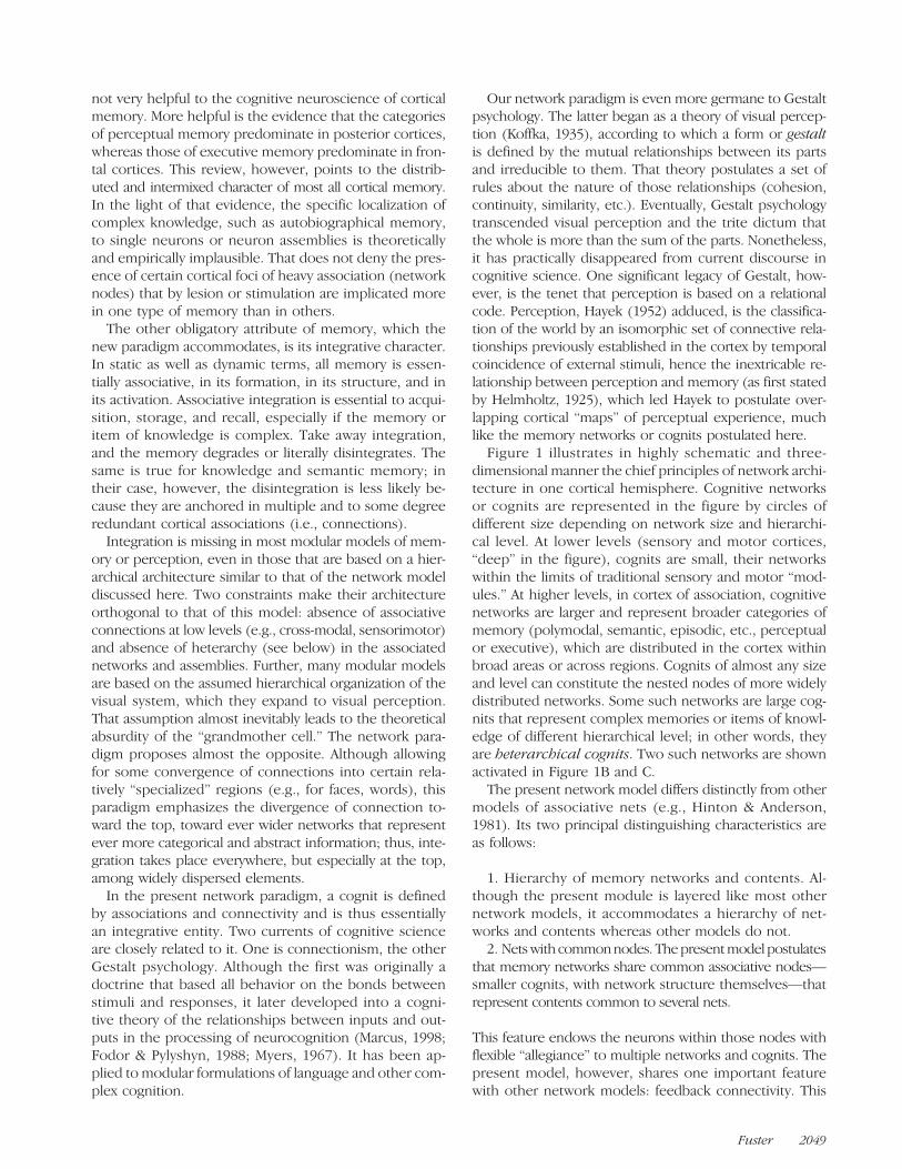
Figure 1 illustrates in highly schematic and three-dimensional manner the chief principles of network archi-tecture in one cortical hemisphere. Cognitive networksor cognits are represented in the figure by circles ofdifferent size depending on network size and hierarchi-cal level. At lower levels (sensory and motor cortices,“deep” in the figure), cognits are small, their networkswithin the limits of traditional sensory and motor “mod-ules.” At higher levels, in cortex of association, cognitivenetworks are larger and represent broader categories ofmemory (polymodal, semantic, episodic, etc., perceptualor executive), which are distributed in the cortex withinbroad areas or across regions. Cognits of almost any sizeand level can constitute the nested nodes of more widelydistributed networks. Some such networks are large cog-nits that represent complex memories or items of knowl-edge of different hierarchical level; in other words, theyare heterarchical cognits. Two such networks are shownactivated in Figure 1B and C.
The present network model differs distinctly from othermodels of associative nets (e.g., Hinton & Anderson,1981). Its two principal distinguishing characteristics areas follows:
1. Hierarchy of memory networks and contents. Al-though the present module is layered like most othernetwork models, it accommodates a hierarchy of net-works and contents whereas other models do not.2. Netswith commonnodes. Thepresentmodel postulates
that memory networks share common associative nodes—smaller cognits, with network structure themselves—thatrepresent contents common to several nets.
This feature endows the neurons within those nodes withflexible “allegiance” to multiple networks and cognits. Thepresent model, however, shares one important featurewith other network models: feedback connectivity. This
Fuster 2049

feature is critical to the dynamic interplay of cognits in theperception–action cycle.
MEMORY CELLS
Some of the most valid inferences about the structure ofa cognit can be drawn from its dynamics in behavior. Itis by studying a memory network in the active state thatwe can glean its structure. Memory networks are acti-vated in a variety of conditions. First and foremost, someare activated, consciously or unconsciously, voluntarily orinvoluntarily, in every act of perception. They are also ac-tivated, more or less voluntarily, in free recall, recogni-tion, new memory acquisition, rehearsal, and workingmemory. The last, I argue, is the most suitable conditionfor the study of active memory networks.
Working memory is the ability to temporarily retain in-formation for a prospective action. The reasons for itssuitability to the study of memory structure and dynamicsare because (1) it is relatively easy to instruct a human oran animal to retain, for a defined period, a specific itemof information that calls for a specific prospective action;(2) it is reasonable to assume that, during that time, theneural substrate for mnemonic retention is in a differentstate than at rest; (3) the physiological measures of dif-fering state probably reflect the physical nature of theinformation in temporary storage, in such a manner thatchanges in the parameters of that information will bereflected by commeasurable changes in network dynam-ics; and (4) the extent of those changes in the cortex andtheir correlations with memory performance will help usdetermine the boundaries and the dynamics of a cognit.
For nearly 40 years, it has been known that in the mon-key performing working-memory tasks, such as delayed re-sponse and delayed matching, neurons in certain corticalregions undergo persistent elevations of firing frequencyduring working memory. Because they were assumed tointervene in a memory process, such neurons were named“memory cells.” Ordinarily, these cells show a transientfrequency change in response to the sensory cue thatthe animal must retain in memory. After the cue has dis-appeared, that change is followed by above-baseline dis-charge for much or all of the ensuing delay or memoryperiod—seconds or minutes—before the motor responseor choice.
Memory cells were first encountered in the pFC of mon-keys performing delayed response (Fuster & Alexander,1971). Subsequent investigations of that cortex revealedmemory cells whose level of memory activity was depen-dent on the physical characteristics of the stimulus cue tobe remembered (memorandum). Some prefrontal cells re-spond preferentially to spatial memoranda (Genovesio,Brasted, & Wise, 2006; Constantinidis, Franowicz, & Goldman-Rakic, 2001; Funahashi, Bruce, &Goldman-Rakic, 1989; Niki,1974), others to visual memoranda (Wilson, Scalaidhe, &Goldman-Rakic, 1993; Fuster,Bauer,& Jervey, 1982), includ-ing faces (Scalaidhe, Wilson, & Goldman-Rakic, 1999), and
still others to auditory (Romanski, 2007; Fuster, Bodner,& Kroger, 2000) or tactile (Romo, Brody, Hernández, &Lemus, 1999) memoranda. Furthermore, in some studies,the relation between prefrontal memory firing and phys-ical stimulus dimension has been found parametric forsuch properties as the location of gaze (Funahashi et al.,1989) or the frequency of mechanical vibration sensedby touch (Romo et al., 1999). In any given prefrontal area,however, the memory cells that show sharp tuning forany given sensory memorandum constitute a minority.Cells preferring memoranda of one sensory modality
or another tend to concentrate in certain domains ofpFC, but these domains are poorly demarcated; cells withany given preference can be found practically anywherein pFC. For example, cells preferring spatial memorandapredominate in dorsolateral areas but are also presentin ventrolateral areas, although in lower numbers, andvice versa for nonspatial memoranda. Likewise, cells pre-ferring and anticipating a reward concentrate in an or-bital domain but are also present in lateral cortex. Somecells are attuned to stimuli of more than one sensorymodality—for example, auditory and visual—that have be-come associated with one another by the learning of aworking-memory task (Artchakov et al., 2007; Romanski,2007; Fuster et al., 2000; Watanabe, 1992; Vaadia, Benson,Hienz, & Goldstein, 1986). Others are attuned to both thespatial and the nonspatial attributes of the memorandum(Fukushima, Hasegawa, & Miyashita, 2004; Rao, Rainer, &Miller, 1997; Fuster et al., 1982).Furthermore, in any prefrontal region, some cells are
also attuned to the motor requirements of the working-memory task (Isomura, Ito, Akazawa, Nambu, & Takada,2003; Akkal, Bioulac, Audin, & Burbaud, 2002; Procyk& Joseph, 2001; Quintana & Fuster, 1999; Carlson, Rämä,Tanila, Linnankoski, & Mansikka, 1997; Fuster et al., 1982)and/or the reward expected or resulting from motor ac-tion (Ichihara-Takeda & Funahashi, 2008; Hikosaka &Watanabe, 2000; Schultz, Tremblay, & Hollerman, 2000;Watanabe, 1996; Rosenkilde, Bauer, & Fuster, 1981). Theprefrontal domains with relatively high concentration ofcells attuned to different modalities, or task attributesare anatomically connected with specialized posteriorcortical areas or subcortical structures (review in Fuster,2008). Visual and auditory domains of ventral pFC are con-nected with corresponding visual and auditory areas oftemporal cortex and reward domains of orbital cortex withtegmental and limbic formations.The memory cells in posterior association cortex gen-
erally show more sensory specificity than those in pFCand tend to cluster in areas of association for the sensorymodality of the memorandum. Thus, cells in inferotempo-ral cortex (Figure 2) are tuned to visual memoranda (Miller,Li, & Desimone, 1991; Miyashita & Chang, 1988; Fuster& Jervey, 1982), whereas cells in parietal cortex are tunedto spatial (Andersen, Bracewell, Barash, Gnadt, & Fogassi,1990) or tactile (Zhou, Ardestani, & Fuster, 2007; Burton& Sinclair, 2000; Zhou & Fuster, 1996) memoranda. As
2050 Journal of Cognitive Neuroscience Volume 21, Number 11
in the pFC, although to a lesser degree (in terms of cellnumbers and response magnitude), cells can be foundin posterior areas that respond to the motor and/or re-ward attributes of memory tasks.The thus far summarized microelectrode evidence
points to the widely distributed character of cortical mem-ory networks, with components in posterior (sensoryor perceptual) cortex and in frontal (executive) cortex(Constantinidis & Procyk, 2004; Fuster, 1995). It further
points to the presence of cells and cell assemblies thatrespond to more than one memorandum or differentcharacteristics of the same memorandum. This propertyof “multiple tuning” argues for the belonging of cells tomultiple networks and for their flexible functional al-legiance to those networks. Next, I discuss how single-unit evidence substantiates the associative character ofworking-memory networks and their structural identitywith long-term memory networks.
WORKING MEMORY FROMLONG-TERM MEMORY
The neuropsychological literature provides indirect evi-dence that the cortical substrate for working memory co-incides with the substrate for long-term memory. Thatevidence derives from a large number of animal and hu-man lesion studies (reviewed in Fuster, 1995), indicatingthat several cortical areas are implicated in the working
Figure 1. Schematic diagram of structural and dynamic principlesof memory network architecture and function. (A) Hierarchies ofperceptual (blue circles) and executive (red circles) networksor cognits of different sizes and hierarchical levels in one corticalhemisphere (three sizes and hierarchical levels have beenarbitrarily chosen); thin lines represent bidirectional connectionsbetween cognits. (B) Stimulus 1 activates a large distributed cognit(Memory 1) made of smaller, more local cognits (three sizes,color-filled circles) connected by bidirectional excitatory pathways(large maroon lines). These smaller cognits constitute nodes ofthe large memory network. (C) Stimulus 2 activates memoryNetwork 2 and its nested component networks. Note thatNetworks 1 and 2 are heterarchical and share common components(nodes). Figure 2. Rasters and spike frequency histograms of the activity of
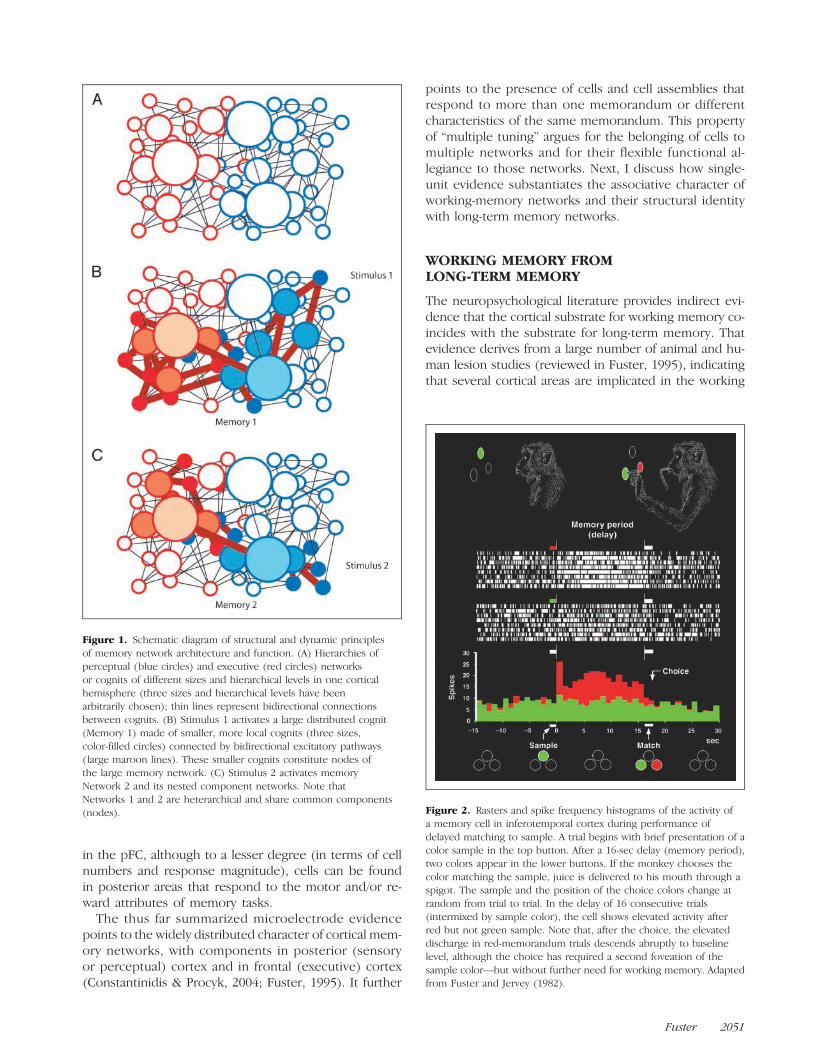
a memory cell in inferotemporal cortex during performance ofdelayed matching to sample. A trial begins with brief presentation of acolor sample in the top button. After a 16-sec delay (memory period),two colors appear in the lower buttons. If the monkey chooses thecolor matching the sample, juice is delivered to his mouth through aspigot. The sample and the position of the choice colors change atrandom from trial to trial. In the delay of 16 consecutive trials(intermixed by sample color), the cell shows elevated activity afterred but not green sample. Note that, after the choice, the elevateddischarge in red-memorandum trials descends abruptly to baselinelevel, although the choice has required a second foveation of thesample color—but without further need for working memory. Adaptedfrom Fuster and Jervey (1982).
Fuster 2051

memory of certain classes of stimuli as well as in theirretention in long-term memory. The lesion of a given as-sociative area in the monkey induces deficits in delayed-response tasks with stimuli of a given modality (visual,spatial, tactile, etc.), as it does also in the recall or recog-nition of stimuli of the same modality. In the human, am-nesias or agnosias for that modality result from lesions ofareas roughly homologous to those of the monkey thatare related to the same modality.
By definition, moreover, working memory determinesa prospective action, commonly a decision between alter-natives (e.g., a selective instrumental response, a verbalresponse, a logical inference, etc.) that is based on long-term memory. Its content and context are inextricablylinked to that memory, whether they include a simplelearned conditional response to a simple sensory stimu-lus, an element of lexicon, or the conclusion of a syllo-gism. Thus, whereas most long-term memory neverenters working memory, all working memory is not onlyanchored in long-term memory but also part of it. Thatpart of long-term memory varies with the circumstancesand is evoked by them; that is, by the associations ofthe material to be retained and the prospective conse-quences of that act of retention, all of them previouslystored in permanent memory.
More direct evidence of the associative nature ofworking-memory networks, and thus their identity to cognits oflong-term memory, comes from electrophysiology. Espe-cially demonstrative are memory cells that show correlatedresponses to stimuli of the same or different modalityassociated with one another by learning. Such cells havebeen found in both frontal (Fuster et al., 2000) andposterior (Schlack & Albright, 2007; Zhou et al., 2007;Messinger, Squire, Zola, & Albright, 2001; Gibson &Maunsell, 1997; Miyashita, 1988) association cortices (Fig-ure 3); tactile–visual cell response correlations can befound even in primary somatosensory cortex (Zhou &Fuster, 1995). The evidence becomes compelling as thecorrelations develop in the process of acquiring the asso-ciations in long-term memory. Further evidence of theidentity of working-memory and long-term memory net-works is the observation that working-memory cells donot respond only to the memoranda but also to assortedsensory and motor inputs that are associated with theperformance of the task at hand (Zhou et al., 2007). In vi-sual processing, the associative inputs from other partsof the sensorium would contribute to a stimulus of whathas been considered context (Bressler & McIntosh, 2007;Albright & Stoner, 2002; Fuster, 1990). Context includesthe history and the meaning of the stimulus in long-termmemory.
Whereas sensory–sensory associations in working mem-ory have been demonstrated in both posterior and fron-tal cortex, sensory–motor and motor–motor associationsappear mainly, if not exclusively, in pFC. There, cells seemto belong to assemblies that encode behavioral sequencesor “temporal motor gestalts” of relatively abstract nature
(Averbeck & Lee, 2007; Shima, Isoda, Mushiake, & Tanji,2007), regardless of their individual motor components(Figure 4). Furthermore, some frontal memory cells areattuned to conditional rules, another indication of theirinvolvement in associative long-term memory (Mansouri,Matsumoto, & Tanaka, 2006; Wallis, Anderson, & Miller,2001; Asaad, Rainer, & Miller, 2000; White & Wise, 1999).The associative character of working-memory cells sub-
stantiates the assumptions (a) that memory cells belongto networks of long-term memory that associate the var-ious sensory and motor features of working-memory de-mands, including but not limited to the memoranda usedby the animal in working-memory tasks, (b) that thosenetworks can incorporate widely dispersed neurons innoncontiguous cortical areas, and (c) that the networksand their associations are formed by experience.Within a given cognitive network, however, the density
of associations appears far from homogeneous. To judgefrom the groupings of cells attuned to the same or similarstimuli, a working-memory network seems to containnodes of heavy association in areas of relative functionalspecialization. Such nodes may be characterized as spe-cial “modules” or “mini-networks” for spatial, visual, audi-tory, tactile, or other working memory. However, theirfunction in the retrieval of long-term memory, as in work-ing memory, is strictly dependent on their previously estab-lished associations with other modules or cells scatteredover the width and the depth of the cortex of association,both frontal and posterior.To sum up, studies of cortical memory cells lead to the
tentative conclusion that working memory consists oftemporary activation of a preexistent network of long-term memory. That network connects with one anotherall the smaller networks and neuronal assemblies that, inthe aggregate, represent the associated features, per-ceptual as well as executive, of the behavioral interactionof the organism with its environment. At the onset of atrial in a working-memory task, the activated networkis updated by the presence of the memorandum. Thus,the updated network of long-term memory becomesoperational, and the process of temporary retention ofthat memorandum begins within the context of the task.Neither the content nor the dynamics of working mem-ory can be separated from the substrate of a sector oflong-term memory and its temporary activation to bridgethe recent past with the proximate future.
MECHANISMS OF WORKING MEMORY
The mechanisms of persistent neuronal activation in work-ing memory are not well understood. One key mechanismappears to be the reverberation of cortical circuits by re-entry of activity from one neuron or neuronal assembly toanother by mutual excitation. That mechanism was firstproposed by Hebb (1949) as the basis of short-term mem-ory. It has never been conclusively proven, although nowit is gathering evidence as the basis of working memory.
2052 Journal of Cognitive Neuroscience Volume 21, Number 11

Cortical reverberation by reentry has a well-recognizedanatomical base. The existence of profuse recurrent axonsin the cortex has been known since Lorente de Nó(1949) described them; the axons make reciprocal excit-atory synapses on neighboring or distant neurons. Mostall pathways between frontal and posterior associationcortices are bidirectional, and so are the connectionsbetween those cortices and thalamic nuclei (Petrides &Pandya, 2002; review in Fuster, 2008). Therefore, thereis ample structural potential in cortical circuitry for sus-tained reverberation in working-memory networks, how-ever separate their neurons may be. Accordingly, reentryis an integral part of the most plausible computational mod-
els of working memory (Warden & Miller, 2007; Wang,2006; Brunel & Wang, 2001; Zipser, Kehoe, Littlewort, &Fuster, 1993). Some of these models assume that reen-try connections are mediated through dopamine, gluta-mate, γ-aminobutyric acid, or N-methyl D-aspartate synaptictransactions.
Most computational models of working memory, how-ever, assume the bistability of their functional architecture:discharge at a certain fixed elevated frequency when in
Figure 3. Cross-temporal association of sound and color in prefrontalcells. Above left: Trial event sequence: (1) brief tone; (2) 10-sec delay;(3) two colors simultaneously presented; (4) animal rewarded forcorrectly (c) choosing the color that matches the tone according to thelearned rules of the task—green if low-pitch tone, red if high-pitchtone. (Tone and color position change at random between trials.)Above right: Monkeyʼs cortex showing in blue the region from whichunits were recorded; Brodmannʼs numeration in frontal areas. Below:Firing frequency histograms from two cells, one (top) selective forhigh tone and red and the other (bottom) selective for low toneand green. Histograms are from a 1-sec period beginning with toneonset (left) and from a 1-sec period preceding choice of color (right).Note the correlation of cell responses to tones and colors in accord withthe rule of the task. Adapted from Fuster et al. (2000), with permission. Figure 4. Prefrontal cell signaling abstract plan of action. The
monkey is trained to perform sequences of three hand movements(push, pull, and turn) in three categorical combinations: (A) “Paired”(one movement repeated, followed by another repeated; e.g.,turn–turn–pull–pull); (B) “alternate” (repeated alternation of twomovements; e.g., push–turn–push–turn); (C) “four repeat” (e.g.,pull–pull–pull–pull). Associated auditory and visual signals instruct theanimal to memorize and to perform the three abstract categories ofsequences. Cell records were taken from lateral pFC before and duringperformance of each sequence. Some cells showed increased firing inpreparation for—planning—all the sequences of one given sequencecategory and not the others. Green dots mark sites from which 0, 1, 2,or 3 category-selective cells were recorded. ARC = arcuate sulcus;PS = principal sulcus. The records are from a cell activated (redhistogram) before “paired-action” sequences, regardless ofcomponent movements. Records are time locked with the GO signal(green triangle) at the start of the first memorized movement in asequence. Adapted from Shima et al. (2007), with permission.
Fuster 2053
memory and reversion to baseline frequency when not.This assumption does not agree with data from the realbrain. Already from early observations (e.g., Fuster, 1973),it has been known that, if the delay (memory period) islong enough (>10 sec), the time course of memory ac-tivity can adopt many forms. The most obvious are thedescending ramps of sensory-coupled cells at the start ofthe delay or the accelerating ramps of motor-coupled cellsduring it (Figure 5), but in any case with considerable var-iability from cell to cell and from trial to trial (Shafi et al.,2007). Many memory cells show mixed temporal patterns.The analysis of interspike intervals (ISIs) reveals, in both thebaseline (intertrial) and the delay (memory) periods, a mul-tiplicity of ISI patterns independent of frequency (Bodner,Shafi, Zhou, & Fuster, 2005). In working memory, the num-ber of observable patterns generally increases.
The multiplicity of firing patterns in working memory isfully consistent with the notion that memory cells belongto multiple networks (Figure 1B and C). One possibility isthat, when the cortex enters working memory, its mem-ory cells fire at a variety of frequencies, each frequencythe expression of activity in a given reentry loop tran-siting through those cells. The cells seem recruited intoa variety of reentry loops, each loop with its own rever-berating frequency. It is reasonable to infer that the cellsbelong to the multiple associative networks that definethe various attributes of the memorandum—including itsprospective attributes related to action and reward. Thosenetworks can be local or far-flung. Depending on the attri-bute in the focus of working memory at any given time, acell would change its allegiance to one network or anotherand thus become attuned to one reverberating frequencyor another.
Whether the reentry is local or far-flung, persistence ofworking-memory discharge may also be caused at leastin part by long-lasting ionic changes at synaptic level. Arecent model (Mongillo, Barak, & Tsodyks, 2008) pos-tulates that working memory is sustained in recurrentneuronal connections by residual calcium released by in-coming spiking, such as that elicited by the memorandum.That residual calcium would increase presynaptically,thereby facilitating synaptic transmission and responsesto subsequent inputs, including further recurrent inputsor replicates of the memorandum (Figure 6). To maintainactive memory, the model would economize on spikes.Also, the following two characteristics make the modelfully compatible with our network assumptions: (1) reen-try within recurrent cortical networks and (2) preexistentmemory code presumably established by Hebbian associa-tive learning and activated by the memorandum. Becauseof their multiple interactions, several networks (long-termassociative memories) can be activated at the same time(Figure 6B).
Corticocortical circuitry can maintain memory active notonly by reverberation but also by tonic influences of onearea upon another or from one part of a cognit uponanother. That circuitry would involve the well-known ana-
tomical connections between prefrontal and posterior cor-tices (Barbas, Ghashghaei, Rempel-Clower, & Xiao, 2002;Petrides & Pandya, 2002; Jones & Powell, 1970). The im-portance of mutual influences between those cortices formemory maintenance is revealed by the results of thefunctional inactivation—by cooling—of pFC on the activityof memory cells in posterior cortex or vice versa (Chafee& Goldman-Rakic, 2000; Quintana, Fuster, & Yajeya, 1989;Fuster, Bauer, & Jervey, 1985). For example (Fuster et al.,1985), cooling the lateral pFC to 20°C during a visual de-layed match-to-sample task diminishes the ability of someinferotemporal cells to discriminate the color of the mem-orandum during the memory period (delay); presumablyas a consequence, the animal commits more errors ofcolor working memory than at normal cortical tempera-ture. Thus, prefrontal cooling diminishes “cognitive control”(Miller & Cohen, 2001) over the inferotemporal cortexin working memory. Alternatively (or concomitantly), theprocedure interrupts the reverberating loop between thetwo cortices that presumably supports active memorymaintenance.In sum, the mechanisms of working memory are not
yet established, but there is increasing evidence that theyinclude the reverberation in circuits within the neuralnetwork, part of long-term memory, that has been acti-vated by the memorandum. Additional evidence is tobe found in the study of patterns and periodicities ofmemory-cell firing.
RHYTHM AND SYNCHRONY
The presence of electrical oscillations in the cortex hasbeen well known since Hans Berger (1929) describedthe EEG on the headʼs surface. Oscillations are encoun-tered not only in the extracranial EEG but also in the localfield potentials (LFPs), which are intracortical signals re-flecting summed dendritic activity in local neuronal as-semblies; essentially, LFPs have the same electrogenesisas the EEG but in more discrete cortical domains thanthose that generate the EEG signal. Many rhythms havebeen related to a wide variety of brain states and psycho-logical conditions. Although their observable range is al-most continuous, those rhythms can be categorized byfrequency into a finite number of discrete classes (Fig-ure 7). The physiological exploration of electrocorticalrhythms is especially productive with regard to twohighly interrelated cognitive functions: perception and at-tention. Both these functions are, in turn, highly relatedto memory. Perceiving is remembering as much as sens-ing, and working memory is attention focused on an inter-nal representation.In the late 1980s, it was discovered that separate groups
of neurons in the visual cortex fired in synchrony (gammarange) in response to visual stimuli (Jagadeesh, Gray, &Ferster, 1992; Engel, König, Kreiter, & Singer, 1991; Gray& Singer, 1989; Eckhorn et al., 1988). One interpretationof this phenomenon was that separate neurons firing
2054 Journal of Cognitive Neuroscience Volume 21, Number 11

in synchrony engaged in what was called “perceptualbinding,” that is, the binding of the diverse visual featuresof the stimulus into a perceptual whole (Engel, Fries, &Singer, 2001; Gray, Konig, Engel, & Singer, 1989). Neural
binding, thus, became a model of perceptual constancy to-ward solving one of the central problems of physiologicalpsychology (Klüver, 1933). Perceptual constancy wouldemerge from the binding of sensory features, namely, from
Figure 5. (A) Task withtemporal and spatial separationbetween cues and responses(below, contingencies betweenthem). The animal faces a panelwith three stimulus–responsebuttons above a pedal, wherethe operant hand rests at alltimes except to respond tostimuli. After a warning signal(flash), one of four colorsappears in the central button(the cue). A period of delayfollows, at the end of which thetwo lateral buttons turn red andgreen, or both white. If thosetwo buttons are colored, theanimal must choose the onematching the cue; if they arewhite, the animal must chooseleft for red cue, right for greencue, right for yellow cue, andleft for blue cue. Thus, inthe delay, if the cue has beenyellow or blue, the animalcan predict the rewardedresponse direction (right orleft, respectively) with 100%probability, whereas if the cuehas been red or green, with only75% probability (left if red, rightif green). Colors and direction of correct choice change at random. c = correct choice. (B) Average discharge of motor-coupled cells duringthe delay of trials with 100% predictable response direction (top graph) and 75% predictable response direction (lower graph). Note that theacceleration of discharge during that memory period is related to predictability. C = cue; R = response. Adapted from Quintana and Fuster (1999),with permission.
Figure 6. Dynamics andanatomy of memory network.(A) Short-term synapticplasticity model. At left, kineticframework with equations forsynaptic variables: δ = Diracdelta function; tsp = time ofpresynaptic spike; Vm =membrane potential. At right,postsynaptic response, throughfacilitating connection, to avolley of presynaptic spikes.During the volley, u increases(facilitation) and x decreases(depression). The product uxmodulates synaptic efficacy.(B) Network architecture.Colored triangles are excitatoryneurons in networks thatencode different memories.Black empty triangles arenonselective neurons. Blackcircles are inhibitory neurons.Adapted from Mongillo et al.(2008), with permission.
Fuster 2055

the mutual functional relationships between cell groupsdespite physical variations in each of those features individu-ally. A relational code would thus emerge in similar manneras a gestalt. Could oscillations play the same role at highercognitive levels, especially in working memory?
The causal relationships between electrocortical oscil-lation and cell spiking are obscure, although there is in-creasing evidence of the coupling of cell spikes and LFPs(Lee, Simpson, Logothetis, & Rainer, 2005; Pesaran, Pezaris,Sahani, Mitra, & Andersen, 2002; Fries, Reynolds, Rorie, &Desimone, 2001). On the one hand, periodic spike trainscan generate periodic dendritic potentials (Reyes, 2003).On the other, dendritic oscillations can change the excit-ability of a cellʼs membrane, thereby biasing its produc-tion of spikes, which may occur in phase at various timeswith respect to the oscillatory cycle (Tsodyks, Skaggs,Sejnowski, & McNaughton, 1996).Both mechanisms mayoperate in the cortex to some degree, in any case lead-ing to temporal correlations between periodic spikes andoscillations.
Synchronous cortical oscillations appear to result fromthe interaction of both, local factors at the membrane ofthe cell and circuit factors at the network in which thecell is embedded. Probably among the latter are the peri-odic inputs from the thalamus (Steriade, 2001; Llinás,1988), the so-called “inhibitory clocking networks” of in-terneurons (Buzsáki, Geisler, Henze, & Wang, 2004), andthe loops of corticocortical connections. In ensuing dis-cussion, I emphasize the last of these factors for its rele-vance to reentry in working memory, without excluding acoadjutant or even primary role for the others.
If we assume that reentry is an important componentof network architecture, length of circuitry should be adeterminant of oscillatory frequency. However, lengthof circuitry does not necessarily mean length of fibers.Braitenberg and Schüz (1998) argued that some long fi-bers (e.g., corticocortical) may actually shorten the ef-fective connectivity within networks. It follows that therecould be an inverse relationship between network size,in terms of effective circuitry, and frequency of oscilla-tion (Buzsáki & Draguhn, 2004; Freeman, Rogers, Holmes,& Silbergeld, 2000). This reasoning accommodates agood amount of electrophysiological data (Buzsáki et al.,2004; Csicsvari, Jamieson, Wise, & Buzsaki, 2003; Steriade,2001).
Complex cortical processing during behavior involvesmany networks, large and small, some nested withinothers. This, in conditions of heightened attention andworking memory, will entail a proliferation of activatednetworks oscillating at multiple frequencies. More gener-ally, the proliferation and the fragmentation of frequenciesare most likely to be at the root of the “desynchronization”of the EEG as the subject awakes from sleep or respondsto sensory stimuli (Basar & Bullock, 2000; Pfurtscheller &Lopes da Silva, 1999). By contrast, at rest or in sleep, whensimple large networks prevail, low frequencies (theta orlower) will predominate. This does not preclude that, by
virtue of their stereotypical functional architecture, certainparts of “ancient cortex” involved in memory, such as thehippocampus, exhibit low oscillatory frequencies even, orespecially, when its networks are highly active (Huxter,Burgess, & OʼKeefe, 2003; Penttonen & Buzsáki, 2003;Huerta & Lisman, 1995).In the sensory association cortex engaged in sharply
focused (“top–down”) attention to the location or char-acteristics of sensory stimuli, high-frequency oscillations(beta and gamma) have been observed in LFPs as wellas in unit discharge (Lakatos, Karmos, Mehta, Ulbert, &Schroeder, 2008; Buschman & Miller, 2007; Saalmann,Pigarev, & Vidyasagar, 2007; Womelsdorf, Fries, Mitra, &Desimone, 2006; Brovelli, Lachaux, Kahane, & Boussaoud,2005; Fries et al., 2001). Presumably, those oscillations re-flect reentrant activity in the small, high-frequency oscil-lating networks that process the item of information thatthe subject attends to. That phenomenon is more appar-ent when attention is extended in time. This is the situa-tion in working memory, which is attention focused onthe internal representation of a recent stimulus for pro-spective action (Fuster, 2003; Baddeley, 1993). Duringworking memory, in sensory or association cortex, oscilla-tory synchrony (Figure 8) commonly appears (Lee et al.,2005; Rizzuto et al., 2003; Pesaran et al., 2002; Tallon-Baudry, Bertrand, & Fischer, 2001). In the human, syn-chrony predominates in the beta and theta ranges. Further,the desynchronizing transition from alpha to beta or higherhas been noted to be selective in different areas depend-ing on the modality or memory load of the memoran-dum (Stipacek, Grabner, Neuper, Fink, & Neubauer, 2003;Klimesch et al., 1996).In higher association cortex, cellular activity during
memory activation is multistable and multivariate, as net-works there profusely intersect. They represent in long-term memory the multiple associated aspects of thememorandum. Accordingly, and supporting our reason-ing for the commonality of anatomical substrates forworking and long-term memory, we have observed cell-firing frequencies attuned to several associated aspectsof a working-memory task (Zhou et al., 2007; Fusteret al., 2000). During the memory period, the analysis ofISIs shows extensive variability (Shafi et al., 2007) anda proliferation of patterns (Bodner et al., 2005), bothsupposedly reflecting the affiliation of cells to multiplememory networks. Thus, the electrocortical recordsfrom human subjects in situations of high cognitive de-mand exhibit a multiplicity of rhythms in areas of asso-ciation. Especially prevalent are oscillations in the theta,alpha, beta, and gamma frequencies (Sehatpour et al.,2008; Gevins & Smith, 2000), in some instances modu-lating one another (Lakatos et al., 2008; Canolty et al.,2006).The most direct electrocortical evidence of the acti-
vation of interregional networks in working memory isthe synchrony of high-frequency (beta and gamma) os-cillations in frontal and posterior regions of association
2056 Journal of Cognitive Neuroscience Volume 21, Number 11

cortex during high attention and working-memory per-formance (Axmacher, Schmitz, Wagner, Elger, & Fell,2008; Sehatpour et al., 2008; Buschman & Miller, 2007;Saalmann et al., 2007; Brovelli et al., 2004; Gross et al.,2004; Brovelli, Battaglini, Naranjo, & Budai, 2002; Tallon-
Baudry et al., 2001; Stein, Rappelsberger, Sarnthein, &Petsche, 1999; Bressler, Coppola, & Nakamura, 1993). Inthe next section, we see in neuroimages the reflectionof that synchrony in posterior and frontal regions duringworking memory. In any case, taken as a whole, theelectrocortical evidence strongly supports the broaderprinciple of widely distributed and overlapping memorynetworks.
NEUROIMAGING OFMEMORY-NETWORK ACTIVATION
The judicious use of functional imaging methods in thehuman has contributed mightily to support the networkparadigm of memory. It has also contributed to the under-standing of the dynamics of memory networks. However,the imaging methodology has considerable limitations. Itis essential to be aware of them before any review of imag-ing data on memory.
The following are the most relevant limitations of PETand fMRI: (1) The neurovascular coupling function is stillpoorly understood. (2) Temporal resolution is inade-quate to measure rapid changes in memory acquisitionand recall. (3) Large individual variability limits conclu-sions on memory distribution or mechanisms. (4) Linearmodels of blood-flow change may not be fully compat-ible with memory functions inherently nonlinear. (5) Tothe extent that memory uses the same cortical networks
Figure 7. Classes of oscillatory activity in the cortex. For eachfrequency band, its range is shown as well as the common term forit. Note the linearity of classes in logarithmic scale. Adapted fromBuzsáki and Draguhn (2004), with permission.
Figure 8. LFP and single-celldischarge at three sites inextrastriate cortex during aworking-memory task. (A) RawLFP signals simultaneouslyrecorded through baselinepretrial, sample, delay, andchoice periods (separated byvertical lines) of a delayedmatching-to-sample trial.(B) Theta-band-filtered LFPs.(C) Single-unit activity (SUA)from each of the same threerecording channels. (D) Eachunit emits action potentials at apreferred angle (radial line) ofthe theta wave. Adapted fromLee et al. (2005), withpermission.
Fuster 2057

as other related cognitive functions (e.g., attention, per-ception, language), it is difficult to disambiguate mem-ory activation from that of those other functions. And(6) the neural inhibition of memory cannot be easilydifferentiated, by imaging, from its activation. Becauseof these limitations, the merits of any imaging study ofmemory depend on the investigatorʼs ability to makeonly indispensable assumptions and use appropriatecontrols and analytical methods. The evidence summa-rized in this section comes from studies that meet thosecriteria.
Neuroimaging does not allow the precise tracing of theboundaries of active memory networks. The conven-tional assessment of cortical activation, in both intensityand extensity, is essentially analogous to that of separat-ing signal from noise. The investigator sets a threshold—calculated from normalized baseline values—and deter-mines the significant deviations that exceed it. Here theproblem is to distinguish the variance of active memoryfrom that of background noise. The presence of “defaultnetworks,” active in the resting state (Fox & Raichle,2007), as well as the increased variance and the dimin-ished activation at the edges of an active memory net-work makes the threshold setting critical yet somewhatarbitrary. If that threshold is too low, the networks ap-pear larger than they are; if it is too high, the networksappear smaller.
Like microelectrode research, the functional neuroimag-ing of working memory focused at first on the pFC, thatis, on the executive sector of memory networks. PET stud-ies showed activation of lateral pFC, especially on theright, in spatial working memory ( Jonides et al., 1993;Petrides, Alivisatos, Evans, & Meyer, 1993). That was sub-sequently corroborated by fMRI studies (Ricciardi et al.,2006; Ranganath, Cohen, Dam, & DʼEsposito, 2004).Both PET (Swartz et al., 1995) and fMRI (Cohen et al.,1994) showed also lateral prefrontal activation in visualnonspatial working memory. A special case of the latteris the memory for faces, which activates not only the lat-eral prefrontal areas but also the cortex of the fusiformgyrus, an area involved in face recognition (Rama &Courtney, 2005; Gazzaley, Rissman, & DʼEsposito, 2004;Ranganath et al., 2004; Mecklinger, Bosch, Gruenewald,Bentin, & Von Cramon, 2000). The pFC, especially on theleft, is activated in verbal working memory (Buchsbaum,Olsen, Koch, & Berman, 2005; Goldstein et al., 2005;Narayanan et al., 2005; Crottaz-Herbette, Anagnoson, &Menon, 2004; Paulesu, Frith, & Frackowiak, 1993; Petrides,Alivisatos, Meyer, & Evans, 1993) and also in working mem-ory for mental arithmetic (De Pisapia, Slomski, & Braver,2007; Kondo et al., 2004).
Whatever the content in working memory, the amountof prefrontal activation is directly proportional to thememory load (Narayanan et al., 2005; Cairo, Liddle,Woodward, & Ngan, 2004; Leung, Seelig, & Gore, 2004;Jaeggi et al., 2003; Linden et al., 2003; Postle, Berger,Goldstein, Curtis, & DʼEsposito, 2001). In other words,
prefrontal activation increases as a function of the num-ber and complexity of items in memory. Practice, how-ever, decreases load-related activation. Is this a sign ofmemory consolidation, which entails economy of synap-tic resources? Or, is it of the migration of executive mem-ory to subcortical structures (e.g., basal ganglia)?Practically all the relevant studies show that the pFC is
not the only region activated in working memory. Almostinvariably, one or more posterior cortical areas are con-comitantly activated. Which posterior area or areas areactivated depends on the modality of the memorandum:inferior temporal areas if it is visual (additionally fusi-form cortex, if it is a face), posterior parietal if it is spa-tial, superior temporal if it is auditory or verbal, andanterior parietal if it is tactile. Homologous areas of cor-tex appear activated in imaging records of the humanas in microelectrode records of the monkey. Prefrontalareas are activated inasmuch as executive memory is in-volved and posterior areas inasmuch as perceptual mem-ory is involved.Consistent with the microelectrode evidence of inter-
actions between prefrontal and posterior associationareas, several imaging studies indicate that those interac-tions underlie the role of pFC in so-called “executive cog-nitive control” or “top–down attention” (Roth, Serences,& Courtney, 2006; Yoon, Curtis, & DʼEsposito, 2006;Buchsbaum et al., 2005; Curtis, Sun, Miller, & DʼEsposito,2005; Postle, 2005; Kondo et al., 2004; Li et al., 2004; Sakai& Passingham, 2004). The medial pFC—especially ante-rior cingulate—seems part of a so-called “anterior atten-tion system,” dedicated to spatial attention (Lenartowicz& McIntosh, 2005; Kondo et al., 2004; Petit, Courtney,Ungerleider, & Haxby, 1998; Posner & Petersen, 1990).However, the attribution of “control” to the pFC, in atten-tion or any other cognitive function, implies for that cortexa role of “central executive,” which makes little sense inbiological terms and leads to an infinite regress (Fuster,2008; McIntosh, 2000).In the light of imaging data, the “central executive” role
of the pFC is in principle reducible to its role of integrat-ing for prospective action a continuous flow of inputsfrom the internal and the external environments. Thememory networks of posterior cortex are part of the in-ternal environment, which in turn can be activated by ex-ternal stimuli and the effects of action, all within theperception–action cycle (below). In that same frame-work, it is possible to understand the role that imagingstudies attribute to the pFC in the retrieval and encodingof memory (Mitchell, Johnson, Raye, & Greene, 2004;Ranganath et al., 2004; Rypma & DʼEsposito, 2003; Lee,Robbins, & Owen, 2000; Buckner et al., 1995; Kapuret al., 1994; Tulving, Kapur, Craik, Moscovitch, & Houle,1994; Tulving, Kapur, Markowitsch, et al., 1994); both re-trieval (except in involuntary or free recall) and encodingare executive acts prompted by external stimuli.Figures 9–12 illustrate schematically the trends of cor-
tical activation on the left hemisphere during perfor-
2058 Journal of Cognitive Neuroscience Volume 21, Number 11

mance of three working-memory tasks: visual, spatial, andverbal. The activation images consist of stills extractedfrom motion pictures constructed by graphic synthesisof data in the following publications (those preceded byan asterisk are based on quantitative meta-analysis of mul-tiple studies): Buchsbaum et al., 2005; Goldstein et al.,2005; *Rajah & DʼEsposito, 2005; Crottaz-Herbette et al.,2004; *Wager & Smith, 2003; *Cabeza & Nyberg, 2000; DʼE-sposito, Postle, & Rypma, 2000; *Duncan & Owen, 2000;Mecklinger et al., 2000; Pollmann & Von Cramon, 2000;*Casey et al., 1998; Petit et al., 1998; and Courtney, Ungerlei-der, Keil, &Haxby, 1997. No attempt wasmade to normalizequantitative differences. The time course of activation,which is unavailable in the majority of publications, wasgrossly estimated based on unit data from the primate inworking-memory tasks.
A reasonable explanation of the joint prefrontal–posterioractivationand functional interdependence inworkingmem-ory is that in the course of behavior—as in reasoning andlanguage—a prefrontal network of executive memory in-teracts with a posterior network of perceptual memory.Both complement each other and cooperate in short-term,long-term, and working memory. Both control each otherreciprocally at the top of the perception–action cycle. Inserial behavior, the control shifts successively in circularfashion between the two. In working memory, that recip-rocal interaction adopts the form of neural reverberation
Figure 9. Approximate location of various cortical areas on thethree-dimensional imaging maps of subsequent figures. Areas inconvexity cortex designated with white labels; those in medial cortexwith gray labels. SMA = supplementary motor area. Below, temporaldisplay of a trial in a typical visual working-memory (WM) task (delayedmatching-to-sample) with faces. First upward inflexion of blue timeline marks the time of presentation of the sample face; secondinflexion, that of the choice faces. Delay—memory—period, betweensample and choice, lasts 20 sec. This and three subsequent figuresare made with the assistance of Allen Ardestani and personnel of theUCLA Laboratory of Neuro Imaging: Arthur Toga (director), AmandaHammond, and Kim Haber.
Figure 10. Relative (abovebaseline) cortical activation atsix moments in time (markedby yellow triangle) in the courseof the visual (face) memory taskoutlined in the previous figure.Activations of convexity cortexin red, of medial cortex in pink.(1) At the sample, activation isrestricted to visual andposterior inferotemporal cortex;(2) in the early delay, it extendsto lateral pFC, anteriorcingulate, anteriorinferotemporal cortex, andfusiform cortex; (3) inmid-delay, it persists inprefrontal, inferotemporal,and fusiform cortex; (4) inlate delay, it migrates topremotor areas, persisting ininferotemporal and fusiformcortex; (5) at the response(choice of sample-matchingface), it covers visual,inferotemporal, and fusiformcortex in the back and extendsto motor areas (including FEFs),SMA, and OFC in the front; and(6) after the trial, activationlingers in anterior cingulateand OFC.
Fuster 2059

Figure 11. Activation in aspatial memory task; thememorandum, in 1, is a starat a certain position on thescreen—eye fixation on center,red cross. Activations ofconvexity cortex in green,of medial cortex in yellow.(1) At the memorandum,activation is restricted to visualcortex; (2) in early delay, itextends to lateral prefrontal,anterior cingulate, and posteriorparietal cortex; (3) in mid-delay,it persists in prefrontal andposterior parietal cortex;(4) in late delay, it migrates topremotor areas (including SMA)and FEFs, persisting in posteriorparietal cortex; (5) at theresponse (eye saccade toposition of the cue), it coversvisual and inferior parietalcortex in the back and extendsto FEFs, SMA, and OFC in thefront; and (6) after the trial,activation lingers in anteriorcingulate and OFC.
Figure 12. Activation in averbal memory task; thememorandum, in 1, is a wordthrough earphones. Activationsof convexity cortex in orange,of medial or sulcal cortexin yellow. (1) At thememorandum, activation isrestricted to auditory cortex,superior temporal gyrus, andinferior frontal cortex; (2) inearly delay, it extends to lateralprefrontal, anterior cingulate,and superior-temporal andparietal association cortex; (3)in mid-delay, it persists inprefrontal and temporo-parietalcortex; (4) in late delay, itpersists in prefrontal andmigrates to premotorareas while persisting intemporo-parietal cortex;(5) at the response (signalingwhether cue word is on thescreen), it covers visual andtemporo-parietal cortex in theback and extends to FEFs, SMA,inferior frontal, and OFC in thefront; and (6) after the trial,activation lingers in anteriorcingulate, OFC, and languageareas.
2060 Journal of Cognitive Neuroscience Volume 21, Number 11

between—and within—the two to retain the memoran-dum and its associations, including the expected responseand reward.In summary, imaging shows that working memory ac-
tivates simultaneously a region of pFC and at least oneother region of posterior cortex. As indicated by theother reviewed methodologies, reverberating reentrybetween the two, at the top of the perception–actioncycle, is probably the key mechanism of working-memorymaintenance. The particular posterior region or regionsmost activated in working memory roughly coincide withthe region(s) containing the most modality-specificmemory cells in the monkey. Neuropsychological dataimplicate those regions in the learning, discrimination,and long-term memory of modality-specific material.Functional imaging, further, supports the conclusionthat working memory is based on the sustained activa-tion of a widespread cortical network of long-term mem-ory or cognit. That network unifies neuron assemblies innoncontiguous cortical areas and represents the asso-ciated aspects of the memorandum, executive as wellas sensory, including the ad hoc trial- or situation-specificinformation.
HOW ARE MEMORY NETWORKS MADEAND ORGANIZED?
At the foundation of all learning and memory, there arecertain changes in the membrane of nerve cells that arecommon to all organisms (Kandel, 2000). These changes,largely mediated by synaptic modulation, usually takeplace in the protein structure of the postsynaptic mem-brane and entail changes in the excitability of the cell.Evidence mostly from invertebrate organisms and fromthe mammalian hippocampus indicates that the synapticmodulation of neural circuits in learning and memoryobeys the principles theoretically formulated in the mid-20th century by Hayek (1952) and Hebb (1949). One suchprinciple is the facilitation of connection between two cellswhen both fire repeatedly together, one exciting theother. Another principle—emphasized by Hayek—is thefacilitation of the response of a cell to two stimuli arrivingto the cell simultaneously (synchronous convergence).Arguably, those two principles are reducible to one, espe-cially if recurrent axons are taken into account. One unify-ing property of synaptic memory-forming mechanisms isembodied in both principles: the temporal coincidenceor the near coincidence of synaptic events.The hippocampus plays a major role in the consolida-
tion of new “declarative”memory (Squire, 1986; Cohen &Squire, 1980), that is, memory accessible to conscious-ness, which includes autobiographical and semantic mem-ories (Tulving, 1987). It is widely assumed that, in theprocess of consolidation, the hippocampus cooperateswith the neocortex, where long-term memory ultimatelysettles. On the basis of psychological testing of hippo-
campal patients, however, the hippocampus has beenexcluded from so-called procedural or “nondeclarative”memory (Zola-Morgan & Squire, 1993). Two basic prob-lems remain unresolved (Squire & Bayley, 2007; Frankland& Bontempi, 2005). One is to construe in the hippocam-pus a temporary “map” or depository of the complex in-formation in autobiographical memories, with its mixtureof new and old, semantic and episodic, explicit and im-plicit. The other is to reconcile a memory-consolidatingrole of the hippocampus with its participation, at least inrodents, in the encoding of spatial locations (Kjelstrupet al., 2008; OʼKeefe & Recce, 1993) and olfactory mem-ory (Eichenbaum, Fagan, Mathews, & Cohen, 1988; Staubli,Fraser, Kessler, & Lynch, 1986). In lower mammals, thehippocampus—ancient cortex—possibly plays with regardto vitally adaptive memories (olfaction, spatial naviga-tion, and touch) the same role that the neocortex playsin higher mammals with regard to more elaborate percep-tual memories.
Whereas the majority of neuropsychological memorystudies of the hippocampus concentrate on memoryacquired through the senses (perceptual or declarative),the hippocampus is most probably also involved in theformation ofmotor or executivememory, that is, themem-ory of actions. The anatomical connections of the hippo-campus with frontal cortex are well developed (Amaral,1987; Van Hoesen, 1982). One of the first formulationsof the synaptic concept of memory applied specifically toexecutive memory (Cajal, 1923). In the absence of experi-mental proof, it is reasonable to speculate that the prin-ciple of temporal coincidence applies to the formation ofmotor memory as well as to that of perceptual memory.In the case of motor memory, the coinciding inputs maybe either proprioceptive or efferent copies of movement(McCloskey, 1981).
In sum, the hippocampus enables memory formationand consolidation in the neocortex. Here, the newlyformed cognits organize themselves (Kohonen, 1984) atvarious hierarchical levels depending on their complexityor abstraction: from the simplest and most concrete at thebottom, in sensory and motor cortices, to the most com-plex and abstract at the top, in the higher associationcortex of the occipital–temporal–parietal and prefrontalregions. As it is acquired, each new memory or item ofknowledge develops from the bottom–up, from the lowestsensory and motor levels to the highest level in cortex ofassociation. That development follows three largely coin-ciding anatomical gradients: (1) a phylogenetic gradientof increasing cortical volume (Northcutt & Kaas, 1995;Rockel, Hiorns, & Powell, 1980) resulting from evolutionaryduplicationof areas by geneticmutation (Fukuchi-Shimogori& Grove, 2001; Rakic, 2001); (2) an ontogenetic gra-dient of increasing myelination and maturation (Barkovich,1995; Conel, 1963); and (3) a connectivity gradient alongeither of the two ascending cortical hierarchies, motorand sensory (Petrides & Pandya, 2002; Felleman & VanEssen, 1991; Jones & Powell, 1970). That connectivity
Fuster 2061
is reciprocal at every step, with feed forward as well asfeedback.
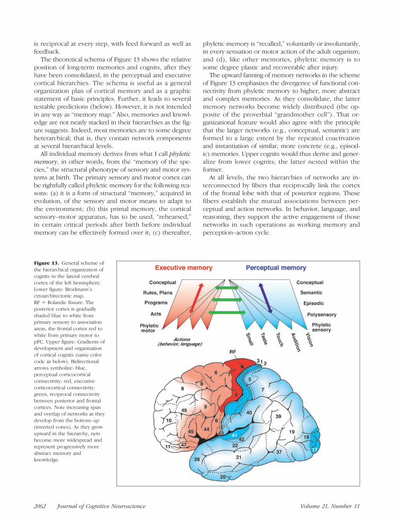
The theoretical schema of Figure 13 shows the relativeposition of long-term memories and cognits, after theyhave been consolidated, in the perceptual and executivecortical hierarchies. The schema is useful as a generalorganization plan of cortical memory and as a graphicstatement of basic principles. Further, it leads to severaltestable predictions (below). However, it is not intendedin any way as “memory map.” Also, memories and knowl-edge are not neatly stacked in their hierarchies as the fig-ure suggests. Indeed, most memories are to some degreeheterarchical; that is, they contain network componentsat several hierarchical levels.
All individual memory derives from what I call phyleticmemory, in other words, from the “memory of the spe-cies,” the structural phenotype of sensory and motor sys-tems at birth. The primary sensory and motor cortex canbe rightfully called phyletic memory for the following rea-sons: (a) it is a form of structural “memory,” acquired inevolution, of the sensory and motor means to adapt tothe environment; (b) this primal memory, the corticalsensory–motor apparatus, has to be used, “rehearsed,”in certain critical periods after birth before individualmemory can be effectively formed over it; (c) thereafter,
phyletic memory is “recalled,” voluntarily or involuntarily,in every sensation or motor action of the adult organism;and (d), like other memories, phyletic memory is tosome degree plastic and recoverable after injury.The upward fanning of memory networks in the scheme
of Figure 13 emphasizes the divergence of functional con-nectivity from phyletic memory to higher, more abstractand complex memories. As they consolidate, the lattermemory networks become widely distributed (the op-posite of the proverbial “grandmother cell”). That or-ganizational feature would also agree with the principlethat the larger networks (e.g., conceptual, semantic) areformed to a large extent by the repeated coactivationand instantiation of similar, more concrete (e.g., episod-ic) memories. Upper cognits would thus derive and gener-alize from lower cognits; the latter nested within theformer.At all levels, the two hierarchies of networks are in-
terconnected by fibers that reciprocally link the cortexof the frontal lobe with that of posterior regions. Thesefibers establish the mutual associations between per-ceptual and action networks. In behavior, language, andreasoning, they support the active engagement of thosenetworks in such operations as working memory andperception–action cycle.
Figure 13. General scheme ofthe hierarchical organization ofcognits in the lateral cerebralcortex of the left hemisphere.Lower figure: Brodmannʼscytoarchitectonic map.RF = Rolandic fissure. Theposterior cortex is graduallyshaded blue to white fromprimary sensory to associationareas, the frontal cortex red towhite from primary motor topFC. Upper figure: Gradients ofdevelopment and organizationof cortical cognits (same colorcode as below). Bidirectionalarrows symbolize: blue,perceptual corticocorticalconnectivity; red, executivecorticocortical connectivity;green, reciprocal connectivitybetween posterior and frontalcortices. Note increasing spanand overlap of networks as theydevelop from the bottom–up(inverted cones). As they growupward in the hierarchy, netsbecome more widespread andrepresent progressively moreabstract memory andknowledge.
2062 Journal of Cognitive Neuroscience Volume 21, Number 11

In summary, the structure of long-term memory in thepresent network memory paradigm has the followingmajor features: (1) it is hierarchical but compatible witha degree of heterarchical organization and dynamics; (2)it contains perceptual cognits mainly in posterior cortex,executive cognits mainly in frontal cortex; and (3) per-ceptual and executive cortices—and their cognits—arejoined by long reentrant and reciprocal fibers, which serveworking memory and the dynamics of the perception–action cycle.The postulated functional architecture of memory
networks leads to the following predictions. Some ofthem have already been partially tested and used tosupport the argument for the new paradigm. They need,however, expanded testing to confirm or reject thisparadigm.
A. Cortical lesions will induce memory deficits depend-ing on the location and extent of the lesion. In theposterior hierarchy, from sensory cortex to associationcortex, lesions will affect the formation, retrieval, andworking memory of progressively higher perceptualcontent. Small lesions at low level (sensory cortex) willaffect simple sensory cognits. Larger lesions at higherlevels (temporal, parietal cortex) will affect larger,more complex cognits (unimodal and polymodal agno-sias, aphasias, and amnesias). Conversely, in the exec-utive hierarchy, from motor to pFC, lesions will affectprogressively higher executive content. Small lesionsof motor cortex will affect the representation of simplemovements by discrete muscle groups. Larger lesionsof premotor cortex will affect the representation ofmovements defined by goal and trajectory. Still largerlesions in the pFC will affect the highest, most complexexecutive memories and knowledge, including rulesand plans.
B.On the assumption of partial commonality of anatomi-cal substrate for long-term and working memory, it canbe predicted that the activation of cognits in workingmemory will elicit electrical and functional imagingsignals from the cortical areas representing the mem-orandum. In those areas, during working memory, mi-croelectrodes will record persistent unit discharge andsynchronous high-frequency LFP oscillations. Sustainedworking memory will elicit imaging signal from thoseareas. By manipulating the category and the context ofthe memorandum, it will be possible to vary the sourceand location of the signals and thus the spread andlocation of the activated cognits. For example, a con-crete sensory memorandum will activate a relativelysmall region of sensory association cortex, such as thesuperior (auditory) or the inferior (visual) temporalgyrus. A more complex stimulus with associations ofmore than one sensory modality will activate multiplesensory association cortices. In all working-memorytasks, the activation will be interregional, involvingsimultaneously prefrontal and posterior cortex, as the
activated cognits will encompass perceptual as well asexecutive networks.
BEHAVIORAL NETWORK DYNAMICS:THE PERCEPTION–ACTION CYCLE
In the cortex, as in the rest of the brain, there are no“systems of memory,” but there is the memory of sys-tems. All cortical systems have their own memory, whichis inextricable from the operations they perform. Thesubstrate for process is inseparable from the substrate forrepresentation. Cognitive networks contribute to behaviorby performing the sensory and the motor functions theyrepresent.
From evidence reviewed, it can be reasonably inferredthat, in goal-directed behavior, posterior and frontal cognitsjoin together to coordinate the action. At high levels ofthe cortical hierarchies, prefrontal networks, which rep-resent broader actions and longer term executive goals,successively activate subjacent networks that representshorter term, intermediate actions and goals. At everystep, action is guided by feedback from previous actions.The entire sequence, with its subordinate steps, is gener-ated and carried out continually by executive networks atvarious levels, integrating stimuli from the environment(internal and external) with feedback signals from thatenvironment, all within the framework of the perception–action cycle.
The perception–action cycle is a basic biological prin-ciple that governs the functional relationships of theorganism with its environment. As a process, it is the cyber-netic circle of sensing and acting that guides the organismto its goals. The concept originated in biology (Uexküll,1926) and eventually entered neurology (Weizsäcker, 1950),cognitive science (Neisser, 1976), and computational neu-roscience (Arbib, 1985). The perception–action cycle op-erates at all levels of the nervous system, from the spinalcord to the cerebral cortex. In the course of complexbehavior, it engages neural networks at every hierarchicallevel of the neocortex, following processing paths thatcourse through the environment and through connec-tions between cortical areas (Figure 14). Action may be in-itiated anywhere in the cycle, in the internal or externalenvironment. Once the cycle is engaged, its networks be-come engaged in series as well as in parallel, with the qual-ification that the interactions between networks may linkdifferent levels heterarchically. Another qualification isthat the cycle is at all levels bidirectional: feed forward isaccompanied by internal feedback. That feedback servesas a kind of corollary discharge (Teuber, 1972) to preparefor impending perception as well as action.
Highly automated, overlearned, or instinctual behav-iors and habits need not engage the cognits of the cerebralcortex. They can be sequentially performed in chainlikefashion through shunts at lower levels of the cycle. Thecortex becomes engaged in the cycle, however, when
Fuster 2063

there are discontinuities in the sequence, especially if thelatter requires temporal integration in the face ofuncertainty or ambiguity—as in working-memory tasks. In-ternal feedback then serves perceptual as well as executiveattention.
Thus, in the course of a demanding temporal gestaltof behavior, language, or reasoning toward a goal, theperception–action cycle orderly recruits a series of mem-ory networks, each modulated by internal feedback. Withthemethods available, the order can be traced only coarsely.A promising analytical method is the computing of Grangercausality on electrical signals. Essentially, this methodallows the study of information flow by analysis of mul-tivariate changes in the interdependence of time seriesfrom several sources (Blinowska, Kus, & Kaminski, 2004;Brovelli et al., 2004). The application of an algorithmictransfer function to the trains of electrical events makesit is possible to predict the directionality of the eventsbetween sources and thus to infer the causal relation-ships between them. Granger causality analysis has beensuccessfully used in tracing electrocortical activity in net-works of frontal and parietal areas controlling hand move-ment in a visuomotor discrimination task (Brovelli et al.,2004).
In neuroimaging, time-series analysis and correlationmethods provide further evidence of the spatial and dy-namic characteristics of large-scale cognitive networks.Bullmore et al. (1996) provided evidence of the activationof vast networks during a task that requires visual andsemantic processing. Those networks show major fociof activation in extrastriate cortex, angular gyrus, superiorand middle temporal gyri, premotor cortex, and Brocaʼsarea. Significantly, there are large functional distancesand discontinuities (negative connectivity) between someof the foci. In other studies (Newman, Just, & Carpenter,2002; Lowe, Dzemidzic, Lurito, Mathews, & Phillips, 2000),correlation and synchronization gradients are detectedbetween the components of a large network in workingmemory. As the collaboration of frontal and parietal re-gions increases, the correlation and the synchronizationbetween them also increase.
Executive frontal networks are activated during the men-tal planning of serial movement (Baker et al., 1996; Roland,Larsen, Lassen, & Skinhøj, 1980; Ingvar & Philipson, 1977).Those networks, therefore, seem to be the depositories ofso-called “memory of the future” (Ingvar, 1985). FulfillingJacksonʼs prediction with regard to anterior frontal areas( Jackson, 1882), those same networks are involved inthe coordination of the planned action, beginning withthe evocation of the objects leading to that action. For ex-ample, whereas the viewing of animals activates posteriorareas, that of tools (“action objects”) activates premotorcortex (Martin, Wiggs, Ungerleider, & Haxby, 1996). Fur-ther, those networks are involved in the actual implemen-tation of the actions, as in the performance of the Tower ofLondon, a test of planning ability (Morris, Ahmed, Syed, &Toone, 1993).
Just as there is an orderly representation of actions infrontal cortex, from the most abstract and complex inpFC to the most concrete in motor cortex, the orderlyexecution of actions follows that trend. Thus, the pro-cessing in the executive memory side of the perception–action cycle descends from the concept and plan of actionin prefrontal networks downward to specific action net-works in motor cortex. Especially persuasive in this re-spect are the studies by Badre and DʼEsposito (2007)and Koechlin, Ody, and Kouneiher (2003). Both use behav-ioral paradigms in which the motor response to a sensorystimulus is contingent on progressively more remote asso-ciations of the stimulus or the response.In the 2003 study (Figure 15), responses depend (a) on
a simple feature (color), (b) on the presence of an addi-tional feature (pattern) that provides the “context,” or (c)on a recent instructional—visual—cue, a prior contin-gency that the authors call “episode.” Thus, from condi-tions a to c, the response to the stimulus is determinedby information of increasing complexity and associativeload. In condition c, the subject must also integrate infor-mation across time. Condition a activates premotor cor-tex; condition b, in addition, activates posterior pFC; andcondition c, in addition, activates anterior pFC. Further,in the third task, the path coefficients of activation sug-gest a processing “cascade” that originates in anteriorpFC and courses through premotor to motor cortex.In sum, the activation of sensorimotor cognits pro-
gresses down the executive hierarchy. Two implicit quali-fications seem necessary, however, to properly interpretthe results of those studies within the network paradigm.One is that, according to the anatomy, the connectivityis reciprocal between all stages of the executive hierar-chy. The other is that the processing toward action isnot only serial, as most hierarchical models imply, butalso parallel.At the end of the previous section, a set of empirical pre-
dictions has been presented that mainly derives from thestructural characteristics of the networkparadigmdiscussedin this review. I finish this section with another set of pre-dictions deriving mainly from its dynamic characteristics:
A. Serial behaviors that require sequential decisions in-formed by the environmental consequences of the sub-jectʼs actions will reveal the temporal alternation ofGranger causality between frontal and posterior areas.This alternation will be revealed not only by LFP rec-ords but also by neuroimaging records. Because thenecessary behavioral and recording methods are bestsuited to the nonhuman primate, those hypothesesshould be tested in monkeys. Neuroimaging recordswill suitably be obtained by near-infrared spectroscopy(NIRS), an optical-imaging method with less spatial res-olution but considerably greater temporal resolutionthan fMRI (Fuster et al., 2005).
B. Working memory will elicit sustained, widespread—interregional—activation of frontal and posterior cortex,
2064 Journal of Cognitive Neuroscience Volume 21, Number 11

Figure 14. Flow of corticaland subcortical connectivityand processing in theperception–action cycle.Empty rhomboids representintermediate areas or subareasof adjacent labeled regions.
Figure 15. Effectiveconnectivity deduced from thefMRI activation of frontal areasduring the performance ofthree levels of associativeresponse to a stimulus, asdescribed in the text. Circlesdenote the approximatelocations of activation in theregions indicated at the right ofthe figure. Frontal activationsand path coefficientssignificantly increasing with thefirst task (stimulus alone), thesecond task (stimulus andcontext), and the third task(“episode,” which includes aprior contingency) are shownin green, yellow, and red,respectively. L = left; R = right;LPFC = lateral pFC. Adaptedfrom Koechlin et al. (2003),with permission.
Fuster 2065

and this will be manifest in multiple-unit, EEG, LFP, andNIRS signals. On the assumption that the activation re-flects reentrant excitation within and between cognits,regions presumed to contain networks representingthe memorandum and the operant response will showhigh degrees of covariance and coherence—especiallyin the high-frequency range.
C.The reversible—for example, cryogenic—lesion of fron-tal cortex will induce a deactivation of posterior cognitsand a deficit in working memory. These will be manifestin multiple-unit and LFP records of posterior cortex andin a concomitant behavioral memory deficit. Thesemanifestations of deficit will result from a dual revers-ible effect of the procedure: (a) interruption of theperception–action cycle at the top and (b) interruptionof reentry loops between frontal and posterior cortexmaintaining active memory.
Conclusions and Future Research
A large body of recent evidence endorses a new networkparadigm of the structure and dynamics of memory inthe cerebral cortex. That evidence suffices to establishthe paradigmʼs basic principles, which sharply distinguishit from other models of cortical memory. According to it,memories as well as items of knowledge consist of dis-tributed and hierarchically organized cortical networks.These memory networks or cognits consist of dispersedneuron populations and the associative connections thatlink them. Those connections, commonly bridging non-contiguous areas of the cortex, are formed, enhanced,and expanded by experience-dependent synaptic modu-lation. Cognits overlap and interconnect extensively with-in and between hierarchical levels (heterarchically). Thus,a neuron or a neuron population can be part of multiplememories or items of knowledge.
Whereas the paradigm is based on vast empirical evi-dence, it is difficult to conceive of one single critical exper-iment to prove it correct. Definitive proof would requirethe simultaneous application to the entire cortex of analyt-ical methods with multiple scales of spatial and temporalresolution, which is now impractical. In the absence ofsuch capability, we can assume that the model is essen-tially correct and attempt to strengthen experimentallythe principles outlined in this review. Nonetheless, oneof the merits of the paradigm is that even with currentmethods, those principles can be proven wrong. Indeed,the paradigm would be falsified if it were shown that (1)specific memories or items or knowledge are exclusivelyrepresented in discrete domains of cortex and not en-dowed with experience-dependent plasticity; (2) memoryor knowledge representations are not interregional andare deprived of hierarchical organization and nesting; (3)anatomical relationships between cell groups are immate-rial to the memory code; or (4) substantial numbers ofcells or cell groups in separate cortical locations are not
attuned to the associated constituent properties of a mem-ory or an item of knowledge.More research is needed for complete description of the
emerging paradigm in physiological and computationalterms. To that end, a number of predictions—advancedin the previous two sections—need testing. Because ofthe modelʼs distributed, associative, and integrative ar-chitecture, special efforts must be made to explore itsconstituent networks with a variety of different methods.Especially useful will be the simultaneous recording of elec-trical and imaging signals from multiple sites in memorytasks. Unit discharge, LFPs, surface EEG, and NIRS shouldbe recorded from monkeys performing working-memorytasks. This combination of signal-recording methods, to-gether with advanced computational methods, shouldallow the exploration of active memory networks withvarying scales and resolution. Signals of different originshould be submitted to computational analysis in multiplespatial and temporal scales, with emphasis on coherence,covariance, and Granger causality. As heretofore, working-memory tasks, with their time-bracketed active memory,should continue to provide the ideal setting for this research.Finally, a pressing issue is the accessibility of long-term
memory, in the active state, to consciousness. The availableevidence indicates that conscious cognition is a gradedfunction, largely dependent on synaptic strength anddegree of cognit activation. These inferences open newavenues for understanding priming and preconscious phe-nomena. Moreover, research on these issues may lead tonew procedures for memory enhancement as well as therehabilitation of old associative paths or the opening ofnew ones.
Acknowledgments
I wish to thank Thomas Albright, Mark DʼEsposito, and StevenBressler for their valuable comments on earlier versions of themanuscript.
Reprint requests should be sent to Joaquín M. Fuster, UCLA SemelInstitute, 740 Westwood Plaza, Los Angeles, CA 90095, or viae-mail: [email protected].
REFERENCES
Akkal, D., Bioulac, B., Audin, J., & Burbaud, P. (2002).Comparison of neuronal activity in the rostral supplementaryand cingulate motor areas during a task with cognitive andmotor demands. European Journal of Neuroscience, 15,887–904.
Albright, T. D., & Stoner, G. R. (2002). Contextual influences onvisual processing. Annual Review of Neuroscience, 25,339–379.
Amaral, D. G. (1987). Memory: Anatomical organizationof candidate brain regions. In F. Plum (Ed.), Handbook ofphysiology; nervous system: Vol. V. Higher functions of thebrain, part 1 (pp. 211–294). Bethesda, MD: AmericanPhysiological Society.
Andersen, R. A., Bracewell, R. M., Barash, S., Gnadt, J. W., &Fogassi, L. (1990). Eye position effects on visual, memory,
2066 Journal of Cognitive Neuroscience Volume 21, Number 11

and saccade-related activity in areas LIP and 7a of macaque.Journal of Neuroscience, 10, 1176–1196.
Arbib, M. A. (1985). Schemas for the temporal organization ofbehaviour. Human Neurobiology, 4, 63–72.
Artchakov, D., Tikhonravov, D., Vuontela, V., Linnankoski, I.,Korvenoja, A., & Carlson, S. (2007). Processing ofauditory and visual location information in the monkeyprefrontal cortex. Experimental Brain Research, 180,469–479.
Asaad, W. F., Rainer, G., & Miller, E. K. (2000). Task-specificneural activity in the primate prefrontal cortex. Journal ofNeurophysiology, 84, 451–459.
Averbeck, B. B., & Lee, D. (2007). Prefrontal neural correlatesof memory for sequences. Journal of Neuroscience, 27,2204–2211.
Axmacher, N., Schmitz, D. P., Wagner, T., Elger, C. E., & Fell, J.(2008). Interactions between medial temporal lobe,prefrontal cortex, and inferior temporal regions duringvisual working memory: A combined intracranial EEG andfunctional magnetic resonance imaging study. Journal ofNeuroscience, 28, 7304–7312.
Baddeley, A. D. (1993). Working memory or working attention?In A. D. Baddeley & L. Weiskrantz (Eds.), Attention: Selection,awareness and control. A tribute to Donald Broadbent(pp. 152–170). Oxford: Clarendon Press.
Badre, D., & DʼEsposito, M. (2007). Functional magneticresonance imaging evidence for a hierarchical organizationof the prefrontal cortex. Journal of Cognitive Neuroscience,19, 2082–2099.
Baker, S. C., Rogers, R. D., Owen, A. M., Frith, C. D., Dolan, R. J.,Frackowiak, R. S. J., et al. (1996). Neural systems engagedby planning: A PET study of the Tower of London task.Neuropsychologia, 34, 515–526.
Barbas, H., Ghashghaei, H. T., Rempel-Clower, N. L., & Xiao, D.(2002). Anatomic basis of functional specialization inprefrontal cortices in primates. In J. Grafman (Ed.),Handbook of neuropsychology (pp. 1–27). Oxford: Elsevier.
Barkovich, A. J. (1995). Pediatric neuroimaging (2nd ed.).New York: Raven Press.
Basar, E., & Bullock, T. H. (2000). Brain dynamics: Progressand perspectives. Berlin: Springer.
Berger, H. (1929). Über das Elektroenzephalogramm desMenschen. Archiv für Psychiatrie und Nervenkrankheiten,87, 527–528.
Blinowska, K. J., Kus, R., & Kaminski, M. (2004). Grangercausality and information flow in multivariate processes.Physical Review. E, Statistical, Nonlinear Soft Matter Physics,70, 050902.
Bodner, M., Shafi, M., Zhou, Y. D., & Fuster, J. M. (2005).Patterned firing of parietal cells in a haptic workingmemory task. European Journal of Neuroscience, 21,2538–2546.
Braitenberg, V., & Schüz, A. (1998). Cortex: Statistics andgeometry of neural connectivity. Heidelberg: Springer.
Bressler, S. L. (1995). Large-scale cortical networks andcognition. Brain Research Reviews, 20, 288–304.
Bressler, S. L., Coppola, R., & Nakamura, R. (1993).Episodic multiregional cortical coherence at multiplefrequencies during visual task performance. Nature, 366,153–156.
Bressler, S. L., & McIntosh, A. R. (2007). The role of neuralcontext in large-scale neurocognitive network operations. InV. K. Jirsa & A. R. McIntosh (Eds.), Springer handbook onbrain connectivity (pp. 403–419). New York: Springer.
Broca, P. (1861). Rémarques sur la siège de la faculté du langagearticulé, suivi dʼune observation dʼaphémie. Bulletins de laSociété Anatomique de Paris, 2, 330–357.
Brovelli, A., Battaglini, P. P., Naranjo, J. R., & Budai, R.
(2002). Medium-range oscillatory network and the 20-Hzsensorimotor induced potential. Neuroimage, 16, 130–141.
Brovelli, A., Ding, M., Ledberg, A., Chen, Y., Nakamura, R., &Bressler, S. L. (2004). Beta oscillations in a large-scalesensorimotor cortical network: Directional influencesrevealed by Granger causality. Proceedings of the NationalAcademy of Sciences, U.S.A., 101, 9849–9854.
Brovelli, A., Lachaux, J. P., Kahane, P., & Boussaoud, D. (2005).High gamma frequency oscillatory activity dissociatesattention from intention in the human premotor cortex.Neuroimage, 28, 154–164.
Brunel, N., & Wang, X. J. (2001). Effects of neuromodulationin a cortical network model of object working memorydominated by recurrent inhibition. Journal ofComputational Neuroscience, 11, 63–85.
Buchsbaum, B. R., Olsen, R. K., Koch, P., & Berman, K. F.(2005). Human dorsal and ventral auditory streamssubserve rehearsal-based and echoic processes duringverbal working memory. Neuron, 48, 687–697.
Buckner, R. L., Petersen, S. E., Ojemann, J. G., Miezin, F. M.,Squire, L. R., & Raichle, M. E. (1995). Functional anatomicalstudies of explicit and implicit memory retrieval tasks.Journal of Neuroscience, 15, 12–29.
Bullmore, E. T., Rabe-Hesketh, S., Morris, R. G., Williams, S. C.,Gregory, L., Gray, J. A., et al. (1996). Functional magneticresonance image analysis of a large-scale neurocognitivenetwork. Neuroimage, 4, 16–33.
Burton, H., & Sinclair, R. J. (2000). Attending to andremembering tactile stimuli: A review of brain imagingdata and single-neuron responses. Journal of ClinicalNeurophysiology, 17, 575–591.
Buschman, T. J., & Miller, E. K. (2007). Top–down versusbottom–up control of attention in the prefrontal andposterior parietal cortices. Science, 315, 1860–1862.
Buzsáki, G., & Draguhn, A. (2004). Neuronal oscillations incortical networks. Science, 304, 1926–1929.
Buzsáki, G., Geisler, C., Henze, D. A., & Wang, X. J. (2004).Interneuron diversity series: Circuit complexity and axonwiring economy of cortical interneurons. Trends inNeurosciences, 27, 186–193.
Cabeza, R., & Nyberg, L. (2000). Imaging cognition II: Anempirical review of 275 PET and MRI studies. Journal ofCognitive Neuroscience, 12, 1–47.
Cairo, T. A., Liddle, P. F., Woodward, T. S., & Ngan, E. T. (2004).The influence of working memory load on phase specificpatterns of cortical activity. Brain Research, CognitiveBrain Research, 21, 377–387.
Cajal, S. R. (1923). Recuerdos de mi Vida. Madrid: Pueyo.Canolty, R. T., Edwards, E., Dalal, S. S., Soltani, M., Nagarajan,
S. S., Kirsch, H. E., et al. (2006). High gamma power isphase-locked to theta oscillations in human neocortex.Science, 313, 1626–1628.
Carlson, S., Rämä, P., Tanila, H., Linnankoski, I., & Mansikka, H.(1997). Dissociation of mnemonic coding and otherfunctional neuronal processing in the monkey prefrontalcortex. Journal of Neurophysiology, 77, 761–774.
Casey, B. J., Cohen, J. D., OʼCraven, K., Davidson, R. J., Irwin,W., Nelson, C. A., et al. (1998). Reproducibility of fMRIresults across four institutions using a spatial workingmemory task. Neuroimage, 8, 249–261.
Chafee, M. V., & Goldman-Rakic, P. S. (2000). Inactivationof parietal and prefrontal cortex reveals interdependenceof neural activity during memory-guided saccades.Journal of Neurophysiology, 83, 1550–1566.
Cohen, J. D., Forman, S. D., Braver, T. S., Casey, B. J.,Servan-Schreiber, D., & Noll, D. C. (1994). Activation ofthe prefrontal cortex in a nonspatial working memory taskwith functional MRI. Human Brain Mapping, 1, 293–304.
Fuster 2067

Cohen, N. J., & Squire, L. R. (1980). Preserved learning andretention of pattern-analyzing skill in amnesia: Dissociation ofknowing how and knowing that. Science, 210, 207–210.
Conel, J. L. (1963). The postnatal development of the humancerebral cortex. Cambridge, MA: Harvard University Press.
Constantinidis, C., Franowicz, M. N., & Goldman-Rakic, P. S.(2001). Coding specificity in cortical microcircuits: Amultiple-electrode analysis of primate prefrontal cortex.Journal of Neuroscience, 21, 3646–3655.
Constantinidis, C., & Procyk, E. (2004). The primate workingmemory networks. Cognitive, Affective & BehavioralNeuroscience, 4, 444–465.
Courtney, S. M., Ungerleider, L. G., Keil, K., & Haxby, J. V.(1997). Transient and sustained activity in a distributedneural system for human working memory. Nature, 386,608–611.
Crottaz-Herbette, S., Anagnoson, R. T., & Menon, V. (2004).Modality effects in verbal working memory: Differentialprefrontal and parietal responses to auditory and visualstimuli. Neuroimage, 21, 340–351.
Csicsvari, J., Jamieson, B., Wise, K. D., & Buzsaki, G. (2003).Mechanisms of gamma oscillations in the hippocampus ofthe behaving rat. Neuron, 37, 311–322.
Curtis, C. E., Sun, F. T., Miller, L. M., & DʼEsposito, M. (2005).Coherence between fMRI time-series distinguishes twospatial working memory networks. Neuroimage, 26,177–183.
De Pisapia, N., Slomski, J. A., & Braver, T. S. (2007). Functionalspecializations in lateral prefrontal cortex associated withthe integration and segregation of information in workingmemory. Cerebral Cortex, 17, 993–1006.
DʼEsposito, M., Postle, B. R., & Rypma, B. (2000). Prefrontalcortical contributions to working memory: Evidence fromevent-related fMRI studies. Experimental Brain Research,133, 3–11.
Duncan, J., & Owen, A. M. (2000). Common regions of thehuman frontal lobe recruited by diverse cognitive demands.Trends in Neurosciences, 23, 475–483.
Eckhorn, R., Bauer, R., Jordan, W., Brosch, M., Kruse, W., Munk,M., et al. (1988). Coherent oscillations: A mechanism offeature linking in the visual cortex? Biological Cybernetics,60, 121–130.
Edelman, G. M., & Mountcastle, V. B. (1978). The mindfulbrain. New York: Plenum Press.
Eichenbaum, H., Fagan, A., Mathews, P., & Cohen, N. J. (1988).Hippocampal system dysfunction and odor discriminationlearning in rats: Impairment or facilitation depending onrepresentational demands. Behavioral Neuroscience, 102,331–339.
Engel, A. K., Fries, P., & Singer, W. (2001). Dynamic predictions:Oscillations and synchrony in top–down processing.Nature Reviews Neuroscience, 2, 704–716.
Engel, A. K., König, P., Kreiter, A. K., & Singer, W. (1991).Interhemispheric synchronization of oscillatory neuronalresponses in cat visual cortex. Science, 252, 1177–1179.
Felleman, D. J., & Van Essen, D. C. (1991). Distributedhierarchical processing in the primate cerebral cortex.Cerebral Cortex, 47, 1047–3211.
Fodor, J. A., & Pylyshyn, Z. W. (1988). Connectionism andcognitive architecture: A critical analysis. In S. Pinker &J. Mehler (Eds.), Connections and symbols (pp. 3–71).Cambridge, MA: MIT Press.
Fox, M. D., & Raichle, M. E. (2007). Spontaneous fluctuations inbrain activity observed with functional magnetic resonanceimaging. Nature Reviews Neuroscience, 8, 700–711.
Frankland, P. W., & Bontempi, B. (2005). The organization ofrecent and remote memories. Nature Reviews Neuroscience,6, 119–130.
Freeman, W. J., Rogers, L. J., Holmes, M. D., & Silbergeld,D. L. (2000). Spatial spectral analysis of humanelectrocorticograms including the alpha and gammabands. Journal of Neuroscience Methods, 95, 111–121.
Fries, P., Reynolds, J. H., Rorie, A. E., & Desimone, R. (2001).Modulation of oscillatory neuronal synchronization byselective visual attention. Science, 291, 1560–1563.
Fukuchi-Shimogori, T., & Grove, E. (2001). Neocortexpatterning by the secreted signaling molecule FGF8. Science,294, 1071–1074.
Fukushima, T., Hasegawa, I., & Miyashita, Y. (2004).Prefrontal neuronal activity encodes spatial targetrepresentations sequentially updated after nonspatialtarget-shift cues. Journal of Neurophysiology, 91,1367–1380.
Funahashi, S., Bruce, C. J., & Goldman-Rakic, P. S. (1989).Mnemonic coding of visual space in the monkeyʼsdorsolateral prefrontal cortex. Journal of Neurophysiology,61, 331–349.
Fuster, J., Guiou, M., Ardestani, A., Cannestra, A., Sheth, S.,Zhou, Y.-D., et al. (2005). Near-infrared spectroscopy (NIRS)in cognitive neuroscience of the primate brain. Neuroimage,26, 215–220.
Fuster, J. M. (1973). Unit activity in prefrontal cortex duringdelayed-response performance: Neuronal correlates oftransient memory. Journal of Neurophysiology, 36, 61–78.
Fuster, J. M. (1990). Inferotemporal units in selective visualattention and short-term memory. Journal ofNeurophysiology, 64, 681–697.
Fuster, J. M. (1995). Memory in the cerebral cortex—Anempirical approach to neural networks in the humanand nonhuman primate. Cambridge, MA: MIT Press.
Fuster, J. M. (2003). Cortex and mind: Unifying cognition.New York: Oxford University Press.
Fuster, J. M. (2008). The prefrontal cortex (4th ed.). London:Academic Press.
Fuster, J. M., & Alexander, G. E. (1971). Neuron activity relatedto short-term memory. Science, 173, 652–654.
Fuster, J. M., Bauer, R. H., & Jervey, J. P. (1982). Cellulardischarge in the dorsolateral prefrontal cortex of the monkeyin cognitive tasks. Experimental Neurology, 77, 679–694.
Fuster, J. M., Bauer, R. H., & Jervey, J. P. (1985). Functionalinteractions between inferotemporal and prefrontal cortex ina cognitive task. Brain Research, 330, 299–307.
Fuster, J. M., Bodner, M., & Kroger, J. K. (2000). Cross-modaland cross-temporal association in neurons of frontal cortex.Nature, 405, 347–351.
Fuster, J. M., & Jervey, J. P. (1982). Neuronal firing in theinferotemporal cortex of the monkey in a visual memory task.Journal of Neuroscience, 2, 361–375.
Gazzaley, A., Rissman, J., & DʼEsposito, M. (2004). Functionalconnectivity during working memory maintenance.Cognitive, Affective & Behavioral Neuroscience, 4,580–599.
Genovesio, A., Brasted, P. J., & Wise, S. P. (2006).Representation of future and previous spatial goals byseparate neural populations in prefrontal cortex. Journal ofNeuroscience, 26, 7305–7316.
Gevins, A., & Smith, M. E. (2000). Neurophysiologicalmeasures of working memory and individual differences incognitive ability and cognitive style. Cerebral Cortex, 10,829–839.
Gibson, J. R., & Maunsell, J. H. R. (1997). Sensory modalityspecificity of neural activity related to memory in visualcortex. Journal of Neurophysiology, 78, 1263–1275.
Goldman-Rakic, P. S. (1988). Topography of cognition: Paralleldistributed networks in primate association cortex. AnnualReview of Neuroscience, 11, 137–156.
2068 Journal of Cognitive Neuroscience Volume 21, Number 11

Goldstein, J. M., Jerram, M., Poldrack, R., Anagnoson, R.,Breiter, H. C., Makris, N., et al. (2005). Sex differences inprefrontal cortical brain activity during fMRI of auditoryverbal working memory. Neuropsychology, 19, 509–519.
Gray, C. M., Konig, P., Engel, A. K., & Singer, W. (1989).Oscillatory responses in cat visual cortex exhibitintercolumnar synchronization which reflects globalstimulus properties. Nature, 338, 334–337.
Gray, C. M., & Singer, W. (1989). Stimulus-specific neuronaloscillations in orientation columns of cat visual cortex.Proceedings of the National Academy of Sciences, U.S.A.,86, 1698–1702.
Gross, J., Schmitz, F., Schnitzler, I., Kessler, K., Shapiro, K.,Hommel, B., et al. (2004). Modulation of long-rangeneural synchrony reflects temporal limitations of visualattention in humans. Proceedings of the National Academyof Sciences, U.S.A., 101, 13050–13055.
Hayek, F. A. (1952). The sensory order. Chicago: University ofChicago Press.
Hebb, D. O. (1949). The organization of behavior. New York:John Wiley & Sons.
Helmholtz, H. V. (1925). Helmholtzʼs treatise on physiologicaloptics ( J. P. C. Southall, Trans). G. Banta, Menasha, WI:The Optical Society of America.
Hikosaka, K., & Watanabe, M. (2000). Delay activity of orbitaland lateral prefrontal neurons of the monkey varying withdifferent rewards. Cerebral Cortex, 10, 263–271.
Hinton, G. E., & Anderson, J. A. (1981). Parallel models ofassociative memory. Hillsdale, NJ: Lawrence ErlbaumAssociates.
Huerta, P. T., & Lisman, J. E. (1995). Bidirectional synapticplasticity induced by a single burst during cholinergic thetaoscillation in CA1 in vitro. Neuron, 15, 1053–1063.
Huxter, J., Burgess, N., & OʼKeefe, J. (2003). Independent rateand temporal coding in hippocampal pyramidal cells. Nature,425, 828–832.
Ichihara-Takeda, S., & Funahashi, S. (2008). Activity of primateorbitofrontal and dorsolateral prefrontal neurons: Effect ofreward schedule on task-related activity. Journal of CognitiveNeuroscience, 20, 563–579.
Ingvar, D. H. (1985). “Memory of the future”: An essay on thetemporal organization of conscious awareness. HumanNeurobiology, 4, 127–136.
Ingvar, D. H., & Philipson, L. (1977). Distribution of cerebralblood flow in the dominant hemisphere during motorideation and motor performance. Annals of Neurology, 2,230–237.
Isomura, Y., Ito, Y., Akazawa, T., Nambu, A., & Takada, M.(2003). Neural coding of “attention for action” and “responseselection” in primate anterior cingulate cortex. Journal ofNeuroscience, 23, 8002–8012.
Jackson, J. H. (1882). On some implications of dissolutionof the nervous system. Medical Press and Circular, ii,411–426.
Jaeggi, S. M., Seewer, R., Nirkko, A. C., Eckstein, D., Schroth, G.,Groner, R., et al. (2003). Does excessive memory loadattenuate activation in the prefrontal cortex? Load-dependentprocessing in single and dual tasks: Functional magneticresonance imaging study. Neuroimage, 19, 210–225.
Jagadeesh, B., Gray, C. M., & Ferster, D. (1992). Visually evokedoscillations of membrane potential in cells of cat visualcortex. Science, 257, 552–554.
Jones, E. G., & Powell, T. P. S. (1970). An anatomical study ofconverging sensory pathways within the cerebral cortex ofthe monkey. Brain, 93, 793–820.
Jonides, J., Smith, E. E., Koeppe, R. A., Awh, E., Minoshima, S.,& Mintun, M. A. (1993). Spatial working memory in humansas revealed by PET. Nature, 363, 623–625.
Kandel, E. R. (2000). Cellular mechanisms of learning andthe biological basis of individuality. In E. R. Kandel,J. H. Schwartz, & T. M. Jessell (Eds.), Principles of neuralscience (pp. 1247–1279). New York: McGraw-Hill.
Kapur, S., Craik, F. I. M., Tulving, E., Wilson, A. A., Houle, S.,& Brown, G. M. (1994). Neuroanatomical correlates ofencoding in episodic memory: Levels of processing effect.Proceedings of the National Academy of Sciences, U.S.A.,91, 2008–2011.
Kjelstrup, K. B., Solstad, T., Brun, V. H., Hafting, T., Leutgeb, S.,Witter, M. P., et al. (2008). Finite scale of spatialrepresentation in the hippocampus. Science, 321, 140–143.
Klimesch, W., Schimke, H., Doppelmayr, M., Ripper, B.,Schwaiger, J., & Pfurtscheller, G. (1996). Event-relateddesynchronization (ERD) and the Dm effect: Does alphadesynchronization during encoding predict later recallperformance? International Journal Psychophysiology,24, 47–60.
Klüver, H. (1933). Behavior mechanisms in monkeys. Chicago:Chicago University Press.
Koechlin, E., Ody, C., & Kouneiher, F. (2003). The architectureof cognitive control in the human prefrontal cortex. Science,302, 1181–1185.
Koffka, K. (1935). Principles of Gestalt psychology. New York:Harcourt, Brace and Company.
Kohonen, T. (1984). Self-organization and associativememory. Berlin: Springer.
Kondo, H., Morishita, M., Osaka, N., Osaka, M., Fukuyama, H.,& Shibasaki, H. (2004). Functional roles of the cingulo-frontalnetwork in performance on working memory. Neuroimage,21, 2–14.
Kuhn, T. S. (1996). The structure of scientific revolutions(3rd ed.). Chicago: The University of Chicago Press.
Lakatos, P., Karmos, G., Mehta, A. D., Ulbert, I., & Schroeder,C. E. (2008). Entrainment of neuronal oscillations as amechanism of attentional selection. Science, 320, 110–113.
Lashley, K. S. (1950). In search of the engram. Symposia of theSociety for Experimental Biology, 4, 454–482.
Lee, A. C., Robbins, T. W., & Owen, A. M. (2000). Episodicmemory meets working memory in the frontal lobe:Functional neuroimaging studies of encoding and retrieval.Critical Reviews in Neurobiology, 14, 165–197.
Lee, H., Simpson, G. V., Logothetis, N. K., & Rainer, G. (2005).Phase locking of single neuron activity to theta oscillationsduring working memory in monkey extrastriate visual cortex.Neuron, 45, 147–156.
Lenartowicz, A., & McIntosh, A. R. (2005). The role of anteriorcingulate cortex in working memory is shaped by functionalconnectivity. Journal of Cognitive Neuroscience, 17,1026–1042.
Leung, H. C., Seelig, D., & Gore, J. C. (2004). The effect ofmemory load on cortical activity in the spatial workingmemory circuit. Cognitive, Affective & BehavioralNeuroscience, 4, 553–563.
Li, Z. H., Sun, X. W., Wang, Z. X., Zhang, X. C., Zhang, D. R., He,S., et al. (2004). Behavioral and functional MRI study ofattention shift in human verbal working memory.Neuroimage, 21, 181–191.
Linden, D. E., Bittner, R. A., Muckli, L., Waltz, J. A., Kriegeskorte,N., Goebel, R., et al. (2003). Cortical capacity constraintsfor visual working memory: Dissociation of fMRI loadeffects in a fronto-parietal network. Neuroimage, 20,1518–1530.
Llinás, R. (1988). The intrinsic electrophysiological properties ofmammalian neurons: Insights into central nervous systemfunctions. Science, 242, 1654–1664.
Lorente de Nó, R. (1949). Cerebral cortex: Architecture,intracortical connections, motor projections. In J. Fulton
Fuster 2069

(Ed.), Physiology of the nervous system (pp. 288–315).New York: New York University Press.
Lowe, M. J., Dzemidzic, M., Lurito, J. T., Mathews, V. P., &Phillips, M. D. (2000). Correlations in low-frequency BOLDfluctuations reflect cortico-cortical connections.Neuroimage, 12, 582–587.
Luria, A. R. (1966). Higher cortical functions in man. NewYork: Basic Books.
Mansouri, F. A., Matsumoto, K., & Tanaka, K. (2006).Prefrontal cell activities related to monkeysʼ success andfailure in adapting to rule changes in a Wisconsin CardSorting Test analog. Journal of Neuroscience, 26,2745–2756.
Marcus, G. F. (1998). Can connectionism save constructivism?Cognition, 66, 153–182.
Martin, A., Wiggs, C. L., Ungerleider, L. G., & Haxby, J. V. (1996).Neural correlates of category-specific knowledge. Nature,379, 649–652.
McClelland, H. A., & Rumelhart, D. E. (1986). Paralleldistributed processing. Cambridge, MA: MIT Press.
McCloskey, D. I. (1981). Corollary discharges: Motor commandsand perception. In V. B. Brooks (Ed.), Handbook ofphysiology: Nervous system (pp. 1415–1447). Bethesda:American Physiological Society.
McIntosh, A. R. (2000). Towards a network theory of cognition.Neural Networks, 13, 861–870.
Mecklinger, A., Bosch, V., Gruenewald, C., Bentin, S., & VonCramon, D. Y. (2000). What have Klingon letters and faces incommon? An fMRI study on content-specific workingmemory systems. Human Brain Mapping, 11, 146–161.
Messinger, A., Squire, L. R., Zola, S. M., & Albright, T. D.(2001). Neuronal representations of stimulus associationsdevelop in the temporal lobe during learning. Proceedingsof the National Academy of Sciences, U.S.A., 98,12239–12244.
Mesulam, M.-M. (1998). From sensation to cognition. Brain,121, 1013–1052.
Miller, E. K., & Cohen, J. D. (2001). An integrative theory ofprefrontal cortex function. Annual Review of Neuroscience,24, 167–202.
Miller, E. K., Li, L., & Desimone, R. (1991). A neural mechanismfor working and recognition memory in inferior temporalcortex. Science, 254, 1377–1379.
Mitchell, K. J., Johnson, M. K., Raye, C. L., & Greene, E. J.(2004). Prefrontal cortex activity associated with sourcemonitoring in a working memory task. Journal of CognitiveNeuroscience, 16, 921–934.
Miyashita, Y. (1988). Neuronal correlate of visual associativelong-term memory in the primate temporal cortex. Nature,335, 817–820.
Miyashita, Y., & Chang, H. S. (1988). Neuronal correlate ofpictorial short-term memory in the primate temporal cortex.Nature, 331, 68–70.
Mongillo, G., Barak, O., & Tsodyks, M. (2008). Synaptic theoryof working memory. Science, 319, 1543–1546.
Morris, R. G., Ahmed, S., Syed, G. M., & Toone, B. K. (1993).Neural correlates of planning ability: Frontal lobe activationduring the Tower of London Test. Neuropsychologia, 31,1367–1378.
Myers, R. E. (1967). Cerebral connectionism and brain function.In C. H. Millikan & F. L. Darley (Eds.), Brain mechanismsunderlying speech and language (pp. 61–72). New York:Grune & Stratton.
Narayanan, N. S., Prabhakaran, V., Bunge, S. A., Christoff, K.,Fine, E. M., & Gabrieli, J. D. (2005). The role of theprefrontal cortex in the maintenance of verbal workingmemory: An event-related FMRI analysis. Neuropsychology,19, 223–232.
Neisser, U. (1976). Cognition and reality: Principles andimplications of cognitive psychology. San Francisco, CA:Freeman.
Newman, S. D., Just, M. A., & Carpenter, P. A. (2002). Thesynchronization of the human cortical working memorynetwork. Neuroimage, 15, 810–822.
Niki, H. (1974). Prefrontal unit activity during delayedalternation in the monkey: I. Relation to direction ofresponse. Brain Research, 68, 185–196.
Northcutt, G., & Kaas, J. H. (1995). The emergence andevolution of mammalian neocortex. Trends inNeurosciences, 18, 373–379.
OʼKeefe, J., & Recce, M. L. (1993). Phase relationship betweenhippocampal place units and the EEG theta rhythm.Hippocampus, 3, 317–330.
Paulesu, E., Frith, C. D., & Frackowiak, R. S. J. (1993). Theneural correlates of the verbal component of workingmemory. Nature, 362, 342–344.
Penttonen, G., & Buzsáki, G. (2003). Thalamus and relatedsystems. Systems, 2, 145.
Pesaran, B., Pezaris, J. S., Sahani, M., Mitra, P. P., & Andersen,R. A. (2002). Temporal structure in neuronal activity duringworking memory in macaque parietal cortex. NatureNeuroscience, 5, 805–811.
Petit, L., Courtney, S. M., Ungerleider, L. G., & Haxby, J. V.(1998). Sustained activity in the medial wall duringworking memory delays. Journal of Neuroscience, 18,9429–9437.
Petrides, M., Alivisatos, B., Evans, A. C., & Meyer, E. (1993).Dissociation of human mid-dorsolateral from posteriordorsolateral frontal cortex in memory processing.Proceedings of the National Academy of Sciences, U.S.A.,90, 873–877.
Petrides, M., Alivisatos, B., Meyer, E., & Evans, A. C. (1993).Functional activation of the human frontal cortex duringthe performance of verbal working memory tasks.Proceedings of the National Academy of Sciences, U.S.A.,90, 878–882.
Petrides, M., & Pandya, D. N. (2002). Association pathwaysof the prefrontal cortex and functional observations. InD. T. Stuss & R. T. Knight (Eds.), Principles of frontallobe function (pp. 31–84). New York: Oxford UniversityPress.
Pfurtscheller, G., & Lopes da Silva, F. H. (1999). Event-relatedEEG/MEG synchronization and desynchronization: Basicprinciples. Clinical Neurophysiology, 110, 1842–1857.
Pollmann, S., & Von Cramon, D. Y. (2000). Object workingmemory and visuospatial processing: Functionalneuroanatomy analyzed by event-related fMRI. ExperimentalBrain Research, 133, 12–22.
Posner, M. I., & Petersen, S. E. (1990). The attention system ofthe human brain. Annual Review of Neuroscience, 13,25–42.
Postle, B. R. (2005). Delay-period activity in the prefrontalcortex: One function is sensory gating. Journal of CognitiveNeuroscience, 17, 1679–1690.
Postle, B. R., Berger, J. S., Goldstein, J. H., Curtis, C. E., &DʼEsposito, M. (2001). Behavioral and neurophysiologicalcorrelates of episodic coding, proactive interference, andlist length effects in a running span verbal working memorytask. Cognitive, Affective & Behavioral Neuroscience, 1,10–21.
Procyk, E., & Joseph, J. P. (2001). Characterization of serialorder encoding in the monkey anterior cingulate sulcus.European Journal of Neuroscience, 14, 1041–1046.
Quintana, J., & Fuster, J. M. (1999). From perception to action:Temporal integrative functions of prefrontal and parietalneurons. Cerebral Cortex, 9, 213–221.
2070 Journal of Cognitive Neuroscience Volume 21, Number 11

Quintana, J., Fuster, J. M., & Yajeya, J. (1989). Effects of coolingparietal cortex on prefrontal units in delay tasks. BrainResearch, 503, 100–110.
Rajah, M. N., & DʼEsposito, M. (2005). Region-specific changesin prefrontal function with age: A review of PET and fMRIstudies on working and episodic memory. Brain, 128,1964–1983.
Rakic, P. (2001). Neurocreationism—Making new cortical maps.Science, 294, 1011–1012.
Rama, P., & Courtney, S. M. (2005). Functional topographyof working memory for face or voice identity. Neuroimage,24, 224–234.
Ranganath, C., Cohen, M., Dam, C., & DʼEsposito, M.(2004). Inferior temporal, prefrontal, and hippocampalcontributions to visual working memory maintenance andassociative memory retrieval. Journal of Neuroscience, 24,3917–3925.
Rao, S. C., Rainer, G., & Miller, E. K. (1997). Integration of whatand where in the primate prefrontal cortex. Science, 276,821–824.
Reyes, A. D. (2003). Synchrony-dependent propagation of firingrate in iteratively constructed networks in vitro. NatureNeuroscience, 6, 593–599.
Ricciardi, E., Bonino, D., Gentili, C., Sani, L., Pietrini, P., &Vecchi, T. (2006). Neural correlates of spatial workingmemory in humans: A functional magnetic resonanceimaging study comparing visual and tactile processes.Neuroscience, 139, 339–349.
Rizzuto, D. S., Madsen, J. R., Bromfield, E. B., Schulze-Bonhage,A., Seelig, D., Schenbrenner-Scheibe, R., et al. (2003).Reset of human neocortical oscillations during a workingmemory task. Proceedings of the National Academy ofSciences, U.S.A., 100, 7931–7936.
Rockel, A. J., Hiorns, R. W., & Powell, T. P. S. (1980). The basicuniformity in structure of the neocortex. Brain, 103,221–244.
Roland, P. E., Larsen, B., Lassen, N. A., & Skinhøj, E. (1980).Supplementary motor area and other cortical areas inorganization of voluntary movements in man. Journal ofNeurophysiology, 43, 118–136.
Romanski, L. M. (2007). Representation and integration ofauditory and visual stimuli in the primate ventral lateralprefrontal cortex. Cerebral Cortex, 17(Suppl. 1), i61–i69.
Romo, R., Brody, C. D., Hernández, A., & Lemus, L. (1999).Neuronal correlates of parametric working memory in theprefrontal cortex. Nature, 399, 470–473.
Rosenkilde, C. E., Bauer, R. H., & Fuster, J. M. (1981). Single cellactivity in ventral prefrontal cortex of behaving monkeys.Brain Research, 209, 375–394.
Roth, J. K., Serences, J. T., & Courtney, S. M. (2006). Neuralsystem for controlling the contents of object workingmemory in humans. Cerebral Cortex, 16, 1595–1603.
Rypma, B., & DʼEsposito, M. (2003). A subsequent-memoryeffect in dorsolateral prefrontal cortex. Brain Research,Cognitive Brain Research, 16, 162–166.
Saalmann, Y. B., Pigarev, I. N., & Vidyasagar, T. R. (2007).Neural mechanisms of visual attention: How top–downfeedback highlights relevant locations. Science, 316,1612–1615.
Sakai, K., & Passingham, R. E. (2004). Prefrontal selectionand medial-temporal lobe reactivation in retrieval ofshort-term verbal information. Cerebral Cortex, 14,914–921.
Scalaidhe, S. P. O., Wilson, F. A. W., & Goldman-Rakic, P. S.(1999). Face-selective neurons during passive viewing andworking memory performance of rhesus monkeys: Evidencefor intrinsic specialization of neuronal coding. CerebralCortex, 9, 459–475.
Schlack, A., & Albright, T. D. (2007). Remembering visualmotion: Neural correlates of associative plasticity and motionrecall in cortical area MT. Neuron, 53, 881–890.
Schultz, W., Tremblay, L., & Hollerman, J. R. (2000). Rewardprocessing in primate orbitofrontal cortex and basal ganglia.Cerebral Cortex, 10, 272–284.
Sehatpour, P., Molholm, S., Schwartz, T. H., Mahoney, J. R.,Mehta, A. D., Javitt, D. C., et al. (2008). A humanintracranial study of long-range oscillatory coherenceacross a frontal-occipital-hippocampal brain network duringvisual object processing. Proceedings of the NationalAcademy of Sciences, U.S.A., 105, 4399–4404.
Shafi, M., Zhou, Y., Quintana, J., Chow, C., Fuster, J., & Bodner,M. (2007). Variability in neuronal activity in primate cortexduring working memory tasks. Neuroscience, 146,1082–1108.
Shima, K., Isoda, M., Mushiake, H., & Tanji, J. (2007).Categorization of behavioural sequences in the prefrontalcortex. Nature, 445, 315–318.
Squire, L. R. (1986). Mechanisms of memory. Science, 232,1612–1619.
Squire, L. R., & Bayley, P. J. (2007). The neuroscience ofremote memory. Current Opinion in Neurobiology, 17,185–196.
Staubli, U., Fraser, D., Kessler, M., & Lynch, G. (1986).Studies on retrograde and anterograde amnesia ofolfactory memory after denervation of the hippocampus byentorhinal cortex lesions. Behavioral and Neural Biology,46, 432–444.
Stein, A., Rappelsberger, P., Sarnthein, J., & Petsche, H. (1999).Synchronization between temporal and parietal cortex duringmultimodal object processing in man. Cerebral Cortex, 9,137–150.
Steriade, M. (2001). Impact of network activities on neuronalproperties in corticothalamic systems. Journal ofNeurophysiology, 86, 1–39.
Stipacek, A., Grabner, R. H., Neuper, C., Fink, A., & Neubauer,A. C. (2003). Sensitivity of human EEG alpha banddesynchronization to different working memory componentsand increasing levels of memory load. Neuroscience Letters,353, 193–196.
Swartz, B. E., Halgren, E., Fuster, J. M., Simpkins, T. F., Gee, M.,& Mandelkern, M. (1995). Cortical activation duringmemorization of abstract pictures. Cerebral Cortex, 5,205–214.
Tallon-Baudry, C., Bertrand, O., & Fischer, C. (2001). Oscillatorysynchrony between human extrastriate areas during visualshort-term memory maintenance. Journal of Neuroscience,21, RC177.
Teuber, H.-L. (1972). Unity and diversity of frontal lobefunctions. Acta Neurobiologiae Experimentalis, 32,625–656.
Tsodyks, M. V., Skaggs, W. E., Sejnowski, T. J., & McNaughton,B. L. (1996). Population dynamics and theta rhythm phaseprecession of hippocampal place cell firing: A spiking neuronmodel. Hippocampus, 6, 271–280.
Tulving, E. (1987). Multiple memory systems andconsciousness. Human Neurobiology, 6, 67–80.
Tulving, E., Kapur, S., Craik, F. I. M., Moscovitch, M., & Houle, S.(1994). Hemispheric encoding/retrieval asymmetry inepisodic memory: Positron emission tomography findings.Proceedings of the National Academy of Sciences, U.S.A.,91, 2016–2020.
Tulving, E., Kapur, S., Markowitsch, H. J., Craik, F. I. M., Habib,R., & Houle, S. (1994). Neuroanatomical correlates ofretrieval in episodic memory: Auditory sentence recognition.Proceedings of the National Academy of Sciences, U.S.A.,91, 2012–2015.
Fuster 2071

Uexküll, J. V. (1926). Theoretical biology. New York: Harcourt,Brace & Co.
Vaadia, E., Benson, D. A., Hienz, R. D., & Goldstein, M. H.(1986). Unit study of monkey frontal cortex: Activelocalization of auditory and of visual stimuli. Journal ofNeurophysiology, 56, 934–952.
Van Hoesen, G. W. (1982). The parahippocampal gyrus. Trendsin Neurosciences, 5, 345–350.
Wager, T. D., & Smith, E. E. (2003). Neuroimaging studies ofworking memory: A meta-analysis. Cognitive, Affective &Behavioral Neuroscience, 3, 255–274.
Wallis, J. D., Anderson, K. C., & Miller, E. K. (2001). Singleneurons in prefrontal cortex encode abstract rules. Nature,411, 953–956.
Wang, X.-J. (2006). A microcircuit model of prefrontal functions:Ying and yang of reverberatory neurodynamics in cognition.In J. Risberg & J. Grafman (Eds.), The prefrontal lobes:Development, function and pathology (pp. 92–127).Cambridge: Cambridge University Press.
Warden, M. R., & Miller, E. K. (2007). The representation ofmultiple objects in prefrontal neuronal delay activity.Cerebral Cortex, 17(Suppl. 1), i41–i50.
Watanabe, M. (1992). Frontal units of the monkey coding theassociative significance of visual and auditory stimuli.Experimental Brain Research, 89, 233–247.
Watanabe, M. (1996). Reward expectancy in primate prefrontalneurons. Nature, 382, 629–632.
Weizsäcker, V. (1950). Der Gestaltkreis. Stuttgart: Thieme.White, I. M., & Wise, S. P. (1999). Rule-dependent neuronal
activity in the prefrontal cortex. Experimental BrainResearch, 126, 315–335.
Wilson, F. A. W., Scalaidhe, S. P. O., & Goldman-Rakic, P. S.(1993). Dissociation of object and spatial processingdomains in primate prefrontal cortex. Science, 260,1955–1958.
Womelsdorf, T., Fries, P., Mitra, P. P., & Desimone, R.(2006). Gamma-band synchronization in visual cortexpredicts speed of change detection. Nature, 439,733–736.
Yoon, J. H., Curtis, C. E., & DʼEsposito, M. (2006).Differential effects of distraction during workingmemory on delay-period activity in the prefrontal cortexand the visual association cortex. Neuroimage, 29,1117–1126.
Young, R. M. (1990). Mind, brain and adaptation in thenineteenth century: Cerebral localization and its biologicalcontext from Gall to Ferrier. New York: Oxford UniversityPress.
Zhou, Y., & Fuster, J. M. (1995). Parietal unit responses in across-modal (visuo-haptic) delay task [Abstract]. Society forNeuroscience Abstracts, 22, 1125.
Zhou, Y., & Fuster, J. M. (1996). Mnemonic neuronal activity insomatosensory cortex. Proceedings of the National Academyof Sciences, U.S.A., 93, 10533–10537.
Zhou, Y. D., Ardestani, A., & Fuster, J. M. (2007). Distributedand associative working memory. Cerebral Cortex,17(Suppl. 1), i77–i87.
Zipser, D., Kehoe, B., Littlewort, G., & Fuster, J. (1993). Aspiking network model of short-term active memory. Journalof Neuroscience, 13, 3406–3420.
Zola-Morgan, S., & Squire, L. R. (1993). Neuroanatomy ofmemory. Annual Review of Neuroscience, 16, 547–563.
2072 Journal of Cognitive Neuroscience Volume 21, Number 11