cortical synaptic arrangements of the third visual pathway ... · cortical synaptic arrangements of...
TRANSCRIPT

Cortical Synaptic Arrangements of the Third Visual Pathway inThree Primate Species: Macaca mulatta, Saimiri sciureus, andAotus trivirgatus
Yuri Shostak,1 Yuchuan Ding,1 Julia Mavity-Hudson,1 and Vivien A. Casagrande1,2,3
Departments of 1Cell Biology, 2Psychology, and 3Ophthalmology and Visual Sciences, Vanderbilt University, Nashville,Tennessee 37232-2175
The koniocellular (K) pathway is one of three pathways from thelateral geniculate nucleus (LGN) to primate visual cortex (V1). Kpathway projections to the cytochrome oxidase (CO) blobs ofV1 suggest involvement in chromatic processing given reportsthat the CO blobs in diurnal primates contain cells selective forcolor. K LGN layers and CO blobs, however, are also welldeveloped in nocturnal primates such as owl monkeys, whichare likely to be color blind. Thus, the K pathway plays eitherdifferent roles in different species or some as yet unidentifiedcommon role(s). Because synaptic arrangements underlie func-tional mechanisms, the purpose of this investigation was tocompare the synaptic circuitry related to the K pathway withinthe CO blobs of two diurnal primates (macaque monkeys andsquirrel monkeys) and one nocturnal primate (owl monkey).Presynaptic K axons were labeled with wheat germ agglutinin-HRP, and presynaptic and postsynaptic profiles in CO blobs
were identified with post-embedding immunocytochemistry forGABA and glutamate. In all three species, K axon terminals areglutamatergic and larger than local axon terminals, suggestingthat they have a greater impact on postsynaptic CO blobtargets than signals arriving via layer IV from the P or M path-ways. A greater proportion of K axons, however, synapse withlarger glutamatergic shafts in the diurnal monkeys than in thenocturnal owl monkey, perhaps reflecting the importance ofcolor within the K pathway of these diurnal species. Alterna-tively, the loss of color vision in the owl monkey could impact Kpathway circuitry earlier in the pathway. The basic similaritiesbetween K axon circuitry within the CO blobs of the threeprimate species examined also could indicate that this pathwayplays some common role or roles across species.
Key words: CO blobs; striate cortex; koniocellular; magno-cellular; parvocellular; electron microscopy
A neurochemically distinct population of koniocellular (K) neu-rons makes up a third channel in the primate lateral geniculatenucleus (LGN). This pathway has been proposed to function invarious sensory as well as modulatory roles (for review, seeCasagrande, 1994, 1999; Hendry and Reid, 2000). Although it isstill unclear exactly what this pathway contributes to vision, anumber of lines of evidence suggest the involvement of the Kpathway in color vision. In particular, it has been shown that someK LGN cells in marmosets and macaque monkeys respond to theonset of a blue stimulus (blue ON cells) (Martin et al., 1997; Reidet al., 1997; White et al., 1998). Moreover, the K pathway is theonly one of the three LGN pathways [koniocellular (K), magno-cellular (M), and parvocellular (P)] that directly terminateswithin the cytochrome oxidase (CO)-rich blobs of layer III ofvisual cortex (V1) (Livingstone and Hubel, 1982; Lachica andCasagrande, 1992; Ding and Casagrande, 1995, 1997). Evidenceexists that the CO blobs in diurnal macaque monkeys contain a
high proportion of cells selective for color [however, see Thomp-son et al., (1979); Livingstone and Hubel (1984); Ts’o and Gilbert(1988); Lennie et al. (1990)]. This information, combined withrecent proposals that midget ganglion cells (retinal input to PLGN cells) may not carry color signals (Calkins and Sterling,1999) and that K LGN cells could carry all of the color informa-tion, has raised major questions concerning the generality of Kpathway function across primates. The problem is that K LGNcells and CO blobs are as numerous and prominent in nocturnalprimates, such as bush babies and owl monkeys, as they are indiurnal primates (Condo and Casagrande, 1990; Casagrande,1994; Xu et al., 2001). Bush babies and owl monkeys have only asingle mid-wavelength (M) cone and no long (L) and short (S)wavelength (i.e., blue) cones (Wikler and Rakic, 1990; Jacobs etal., 1993). In fact, it is highly likely that both bush babies and owlmonkeys are color blind (G. H. Jacobs, personal communication)and use their single cones simply to extend their dynamic visualrange. These facts raise questions about the functional generalityof the K pathway across species. If K LGN cells provide a uniqueconduit for color signals in diurnal primates, what are they doingin nocturnal primates? Available data suggest that connectionsand neurochemistry of the K pathway are quite similar in diurnaland nocturnal primates (for review, see Casagrande, 1994). Per-haps the K pathway simply does different things in diurnal andnocturnal primates. Alternatively, the K pathway may play a moregeneral role in all primates that remains to be identified. Thepurpose of this study was to investigate the resemblance anddifferences in the synaptic circuitry related to the K pathwaywithin the CO blobs of two diurnal primates (the Old World
Received Aug. 16, 2001; revised Jan. 2, 2002; accepted Jan. 9, 2002.This research was supported by National Institutes of Health Grant EY01778
(V.A.C.) and core Grants EY08126 and HD15052. We are especially grateful toMary Feurtado for her excellent assistance with animal care and anesthesia and toDr. Gyula Sary for expert assistance in surgery. We also thank Dr. Jeffrey Schall, Dr.Jon Kaas, Dr. Amy Wiencken-Barger, Jennifer Ichida, Xiangmin Xu, Zhuang Song,and David Royal for helpful comments on this manuscript.
Correspondence should be addressed to Dr. Vivien A. Casagrande, Department ofCell Biology, Vanderbilt University Medical School, Medical Center North C-2310,Nashville, TN 372327-2175. E-mail: [email protected].
Y. Ding’s present address: Department of Neurological Surgery, Wayne StateUniversity Medical School, University Health Center, 4201 St. Antonine 6E, De-troit, MI 48201.Copyright © 2002 Society for Neuroscience 0270-6474/02/222885-09$15.00/0
The Journal of Neuroscience, April 1, 2002, 22(7):2885–2893

macaque monkeys and the New World squirrel monkeys) and anocturnal primate (the New World owl monkeys).
MATERIALS AND METHODSAnimals. For this study we used two adult squirrel monkeys (Saimirisciureus), two adult macaque monkeys (Macaca mulatta), and four adultowl monkeys (Aotus trivirgatus). All of the animals were cared foraccording to the National Institutes of Health Guide for the Care and Useof Laboratory Animals and the guidelines of the Vanderbilt UniversityAnimal Care Committee.
K axon labeling. Details of the surgery are similar to those describedearlier (Lachica and Casagrande, 1992; Lachica et al., 1993; Ding andCasagrande, 1997, 1998). Briefly, before surgery, atropine sulfate (0.1mg/kg) was given to inhibit salivation. To inject tracer into the LGN Klayers, monkeys were deeply anesthetized with isofluorane (3–4%) inoxygen and maintained with the same gas mixture at 1–2% during thesurgery. All procedures were performed under aseptic conditions whilethe animals were deeply anesthetized. Heart and respiration rates weremonitored continuously, and reflexes were tested periodically; animalswere kept warm with a water-circulating heating pad throughout surgery.Once deeply anesthetized, the monkeys were secured in a stereotaxicapparatus, the skull was exposed, a craniotomy was performed, and adural flap was elevated. For identification of LGN layers, responsesevoked by a flashing light were recorded extracellularly through a tung-
sten electrode (5 M�; FHC Inc., Bowdoinham, ME). When the K3 layerof LGN was identified on the basis of changes in eye dominance, theelectrode was removed and a glass pipette (20–30 �m inner tip diameter)filled with 1% wheat germ agglutinin conjugated to horseradish peroxi-dase (WGA-HRP) in saline was inserted at the same location. Record-ings then were made to verify the LGN laminar position of the pipette forcentering the injections within the K3 layer. Next, WGA-HRP (�4 �l)was pressure injected slowly over 8 min. Each injection was large enoughto cover all LGN layers within zones approximately one-third to one-halfthe volume of the LGN. Figure 1 shows an example of one of ourinjections made in macaque monkey LGN. When the injection wascomplete, the pipette was removed, the dural flap was repositioned, andthe skin was sutured.
After the operation, all animals were given 0.01 mg/kg of buprenor-phin as analgesic and 300,000 U/kg of long-acting penicillin (Flocillin)and monitored carefully until they were fully conscious and capable ofeating and drinking on their own. At that point, animals were returnedto their home cages and provided with soft palatable foods and water.
Histolog ical procedures. After a 2 d survival period, the animals weredeeply anesthetized with an overdose of Nembutal. The animals wereinitially perfused transcardially with a brief rinse of oxygenated saline,then perfused with 2.0% paraformaldehyde/1.5% glutaraldehyde in 0.1 Mphosphate buffer, pH 7.4, at 4°C, and finally with the same fixativecontaining 4.0% sucrose. Total duration of the perfusion was �15 min.The brains were removed and post-fixed in 4.0% sucrose in the fixative at4°C for 1 hr. They were then rinsed three times in 0.1 M phosphate buffer,pH 7.4, and placed in 4.0% sucrose in the same buffer at 4°C overnight.The following day, visual cortex was blocked anterior to V2 and dissectedfrom the remainder of the cortical hemisphere. Then, parasagittal 80 �msections were cut on a vibratome. The thalamus was removed, frozen, andsectioned parasagittally at 52 �m on a freezing microtome.
WGA-HRP histochemistry. All cortical and LGN sections were treatedwith a modified tetramethyl benzidine (TMB) and stabilization proce-dure (Mesulam, 1978; Horn and Hoffman, 1987). Sections to be used forlight microscopy were then mounted on gelatin-coated slides, air dried,defatted, dipped briefly in a clearing agent (Histo-Clear), and cover-slipped. Counterstaining was unnecessary in these cases because LGNand cortical layers were clearly visible in the TMB-treated sections. AllLGN injection sites were reconstructed from serial sections using amicroprojector at low magnification (170�) to document the location andextent of the WGA-HRP label.
Electron microscopic post-embedding immunocytochemistry. All corticalsections were post-fixed with 1.0% osmium tetroxide in 0.1 M phosphatebuffer at 4°C and stained en bloc with uranyl acetate (2.0% solution in70% ethanol, for 1 hr at 4°C), subsequently dehydrated in an ascendingseries of graded ethanols, and embedded in Epon resin overnight. Afterpolymerization at 60°C for 2 d and before ultrathin sectioning, slicescontaining WGA-HRP-labeled K axons in cortical layer IIIB were dis-sected from the remainder of the tissue with a NeuroPunch (Ted Pella,Redding, CA). The neuropunches were selected from cortical regionswith the most intense label. Within these cortical areas, clear axonal labelwas evident within layers VI, both halves of layer IVC, IVA, and IIIBblobs, and layer I, demonstrating that all P, M, and K LGN layers wereinvolved in the injection. The neuropunches were centered on thepatches of WGA-HRP label within layer IIIB. Because K axons termi-nate as patches that colocalize with the CO blobs in all species that havebeen examined, including macaque monkey (Livingstone and Hubel,1982; Fitzpatrick et al., 1983; Lachica and Casagrande, 1992; Casagrandeet al., 1997; Ding and Casagrande, 1997, 1998), we made the assumptionin this study that the patches of label visualized within layer IIIB of V1
Figure 1. Injection site within the macaque LGN. Note that the tip of thepipette (indicated by an arrow) is placed within K3 but that all LGN layersare labeled in one portion of the LGN. K layers are indicated as K1, K2,K3, K4, K5, and K6. Scale bar, 1 mm.
Table 1. Postsynaptic targets of K axon terminals
Species K terminals
Postsynaptic targets
Dendritic shafts Spines
Glutamatergic GABAergic Glutamatergic No label
Owl monkey n � 46 n � 11 n � 3 n � 33 n � 4Squirrel monkey n � 66 n � 21 n � 9 n � 31 n � 5Macaque monkey n � 53 n � 18 n � 3 n � 29 n � 3
Total CO blob area examined: squirrel monkey � 1805.76 �m2; owl monkey � 1787.52 �m2; macaque monkey � 1696.32 �m2. The mismatch between the numbers of Kterminals and their postsynaptic targets reflects the fact that some K axons make contact with more than one postsynaptic target. n expresses the total number of the profiles.
2886 J. Neurosci., April 1, 2002, 22(7):2885–2893 Shostak et al. • K Pathway Synaptic Arrangements in V1

originated from K axons. A small portion of cortical layers IIIA and IIICwas included within these punches but was removed later when the blockwas trimmed before ultrathin sectioning. Ultrathin sections (�70 nm) werecut with a diamond knife using an Ultracut E (AO/Reichert) and collectedon uncoated 200-mesh nickel grids (EM Sciences, Washington, PA).
A modification of the technique originally described by Phend et al.(1992) was used in this study to optimize post-embedding double immu-nocytochemistry for glutamate and GABA. Briefly, ultrathin sections ongrids were rinsed for 5 min in TBST (0.9% NaCl, 0.1% Triton X-100 in0.05 M Tris, pH 7.6) and then incubated overnight with anti-glutamateprimary antibody (rabbit polyclonal; Chemicon International, Temecula,CA) diluted 1:50 in TBST. After rinsing in TBST, pH 7.6, and TBST, pH8.2, sections were incubated in secondary goat anti-rabbit antibody con-jugated to 10 nm gold (Ted Pella) diluted 1:20 in TBST, pH 8.2, for 1 hr.After completion of the glutamate immunostaining, the binding siteswere deactivated by exposure to paraformaldehyde vapors in an 80°Coven for 1 hr. Sections were then incubated with anti-GABA antibody(rabbit polyclonal; Sigma, St. Louis, MO) diluted 1:2000 in TBST, pH7.6, for 24 hr, followed by incubation in a goat anti-rabbit IgG conjugatedto 30 nm gold (Ted Pella) diluted 1:20 for 1 hr. After washing indeionized water, the sections were counterstained with uranyl acetateand lead citrate (Reynolds, 1963) and examined using a Hitachi H-800transmission electron microscope with an acceleration voltage of 100 kV.As controls, sections were incubated in solutions omitting each step fromthe regular staining sequence, keeping the rest of the procedure the sameas described above. Sections processed in this way were totally devoid ofgold particles. Controls were also done to determine denaturation of freeanti-IgG binding sites for the first secondary antibody (goat anti-rabbitIgG conjugated to 10 nm gold). The efficacy of the latter deactivation wasalmost 100%.
Data collection and analysis. All labeled K axon terminals (WGA-HRPpositive) making synaptic contact were analyzed from all three species inat least four to five different punches (i.e., separate blocks) with 10sections for each punch. Different punches were taken from different COblobs. Terminal labeling and synapse identification were confirmed byexamining at least two adjacent sections. All labeled terminals werephotographed at 20,000�. The total CO blob area examined for all three
Figure 2. K axons ( K ), identified by black diffuse electron-dense reactionproduct, mainly make single asymmetric synaptic contacts (examples areindicated by arrows) with glutamatergic dendritic spines (sp). Small goldparticles (10 nm) showing immunoreactivity for glutamate are seen assmall black dots. A, Squirrel monkey; B, macaque monkey; C, owl monkey.Scale bar, 0.5 �m.
Figure 3. Squirrel monkey. A WGA-HRP-labeled K axon (K ), identifiedby black diffuse electron-dense reaction product, makes an asymmetricperforated synapse (arrow) onto a glutamatergic dendritic shaft ( d).Small gold particles (10 nm) showing immunoreactivity for glutamate areseen as small black dots. Scale bar, 0.5 �m.
Shostak et al. • K Pathway Synaptic Arrangements in V1 J. Neurosci., April 1, 2002, 22(7):2885–2893 2887
species of monkey was the same (for squirrel monkeys, 1805.76 �m 2; forowl monkeys, 1787.52 �m 2; for macaque monkeys, 1696.32 �m 2). Typ-ically, glutamatergic profiles contain only small gold particles, andGABAergic profiles contain only big gold particles with a few small goldparticles, indicative of glutamate as a precursor in GABAergic cells.Presynaptic and postsynaptic profiles were considered immunoreactivefor glutamate if the ratio of big (30 nm) to small (10 nm) gold particleswas �0.0057/1 �m 2 and immunoreactive for GABA if the ratio was�1.047/1 �m 2 ( p � 0.013; t test). The above-mentioned ratios forglutamate and GABA were estimated on photographs taken from fivemorphologically distinctive glutamatergic and GABAergic cells. Thetotal area of dendrites and synapses from these cells was measured, andthe total numbers of big and small particles in that area were counted toobtain the gold ratios per 1.0 �m 2 of profile area for glutamatergic andGABAergic cells.
In the present study, we used the criteria proposed originally byFreund et al. (1989) to distinguish dendritic spines from shafts. Accord-ing to their criteria, all dendritic profiles that contain mitochondria andmicrotubules were classified as dendritic shafts regardless of their diam-eter, and all profiles lacking mitochondria and microtubules were classi-fied as dendritic spines.
On the basis of the ultrastructural “size principle” proposed by Pierceand Mendell (1993), in which morphological features associated withsynaptic release scale directly in proportion to terminal size, the cross-sectional areas of all labeled terminals and postsynaptic dendritic shafts,in which both presynaptic and postsynaptic profiles were distinct, weremeasured by tracing the contour of the profiles using the BioQuant-IVimage analysis system (R&M Biometric, Nashville, TN). We comparedboth K axon terminal sizes with all other axon terminal sizes within thelocal area and dendritic shafts postsynaptic to K axons with thosepostsynaptic to all local axons. The size of the local area was 27.36 �m 2
for all measurements. Results were analyzed using a Student t test forcomparisons of two groups and an ANOVA with a post hoc t test forcomparison of three or more groups ( p � 0.05 was considered signifi-cant). No efforts were made to quantitatively compare data across thethree species of monkeys because of variation in the overall size ofprocesses and differences in the shrinkage coefficient for each species ofmonkey.
RESULTSBoth similarities and differences in the synaptic arrangements ofK axons were found across the three primate species. In all threespecies, K axon terminals contain glutamate, are larger than thelocal population of axon terminals, and contact mainly spines; asignificant minority also contact glutamatergic dendritic shafts.Approximately 15% of K axons synapse on GABAergic den-drites. The most intriguing differences between the two diurnalmonkeys and the one nocturnal monkey were the following: in themacaque and squirrel monkeys, a greater percentage of K axonsterminated on dendritic shafts than in the owl monkey, and theglutamatergic dendritic shafts postsynaptic to K axons were sig-nificantly larger than the glutamatergic dendritic shafts postsyn-aptic to the local population of axons only in the diurnal monkeys.These results are discussed in more detail below.
Similarities between speciesA total of 165 WGA-HRP-labeled K axon terminal profiles wereidentified (46 from owl monkeys, 66 from squirrel monkeys, and53 from macaque monkeys) (Table 1) by the presence of electron-dense reaction product. These geniculocortical K terminals, con-taining one to several mitochondria, were loosely packed withclear round vesicles (Fig. 2) and were all immunopositive only forglutamate, as identified by the presence of an appropriate numberof small (10 nm) gold particles (Figs. 2–7) and the absence of large(30 nm) gold particles. These K axon terminals were exclusivelypresynaptic and usually formed small, single, asymmetric synapticcontacts with dendritic profiles, both spines (Fig. 2) and shafts(Fig. 3), but not with cell bodies or other axons.
Using the criteria described by Freund and colleagues (1989),
we determined that most of the K axons in all three species ofmonkeys (80.4% in owl monkey, 54.6% in squirrel monkey, and60.4% in macaque monkey) terminate on dendritic spines (Fig.2), with the remainder terminating on dendritic shafts (Figs. 3, 4,Table 1).
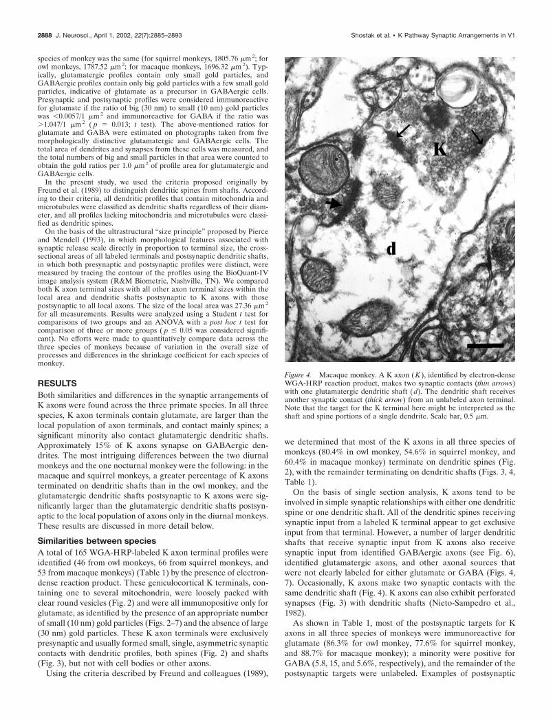
On the basis of single section analysis, K axons tend to beinvolved in simple synaptic relationships with either one dendriticspine or one dendritic shaft. All of the dendritic spines receivingsynaptic input from a labeled K terminal appear to get exclusiveinput from that terminal. However, a number of larger dendriticshafts that receive synaptic input from K axons also receivesynaptic input from identified GABAergic axons (see Fig. 6),identified glutamatergic axons, and other axonal sources thatwere not clearly labeled for either glutamate or GABA (Figs. 4,7). Occasionally, K axons make two synaptic contacts with thesame dendritic shaft (Fig. 4). K axons can also exhibit perforatedsynapses (Fig. 3) with dendritic shafts (Nieto-Sampedro et al.,1982).
As shown in Table 1, most of the postsynaptic targets for Kaxons in all three species of monkeys were immunoreactive forglutamate (86.3% for owl monkey, 77.6% for squirrel monkey,and 88.7% for macaque monkey); a minority were positive forGABA (5.8, 15, and 5.6%, respectively), and the remainder of thepostsynaptic targets were unlabeled. Examples of postsynaptic
Figure 4. Macaque monkey. A K axon (K ), identified by electron-denseWGA-HRP reaction product, makes two synaptic contacts (thin arrows)with one glutamatergic dendritic shaft (d). The dendritic shaft receivesanother synaptic contact (thick arrow) from an unlabeled axon terminal.Note that the target for the K terminal here might be interpreted as theshaft and spine portions of a single dendrite. Scale bar, 0.5 �m.
2888 J. Neurosci., April 1, 2002, 22(7):2885–2893 Shostak et al. • K Pathway Synaptic Arrangements in V1

labeling for glutamate and GABA are shown in Figures 2-6 and7, respectively.
We made a number of additional measurements in an effort toprovide more detailed information about similarities and differ-ences between the synaptic arrangements made by K LGN axonsand other local axons (e.g., axons from layer IV carrying indirectsignals from the LGN M and P pathways). First, we evaluated thecross-sectional area of all labeled K axon terminals and postsyn-aptic dendritic shafts in which both presynaptic and postsynapticprofiles were distinct. K axon terminal and target sizes werecompared with the sizes of all other local glutamatergic axonalprofiles and their dendritic shaft targets. Only dendritic shaftsthat could be clearly identified as containing glutamate or GABAas a transmitter were included in the measurements. Table 2
shows that for all three primate species, K axon terminals weresignificantly larger in area (on average twice the size) whencompared with the remaining local population of glutamatergicaxon terminals. The size distribution of K axon terminals incomparison to the local population of glutamate containing axonterminals is shown in Figure 8.
Second, we compared the size of K axon terminals that madesynapses with spines with those that made synapses with dendriticshafts containing either glutamate or GABA. Comparisons weremade separately for each of the three primate species. No differ-ences were found using an ANOVA ( p � 0.98 for owl monkeys;p � 0.88 for squirrel monkeys; p � 0.97 for macaque monkeys).
A similar comparison was made between the sizes of GABAer-gic dendritic shafts receiving synapses from K axons and those
Figure 5. Squirrel monkey. A K axon ( K ) makes three synaptic contacts with spines (sp); one of them is a perforated synapse. In this case, two spineswere identified as glutamatergic on the basis of the presence of small gold particles, and one was unlabeled (asterisk). This K axon likely makes anothersynaptic contact with the GABAergic (note large gold particles) dendritic shaft (GABA d), but the synapse is not visible in this plane of section. Largergold particles (30 nm) showing immunoreactivity for GABA are seen as larger black dots. Scale bar, 0.5 �m.
Shostak et al. • K Pathway Synaptic Arrangements in V1 J. Neurosci., April 1, 2002, 22(7):2885–2893 2889

receiving input from other local glutamatergic axons. We foundthat in all three primate species, K axons tend to make contactwith larger postsynaptic GABAergic dendritic shafts than doother local axons (1.63 � 0.09 �m2, n � 3 vs 0.82 � 0.07 �m2, n �69 for owl monkeys; 2.24 � 0.63 �m2, n � 9 vs 1.65 � 0.19 �m2,n � 12 for squirrel monkeys; and 1.44 � 0.58 �m2, n � 3 vs 0.74 �0.08 �m2, n � 53 for macaque monkeys for all local GABAergicdendritic shafts, respectively). Larger samples will be required todetermine whether these differences are significant.
Differences between speciesTwo measures appeared to distinguish the diurnal monkeys (ma-caque and squirrel monkeys) from the nocturnal monkey (owlmonkey). In the diurnal species a much larger percentage (38–40%) of the total population of K axons terminated on glutama-tergic dendritic shafts (as opposed to spines) than in the nocturnalowl monkey (25%). In addition, as shown in Table 3, when wecompared the sizes of postsynaptic glutamatergic dendritic shaftscontacted by K axons with the sizes of all glutamatergic dendriticshafts in the local area, we found that K axons in both diurnalprimates terminate on relatively larger glutamatergic postsynap-tic dendritic shafts than do K axons in the nocturnal owl monkeys.
In addition to differences that appeared to correlate with visuallifestyle (diurnal versus nocturnal), we found one species differ-ence that correlated with evolutionary distance. In the NewWorld simians, a small percentage of K axons makes multiplesynaptic contacts. In owl monkeys 15.2% of K axons (7 of 46) andin squirrel monkeys 9.1% of K axons (6 of 66) make contacts withseveral postsynaptic targets: with one spine and one shaft (Fig. 7)or with more than one spine (Fig. 5). In contrast, none of the
53 K axons in macaque monkeys made multiple contacts withpostsynaptic targets. These percentages, however, are likely to bean underestimate because serial reconstructions through the en-tire bouton were not performed.
A final species difference that did not correlate with either lifestyle or evolutionary similarity was the finding that �50% of Kaxons in squirrel monkeys make perforated synaptic contacts withtheir targets versus �5–6% in owl and macaque monkeys.
Figure 6. Squirrel monkey. A glutamatergic dendrite (d) with spine (sp)receives two synaptic contacts, one from a K axon (thick arrow) andanother from a GABAergic terminal (GABA term; thin arrow). Blackamorphous material indicates WGA-HRP electron-dense reaction prod-uct in the K axon ( K ). Scale bar, 0.5 �m.
Figure 7. Squirrel monkey. Some K axons are involved in complexsynaptic arrangements. This K axon (K ) synapses (indicated by a thickarrows) on both an inhibitory GABAergic dendritic shaft (GABA d) anda spine (sp). Larger gold particles (30 nm) showing immunoreactivity forGABA are seen as larger black dots. Note that the same GABAergicdendritic shaft receives additional input from two other glutamatergicaxon terminals (thin arrows). Black amorphous material indicates WGA-HRP electron-dense reaction product in the K axon. Scale bar, 0.5 �m.
Table 2. Sizes of axon terminals
Species K terminals All glutamatergic terminals
Owl monkey 1.39 � 0.12* (n � 46) 0.70 � 0.04 (n � 137)Squirrel monkey 1.79 � 0.16* (n � 66) 0.95 � 0.10 (n � 54)Macaque monkey 1.03 � 0.09* (n � 53) 0.53 � 0.03 (n � 94)
All data are expressed in micrometers squared as means � SEM; *indicates that Kaxon terminals are significantly different from the remaining population of glutama-tergic terminals (t test; p � 0.05).
2890 J. Neurosci., April 1, 2002, 22(7):2885–2893 Shostak et al. • K Pathway Synaptic Arrangements in V1

DISCUSSIONCommon features of K LGN circuits across primatesAlthough CO blobs are recognized as a ubiquitous feature ofprimate V1, the role of the CO blobs in vision remains contro-versial. At one extreme are arguments that regions exhibitingdense CO staining are simply zones of thalamic input where themetabolism in cortex is slightly higher; CO blobs are the targetsfor K axons, just as layer IV (another CO-dense layer) is thetarget of M and P axons. At the other extreme is the proposal thatCO blobs are artifacts of activity-dependent rules of development(Purves and LaMantia, 1990). The most popular proposal stillremains that CO blobs reflect functional specialization for colorin diurnal primates (Livingstone and Hubel, 1984; Ts’o andGilbert, 1988). Support for a role in color also comes frompublished data on K LGN cells in marmosets (Martin et al., 1997;White et al., 1998) and preliminary data in macaque monkeys(Reid et al., 1997) suggesting that a percentage of K cells (20% inmarmosets) responds to signals from S cones (blue ON); however,no LGN K cells have been identified that respond to red/green(i.e., L and M cones). Therefore, if CO blobs are part of a colorchannel, they must combine chromatic signals from P and K cells.If this is true, then we still need to understand what CO blobs aredoing in nocturnal primates, such as owl monkeys, that do notexpress the S cone pigment gene (Jacobs et al., 1993) and are mostlikely color blind (Jacobs et al., 1996). Interestingly, in many
respects (morphology and connectivity), the S cone (blue) systemmore closely resembles the rod system than the L and M conechannels in vision (Kolb et al., 1997). In fact, some evidencesuggests that rods participate in the “blue” mechanism (Trezona,1970) and signals from rods and S cones can interact (Naarendorpet al., 1996) and share the same pathway. Virsu and colleagues(1987) reported such sharing of signals from rods and S cones inmacaque LGN, although this result remains controversial (Lee etal., 1997). If true, it could suggest a mechanism to explain why theK pathway is preserved in nocturnal primates. Perhaps the por-tion of the K pathway that is dominated by S cones in diurnalmonkeys becomes part of a second rod-dominated pathway innocturnal primates. This is in agreement with an older proposalthat cells in the CO blobs are concerned, in general, with contrast(color or brightness) as opposed to cells in the interblobs that aremore concerned with geometrical variables (Allman and Zucker,1990; DeBruyn et al., 1993). This idea would also agree with ourfinding that the K pathway circuitry is quite similar across diurnaland nocturnal primates. Thus, under nocturnal conditions the KCO blob pathway may simply switch in nocturnal primates to arod pathway.
In fact, the similarities in the K pathway far outweigh thedifferences, suggesting that there has not been a major change inthe K pathway circuitry within the CO blobs that correlates withthe shift from color vision to lack of color vision and that thecommon structural elements of the K pathway across primatesreflect some function that they share or that all LGN inputpathways share. The similarities that we see between K axonpopulations include the demonstration that K axons are (1) glu-tamatergic, (2) make asymmetric (Gray’s type I) synapses mainlywith spines, and (3) engage in simple synaptic arrangements withglutamatergic postsynaptic targets typically involving single syn-aptic contacts. The tendency for K geniculocortical axons tocontact predominantly spines of excitatory cells extends also toboth cat X and Y geniculocortical boutons in area 17 and cat Yaxons in area 18, and M and P axons in layer IV of primates(Garey and Powell, 1971; Tigges and Tigges, 1979; Winfield et al.,1982; Winfield and Powell, 1983; Freund et al., 1989; Ding andCasagrande, 1998), as well as to rat geniculocortical axons in area17 (Garey and Powell, 1971; LeVay and Gilbert, 1976; Winfieldand Powell, 1976, 1983; Peters and Feldman, 1977; Freund et al.,1985, Kharazia and Weinberg, 1994), suggesting that these aregeneral features of all geniculocortical axons in mammals.
In addition to spines and dendritic shafts containing glutamate,a minority (5–15%) of K axons in all three species makes synapseswith the dendritic shafts of inhibitory (GABAergic) interneurons.This percentage of contacts with inhibitory interneurons is sim-ilar to that of the local population of axons, suggesting that Kaxons do not relate in unique ways to the GABAergic circuitry,with the exception that K axons tend to contact larger GABAer-gic dendrites than the local population of axons (see also below).Approximately 15–20% of all cortical neurons are GABAergic(Fitzpatrick et al., 1987; Hendry et al., 1987), and the proportionof GABAergic dendrites reported in the CO blob neuropil ofmacaque monkeys is similar (Beaulieu et al., 1992; Nie andWong-Riley 1995).
Comparison among K, M, and P LGN synaptic circuitsThe CO blobs in layer IIIB receive numerous intrinsic and agreater variety of extrinsic connections than have been reportedin layer IV. In addition to axons from cells in both IV� and IV�layers (the M and P recipient sublayers), CO blobs receive input
Table 3. Sizes of postsynaptic glutamatergic dendritic shafts
Species
Postsynaptic to Kaxons–glutamatergicdendritic shafts
All other glutamatergicdendritic shafts
Owl monkey 0.75 � 0.08 (n � 11) 0.73 � 0.04 (n � 189)Squirrel monkey 1.73 � 0.32* (n � 21) 0.87 � 0.08 (n � 65)Macaque monkey 1.21 � 0.18* (n � 18) 0.51 � 0.03 (n � 276)
All data are expressed in micrometers squared as means � SEM; *indicates that themeans are significantly different from the remaining population of glutamatergicdendritic shafts (t test; p � 0.05).
Figure 8. Squirrel monkey. K axon terminals (black bars) are on averagelarger in size than local axon terminals (white bars) in the local area of COblobs. The arrows and the numbers above them show, respectively, themean size of K axon terminals (1.79 � 0.16 �m 2) and the mean size of alllocal glutamatergic axon terminals (0.95 � 0.10 �m 2). See Results fordetails.
Shostak et al. • K Pathway Synaptic Arrangements in V1 J. Neurosci., April 1, 2002, 22(7):2885–2893 2891

from cells in all cortical layers except layer I, as well as input fromlocal cells (Lund and Yoshioka, 1991; Peters and Sethares, 1991;Casagrande et al., 1992; Lachica et al., 1992, 1993; Boyd et al.,2000). Extrinsic connections of the CO blobs include not only theLGN K pathway, but also input from a number of extrastriatevisual areas (e.g., feedback from V2), as well as input from thepulvinar (Kaas and Huerta, 1988; Desimone et al., 1990). Withinthe CO blobs there are no spiny stellate cells (Lund, 1984).Therefore, in the CO blobs, all dendritic spines arise from eitherlocal pyramidal cells or pyramidal cells from other layers, espe-cially layer V (Peters and Sethares, 1991). These relationshipsindicate that K axons could have direct access to output neurons,unlike the axons of M and P cells, which terminate exclusively onspiny stellate cells in layer IV and avoid pyramidal dendrites(Lund, 1984). Our quantitative comparison reveals that K axonterminals are larger in area than local glutamatergic axon termi-nals. This suggests that K axons have a greater impact on V1output neurons, unless the terminals arriving from layer IV(reflecting the indirect inputs from the P and M pathways) areskewed in size away from the local mean. Although the propor-tion of GABAergic target elements contacted by the K pathway(5–15%) is similar to that described in the literature for M and Pcells (5–10%) (Freund et al., 1989), it is intriguing that K axons inall three monkey species contact significantly larger postsynapticGABAergic dendritic shafts than do local axons, suggesting thatK axons terminate closer to the cell bodies of these inhibitorycells than axons carrying M and P signals from layer IV.
Taken together, these comparisons among the synaptic rela-tionships of K, M, and P axons suggest that all three parallel LGNstreams use similar strategies at the first stage of informationtransfer to cortical circuits, but that the K pathway has a strongerthan suspected impact on messages outgoing from V1 given thelocation and size of its axons. Additionally, the K pathway ap-pears to be in a position to more efficiently activate inhibitoryneurons. This could allow signals arriving from K axons to bemore rapidly shut down in comparison to signals arriving via Mand P axons, at least according to models that propose feedfor-ward inhibition of incoming LGN afferent signals (Somogyi, 1989;Berman et al., 1991, 1992; Douglas and Martin, 1991).
Differences in K LGN circuits across primatesDespite the many across-species similarities in K pathway orga-nization, there are intriguing differences. The most interesting ofthese differences are those that could correlate with the presenceor absence of color vision. Comparison of the diurnal monkeys(macaque and squirrel monkeys) that have color vision with anocturnal owl monkey that lacks color vision showed that agreater proportion of K axons in the diurnal monkeys synapsewith dendritic shafts (as opposed to spines) as well as with largerglutamatergic dendritic shafts. This shift could reflect a loss ofone population of K axons in owl monkeys, a population convey-ing information from the missing S cone pathway (Jacobs et al.,1996). This change also fits with data in marmosets showing thatonly a small proportion of K axons (20%) actually carry colorsignals (i.e., S cone signals); the remainder do something else(Martin et al., 1997; White et al., 1998). Such a scenario wouldalso fit the proposed evolutionary history of the owl monkey,suggesting that the ancestors of owl monkeys had S cones (andlikely color vision) and that they only recently lost S cones undersome pressure to become nocturnal (Martin, 1990). Anotherconclusion that could be drawn from these data is that the Kpathway input to CO blob glutamatergic dendrites in diurnal
primates occurs at sites that can exert relatively greater domi-nance compared with other afferents, and that this distinctivefeature of the K pathway has been minimized in the nocturnal owlmonkey, perhaps as a consequence to the loss of color vision. Inthat case the prediction would be that other diurnal primates withS cones (e.g., marmosets) should show the pattern that we havefound in the macaque and squirrel monkeys, whereas other noc-turnal primates that lack S cones (e.g., bush babies) should showthe pattern seen in the owl monkey. Nevertheless, these diurnal /nocturnal differences account for only a small percentage of the Kaxons in these three species (�20%). These findings support theconclusion that most of the signals that the K pathway conveys tothe CO blobs are concerned with something other than color.
REFERENCESAllman J, Zucker S (1990) Cytochrome oxidase and functional coding
in primate striate cortex: a hypothesis. Cold Spring Harb Symp QuantBiol 55:979–982.
Beaulieu C, Kisvarday Z, Somogyi P, Cynader M, Cowey A (1992)Quantitative distribution of GABA-immunopositive and -immuno-negative neurons and synapses in the monkey striate cortex (area 17).Cereb Cortex 2:295–309.
Berman NJ, Douglas RJ, Martin KA, Whitteridge D (1991) Mechanismsof inhibition in cat visual cortex. J Physiol (Lond) 440:697–722.
Berman NJ, Douglas RJ, Martin KA (1992) GABA-mediated inhibitionin the neural networks of visual cortex. Prog Brain Res 90:443–476.
Boyd JD, Mavity-Hudson JA, Casagrande VA (2000) The connectionsof layer 4 subdivisions in the primary visual cortex (V1) of the owlmonkey. Cereb Cortex 10:644–662.
Calkins DJ, Sterling P (1999) Evidence that circuits for spatial and colorvision segregate at the first retinal synapse. Neuron 24:313–321.
Casagrande VA (1994) A third parallel visual pathway to primate areaV1. Trends Neurosci 17:305–310.
Casagrande VA (1999) The mystery of the visual system K pathway.J Physiol (Lond) 517:630.
Casagrande VA, Kaas JH (1994) The afferent, intrinsic, and efferentconnections of primary visual cortex in primates. In: Cerebral cortex,Vol. 10, primary visual cortex of primates (Peters A, Rockland K, eds),pp 201–259. New York: Plenum.
Casagrande VA, Mavity-Hudson JA, Taylor JG (1992) Intrinsic connec-tions of owl monkey striate cortex: difference between cytochromeoxidase (CO) blobs and interblobs. Soc Neurosci Abstr 18:389.
Casagrande VA, Ding Y, Boyd JD (1997) The morphology of LGNaxons from different K layers in V1 of macaque monkey. Soc NeurosciAbstr 23:2361.
Condo GJ, Casagrande VA (1990) Organization of cytochrome oxidasestaining in the visual cortex of nocturnal primates (Galago crassicau-datus and Galago senegalensis): I. Adult patterns. J Comp Neurol293:632–645.
DeBruyn EJ, Casagrande VA, Beck PD, Bonds AB (1993) Visual reso-lution and sensitivity of single cells in the primary visual cortex (V1) ofa nocturnal primate (bush baby): correlations with cortical layers andcytochrome oxidase patterns. J Neurophysiol 69:3–18.
Desimone R, Wessinger M, Thomas L, Schneider W (1990) Attentionalcontrol of visual perception: cortical and subcortical mechanisms. ColdSpring Harb Symp Quant Biol 55:963–971.
Ding Y, Casagrande VA (1995) The morphology of LGN axons thatterminate in the CO blobs and primate VI. Soc Neurosci Abstr 21:394.
Ding Y, Casagrande VA (1997) The distribution and morphology ofLGN K pathway axons within the layers and CO blobs of owl monkeyV1. Vis Neurosci 14:691–704.
Ding Y, Casagrande VA (1998) Synaptic and neurochemical character-ization of parallel pathways to the cytochrome oxidase blobs of primatevisual cortex. J Comp Neurol 391:429–443.
Douglas RJ, Martin KA (1991) A functional microcircuit for cat visualcortex. J Physiol (Lond) 440:735–769.
Fitzpatrick D, Itoh K, Diamond IT (1983) The laminar organization ofthe lateral geniculate body and the striate cortex in the squirrel monkey(Saimiri sciureus). J Neurosci 3:673–702.
Fitzpatrick D, Lund JS, Schmechel DE, Towles AC (1987) Distributionof GABAergic neurons and axon terminals in the macaque striatecortex. J Comp Neurol 264:73–91.
Freund TF, Martin KA, Somogyi P, Whitteridge D (1985) Innervationof cat visual areas 17 and 18 by physiologically identified X- and Y-typethalamic afferents. II. Identification of postsynaptic targets by GABAimmunocytochemistry and Golgi impregnation. J Comp Neurol242:275–291.
Freund TF, Martin KAC, Soltesz I, Somogyi P, Whitteridge D (1989)Arborization pattern and postsynaptic targets of physiologically iden-
2892 J. Neurosci., April 1, 2002, 22(7):2885–2893 Shostak et al. • K Pathway Synaptic Arrangements in V1

tified thalamocortical afferents in striate cortex of the macaque monkey.J Comp Neurol 289:315–336.
Garey LJ, Powell TPS (1971) An experimental study of the terminationof the lateral geniculo-cortical pathway in the cat and monkey. Proc RSoc Lond B Biol Sci 179:41–63.
Hendry SH, Reid C (2000) The koniocellular pathway in primate vision.Annu Rev Neurosci 23:127–153.
Hendry SH, Schwark HD, Jones EG, Yan J (1987) Numbers and pro-portions of GABA-immunoreactive neurons in different areas of mon-key cerebral cortex. J Neurosci 7:1503–1519.
Horn AKE, Hoffman KP (1987) Combined GABA-immunocyto-chemistry and TMB-HRP histochemistry of pretectal nuclei projectingto the inferior olive in rats, cats and monkeys. Brain Res 409:133–138.
Jacobs GH, Deegan JF, Neitz J, Crognale MA, Neitz M (1993) Pho-topigments and color vision in the nocturnal monkey, Aotus. VisionRes 33:773–783.
Jacobs GH, Neitz M, Neitz J (1996) Mutations in S-cone pigment genesand the absence of colour vision in two species of nocturnal primate.Proc R Soc Lond B Biol Sci 263:705–710.
Kaas JH, Huerta MF (1988) Subcortical visual system of primates. In:Comparative primate biology, Vol 4 (Steklis HP, ed), pp 327–391. NewYork: Wiley.
Kharazia VN, Weinberg RJ (1994) Glutamate in thalamic fibers termi-nating in layer IV of primary sensory cortex. J Neurosci 14:6021–6032.
Kolb H, Goede P, Roberts S, McDermott E, Gouras P (1997) Unique-ness of the S-cone pedicle in the human retina and consequences forcolor processing. J Comp Neurol 386:443–460.
Lachica EA, Casagrande VA (1992) Direct W-like geniculate projec-tions to the cytochrome oxidase (CO) blobs in primate visual cortex:axon morphology. J Comp Neurol 319:141–158.
Lachica EA, Beck PD, Casagrande VA (1992) Parallel pathways in ma-caque monkey striate cortex: anatomically defined columns in layer III.Proc Natl Acad Sci USA 89:3566–3570.
Lachica EA, Beck PD, Casagrande VA (1993) Intrinsic connections oflayer III of striate cortex in squirrel monkey and bush baby: correlationswith patterns of cytochrome oxidase. J Comp Neurol 329:163–187.
Lee BB, Smith VC, Pokorny J, Kremers J (1997) Rod inputs to macaqueganglion cells. Vision Res 37:2813–2828.
Lennie P, Krauskopf J, Sclar G (1990) Chromatic mechanisms in striatecortex of macaque. J Neurosci 10:649–669.
LeVay S, Gilbert CD (1976) Laminar patterns of geniculocortical pro-jection in the cat. Brain Res 113:1–19.
Livingstone MS, Hubel DH (1982) Thalamic inputs to cytochrome oxi-dase rich regions in monkey visual cortex. Proc Natl Acad Sci USA79:6098–6101.
Livingstone MS, Hubel DH (1984) Anatomy and physiology of a colorsystem in the primate visual cortex. J Neurosci 4:309–356.
Lund JS (1984) Spiny stellate cells. In: Cerebral cortex, Vol 1, cellularcomponents of the cerebral cortex (Peter A, Jones EG, eds), pp 255–308. New York: Plenum.
Lund JS, Yoshioka T (1991) Local circuit neurons of macaque monkeystriate cortex: III. Neurons of laminae 4B, 4A and 3B. J Comp Neurol311:234–258.
Martin PR, White AJR, Goodchild AK, Wilder HD, Sefton AE (1997)Evidence that blue-on cells are part of the third geniculocortical path-way in primates. Eur J Neurosci 9:1536–1541.
Martin RD (1990) Primate origins and evolution. Princeton: PrincetonUP.
Mesulam MM (1978) Tetramethyl benzidine for horseradish peroxidaseneurohistochemistry: a non-carcinogenic blue reaction product withsuperior sensitivity for visualizing neural afferents and efferents. J His-tochem Cytochem 26:106–117.
Naarendorp F, Rice KS, Sieving PA (1996) Summation of rod and Scone signal at threshold in human observers. Vision Res 36:2681–2688.
Nie F, Wong-Riley MT (1995) Double labeling of GABA and cyto-chrome oxidase in the macaque visual cortex: quantitative EM analysis.J Comp Neurol 356:115–131.
Nieto-Sampedro M, Hoff SF, Cotman CW (1982) Perforated postsynap-tic densities: probable intermediates in synapse turnover. Proc NatlAcad Sci USA 79:5718–5722.
Peters A, Feldman ML (1977) The projection of the lateral geniculatenucleus to area 17 of the rat cerebral cortex. IV. Terminations on spinydendrites. J Neurocytol 6:669–689.
Peters A, Sethares C (1991) Organization of pyramidal neurons in area17 of monkey visual cortex. J Comp Neurol 306:1–23.
Phend KD, Weinberg RJ, Rustioni A (1992) Techniques to optimizepost-embedding single and double staining for amino acid neurotrans-mitters. J Histochem Cytochem 40:1011–1020.
Pierce JP, Mendell LM (1993) Quantitative ultrastructure of la boutonsin the ventral horn: scaling and positional relationships. J Neurosci13:4748–4763.
Purves D, LaMantia AS (1990) Numbers of “blobs” in the primaryvisual cortex of neonatal and adult monkeys. Proc Natl Acad Sci USA87:5764–5767.
Reid RC, Alonso JM, Hendry SHC (1997) S-cone input is relayed tovisual cortex from two koniocellular layers of macaque LGN. SocNeurosci Abstr 23:13.
Reynolds ES (1963) The use of lead citrate at high pH as an electronopaque stain in electron microscopy. J Cell Biol 17:208–212.
Somogyi P (1989) Synaptic organization of GABAergic neurons andGABAA receptors in the lateral geniculate nucleus and visual cortex.In: Neural mechanisms of visual perception (Lam DKT, Gilbert CD,eds), pp 35–62. Houston: Portfolio Publishing.
Thompson AL, Bogen JE, Marsh JF (1979) Cultural hemisphericity:evidence from cognitive tests. Int J Neurosci 9:37–43.
Tigges M, Tigges J (1979) Types of degenerating geniculocortical axonterminals and their contribution to layer IV of area 17 in the squirrelmonkey (Saimiri). Cell Tissue Res 196:471–486.
Trezona PW (1970) Rod participation in the blue mechanism and itseffect on colour matching. Vision Res 10:317–332.
Ts’o DY, Gilbet CD (1988) The organization of chromatic and spatialinteractions in the primate striate cortex. J Neurosci 8:1712–1727.
Virsu V, Lee BB, Creutzfeldt OD (1987) Mesopic spectral responsesand the Purkinje shift of macaque lateral geniculate nucleus cells.Vision Res 27:191–200.
White AJ, Wilder HD, Goodchild AK, Sefton AJ, Martin PR (1998)Segregation of receptive field properties in the lateral geniculate nu-cleus of a New-World monkey, the marmoset Callithrix jacchus. J Neu-rophysiol 80:2063–2076.
Wikler KC, Rakic P (1990) Distribution of photoreceptor subtypes inthe retina of diurnal and nocturnal primates. J Neurosci 10:3390–3401.
Winfield DA, Powell TP (1976) The termination of thalamo-corticalfibres in the visual cortex of the cat. J Neurocytol 5:269–281.
Winfield DA, Powell TP (1983) Laminar cell counts and geniculo-cortical boutons in area 17 of cat and monkey. Brain Res 277:223–229.
Winfield DA, Rivera-Dominguez M, Powell TP (1982) The terminationof geniculocortical fibers in area 17 of the visual cortex in the macaquemonkey. Brain Res 231:19–32.
Xu X, Ichida JM, Allison JD, Boyd JD, Bonds AB, Casagrande VA(2001) A comparison of koniocellular (K), magnocellular (M), andparvocellular (P) receptive field properties in the lateral geniculatenucleus (LGN) of the owl monkey (Aotus trivirgatus). J Physiol (Lond)531:203–218.
Shostak et al. • K Pathway Synaptic Arrangements in V1 J. Neurosci., April 1, 2002, 22(7):2885–2893 2893