cpd and contracted pelvis
DESCRIPTION
cpd and contracted pelvisTRANSCRIPT

Class Teaching
Ms. Shalini JoshiM.Sc nsgS.C.O.N. Dehradun


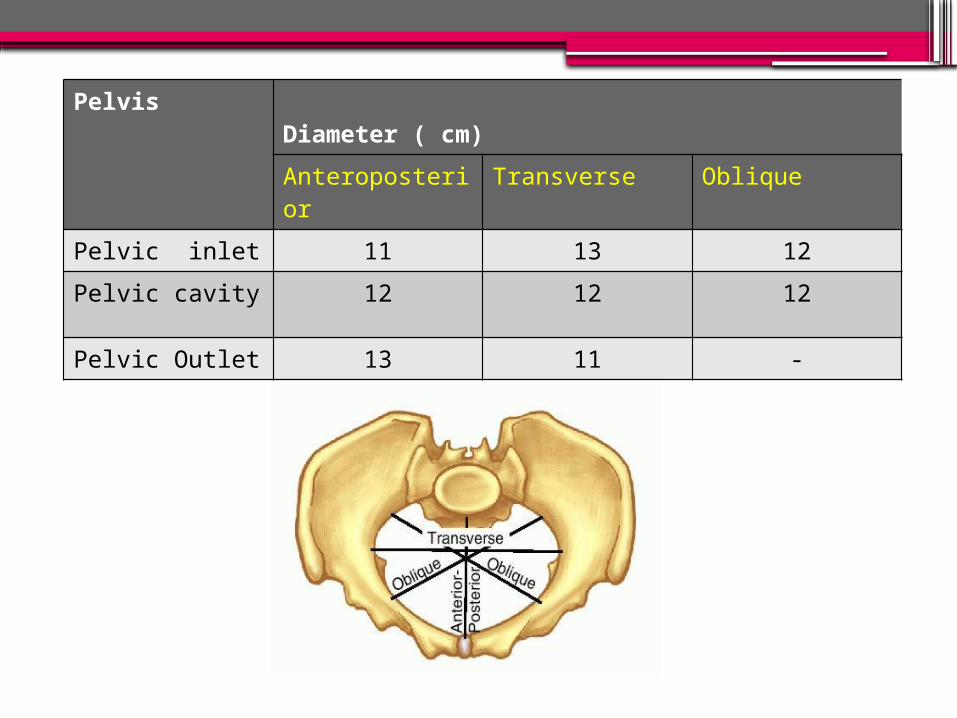
Pelvis Diameter ( cm)Anteroposterior
Transverse Oblique
Pelvic inlet 11 13 12Pelvic cavity 12 12 12
Pelvic Outlet 13 11 -

CEPHALO PELVIC DISPROPORTION
and CONTRACTED PELVIS

•To define CPD and contracted pelvis.•To describe the causes and degree of CPD
and contracted pelvis•To discuss the classification of contracted
pelvis.•To explain the diagnosis of CPD and
contracted pelvis•To enumerate the effects of contracted
pelvis.•To describe the management of the CPD
and contracted pelvis.•To enlist the complication of the CPD.
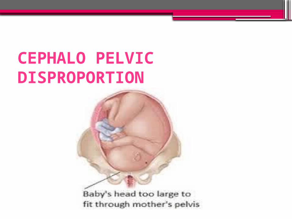
CEPHALO PELVIC DISPROPORTION

DEFINITION •Cephalo pelvic disproportion is the disparity in relation between the head of baby and the
mother’s pelvis.
• It is a pelvis in which one or more of its diameter is reduced below the normal by one
or more centimeter
DEGREE OF DISPROPORTION
• It is based on clinical findings and pelvimetry:-
a) Severe disproportion:- when the obstetric conjugate is less than 7.5 cm (3”) then it is said to be severe disproportion.
b) Borderline disproportion:- when the obstetric conjugate is between 9.5 and 10 cm. In inlet the anterior posterior diameter is less than 10 cm and transverse diameter is less than 12 cm.

INCIDENCE According to American College of Nursing
Midwives, occur 20 out of 250 pregnancy.
“It has been seen through studies that 65% of women who have been diagnosed
with CPD in previous pregnancies, deliver vaginally in subsequent pregnancies.”

CAUSES •Nutritional deficiency
•Disease / injury to pelvic bones
•Developmental defects
•A large size baby
•Abnormal fetal position
•Problem with genital tract

CLASSIFICATION OF CAUSESAbsolute causes:- it is a true mechanical
obstruction due to:- Permanent maternal cause such as
contracted pelvis, anterior sacrococcygeal tumor.
Temporary fetal causes such as hydrocephalus, large baby etc.
Relative cause:- the relative cause includes brow presentation, face presentation, mento posterior, occipito posterior position, deflexed head in vertex presentation

CONTRACTED PELVIS

DEFINITION•Anatomical - It is a pelvis in which one or
more of its diameters is reduced below the normal by one or more centimeters.
•Obstetric - It is a pelvis in which one or more of its diameters is reduced so that it interferes with the normal mechanism of labor.

ETIOLOGY• Common causes of contracted pelvis are:-Nutritional and environmental defects:- minor variation;- common major :- rachitic and osteomalacic –rare
Disease or injury affecting the bone of the pelvis:- fracture ,tumors, tubercular artheritis.
spine:- kyphosis, scoliosis, coccygeal deformity lower limbs:- poliomyeitis, hip joint disease
Developmental defects:- naegele’s pelvis, robert’s pelvis

CLASSIFICATIONClassified by:-•A) type of distortion of pelvic architecture•B) degree of contraction

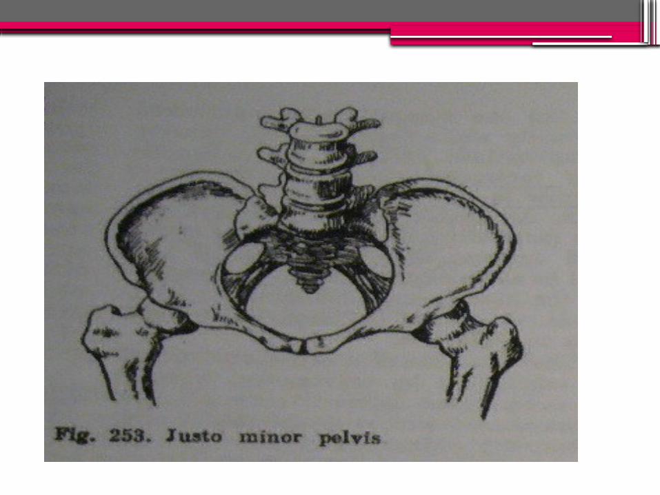
A) Classification by Pelvic Architecture1. Pelvis aequabiliter justo minor•Characterized by general reduction of all
diameters; equally shortened usually by 1-2cm
•Occurs in short. Also occurs in women with massive skeletal bones and developed muscles, the pelvis has masculine features such as narrow sacrum, narrow pubic outlet {funnel-shaped)

2. Flat Pelvis•reduced anteroposterior diameters with
normal transverse and oblique diameters•Has 2 types of contracture
a) Simple flat (or platypellic) pelvis Entire sacral platform is dislocated
toward the symphysis hence all the anteroposterior diameters of all pelvic planes are reduced

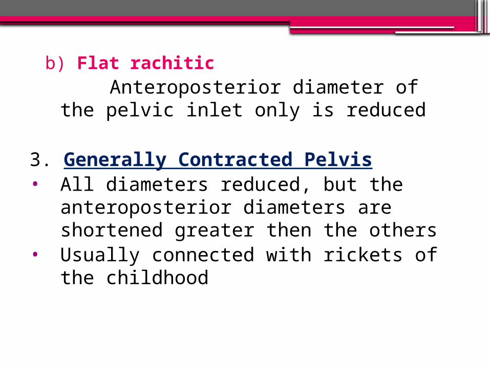
b) Flat rachitic Anteroposterior diameter of the pelvic
inlet only is reduced
3. Generally Contracted Pelvis• All diameters reduced, but the
anteroposterior diameters are shortened greater then the others
• Usually connected with rickets of the childhood


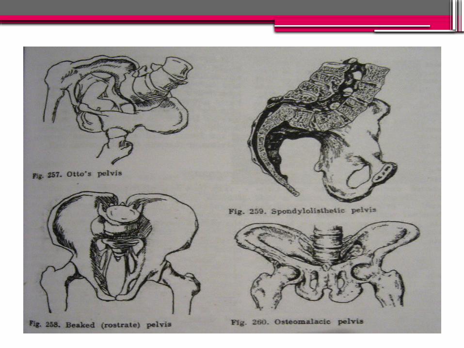
Rare forms of contracted pelvis•Otto’s pelvis – develop as result of
inflammatory process in the hip or knee•Beaked (rostrate) pelvis – under
development of both sacral wings•Spondylolithetic pelvis – formed due to
partial dislocation of last lumbar vertebra in front of 1st sacral vertebra
•Osteomalacic pelvis•Scoliotic pelvis – only the lumber region
cause deformity of the pelvis. The acetabulum is pushed inwards on the weight bearing side.

B) Classification by degree of contracture 4 degreesi. First degree: true conjugate <11cm but not
<9cm, spontaneous delivery is possibleii. Second degree: true conjugate = 9-7.5cm
spontaneous delivery possible but complications may arise
iii.Third degree: true conjugate 7.5-6cm spontaneous delivery impossible, use C-section
iv. Fourth degree: true conjugate <6cm, impossible delivery, only way is C-section ; also known as absolutely contracted pelvis

DiagnosisA. History
•Rickets: is expected if there is a history of delayed walking and dentition.
•Trauma or diseases: of the pelvis, spines or lower limbs.
•Infantilism
•Previous tuberculosis of bones and joints

•Bad obstetric history: e.g. prolonged labour ended by;▫difficult forceps,▫caesarean section or▫still birth.Weight of the baby,Evidence of maternal injuries such as
complete perineal tear, vesico vaginal istula, recto vaginal fistula

B. General examination Abnormal gait :- Assess woman for stockily built with
bull neck. Broad shoulder and short thigh Obese and male distribution of hair
Stature :women < 150 cm or 5 feet

C. Abdomen examinationPendulous abdomen in primigravida
fetal head fails to enter a contracted pelvis at the end of pregnancy and floats high above inlet, failed growth of uterus deviates upward and anteriorly
Non engagement in last 3-4 wks in primigravida
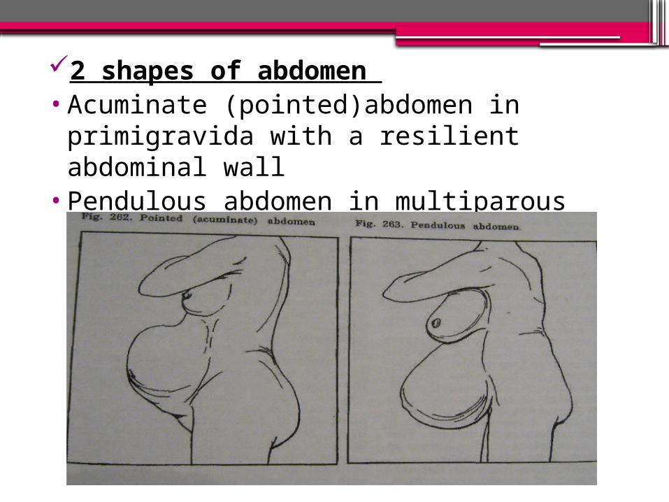
2 shapes of abdomen •Acuminate (pointed)abdomen in
primigravida with a resilient abdominal wall
•Pendulous abdomen in multiparous women

ABDOMINAL METHOD IN CPD
•Patient is placed in dorsal position with thigh flexes and separated.
•The head is grasped by the left hand.
•2 fingers (index and middle) of theright hand are placed above the symphysis pubis to note the degree of overlapping. If when the head is pushed downward and backward.

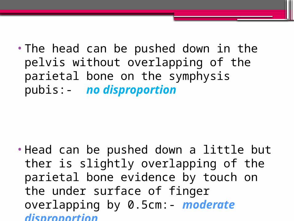
•The head can be pushed down in the pelvis without overlapping of the parietal bone on the symphysis pubis:- no disproportion
•Head can be pushed down a little but ther is slightly overlapping of the parietal bone evidence by touch on the under surface of finger overlapping by 0.5cm:- moderate disproportion
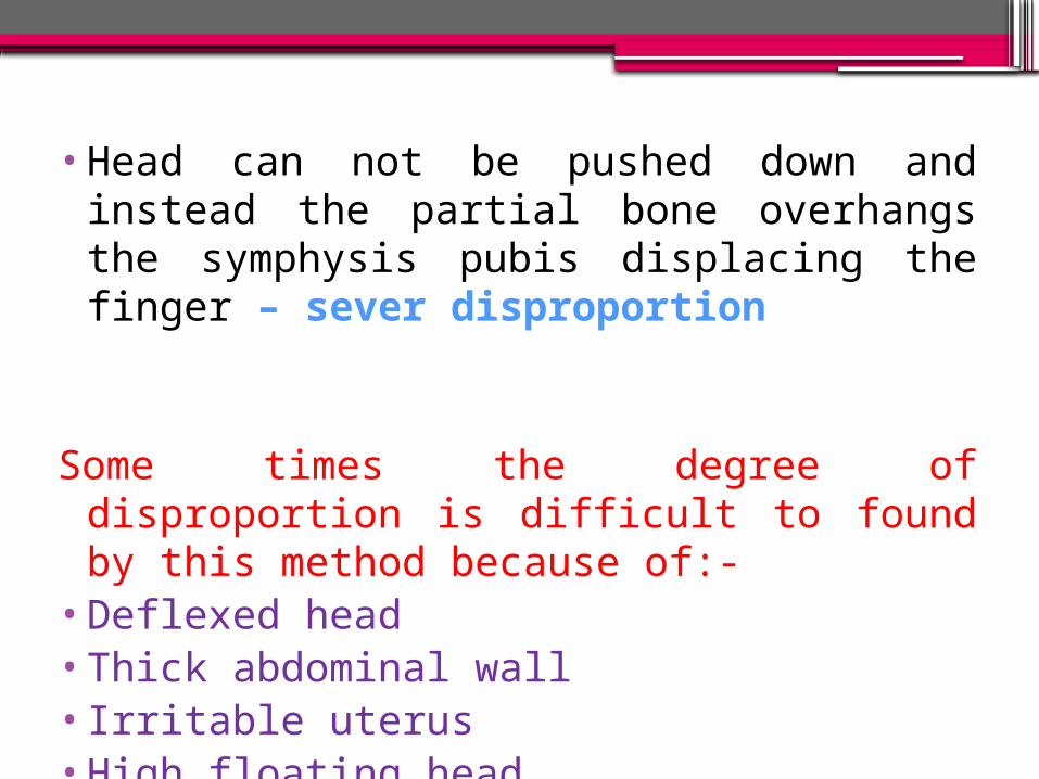
•Head can not be pushed down and instead the partial bone overhangs the symphysis pubis displacing the finger – sever disproportion
Some times the degree of disproportion is difficult to found by this method because of:-
•Deflexed head•Thick abdominal wall•Irritable uterus•High floating head
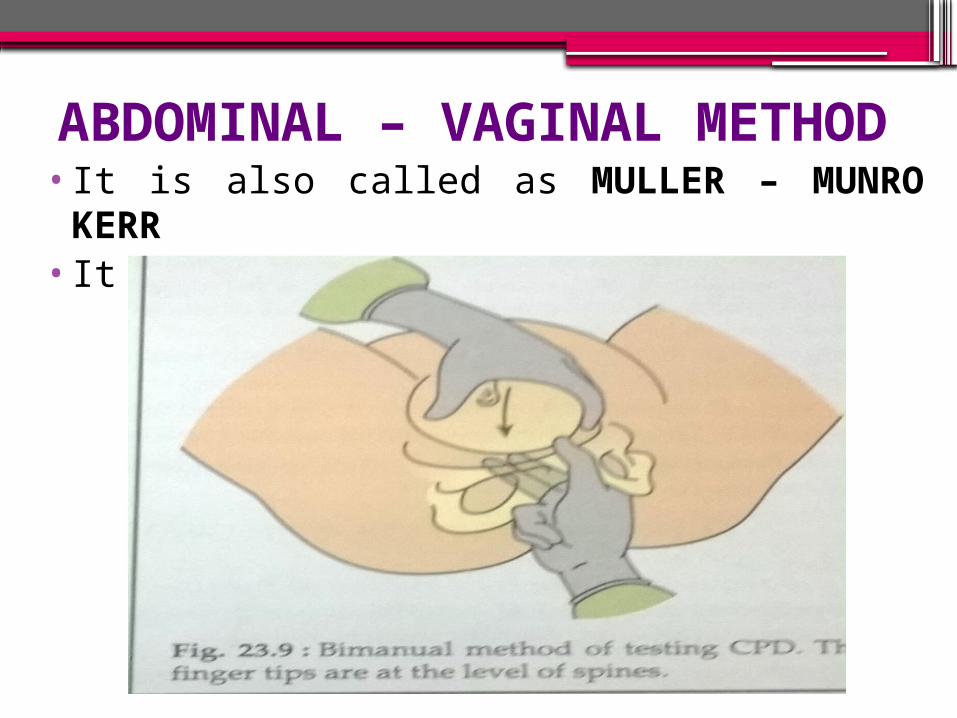
ABDOMINAL – VAGINAL METHOD•It is also called as MULLER – MUNRO KERR
•It is bimanual method.

Results :- •the head can be pushed down up to the
level of ischial spines and there is no overlapping of the parietal bone over the symphysis pubis:- no disproportion
•The head can be pushed down a little but not up to the level of ischial spine and ther is slight overlapping of the parietal bone:- slight or moderate disproportion
• The head can not be pushed down and instead the parietal bone overhangs the symphysis pubis displacing the thumb:- sever disproportion.

D. Pelvimetry• It is assessment of the pelvic diameters and
capacity done at 38-39 weeks. It includes:• Clinical pelvimetry:
▫Internal pelvimetry for: inlet, cavity, and outlet.
▫External pelvimetry for: inlet and outlet.
• Imaging pelvimetry:▫X-ray.▫Computerised tomography (CT).▫Magnetic resonance imaging (MRI) .


EFFECT OF CONTRACTED
PELVIS
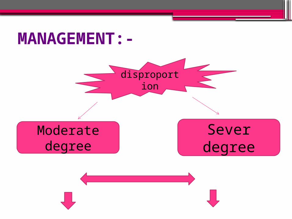
MANAGEMENT:-
disproportion
Moderate degree
Sever degree

Preterm
laborTerm labor
Induction of labor
Cesarean section
Trial labor
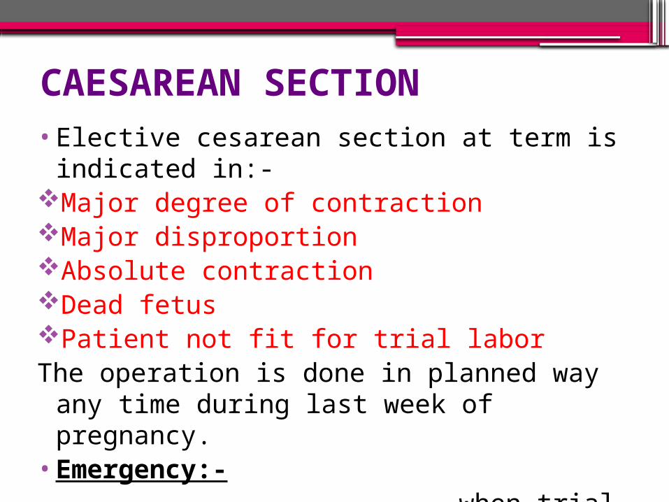
CAESAREAN SECTION•Elective cesarean section at term is
indicated in:-Major degree of contractionMajor disproportionAbsolute contractionDead fetusPatient not fit for trial laborThe operation is done in planned way any
time during last week of pregnancy.•Emergency:- when trial labor is failed

•Trial labor:-•It is the conduction of spontaneous labor
in a moderate degree of disproportion, in an institution under supervision with watchful expectancy hoping for a vaginal delivery
or Trial of labor is a test of labor allowing the
patient to enter into active labor putting all variable
( power, passage and passenger) into test and determine whether vaginal delivery is possible or not.

•CONDUCTION OF A TRIAL OF LABOR:-
Careful fetal and maternal monitoring by electronic fetal monitoring and non stress test
Oral feeding remain suspended and hydration is maintained by intravenous drip
Adequate analgesic is administered
Augmentation of labor by pitocin

The progress of labor is mapped with partograph:-
i) progressive descent of the head ii) progressive dilatation of the
cervixAfter the membrane rupture, pelvic
examination is to be done:- i) to exclude cord prolapse ii) to note the color of liquor iii) to assess the pelvis once or more iv) to note the condition of the
cervix including pressure of the presenting part of the cervix

in favorable cases, end spontaneously, low forcep and low ventose.
In unfavorable cases, do caesarean section.
Successful trial:-
A trial is called successful, if a healthy baby is born vaginally, spontaneous or by forcep or ventose with the mother in good condition
Failure of trial labor:-
Delivery is by cesarean section or delivery of a dead baby spontaneously or by craniotomy is called failure of trial labor

ADVANTAGES OF TRIAL LABOR•Lower incidence of cesarean section.
•A successful trial ensures the women a good future obstetrics.

DISADVANTAGES OF TRIAL LABOR•May end before full cervix dilatation
•Increased fetal mortality and morbidity
•In failed trial operative risk increases.

NURSING MANAGEMENT:-•Check vitals every 4 hourly•Monitor both contraction and fetus
continuously•Report immediately the sign of fetal distress•Position the mother in ways to increase the
pelvic diameter such as sitting or squatting which increase the outlet diameter and also aid in fetal descent
•Assess the fetus for hypoxia•Provide support to the client and the family
members in coping with stress of a complicated labor
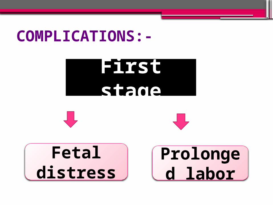
COMPLICATIONS:-
First stage
Fetal distress
Prolonged labor

Second stage
Delayed second stage
Shoulder dystocia

Third stage
Retained placenta
Maternal injuryPPH

SUMMARY