cranial avm: classification and management. av malformations high flow cerebrovascular lesions...
TRANSCRIPT

CRANIAL AVM: CLASSIFICATION AND MANAGEMENT

AV malformations
• High flow cerebrovascular lesions• Prevalence 0.04-0.52%• Sporadic (98%)• Syndromic (2%)
• Hereditary hemorrhagic telengiectasia (Osler Weber Rendu)
• Cerebrofacial AV metameric syndromes (CAMS)
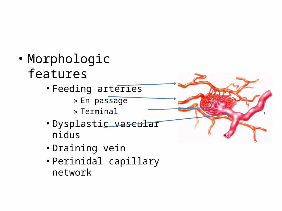
• Morphologic features• Feeding arteries
» En passage» Terminal
• Dysplastic vascular nidus• Draining vein• Perinidal capillary network
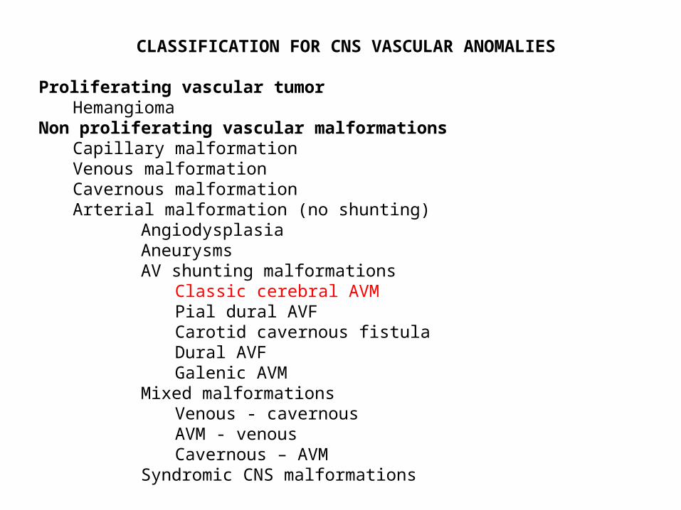
CLASSIFICATION FOR CNS VASCULAR ANOMALIES
Proliferating vascular tumorHemangioma
Non proliferating vascular malformationsCapillary malformationVenous malformationCavernous malformationArterial malformation (no shunting)
AngiodysplasiaAneurysms AV shunting malformations
Classic cerebral AVMPial dural AVFCarotid cavernous fistulaDural AVFGalenic AVM
Mixed malformationsVenous - cavernousAVM - venousCavernous – AVM
Syndromic CNS malformations
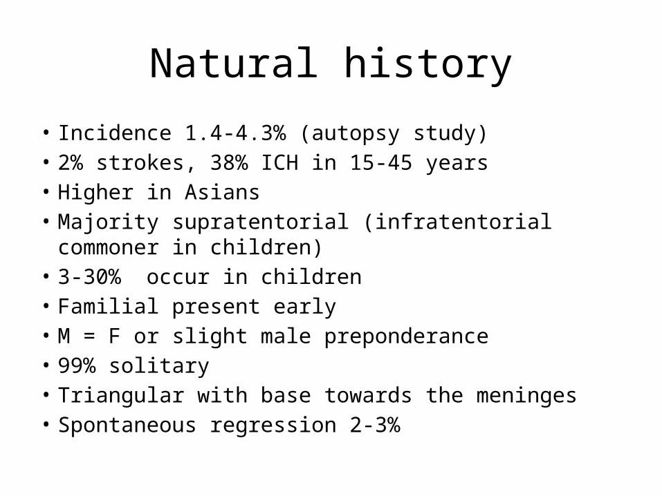
Natural history
• Incidence 1.4-4.3% (autopsy study)• 2% strokes, 38% ICH in 15-45 years• Higher in Asians• Majority supratentorial (infratentorial commoner in children)• 3-30% occur in children• Familial present early• M = F or slight male preponderance• 99% solitary• Triangular with base towards the meninges• Spontaneous regression 2-3%
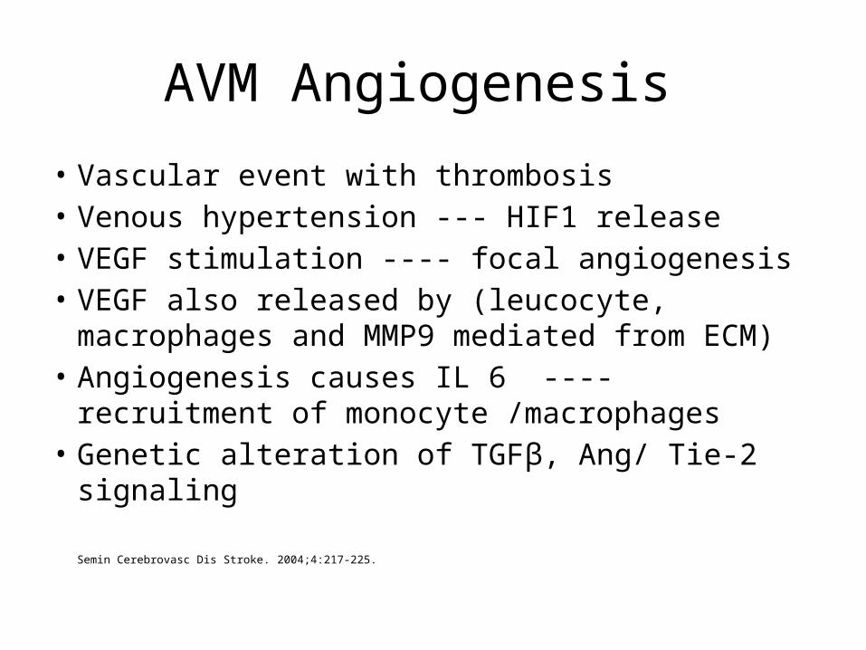
AVM Angiogenesis
• Vascular event with thrombosis• Venous hypertension --- HIF1 release• VEGF stimulation ---- focal angiogenesis• VEGF also released by (leucocyte, macrophages and
MMP9 mediated from ECM)• Angiogenesis causes IL 6 ---- recruitment of
monocyte /macrophages• Genetic alteration of TGFβ, Ang/ Tie-2 signaling
Semin Cerebrovasc Dis Stroke. 2004;4:217-225.
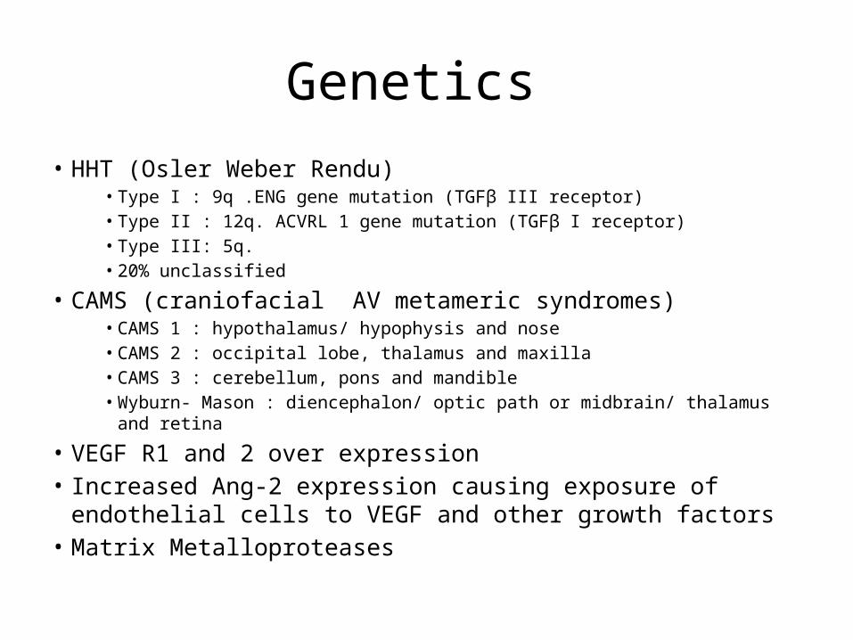
Genetics • HHT (Osler Weber Rendu)
• Type I : 9q .ENG gene mutation (TGFβ III receptor)• Type II : 12q. ACVRL 1 gene mutation (TGFβ I receptor)• Type III: 5q.• 20% unclassified
• CAMS (craniofacial AV metameric syndromes)• CAMS 1 : hypothalamus/ hypophysis and nose• CAMS 2 : occipital lobe, thalamus and maxilla• CAMS 3 : cerebellum, pons and mandible• Wyburn- Mason : diencephalon/ optic path or midbrain/ thalamus and retina
• VEGF R1 and 2 over expression• Increased Ang-2 expression causing exposure of endothelial cells to
VEGF and other growth factors• Matrix Metalloproteases

Presentation
• Asymptomatic
• Haemorrhage
• Seizures
• Headache
• Neurological defecits
• Congestive heart failure
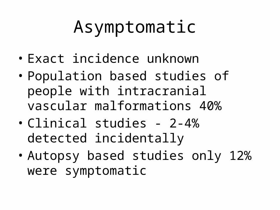
Asymptomatic
• Exact incidence unknown• Population based studies of people with
intracranial vascular malformations 40%• Clinical studies - 2-4% detected incidentally• Autopsy based studies only 12% were
symptomatic
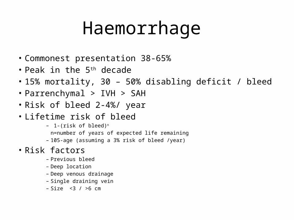
Haemorrhage • Commonest presentation 38-65%• Peak in the 5th decade• 15% mortality, 30 – 50% disabling deficit / bleed• Parrenchymal > IVH > SAH• Risk of bleed 2-4%/ year• Lifetime risk of bleed
– 1-(risk of bleed)n
n=number of years of expected life remaining– 105-age (assuming a 3% risk of bleed /year)
• Risk factors– Previous bleed– Deep location– Deep venous drainage– Single draining vein– Size <3 / >6 cm
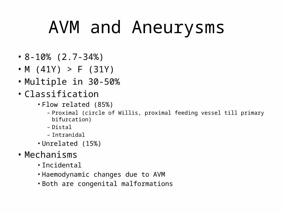
AVM and Aneurysms
• 8-10% (2.7-34%)• M (41Y) > F (31Y)• Multiple in 30-50%• Classification
• Flow related (85%)– Proximal (circle of Willis, proximal feeding vessel till primary bifurcation)– Distal – Intranidal
• Unrelated (15%)
• Mechanisms• Incidental • Haemodynamic changes due to AVM• Both are congenital malformations
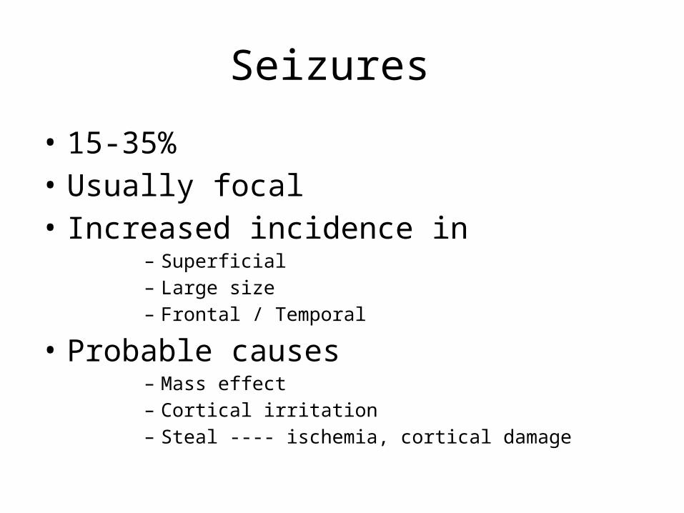
Seizures
• 15-35%• Usually focal• Increased incidence in
– Superficial– Large size– Frontal / Temporal
• Probable causes– Mass effect– Cortical irritation– Steal ---- ischemia, cortical damage
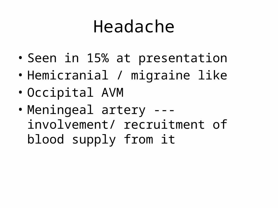
Headache
• Seen in 15% at presentation• Hemicranial / migraine like• Occipital AVM• Meningeal artery --- involvement/ recruitment
of blood supply from it

Neurological deficits
• < 10% at presentation• Transient, permanent, progressive• Learning disorders in 66% of adults• Functional decline 1.5%/ Yr (not due to bleed)• Mechanism
• Recurrent bleed• Mass effect• Hydrocephalus• Ischemia• Steal phenomenon
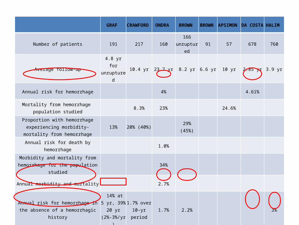
GRAF CRAWFOR
D ONDRA BROWN BROWN APSIMON
DA COSTA
HALIM
Number of patients 191 217 160166
unruptured91 57 678 760
Average follow-up4.8 yr for
unruptured10.4 yr 23.7 yr 8.2 yr 6.6 yr 10 yr 2.85 yr 3.9 yr
Annual risk for hemorrhage 4% 4.61%
Mortality from hemorrhage population studied
8.3% 23% 24.6%
Proportion with hemorrhage experiencing morbidity-mortality from hemorrhage
13% 20% (40%) 29% (45%)
Annual risk for death by hemorrhage 1.0%
Morbidity and mortality from hemorrhage for the population studied
34%
Annual morbidity and mortality 2.7%
Annual risk for hemorrhage in the absence of a hemorrhagic history
14% at 5 yr, 39% 20 yr (2%-3%/yr)
1.7% over 10-yr period
1.7% 2.2% 3%
Annual risk for hemorrhage with a recent hemorrhagic history
Cumulative: 6% at 1 yr,
13% at 5 yr, 47% at 20 yr
3.6% over 10-yr period
7.48% 7%
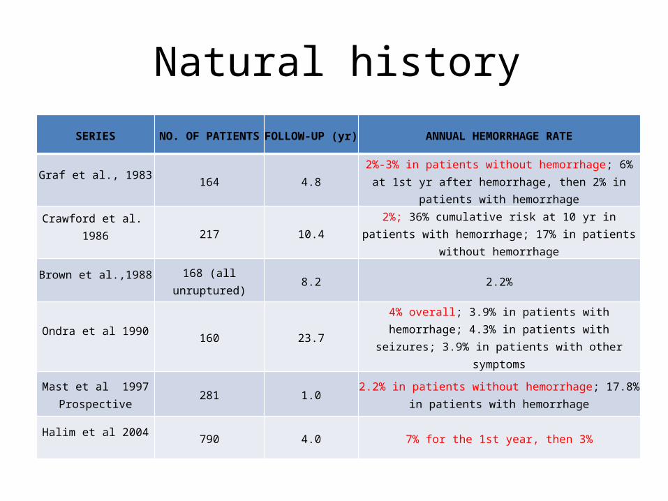
Natural history
SERIES NO. OF PATIENTS FOLLOW-UP (yr) ANNUAL HEMORRHAGE RATE
Graf et al., 1983 164 4.82%-3% in patients without hemorrhage; 6% at 1st yr
after hemorrhage, then 2% in patients with hemorrhage
Crawford et al. 1986 217 10.42%; 36% cumulative risk at 10 yr in patients with hemorrhage; 17% in patients without hemorrhage
Brown et al.,1988 168 (all unruptured) 8.2 2.2%
Ondra et al 1990 160 23.74% overall; 3.9% in patients with hemorrhage; 4.3% in
patients with seizures; 3.9% in patients with other symptoms
Mast et al 1997Prospective
281 1.02.2% in patients without hemorrhage; 17.8% in patients
with hemorrhage
Halim et al 2004 790 4.0 7% for the 1st year, then 3%
Diagnosis
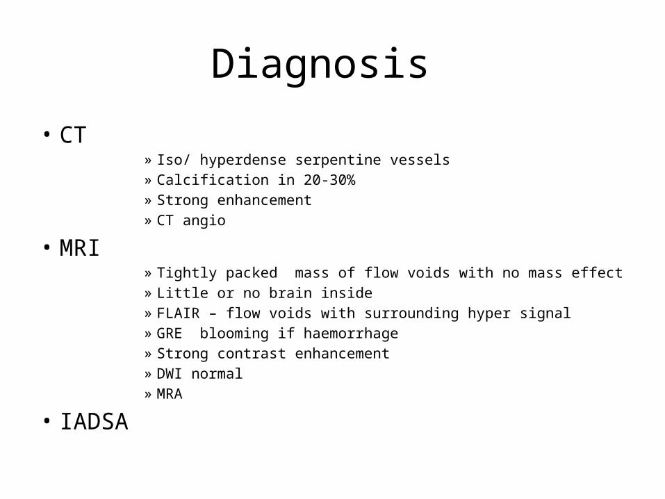
• CT» Iso/ hyperdense serpentine vessels» Calcification in 20-30%» Strong enhancement» CT angio
• MRI» Tightly packed mass of flow voids with no mass effect» Little or no brain inside» FLAIR – flow voids with surrounding hyper signal» GRE blooming if haemorrhage» Strong contrast enhancement» DWI normal» MRA
• IADSA
Diagnosis
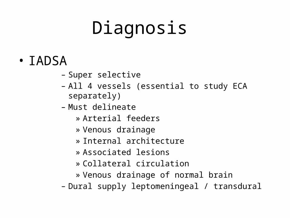
• IADSA– Super selective – All 4 vessels (essential to study ECA separately)– Must delineate
» Arterial feeders» Venous drainage» Internal architecture» Associated lesions» Collateral circulation» Venous drainage of normal brain
– Dural supply leptomeningeal / transdural
Classification
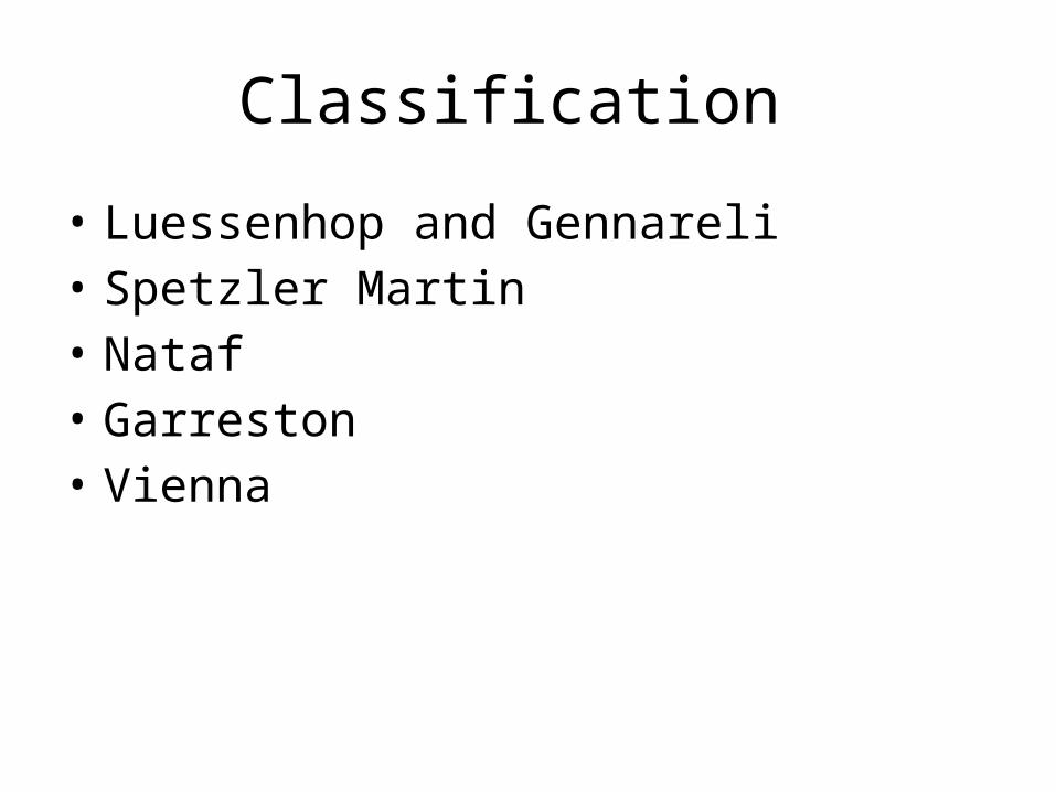
• Luessenhop and Gennareli• Spetzler Martin• Nataf• Garreston• Vienna

Management
• Observation• Microsurgery• Gamma knife• Embolization• Combination • One modality or a sequential combination is
the ideal treatment for a patient, different modalities are not interchangeable
Microsurgery
• Patient related» Age» General condition» Neurological status» Occupation and lifestyle
• AVM related» Size and configuration» Location» AVM anatomy and aneurysms
• Surgeon related» Experience» Availability and familiarity with all modalities
Microsurgery
• Always elective surgery• If hematoma – conservative evacuation• All brain is eloquent, some is more eloquent• Large craniotomy for superficial lesions• Positioning to minimize retraction• Major draining veins controlled last
Dissection technique
• Open arachnoid, sulci and fissures• Circumferential dissection• Arteries tackled first• Follow till the nidus and confirm the entry to the AVM• Transect close to nidus• No tamponade except on AVM• Skeletonize superficial major draining vein• Post resection
• Hypertensive challenge• Cottonoid rub• Intraop / postop angiography
Microsurgery complications
• Intra operative– Bleeding– Parenchymal damage– Retraction injury– Visual radiation injury
• Post operative– Hemorrhage– New onset Seizures 15% (55% improve, 35% unchanged)– NPPB– Retrograde venous/ arterial thrombosis– Vasospasm (rare)
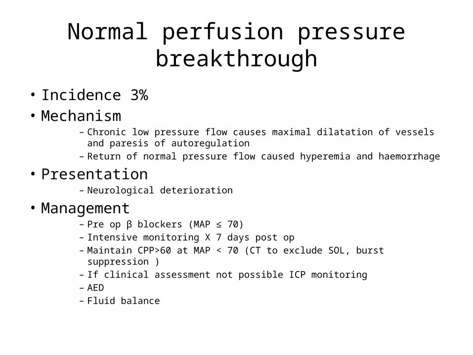
Normal perfusion pressure breakthrough
• Incidence 3%• Mechanism
– Chronic low pressure flow causes maximal dilatation of vessels and paresis of autoregulation
– Return of normal pressure flow caused hyperemia and haemorrhage
• Presentation – Neurological deterioration
• Management – Pre op β blockers (MAP ≤ 70)– Intensive monitoring X 7 days post op– Maintain CPP>60 at MAP < 70 (CT to exclude SOL, burst suppression )– If clinical assessment not possible ICP monitoring– AED– Fluid balance
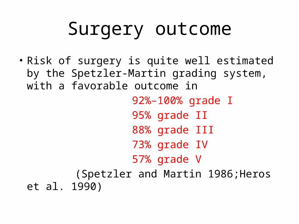
Surgery outcome
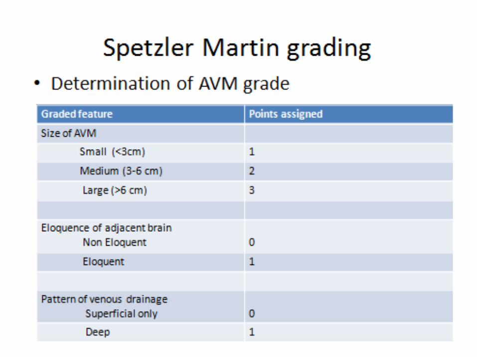
• Risk of surgery is quite well estimated by the Spetzler-Martin grading system, with a favorable outcome in
92%–100% grade I 95% grade II 88% grade III 73% grade IV 57% grade V (Spetzler and Martin 1986;Heros et al. 1990)
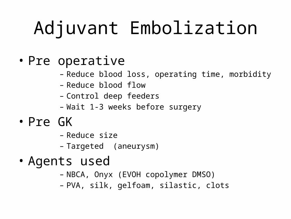
Adjuvant Embolization
• Pre operative– Reduce blood loss, operating time, morbidity– Reduce blood flow– Control deep feeders– Wait 1-3 weeks before surgery
• Pre GK– Reduce size– Targeted (aneurysm)
• Agents used– NBCA, Onyx (EVOH copolymer DMSO)– PVA, silk, gelfoam, silastic, clots

Curative Embolization
• Mortality 1-4%• Morbidity 0 - 50%• Defecits 10 – 14% (disabling 2-5%)• Cure rates 5-20% (0-70%, gyral 12.5% sulcal
60%)• Success
• Nidus accessible to catheter• <3 feeders• Nidus < 3 cm
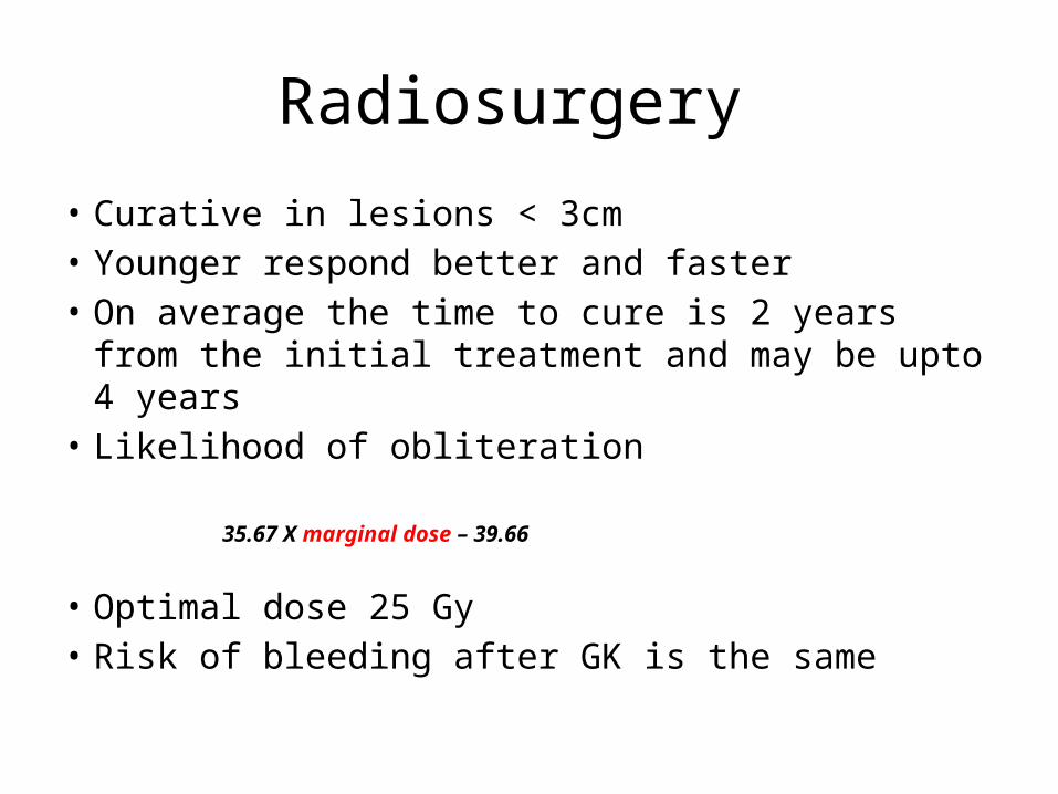
Radiosurgery
• Curative in lesions < 3cm• Younger respond better and faster• On average the time to cure is 2 years from the initial
treatment and may be upto 4 years• Likelihood of obliteration
35.67 X marginal dose – 39.66
• Optimal dose 25 Gy• Risk of bleeding after GK is the same
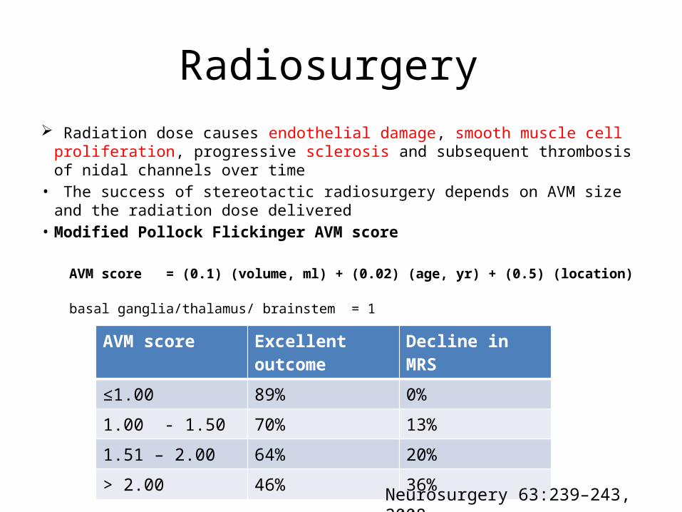
Radiosurgery Radiation dose causes endothelial damage, smooth muscle cell proliferation,
progressive sclerosis and subsequent thrombosis of nidal channels over time• The success of stereotactic radiosurgery depends on AVM size and the
radiation dose delivered• Modified Pollock Flickinger AVM score
AVM score = (0.1) (volume, ml) + (0.02) (age, yr) + (0.5) (location) basal ganglia/thalamus/ brainstem = 1
AVM score Excellent outcome Decline in MRS
≤1.00 89% 0%
1.00 - 1.50 70% 13%
1.51 – 2.00 64% 20%
> 2.00 46% 36%
Neurosurgery 63:239–243, 2008
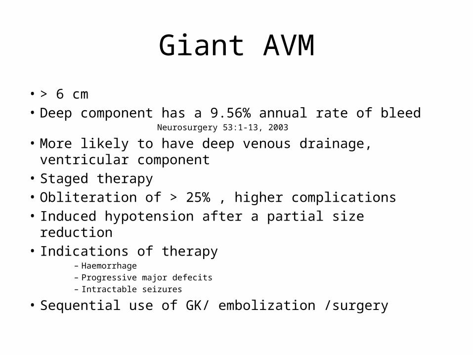
Giant AVM• > 6 cm• Deep component has a 9.56% annual rate of bleed
Neurosurgery 53:1-13, 2003
• More likely to have deep venous drainage, ventricular component• Staged therapy • Obliteration of > 25% , higher complications• Induced hypotension after a partial size reduction• Indications of therapy
– Haemorrhage– Progressive major defecits– Intractable seizures
• Sequential use of GK/ embolization /surgery
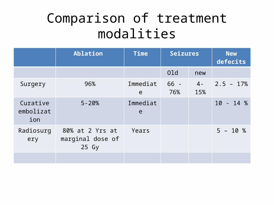
Comparison of treatment modalities
Ablation Time Seizures New defecits
Old new
Surgery 96% Immediate 66 -76% 4-15% 2.5 – 17%
Curative embolization
5-20% Immediate 10 - 14 %
Radiosurgery 80% at 2 Yrs at marginal dose of 25 Gy
Years 5 – 10 %
J Neurosurg. 2009 May;110(5):1003-9.Outcome after hemorrhage following Gamma Knife surgery for cerebral arteriovenous malformations.Kasliwal MK, Kale SS, Gupta A, Kiran NA, Sharma MS, Sharma BS, Mahapatra AK.
J Neurosurg. 2007 Dec;107(6 Suppl):479-84.Gamma Knife surgery for intracranial arteriovenous malformations in children: a retrospective study in 103 patients.Kiran NA, Kale SS, Vaishya S, Kasliwal MK, Gupta A, Sharma MS, Sharma BS, Mahapatra AK.
For many patients with large AVMs, discretion may be the better part of valour. As patients and their surgeons age, the vigour with which multimodality management strategies are pursued begins to wane.
L Dade LunsfordCommentsNeurosurgery 53:1-13, 2003
We now recommend no treatment for most Grade IV and V AVMs. In fact, partial treatment may even worsen outcomes compared with the natural history of AVMs. We do not support palliative treatment of AVMs except in the specific circumstances of arterial or intranidal aneurysms or progressive neurological deficits related to vascular steal.
Robert F SpetzlerCommentsNeurosurgery 53:1-13, 2003
Most Grade V AVMs and many Grade IV AVMs should be treated conservatively since they are generally too large for radiosurgery, present unacceptable surgical morbidity, can only rarely be completely occluded by embolization and incomplete embolization, which is risky, does not improve and may worsen the natural history.
Roberto C HerosYoumans Neurological Surgery 6th edition

THANK YOU