critical factors in the biology of human cancer - cancer research
TRANSCRIPT

(CANCER RESEARCH 50. 6130-6138, October l, 1990]
Special Lecture
Critical Factors in the Biology of Human Cancer Metastasis: Twenty-eighthG. H. A. Clowes Memorial Award Lecture1
Isaiah J. FidlerDepartment of Cell Biology, The University of Texas M. D. Anderson Cancer Center, Houston, Texas 77030
Abstract
The process of metastasis is not random. Rather, it consists of a seriesof linked, sequential steps that must be completed by tumor cells if ametastasis is to develop. Although some of the steps in this processcontain stochastic elements, as a whole, metastasis favors the survivaland growth of a few subpopulations of cells that preexist within theparent neoplasm. Moreover, métastasescan have a clonal origin, anddifferent métastasescan originate from the proliferation of single cells.The outcome of metastasis depends on the interaction of metastatic cellswith different organ environments. Organ-specific métastaseshave beendemonstrated in a variety of experimental tumor systems. Moreover, wehave found tumor growth that is specific to a particular site within oneorgan. Whether the same conclusions can be reached for human cancersremained unanswered until very recently.
Studies from our laboratory and from others have shown that theimplantation of human cancer cells derived from surgical specimens intocorrect anatomical sites of nude mice can provide a suitable model ofmetastasis of human tumors. Clonal analysis of a human renal carcinoma,colon carcinomas, and melanomas has revealed that these tumors areindeed heterogeneous for metastatic properties, an observation made onlyafter orthotopic implantation. Thus, growth in the environment of specificorgans can be selective and the environment per se influences this process.
While it is clear that vascularity and local immunity can facilitate orretard tumor growth, we have concentrated on understanding how damageto an organ and the subsequent repair process can facilitate tumor cellproliferation. Accelerated growth of human colon cancer cells was foundin hepatectomized nude mice, whereas accelerated growth of human renalcancer cells was found in nephrectomized nude mice. These data suggestthat systemic physiological signals can be recognized by neoplastic cellspresumably by mechanisms similar to those shared by their normal cellcounterparts. In summary, the critical factors that regulate metastasisare the intrinsic properties of metastatic cells and host factors involvedin homeostasis. The recent increase in our understanding of metastasisshould provide important leads for developing more effective approachesto the treatment of disseminated cancer.
Introduction
To be a recipient of the G. H. A. Clowes Memorial Award isa distinct honor which I shall always cherish. I consider myselfa representative of a larger group of scientists whose endeavorsin elucidating the mysteries of cancer metastasis you haverecognized. During the last 20 years, I have devoted myself tothe understanding of the biology and therapy of cancer metastasis. My interest in this field was stimulated by my teacher,Irving Zeidman, who taught me to ask relevant questions, tosearch for answers, and to even sometimes ignore dogmas. Iam also grateful to many friends, colleagues, and students withwhom I have worked. Their contributions will become clearduring my presentation. In particular, I thank Margaret Kripke,George Poste, Garth Nicolson, Ian Hart, Avraham Raz, Robert
Received 6/26/90; accepted 6/27/90.' Financial support of this research was provided by Grant R35-CA 42107
from the National Cancer Institute. NIH. Presented at the Seventy-ninth AnnualMeeting of the American Association for Cancer Research, May 26. 1988, inNew Orleans, LA.
Kerbel, Philip Frost, Michael Hanna, David Tarin, and Frederick Becker for productive collaboration and warm friendship.I also take this opportunity to thank the National CancerInstitute and other funding sources for providing the necessarysupport without which hypotheses cannot become reality.
I am extremely pleased that Dr. Henry Mihich introducedmy presentation. In 1978, when he was the Program Chairmanof the Annual Meeting, he introduced me as a speaker on atopic of current interest: "Tumor heterogeneity and the biologyof cancer invasion and metastasis." At that time, Garth Nicol
son, George Poste, Gloria Heppner, and I, among others, hadjust begun to understand these processes, and I remember howhonored I was to be chosen to describe some of the early findingsin this emerging field (1). During the last decade, our collectiveknowledge has advanced considerably, and in this presentationI shall describe some of the progress accomplished by us andothers.
My lecture is divided into three parts. First, I shall discussthe pathogenesis of metastasis and describe some of the mechanisms that regulate this process. Second, I shall describe thedevelopment of animal models to study the biology of humancancer metastasis. Finally, I shall describe preliminary data onthe role of homeostatic factors in the pathogenesis of metastasis.
Metastasis, the spread of cells from the primary neoplasm todistant sites and their growth there, is the most fearsome aspectof cancer. This fear is well founded. Despite significant improvements in early diagnosis, surgical techniques, general patient care, and local and systemic adjuvant therapies, mostdeaths from cancer are due to métastasesthat are resistant toconventional therapies (1-9). In a large number of patients withcancer, metastasis may well have occurred by the time ofdiagnosis (4, 6, 8, 9). The métastasescan be located in differentlymph nodes and visceral organs and in various regions of thesame organ, thus complicating their treatment. Furthermore,the specific organ environment can modify the response of ametastatic tumor cell to systemic therapy and alter the efficiencyof anticancer agents.
The major barrier to the treatment of métastasesis thebiological heterogeneity of cancer cells in primary and secondary neoplasms. This heterogeneity is exhibited in a wide rangeof genetic, biochemical, immunologie-ai, and biological charac
teristics, such as cell surface receptors, enzymes, karyotypes,cell morphologies, growth properties, sensitivities to varioustherapeutic agents, and ability to invade and produce metastasis(1-5, 10-15). Moreover, it is important to remember the term"cancer" denotes a collection of malignancies, with each cancer
of each organ consisting of numerous subsets. This tremendousheterogeneity is probably due to the different etiologies, origins,and selection pressures of different cancers.
Continual empiricism in the treatment of cancer is unlikelyto produce significant improvement. Therefore understanding
6130
on April 4, 2019. © 1990 American Association for Cancer Research. cancerres.aacrjournals.org Downloaded from

CRITICAL FACTORS IN HUMAN CANCER METASTASIS
the mechanisms responsible for the development of biological extravasation occurs next, probably by the same mechanismsheterogeneity in primary cancers and in métastasesand theprocess by which tumor cells invade local tissues and spread todistant organs is a primary goal of cancer research. Only froma better understanding will come the ability to design moreeffective therapy for different cancers and improvements in theway physicians deal with cancer metastasis. My lecture, therefore, reviews recent data that provide some answers to thesedifficult questions.
The Pathogenesis of a Metastasis
The process of cancer metastasis consists of a long series ofsequential interrelated steps, each of which can be rate limitingsince a failure or an insufficiency at any of the steps aborts theprocess (7). The outcome of the process is dependent on boththe intrinsic properties of the tumor cells and the responses ofthe host; the balance of these interactions can vary amongdifferent patients (2, 6, 9). In principle, the steps or eventsrequired for the formation of a metastasis are the same in alltumors, and I therefore illustrate the process by using melanoma as the model (Fig. 1).
Major steps in the formation of a metastasis are as follows:(a) after the initial transforming event, either unicellular ormulticellular, growth of neoplastic cells must be progressive;(b) extensive vascularization must occur if a tumor mass is toexceed 2 mm in diameter (16). The synthesis and secretion ofseveral angiogenesis factors play a key role in establishing aneocapillary network from the surrounding host tissue (17,18);(c) local invasion of the host stroma by some tumor cells couldoccur by several mechanisms that are not mutually exclusive(19-22). Thin-walled venules, like lymphatic channels, offervery little resistance to penetration by tumor cells and providethe most common pathways for tumor cell entry into thecirculation. Although clinical observations have suggested thatcarcinomas frequently metastasize and grow via the lymphaticsystem, whereas malignant tumors of mesenchymal origin, e.g.,melanoma, more often spread by the hematogenous route, thepresence of numerous venolymphatic anastomoses invalidatesthis belief (23); (¡I)detachment and embolization of smalltumor cell aggregates occurs next; (e) tumor cells that survivethe circulation must arrest in the capillary beds of organs; (/)
The Metastatic Process
Primarymalignant Neovascularization Invasion Embolismneoplasm
Lymphatics, MulticeliaggregatesVenules.capillaries {Lymphocyte,platelets)
Responsetomicroenvironment
Tumorcellproliferation
-~ \^f»j
Extravasation Adherence Arrestin distantorgancapillarybed
"^BtBWBP ^MetastasisofMétastases
Metastases
Fig. 1. The pathogenesis of a metastasis. To produce métastases,tumor cellsin a primary neoplasm must complete a series of sequential and selective steps,each of which can be rate limiting since a failure or an insufficiency at any of thesteps aborts the process.
that influence initial invasion; (g) proliferation within the organparenchyma completes the metastatic process. To produce detectable lesions, the métastasesmust develop a vascular network, evade the host immune system (2), and respond to organ-specific factors that influence their growth (24-28). Once theydo so, the cells can invade host stroma, penetrate blood vessels,and enter the circulation to produce secondary métastases,theso-called "metastasis of métastases"(7-9).
When I described the metastatic process, 1 emphasized thatonly a few cells in a primary tumor can give rise to a metastasis.This is due in part to the elimination of any disseminatingtumor cell that fails to complete any step in metastasis. Thecomplexity of the pathogenesis of metastasis explains, in part,why the process is deemed to be inefficient (9, 29). For example,the presence of tumor cells in the circulation does not predictthat metastasis will occur, because most tumor cells that enterthe blood stream are rapidly eliminated (30). Using radiolabeledB16 melanoma cells, I observed that by 24 h after entry intothe circulation, <1% of the cells are still viable, and <0.1% oftumor cells placed into the circulation survived to producemétastases(30). Observations such as this prompted me to askwhether the development of métastasesrepresents the fortuitous survival and growth of very few neoplastic cells or whetherit represents the selective growth of unique subpopulations ofmalignant cells endowed with special properties. In other words,can all cells growing in a primary neoplasm produce secondary-
lesions, or do only specific and unique cells possess the appropriate properties that would enable them to survive the potentially destructive journey from the primary tumor to the sitesof future métastases?Most recent data indeed show that neoplasms are biologically heterogeneous and that the process ofmetastasis is selective.
Metastatic Heterogeneity
Cells with different metastatic properties have been isolatedfrom the same parent tumor, thus supporting the hypothesisthat not all the cells in a primary tumor can successfullydisseminate. Two general approaches have been used to isolatepopulations of cells that differ from the parent neoplasm inmetastatic capacity. In the first, metastatic cells are selected invivo: tumor cells are implanted s.c., i.m., or into other tissues,or they are injected i.v. into mice, and metastasis is allowed tooccur. The metastatic lesions are harvested, and the cells thatare recovered can first be expanded in culture or used immediately to repeat the process. The cycle is repeated several times.The behavior of the cycled cells is compared with that of thecells of the parent tumor to determine whether the selectionprocess enhanced metastatic capacity (31), and the increase inmetastatic capacity of the recovered cells did not result fromthe adaptation of tumor cells to preferential growth in a particular organ (32-34). This procedure was originally used toisolate the B16-F10 line from the wild-type B16 melanoma(31). It has also been successfully used to produce tumor celllines with increased metastatic capacity from many of theexperimental tumors tested (35-39).
In the second approach, cells are selected for the enhancedexpression of a phenotype believed to be important in one oranother step of the metastatic sequence, and then they aretested in the appropriate host to determine whether concomitant metastatic potential has been increased or decreased. Thismethod has been used to examine whether properties as diverse
6131
on April 4, 2019. © 1990 American Association for Cancer Research. cancerres.aacrjournals.org Downloaded from

CRITICAL FACTORS IN HUMAN CANCER METASTASIS
as resistance to T-lymphocytes (40,41), adhesive characteristics(42, 43), invasive capacity (36, 44-46), lectin resistance (43, 47,48), and resistance to natural killer cells (49) are important inmetastasis.
One obvious criticism of these studies is that the survivingisolated tumor line may have arisen as a result of adaptiverather than selective processes. The first experimental proof formetastatic heterogeneity in neoplasms was provided by Margaret Kripke and me in 1977 in work with the mouse B16melanoma (50). Using the modified fluctuation assay of Luriaand Delbruck (51), we showed that different tumor cell clones,each derived from individual cells isolated from the parenttumor, vary dramatically in their ability to form pulmonarynodules following i.v. inoculation into syngeneic mice. Controlsubcloning procedures demonstrated that the observed diversitywas not a consequence of the cloning procedure (50).
To exclude the possibility that the metastatic heterogeneityfound in the B16 melanoma might have been introduced as aresult of the lengthy in vivo and in vitro cultivation, we studiedthe biological and metastatic heterogeneity in a mouse melanoma induced in C3H mice by chronic exposure to UV Bradiation and painting with croton oil (52). One mouse thustreated developed a melanoma designated K-1735. The originalK-1735 melanoma was established in culture and immediately
cloned (53, 54). In an experiment similar in design to the onedescribed for the B16 melanoma (50), we found that the clonesdiffered greatly from each other and from the parent tumor intheir ability to produce lung métastases.In addition to differences in the number of métastases,we also found significantvariability in the size and pigmentation of the métastases.Métastasesto the lymph nodes, brain, heart, liver, and skinwere found in addition to lung métastases;those growing in thebrain were uniformly melanotic, whereas those growing in otherorgans generally were not (27, 53, 54).
To determine whether the absence of metastasis productionby some clones of the K-1735 melanoma was a consequence oftheir immunological rejection by the normal host (55-58),Nabil Hanna and I examined their metastatic behavior in youngnude mice (59, 60). In such recipients, the immunologicalbarrier to metastatic cells that also may be highly immunogenicis removed, and antigenic metastatic cells may thus successfullycomplete the process. Cells of two clones that did not producemétastasesin normal syngeneic mice produced tumor foci inthe young nude mice. However, most of the nonmetastaticclones were nonmetastatic in both normal syngeneic and nuderecipients. Therefore, the failure of the clones to metastasize insyngeneic mice was probably not caused by their immunologicalrejection by the host but by other deficiencies that prevented
BENIGN MALIGNANT
Fig. 2. Metastatic and nonmetastatic phenotypes. Failure to produce a metastasis can be due to a single or to multiple deficiencies. Therefore, not allnonmetastatic cells share identical phenotypes.
completion of one or another step in the complex metastaticprocess (Fig. 2).
The finding that preexisting tumor cell subpopulations proliferating in the same tumor exhibit heterogeneous metastaticpotential has since been confirmed in many laboratories, witha wide range of experimental animal tumors of different histories and histological origins (1-4,7, 10,11, 13-15). In addition,studies using young nude mice as models for metastasis ofhuman neoplasms have shown that several human tumor linesand freshly isolated tumors, such as colon carcinoma and renalcarcinoma, also contain subpopulations of cells with widelydiffering metastatic properties (60).
James Talmadge and I also addressed the question of whetherthe cells that survive to form métastasespossess a greatermetastatic capacity than most cells in an unselected neoplasm(31, 34, 37). Most lines derived from metastatic deposits produced significantly more métastasesthan cells of the parentline. Studies with heterogeneous, unselected neoplasms havetherefore led us to conclude that metastasis is a selective processregulated by a number of different mechanisms.
Role of the Organ Environment in the Pathogenesis of Metastasis
Clinical observations of cancer patients and studies withexperimental rodent tumors have revealed that certain tumorsproduce metastasis to specific organs independent of vascularanatomy, rate of blood flow, and number of tumor cells delivered to each organ. The distribution and fate of hematogenouslydisseminated, radiolabeled melanoma cells in experimental rodent systems amply demonstrate that tumor cells reach themicrovasculature of many organs (30, 61-63). Extravasationinto the organ parenchyma and proliferation of tumor cellsoccur in only some organs. Therefore, the mere presence ofviable tumor cells in a particular organ does not always predictthat the cells will proliferate to produce métastases(24-26, 64).
The search for the mechanisms that regulate the pattern ofmetastasis began a century ago. In 1889, Paget (65) questionedwhether the distribution of métastaseswas due to chance. Hetherefore analyzed 735 autopsy records of women with breastcancer. The nonrandom pattern of visceral métastasessuggestedto Paget that the process was not due to chance but, rather,that certain tumor cells (the "seed") had a specific affinity forthe milieu of certain organs (the "soil"). Métastasesresulted
only when the seed and soil were matched (65).Experimental data supporting the "seed and soil" hypothesis
of Paget were derived from studies on the preferential invasionand growth of B16 melanoma métastasesin specific organs (66,67). Ian Hart and I injected B16 melanoma cells into thecirculation of syngeneic C57BL/6 mice. Tumor growths developed in the lungs and in fragments of pulmonary or ovariantissue implanted i.m. In contrast, metastatic lesions did notdevelop in renal tissue implanted as a control or at the site ofsurgical trauma (67). This study confirmed that sites of metastasis are determined not solely by the characteristics of theneoplastic cells but also by the microenvironment of the hosttissue. In vitro experiments demonstrating organ-selective adhesion, invasion, and growth also support Paget's hypothesis (65).
With the B16 melanoma system, cells that are selective fororgan adhesion, invasion, and growth have been isolated (24,26, 31-39, 68). Moreover, experiments with organ tissue-derived soluble growth factors indicate that soil factors can haveprofound effects on certain tumor cell subpopulations (24).
There is no question that the circulatory anatomy influences6132
on April 4, 2019. © 1990 American Association for Cancer Research. cancerres.aacrjournals.org Downloaded from

CRITICAL FACTORS IN HUMAN CANCER METASTASIS
the dissemination of many malignant cells (6, 8, 9); however, itcannot, as Ewing proposed (69), fully explain the patterns ofdistribution of numerous tumors. Ethical considerations ruleout the experimental analysis of cancer metastasis in patientsas studied in laboratory animals, by which either Paget or Ewingmight be proved correct. The introduction of peritoneovenousshunts for palliation of malignant ascites has, however, provided an opportunity to study some of the factors affectingmetastatic spread in humans. David Tarin and colleagues havedescribed the outcome in patients with malignant ascites draining into the venous circulation, with the resulting entry of viabletumor cells into the jugular veins (70, 71 ). Good palliation withminimal complications was reported for 29 patients with different neoplasms. The autopsy findings in 15 patients substantiated the clinical observations that the shunts do not significantly increase the risk of metastasis. In fact, despite continuousentry of millions of tumor cells into the circulation, métastasesin the lung (the first capillary bed encountered) were rare (70,71). These results provide compelling verification of the venerable "seed and soil" hypothesis (65).
An interesting demonstration for organ-specific metastasis
comes from recent studies. Gabriele Schackert and I describedthe development of a mouse model with which to study cerebralmetastasis after injection of syngeneic tumor cells into theinternal carotid artery (72, 73). A direct, i.e.2 injection of tumor
cells was used to determine tumorigenicity. The injection ofcells into the internal carotid artery of mice simulates thehematogenous spread of tumor emboli to the brain. Thus, thistechnique can examine the last steps of the metastatic process:release of tumor cells into the circulation; arrest of tumor cellsin capillaries; penetration and extravasation of the tumor cellsinto the brain through the blood-brain barrier; and continuousgrowth of the cells in the tissue.
We found a remarkable difference between two murine melanomas in patterns of brain metastasis; the K-1735 melanomaproduced lesions only in the brain parenchyma, whereas theB16 melanoma produced only meningea! growths (73). Theseresults demonstrate specificity for metastatic growth in differentregions within a single organ. The results from site distributionanalysis of radiolabeled murine melanoma cells injected intothe internal carotid artery ruled out that the patterns of initialcell arrest in the microvasculature of the brain predicted theeventual sites of growth. Thus, an alternative explanation forthe different sites of tumor growth involves interactions betweenthe metastatic cells and the organ environment, possibly interms of specific binding to endothelial cells and responses tolocal growth factors. In other words, organ-specific métastasesare produced by tumor cells that are receptive to their newenvironment.
Models for Human Cancer Metastasis
An appropriate model for studies of human cancer metastasismust meet two rigid demands: it must use metastatic cells(seed), and it must grow in the relevant organ environment(soil). In 1983, when I joined the faculty of the University ofTexas M. D. Anderson Cancer Center, I began a collaborativestudy with Drs. Kim Jessup, Raffaella Giavazzi, and laterKiyoshi Morikawa to design a bioassay of the malignant potential of cells from surgical specimens of HCCs. There was and
2The abbreviations used are: i.e.. intracranial; i.s., intrasplenic: HCC. human
colon carcinoma; CEA. carcinoembryonic antigen: HRCC, human renal cellcarcinoma.
still is an urgent need for such a model because the major goalof oncologists is the prevention or eradication of métastases,and in many patients with colon cancer, metastasis is likely tohave occurred prior to diagnosis and surgical resection of theprimary tumor (74).
Vince Pollack and I have previously shown that nude micecan be used to ascertain the metastatic potential of allogeneictumors and that metastatic variants can be selected in nudemice from heterogeneous mouse tumors (75). We thereforedecided to use similar techniques to investigate the metastaticpotential of HCC. We reasoned that if a HCC contained metastatic cells, these cells would produce metastasis in nude mice,whereas HCCs that do not contain metastatic cells will not. Asyou shall soon see, this was a gross oversimplification.
Correlation of Experimental Metastatic Behavior with ClinicalStaging
In initial studies, four tumor lines were derived from primaryHCCs, three from hepatic métastasesand one from a mesentericlymph node metastasis. Tumor cells of each line were injectedinto multiple sites in nude mice: the spleen; the subcutis;muscle; and the venous system (76, 77). All the inoculi consistedof single-cell suspensions obtained by enzymatic dissociation ofsolid tumors. In the course of these experiments, we examinedby autopsy approximately 600 mice that had growing HCC.Injection s.c., although successful in initiating local tumorgrowth in only one case yielded visceral metastasis to the lung.In only 10 did histológica! examination reveal tumor growth inlymph nodes draining the injection site (76, 77).
Métastasesof colorectal cancer may occur late in the disease,often after surgical resection of the primary tumor. In somereports of experimental tumor systems in rodents, multiplemétastasesdeveloped subsequent to surgical removal of a localtumor. Similarly, the incidence of lung métastaseswas shownto increase with the prolonged survival of nude mice that hadlocally recurrent HCC at the site of injection-resection (78). Inour experiments with nude mice, we injected HCC cells into ahind thigh and amputated the leg when the tumors reached1.5-2 cm. Although most of the mice developed recurrenttumor at the incision site, métastaseswere found in lungs ofonly two mice, even though all mice survived for 6 months afterthe initial tumor cell injection. Neither cells from primaryHCCs nor cells from métastasesproduced metastasis in nudemice subsequent to s.c. or i.m. implantation. When the HCCcells were administered i.V., no correlation was found betweenthe experimental lung métastasesand the clinical stage of theoriginal neoplasms.
We next made a critical decision. Hepatic métastasesaccountfor many of the deaths from colorectal carcinoma. To developa reproducible model of hepatic metastasis, tumor cells havebeen implanted into the spleens of nude mice. From this site ofinjection, tumor cells gain access to the blood stream and thenreach the liver to proliferate into secondary tumor colonies. Inour laboratory, James Kozlowski and coworkers (79) investigated the metastatic behavior of 11 human cell lines of differenthistológica! origin and the production of lung and liver métastases in the nude mouse. The extent of metastasis depended onthe nature of the tumor cells, with the most dramatic expressionof malignancy found for two variants of the HT-29 HCC cellline subsequent to i.s. injection (79). Merely implanting humantumor cells into the spleens of nude mice does not guaranteethat metastasis to the liver will occur. A more recent study from
6133
on April 4, 2019. © 1990 American Association for Cancer Research. cancerres.aacrjournals.org Downloaded from

CRITICAL FACTORS IN HUMAN CANCER METASTASIS
our laboratory demonstrated that variant lines established froma surgical specimen of a human renal cell carcinoma producedextensive metastasis if the cells were implanted into the kidneyof nude mice. In contrast, i.s. implantation of these cells produced only splenic tumors and not metastasis (80).
The i.s. injection of HCC cells followed by the formation oftumor lesions in the liver allowed us to distinguish HCC withdifferent malignant potentials. Thirty days after the injectionof tumor cells derived from liver métastasesof HCC (Dukes'
stage D), the mice became moribund and were then killed. Atautopsy, their livers were completely replaced by tumor. Micegiven injections of cells from primary colorectal carcinomasdeveloped few visible tumor foci in the liver by 90 days afteri.s. injection. Cells of one primary tumor produced visible livertumor in all the injected mice, but this required 90 days (76,77). The cells recovered from the liver lesions were of humanorigin (karyotype and isoenzyme analyses).
The previous studies demonstrated that the i.s. injection ofHCC cells can lead to the production of discrete tumor nodulesin the liver. To further delineate the malignancy of tumors ofdifferent origin, Kiyoshi Morikawa compared the behavior ofHCC cells enzymatically dissociated from surgical specimensof a primary HCC classified as Dukes' stage, B2, a primaryHCC classified as Dukes' stage D, and one liver metastasis.
The cells were implanted into the subcutis or spleen of differentnude mice or were established in culture. Tumors developed inboth sites of implantation, but hepatic tumor nodules werefound only in mice given injections of HCC cells into the spleen(81, 82). Once again, cells from surgical specimen of Dukes'
stage D tumors produced a significantly higher number of HCCcolonies in the liver of nude mice than cells from the Dukes'
stage B tumor.As I mentioned earlier, we began these studies nearly 5 years
ago, a sufficient time to be able to determine whether theproduction of experimental hepatic métastasesin athymic nudemice by HCC correlated with the clinical outcome in patients.HCC cells from 82 patients were injected into groups of nudemice, either in the flank to assess tumorigenicity or into thespleen to produce experimental metastasis in the liver. Growthin mice was recorded and compared with clinicopathologicalfactors and clinical outcome. Growth of HCC in either theHanks or the livers of nude mice was associated with the timeto recurrence of disease (postsurgery) in a Wilcoxon analysis.Analysis of the outcome data in a Cox proportional hazardsmodel suggested that there was an interaction between tumorigenicity and metastatic potential of HCC in nude mice andserum carcinoembryonic antigen (CEA) concentration in thepatient and the stage of disease. A univariate analysis indicatedthat both tumorigenicity and metastatic potential of HCC innude mice were significantly associated with the serum CEAconcentration of the patient but not with the other variablessuch as stage of disease, mucin production, local tissue invasion,or state of differentiation. A subset of 57 patients was operatedupon for cure and followed prospectively for up to 61 months.Tumorigenicity in nude mice and experimental metastatic potential were associated with disease recurrence in 23 of thesepatients. Seventy-eight % of the subset of patients who wereoperated upon for cure developed liver metastasis. Collectively,the ability of HCC cells isolated from surgical specimens togrow in athymic nude mice correlated with the development ofadvanced disease in patients (83, 84).
These results, however, were somewhat disappointing because we did not succeed in assessing metastatic potential in
every tumor and, therefore, failed in the mission of developinga bioassay to improve prognosis and recommend a course oftherapy. We did, however, learn a valuable lesson, the principleof orthotopic transplantation. The orthotopic transplantationof colon tumor has been described for a chemically inducedmurine adenocarcinoma (85-87). Colon tumor cells were injected into different sites along the small and large intestinesof syngeneic mice. The highest rate of tumor take occurred inthe cecum, and about 50% of mice with local tumors developedliver métastases.The same tumor implanted s.c. grew locallyand produced pulmonary but not hepatic métastases(78, 88).
Kiyoshi Morikawa and I next began experiments with ortho-topic implantations to select and isolate cells with increasedliver-metastasizing potential from heterogeneous primaryHCCs. Cells derived from a surgical specimen of a primaryHCC classified as Dukes' stage B2 were directly established in
culture or were injected into the subcutis, cecum, or spleen ofnude mice. Progressively growing tumors were excised, dissociated, and established in culture. Subsequent to implantationinto the cecum or spleen of nude mice, cells from all 4 linesproduced only a few liver tumor foci. HCC cells from the fewliver métastaseswere expanded in culture and then injectedinto the spleen of nude mice to provide a source for furthercycles of selection. With each successive in vivo selection cycle,the metastatic ability of the isolated-propagated cells increased.Four cycles of selection yielded cell lines with a very highmetastatic efficiency in nude mice (81). In parallel studies usinganother surgical specimen of a primary HCC classified asDukes' stage D, we isolated cell lines that were highly metastatic
in nude mice. Successive selection cycles for growth in the liveronly slightly increased metastatic properties (81).
The metastatic potential of the HCC cells in nude mice wasdetermined by two different assays. The first involved theimplantation of cells into the spleen and the production of livertumor foci (experimental metastasis). The second assay measured the ability of HCC cells to produce lymph node and livermétastasessubsequent to implantation into the wall of thececum (spontaneous metastasis). In general, there was agreement on the results of the two assays; a cell line highly metastatic to the liver after i.e. injection was also highly metastaticto the mesenteric lymph nodes after intracecal injection. Neither assay revealed HCC métastasesto the lung (81, 82).
The nude mouse model had been used to select metastaticcells from heterogeneous nonselected murine neoplasms (75).Our present results confirm these findings and show that thenude mouse can be used to isolate and expand metastaticsubpopulations of cells from HCCs. The classification of aHCC as Dukes' B tumor denotes that the lesion is confined to
the wall of the colon without any evidence for metastatic disease(74). In contrast, a HCC classified as Dukes' D tumor presents
with obvious métastasesin lymph nodes and visceral organs(74). In general, Dukes' B tumors are likely to be an earliermanifestation of HCC than Dukes' D tumors. Clinical obser
vations of various neoplasms have suggested that tumors tendto evolve with the passage of time. Neoplasms that are firstdiagnosed as noninvasive-nonmetastatic can progress to become metastatic (1-4). In the case of HCCs, it is entirelypossible that an early Dukes' B neoplasm can progress tobecome a Dukes' D neoplasm. If such were the case, Dukes' Btumors should contain but few metastatic cells, whereas Dukes'
D tumors should contain a large number of metastatic cells.Our present data support this assumption (81, 82).
6134
on April 4, 2019. © 1990 American Association for Cancer Research. cancerres.aacrjournals.org Downloaded from

CRITICAL FACTORS IN HUMAN CANCER METASTASIS
Studies with a Human Renal Cell Carcinoma
HRCC is not a very common cancer. The prognosis of apatient with this cancer is poor because no effective therapy hasbeen established for advanced stages of this disease. Althoughseveral investigators have reported the successful transplantation of HRCC cells into the subcutis of nude mice (89-91), theusefulness of this model has been limited. Like other tumorcells, transplanted HRCC cells rarely metastasize in nude mice,regardless of their degree of malignancy in the patient (60). Thefinding that the growth rate and incidence of cancer metastasisin nude mice can be increased by manipulation of the route oftumor cell implantation and organ implantation sites (76, 78)prompted Seiji Naito to investigate whether the orthotopicimplantation of HRCC into the kidney of nude mice wouldallow the expression of malignant potential.
The purpose of the initial study was to determine whetherthe methods for isolating cells from a surgical specimen of aHRCC influence the biological behavior of the cancer cells.HRCC obtained from a surgical specimen was dissociated byenzymatic treatment (80, 92), and cells were plated into culturedishes or injected s.c. and into the kidney of nude mice. Aresultant kidney tumor also produced liver métastasesandascites. All tumors growing in different organs of nude micewere then established in culture. The human origin of all fivelines was ascertained by karyotypic and isoenzyme analysis.Because the five cell lines of HRCCs were isolated by fivedifferent methods (culture, s.c. tumor, renal tumor, liver metastasis from the renal tumor, and ascitic cells from the renaltumor), we asked whether the cell lines exhibited biologicalheterogeneity, including differences in metastatic potential, andwhether different implantation sites in nude mice would influence such behavior.
Cells from all lines were injected s.c., i.p., i.v., i.s., andbeneath the renal capsule of nude mice. All the lines weretumorigenic after s.c. or renal subcapsule injection, althoughthe rate of tumor growth varied among the five lines. Themetastatic behavior of the cells differed significantly among the5 lines. Some cells exhibited invasive and metastatic propertiesregardless of organ site for their implantation (s.c., i.v., ¡.p.,orkidney). In contrast, cells of the other lines were poorly metastatic regardless of the site of implantation. Second, the highest incidence of metastasis by the HRCC lines was produced bytumors growing in the kidney. The injection of cells into theperitoneum, spleen, or subcutis was associated with tumori-
genicity but not metastasis (80).The work with HRCC is important for two reasons. First,
the site of implantation of human tumor cells can promote thegrowth of different subpopulations of cells from a heterogeneous tumor. Cell lines established from subcutaneous tumorsor kidney tumors differed in their biological properties, andpreliminary cytogenetic analysis by Sen Pathak suggests thateach cell line has a distinct karyotype. This finding raises aquestion as to how human tumor cell lines should be isolatedfor the study of biology and therapy. Second, the implantationof cells from HRCC cell lines into the kidney of nude mice wasassociated with invasive and metastatic behavior. In contrast,the s.c. implantation of the same cells was associated with theformation of a dense fibrous capsule surrounding the localtumor. This finding indicates that the appropriate nude mousemodel for studying the biology and therapy of human cancersmust be based on orthotopic implantation of tumor cells.
Orthotopic versus Ectopie Implantations for Studies of Spontaneous Metastasis
The implantation of human tumor cells into the subcutis ofnude mice has been reported by many investigators, but thegrowing tumors fail to produce metastasis (20). Therefore, evenif human tumors growing s.c. in nude mice maintain theiroriginal morphological and biochemical characteristics, it isquestionable whether the s.c. environment of the nude mouseis the most appropriate site for the growth of human tumors(except skin cancers and melanomas). The ideal in vivo modelfor studying this disease should allow the interaction of thetumor cells with their relevant organ environment.
The data I just discussed raised the question of which organsite of nude mice should be used for the transplantation ofheterogeneous human tumors. Although the routine transplantation of human tumors into the s.c. space of nude mice isrelatively easy, it may not yield cell lines that resemble theoriginal human tumor. If a human tumor is biologically heterogeneous, some of its cells may possess a growth advantage,depending on whether it is transplanted to the skin, the cecum,the liver, or the kidney of nude mice.
Orthotopic implantation in nude mice of human tumor cellsrecovered from surgical specimens is mandatory for accurateassessment of metastatic potential. This is the case not onlywith human colon (76, 77, 81, 82, 84, 87, 89, 93, 94) and renalcell carcinomas (80, 92), but also melanomas (into the skin)(95-98), mammary carcinomas (into the mammary fat pad)(99-101), bladder carcinomas (into the bladder wall) ( 102,103),prostatic carcinoma (into the peritoneum) (79), pancreatic carcinoma (into the pancreas) (104-108), and lung cancer (intothe bronchi) (109). All result in rapid growth of local tumorsand, in many instances, in distant metastasis.
In sharp contrast, the implantation of these very same humancancer cells at ectopie sites (usually s.c. or i.m.) results in slowgrowth of local tumors and only rarely in metastasis. Thesefindings are by no means restricted to human neoplasms innude mice. Similar data show that the implantation site ofmouse neoplasms, such as fibrosarcoma (110), melanomas(111), mammary carcinomas (112), and the Lewis lung carcinoma (113) greatly influences the biological behavior of theneoplasms.
Homeostasis and Cancer Metastasis
For the final (and brief) portion of my presentation, I wishto speculate about a mechanism that may influence the growthand behavior of tumor cells at orthotopic sites. During mylecture, I have emphasized that the outcome of metastasis isdetermined by both the intrinsic properties of tumor cells andby host factors. The latter are likely to be important in maintenance of self, i.e., homeostasis. Factors that control the processes of organ repair and/or regeneration are known to be organspecific. For example, subsequent to a partial hepatectomy(60%), the liver undergoes rapid cell division termed "regeneration." In a hepatectomized mouse, however, no similar cell
division can be found in the kidneys. In contrast, the mousekidney compensates for unilateral nephrectomy by hypertrophyand hyperplasia, whereas the liver does not regenerate afternephrectomy.
Janet Price, Jerald Killion, Hans Schackert, and I carried outtransplantation experiments on human colon carcinomas andhuman renal cell carcinomas in nude mice that have subse-
6135
on April 4, 2019. © 1990 American Association for Cancer Research. cancerres.aacrjournals.org Downloaded from
CRITICAL FACTORS IN HUMAN CANCER METASTASIS
quently been subjected to either nephrectomy, hepatectomy(60%), or abdominal surgery (as a trauma control). The resultswere intriguing. Human colon carcinoma cells implanted s.c.demonstrated accelerated growth in partially hepatectomizedmice but not in nephrectomized mice. Human renal cell carcinoma cells established as micrometastases in the lung of nudemice underwent a significant growth acceleration subsequent tounilateral nephrectomy but not hepatectomy. In other words,liver regeneration in nude mice stimulated growth of HCC cells,whereas hypertrophy-hyperplasia of the kidney stimulated thegrowth of human renal cancer cells. In both studies, the humancancer cells were recent isolates from surgical specimens ofhuman cancers.
These results indicate that metastatic cells can respond tophysiological signals produced when homeostasis is disturbed,i.e., the processes of repair and regeneration. Damage to theorgans is followed first by inflammation. Subsequent repairthen stimulates the growth of normal cells. Tumor cells thateither originate from or have an affinity for growth in thisparticular organ can also respond to these signals.
Conclusions
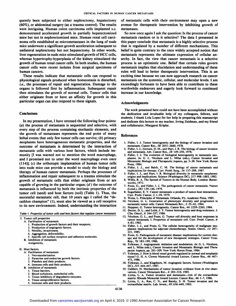
In my presentation, I have stressed the following four points:(a) the process of metastasis is sequential and selective, withevery step of the process containing stochastic elements, andthe growth of métastasesrepresents the end point of manylethal events that only few tumor cells can survive; (b) primaryneoplasms have heterogeneous metastatic properties, and theoutcome of metastasis is determined by the interaction ofmetastatic cells with various host factors, which include immunity [I was warned not to mention the word macrophage,and I promised not to utter the word macrophage even once(114)]; (c) the orthotopic implantation of human tumor cellsinto nude mice can provide a model to study the biology andtherapy of human cancer metastasis. Perhaps the processes ofinflammation and repair subsequent to a trauma stimulate thegrowth of metastatic cells that either originate from or arecapable of growing in the particular organ; (d) the outcome ofmetastasis is influenced by both the intrinsic properties of thetumor cell (seed) and host factors (soil) (Table 1). Thus, thesuccessful metastatic cell, that 10 years ago I called the "decathlon champion" (1), must also be viewed as a cell receptive
to its new environment. Indeed, understanding the interaction
Table 1 Properties of tumor cells and host factors that regulate cancer metastasis
I. Tumor cell propertiesA. Facilitation of metastasis
1. Production of growth factors and their receptors.2. Production of angiogenic factors.3. Motility. invasiveness.4. Aggregation, deformibility.5. Specific cell surface receptors and adhesion molecules.
B. Inhibition of metastasisAmigenicity.
II. Host factorsA. Facilitation of metastasis
1. Neovascularization.2. Paracrine and endocrine growth factors.3. Platelets and their products.4. Immune cells and their products.
B. Inhibition of metastasis1. Tissue barriers.2. Blood turbulence, endothelial cells.3. Tissue inhibitors of degradative enzymes.4. Tissue antiproliferative factors.5. Immune cells and their products.
of metastatic cells with their environment may open a newavenue for therapeutic intervention by inhibiting growth ofmétastases.
So now once again I ask the question: Is the process of cancermetastasis random or is it selective? The data I presented inthis paper conclude that metastasis is a highly selective processthat is regulated by a number of different mechanisms. Thisbelief is quite contrary to the once widely accepted notion thatmetastasis represents the ultimate expression of cellular anarchy. In fact, the view that cancer metastasis is a selectiveprocess is an optimistic one. Belief that certain rules governmetastasis implies that elucidation and understanding of theserules will lead to better therapeutic intervention. This is anexciting time because we can now approach research on cancermetastasis on the systemic, cellular, and molecular levels. I amexceedingly fortunate to have been able to contribute to theseworthwhile endeavors and eagerly look forward to continuedincrease in our knowledge.
Acknowledgments
The work presented here could not have been accomplished withoutthe dedication and invaluable help of my colleagues, fellows, andstudents. I thank Lola Lopez for her help in preparing this manuscriptand dedicate this lecture to my teacher, Irving Zeidman, and my friendand collaborator, Margaret Kripke.
References
1. Fidler, I. J. Tumor heterogeneity and the biology of cancer invasion andmetastasis. Cancer Res., 38: 2651-2660. 1978.
2. Fidler, I. J., Gersten, D. M., and Hart, I. R. The biology of cancer invasionand metastasis. Adv. Cancer Res., 28: 149-250, 1978.
3. Fidler, I. J. The evolution of biological heterogeneity in metastatic neoplasms. In: G. L. Nicolson and L. Milas (eds.). Cancer Invasion andMetastasis: Biologic and Therapeutic Aspects, pp. 5-30. New York: RavenPress, 1984.
4. Fidler. I. J., and Balch, C. M. The biology of cancer metastasis andimplications for therapy. Curr. Probi. Surg., 24: 137-209, 1987.
5. Fidler. I. J., and Hart. I. R. Biological diversity in metastatic neoplasms:origins and implications. Science (Washington DC), 217: 998-1003, 1982.
6. Willis, R. A. The Spread of Tumors in the Human Body. London: Butter-worths, 1972.
7. Poste, G., and Fidler, I. J. The pathogenesis of cancer metastasis. Nature(Lond.), 283: 139-146, 1979.
8. Sugarbaker, E. V. Cancer metastasis: a product of tumor-host interactions.Curr. Probi. Cancer, 3: 1-59, 1979.
9. Weiss, L. Principles of Metastasis. Orlando, FL: Academic Press, 1985.10. Nicolson, G. L. Generation of phenotypic diversity and progression in
metastatic tumor cells. Cancer Metastasis Rev., 3: 25-42, 1984.11. Heppner, G. Tumor heterogeneity. Cancer Res., 44: 2259-2265, 1984.12. Dexter, D. L., and Leith, J. T. Tumor heterogeneity and drug resistance. J.
Clin. Oncol.. 4: 244-257, 1986.13. Nicolson, G. L., and Poste, G. Tumor cell diversity and host responses in
cancer metastasis. I. Properties of metastatic cell. Curr. Probi. Cancer, 6:4-83, 1982.
14. Fidler. I. J., and Poste, G. The cellular heterogeneity of malignant neoplasms: implications for adjuvant chemotherapy. Semin. Oncol.. 12: 207-221, 1985.
15. Poste, G. Pathogenesis of metastatic disease: implications for current therapy and for the development of new therapeutic strategies. Cancer Treat.Rep., 70: 183-199, 1986.
16. Folkman. J. Angiogenesis: initiation and modulation. In: G. L. Nicolson,and L. Milas (eds.). Cancer Invasion and Metastasis: Biologic and Therapeutic Aspects, pp. 201-209. New York: Raven Press, 1984.
17. Folkman, J. How is blood vessel growth regulated in normal and neoplastictissue? G. H. A. Clowes Memorial Award Lecture. Cancer Res., 46: 467-473, 1986.
18. Folkman, J., and Klagsburn, M. Angiogenic factors. Science (WashingtonDC), 235: 444-447. 1987.
19. Gabbert, H. Mechanisms of tumor invasion: evidence from in vivo observations. Cancer Metastasis Rev., 4: 283-310, 1985.
20. Liotta, L. A. Tumor invasion and metastasis—role of the extracellularmatrix: Rhoads Memorial Award Lecture. Cancer Res., 46: 1-7, 1986.
21. Liotta. L. A., Rao, C. N., and Barsky. S. H. Tumor invasion and theextracellular matrix. Lab. Invest.. 49: 636-649, 1983.
6136
on April 4, 2019. © 1990 American Association for Cancer Research. cancerres.aacrjournals.org Downloaded from

CRITICAL FACTORS IN HUMAN CANCER METASTASIS
22. Marcel. M. Invasion in vitro: methods of analysis. Cancer Metastasis Rev.,2:201-209, 1983. 55.
23. Fisher, B., and Fisher, E. R. The interrelationship of hematogenous andlymphatic tumor cell dissemination. Surg. Gynecol. Obstet., 122: 791-797,1966. 56.
24. Nicolson, G. L. Cancer metastasis: tumor cell and host organ propertiesimportant in metastasis to specific secondary sites. Biochim. Biophys. Acta, 57.948: 175-224. 1988.
25. Horak, E.. Darling, D. L.. and Tarin. D. Analysis of organ-specific effectson metastatic tumor formation by studies in vitro. 3. Nati. Cancer Inst., 76: 58.913-922, 1986.
26. Naito. S., Giavazzi, R., and Fidler. I. J. Correlation between the in vitro 59.interaction of tumor cells with an organ environment and metastatic behavior in rivo. Invasion Metastasis. 7: 16-29, 1987.
27. Price, J. E.. Tarin. D., and Fidler. I. J. The influence of organ microenvi- 60.ronment on pigmentation of a metastatic murine melanoma. Cancer Res.,48: 2258-2264, 1988.
28. Price, J. E., Naito, S., and Fidler. I. J. The role of the organ microenviron- 61.ment in the selective process of metastasis. Clin. Exp. Metastasis, 6: 91-102, 1988.
29. Weiss, L. Metastatic inefficiency: causes and consequences. Cancer Rev., 3: 62.1-24, 1986.
30. Fidler. I. J. Metastasis: quantitative analysis of distribution and fate oftumor cell emboli labeled with '"l-5-iododeoxyuridine. J. Nati. Cancer 63.Inst., 45: 773-782. 1970.
31. Fidler. I. J. Selection of successive tumor lines for metastasis. Nat. NewBiol.. 242: 148-149. 1973. 64.
32. Nicolson, G. L., and Dulski. K. M. Organ specificity of metastatic tumorcolonization is related to organ-selective growth properties of malignantcells. Int. J. Cancer. 38: 289-294, 1986. 65.
33. Raz. A.. Hanna. N.. and Fidler, I. J. In vivo isolation of a metastatic tumorcell variant involving selective and nonadaptive processes. J. Nati. Cancer 66.Inst., 66: 183-194. 1981.
34. Talmadge. J. E.. and Fidler. 1. J. Cancer metastasis is selective or random 67.depending on the parent tumor population. Nature (Lond.), 27: 593-594,1978. 68.
35. Fidler, I. J. General considerations for studies of experimental cancermetastasis. In: H. Bush (ed.). Methods in Cancer Research, pp. 399-439.New York: Academic Press, 1978. 69.
36. Poste. G. Experimental systems for analysis of the malignant phenotype.Cancer Metastasis Rev., /: 141-200, 1982. 70.
37. Talmadge. J. E., and Fidler. I. J. Enhanced metastatic potential of tumorcells harvested from spontaneous métastasesof heterogeneous murine tumors. J. Nati. Cancer Inst.. 69: 975-980. 1982. 71.
38. Raz. A., and Hart. I. Murine melanoma: a model for intracranial metastasis.Br. J. Cancer. 42: 331-341. 1980.
39. Brunson. K. \V., and Nicolson, G. L. Selection and biologic properties ofmalignant variants of a murine lymphosarcoma. J. Nati. Cancer Inst.. 61: 72.1499-1503, 1978.
40. Fidler. I. J.. Gersten, D. M., and Budmen. M. B. Characterization in vivo 73.and in vitro of tumor cells selected for resistance to syngeneic lymphocyte-mediated cytotoxicity. Cancer Res.. 36: 3160-3165. 1976.
41. Fidler. I. J., and Bucana. C. D. Mechanism of tumor cell resistance to lysis 74.by syngeneic lymphocytes. Cancer Res., 37: 3945-3956, 1977.
42. Briles. E. B.. and Kornfeld. S. Isolation and metastatic properties of detachment variants of B16 melanoma cells. J. Nati. Cancer Inst., 60:1217-1222, 75.1978.
43. Reading. C. L., and Hutchins. J. T. Carbohydrate structure in tumorimmunity. Cancer Metastasis Rev., 4: 221-260. 1985. 76.
44. Hart. I. R. The selection and characterization of an invasive variant of theB16 melanoma. Am. J. Palhol.. 97: 587-600. 1979.
45. Poste. G.. Doll. J.. Hart. I. R.. and Fidler. I. J. In vitro selection of murineBI6 melanoma variants with enhanced tissue invasive properties. Cancer 77.Res.. 40: 1636-1644. 1980.
46. Sloane. B. F., and Honn. K. V. Cysteine proteinase and metastasis. CancerMetastasis Rev.. 3: 249-265, 1984. 78.
47. Kerbel. R. S. Immunologie studies of membrane mutants of a highlymctastalic murine tumor. Am. J. Pathol.. 97:609-616. 1979.
48. Kerbel, R. S., Dennis, J. W., Lagarde, A. E., and Frost, P. Tumor progression in metastasis: an experimental approach using lectin-resistant tumor 79.variants. Cancer Metastasis Rev., /: 99-140, 1982.
49. Hanna. N. Role of natural killer cell in control of cancer metastasis. CancerMetastasis Rev., /: 45-65. 1982. 80.
50. Fidler. I. J., and Kripke. M. L. Metastasis results from pre-existing variantcells within a malignant tumor. Science (Washington DC). 797: 893-895,1977.
51. I mia. S. E., and Delbruck. M. Mutations of bacteria from virus sensitive 81.to virus resistance. Genetics, 28: 491-511. 1943.
52. Kripke. M. L. Speculation on the role of ultraviolet radiation in thedevelopment of malignant melanoma. J. Nati. Cancer Inst., 63: 541-545,1979. 82.
53. Fidler. I. J.. and Kripke. M. L. Metastatic heterogeneity of cells from theK-1735 melanoma. In: E. Grundmann (ed.). Metastatic Tumor Growth, pp.71-81. Stuttgart: Gustav Fischer Verlag, 1980.
54. Fidler, I. J., Gruys, E.. Cifone, M. A., Barnes, Z., and Bucana. C. Demon- 83.stration of multiple phenotype diversity in a murine melanoma of recent
6137
origin. J. Nati. Cancer Inst., 67: 947-956. 1981.Fidler, I. J., Gersten, D. M., and Kripke, M. L. Influence of immune statuson the metastasis of three murine fìbrosarcomasof different immunogenic-ities. Cancer Res.. 39: 3816-3821. 1979.Kripke, M. L. Immunoregulation and carcinogenesis: past, present, andfuture. J. Nati. Cancer Inst., 80: 722-727. 1988.Frost. P.. and Kerbel, R. S. Immunology of metastasis: can the immuneresponse cope with disseminated tumor? Cancer Metastasis Rev., 2: 239-256, 1983.Fidler, I. J., and Kripke, M. L. Tumor cell antigenicity, host immunity, andcancer metastasis. Cancer Immunol. Immunother., 7: 201-205, 1980.Aukerman, S. L., Price, J. E., and Fidler. I. J. Different deficiencies in theprevention of tumorigenic-low-metastatic murine K-1735 melanoma cellsfrom producing métastases.J. Nati. Cancer Inst., 77: 915-924, 1986.Fidler, I. J. Rationale and methods for the use of nude mice to study thebiology and therapy of human cancer metastasis. Cancer Metastasis Rev.,5:29-49, 1986.Price, J. E., Aukerman, S. L., and Fidler, 1. J. Evidence that the process ofmurine melanoma metastasis is sequential and selective and contains stochastic elements. Cancer Res., 46: 5172-5178, 1986.Fidler. I. J., Gersten. D. M., and Riggs. C. W. Relationship of host immunestatus to tumor cell arrest, distribution, and survival in experimental animals. Cancer (Phila.), 40: 46-55, 1977.Hart, I. R., Talmadge, J. E., and Fidler, I. J. Metastatic behavior of amurine reticulum cell sarcoma exhibiting organ-specific growth. CancerRes., 41: 1281-1287. 1981.Fidler, I. J., and Talmadge, J. E. Evidence that intravenously derived murinepulmonary métastasescan originate from the expansion of a single tumorcell. Cancer Res., 46: 5167-5171, 1986.Page!. S. The distribution of secondary growths in cancer of the breast.Lancet. /: 571-573, 1889.Hart, I. R. "Seed and soil" revisited: mechanisms of site-specific metastasis.Cancer Metastasis Rev., /: 5-17, 1982.Hart, I. R., and Fidler, I. J. Role of organ selectivity in the determinationof metastalic patterns of B16 melanoma. Cancer Res., 41:1281-1287,1981.Netland. P. A., and Zetter, B. R. Metastatic potential of B16 melanomacells after in vitro selection for organ-specific adherence. Cell Biol., 101:720-724, 1985.Ewing, J. Neoplastic Diseases, Ed. 6. Philadelphia: W. B. Saunders Co..1928.Tarin. D., Price. J. E.. Kettlewell. M. G. W., Souter, R. G., Vass, A. C. R.,and Crossley. B. Mechanisms of human tumor metastasis studied in patientswith peritoneovenous shunts. Cancer Res., 44: 3584-3592, 1984.Tarin, D., Price, J. E., Kettlewell, M. G. W., Souter, R. G., Vass, A. C. R.,and Crossley, B. Clinicopathological observations on metastasis in manstudied in patients treated with peritoneovenous shunts. Br. Mcd. J., 288:749-751, 1984.Schackert. G., and Fidler, 1. J. Development of in vivo models for studiesof brain metastasis. Int. J. Cancer, 41: 589-594, 1988.Schackert, G., and Fidler, I. J. Site-specific metastasis of mouse melanomasand a fibrosarcoma in the brain or the meninges of syngeneic animals.Cancer Res.. 48: 3478-3483, 1988.August, D. A., Ottow, R. T., and Sugarbaker, P. H. Clinical perspectives ofhuman colorectal cancer metastasis. Cancer Metastasis Rev., 3: 303-325,1984.Pollack. V. A., and Fidler, I. J. Use of young nude mice for selection ofsubpopulations of cells with increased metastalic potential from nonsynge-neic neoplasms. J. Nati. Cancer Inst., 69: 137-141, 1982.Giavazzi, R., Campbell, D. E., Jessup, J. M. Cleary, K.. and Fidler, I. J.Metastatic behavior of tumor cells isolated from primary and metastatichuman coloréela!carcinomas implanted into different sites of nude mice.Cancer Res., 46: 1928-1933. 1986.Giavazzi. R.. Jessup. J. M., Campbell, D. E., Walker. S. M., and Fidler, l.J. Experimental nude model of human colorectal cancer liver metastasis. J.Nati. Cancer Inst., 77: 1303-1308, 1986.Sordat, B. C. M., Ueyama, Y., and Fogh, J. Metastasis of tumor xenograftsin the nude mouse. In: 3. Fogh and B. C. Giovanella (eds.). The Nude Mousein Experimental and Clinical Research, Vol. 2, pp. 95-147. New York:Academic Press, 1982.Kozlowski. J. M., Fidler, I. J., Campbell, D., Xu, Z., Kaighn, M. E., andHart. I. R. Metastatic behavior of human tumor cell lines grown in the nudemouse. Cancer Res., 44: 3522-3529, 1984.Naito, S., von Eschenbach, A. C., Giavazzi, R., and Fidler, I. J. Growthand metastasis of tumor cells isolated from a human renal cell carcinomaimplanted into different organs of nude mice. Cancer Res., 46: 4109-4115,1986.Morikawa. K.. Walker. S. M., Jessup, J. M., and Fidler, I. J. In vivoselection of highly metastatic cells from surgical specimens of differenthuman colon carcinomas implanted into nude mice. Cancer Res., 48: 1943-1948. 1988.Morikawa. K., Walker, S. M., Nakajima, M., Pathak. S., Jessup. J. M., andFidler. I. J. Influence of organ environment on the growth, selection, andmetastasis of human colon carcinoma cells in nude mice. Cancer Res., 48:6863-6871, 1988.Jessup. J. M., Giavazzi, R., Campbell, D., Cleary, K. R., Morikawa, K.,Hosteller, R., Atkinson. E. N., and Fidler, I. J. Metastatic potential of
on April 4, 2019. © 1990 American Association for Cancer Research. cancerres.aacrjournals.org Downloaded from

CRITICAL FACTORS IN HUMAN CANCER METASTASIS
human colorectal carcinomas implanted into nude mice: prediction of clinical outcome in patients operated upon for cure. Cancer Res., 49: 6906-6910. 1989.
84. Jessup, J. M. Giavazzi, R., Campbell. D., Clear), K.. Morikawa, K.. andFidler. 1.J. Growth potential of human colorectal carcinomas in nude mice:association with preoperative serum concentration of carcinoembryonicantigen. Cancer Res.. 48: 1689-1692. 1988.
85. Thombre, P. S.. and Doedhar. S. D. Inhibition of liver métastasesin murinecolon adenocarcinoma by liposomes containing human C-reactive proteinor crude lymphokines. Cancer Immunol. Immunother., 16: 145-150, 1984.
86. Goldroscn, M. H. Murine colon adenocarcinoma immunobiology of métastases. Cancer (Phila.). 45: 1223-1228, 1980.
87. Bresalier. R. S., Raper. S. E., Hujanen. E. S.. and Kim. V. S. A new modelfor human colon cancer metastasis. Int. J. Cancer, 39: 625-630, 1987.
88. Sordat. B.. and Wang. W. R. Human colorectal tumor xenografts in nudemice: expression of malignancy. Behring Inst. Mitt.. 74: 291-300, 1984.
89. dayman, R. V., Figenshau. R. S., and Bear, A. Transplantation of humanrenal carcinomas into athymic mice. Cancer Res., 45: 2650-2653, 1985.
90. Hoehn. W. A., and Schroeder. F. H. Renal cell carcinoma: two new celllines and a serially transplantable nude mouse tumor (NC 65). Invest. Urol.,16: 106-112, 1978.
91. Naito. S., Kanamori. T., and Hisano, S. Human renal cell carcinoma:establishment and characterization of two new cell lines. J. Urol., 128:1117-1121, 1982.
92. Naito, S., von Eschenbach. A. C.. and Fidler. 1. J. Different growth patternand biologic behavior of human and renal cell carcinoma implanted intodifferent organs of nude mice. J. Nail. Cancer Inst., 78: 377-385, 1987.
93. Wang, W. R., Sordat. B., Paiguet, D., and Sordat, M. Human colon tumorsin nude mice: implantation site and expression of the invasive phenotype.In: B. Sordat (ed.). Immune-deficient Animals, pp. 239-245. Basel: S.Karger AG. 1984.
94. Schackert. H.. and Fidler. I. J. Development of an animal model to studythe biology of recurrent colorectal cancer originating from mesenteric lymphsystem métastases.Int. J. Cancer. 44: 177-181, 1989.
95. Kozlowski. J. M., Hart, I. R., Fidler. I. J.. and Hanna. N. A humanmelanoma line heterogeneous with respect to metastatic capacity in athymicnude mice. J. Nati. Cancer Inst.. 72: 913-917. 1984.
96. Kerbel. R. S., Man, M. S., and Dexter, D. A model of human cancermetastasis: extensive spontaneous and artificial metastasis of a humanpigmentcd melanoma and derived variant sublines in nude mice. J. Nati.Cancer Inst.. 72:93-108, 1984.
97. Kerbel. R. S., and Man, M. S. Single-step selection of unique humanmelanoma variants displaying unusually aggressive metastatic behavior innude athymic mice. Invasion Metastasis, 4: 31-43, 1984.
98. Cornil, T., Man. M. S., Fernandez, B., and Kerbel. R. S. Enhanced tumor-igenicity. melanogenesis and metastasis of the human malignant melanomaobserved after subdermal implantation in nude mice. J. Nati. Cancer Inst..81: 938-944. 1989.
99. Shafie, S. M.. and Liotta. L. A. Formation of metastasis by human breastcarcinoma cells (MCF-7) in nude mice. Cancer Lett.. //: 81-87. 1980.
100. Price. J. E.. Polyzos, A., Zhang. R. D.. and Daniels. L. M. Tumorigenicityand metastasis of human breast carcinoma cell lines in nude mice. CancerRes., 50:717-721. 1990.
101. Levy, J. A., White, A. C.. and McGrath, C. M. Growth and histology of ahuman mammary-carcinoma cell line at different sites in the athymic mouse.Br. J. Cancer. 45: 375-383, 1982.
102. Ibrahiem. E. H. I., Nigam. V. N., Brailovsky. C. A., Madamas. P.. andMiniali. M. Orthotopic implantation of primary (iV-[4-(5-nitro-2-furyl)-2-thiazolyljformamide-induced bladder cancer in bladder submucosa: an animal model for bladder cancer study. Cancer Res., 43: 617-622, 1983.
103. Ahlering, T. E.. Dubeau. L.. and Jones. P. A. A new in vivo model to studyinvasion and metastasis of human bladder carcinoma. Cancer Res., 47:6660-6665, 1987.
104. Kajiji, S. M., Meitner, P. A., Bogaars. H. A., Dexter, D. L., Calabresi, P.,and Turner, M. D. Metastasis of a human pancreatic adenocarcinoma(RWP-1) in nude mice. Br. J. Cancer. 40:970-975, 1982.
105. Vezeridis, M. P., Turner, M. D., Kajiji, S. M., Yankee. R. A., and Meither.P. A. Metastasis of human pancreatic cancer (HPC) to liver in nude mice.Proc. Am. Assoc. Cancer Res., 26: 53, 1985.
106. Tan, M. H., and Chu, T. M. Characterization of the tumorigenic andmetastatic properties of a human pancreatic tumor cell line (ASPC-1)implanted orthotopically into nude mice. Tumour Biol., 6: 89-98. 1985.
107. Marincola. F. M.. Drucker, B. J., Siao, D. Y„Hough. K. L., and Holder.W. D.. Jr. The nude mouse as a model for the study of human pancreaticcancer. J. Surg. Res.. 47: 520-529, 1989.
108. Vezeridis, M. P., Doremus, C. M., Tibbetts, L. M., Tzanakakis. G., andJackson. B. T. Invasion and metastasis following orthotopic transplantationof human pancreatic cancer in the nude mouse. J. Surg. Oncol.. 40: 261-265. 1989.
109. McLemore. T. L., Liu. M. C. Blacker, P. C., Gregg, M.. Alley, M. C.,Abbott, B. J., Shoemaker, R. H.. Bohlman. M. E.. Litters!, C. C., Hubbard.W. C., Brennan. R. H.. McMahon, J. B., Fine, D. L.. Eggleston. J. C,Mayo. J. G.. and Boyd, M. R. Novel intrapulmonar)' model for orthotopicpropagation of human lung cancers in athymic nude mice. Cancer Res., 47:5132-5140, 1987.
110. Meyvisch, C. Influence of implantation site on formation of metastasis.Cancer Metastasis Rev.. 2: 295-306, 1983.
111. Poste, G. Experimental systems for analysis of the malignant phenotype.Cancer Metastasis Rev., /: 141-199. 1982.
112. Miller. F. R. Comparison of metastasis of mammary tumors growing in themammary fatpad versus the subcutis. Invasion Metastasis, 7:220-226,1981.
113. Malave, !.. Blanca, I., and Fuji. H. Influence of inoculation site on development of the Lewis lung carcinoma and suppressor cell activity in syngeneicmice. J. Nati. Cancer Inst.. 62: 83-88. 1979.
114. Fidler, I. J. Macrophages and metastasis: a biological approach to cancertherapy. Cancer Res., 45: 4714-4726. 1985.
6138
on April 4, 2019. © 1990 American Association for Cancer Research. cancerres.aacrjournals.org Downloaded from

1990;50:6130-6138. Cancer Res Isaiah J. Fidler Twenty-eighth G. H. A. Clowes Memorial Award LectureCritical Factors in the Biology of Human Cancer Metastasis:
Updated version
http://cancerres.aacrjournals.org/content/50/19/6130
Access the most recent version of this article at:
E-mail alerts related to this article or journal.Sign up to receive free email-alerts
Subscriptions
Reprints and
To order reprints of this article or to subscribe to the journal, contact the AACR Publications
Permissions
Rightslink site. Click on "Request Permissions" which will take you to the Copyright Clearance Center's (CCC)
.http://cancerres.aacrjournals.org/content/50/19/6130To request permission to re-use all or part of this article, use this link
on April 4, 2019. © 1990 American Association for Cancer Research. cancerres.aacrjournals.org Downloaded from