cross talk between tetanus neurotoxin-insensitive vesicle ...edoc.mdc-berlin.de/6818/1/6818oa.pdfthe...
TRANSCRIPT
Molecular Biology of the CellVol. 14, 4207–4220, October 2003
Cross Talk between Tetanus Neurotoxin-insensitiveVesicle-associated Membrane Protein-mediatedTransport and L1-mediated Adhesion□D□V
Philipp Alberts,* Rachel Rudge,* Ina Hinners,† Aude Muzerelle,‡Sonia Martinez-Arca,* Theano Irinopoulou,* Veronique Marthiens,§Sharon Tooze,† Fritz Rathjen,� Patricia Gaspar,‡ and Thierry Galli*¶
*Membrane Traffic and Neuronal Plasticity, Institut National de la Sante et de la Recherche MedicaleU536, F-75005 Paris, France; †Secretory Pathways Laboratory, Cancer Research UK, London ResearchInstitute, London WC2A 3PX, United Kingdom; ‡Institut National de la Sante et de la RechercheMedicale U106, Hopital Salpetriere, F-75651, Paris, France; §Institut National de la Sante et de laRecherche Medicale U440, F-75005 Paris, France; and �Max-Delbrueck-Centrum fuer MolekulareMedizin, D-13092 Berlin, Germany
Submitted March 14, 2003; Revised June 2, 2003; Accepted June 4, 2003Monitoring Editor: Jennifer Lippincott-Schwartz
The membrane-trafficking pathway mediated by tetanus neurotoxin-insensitive vesicle-associated membrane protein(TI-VAMP) in neurons is still unknown. We show herein that TI-VAMP expression is necessary for neurite outgrowth inPC12 cells and hippocampal neurons in culture. TI-VAMP interacts with plasma membrane and endosomal target solubleN-ethylmaleimide-sensitive factor attachment protein receptors, suggesting that TI-VAMP mediates a recycling pathway.L1, a cell-cell adhesion molecule involved in axonal outgrowth, colocalized with TI-VAMP in the developing brain,neurons in culture, and PC12 cells. Plasma membrane L1 was internalized into the TI-VAMP–containing compartment.Silencing of TI-VAMP resulted in reduced expression of L1 at the plasma membrane. Finally, using the extracellulardomain of L1 and N-cadherin immobilized on beads, we found that the silencing of TI-VAMP led to impaired L1- but notN-cadherin–mediated adhesion. Furthermore, TI-VAMP- but not synaptobrevin 2-containing vesicles accumulated at thesite of the L1 bead-cell junction. We conclude that TI-VAMP mediates the intracellular transport of L1 and thatL1-mediated adhesion controls this membrane trafficking, thereby suggesting an important cross talk between membranetrafficking and cell-cell adhesion.
INTRODUCTION
The development of the brain depends on the differentiationof neurons and particularly on the extension, branching, andconnection of their axons and dendrites (Prochiantz, 1995).The outgrowth of axons and dendrites requires massivetransport of lipids and proteins from the Golgi apparatus tothese extensions of the plasma membrane, but the underly-ing molecular mechanism is largely unknown (Futermanand Banker, 1996). Neurite outgrowth also depends on therecycling of plasma membrane proteins, particularly cellsubstrate adhesion molecules (CAMs) such as integrins andCAMs of the immunoglobulin superfamily (IgCAMs) (Ka-
miguchi and Lemmon, 2000a). Current knowledge indicatesthat the exocytosis mediating neurite outgrowth differs fromthe exocytosis mediating neurotransmitter release, suggest-ing that separate subcellular and molecular pathways un-derlie each process. Syntaxin (Stx) 1, synaptosomal-associ-ated protein 25 kd (SNAP25), and synaptobrevin 2 (Syb 2),the components of the synaptic soluble N-ethylmaleimide-sensitive factor attachment protein receptor (SNARE) com-plex, mediate the fusion of synaptic vesicles with the plasmamembrane (for review, see Galli and Haucke, 2001). Accord-ingly, genetic ablation of SNAP25 leads to a blockade ofevoked neurotransmitter release (Washbourne et al., 2002),and Syb 2 genetic inactivation causes an almost completeloss of spontaneous and evoked release (Schoch et al., 2001).However, the development of the brain seems normal inboth Syb 2 and SNAP25 knockout mice (Schoch et al., 2001;Washbourne et al., 2002), indicating that these molecules arenot necessary for neurite outgrowth. Similar results werealso obtained in the case of Munc-18, an important regulatorof syntaxin 1 (Verhage et al., 2000). Furthermore, clostridialneurotoxins that cleave Syb have no effect on the develop-ment of neurons in culture (Osen-Sand et al., 1996; Grosse etal., 1999). However, former data have reported that botuli-num neurotoxins that cleave SNAP25 do inhibit neuriteoutgrowth (Osen-Sand et al., 1996). This discrepancy with
Article published online ahead of print. Mol. Biol. Cell 10.1091/mbc.E03–03–0147. Article and publication date are available atwww.molbiolcell.org/cgi/doi/10.1091/mbc.E03–03–0147.
□D □V Online version of this article contains both videos andsupplementary figure material for some figures. Onlineversion is available at www.molbiolcell.org.
¶ Corresponding author. E-mail address: [email protected] used: CAM, cell adhesion molecule; IgCAM, celladhesion molecule of the immunoglobulin superfamily; Stx,syntaxin; siRNA, small interfering double-stranded RNA; TI-VAMP, tetanus neurotoxin-insensitive vesicle-associated mem-brane protein.
© 2003 by The American Society for Cell Biology 4207
the findings in the SNAP25 knockout mouse could be owingto the expression of SNAP23, a close homolog that may beable to functionally replace SNAP25. In any case, theseresults clearly indicate that the molecules involved in themembrane-trafficking pathway mediating neurite out-growth are, to a large extent, different from those involved inneurotransmitter release.
The finding that neurite outgrowth and apical transport inepithelial cells are insensitive to tetanus neurotoxin led us toinvestigate the function of tetanus neurotoxin-insensitivevesicle-associated membrane protein (TI-VAMP) (Galli et al.,1998). We previously found that TI-VAMP defines a novelcompartment, particularly enriched at the tip of growthcones in developing neurons (Coco et al., 1999). TI-VAMPshows the same general structure as classical VAMPs, i.e., aSNARE motif, a carboxy-terminal transmembrane domain,and a short lumenal domain, but in addition carries anamino-terminal extension called the Longin domain (Filip-pini et al., 2001). This domain inhibits the SNARE motif frominteracting with cognate SNARE complexes both in vitroand in vivo (Martinez-Arca et al., 2000, 2003b). Expression ofthe Longin domain of TI-VAMP results in strong inhibitionof neurite outgrowth in PC12 cells (Martinez-Arca et al.,2000) and both axonal and dendritic outgrowths in neuronsin culture (Martinez-Arca et al., 2001).
These results prompted us to further characterize the ve-sicular compartment containing TI-VAMP and its functionin neurons. We show herein that TI-VAMP is essential forneurite outgrowth and interacts with plasma membrane andendosomal t-SNAREs. We found that L1, a cell-cell adhesionmolecule involved in axonal outgrowth (Brummendorf et al.,1998) and cell motility (Mechtersheimer et al., 2001) colocal-ized with TI-VAMP in the developing brain, neurons inculture, and PC12 cells. Using the extracellular domain of L1and N-cadherin immobilized on beads, we found that thesilencing of TI-VAMP led to impaired L1- but not N-cad-herin–mediated adhesion and that TI-VAMP–containingvesicles accumulated at the site of the L1 bead-cell junction.Therefore, our results demonstrate a cross talk betweenTI-VAMP–mediated transport and L1-mediated adhesion.
MATERIALS AND METHODS
Antibodies, Clones, and ReagentsThe mouse monoclonal antibody (mAb) (clone 158.2) directed against TI-VAMP has been described elsewhere (Muzerelle et al., 2003). Polyclonalanti-green fluorescent protein (GFP) antibody was described previously (Mar-tinez-Arca et al., 2001). Mouse monoclonal antibodies directed against Syb 2(clone 69.1) and syntaxin 1 (HPC-1) and the polyclonal antibody againstsyntaxin 7 were generous gifts from R. Jahn (Max Planck Institute, Goettin-gen, Germany), C. Barnstable (Yale University, New Haven, CT), and Dr. W.Hong (Institute of Molecular and Cell Biology, Singapore, Republic of Singa-pore), respectively. Polyclonal rabbit antibodies against mouse L1 and Fabfragments generated from this antibody were described previously (Rathjenand Rutishauser, 1984). The polyclonal anti-Syb 2 antibody (MC23) wasdescribed previously (Chilcote et al., 1995). The following commercial anti-bodies were used in this study: monoclonal anti-Na/K-ATPase a-1 (cloneC464.6) from Upstate Biotechnology (Charlottesville, VA), monoclonal anti-syntaxin 13 (clone 15G2) from StressGen (San Diego, CA), and monoclonalanti-Vti1b (Transduction Laboratories clone 7) from BD Biosciences (FranklinLakes, NJ). Affinity-purified Alexa 488 and Cy3-coupled goat anti-rabbit andanti-mouse immunoglobulins were from Jackson Immunoresearch Laborato-ries (West Grove, PA). Horseradish peroxidase- and alkaline phosphatase-coupled streptavidin, sheep anti-mouse and anti-rabbit IgGs were from Pro-mega (Madison, WI). Nerve growth factor (NGF) was supplied by AlomoneLabs (Rehovot, Israel). All other reagents were from Sigma-Aldrich (SaintQuentin Fallavier, France) unless specified.
DNA ConstructionsThe cDNA coding for L1-Fc chimera has been described previously (DeAngelis et al., 1999). Expression vectors coding for GFP and TI-VAMP fusedto GFP have been described previously (Martinez-Arca et al., 2000).
Cell CulturePC12 cells were cultured and plated on collagen-coated glass coverslips asdescribed previously (Martinez-Arca et al., 2000). Cortical and striatal neuronswere prepared from rat E16 embryos as described previously (Rousselet et al.,1990). Hippocampal neurons from E18 rat brain were prepared as describedpreviously (Chang and De Camilli, 2001). Purification of L1-Fc chimera wasperformed as described previously (De Angelis et al., 1999). Briefly, Cos-7 cellswere transiently transfected with cDNA coding for L1-Fc chimera by usingLipofectAMINE-2000. Secreted L1-Fc protein was allowed to accumulate forseveral days in Ig-free medium. The supernatant was collected and recombi-nant protein was purified using protein A-Sepharose. To coat coverslips withL1-Fc chimera, coverslips were incubated for 2 h at 37°C with 20 �g/ml goatanti-human Fc-specific antibody (Sigma, Saint Quentin Fallavier, France) inLeibovitz medium, pH 8 (Invitrogen, Cergy Pontoise, France). Coverslipswere rinsed and incubated with 3 �g/ml L1-Fc fragment in Leibovitz mediumfor another 2 h at 37°C.
Small Interfering Double-Stranded RNA (siRNA)Treatment of PC12 CellsPC12 cells grown on collagen-coated 35-mm dishes were transfected by using10 �l LipofectAMINE-2000 twice on two consecutive days with 0.5 �g of GFPplus either 4 �g of siRNA molecules corresponding to the rat TI-VAMPsequence, base pairs 486–506: AACCTCGTAGATTCGTCCGTC (siRNAr;Dharmacon, Lafayette, CO) or control siRNA molecules corresponding to theequivalent sequence of canine TI-VAMP: AATCTTGTGGATTCGTCTGTC(siRNAd; Proligo-Genset, Paris, France). The canine TI-VAMP sequence wasobtained by reverse transcription-polymerase chain reaction of RNA fromMadin-Darby canine kidney cells. The next day, cells were split and aliquotscultured on either collagen-coated coverslips or put back on collagen-coatedculture dishes. For the neurite outgrowth assay, the cells were differentiatedby the addition of 100 nM staurosporine 48 h after the second transfection,fixed the next day (i.e., 96 h after the first transfection), and processed forimmunofluorescence with anti-GFP and anti-TI-VAMP antibodies. Thirtyrandomly chosen images for each condition were taken based on the GFPsignal with a MicroMax charge-coupled device (CCD) camera (PrincetonInstruments, Princeton, NJ), resulting in the analysis of at least 50 GFP-positive cells. A neurite was defined as a thin process �10 �m long. Thelength of each neurite, from the cell body limit until the tip of the process, wasmeasured in each case by using MetaMorph software (Princeton Instru-ments). Differences were evaluated statistically with the chi square test. Allthe recordings and the MetaMorph analysis were done blind.
siRNA Treatment of Hippocampal NeuronsNeurons were dissected as described and plated on poly-d-lysine–coated14-mm coverslips at a density of 25,000 cells/coverslip (16,500 cell/cm2). Fourhours after plating, the media were replaced with neurobasal media plus 4%B27, and cells were transfected with 0.25 �g of GFP alone or cotransfectedwith 0.8 �g of siRNAr or siRNAd by using 0.5 �l of LipofectAMINE 2000.Four hours after transfection, the media were changed again to prevent anytoxic effects of the LipofectAMINE. Neurons were transfected for a secondtime 24 h after plating; again, the media were replaced 4 h after transfection.Three days after dissection, neurons were fixed and processed for immuno-fluorescence with anti-GFP and anti-TI-VAMP antibodies. Transfected cellsfrom three independent experiments were imaged with a MicroMax CCDcamera (Princeton Instruments), resulting in the analysis of �75 neurons foreach of the control conditions and �150 neurons treated with siRNAr. Neuriteoutgrowth measurements and statistical analysis were performed as de-scribed above.
Antibody Uptake AssayPC12 cells were plated on collagen-coated glass coverslips and treated withNGF at a concentration of 50 ng/ml for 2–3 d. Cells were incubated with Fabfragments directed against L1 in NGF-containing medium at a concentrationof 25 �g/ml for 60 min at 37°C. The cells were fixed after washes in coldphosphate-buffered saline with 1 mg/ml bovine serum albumin (BSA) andacid stripping with 0.2 M acetic acid/0.5 M NaCl for 2 min at 4°C. Antibodyuptake on neurons grown on collagen-coated glass coverslips was performedessentially as described for PC12 cells. No acid stripping was performed, butafter removal of Fab fragments and two washes of the cells in medium,neurons were reincubated for 60 min at 37°C.
ImmunocytochemistryDevelopmental localization of TI-VAMP and L1 was analyzed in Sprague-Dawley rats. Embryos (E13, E15, and E19) were extracted from timed preg-
P. Alberts et al.
Molecular Biology of the Cell4208
nant dams after chloral hydrate anesthesia (E0, day of mating) and fixed in 4%paraformaldehyde overnight. After 24-h cryoprotection in 10% sucrose inphosphate buffer, serial cryostat sections (15 �m in thickness) were takenfrom the whole head in the sagittal plane for embryos. Sections were incu-bated overnight at room temperature in TI-VAMP mAb (clone 158.2; 1/500)alone or in combination with the L1 antibody (1/500) and processed forimmunofluorescence.
Cells in culture were fixed with 4% paraformaldehyde/4% sucrose andprocessed for immunofluorescence as described previously (Coco et al., 1999).Optical conventional fluorescence microscopy was performed on a Provismicroscope (Olympus, Tokyo, Japan) equipped with a MicroMax CCD cam-era (Princeton Instruments). Confocal laser scanning microscopy was per-formed using a SP2 confocal microscope (Leica Microsystems, Mannheim,Germany). Images were assembled using Adobe Photoshop (Adobe Systems,San Jose, CA).
Quantification of surface L1 signal was performed with a semiautomaticprogram based on the MetaMorph software (Universal Imaging, Downing-town, PA). Cells were segmented automatically, and the mask obtained by thesegmentation procedure was used to compute the average intensity of thefluorescence over each cell. All acquisition and segmentation parameters werekept constant for all images. Differences were evaluated statistically with theMann-Whitney nonparametric test.
ImmunoprecipitationImmunoprecipitation from rat brain was performed as described previously(Martinez-Arca et al., 2000). Monoclonal anti-TI-VAMP antibody 158.2 andcontrol mouse IgG were covalently coupled to protein G-Sepharose accordingto Harlow and Lane (1988). Rat brain lysate was incubated with immuno-beads overnight, and the beads were washed five times in homogenizationbuffer with 0.1% Triton X-100. Bound material was separated by SDS-PAGE(Schagger and von Jagow, 1987) followed by Western blotting.
Subcellular FractionationPC12 cells were homogenized with an EMBL-cell cracker in 10 mM HEPES-KOH pH 7.2, 250 mM sucrose, 1 mM EDTA, 1 mM MgOAc, and proteaseinhibitors. The homogenate was centrifuged at 800 � g for 10 min, and 1.5 mlof the supernatant was loaded onto a 0.3–1.2 M linear sucrose gradient andspun for 15 min in a SW40 rotor at 25,000 rpm (velocity gradient). Then, 1-mlfractions were unloaded from the top; membranes were pelleted and resus-pended in sample buffer. Equal volumes of each fraction were separated bySDS-PAGE (Laemmli, 1970) and analyzed by Western blotting. For equilib-rium gradient analysis the light fractions 1–4 of the velocity gradient werepooled and loaded on a sucrose step gradient of the following composition: 1ml 0.8 M, 2 ml 1 M, 2 ml 1.2 M, 2 ml 1.4 M, and 1 ml 1.6 M. After centrifugationin SW40 at 25,000 rpm overnight, the gradient was unloaded and analyzed asdescribed above.
Rat brain was homogenized as described above. The supernatant (S1) wasrecentrifuged for 15 min at 9250 � g to yield a supernatant (S2) and pellet (P2).The S2 fraction was further separated by velocity gradient centrifugation. Analiquot was loaded on top of a sucrose gradient ranging from 0.32 to 1.2 Msucrose and spun for 20 min at 25,000 rpm in a SW41 rotor. The gradient wasunloaded by upward displacement, and 1-ml fractions were collected and themembranes were pelleted and resuspended in equal volumes of samplebuffer. Equal volumes of each fraction were analyzed by SDS-PAGE andWestern blotting. The first two fractions of the velocity gradient were furtheranalyzed by equilibrium gradient centrifugation. The fractions were adjustedto a sucrose concentration of 0.6 M sucrose and mixed with 1.4 M sucrose toyield a linear gradient ranging from 0.6 to 1.4 M sucrose. Samples were spunovernight at 25,000 rpm in a SW41 rotor. Gradients were unloaded as de-scribed above. The membranes were pelleted and resuspended in equalvolumes of sample buffer, and analysis was performed as described above.
Bead-Cell Adhesion AssayBead adhesion to PC12 cells was performed essentially as described previ-ously (Lambert et al., 2000). For coating of L1-Fc chimera, surfactant-freelatex-sulfate microspheres (diameter 6.2 �m for PC12 cells and 4 �m forhippocampal neurons; Interfacial Dynamics, Portland, OR) were incubatedwith goat anti-human Fc antibody (Sigma) at a ratio of 1 volume of IgGs/9volumes of beads overnight at 4°C in 0.1 M borate buffer pH 8. Beads werethen blocked with borate buffer containing 1% BSA for 15 min at roomtemperature in the case of PC12 cells or with BlockAid (Molecular Probes,Leiden, The Netherlands) in the case of hippocampal neurons. Blocked beadswere incubated with L1-Fc (80 �g/ml) at a ratio of 1 volume of L1-Fc/4volumes of beads in borate/BSA buffer for 2 h at room temperature or withborate/BSA buffer alone (control beads). For hippocampal neurons, beadswere incubated with either L1-Fc fragment or with recombinant human Fcfragment at the same concentration (Jackson Immunoresearch Laboratories).For coupling N-cadherin-Fc chimera to beads, anti-mouse Fc antibodies wereused (Jackson Immunoresearch Laboratories).
To test bead-cell adhesion, PC12 cells were seeded on collagen-coatedcoverslips (diameter 12 mm) at intermediate density. The next day, beadswere added to the cells (2 �l of beads in 200 �l of complete medium) and leftfor 45 min at 37°C. Cells were washed with 3 ml of prewarmed medium,fixed, and analyzed by immunofluorescence. Neurons were plated at a den-sity of 2 � 104cells/14-mm coverslip and incubated with beads at day 3 invitro in neurobasal media plus 4% B27 containing 1% BSA.
To test the role of TI-VAMP in L1 or N-cadherin–mediated bead-celljunctions, PC12 cells were treated with siRNA as described above. The bead-cell adhesion assay was performed as described above, and cells were stainedwith 158.2 and anti-L1-polyclonal antibody. For L1 beads images were ac-quired by conventional epifluorescence at a magnification of 40�. The choiceof cells was based exclusively on the 158.2 signal, thus blind for the signalcorresponding to cell-associated L1 beads. N-Cadherin beads were detectedwith anti-mouse Cy3-conjugated secondary antibody. In that case, choice ofcells was based on the L1 signal thus blind for cell associated cadherin beads.The number of beads in each image was counted and differences wereevaluated statistically with the Mann-Whitney nonparametric test.
The immunofluorescence intensities of the staining for TI-VAMP and Syb 2in growth cones contacting an L1-coated bead were quantified using the“Region Measurement” function of the MetaMorph software (Universal Im-aging). Growth cones were chosen on the basis of TI-VAMP signal thus blindfor synaptorevin 2 signal. The average pixel intensity was computed overidentical regions of identical basal and apical confocal sections for the TI-VAMP and Syb 2 signals. Differences were evaluated statistically with theMann-Whitney nonparametric test.
VideomicroscopyPC12 cells were transiently transfected with TI-VAMP-GFP by using Lipo-fectAMINE 2000. The next day cells were incubated with L1- or N-cadherinbeads in an appropriate chamber equilibrated to 37°C and 5% CO2. TI-VAMP-GFP–positive cells with associated bead were recorded every 10 s (exposuretime 300 ms) over a time period of 20 min by using a Leica DMIRE2 micro-scope equipped with a Cascade camera (Roper Scientific, San Antonio, TX).
Online Supplemental MaterialThe videos available in the online version correspond to the video stillsshown in Figure 8B. PC12 cells expressing TI-VAMP-GFP were incubatedwith either L1-coated beads (Video 1) or N-cadherin–coated beads (Video 2),and the GFP signal of cells associated with beads was acquired every 10 s for300 ms/acquisition. Seventy-seven (Video 1) and 83 (Video 2) frames areshown at 1 frame per 1/6 s.
RESULTS
TI-VAMP Is Essential for Neurite OutgrowthWe have previously proposed that TI-VAMP mediates neu-rite outgrowth on the basis of the effect of overexpression ofdominant-positive and -negative forms of the protein inPC12 cells and neurons (Martinez-Arca et al., 2000, 2001). Inan independent approach, we sought to further analyze therole of TI-VAMP–mediated trafficking in neurite outgrowth.To this end, we designed siRNA (Elbashir et al., 2001) equiv-alent to the sequence of either rat or, as a control, dogTI-VAMP to silence the expression of the TI-VAMP proteinin rat cells. Extracts of transfected cells were analyzed byWestern blotting with antibodies against TI-VAMP, Syb,IgCAM L1, and transferrin receptor, and densitometric anal-ysis of the corresponding signals was performed (Figure 1B).In siRNAr-transfected cells, total TI-VAMP expression wasreduced to 30% (average more than three independent ex-periments) of that of siRNAd-transfected cells. No differencein the expression level of L1, transferrin receptor, and Sybwas observed. To analyze the effect of silencing TI-VAMPexpression on neurite outgrowth, we transfected PC12 cellswith siRNAr or siRNAd and induced differentiation of PC12cells by treatment with staurosporine for 24 h. To clearlyidentify transfected cells, we cotransfected small amounts ofenhanced green fluorescent protein (EGFP) plasmid DNAwith the siRNAs. By immunofluorescence, we found that thesiRNAr-transfected GFP-positive cells were virtually devoidof TI-VAMP immunoreactivity, whereas no effect on TI-VAMP expression was seen in siRNAd-transfected cells
Membrane Traffic and Adhesion
Vol. 14, October 2003 4209
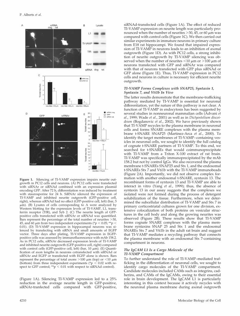
(Figure 1A). Silencing TI-VAMP expression led to a 25%reduction in the average neurite length in GFP-positive,siRNAr-tranfected cells compared with GFP-positive,
siRNAd-transfected cells (Figure 1A). The effect of reducedTI-VAMP expression on neurite length was particularly pro-nounced when the number of neurites �30, 45, or 60 �m wascompared with control cells (Figure 1C). We then carried outsimilar experiments in immature neurons in primary culturefrom E18 rat hippocampi. We found that impaired expres-sion of TI-VAMP in neurons leads to an inhibition of axonaloutgrowth (Figure 1D). As with PC12 cells, a strong inhibi-tion of neurite outgrowth by TI-VAMP silencing was ob-served when the number of neurites �10 �m or �100 �m ofneurons transfected with GFP and siRNAr was comparedwith that of neurons transfected with GFP plus siRNAd orGFP alone (Figure 1E). Thus, TI-VAMP expression in PC12cells and neurons in culture is necessary for efficient neuriteoutgrowth.
TI-VAMP Forms Complexes with SNAP25, Syntaxin 1,Syntaxin 7, and Vti1b In VivoThe latter results demonstrate that the membrane-traffickingpathway mediated by TI-VAMP is essential for neuronaldifferentiation, yet the nature of this pathway is not clear. Afunction of TI-VAMP in endocytosis has been suggested byseveral studies in nonneuronal mammalian cells (Advani etal., 1999; Wade et al., 2001) as well as in Dictyostelium discoi-deum (Bogdanovic et al., 2002). We have previously shownthat TI-VAMP recycles to the plasma membrane in neuronalcells and forms SNARE complexes with the plasma mem-brane t-SNARE SNAP25 (Martinez-Arca et al., 2000). Toidentify the target membranes of TI-VAMP–containing ves-icles in neuronal cells, we sought to identify the full catalogof cognate t-SNARE partners of TI-VAMP. To this end, wesearched for t-SNAREs that would coimmunoprecipitatewith TI-VAMP from a Triton X-100 extract of rat brain.TI-VAMP was specifically immunoprecipitated by the mAb158.2 but not by control IgGs. We also recovered the plasmamembrane t-SNAREs SNAP25 and Stx 1, and the endosomalt-SNAREs Stx 7 and Vti1b with the TI-VAMP immunobeads(Figure 2A). Importantly, we did not observe complex for-mation with another endosomal t-SNARE, syntaxin 13. Therecombinant forms of syntaxin 13 and TI-VAMP are able tointeract in vitro (Yang et al., 1999); thus, the absence ofsyntaxin 13 in our assay suggests that the complexes weisolated were not formed during the process of detergentsolubilization of the tissue. Furthermore, when we deter-mined the subcellular distribution of TI-VAMP and Stx 7 inprimary corticostriatal cultures grown for one division, ex-tensive colocalization of both proteins on vesicular struc-tures in the cell body and along the growing neurites wasobserved (Figure 2B). These results show that TI-VAMPforms cognate SNARE complexes with the plasma mem-brane syntaxins SNAP 25 and Stx 1 and the endosomalSNAREs Stx 7 and Vti1b in the adult rat brain and suggestthat TI-VAMP mediates a recycling pathway that connectsthe plasma membrane with an endosomal Stx 7-containingcompartment in neurons.
The IgCAM L1 Is a Cargo Molecule of theTI-VAMP CompartmentTo further understand the role of TI-VAMP–mediated traf-ficking in the differentiation of neuronal cells, we sought toidentify cargo molecules of the TI-VAMP compartment.Candidate molecules included CAMs such as integrins, cad-herins, and CAMs of the IgCAMs, owing to their essentialrole in brain development. The IgCAM L1 is particularlyinteresting in this context because it actively recycles withthe neuronal plasma membrane during axonal outgrowth
Figure 1. Silencing of TI-VAMP expression impairs neurite out-growth in PC12 cells and neurons. (A) PC12 cells were transfectedwith siRNAr or siRNAd combined with an expression plasmidencoding GFP. After 72 h, differentiation was induced by treatmentwith staurosporine for 24 h. SiRNAr silenced the expression ofTI-VAMP and inhibited neurite outgrowth (GFP-positive cell,right), whereas siRNAd had no effect (GFP-positive cell, left) (bar, 5�m). (B) Lysates of cells corresponding to A were analyzed byWestern blotting for the expression levels of TI-VAMP, L1, trans-ferrin receptor (TfR), and Syb 2. (C) The neurite length of GFP-positive cells transfected with siRNAr or siRNAd was quantified.Bars represent the percentage of the total number of neurites �30,45, and 60 �m from two independent experiments (*p � 0.05; **p �0.01). (D) TI-VAMP expression in hippocampal neurons was si-lenced by transfecting with siRNA and small amounts of EGFPvector. Three days after plating, TI-VAMP expression in EGFP-positive cells was assessed by immunofluorescence with mAb 158.2.As in PC12 cells, siRNAr decreased expression levels of TI-VAMPand inhibited neurite outgrowth (GFP-positive cell, right) comparedwith control cells (GFP-positive cell, left) (bar, 10 �m). (E) Quanti-fication of axon lengths in neurons cotransfected with siRNAd orsiRNAr and EGFP or transfected with EGFP alone is shown. Barsrepresent the percentage of total axons �100 �m (top) or �10 �m(bottom) from three independent experiments (*p � 0.02 with re-spect to GFP control; **p � 0.01 with respect to siRNAd control).
P. Alberts et al.
Molecular Biology of the Cell4210
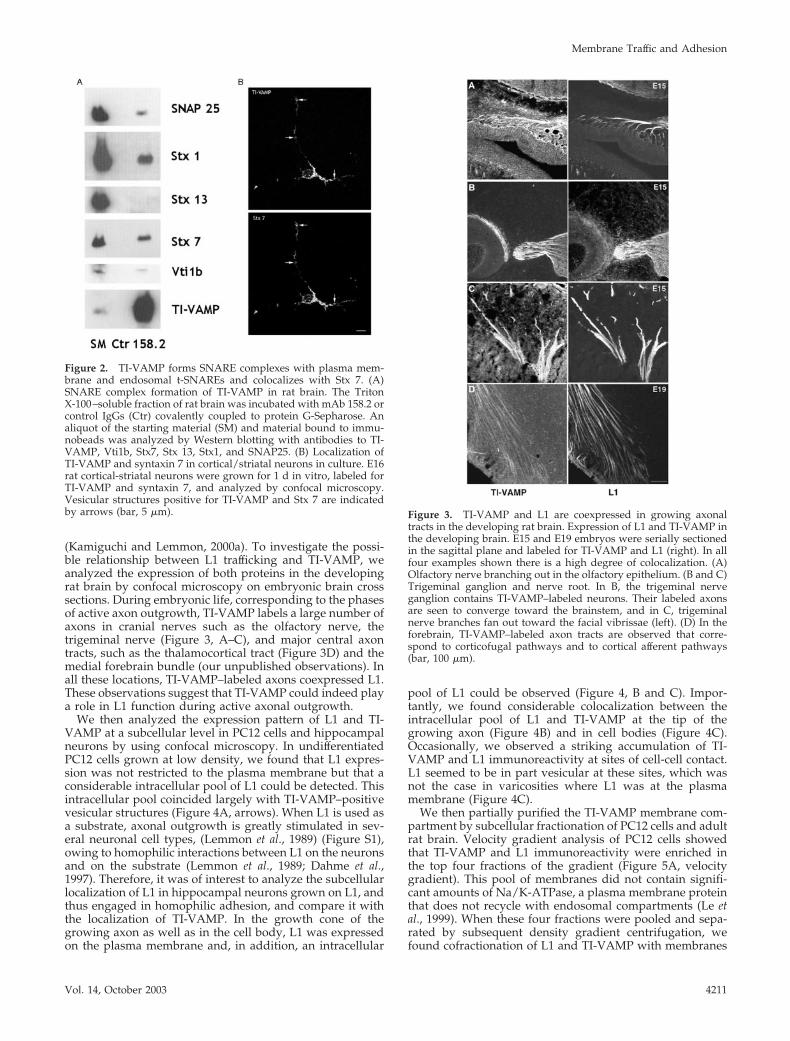
(Kamiguchi and Lemmon, 2000a). To investigate the possi-ble relationship between L1 trafficking and TI-VAMP, weanalyzed the expression of both proteins in the developingrat brain by confocal microscopy on embryonic brain crosssections. During embryonic life, corresponding to the phasesof active axon outgrowth, TI-VAMP labels a large number ofaxons in cranial nerves such as the olfactory nerve, thetrigeminal nerve (Figure 3, A–C), and major central axontracts, such as the thalamocortical tract (Figure 3D) and themedial forebrain bundle (our unpublished observations). Inall these locations, TI-VAMP–labeled axons coexpressed L1.These observations suggest that TI-VAMP could indeed playa role in L1 function during active axonal outgrowth.
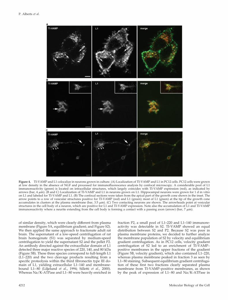
We then analyzed the expression pattern of L1 and TI-VAMP at a subcellular level in PC12 cells and hippocampalneurons by using confocal microscopy. In undifferentiatedPC12 cells grown at low density, we found that L1 expres-sion was not restricted to the plasma membrane but that aconsiderable intracellular pool of L1 could be detected. Thisintracellular pool coincided largely with TI-VAMP–positivevesicular structures (Figure 4A, arrows). When L1 is used asa substrate, axonal outgrowth is greatly stimulated in sev-eral neuronal cell types, (Lemmon et al., 1989) (Figure S1),owing to homophilic interactions between L1 on the neuronsand on the substrate (Lemmon et al., 1989; Dahme et al.,1997). Therefore, it was of interest to analyze the subcellularlocalization of L1 in hippocampal neurons grown on L1, andthus engaged in homophilic adhesion, and compare it withthe localization of TI-VAMP. In the growth cone of thegrowing axon as well as in the cell body, L1 was expressedon the plasma membrane and, in addition, an intracellular
pool of L1 could be observed (Figure 4, B and C). Impor-tantly, we found considerable colocalization between theintracellular pool of L1 and TI-VAMP at the tip of thegrowing axon (Figure 4B) and in cell bodies (Figure 4C).Occasionally, we observed a striking accumulation of TI-VAMP and L1 immunoreactivity at sites of cell-cell contact.L1 seemed to be in part vesicular at these sites, which wasnot the case in varicosities where L1 was at the plasmamembrane (Figure 4C).
We then partially purified the TI-VAMP membrane com-partment by subcellular fractionation of PC12 cells and adultrat brain. Velocity gradient analysis of PC12 cells showedthat TI-VAMP and L1 immunoreactivity were enriched inthe top four fractions of the gradient (Figure 5A, velocitygradient). This pool of membranes did not contain signifi-cant amounts of Na/K-ATPase, a plasma membrane proteinthat does not recycle with endosomal compartments (Le etal., 1999). When these four fractions were pooled and sepa-rated by subsequent density gradient centrifugation, wefound cofractionation of L1 and TI-VAMP with membranes
Figure 2. TI-VAMP forms SNARE complexes with plasma mem-brane and endosomal t-SNAREs and colocalizes with Stx 7. (A)SNARE complex formation of TI-VAMP in rat brain. The TritonX-100–soluble fraction of rat brain was incubated with mAb 158.2 orcontrol IgGs (Ctr) covalently coupled to protein G-Sepharose. Analiquot of the starting material (SM) and material bound to immu-nobeads was analyzed by Western blotting with antibodies to TI-VAMP, Vti1b, Stx7, Stx 13, Stx1, and SNAP25. (B) Localization ofTI-VAMP and syntaxin 7 in cortical/striatal neurons in culture. E16rat cortical-striatal neurons were grown for 1 d in vitro, labeled forTI-VAMP and syntaxin 7, and analyzed by confocal microscopy.Vesicular structures positive for TI-VAMP and Stx 7 are indicatedby arrows (bar, 5 �m). Figure 3. TI-VAMP and L1 are coexpressed in growing axonal
tracts in the developing rat brain. Expression of L1 and TI-VAMP inthe developing brain. E15 and E19 embryos were serially sectionedin the sagittal plane and labeled for TI-VAMP and L1 (right). In allfour examples shown there is a high degree of colocalization. (A)Olfactory nerve branching out in the olfactory epithelium. (B and C)Trigeminal ganglion and nerve root. In B, the trigeminal nerveganglion contains TI-VAMP–labeled neurons. Their labeled axonsare seen to converge toward the brainstem, and in C, trigeminalnerve branches fan out toward the facial vibrissae (left). (D) In theforebrain, TI-VAMP–labeled axon tracts are observed that corre-spond to corticofugal pathways and to cortical afferent pathways(bar, 100 �m).
Membrane Traffic and Adhesion
Vol. 14, October 2003 4211
of similar density, which were clearly different from plasmamembrane (Figure 5A, equilibrium gradient; and Figure S2).We then applied the same approach to fractionate adult ratbrain. The supernatant of a low-speed centrifugation of ratbrain homogenate (S1) was separated by medium-speedcentrifugation to yield the supernatant S2 and the pellet P2.An antibody directed against the extracellular domain of L1detected three major reactive species of 220, 140, and 80 kDa(Figure 5B). These three species correspond to full-length L1(L1–220) and the two cleavage products resulting from aspecific proteolysis within the third fibronectin type III do-main of L1, yielding extracellular L1–140 and membrane-bound L1–80 (Liljelund et al., 1994; Silletti et al., 2000).Whereas Na/K-ATPase and L1–80 were heavily enriched in
fraction P2, a small pool of L1–220 and L1–140 immunore-activity was detectable in S2. TI-VAMP showed an equaldistribution between S2 and P2. Because S2 was poor inplasma membrane proteins, we decided to further analyzethe membrane population of S2 by velocity and equilibriumgradient centrifugation. As in PC12 cells, velocity gradientcentrifugation of S2 led to an enrichment of TI-VAMP–positive membranes in the upper fractions of the gradient(Figure 5B, velocity gradient), which also contained L1–220,whereas plasma membrane peaked in fraction 3 as seen byL1–80 staining. Subsequent equilibrium gradient centrifuga-tion of these first two fractions clearly separated plasmamembrane from TI-VAMP–positive membranes, as shownby the peak of expression of L1–80 and Na/K-ATPase in
Figure 4. TI-VAMP and L1 colocalize in neurons grown in culture. (A) Localization of TI-VAMP and L1 in PC12 cells. PC12 cells were grownat low density in the absence of NGF and processed for immunofluorescence analysis by confocal microscopy. A considerable pool of L1immunoreactivity (green) is located on intracellular structures, which largely coincides with TI-VAMP expression (red), as indicated byarrows (bar, 4 �m). (B and C) Localization of TI-VAMP and L1 in neurons grown on L1. Hippocampal neurons were grown for 1 d in vitroon L1 and labeled for TI-VAMP and L1. (B) The confocal sections were taken from the apical part of the growth cone shown in the inset. Thearrow points to a row of vesicular structures positive for TI-VAMP (red) and L1 (green); most of L1 (green) at the tip of the growth coneaccumulates in clusters at the plasma membrane (bar, 3.5 �m). (C) Two contacting neurons are shown. The arrowheads point at vesicularstructures in the cell body of a neuron, which are positive for L1 and TI-VAMP expression. Note also the accumulation of L1 and TI-VAMPimmunoreactivity where a neurite extending from the cell body is forming a contact with a passing axon (arrow) (bar, 7 �m).
P. Alberts et al.
Molecular Biology of the Cell4212
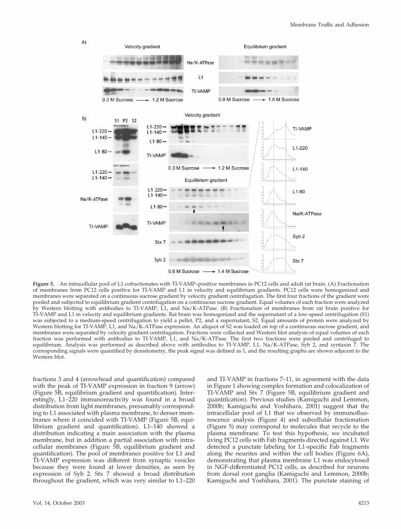
fractions 3 and 4 (arrowhead and quantification) comparedwith the peak of TI-VAMP expression in fraction 9 (arrow)(Figure 5B, equilibrium gradient and quantification). Inter-estingly, L1–220 immunoreactivity was found in a broaddistribution from light membranes, presumably correspond-ing to L1 associated with plasma membrane, to denser mem-branes where it coincided with TI-VAMP (Figure 5B, equi-librium gradient and quantification). L1–140 showed adistribution indicating a main association with the plasmamembrane, but in addition a partial association with intra-cellular membranes (Figure 5B, equilibrium gradient andquantification). The pool of membranes positive for L1 andTI-VAMP expression was different from synaptic vesiclesbecause they were found at lower densities, as seen byexpression of Syb 2. Stx 7 showed a broad distributionthroughout the gradient, which was very similar to L1–220
and TI-VAMP in fractions 7–11, in agreement with the datain Figure 1 showing complex formation and colocalization ofTI-VAMP and Stx 7 (Figure 5B, equilibrium gradient andquantification). Previous studies (Kamiguchi and Lemmon,2000b; Kamiguchi and Yoshihara, 2001) suggest that theintracellular pool of L1 that we observed by immunofluo-rescence analysis (Figure 4) and subcellular fractionation(Figure 5) may correspond to molecules that recycle to theplasma membrane. To test this hypothesis, we incubatedliving PC12 cells with Fab fragments directed against L1. Wedetected a punctate labeling for L1-specific Fab fragmentsalong the neurites and within the cell bodies (Figure 6A),demonstrating that plasma membrane L1 was endocytosedin NGF-differentiated PC12 cells, as described for neuronsfrom dorsal root ganglia (Kamiguchi and Lemmon, 2000b;Kamiguchi and Yoshihara, 2001). The punctate staining of
Figure 5. An intracellular pool of L1 cofractionates with TI-VAMP–positive membranes in PC12 cells and adult rat brain. (A) Fractionationof membranes from PC12 cells positive for TI-VAMP and L1 in velocity and equilibrium gradients. PC12 cells were homogenized andmembranes were separated on a continuous sucrose gradient by velocity gradient centrifugation. The first four fractions of the gradient werepooled and subjected to equilibrium gradient centrifugation on a continuous sucrose gradient. Equal volumes of each fraction were analyzedby Western blotting with antibodies to TI-VAMP, L1, and Na/K-ATPase. (B) Fractionation of membranes from rat brain positive forTI-VAMP and L1 in velocity and equilibrium gradients. Rat brain was homogenized and the supernatant of a low-speed centrifugation (S1)was subjected to a medium-speed centrifugation to yield a pellet, P2, and a supernatant, S2. Equal amounts of protein were analyzed byWestern blotting for TI-VAMP, L1, and Na/K-ATPase expression. An aliquot of S2 was loaded on top of a continuous sucrose gradient, andmembranes were separated by velocity gradient centrifugation. Fractions were collected and Western blot analysis of equal volumes of eachfraction was performed with antibodies to TI-VAMP, L1, and Na/K-ATPase. The first two fractions were pooled and centrifuged toequilibrium. Analysis was performed as described above with antibodies to TI-VAMP, L1, Na/K-ATPase, Syb 2, and syntaxin 7. Thecorresponding signals were quantified by densitometry, the peak signal was defined as 1, and the resulting graphs are shown adjacent to theWestern blot.
Membrane Traffic and Adhesion
Vol. 14, October 2003 4213
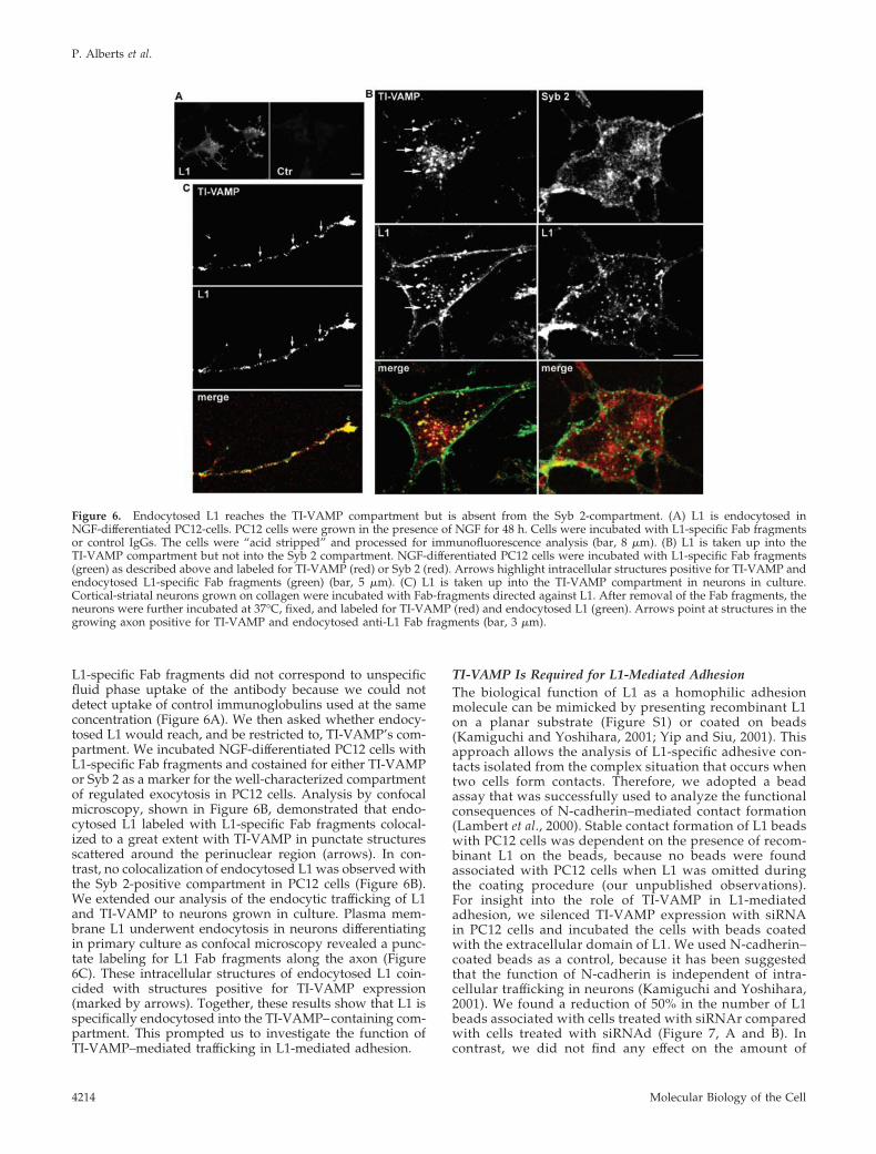
L1-specific Fab fragments did not correspond to unspecificfluid phase uptake of the antibody because we could notdetect uptake of control immunoglobulins used at the sameconcentration (Figure 6A). We then asked whether endocy-tosed L1 would reach, and be restricted to, TI-VAMP’s com-partment. We incubated NGF-differentiated PC12 cells withL1-specific Fab fragments and costained for either TI-VAMPor Syb 2 as a marker for the well-characterized compartmentof regulated exocytosis in PC12 cells. Analysis by confocalmicroscopy, shown in Figure 6B, demonstrated that endo-cytosed L1 labeled with L1-specific Fab fragments colocal-ized to a great extent with TI-VAMP in punctate structuresscattered around the perinuclear region (arrows). In con-trast, no colocalization of endocytosed L1 was observed withthe Syb 2-positive compartment in PC12 cells (Figure 6B).We extended our analysis of the endocytic trafficking of L1and TI-VAMP to neurons grown in culture. Plasma mem-brane L1 underwent endocytosis in neurons differentiatingin primary culture as confocal microscopy revealed a punc-tate labeling for L1 Fab fragments along the axon (Figure6C). These intracellular structures of endocytosed L1 coin-cided with structures positive for TI-VAMP expression(marked by arrows). Together, these results show that L1 isspecifically endocytosed into the TI-VAMP–containing com-partment. This prompted us to investigate the function ofTI-VAMP–mediated trafficking in L1-mediated adhesion.
TI-VAMP Is Required for L1-Mediated AdhesionThe biological function of L1 as a homophilic adhesionmolecule can be mimicked by presenting recombinant L1on a planar substrate (Figure S1) or coated on beads(Kamiguchi and Yoshihara, 2001; Yip and Siu, 2001). Thisapproach allows the analysis of L1-specific adhesive con-tacts isolated from the complex situation that occurs whentwo cells form contacts. Therefore, we adopted a beadassay that was successfully used to analyze the functionalconsequences of N-cadherin–mediated contact formation(Lambert et al., 2000). Stable contact formation of L1 beadswith PC12 cells was dependent on the presence of recom-binant L1 on the beads, because no beads were foundassociated with PC12 cells when L1 was omitted duringthe coating procedure (our unpublished observations).For insight into the role of TI-VAMP in L1-mediatedadhesion, we silenced TI-VAMP expression with siRNAin PC12 cells and incubated the cells with beads coatedwith the extracellular domain of L1. We used N-cadherin–coated beads as a control, because it has been suggestedthat the function of N-cadherin is independent of intra-cellular trafficking in neurons (Kamiguchi and Yoshihara,2001). We found a reduction of 50% in the number of L1beads associated with cells treated with siRNAr comparedwith cells treated with siRNAd (Figure 7, A and B). Incontrast, we did not find any effect on the amount of
Figure 6. Endocytosed L1 reaches the TI-VAMP compartment but is absent from the Syb 2-compartment. (A) L1 is endocytosed inNGF-differentiated PC12-cells. PC12 cells were grown in the presence of NGF for 48 h. Cells were incubated with L1-specific Fab fragmentsor control IgGs. The cells were “acid stripped” and processed for immunofluorescence analysis (bar, 8 �m). (B) L1 is taken up into theTI-VAMP compartment but not into the Syb 2 compartment. NGF-differentiated PC12 cells were incubated with L1-specific Fab fragments(green) as described above and labeled for TI-VAMP (red) or Syb 2 (red). Arrows highlight intracellular structures positive for TI-VAMP andendocytosed L1-specific Fab fragments (green) (bar, 5 �m). (C) L1 is taken up into the TI-VAMP compartment in neurons in culture.Cortical-striatal neurons grown on collagen were incubated with Fab-fragments directed against L1. After removal of the Fab fragments, theneurons were further incubated at 37°C, fixed, and labeled for TI-VAMP (red) and endocytosed L1 (green). Arrows point at structures in thegrowing axon positive for TI-VAMP and endocytosed anti-L1 Fab fragments (bar, 3 �m).
P. Alberts et al.
Molecular Biology of the Cell4214
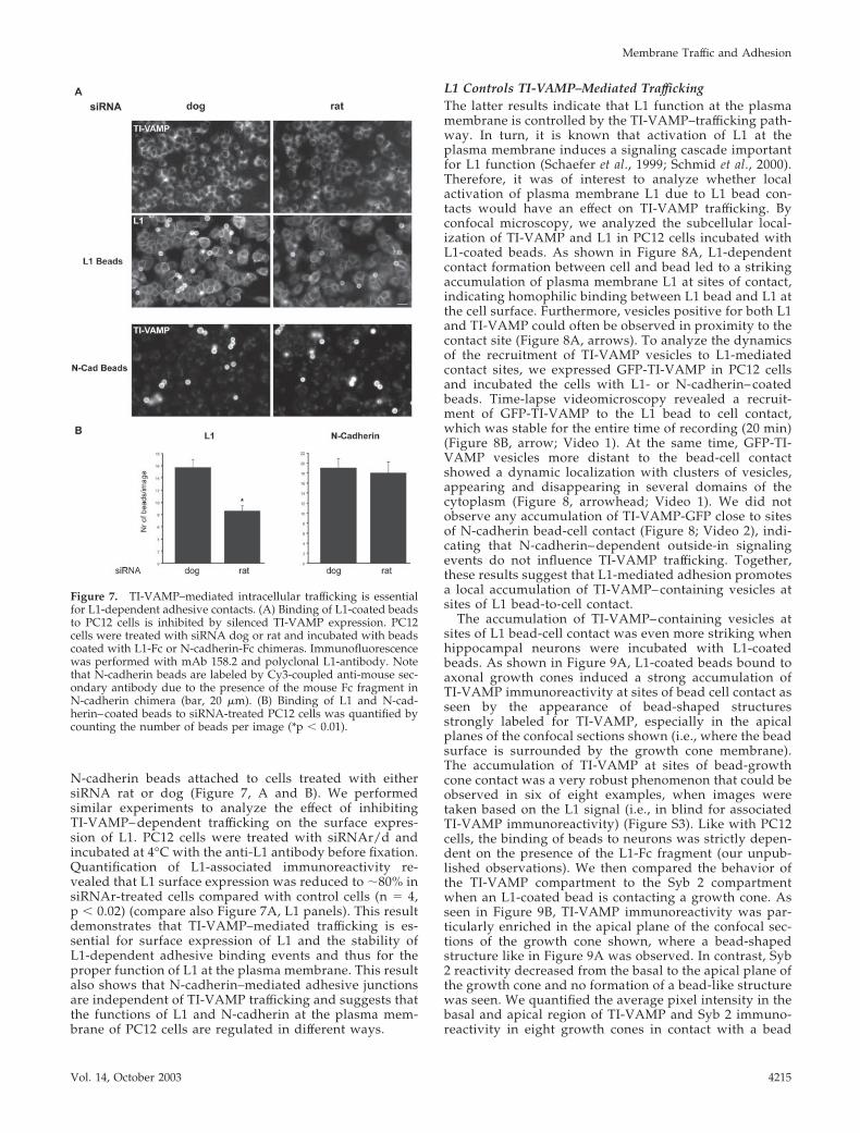
N-cadherin beads attached to cells treated with eithersiRNA rat or dog (Figure 7, A and B). We performedsimilar experiments to analyze the effect of inhibitingTI-VAMP– dependent trafficking on the surface expres-sion of L1. PC12 cells were treated with siRNAr/d andincubated at 4°C with the anti-L1 antibody before fixation.Quantification of L1-associated immunoreactivity re-vealed that L1 surface expression was reduced to �80% insiRNAr-treated cells compared with control cells (n � 4,p � 0.02) (compare also Figure 7A, L1 panels). This resultdemonstrates that TI-VAMP–mediated trafficking is es-sential for surface expression of L1 and the stability ofL1-dependent adhesive binding events and thus for theproper function of L1 at the plasma membrane. This resultalso shows that N-cadherin–mediated adhesive junctionsare independent of TI-VAMP trafficking and suggests thatthe functions of L1 and N-cadherin at the plasma mem-brane of PC12 cells are regulated in different ways.
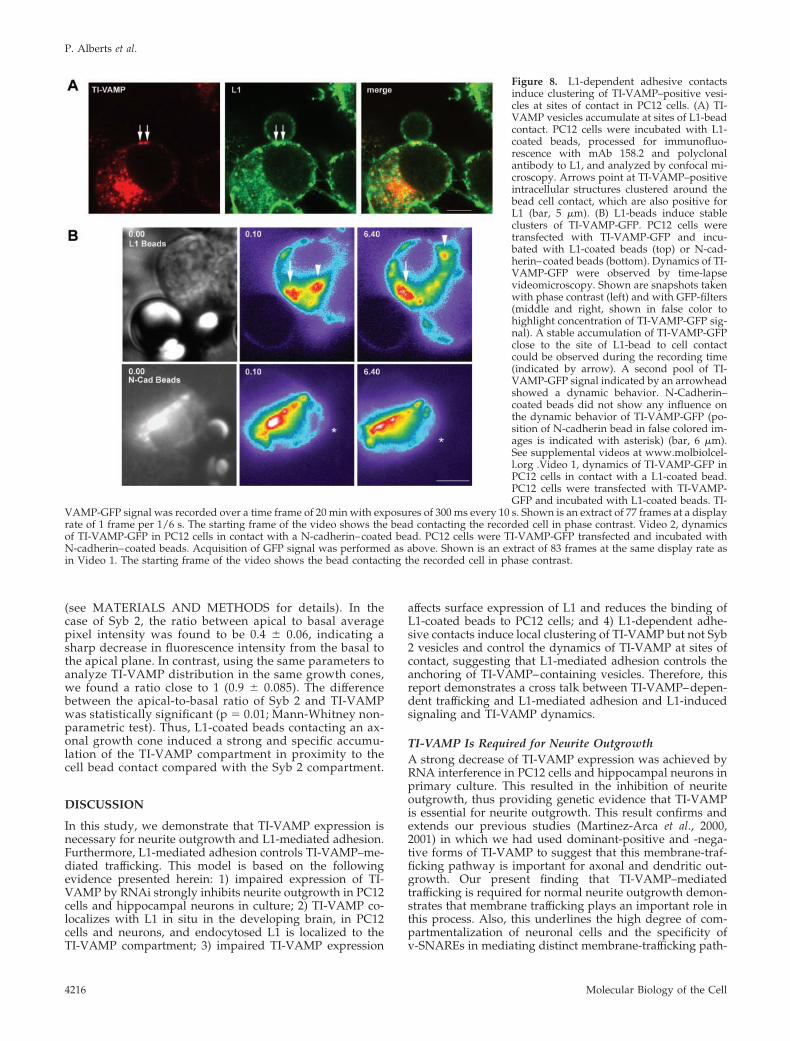
L1 Controls TI-VAMP–Mediated TraffickingThe latter results indicate that L1 function at the plasmamembrane is controlled by the TI-VAMP–trafficking path-way. In turn, it is known that activation of L1 at theplasma membrane induces a signaling cascade importantfor L1 function (Schaefer et al., 1999; Schmid et al., 2000).Therefore, it was of interest to analyze whether localactivation of plasma membrane L1 due to L1 bead con-tacts would have an effect on TI-VAMP trafficking. Byconfocal microscopy, we analyzed the subcellular local-ization of TI-VAMP and L1 in PC12 cells incubated withL1-coated beads. As shown in Figure 8A, L1-dependentcontact formation between cell and bead led to a strikingaccumulation of plasma membrane L1 at sites of contact,indicating homophilic binding between L1 bead and L1 atthe cell surface. Furthermore, vesicles positive for both L1and TI-VAMP could often be observed in proximity to thecontact site (Figure 8A, arrows). To analyze the dynamicsof the recruitment of TI-VAMP vesicles to L1-mediatedcontact sites, we expressed GFP-TI-VAMP in PC12 cellsand incubated the cells with L1- or N-cadherin– coatedbeads. Time-lapse videomicroscopy revealed a recruit-ment of GFP-TI-VAMP to the L1 bead to cell contact,which was stable for the entire time of recording (20 min)(Figure 8B, arrow; Video 1). At the same time, GFP-TI-VAMP vesicles more distant to the bead-cell contactshowed a dynamic localization with clusters of vesicles,appearing and disappearing in several domains of thecytoplasm (Figure 8, arrowhead; Video 1). We did notobserve any accumulation of TI-VAMP-GFP close to sitesof N-cadherin bead-cell contact (Figure 8; Video 2), indi-cating that N-cadherin– dependent outside-in signalingevents do not influence TI-VAMP trafficking. Together,these results suggest that L1-mediated adhesion promotesa local accumulation of TI-VAMP– containing vesicles atsites of L1 bead-to-cell contact.
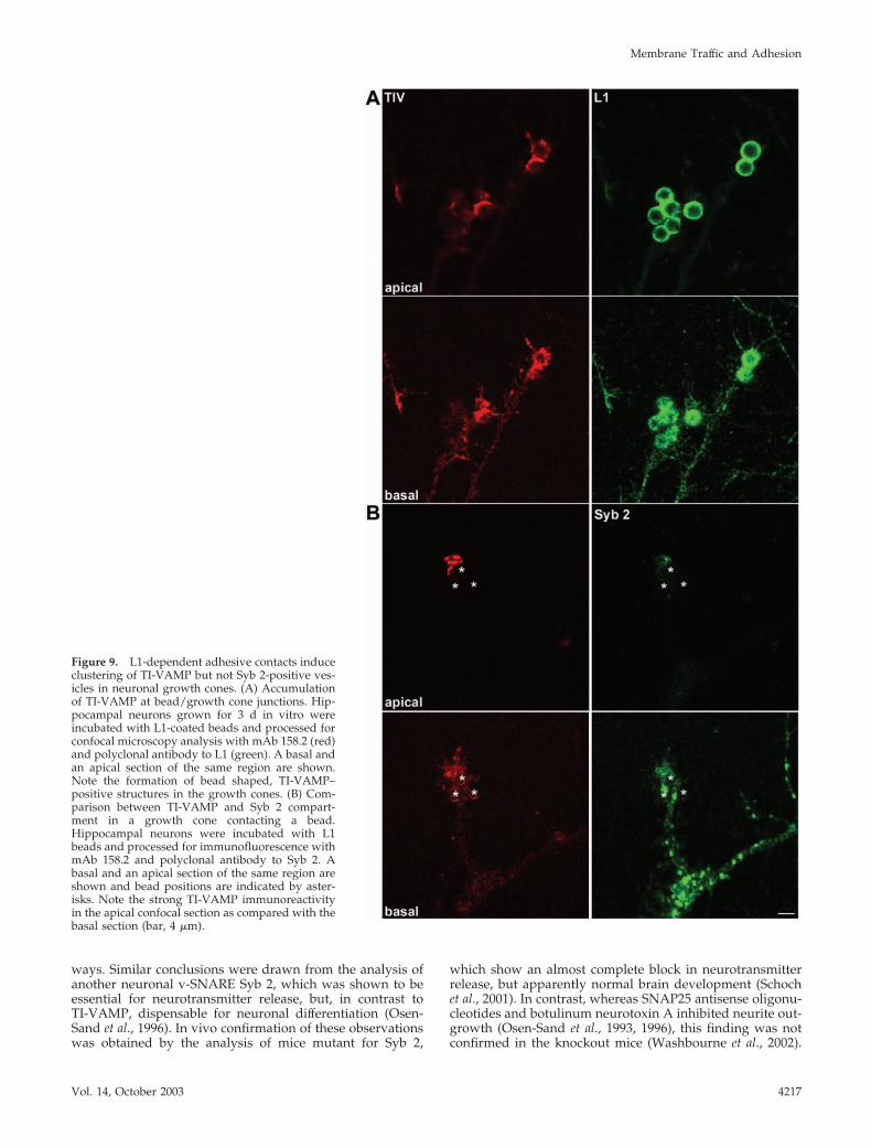
The accumulation of TI-VAMP– containing vesicles atsites of L1 bead-cell contact was even more striking whenhippocampal neurons were incubated with L1-coatedbeads. As shown in Figure 9A, L1-coated beads bound toaxonal growth cones induced a strong accumulation ofTI-VAMP immunoreactivity at sites of bead cell contact asseen by the appearance of bead-shaped structuresstrongly labeled for TI-VAMP, especially in the apicalplanes of the confocal sections shown (i.e., where the beadsurface is surrounded by the growth cone membrane).The accumulation of TI-VAMP at sites of bead-growthcone contact was a very robust phenomenon that could beobserved in six of eight examples, when images weretaken based on the L1 signal (i.e., in blind for associatedTI-VAMP immunoreactivity) (Figure S3). Like with PC12cells, the binding of beads to neurons was strictly depen-dent on the presence of the L1-Fc fragment (our unpub-lished observations). We then compared the behavior ofthe TI-VAMP compartment to the Syb 2 compartmentwhen an L1-coated bead is contacting a growth cone. Asseen in Figure 9B, TI-VAMP immunoreactivity was par-ticularly enriched in the apical plane of the confocal sec-tions of the growth cone shown, where a bead-shapedstructure like in Figure 9A was observed. In contrast, Syb2 reactivity decreased from the basal to the apical plane ofthe growth cone and no formation of a bead-like structurewas seen. We quantified the average pixel intensity in thebasal and apical region of TI-VAMP and Syb 2 immuno-reactivity in eight growth cones in contact with a bead
Figure 7. TI-VAMP–mediated intracellular trafficking is essentialfor L1-dependent adhesive contacts. (A) Binding of L1-coated beadsto PC12 cells is inhibited by silenced TI-VAMP expression. PC12cells were treated with siRNA dog or rat and incubated with beadscoated with L1-Fc or N-cadherin-Fc chimeras. Immunofluorescencewas performed with mAb 158.2 and polyclonal L1-antibody. Notethat N-cadherin beads are labeled by Cy3-coupled anti-mouse sec-ondary antibody due to the presence of the mouse Fc fragment inN-cadherin chimera (bar, 20 �m). (B) Binding of L1 and N-cad-herin–coated beads to siRNA-treated PC12 cells was quantified bycounting the number of beads per image (*p � 0.01).
Membrane Traffic and Adhesion
Vol. 14, October 2003 4215
(see MATERIALS AND METHODS for details). In thecase of Syb 2, the ratio between apical to basal averagepixel intensity was found to be 0.4 � 0.06, indicating asharp decrease in fluorescence intensity from the basal tothe apical plane. In contrast, using the same parameters toanalyze TI-VAMP distribution in the same growth cones,we found a ratio close to 1 (0.9 � 0.085). The differencebetween the apical-to-basal ratio of Syb 2 and TI-VAMPwas statistically significant (p � 0.01; Mann-Whitney non-parametric test). Thus, L1-coated beads contacting an ax-onal growth cone induced a strong and specific accumu-lation of the TI-VAMP compartment in proximity to thecell bead contact compared with the Syb 2 compartment.
DISCUSSION
In this study, we demonstrate that TI-VAMP expression isnecessary for neurite outgrowth and L1-mediated adhesion.Furthermore, L1-mediated adhesion controls TI-VAMP–me-diated trafficking. This model is based on the followingevidence presented herein: 1) impaired expression of TI-VAMP by RNAi strongly inhibits neurite outgrowth in PC12cells and hippocampal neurons in culture; 2) TI-VAMP co-localizes with L1 in situ in the developing brain, in PC12cells and neurons, and endocytosed L1 is localized to theTI-VAMP compartment; 3) impaired TI-VAMP expression
affects surface expression of L1 and reduces the binding ofL1-coated beads to PC12 cells; and 4) L1-dependent adhe-sive contacts induce local clustering of TI-VAMP but not Syb2 vesicles and control the dynamics of TI-VAMP at sites ofcontact, suggesting that L1-mediated adhesion controls theanchoring of TI-VAMP–containing vesicles. Therefore, thisreport demonstrates a cross talk between TI-VAMP–depen-dent trafficking and L1-mediated adhesion and L1-inducedsignaling and TI-VAMP dynamics.
TI-VAMP Is Required for Neurite OutgrowthA strong decrease of TI-VAMP expression was achieved byRNA interference in PC12 cells and hippocampal neurons inprimary culture. This resulted in the inhibition of neuriteoutgrowth, thus providing genetic evidence that TI-VAMPis essential for neurite outgrowth. This result confirms andextends our previous studies (Martinez-Arca et al., 2000,2001) in which we had used dominant-positive and -nega-tive forms of TI-VAMP to suggest that this membrane-traf-ficking pathway is important for axonal and dendritic out-growth. Our present finding that TI-VAMP–mediatedtrafficking is required for normal neurite outgrowth demon-strates that membrane trafficking plays an important role inthis process. Also, this underlines the high degree of com-partmentalization of neuronal cells and the specificity ofv-SNAREs in mediating distinct membrane-trafficking path-
Figure 8. L1-dependent adhesive contactsinduce clustering of TI-VAMP–positive vesi-cles at sites of contact in PC12 cells. (A) TI-VAMP vesicles accumulate at sites of L1-beadcontact. PC12 cells were incubated with L1-coated beads, processed for immunofluo-rescence with mAb 158.2 and polyclonalantibody to L1, and analyzed by confocal mi-croscopy. Arrows point at TI-VAMP–positiveintracellular structures clustered around thebead cell contact, which are also positive forL1 (bar, 5 �m). (B) L1-beads induce stableclusters of TI-VAMP-GFP. PC12 cells weretransfected with TI-VAMP-GFP and incu-bated with L1-coated beads (top) or N-cad-herin–coated beads (bottom). Dynamics of TI-VAMP-GFP were observed by time-lapsevideomicroscopy. Shown are snapshots takenwith phase contrast (left) and with GFP-filters(middle and right, shown in false color tohighlight concentration of TI-VAMP-GFP sig-nal). A stable accumulation of TI-VAMP-GFPclose to the site of L1-bead to cell contactcould be observed during the recording time(indicated by arrow). A second pool of TI-VAMP-GFP signal indicated by an arrowheadshowed a dynamic behavior. N-Cadherin–coated beads did not show any influence onthe dynamic behavior of TI-VAMP-GFP (po-sition of N-cadherin bead in false colored im-ages is indicated with asterisk) (bar, 6 �m).See supplemental videos at www.molbiolcel-l.org .Video 1, dynamics of TI-VAMP-GFP inPC12 cells in contact with a L1-coated bead.PC12 cells were transfected with TI-VAMP-GFP and incubated with L1-coated beads. TI-
VAMP-GFP signal was recorded over a time frame of 20 min with exposures of 300 ms every 10 s. Shown is an extract of 77 frames at a displayrate of 1 frame per 1/6 s. The starting frame of the video shows the bead contacting the recorded cell in phase contrast. Video 2, dynamicsof TI-VAMP-GFP in PC12 cells in contact with a N-cadherin–coated bead. PC12 cells were TI-VAMP-GFP transfected and incubated withN-cadherin–coated beads. Acquisition of GFP signal was performed as above. Shown is an extract of 83 frames at the same display rate asin Video 1. The starting frame of the video shows the bead contacting the recorded cell in phase contrast.
P. Alberts et al.
Molecular Biology of the Cell4216
ways. Similar conclusions were drawn from the analysis ofanother neuronal v-SNARE Syb 2, which was shown to beessential for neurotransmitter release, but, in contrast toTI-VAMP, dispensable for neuronal differentiation (Osen-Sand et al., 1996). In vivo confirmation of these observationswas obtained by the analysis of mice mutant for Syb 2,
which show an almost complete block in neurotransmitterrelease, but apparently normal brain development (Schochet al., 2001). In contrast, whereas SNAP25 antisense oligonu-cleotides and botulinum neurotoxin A inhibited neurite out-growth (Osen-Sand et al., 1993, 1996), this finding was notconfirmed in the knockout mice (Washbourne et al., 2002).
Figure 9. L1-dependent adhesive contacts induceclustering of TI-VAMP but not Syb 2-positive ves-icles in neuronal growth cones. (A) Accumulationof TI-VAMP at bead/growth cone junctions. Hip-pocampal neurons grown for 3 d in vitro wereincubated with L1-coated beads and processed forconfocal microscopy analysis with mAb 158.2 (red)and polyclonal antibody to L1 (green). A basal andan apical section of the same region are shown.Note the formation of bead shaped, TI-VAMP–positive structures in the growth cones. (B) Com-parison between TI-VAMP and Syb 2 compart-ment in a growth cone contacting a bead.Hippocampal neurons were incubated with L1beads and processed for immunofluorescence withmAb 158.2 and polyclonal antibody to Syb 2. Abasal and an apical section of the same region areshown and bead positions are indicated by aster-isks. Note the strong TI-VAMP immunoreactivityin the apical confocal section as compared with thebasal section (bar, 4 �m).
Membrane Traffic and Adhesion
Vol. 14, October 2003 4217
However, neurons express several syntaxins and bothSNAP23 and SNAP25 (Chen et al., 1999), so it can be antic-ipated that only the genetic invalidation of several plasmamembrane SNAREs (at least SNAP25 and SNAP23) willresult in defects in the development of the brain. Futurestudies are required to unravel the exact composition ofSNAREs and their regulators involved in the differentiationof neurons.
L1 Recycles in the TI-VAMP CompartmentWe have previously shown that TI-VAMP defines a newtubulo-vesicular compartment particularly concentratedat the leading edge of growth cones in hippocampal neu-rons developing in vitro. We also showed that TI-VAMPdoes not colocalize with either markers of synaptic vesi-cles or with several markers of endocytic compartments,except CD63 (Coco et al., 1999). Like other tetraspanins,CD63 has been implicated in integrin-mediated cell mi-gration (Berditchevski, 2001). Studies on the function ofTI-VAMP suggested an involvement of TI-VAMP in lateendocytic pathways in nonneuronal mammalian cells(Advani et al., 1999; Wade et al., 2001) as well as in D.discoideum, where it was shown to participate in an endo-somal SNARE complex composed of Stx 7 and 8 and vti1b(Bogdanovic et al., 2002). In agreement with these find-ings, we show herein that internalized L1 reaches theTI-VAMP compartment in neuronal cells and that TI-VAMP forms a SNARE complex with syntaxin 7 and vti1band colocalizes with the endosomal t-SNARE syntaxin 7.Yet, our data point to a function of TI-VAMP that is morelikely to correspond to a recycling rather than a degrada-tive pathway in neuronal cells. This is supported by thefinding that TI-VAMP forms SNARE complexes with theplasma membrane SNAREs syntaxin 1 and SNAP25. Im-portantly, TI-VAMP controls the surface expression of L1,thus clearly indicating that TI-VAMP mediates an exo-cytic pathway in neuronal cells. The previous findingsthat TI-VAMP colocalizes with CD63 in PC12 cells (Cocoet al., 1999) and HeLa cells (Martinez-Arca et al., 2003b)could suggest that TI-VAMP’s compartment correspondsto a specialized recycling endosome to which L1 (L1–220and possibly L1–140) would be targeted after internaliza-tion and from which it would then be recycled to theplasma membrane. The TI-VAMP compartment could berelated to the recently identified enlargosome, a compart-ment involved in membrane repair whose exocytosis istetanus neurotoxin-insensitive (Borgonovo et al., 2002). Itwill be important to get a more complete picture of thecargo proteins targeted to the TI-VAMP compartment tofurther understand the nature and molecular compositionof this compartment.
Functional Cross Talk between TI-VAMP and L1Our finding that L1-dependent adhesion is impaired byreduced expression of TI-VAMP clearly points to an es-sential role of TI-VAMP– dependent trafficking in thefunction of L1 at the plasma membrane. Interestingly,neuronal L1 harbors an additional short exon that yields atyrosine-based motif YRSLE. This motif mediates bindingto the AP-2 adaptor complex and clathrin-dependent en-docytosis and accelerates the recycling of neuronal L1(Kamiguchi et al., 1998) suggesting a role for intracellulartrafficking in L1 function, especially in the course of neu-ronal development. In analogy to migrating cells, it wassuggested that L1 trafficking in the axonal growth coneestablishes a dynamic gradient of L1 adhesivity necessary
to move along the substrate (Kamiguchi and Lemmon,2000b). Indeed, such a gradient was recently detected andits maintenance was dependent on endocytic trafficking(Kamiguchi and Yoshihara, 2001). Our results suggest thatthe TI-VAMP– dependent intracellular trafficking of L1may be necessary to stabilize and regulate adhesive con-tacts of neuronal cells. Clearly, the demonstration thatTI-VAMP is an essential molecular player involved in thetrafficking of L1 will greatly facilitate the analysis of thecomplex function and regulation of this highly importantmolecule in brain development.
The functional cross talk between TI-VAMP and L1 ishighlighted by our finding that L1-mediated adhesion trig-gers the accumulation of TI-VAMP and L1-containing vesi-cles at the site of contact. The novelty of this finding raisesseveral important questions. Are these vesicles involved inthe establishment and/or maintenance of the contacts? Re-cent results on the regulation of L1-trafficking show thatendocytosis of L1 is directly linked to tyrosine dephosphor-ylation of neuronal L1 on the YRSLE-motif (Schaefer et al.,2002). Interestingly, this dephosphorylation seems to occurlocally at sites where L1 is engaged in cell-cell contacts oraxonal outgrowth on L1 substrate (Schaefer et al., 2002). Theconcentration of TI-VAMP vesicles at L1-dependent contactsmight therefore provide the trafficking machinery necessaryfor L1 to recycle locally and thus perform its function. Howdoes L1-dependent signaling control membrane trafficking?Activation of L1 by homophilic interaction stimulates a sig-naling pathway involving src, PI3Kinase, Rac1, and the mi-togen-activated protein kinases, and activation of src seemsto play an essential role in L1-dependent neurite outgrowth(Ignelzi et al., 1994; Schaefer et al., 1999; Schmid et al., 2000).Also, L1 interacts directly with scaffolding proteins such asAnkyrin and Ezrin (Davis and Bennett, 1994; Dickson et al.,2002). Future studies should test how far the recruitment ofTI-VAMP vesicles to L1-dependent contacts depends on anyof the proteins mentioned above or whether an as yet un-known mechanism is recruiting and stabilizing TI-VAMPvesicles.
Our findings have important implications for our under-standing of the role of adhesion molecules such as L1 in thedifferentiation of neurons and the development of the brain.We propose that the function of L1 does not solely dependon its capacity to transduce intracellular signals and to me-diate cell-to-cell adhesion but also consists in the targeting ofthe exocytic organelles involved in neurite outgrowth to theleading edge of growing axons and to sites of contact for-mation between cells. In light of the recent finding thatN-CAM promotes the accumulation of trans-Golgi networkorganelles at sites of cell-cell contacts, a process that mayactivate the formation of synapses (Sytnyk et al., 2002) andthe link between TI-VAMP–mediated trafficking and L1-mediated adhesion unraveled herein, we propose that crosstalk between membrane trafficking and cell-cell and cell-matrix adhesion molecules may be crucial for neuronal dif-ferentiation.
ACKNOWLEDGMENTS
We are indebted to B. Allinquant, and A. Prochiantz for initial experiments inneurons, to R. Jahn, and W. Hong for kindly providing reagents. This workwas supported in part by grants from the Human Frontier Science Program(RGY0027/2001-B101), European Community (QLG3-CT-2001-02430_Retro-grade Signaling), the Association pour la Recherche sur le Cancer (ARC no.5873 and no. 4762), and the Ministere de la Recherche (ACI-JC5254 andACI-BDPI128) to T.G.
P. Alberts et al.
Molecular Biology of the Cell4218
REFERENCES
Advani, R.J., Yang, B., Prekeris, R., Lee, K.C., Klumperman, J., and Scheller,R.H. (1999). VAMP-7 mediates vesicular transport from endosomes to lyso-somes. J. Cell Biol. 146, 765–775.
Berditchevski, F. (2001). Complexes of tetraspanins with integrins: more thanmeets the eye. J. Cell Sci. 114, 4143–4151.
Bogdanovic, A., Bennett, N., Kieffer, S., Louwagie, M., Morio, T., Garin, J.,Satre, M., and Bruckert, F. (2002). Syntaxin 7, Syntaxin 8, Vti1 and VAMP7form an active SNARE complex for early macropinocytic compartment fusionin Dictyostelium discoideum. Biochem J. 368, 29–39.
Borgonovo, B., Cocucci, E., Racchetti, G., Podini, P., Bachi, A., and Meldolesi,J. (2002). Regulated exocytosis: a novel, widely expressed system. Nat. CellBiol. 4, 955–962.
Brummendorf, T., Kenwrick, S., and Rathjen, F.G. (1998). Neural cell recog-nition molecule L 1, from cell biology to human hereditary brain malforma-tions. Curr. Opin. Neurobiol. 8, 87–97.
Chang, S., and De Camilli, P. (2001). Glutamate regulates actin-based motilityin axonal filopodia. Nat. Neurosci. 4, 787–793.
Chen, D., Minger, S.L., Honer, W.G., and Whiteheart, S.W. (1999). Organiza-tion of the secretory machinery in the rodent brain: distribution of the t-SNAREs, SNAP-25 and SNAP-23. Brain Res. 831, 11–24.
Chilcote, T.J., Galli, T., Mundigl, O., Edelmann, L., McPherson, P.S., Takei, K.,and De Camilli, P. (1995). Cellubrevin and synaptobrevins: similar subcellularlocalization and biochemical properties in PC12 cells. J. Cell Biol. 129, 219–231.
Coco, S., Raposo, G., Martinez, S., Fontaine, J.J., Takamori, S., Zahraoui, A.,Jahn, R., Matteoli, M., Louvard, D., and Galli, T. (1999). Subcellular localiza-tion of tetanus neurotoxin-insensitive vesicle-associated membrane protein(VAMP)/VAMP7 in neuronal cells: evidence for a novel membrane compart-ment. J. Neurosci. 19, 9803–9812.
Dahme, M., Bartsch, U., Martini, R., Anliker, B., Schachner, M., and Mantei, N.(1997). Disruption of the mouse L1 gene leads to malformations of thenervous system. Nat. Genet. 17, 346–349.
Davis, J.Q., and Bennett, V. (1994). Ankyrin binding activity shared by theneurofascin/L1/NrCAM family of nervous system cell adhesion molecules.J. Biol. Chem. 269, 27163–27166.
De Angelis, E., MacFarlane, J., Du, J.S., Yeo, G., Hicks, R., Rathjen, F.G.,Kenwrick, S., and Brummendorf, T. (1999). Pathological missense mutationsof neural cell adhesion molecule L1 affect homophilic and heterophilic bind-ing activities. EMBO J. 18, 4744–4753.
Dickson, T.C., Mintz, C.D., Benson, D.L., and Salton, S.R. (2002). Functionalbinding interaction identified between the axonal CAM L1 and members ofthe ERM family. J. Cell Biol. 157, 1105–1112.
Elbashir, S.M., Harborth, J., Lendeckel, W., Yalcin, A., Weber, K., and Tuschl,T. (2001). Duplexes of 21-nucleotide RNAs mediate RNA interference incultured mammalian cells. Nature 411, 494–498.
Filippini, F., Rossi, V., Galli, T., Budillon, A., D’Urso, M., and D’Esposito, M.(2001). Longins: a new evolutionary conserved VAMP family sharing a novelSNARE domain. Trends Biochem. Sci. 26, 407–409.
Futerman, A.H., and Banker, G.A. (1996). The economics of neurite out-growth–the addition of new membrane to growing axons. Trends Neurosci.19, 144–149.
Galli, T., and Haucke, V. (2001). Cycling of synaptic vesicles: how far? Howfast! Sci STKE 2001, RE1. Available at http://stke.sciencemag.org/. AccessedSeptember 11, 2003.
Galli, T., Zahraoui, A., Vaidyanathan, V.V., Raposo, G., Tian, J.M., Karin, M.,Niemann, H., and Louvard, D. (1998). A novel tetanus neurotoxin-insensitivevesicle-associated membrane protein in SNARE complexes of the apicalplasma membrane of epithelial cells. Mol. Biol. Cell 9, 1437–1448.
Grosse, G., Grosse, J., Tapp, R., Kuchinke, J., Gorsleben, M., Fetter, I.,HohneZell, B., Gratzl, M., and Bergmann, M. (1999). SNAP-25 requirement fordendritic growth of hippocampal neurons. J. Neurosci. Res. 56, 539–546.
Harlow, E., and Lane, D. (1988). Antibodies: A Laboratory Manual. ColdSpring Harbor, NY: Cold Spring Harbor Laboratory.
Ignelzi, M.A., Jr., Miller, D.R., Soriano, P., and Maness, P.F. (1994). Impairedneurite outgrowth of src-minus cerebellar neurons on the cell adhesion mol-ecule L1. Neuron 12, 873–884.
Kamiguchi, H., and Lemmon, V. (2000a). IgCAMs: bidirectional signals un-derlying neurite growth. Curr. Opin. Cell Biol. 12, 598–605.
Kamiguchi, H., and Lemmon, V. (2000b). Recycling of the cell adhesionmolecule L1 in axonal growth cones. J. Neurosci. 20, 3676–3686.
Kamiguchi, H., Long, K.E., Pendergast, M., Schaefer, A.W., Rapoport, I.,Kirchhausen, T., and Lemmon, V. (1998). The neural cell adhesion moleculeL1 interacts with the AP-2 adaptor and is endocytosed via the clathrin-mediated pathway. J. Neurosci. 18, 5311–5321.
Kamiguchi, H., and Yoshihara, F. (2001). The role of endocytic l1 trafficking inpolarized adhesion and migration of nerve growth cones. J. Neurosci. 21,9194–9203.
Laemmli, U.K. (1970). Cleavage of structural proteins during the assembly ofthe head of bacteriophage T4. Nature 227, 680–685.
Lambert, M., Padilla, F., and Mege, R.M. (2000). Immobilized dimers ofN-cadherin-Fc chimera mimic cadherin-mediated cell contact formation: con-tribution of both outside-in and inside-out signals. J. Cell Sci. 113, 2207–2219.
Le, T.L., Yap, A.S., and Stow, J.L. (1999). Recycling of E-cadherin: a potentialmechanism for regulating cadherin dynamics. J. Cell Biol. 146, 219–232.
Lemmon, V., Farr, K.L., and Lagenaur, C. (1989). L1-mediated axon out-growth occurs via a homophilic binding mechanism. Neuron 2, 1597–1603.
Liljelund, P., Ghosh, P., and van den Pol, A.N. (1994). Expression of the neuralaxon adhesion molecule L1 in the developing and adult rat brain. J. Biol.Chem. 269, 32886–32895.
Martinez-Arca, S., Rudge, R.E., Vacca, M., Camonis, J., Daviet, L., Form-stecher, E., Hamburger, A., Filippini, F., D’Esposito, M., and Galli, T. (2003b).A novel dual mechanism controlling the localization and function of exocyticv-SNAREs. Proc. Nat. Acad. Sci. USA. 100, 9011–9016.
Martinez-Arca, S., Alberts, P., Zahraoui, A., Louvard, D., and Galli, T. (2000).Role of tetanus neurotoxin insensitive vesicle-associated membrane protein(TI-VAMP) in vesicular transport mediating neurite outgrowth. J. Cell Biol.149, 889–899.
Martinez-Arca, S., Coco, S., Mainguy, G., Schenk, U., Alberts, P., Bouille, P.,Mezzina, M., Prochiantz, A., Matteoli, M., Louvard, D., and Galli, T. (2001). Acommon exocytotic mechanism mediates axonal and dendritic outgrowth.J. Neurosci. 21, 3830–3838.
Martinez-Arca, S., Proux-Gillardeaux, V., Alberts, P., Louvard, D., and Galli,T. (2003a). Ectopic expression of syntaxin 1 in the ER redirects TI-VAMP- andcellubrevin-containing vesicles. J. Cell Sci. 116, 2805–2816.
Mechtersheimer, S., Gutwein, P., Agmon-Levin, N., Stoeck, A., Oleszewski,M., Riedle, S., Fogel, M., Lemmon, V., and Altevogt, P. (2001). Ectodomainshedding of L1 adhesion molecule promotes cell migration by autocrinebinding to integrins. J. Cell Biol. 155, 661–673.
Muzerelle, A., Alberts, P., Martinez Arca, S., Jeannequin, O., Lafaye, P., Mazie,J.-C., Galli, T., and Gaspar, P. (2003). Identification of a presynaptic membranecompartment containing tetanus neurotoxin-insensitive vesicle associatedmembrane protein in the rat brain. Neuroscience. (in press).
Osen-Sand, A., Catsicas, M., Staple, J.K., Jones, K.A., Ayala, G., Knowles, J.,Grenningloh, G., and Catsicas, S. (1993). Inhibition of axonal growth bySNAP-25 antisense oligonucleotides in vitro and in vivo. Nature 364, 445–448.
Osen-Sand, A., Staple, J.K., Naldi, E., Schiavo, G., Rossetto, O., Petitpierre, S.,Malgaroli, A., Montecucco, C., and Catsicas, S. (1996). Common and distinctfusion proteins in axonal growth and transmitter release. J. Comp. Neurol.367, 222–234.
Prochiantz, A. (1995). Neuronal polarity: giving neurons heads and tails.Neuron 15, 743–746.
Rathjen, F.G., and Rutishauser, U. (1984). Comparison of two cell surfacemolecules involved in neural cell adhesion. EMBO J. 3, 461–465.
Rousselet, A., Autillo-Touati, A., Araud, D., and Prochiantz, A. (1990). In vitroregulation of neuronal morphogenesis and polarity by astrocyte-derived fac-tors. Dev. Biol. 137, 33–45.
Schaefer, A.W., Kamei, Y., Kamiguchi, H., Wong, E.V., Rapoport, I., Kirch-hausen, T., Beach, C.M., Landreth, G., Lemmon, S.K., and Lemmon, V. (2002).L1 endocytosis is controlled by a phosphorylation-dephosphorylation cyclestimulated by outside-in signaling by L1. J. Cell Biol. 157, 1223–1232.
Schaefer, A.W., Kamiguchi, H., Wong, E.V., Beach, C.M., Landreth, G., andLemmon, V. (1999). Activation of the MAPK signal cascade by the neural celladhesion molecule L1 requires L1 internalization. J. Biol. Chem. 274, 37965–37973.
Schagger, H., and von Jagow, G. (1987). Tricine-sodium dodecyl sulfate-polyacrylamide gel electrophoresis for the separation of proteins in the rangefrom 1 to 100 kDa. Anal. Biochem. 166, 368–379.
Membrane Traffic and Adhesion
Vol. 14, October 2003 4219
Schmid, R.S., Pruitt, W.M., and Maness, P.F. (2000). A MAP kinase-signalingpathway mediates neurite outgrowth on L1 and requires Src-dependent en-docytosis. J. Neurosci. 20, 4177–4188.
Schoch, S., Deak, F., Konigstorfer, A., Mozhayeva, M., Sara, Y., Sudhof, T.C.,and Kavalali, E.T. (2001). SNARE function analyzed in synaptobrevin/VAMPknockout mice. Science 294, 1117–1122.
Silletti, S., Mei, F., Sheppard, D., and Montgomery, A.M. (2000). Plasmin-sensitive dibasic sequences in the third fibronectin-like domain of L1-celladhesion molecule (CAM) facilitate homomultimerization and concomitantintegrin recruitment. J. Cell Biol. 149, 1485–1502.
Sytnyk, V., Leshchynska, I., Delling, M., Dityateva, G., Dityatev, A., andSchachner, M. (2002). Neural cell adhesion molecule promotes accumulationof TGN organelles at sites of neuron-to-neuron contacts. J. Cell Biol. 159,649–661.
Verhage, M., et al. (2000). Synaptic assembly of the brain in the absence ofneurotransmitter secretion. Science 287, 864–869.
Wade, N., Bryant, N.J., Connolly, L.M., Simpson, R.J., Luzio, J.P., Piper, R.C.,and James, D.E. (2001). Syntaxin 7 complexes with mouse Vps10p tail inter-actor 1b, Syntaxin 6, vesicle-associated membrane protein (VAMP)8, andVAMP7 in B16 melanoma cells. J. Biol. Chem. 276, 19820–19827.
Washbourne, P., et al. (2002). Genetic ablation of the t-SNARE SNAP-25distinguishes mechanisms of neuroexocytosis. Nat. Neurosci. 5, 19–26.
Yang, B., Gonzalez, L., Prekeris, R., Steegmaier, M., Advani, R.J., and Scheller,R.H. (1999). SNARE interactions are not selective - Implications for membranefusion specificity. J. Biol. Chem. 274, 5649–5653.
Yip, P.M., and Siu, C.H. (2001). PC12 cells utilize the homophilic binding siteof L1 for cell-cell adhesion but L1-alphavbeta3 interaction for neurite out-growth. J. Neurochem. 76, 1552–1564.
P. Alberts et al.
Molecular Biology of the Cell4220