crossm · higherthanthemeanphylumvalue)andinthetricarboxylicacid(tca)cycle(28.3% ......
TRANSCRIPT
Expanding Primary Metabolism Helps Generate the MetabolicRobustness To Facilitate Antibiotic Biosynthesis inStreptomyces
Jana K. Schniete,a Pablo Cruz-Morales,b Nelly Selem-Mojica,b Lorena T. Fernández-Martínez,c Iain S. Hunter,a
Francisco Barona-Gómez,b Paul A. Hoskissona
aStrathclyde Institute of Pharmacy and Biomedical Sciences, University of Strathclyde, Glasgow, UnitedKingdom
bEvolution of Metabolic Diversity Laboratory, Langebio, Guanajuato, MexicocDepartment of Biology, Edge Hill University, Ormskirk, Lancashire, United Kingdom
ABSTRACT The expansion of the genetic repertoire of an organism by gene dupli-cation or horizontal gene transfer (HGT) can aid adaptation. Streptomyces bacteriaare prolific producers of bioactive specialized metabolites that have adaptive func-tions in nature and have found extensive utility in human medicine. While the bio-synthesis of these specialized metabolites is directed by dedicated biosynthetic geneclusters, little attention has been focused on how these organisms have evolved ro-bustness in their genomes to facilitate the metabolic plasticity required to providechemical precursors for biosynthesis during the complex metabolic transitions fromvegetative growth to specialized metabolite production and sporulation. Here, weexamine genetic redundancy in actinobacteria and show that specialized metabolite-producing bacterial families exhibit gene family expansion in primary metabolism.Focusing on a gene duplication event, we show that the two pyruvate kinases in thegenome of Streptomyces coelicolor arose by an ancient duplication event and thateach has evolved altered enzymatic kinetics, with Pyk1 having a 20-fold-higher kcat
than Pyk2 (4,703 s�1 compared to 215 s�1, respectively), and yet both are constitu-tively expressed. The pyruvate kinase mutants were also found to be compromisedin terms of fitness compared to wild-type Streptomyces. These data suggest that ex-panding gene families can help maintain cell functionality during metabolic pertur-bation such as nutrient limitation and/or specialized metabolite production.
IMPORTANCE The rise of antimicrobial-resistant infections has prompted a resur-gence in interest in understanding the production of specialized metabolites, suchas antibiotics, by Streptomyces. The presence of multiple genes encoding the sameenzymatic function is an aspect of Streptomyces biology that has received little at-tention; however, understanding how the metabolic expansion influences these or-ganisms can help enhance production of clinically useful molecules. Here, we showthat expanding the number of pyruvate kinases enables metabolic adaptation, in-creases strain fitness, and represents an excellent target for metabolic engineering ofindustrial specialized metabolite-producing bacteria and the activation of crypticspecialized metabolites.
KEYWORDS actinobacteria, primary metabolism, Streptomyces, antibiotics, evolution,pyruvate kinase
A remarkable feature of specialized metabolite-producing actinobacterial genomesis the annotation of multiple genes that encode the same putative biochemical
function (1, 2). This expansion of gene families by gene duplication or horizontal genetransfer (HGT) is thought to introduce robustness into biological systems, which in turn
Received 12 December 2017 Accepted 9January 2018 Published 6 February 2018
Citation Schniete JK, Cruz-Morales P, Selem-Mojica N, Fernández-Martínez LT, Hunter IS,Barona-Gómez F, Hoskisson PA. 2018.Expanding primary metabolism helps generatethe metabolic robustness to facilitate antibioticbiosynthesis in Streptomyces. mBio 9:e02283-17. https://doi.org/10.1128/mBio.02283-17.
Editor Sang Yup Lee, Korea Advanced Instituteof Science and Technology
Copyright © 2018 Schniete et al. This is anopen-access article distributed under the termsof the Creative Commons Attribution 4.0International license.
Address correspondence to Paul A. Hoskisson,[email protected].
RESEARCH ARTICLE
crossm
January/February 2018 Volume 9 Issue 1 e02283-17 ® mbio.asm.org 1
on February 10, 2019 by guest
http://mbio.asm
.org/D
ownloaded from

facilitates evolvability and adaptation (3–5). The expansion of gene families results inrelaxed selection following the gene duplication or HGT event, which allows theaccumulation of mutations which enable diversification of function to occur (6). Thissuggests that gene family expansion within genomes is a key driver of biologicalinnovation by facilitating adaptation (7). The production of extensive specialized me-tabolites by certain actinobacterial lineages is thought to be a key adaptive responseto life in complex, highly competitive environments such as soil (8–10) and, as such,may drive the expansion of primary metabolic capability, providing the metabolicrobustness that facilitates the evolution of novel biosynthetic functions.
Surprisingly, many central metabolic enzymes are nonessential for survival due togenetic redundancy through the presence of isoenzymes or metabolic bypasses. Theredundancy allows cells to adapt to a variety of habitats and dynamic environmentalconditions through provision of metabolic plasticity (11). While this has been studied inthe unicellular enteric bacterium Escherichia coli and the yeast Saccharomyces cerevisiae(12), little attention has been paid to organisms with an extensive specialized metab-olism such as Streptomyces. Gene families of actinobacterial developmental genes havebeen studied at the genetic level (7, 13, 14), but little attention has been paid to eithercentral or specialized metabolism (15) and how the supply of biosynthetic precursorsis maintained during the adaptive response under challenging environmental condi-tions.
In actinobacteria, production of specialized metabolites is frequently growth phasedependent and usually in response to nutrient starvation and during entry into thesporulation phase of the life cycle (16). This creates a potential metabolic conflict for anorganism, where declining availability of metabolites may constrain certain cellularprocesses in favor of others, such as reducing cellular pools of metabolites that are useddirectly for specialized metabolite biosynthesis. Under these conditions, it is likely thatgenetic redundancy can promote robustness and plasticity that helps to maintaincellular function in the face of perturbation (17, 18), while allowing sporulation andspecialized metabolite production to occur.
Here, we systematically examine the genetic redundancy within the genomes ofspecialized metabolite-producing actinobacteria to understand how genetic robust-ness enables the evolution of extensive specialized metabolism. Moreover, a detailedfunctional analysis of a redundant, duplicated pyruvate kinase (PK) gene pair fromStreptomyces coelicolor A3(2) indicates that biochemical diversification at the enzymelevel facilitates the evolution of distinct physiological roles which enable functionalityduring the metabolic reprogramming that is associated with physiological differentia-tion and increased fitness.
RESULTSGene expansion events are overrepresented in specialized metabolite-
producing actinobacteria. To determine if gene expansion events in central metab-olism occur with greater frequency in specialized metabolite-producing organisms, adatabase of 612 actinobacterial genomes spanning 80 genera was compiled (seeTable S1 at https://doi.org/10.6084/m9.figshare.5772963.v1). All genomes were retrievedfrom GenBank, reannotated with RAST (19) to ensure consistency of annotation across thedatabase, and then analyzed in a bespoke bioinformatics pipeline based on EvoMining (20,21) (see Fig. S1 and Table S1 at https://doi.org/10.6084/m9.figshare.5772963.v1). It washypothesized that, if precursor-supplying pathways are a contributing factor to theadaptive response of specialized metabolite production, then the enzymatic nodescontributing to precursor supply should be overrepresented in the database (Table 1;also see Tables S1 and S2 at https://doi.org/10.6084/m9.figshare.5772963.v1).
Expansion events were defined as cases where the number of enzyme familymembers per suborder had a value equal to or higher than the mean number ofmembers per phylum plus its standard deviation. The glycolytic pathway showed thehighest number of gene expansion events, in the Streptomycineae and Catenulispori-neae, with 23.3% and 25.0% more genes encoding glycolytic function than was average
Schniete et al. ®
January/February 2018 Volume 9 Issue 1 e02283-17 mbio.asm.org 2
on February 10, 2019 by guest
http://mbio.asm
.org/D
ownloaded from
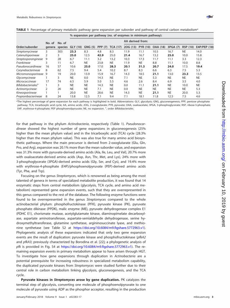
for that pathway in the phylum Actinobacteria, respectively (Table 1). Pseudonocar-dineae showed the highest number of gene expansions in gluconeogenesis (25%higher than the mean phylum value) and in the tricarboxylic acid (TCA) cycle (28.3%higher than the mean phylum value). This was also true for many amino acid biosyn-thetic pathways. Where the main precursor is derived from 2-oxoglutarate (Glu, Gln,Pro, and Arg), expansion was 20.1% more than the mean suborder value, and expansionwas 31.3% more with pyruvate-derived amino acids (Ala, Ile, Leu, and Val), 20.7% morewith oxaloacetate-derived amino acids (Asp, Asn, Thr, Met, and Lys), 24% more with3-phosphoglycerate (3PGA)-derived amino acids (Gly, Ser, and Cys), and 19.4% morewith erythrose-4-phosphate (E4P)/phosphoenolpyruvate (PEP)-derived amino acids(Tyr, Phe, and Trp).
Focusing on the genus Streptomyces, which is renowned as being among the mosttalented of genera in terms of specialized metabolite production, it was found that 14enzymatic steps from central metabolism (glycolysis, TCA cycle, and amino acid me-tabolism) represented gene expansion events, such that they are overrepresented inthis genus compared to the rest of the database. The following enzyme functions werefound to be overrepresented in the genus Streptomyces compared to the wholeactinobacterial phylum: phosphofructokinase (PFK), pyruvate kinase (PK), pyruvatephosphate dikinase (PPDK), malic enzyme (ME), pyruvate dehydrogenase complex E1(PDHC E1), chorismate mutase, acetylglutamate kinase, diaminopimelate decarboxyl-ase, aspartate aminotransferase, aspartate-semialdehyde dehydrogenase, serine hy-droxymethyltransferase, glutamine synthetase, argininosuccinate lyase, and methio-nine synthetase (see Table S2 at https://doi.org/10.6084/m9.figshare.5772963.v1).Phylogenetic analysis of these expansions indicated that only two gene expansionevents are the result of duplication: pyruvate kinase and phosphofructokinase (pfkA2and pfkA3; previously characterized by Borodina et al. [22]; a phylogenetic analysis ofpfk is provided in Fig. S4 at https://doi.org/10.6084/m9.figshare.5772963.v1). The re-maining expansion events in primary metabolism appear to have arisen through HGT.To investigate how gene expansions through duplication in Actinobacteria are apotential prerequisite for increasing robustness in specialized metabolism capability,the duplicated pyruvate kinases from Streptomyces were studied further due to theircentral role in carbon metabolism linking glycolysis, gluconeogenesis, and the TCAcycle.
Pyruvate kinases in Streptomyces arose by gene duplication. PK catalyzes theterminal step of glycolysis, converting one molecule of phosphoenolpyruvate to onemolecule of pyruvate using ADP as the phosphor acceptor, resulting in the production
TABLE 1 Percentage of primary metabolic pathway gene expansion per suborder and pathway of central carbon metabolisma
Order/suborderNo. ofgenera
No. ofspecies
% expansion per pathway (no. of enzymes in minimum pathway):
GLY (10) GNG (5) PPP (7) TCA (17)
AA derived from:
2OG (13) PYR (10) OAA (18) 3PGA (7) R5P (10) E4P/PEP (18)
Streptomycineae 3 303 23.3 8.3 4.8 8.3 11.9 11.1 10.5 16.7 NE 14.0Catenulisporineae 2 3 25.0 12.5 42.9 25.0 21.4 16.7 13.2 25.0 10.0 15.8Streptosporangineae 9 28 6.7 11.1 3.2 13.2 10.3 17.3 11.7 11.1 3.3 12.3Frankineae 3 11 6.7 NE 23.8 NE 11.9 NE 8.8 11.1 10.0 8.8Pseudonocardineae 16 57 10.6 25.0 17.0 28.3 20.1 31.3 20.7 24.0 17.5 19.4Corynebacterineae 8 62 7.5 9.4 5.4 10.2 8.7 8.3 6.6 6.3 7.5 5.3Micromonosporineae 9 19 20.0 13.9 15.9 16.7 14.3 18.5 21.1 13.0 23.3 13.5Glycomycineae 1 3 NE 0.0 14.3 NE 7.1 NE 5.3 NE NE NEMicrococcineae 17 74 6.5 5.9 5.0 5.5 4.6 2.6 8.4 6.9 3.5 4.0Bifidobacteriales* 1 3 NE NE 14.3 NE 0.0 11.1 21.1 NE 10.0 NEActinomycineae 2 26 NE NE 7.1 NE 0.0 NE NE NE NE 5.3Kineosporiineae 1 1 20.0 NE 28.6 NE 14.3 NE 21.1 NE 20.0 5.3Propionibacterineae 8 24 13.8 12.5 7.1 9.4 7.1 18.1 11.8 12.5 7.5 4.6aThe highest percentage of gene expansion for each pathway is highlighted in bold. Abbreviations: GLY, glycolysis; GNG, gluconeogenesis; PPP, pentose phosphatepathway; TCA, tricarboxylic acid cycle; AA, amino acids; 2OG, 2-oxoglutarate; PYR, pyruvate; OAA, oxaloacetate; 3PGA, 3-phosphoglycerate; R5P, ribose-5-phosphate;E4P, erythrose-4-phosphate; PEP phosphoenolpyruvate; NE, no expansion. *, order Bifidobacteriales.
Metabolic Robustness in Streptomyces ®
January/February 2018 Volume 9 Issue 1 e02283-17 mbio.asm.org 3
on February 10, 2019 by guest
http://mbio.asm
.org/D
ownloaded from
of ATP. PK therefore plays a key role in linking glycolysis and the citric acid cycle.Moreover, it results in the formation of a direct precursor of acetyl coenzyme A(acetyl-CoA), which feeds directly into polyketide specialized metabolites. A high-resolution species-level phylogeny of the Actinobacteria was constructed using the �
subunit of RNA polymerase (RpoB [23, 24]), which allowed the segregation of thephylum into three distinct phylogenetic branches: one composed of Streptomycineaeand Catenulisporineae; a second including Propionibacterineae, Actinomycineae, Bifido-bacteriales, and Micrococcineae; and the third one with Micromonosporineae, Glycomy-cineae, Corynebacterineae, Pseudonocardineae, Frankineae, and Streptosporangineae(Fig. 1A).
A second phylogeny of the annotated PKs of the Actinobacteria was constructed. Itindicated that there is a high level of congruence with the RpoB phylogeny, asexpected for a central metabolic enzyme (Fig. 1B). However, a bifurcating topologywithin the Streptomycetaceae family was found, which contained the two genes en-coding the putative PKs. This topology indicates that a gene duplication event oc-curred, giving rise to two PKs within this group. Analysis of 286 Streptomyces speciesshowed that 281 species have duplicate copies of PK, three species possess a singlecopy (Streptomyces somaliensis, Streptomyces sp. strain NRRL F5135, and Streptomy-ces scabrisporus), two species have three copies (Streptomyces olindensis and Strepto-myces sp. strain AcH505), and a single species has four copies (Streptomyces resistomy-cificus). Interestingly, Streptomyces sp. AcH505 and S. resistomycificus had one copy ofpyk in each main branch of the PK tree and additional copies were found to bephylogenetically distant (orange branches in Fig. 1B), suggesting that these copies wereacquired through horizontal gene transfer (HGT). Overall, 92% (302 of 327) of actino-bacterial genomes outside the genus Streptomyces encoded a single PK, reinforcing theuniqueness of the duplication in this genus (Fig. 1B).
To determine if the duplicate PKs annotated in the Streptomyces genome havepyruvate kinase activity, we used the two PKs from the model streptomycete S. coeli-color A3(2) in genetic complementation tests of PK mutants of Escherichia coli. E. colialso has two PKs: a primary enzyme encoded by pykF, which is a type I enzyme,regulated allosterically by fructose-1,6-biphosphate (FBP), and a distinct secondary typeII PK (pykA), regulated allosterically by AMP (25). In Streptomyces, both PKs (Pyk1 andPyk2) are type II enzymes regulated by AMP. To test for functional complementation,E. coli mutants (�pykA, �pykF, and a �pykA�pykF double mutant [see Table S4 athttps://doi.org/10.6084/m9.figshare.5772963.v1]) were tested, along with the isogenicparental strain (E. coli BW25113) for their ability to grow under a range of physiologicalconditions (Fig. 1C). In LB (for which PK is dispensable for growth) and M9 plus acetateas the sole carbon source (where PK is also dispensable for growth), little difference wasobserved in the specific growth rate (hour�1) of the strains. When the strains weregrown in M9 plus glucose as the sole carbon source (where PK is essential for growth),the E. coli �pykA �pykF double mutant was unable to grow but could be geneticallycomplemented with either pyk1 (SCO2014) or pyk2 (SCO5423) from S. coelicolor. Theindividual E. coli ΔpykA and ΔpykF mutants had reduced specific growth rates (around50% of the rate of the isogenic parent strain) in M9 plus glucose. Genetic complemen-tation with pyk1 or pyk2 from S. coelicolor was able to restore growth of an E. coli �pykFmutant as expected. The E. coli �pykA mutant could be complemented only with pyk2from S. coelicolor, suggesting a much more limited physiological role for pykA in E. coli(Fig. 1C). Remarkably, pyk2 is the closest BLAST homologue of pykA in the S. coelicolorgenome (43% identity). These data confirm that both pyk1 and pyk2 from S. coelicolorhave retained PK activity following the duplication event but that each has divergedand evolved different physiological roles.
Given that the PKs in S. coelicolor have diverged following duplication, we assessedthe level of selection imposed on the PKs of Streptomyces by calculating the ratio ofnonsynonymous changes (dN) to synonymous changes (dS). Twenty PK sequences from10 Streptomyces genomes were chosen to calculate the dN, dS, and dN/dS values. ThedN/dS ratio for pairs of pyk sequences for each of the genomes yielded dN/dS ratios
Schniete et al. ®
January/February 2018 Volume 9 Issue 1 e02283-17 mbio.asm.org 4
on February 10, 2019 by guest
http://mbio.asm
.org/D
ownloaded from
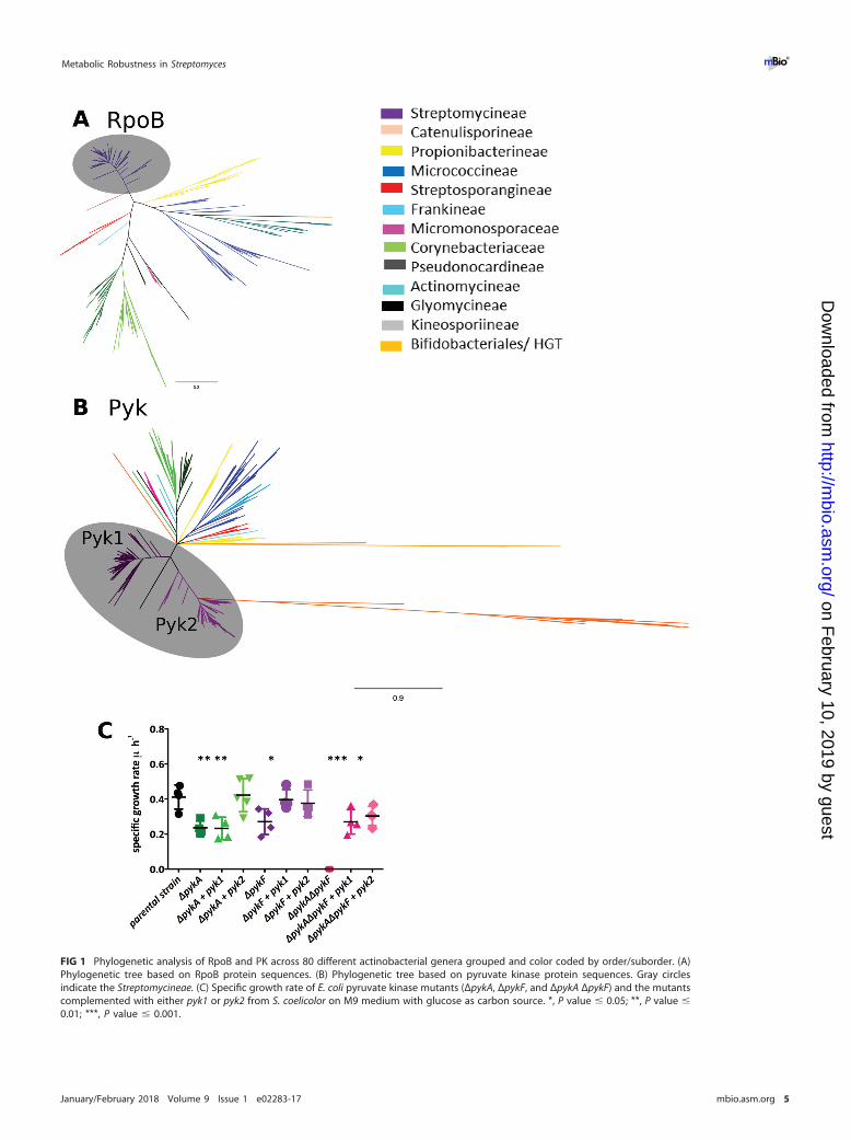
FIG 1 Phylogenetic analysis of RpoB and PK across 80 different actinobacterial genera grouped and color coded by order/suborder. (A)Phylogenetic tree based on RpoB protein sequences. (B) Phylogenetic tree based on pyruvate kinase protein sequences. Gray circlesindicate the Streptomycineae. (C) Specific growth rate of E. coli pyruvate kinase mutants (ΔpykA, ΔpykF, and ΔpykA ΔpykF) and the mutantscomplemented with either pyk1 or pyk2 from S. coelicolor on M9 medium with glucose as carbon source. *, P value � 0.05; **, P value �0.01; ***, P value � 0.001.
Metabolic Robustness in Streptomyces ®
January/February 2018 Volume 9 Issue 1 e02283-17 mbio.asm.org 5
on February 10, 2019 by guest
http://mbio.asm
.org/D
ownloaded from
ranging from 0.407 to 0.500, suggesting that PKs in Streptomyces are under strongpurifying selection (see Table S3 at https://doi.org/10.6084/m9.figshare.5772963.v1).Such high levels of purifying selection indicate that the duplication event in Strepto-myces is likely to be ancient and is consistent with the PK tree topology (Fig. 1B).
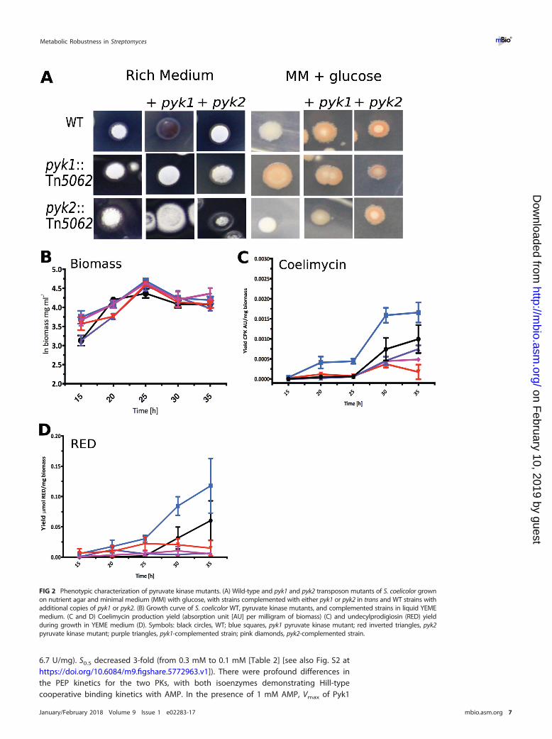
The two pyruvate kinases in Streptomyces have distinct physiological roles. Todetermine the roles played by the PKs in growth, development, and antibiotic produc-tion, a series of mutant S. coelicolor strains was constructed and genetically comple-mented (see Table S4, Table S5, and Fig. S2 at https://doi.org/10.6084/m9.figshare.5772963.v1). Deletion mutants and transposon insertion mutants showed similarphenotypes (see Fig. S2 at https://doi.org/10.6084/m9.figshare.5772963.v1), and allsubsequent work was carried out with transposon insertion mutants. Growth onnutrient agar showed no differences between the strains, except when an additionalcopy of pyk1 was present in the wild type (WT) in trans, resulting in overproduction ofactinorhodin (ACT) in rich medium and undecylprodigiosin (RED) in minimal medium(Fig. 2A). During culture on solid minimal medium with 1% glucose as carbon source,the strains showed no growth defects compared to the wild type (Fig. 2A). Interestingly,the pyk1::Tn5062 mutant showed an increase in specialized metabolite production(Fig. 2A, C, and D). The pyk2::Tn5062 strain was marginally affected in growth andshowed no overexpression of specialized metabolites (Fig. 2A). No changes in growthrate were observed in rich medium (YEME medium) for the WT, mutants, or comple-mented strains (Fig. 2B). However, growth of the strains in this medium showed anincrease in production of the polyketide coelimycin (26) and RED in the pyk1::Tn5062mutant (Fig. 2C), whereas a pyk2::Tn5062 mutant showed reduced antibiotic yields(Fig. 2C and D). These data suggest that each PK isoenzyme plays a distinct physio-logical role in growth of Streptomyces and that perturbation of central metabolism bytheir deletion or addition affects specialized metabolite biosynthesis. This would sug-gest that the PKs either are regulated differently at key stages of the Streptomyces lifecycle at the level of transcription or are regulated at the posttranscriptional/transla-tional level.
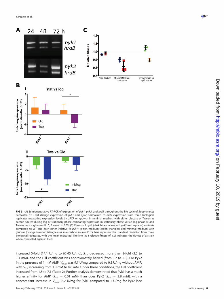
To test this hypothesis, we used semiquantitative reverse transcription-PCR (RT-PCR)to examine the expression of the PKs from S. coelicolor throughout growth, relative tothe multiplexed vegetative sigma factor encoded by hrdB. We found that both geneswere constitutively expressed throughout growth (vegetative hyphae and aerial hy-phae and during sporulation) relative to hrdB (Fig. 3A). To further characterize tran-scription, we used quantitative real time PCR (qRT-PCR) at two time points during logand stationary phase during either glycolytic growth (glucose as sole carbon source) oranaplerotic/gluconeogenic growth (Tween as sole carbon source [27]) to analyze theexpression levels of pyk1, pyk2, and hrdB. Normalizing expression to hrdB, there was anexpected decrease in pyk1 expression on Tween compared to glucose during the logand stationary phases of growth (3-fold and 8-fold, respectively [Fig. 3A and B]).Comparison of pyk1 and pyk2 during the logarithmic growth phase indicated that pyk2had a 1.5-fold-lower level of expression when grown on Tween as the sole carbonsource than it did in stationary phase, with all other conditions showing no significantchanges in expression between pyk1 and pyk2 (Fig. 3B, i), suggesting that activity of PKsin Streptomyces is likely to be controlled at the posttranslational level. Expression ofpyk2 also showed a decrease in expression on Tween compared to that on glucoseduring both phases, but the change was not significant (Fig. 3B, ii).
Pyk1 and Pyk2 in Streptomyces have key substrate affinity differences andspecific effector molecules. To understand the biochemical control of the two PKs ofS. coelicolor, we purified each enzyme and studied its biochemical characteristics. BothPyk1 and Pyk2 were activated by the effector molecule AMP, and they both showedMichaelis-Menten-type kinetics for the substrate ADP. For Pyk1, S0.5 (substrate concen-tration at half Vmax) was 3-fold lower in the presence of 1 mM AMP (0.6 mM down to0.2 mM [Table 2]), while Vmax also increased 3.5-fold (from 21 U/mg to 73.3 U/mg). Pyk2showed a 5-fold increase of Vmax in the presence of 1 mM AMP (from 1.2 U/mg to
Schniete et al. ®
January/February 2018 Volume 9 Issue 1 e02283-17 mbio.asm.org 6
on February 10, 2019 by guest
http://mbio.asm
.org/D
ownloaded from
6.7 U/mg). S0.5 decreased 3-fold (from 0.3 mM to 0.1 mM [Table 2] [see also Fig. S2 athttps://doi.org/10.6084/m9.figshare.5772963.v1]). There were profound differences inthe PEP kinetics for the two PKs, with both isoenzymes demonstrating Hill-typecooperative binding kinetics with AMP. In the presence of 1 mM AMP, Vmax of Pyk1
FIG 2 Phenotypic characterization of pyruvate kinase mutants. (A) Wild-type and pyk1 and pyk2 transposon mutants of S. coelicolor grownon nutrient agar and minimal medium (MM) with glucose, with strains complemented with either pyk1 or pyk2 in trans and WT strains withadditional copies of pyk1 or pyk2. (B) Growth curve of S. coelicolor WT, pyruvate kinase mutants, and complemented strains in liquid YEMEmedium. (C and D) Coelimycin production yield (absorption unit [AU] per milligram of biomass) (C) and undecylprodigiosin (RED) yieldduring growth in YEME medium (D). Symbols: black circles, WT; blue squares, pyk1 pyruvate kinase mutant; red inverted triangles, pyk2pyruvate kinase mutant; purple triangles, pyk1-complemented strain; pink diamonds, pyk2-complemented strain.
Metabolic Robustness in Streptomyces ®
January/February 2018 Volume 9 Issue 1 e02283-17 mbio.asm.org 7
on February 10, 2019 by guest
http://mbio.asm
.org/D
ownloaded from
increased 5-fold (14.1 U/mg to 65.45 U/mg), S0.5 decreased more than 3-fold (3.5 to1.1 mM), and the Hill coefficient was approximately halved (from 3.7 to 1.8). For Pyk2in the presence of 1 mM AMP, Vmax was 9.1 U/mg compared to 0.5 U/mg without AMP,with S0.5 increasing from 1.3 mM to 8.6 mM. Under these conditions, the Hill coefficientincreased from 1.5 to 7.1 (Table 2). Further analysis demonstrated that Pyk1 has a muchhigher affinity for AMP (S0.5 � 0.01 mM) than does Pyk2 (S0.5 � 3.8 mM), with aconcomitant increase in Vmax (8.2 U/mg for Pyk1 compared to 1 U/mg for Pyk2 [see
FIG 3 (A) Semiquantitative RT-PCR of expression of pyk1, pyk2, and hrdB throughout the life cycle of Streptomycescoelicolor. (B) Fold change expression of pyk1 and pyk2 normalized to hrdB expression from three biologicalreplicates measuring expression levels by qPCR on growth in minimal medium with either glucose or Tween ascarbon source during log or stationary phase comparing expression in stationary phase versus log phase (i) andTween versus glucose (ii). *, P value � 0.05. (C) Fitness of pyk1 (dark blue circles) and pyk2 (red squares) mutantscompared to WT and each other (relative to pyk1) in rich medium (green triangles) and minimal medium withglucose (orange inverted triangles) as sole carbon source. Error bars represent the standard deviation from threebiological replicates, with the mean indicated. The line (at a relative fitness of 1.0) indicates the fitness of a strainwhen competed against itself.
Schniete et al. ®
January/February 2018 Volume 9 Issue 1 e02283-17 mbio.asm.org 8
on February 10, 2019 by guest
http://mbio.asm
.org/D
ownloaded from
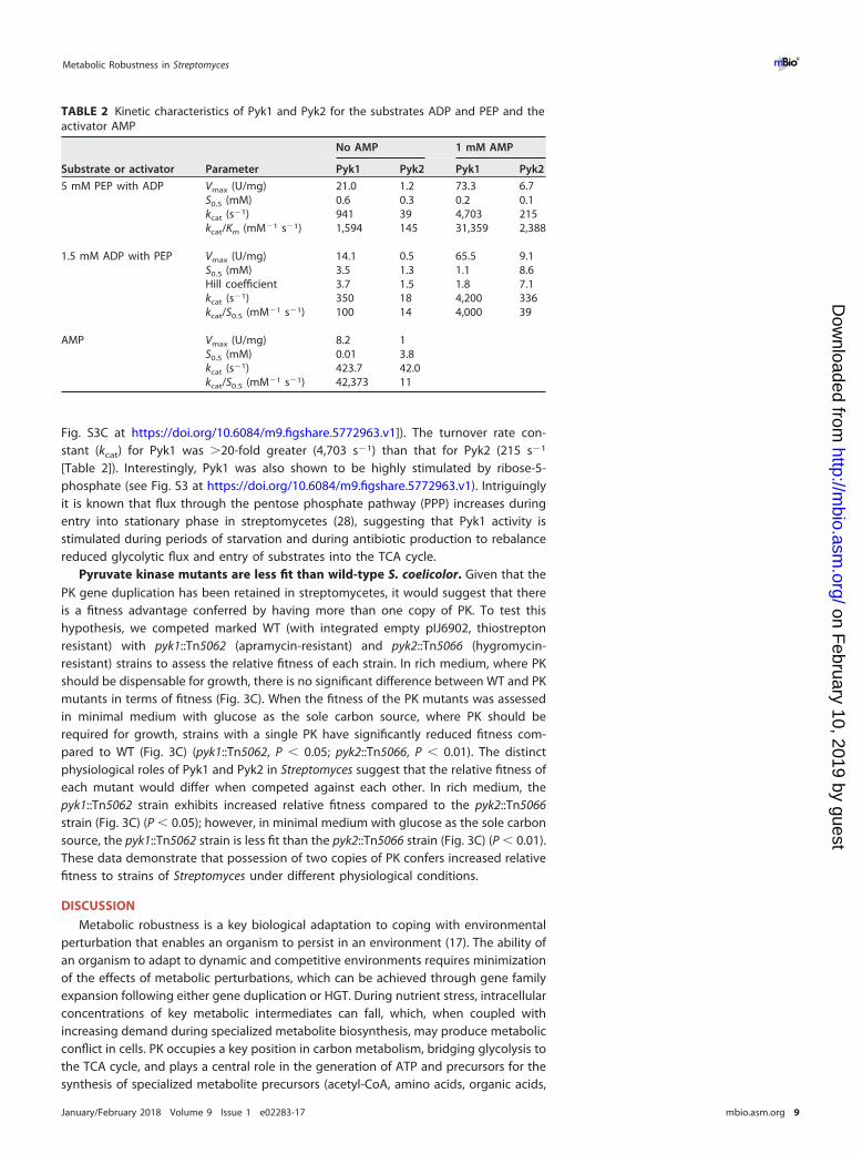
Fig. S3C at https://doi.org/10.6084/m9.figshare.5772963.v1]). The turnover rate con-stant (kcat) for Pyk1 was �20-fold greater (4,703 s�1) than that for Pyk2 (215 s�1
[Table 2]). Interestingly, Pyk1 was also shown to be highly stimulated by ribose-5-phosphate (see Fig. S3 at https://doi.org/10.6084/m9.figshare.5772963.v1). Intriguinglyit is known that flux through the pentose phosphate pathway (PPP) increases duringentry into stationary phase in streptomycetes (28), suggesting that Pyk1 activity isstimulated during periods of starvation and during antibiotic production to rebalancereduced glycolytic flux and entry of substrates into the TCA cycle.
Pyruvate kinase mutants are less fit than wild-type S. coelicolor. Given that thePK gene duplication has been retained in streptomycetes, it would suggest that thereis a fitness advantage conferred by having more than one copy of PK. To test thishypothesis, we competed marked WT (with integrated empty pIJ6902, thiostreptonresistant) with pyk1::Tn5062 (apramycin-resistant) and pyk2::Tn5066 (hygromycin-resistant) strains to assess the relative fitness of each strain. In rich medium, where PKshould be dispensable for growth, there is no significant difference between WT and PKmutants in terms of fitness (Fig. 3C). When the fitness of the PK mutants was assessedin minimal medium with glucose as the sole carbon source, where PK should berequired for growth, strains with a single PK have significantly reduced fitness com-pared to WT (Fig. 3C) (pyk1::Tn5062, P � 0.05; pyk2::Tn5066, P � 0.01). The distinctphysiological roles of Pyk1 and Pyk2 in Streptomyces suggest that the relative fitness ofeach mutant would differ when competed against each other. In rich medium, thepyk1::Tn5062 strain exhibits increased relative fitness compared to the pyk2::Tn5066strain (Fig. 3C) (P � 0.05); however, in minimal medium with glucose as the sole carbonsource, the pyk1::Tn5062 strain is less fit than the pyk2::Tn5066 strain (Fig. 3C) (P � 0.01).These data demonstrate that possession of two copies of PK confers increased relativefitness to strains of Streptomyces under different physiological conditions.
DISCUSSION
Metabolic robustness is a key biological adaptation to coping with environmentalperturbation that enables an organism to persist in an environment (17). The ability ofan organism to adapt to dynamic and competitive environments requires minimizationof the effects of metabolic perturbations, which can be achieved through gene familyexpansion following either gene duplication or HGT. During nutrient stress, intracellularconcentrations of key metabolic intermediates can fall, which, when coupled withincreasing demand during specialized metabolite biosynthesis, may produce metabolicconflict in cells. PK occupies a key position in carbon metabolism, bridging glycolysis tothe TCA cycle, and plays a central role in the generation of ATP and precursors for thesynthesis of specialized metabolite precursors (acetyl-CoA, amino acids, organic acids,
TABLE 2 Kinetic characteristics of Pyk1 and Pyk2 for the substrates ADP and PEP and theactivator AMP
Substrate or activator Parameter
No AMP 1 mM AMP
Pyk1 Pyk2 Pyk1 Pyk2
5 mM PEP with ADP Vmax (U/mg) 21.0 1.2 73.3 6.7S0.5 (mM) 0.6 0.3 0.2 0.1kcat (s�1) 941 39 4,703 215kcat/Km (mM�1 s�1) 1,594 145 31,359 2,388
1.5 mM ADP with PEP Vmax (U/mg) 14.1 0.5 65.5 9.1S0.5 (mM) 3.5 1.3 1.1 8.6Hill coefficient 3.7 1.5 1.8 7.1kcat (s�1) 350 18 4,200 336kcat/S0.5 (mM�1 s�1) 100 14 4,000 39
AMP Vmax (U/mg) 8.2 1S0.5 (mM) 0.01 3.8kcat (s�1) 423.7 42.0kcat/S0.5 (mM�1 s�1) 42,373 11
Metabolic Robustness in Streptomyces ®
January/February 2018 Volume 9 Issue 1 e02283-17 mbio.asm.org 9
on February 10, 2019 by guest
http://mbio.asm
.org/D
ownloaded from
etc. [27]). At key branch points in metabolism, such as here, the levels of metabolitesare tightly controlled (29), and in E. coli, the flux between PEP and pyruvate is stablymaintained by two phylogenetically distinct PKs (25, 30). In the streptomycetes, aduplication event has led to the evolution of altered substrate affinity and enzymeefficiency in the two duplicate copies which contribute to metabolic robustnessenabling cellular processes to occur during times of perturbation, such as at the entryinto stationary phase and specialized metabolite production.
Enzyme family expansion in central metabolism is widespread in the Actinobacteria,in particular with those genera that are extensive producers of specialized metabolites(21) (see Table S1 at https://doi.org/10.6084/m9.figshare.5772963.v1), with the Strepto-mycineae showing extensive enzyme family expansion in glycolysis. Within glycolysis,only two enzyme families show duplication as a route to expansion. Borodina et al. (22)showed that PfkA2 was the primary phosphofructokinase in S. coelicolor but did notstudy the wider role of duplication or a detailed kinetic comparison of the expansionevent. The duplication of pyruvate kinase is ancient and has permitted subsequentdivergence of the gene pair to evolve distinct physiological roles, where Pyk2 appearsto function as a housekeeping PK, with a higher affinity for PEP (when AMP is low), andPyk1 exhibits strong activation as AMP concentrations rise. An increased AMP concen-tration is a well-established starvation signal in bacteria (31) and may serve to increaseflux through the terminal end of glycolysis during starvation to facilitate precursorsupply for specialized metabolites. Moreover, the PPP intermediate ribose-5-phosphatestimulates Pyk1 activity, providing a physiological link between the PPP and theassociated generation of NADPH, which has established links to synthesis of specializedmetabolites, including those overproduced by strains engineered in this work (28).Disruption of pyk1 in S. coelicolor leads to increased levels of coelimycin and undecyl-prodigiosin when grown in rich medium (such as in industrial situations) but nosignificant increase in the yield of the polyketide antibiotic actinorhodin, which mayreflect the ranges of different metabolic control points affecting biosynthesis of chem-ically similar specialized metabolites. The regulation of this node is known to becontrolled by at least one global regulator, ScbR2, which is encoded in the coelimycin(cpk) biosynthetic gene cluster (BGC). ScbR2 expressed in mid-log phase has beenshown to bind to pyk2 and reduce its expression. ScbR2 is also a regulator of ACT andRED biosynthesis in addition to the angucyclines (32). Interestingly, coelimycin wasoriginally considered to be a cryptic BGC and the disruption of PK activity results inincreased production of this molecule, suggesting that strategies that affect precursorsupply may activate the plethora of cryptic biosynthetic pathways in Actinobacteria andmay provide a useful strategy for studying these molecules.
Our data demonstrate that the duplication of pyruvate kinase promotes metabolicrobustness through altered kinetic parameters which increase the relative fitness ofstrains and influence the production of specialized metabolites. Understanding theevolution of central metabolism in conjunction with specialized metabolism can con-tribute to our fundamental understanding of the ability of Actinobacteria to produce aplethora of useful molecules and can help inform on novel approaches to metabolicengineering and genome mining (21).
MATERIALS AND METHODSDatabase generation and bioinformatics analysis. The NCBI database (https://www.ncbi.nlm.nih
.gov/genbank/wgs) was the source of actinobacterial genomes having a minimum coverage of 25� andless than 30 contigs per mega-base pair. To ensure a wide range of phylogeny, a selection of 612 speciesfrom 80 genera were included. Each genome was reannotated using RAST (19), and the annotation fileswere used to determine the frequency of each functional annotation. The mean number of occurrencesof each functional role was calculated per genus, and examples which had a value equal to or higher thanthe mean plus its standard deviation were defined as a “gene expansion event.” Each candidate proteinsequence was extracted from the actinobacterial genome database to form a BLASTP analysis workingdatabase. The sequences were then aligned with MUSCLE v3.8.31 (33), and alignments were scrutinizedusing Jalview v2.10.1 to ensure that at least 25% coverage with the query was achieved; if not, sequencesand expansions were discarded from the working database. Phylogenetic analysis of the alignments wasconducted using MrBayes (34) v3.2.3 with trees visualized in FigTree v1.4.2 (http://tree.bio.ed.ac.uk/software/figtree/). Sequences were obtained from the NCBI gene database and aligned with ClustalW
Schniete et al. ®
January/February 2018 Volume 9 Issue 1 e02283-17 mbio.asm.org 10
on February 10, 2019 by guest
http://mbio.asm
.org/D
ownloaded from

algorithm in MEGA v6.06 (35), and the synonymous (dS) and nonsynonymous (dN) changes weredetermined using the distance model function with the Nei-Gojobori method (36) with the Jukes-Cantoralgorithm (37) bootstrap.
Growth and mutant construction in Streptomyces. A full list of strains, plasmids and oligonucle-otides used in this study is provided in Tables S4, S5, and S6 at https://doi.org/10.6084/m9.figshare.5772963.v1. Routine growth, spore generation, and conjugation of Streptomyces strains were carried outaccording to the work of Kieser et al. (38). Antibiotic titers were determined according to the work ofGomez-Escribano et al. (26) for coelimycin and the work of Kieser et al. (38) for RED and ACT. S. coelicolorgene knockout mutants (�pyk1 and �pyk2) were constructed using PCR-targeted gene replacement withan apramycin resistance cassette [acc(3)IV] using the Redirect system (39) and the primers reported inTable S6 at https://doi.org/10.6084/m9.figshare.5772963.v1. S. coelicolor transposon insertion mutants(pyk1::Tn5062 and pyk2::Tn5062) were constructed using Tn5062 mutagenized cosmids as described inthe work of Fernández-Martínez et al. (40). Each cosmid was first verified by restriction analysis beforebeing conjugated into Streptomyces. All strains were verified by PCR and sequencing of the respectiveproducts. Growth curves were performed at a 400-ml scale in minimal medium (28) with either 1%glucose or Tween 80 as the carbon source in 2-liter flasks containing a metal spring at 30°C and 250 rpm.
Interspecies complementation. E. coli single mutants were from the Keio collection of E. coliBW25113 (41), and the double mutant was constructed using � Red recombination of pykF according tothe work of Datsenko and Wanner (42). Complementation studies used the Streptomyces pyk1 and pyk2genes cloned into pET100_TOPO (Invitrogen).
E. coli growth curves were carried out in 250-ml flasks with a working volume of 50 ml of either LBor M9 medium with 1% (wt/vol) glucose or 0.4% (wt/vol) sodium acetate as carbon source. Flasks wereinoculated from an overnight culture (1% [vol/vol]) including the appropriate antibiotics and 1 mMisopropyl-�-D-thiogalactopyranoside (IPTG) to induce expression of the PKs. Growth was followed asoptical density at 600 nm (OD600) at 37°C with shaking at 250 rpm. The specific growth rate wasdetermined from the semilogarithmic plot of biomass concentration.
Protein overexpression and purification. The coding sequence of pyk1 was codon optimized forE. coli and amplified from the vector pEX-K4 using the primers in Table S6 at https://doi.org/10.6084/m9.figshare.5772963.v1. The native version of pyk2 was used to amplify the coding sequence using theprimers in Table S6 at https://doi.org/10.6084/m9.figshare.5772963.v1. Both coding sequences werecloned into the pET100_TOPO vector (Invitrogen) according to the manufacturer’s instructions. Overex-pression of pyk1 was in E. coli Origami B on LB with 1% (wt/vol) glucose at 30°C until an OD600 of 0.4 wasreached, and the expression was induced with 0.05 mM IPTG at 18°C overnight. Pyk2 overexpression wasin E. coli Rosetta using autoinduction medium [components per liter: 10 g tryptone, 5 g yeast extract,3.3 g (NH4)2SO4, 6.8 g KH2PO4, 7.1 g Na2HPO4, 0.5 g glucose, 2.0 g �-lactose, 0.15 g MgSO4]; cells weregrown at 37°C for 2 h and then reduced to 18°C for overnight cultivation. Cells were disrupted bysonication. Pyk1 and Pyk2 were purified by nickel affinity chromatography using HisTrap FF crude (GEHealthcare) with binding buffer (100 mM KH2PO4 pH 7.2, 10% [vol/vol] glycerol, 100 mM NaCl, 20 mMimidazole). Tagged proteins were eluted with increasing imidazole concentrations (elution buffer:100 mM KH2PO4 [pH 7.2], 10% [vol/vol] glycerol, 100 mM NaCl, 1 M imidazole). Fractions (1 ml) werecollected, and the highest concentrations of protein were pooled.
Kinetic characteristics of each pyruvate kinase were determined using purified protein samplesaccording to the method of Bergmeyer et al. (43). Assays to determine the enzyme kinetics for each PKunder each condition were carried out in triplicate, and results were analyzed using GraphPad Prismusing the Michaelis-Menten or Hill equation where appropriate.
RNA extraction, semiquantitative RT-PCR, and qPCR analysis. Biomass of S. coelicolor came fromliquid cultures, and semiquantitative RT-PCR was carried out according to the method of Clark andHoskisson (7). Total RNA was used as the template for cDNA synthesis using a qPCRBio cDNA synthesiskit (PCR Biosystems). All cDNA samples were diluted to a concentration of 10 ng/�l with each quanti-tative PCR (qPCR) mixture containing 10 ng of cDNA (1 �l) and were then mixed with 10 �l MasterMix(2� qPCRBio SyGreen mix Lo-ROX kit from PCR Biosystems) and 2.5 �l of each primer to a final reactionvolume of 20 �l using the Corbett Research 6000 analyzer (Qiagen).
Fitness experiments. Competitive fitness of PK mutants versus WT was estimated based on the workof Lenski et al. (44). Briefly, spores of each strain were separately pregerminated in 2� YT medium (38)for 7 h at 30°C and washed twice in the culture medium to be used in the fitness experiment. GermlingOD600 was determined, and a 10-ml culture was inoculated with approximately 1 � 105 CFU of eachstrain. Initial and final strain densities (after 68 h of growth) were determined by plating on nutrient agarwith the appropriate selection. The net growth rate of each competitor was calculated from the data, andthe relative fitness of a strain is expressed as the ratio of its growth rate to that of its competitor. Assayswere performed in triplicate.
ACKNOWLEDGMENTSWe thank Ian Henderson and Faye Morris (University of Birmingham) for the gift of
the E. coli Keio collection mutants and David Hodgson for helpful discussions.We thank the Scottish Universities Life Science Alliance (SULSA) for BioSkape PhD
funding to J.K.S., the Mac Robertson Travelling Scholarship awarded to J.K.S. to visit thelaboratory of F.B.-G., and the Natural Environment Research Council for grant NE/M001415/1 to P.A.H. Work in the laboratory of F.B.-G. was funded by CONACyT, Mexico
Metabolic Robustness in Streptomyces ®
January/February 2018 Volume 9 Issue 1 e02283-17 mbio.asm.org 11
on February 10, 2019 by guest
http://mbio.asm
.org/D
ownloaded from
(grant 177568), and Langebio institutional funds to support P.C.-M. as a postdoctoralfellow. L.T.F.-M. acknowledges the support of BBSRC/NPRONET (NPRONET POC028), theBritish Council (275898511), and Edge Hill University for funding.
The funders had no role in study design, data collection and interpretation, or thedecision to submit the work for publication.
REFERENCES1. Bentley SD, Chater KF, Cerdeño-Tárraga AM, Challis GL, Thomson NR,
James KD, Harris DE, Quail MA, Kieser H, Harper D, Bateman A, Brown S,Chandra G, Chen CW, Collins M, Cronin A, Fraser A, Goble A, Hidalgo J,Hornsby T, Howarth S, Huang CH, Kieser T, Larke L, Murphy L, Oliver K,O’Neil S, Rabbinowitsch E, Rajandream MA, Rutherford K, Rutter S,Seeger K, Saunders D, Sharp S, Squares R, Squares S, Taylor K, Warren T,Wietzorrek A, Woodward J, Barrell BG, Parkhill J, Hopwood DA. 2002.Complete genome sequence of the model actinomycete Streptomycescoelicolor A3(2). Nature 417:141–147. https://doi.org/10.1038/417141a.
2. Hiltner JK, Hunter IS, Hoskisson PA. 2015. Tailoring specialized metabo-lite production in Streptomyces. Adv Appl Microbiol 91:237–255. https://doi.org/10.1016/bs.aambs.2015.02.002.
3. Wagner A. 2008. Robustness and evolvability: a paradox resolved. ProcBiol Sci 275:91–100. https://doi.org/10.1098/rspb.2007.1137.
4. Wagner A. 2008. Gene duplications, robustness and evolutionary inno-vations. Bioessays 30:367–373. https://doi.org/10.1002/bies.20728.
5. Treangen TJ, Rocha EPC. 2011. Horizontal transfer, not duplication,drives the expansion of protein families in prokaryotes. PLoS Genet7:e1001284. https://doi.org/10.1371/journal.pgen.1001284.
6. McLoughlin SY, Copley SD. 2008. A compromise required by genesharing enables survival: implications for evolution of new enzymeactivities. Proc Natl Acad Sci U S A 105:13497–13502. https://doi.org/10.1073/pnas.0804804105.
7. Clark LC, Hoskisson PA. 2011. Duplication and evolution of devA-likegenes in Streptomyces has resulted in distinct developmental roles. PLoSOne 6:e25049. https://doi.org/10.1371/journal.pone.0025049.
8. Clark LC, Seipke RF, Prieto P, Willemse J, van Wezel GP, Hutchings MI,Hoskisson PA. 2013. Mammalian cell entry genes in Streptomyces mayprovide clues to the evolution of bacterial virulence. Sci Rep 3:1109.https://doi.org/10.1038/srep01109.
9. Challis GL, Hopwood DA. 2003. Synergy and contingency as drivingforces for the evolution of multiple secondary metabolite production byStreptomyces species. Proc Natl Acad Sci U S A 100(Suppl 2):14555–14561. https://doi.org/10.1073/pnas.1934677100.
10. Chater KF, Biró S, Lee KJ, Palmer T, Schrempf H. 2010. The complexextracellular biology of Streptomyces. FEMS Microbiol Rev 34:171–198.https://doi.org/10.1111/j.1574-6976.2009.00206.x.
11. Kim J, Copley SD. 2007. Why metabolic enzymes are essential or non-essential for growth of Escherichia coli K12 on glucose. Biochemistry46:12501–12511. https://doi.org/10.1021/bi7014629.
12. Teichmann SA, Babu MM. 2004. Gene regulatory network growth byduplication. Nat Genet 36:492– 496. https://doi.org/10.1038/ng1340.
13. Girard G, Traag BA, Sangal V, Mascini N, Hoskisson PA, Goodfellow M,van Wezel GP. 2013. A novel taxonomic marker that discriminatesbetween morphologically complex actinomycetes. Open Biol 3:130073.https://doi.org/10.1098/rsob.130073.
14. Chater KF, Chandra G. 2006. The evolution of development in Strepto-myces analysed by genome comparisons. FEMS Microbiol Rev 30:651– 672. https://doi.org/10.1111/j.1574-6976.2006.00033.x.
15. Tokovenko B, Rebets Y, Luzhetskyy A. 2016. Automating assessment ofthe undiscovered biosynthetic potential of Actinobacteria. bioRxiv https://doi.org/10.1101/036087.
16. Bibb MJ. 2005. Regulation of secondary metabolism in streptomycetes.Curr Opin Microbiol 8:208 –215. https://doi.org/10.1016/j.mib.2005.02.016.
17. Lenski RE, Barrick JE, Ofria C. 2006. Balancing robustness and evolvabil-ity. PLoS Biol 4:e428. https://doi.org/10.1371/journal.pbio.0040428.
18. Nowak MA, Boerlijst MC, Cooke J, Smith JM. 1997. Evolution of geneticredundancy. Nature 388:167–171. https://doi.org/10.1038/40618.
19. Aziz RK, Bartels D, Best AA, DeJongh M, Disz T, Edwards RA, Formsma K,Gerdes S, Glass EM, Kubal M, Meyer F, Olsen GJ, Olson R, Osterman AL,Overbeek RA, McNeil LK, Paarmann D, Paczian T, Parrello B, Pusch GD,Reich C, Stevens R, Vassieva O, Vonstein V, Wilke A, Zagnitko O. 2008.
The RAST server: rapid annotations using subsystems technology. BMCGenomics 9:75. https://doi.org/10.1186/1471-2164-9-75.
20. Barona-Gómez F, Cruz-Morales P, Noda-García L. 2012. What cangenome-scale metabolic network reconstructions do for prokaryoticsystematics? Antonie Van Leeuwenhoek 101:35– 43. https://doi.org/10.1007/s10482-011-9655-1.
21. Cruz-Morales P, Kopp JF, Martínez-Guerrero C, Yáñez-Guerra LA,Selem-Mojica N, Ramos-Aboites H, Feldmann J, Barona-Gómez F.2016. Phylogenomic analysis of natural products biosynthetic geneclusters allows discovery of arseno-organic metabolites in modelstreptomycetes. Genome Biol Evol 8:1906 –1916. https://doi.org/10.1093/gbe/evw125.
22. Borodina I, Siebring J, Zhang J, Smith CP, van Keulen G, Dijkhuizen L,Nielsen J. 2008. Antibiotic overproduction in Streptomyces coelicolorA3(2) mediated by phosphofructokinase deletion. J Biol Chem 283:25186 –25199. https://doi.org/10.1074/jbc.M803105200.
23. Case RJ, Boucher Y, Dahllöf I, Holmström C, Doolittle WF, Kjelleberg S.2007. Use of 16S rRNA and rpoB genes as molecular markers for micro-bial ecology studies. Appl Environ Microbiol 73:278 –288. https://doi.org/10.1128/AEM.01177-06.
24. Kämpfer P, Glaeser SP. 2012. Prokaryotic taxonomy in the sequencingera—the polyphasic approach revisited. Environ Microbiol 14:291–317.https://doi.org/10.1111/j.1462-2920.2011.02615.x.
25. Muñoz ME, Ponce E. 2003. Pyruvate kinase: current status of regulatoryand functional properties. Comp Biochem Physiol B Biochem Mol Biol135:197–218. https://doi.org/10.1016/S1096-4959(03)00081-2.
26. Gomez-Escribano JP, Song L, Fox DJ, Yeo V, Bibb MJ, Challis GL. 2012.Structure and biosynthesis of the unusual polyketide alkaloid coelimycinP1, a metabolic product of the cpk gene cluster of Streptomyces coeli-color M145. Chem Sci 3:2716. https://doi.org/10.1039/c2sc20410j.
27. Hodgson DA. 2000. Primary metabolism and its control instreptomycetes: a most unusual group of bacteria. Adv Microb Physiol42:47–238. https://doi.org/10.1016/S0065-2911(00)42003-5.
28. Obanye AIC, Hobbs G, Gardner DCJ, Oliver SG. 1996. Correlation be-tween carbon flux through the pentose phosphate pathway and pro-duction of the antibiotic methylenomycin in Streptomyces coelicolorA3(2). Microbiology 142:133–137. https://doi.org/10.1099/13500872-142-1-133.
29. Sauer U, Eikmanns BJ. 2005. The PEP-pyruvate-oxaloacetate node as theswitch point for carbon flux distribution in bacteria. FEMS Microbiol Rev29:765–794. https://doi.org/10.1016/j.femsre.2004.11.002.
30. Emmerling M, Dauner M, Ponti A, Fiaux J, Hochuli M, Szyperski T,Wüthrich K, Bailey JE, Sauer U. 2002. Metabolic flux responses to pyru-vate kinase knockout in Escherichia coli. J Bacteriol 184:152–164. https://doi.org/10.1128/JB.184.1.152-164.2002.
31. Brauer MJ, Yuan J, Bennett BD, Lu W, Kimball E, Botstein D, RabinowitzJD. 2006. Conservation of the metabolomic response to starvation acrosstwo divergent microbes. Proc Natl Acad Sci U S A 103:19302–19307.https://doi.org/10.1073/pnas.0609508103.
32. Li X, Wang J, Li S, Ji J, Wang W, Yang K. 2015. ScbR and ScbR2-mediatedsignal transduction networks coordinate complex physiological re-sponses in Streptomyces coelicolor. Sci Rep 5:14831. https://doi.org/10.1038/srep14831.
33. Edgar RC. 2004. MUSCLE: a multiple sequence alignment method withreduced time and space complexity. BMC Bioinformatics 5:113. https://doi.org/10.1186/1471-2105-5-113.
34. Ronquist F, Teslenko M, van der Mark P, Ayres DL, Darling A, HöhnaS, Larget B, Liu L, Suchard MA, Huelsenbeck JP. 2012. MrBayes 3.2:efficient Bayesian phylogenetic inference and model choice across alarge model space. Syst Biol 61:539 –542. https://doi.org/10.1093/sysbio/sys029.
35. Kumar S, Tamura K, Nei M. 2004. MEGA3: integrated software for
Schniete et al. ®
January/February 2018 Volume 9 Issue 1 e02283-17 mbio.asm.org 12
on February 10, 2019 by guest
http://mbio.asm
.org/D
ownloaded from
molecular evolutionary genetics analysis and sequence alignment.Brief Bioinform 5:150 –163. https://doi.org/10.1093/bib/5.2.150.
36. Nei M, Gojobori T. 1986. Simple methods for estimating the numbers ofsynonymous and nonsynonymous nucleotide substitutions. Mol Biol Evol3:418–426. https://doi.org/10.1093/oxfordjournals.molbev.a040410.
37. Jukes TH, Cantor CR. 1969. Evolution of protein molecules, p 21–132. InMunro HN (ed), Mammalian protein metabolism, vol 3. Academic Press,New York, NY.
38. Kieser T, Bibb MJ, Buttner MJ, Chater KF, Hopwood DA. 2000. PracticalStreptomyces genetics. John Innes Foundation, Norwich, UnitedKingdom.
39. Gust B, Challis GL, Fowler K, Kieser T, Chater KF. 2003. PCR-targetedStreptomyces gene replacement identifies a protein domain needed forbiosynthesis of the sesquiterpene soil odor geosmin. Proc Natl Acad SciU S A 100:1541–1546. https://doi.org/10.1073/pnas.0337542100.
40. Fernández-Martínez LT, Del Sol R, Evans MC, Fielding S, Herron PR,Chandra G, Dyson PJ. 2011. A transposon insertion single-gene knockout
library and new ordered cosmid library for the model organism Strep-tomyces coelicolor A3(2). Antonie Van Leeuwenhoek 99:515–522. https://doi.org/10.1007/s10482-010-9518-1.
41. Baba T, Ara T, Hasegawa M, Takai Y, Okumura Y, Baba M, Datsenko KA,Tomita M, Wanner BL, Mori H. 2006. Construction of Escherichia coli K-12in-frame, single-gene knockout mutants: the Keio collection. Mol SystBiol 2:2006.0008. https://doi.org/10.1038/msb4100050.
42. Datsenko KA, Wanner BL. 2000. One-step inactivation of chromosomalgenes in Escherichia coli K-12 using PCR products. Proc Natl Acad SciU S A 97:6640 – 6645. https://doi.org/10.1073/pnas.120163297.
43. Bergmeyer HU, Gawehn K, Grassl M. 1974. Enzymatic assay of pyruvatekinase, p 509 –510. In Bergmeyer HU (ed), Methods of enzymatic analysis,2nd ed, vol 1. Academic Press, New York, NY.
44. Lenski RE, Rose MR, Simpson SC, Tadler SC. 1991. Long-term experimen-tal evolution in Escherichia coli. I. Adaptation and divergence during2,000 generations. Am Nat 138:1315–1341. https://doi.org/10.1086/285289.
Metabolic Robustness in Streptomyces ®
January/February 2018 Volume 9 Issue 1 e02283-17 mbio.asm.org 13
on February 10, 2019 by guest
http://mbio.asm
.org/D
ownloaded from