crystal structures of the gon7 pcc1 and cgi121 complexes ... · on kae1 but the function of the...
TRANSCRIPT
3358–3372 Nucleic Acids Research, 2015, Vol. 43, No. 6 Published online 03 March 2015doi: 10.1093/nar/gkv155
Crystal structures of the Gon7/Pcc1 andBud32/Cgi121 complexes provide a model for thecomplete yeast KEOPS complexWenhua Zhang1, Bruno Collinet1,2, Marc Graille1, Marie-Claire Daugeron3,Noureddine Lazar1, Domenico Libri4, Dominique Durand1 and Herman van Tilbeurgh1,*
1Institut de Biologie Integrative de la Cellule, UMR 9198, CNRS, Universite de Paris Sud XI, Batiment 430, 91405Orsay, France, 2Sorbonne Universites, UPMC Univ Paris 06, UFR 927, Sciences de la vie, F-75005, Paris, France,3Domenico Libri 33 Institut Jacques Monod, CNRS, UMR 7592, Universite de Paris Diderot, Sorbonne Paris Cite,75205 Paris, France and 4Institut Jacques Monod, CNRS, UMR 7592, Universite de Paris Diderot, Sorbonne ParisCite, 75205 Paris, France
Received January 13, 2015; Revised February 16, 2015; Accepted February 18, 2015
ABSTRACT
The yeast KEOPS protein complex comprising Kae1,Bud32, Cgi121, Pcc1 and Gon7 is responsible for theessential tRNA threonylcarbamoyladenosine (t6A)modification. Deletion of genes coding for theKEOPS subunits also affects telomere elongationand transcriptional regulation. In the present work,the crystal structure of Bud32/Cgi121 in complexwith ADP revealed that ADP is bound in the catalyticsite of Bud32 in a canonical manner characteristicof Protein Kinase A (PKA) family proteins. We foundthat Gon7 forms a stable heterodimer with Pcc1 andreport the crystal structure of the Pcc1-Gon7 het-erodimer. Gon7 interacts with the same Pcc1 regionengaged in the archaeal Pcc1 homodimer. We furthershow that yeast KEOPS, unlike its archaeal counter-part, exists as a heteropentamer in which Gon7, Pcc1,Kae1, Bud32 and Cgi121 also adopt a linear arrange-ment. We constructed a model of yeast KEOPS thatprovides structural insight into the role of Gon7. Themodel also revealed the presence of a highly pos-itively charged crater surrounding the entrance ofKae1 that likely binds tRNA.
INTRODUCTION
The N6-threonylcarbamoyladenosine (t6A) modification isuniversally present at position 37 of tRNAs that recog-nize ANN-codons, with N being any nucleotide (1,2). Thet6A modification enhances the codon–anti-codon interac-tion, is required for recognition of the AUG start codon
and is important for maintaining the translational fidelity(3,4). Abolition of the t6A modification leads to increasedframe shift events and errors in start codon recognition (4).Impaired t6A modification therefore affects the translationof many proteins and hence might indirectly impact mul-tiple cellular processes (5,6). Comparative genomics andbiochemical experiments identified the enzymes involvedand provided insight into the reaction mechanism. Thet6A biosynthesis reaction proceeds in two steps. The uni-versal Sua5 enzyme first synthesizes an unstable threonyl-carbamoyladenylate (TCA) intermediate from adenosinetriphosphate (ATP), threonine and bicarbonate. In the sec-ond step, the threonylcarbamoyl-moiety of TCA is trans-ferred onto A37 of substrate tRNA. The TCA transfer iscatalyzed in archaea and eukaryotes by the multi-subunitcomplex KEOPS (6–8), which in yeast is composed of fiveproteins: Gon7, Pcc1, Kae1, Bud32 and Cgi121 (19). Thereexists ample evidence that the TCA transfer takes placeon Kae1 but the function of the other KEOPS subunitsremains largely unclear. Qri7 is the yeast mitochondrialparalog of Kae1 and is able to complement Kae1 in thet6A biosynthesis (4). A recent study showed that Sua5 andQri7 constitute the minimal system for the in vitro t6Abiosynthesis (8). In bacteria, the in vitro t6A biosynthesis re-quires YrdC (Sua5 ortholog), YgjD (TsaD, Kae1 ortholog)and two bacterial-specific proteins YeaZ (TsaB) and YjeE(TsaE)(7,10,11).
The crystal structures of archaeal Kae1 (12), mi-tochondrial Qri7 (8) and bacterial YgjD (13) showedthat they belong to the ASKHA (acetate and sugarkinases/Hsp70/actin) superfamily (14). A non-hemeFe(III) sits in the active site of Pyrococcus abyssi Kae1,liganded by two histidines and one aspartate, which form
*To whom correspondence should be addressed. Tel: +33 1 69 15 31 55; Email: [email protected] may also be addressed to Bruno Collinet. Tel: +33 1 69 15 79 68; Email: [email protected] address: Marc Graille, Laboratoire de Biochimie, CNRS, UMR 7654, Ecole Polytechnique, F-91128 Palaiseau Cedex, France.
C© The Author(s) 2015. Published by Oxford University Press on behalf of Nucleic Acids Research.This is an Open Access article distributed under the terms of the Creative Commons Attribution License (http://creativecommons.org/licenses/by-nc/4.0/), whichpermits non-commercial re-use, distribution, and reproduction in any medium, provided the original work is properly cited. For commercial re-use, please [email protected]

Nucleic Acids Research, 2015, Vol. 43, No. 6 3359
a conserved metal-binding motif in Kae1/Qri7/YgjD (4).The metal directly contacts the � - and �-phosphates ofthe AMPPNP in archaeal Kae1 (15,16) and the ATP�S inSalmonella typhimurium and Escherichia coli YgjD (11,13).The crystal structures of archaeal Pcc1, Kae1, Bud32 andCgi121 as well as those of a few KEOPS subcomplexeshave been determined (16,17). The ensemble of structuresof subcomplexes allowed reconstructing a structural modelof KEOPS from archaea (17). KEOPS displays a linearrearrangement of subunits, with Pcc1 at one end, Kae1and Bud32 in the middle and Cgi121 at the other end.The N-terminal domain of Kae1 binds to Pcc1 and itsC-terminal domain protrudes into the two lobes of theBud32 kinase protein. Bud32 is structurally related tothe small-size atypical RIO kinases that lack the longactivation loop and are involved in ribosome biogenesis(18). Cgi121 binds to the N-terminal lobe of Bud32 and haspresently no structural analogs. Archaeal Pcc1 functionsas a dimerization module that further mediates the for-mation of a dimer of heterotetrameric KEOPS, exhibitinga V-shape molecular architecture in which Pcc1 dimer issituated at the base connecting the two arms (17). Finally,the fifth component of KEOPS complex from yeast is Gon7for which no orthologs have been found outside the fungallineage. Nothing is known about its specific function,though it is required for t6A biosynthesis and telomerelength maintenance (6,19). Gon7 gene expression can beinduced during osmotic stress (20) and the Gon7-knockoutyeast strain shows a defect in mannosyl phosphorylationof mannoprotein-linked oligosaccharides (21).
Telomeres are the physical ends of linear chromosomesand they shorten along with genome replication during celldivision. Telomere capping in yeast is carried out by a pro-tein complex containing Cdc13, Stn1 and Ten1 (22,23). Ina search for new Saccharomyces cerevisiae genes involvedin capping, KEOPS was shown to promote both telomereuncapping and elongation (17,19). In a parallel-unrelatedstudy, Kisseleva-Romanova et al. identified the same com-plex and documented its role in transcriptional regulationof GAL genes in yeast (24). Interaction of the KEOPS com-plex with chromatin suggested that it could also functionas a transcription factor (24). The functional importanceof the integrity of the yeast KEOPS complex was tested invivo. Mutations that lead to disruption of the Kae1/Bud32and Pcc1/Kae1 complexes had pleiotropic phenotypes in-cluding slow growth, telomere shortening, defective tran-scriptional regulation, similar to effects caused by deletionof either KAE1 gene or BUD32 gene (5,16,17). Disrup-tion of the Cgi121/Bud32 also caused slow growth andshort telomeres but the effect was milder than that forKae1/Bud32 (17). It is for the moment not known whetherthe pleiotropic effects of the KEOPS deletion mutations area consequence of the defects in t6A metabolism.
The mechanism of tRNA t6A modification is still poorlyunderstood. For instance it is not clear what might bethe role of the non-catalytic subunits of the KEOPS com-plex, since the mitochondrial Qri7 protein is capable onits own to catalyze the TCA transfer reaction onto tRNA.This may reflect requirements for t6A biosynthesis that aredifferent in the cytoplasm compared to those in the mi-tochondria. To answer these questions, structural data of
KEOPS–tRNA complexes and more detailed reaction ki-netics are needed. We determined the crystal structure of theGon7/Pcc1 heterodimer and investigated the role of Gon7within the KEOPS complex. We show that Gon7 interactswith Pcc1 and binds to Pcc1 at the opposite site of that be-tween Pcc1 and Kae1, structurally mimicking the secondPcc1 subunit as in the archaeal Pcc1 homodimer. We furtherdetermined the structure of the yeast Cgi121/Bud32 sub-complex and construct a 3D model for the complete yeastKEOPS complex. This model revealed the presence of adeep conserved crater leading to the active site of Kae1 andsurrounded by residues from Gon7, Pcc1, Kae1 and Bud32.
MATERIALS AND METHODS
Cloning, expression and purification of yeast KEOPS com-plex and subcomplexes
An updated sequence of the CGI121 gene published in 2007contains a 106-bp intron (25). In order to remove this in-tron, the coding sequence has been cloned by using a 3 poly-merase chain reaction (PCR) strategy with the followingoligonucleotides (in bold is shown the NdeI restriction siteon ‘ex1 fw’ and NotI restriction site on ‘ex2 rv’; underlinedis shown for the six histidine codons introduced at the 3′ endjust before the Stop codon in order to graft a 6His-tag at theC-terminal end of Cgi121):
ex1 fw 5′-GGGAACATATGGTAGTATCCATCATACCGC-3′ex1 rv 5′-CTTAATTTATAGATTTTTCTTATGAGCGCTTC
GTC-3′ex2 fw 5′-AGCGCTCATAAGAAAAATCTATAAATTAAG
TGATGA-3′ex2 rv 5′-TTTTTGCGGCCGCTT
AATGGTGATGGTGATGGTGCACACCCCTCAATTGAATAGC-3′
Using S288C S. cerevisiae genomic DNA as a template,the first exon of CGI121 gene was constructed by PCR us-ing primers ‘ex1 fw’ and ‘ex1 rv’ while the second exon withprimers ‘ex2 fw’ and ‘ex2 rv’. Two microliter of each PCRproducts were then polymerized by overhang extension andused as a template in the third PCR carried out with primers‘ex1 fw’ and ‘ex2 rv’. Finally a 585-bp DNA fragment hasbeen obtained, purified on agarose gel and double-digestedwith NdeI and NotI. This fragment has then been ligatedinto a pET9-derived expression vector previously digestedwith the same restriction enzymes. The ligation producthas been used to transform XL1 blue MRF′ E. coli strain(Stratagen). After screening procedure, the selected pET9-Cgi121his plasmid has been sequenced on both strands tocheck for the absence of unwanted mutations.
We designed a 3.5-kb DNA synthetic fragment that con-tains five Open Reading Frames (ORFs) of S. cerevisiaeKEOPS subunits and subcloned it into a pUC backbonevector (https://www.dna20.com/). In this expression vectornamed pJ241 KEOPS/EKC-WT, the five ORFs of KEOPSsubunits have been placed in the following order from the5′ end to the 3′ end: yKae1, yBud32, yCgi121, yPcc1 andyGon7–6His (26). Each ORF possesses its own RBS (ribo-some binding site) and stop codon and only one T7 pro-moter and one terminator sequence have been added, re-spectively, at the 5′ and 3′ end of the synthetic gene. The
3360 Nucleic Acids Research, 2015, Vol. 43, No. 6
cloning of Bud32–Cgi121 complex was carried out usingthe same strategy as for KEOPS complex. Briefly, the newexpression vector named 6-pK-Sc-Bu,Cg-a contains the up-stream BUD32 and downstream CGI121 with six extra his-tidine codons at its 3′ end. In short, two other polycistronicvectors expressing Gon7-Pcc1-6His and Gon7-strep werechemically synthesized by genscript (Piscataway, NJ, USA)and subcloned into pET21a vector, which gave rise to theplasmids pET21a-BC1 and pET21a-BC4, respectively.
Cgi121-6His, KEOPS-6His and Bud32-Cgi121-6Hiswere heterologously produced using E. coli BL21 RosettapLysS (Novagen), E. coli BL21 AI cells (Invitrogen) andE. coli BL21 Rosetta pLysS (Novagen) transformed withpET9-Cgi121his, pJ241 KEOPS/EKC-WT and 6-pK-Sc-Bu,Cg-a, respectively. The expression of KEOPS wasinduced by adding 0.2% of arabinose and isopropyl �-D-1-thiogalactopyranoside (IPTG) to a final concentrationof 0.5 mM in 2YT media (Life Technologies), followed bygrowing the culture overnight at 15◦C. The expression ofCgi121-6His and Bud32-Cgi121 was induced by addingIPTG (Sigma) to a final concentration of 0.5 mM for 3.5h at 37◦C in 2YT media (Life Technologies). Convention-ally, SeMet-substituted Cgi121-6His was heterologouslyproduced using E. coli BL21 Rosetta pLysS (Novagen)transformed with pET9-Cgi121-6His. The expression wasinduced at 15◦C by adding IPTG to a final concentrationof 0.5 mM for 72 h in a minimal media supplementedwith amino acids and seleno-methionine. Native andSeMet-substituted Gon7-Pcc1-6His and N15-labeledGon7-strep were produced in E. coli RosettaBlue (DE3)pLysS Cells (Novagen) transformed with pET21a-BC1and pET21a-BC4, respectively. The expression of nativeGon7-Pcc1-6His was induced by adding IPTG to a finalconcentration of 0.5 mM in 2YT media (Life Technologies)overnight at 15◦C. The expression of SeMet-substitutedGon7-Pcc1-6His and N15-labeled Gon7-strep was inducedby 0.5 mM IPTG in a minimal media supplemented withseleno-methionine and 15NH4Cl, respectively, followed bygrowing the cells for 72 h at 15◦C.
Cells were harvested by centrifugation and suspended in30 ml of lysis buffer (20-mM Tris-HCl pH 7.5, 200 mMNaCl and 5 mM 2-mercaptoethanol) and either stored at−20◦C or directly used for protein purification. Cells werefirst lysed by sonication (branson sonifier 250) and cen-trifuged at 20 000g for 30 min. For Cgi121-6His, KEOPS-6His, Bud32-Cgi121-6His and Gon7-Pcc1-6His, the super-natant (soluble fraction) was then applied on immobilizedmetal affinity chromatography (IMAC) using Ni-IDA resin(Macherey Nagel). Following a washing step with 25–30 mlof lysis buffer, elution of the five proteins was performedby loading successive fractions of 1 ml of lysis buffer sup-plemented with increasing concentrations of imidazole. Asecond size-exclusion chromatography step was performedon HiLoad Superdex 200 (for KEOPS-6His) or HiLoad Su-perdex 75 (GE-Healthcare) using preparative buffer com-prising 20 mM Tris-HCl pH 7.5, 300 mM NaCl and 5mM 2-mercaptoethanol. In some cases, an extra mono Qanion-exchange purification step was applied (buffers arecomposed of 30 mM Tris-HCl pH 8.5 and NaCl at a con-centration of 100 mM and 1.0 M) in conjunction with asize-exclusion chromatography purification step. Appropri-
ate fractions were pooled, concentrated, flash frozen andstored at −80◦C or directly used for subsequent experi-ments. The purification of N15-labeled Gon7-Strep used theStrep-Tactin resin (IBA), according to the manufacturer’sinstructions. The final size-exclusion chromatography stepwas performed on a HiLoad Superdex 75 column (20 mMpotassium phosphate pH 4.5 and 200 mM NaCl). Fractionscontaining Gon7-Strep were pooled and prepared for im-mediate use.
Crystallization and structure determination
Both native and SeMet-substituted Cgi121 proteins werestored in 20 mM Tris-HCl pH 7.5, 300 mM NaCl, 5 mM2-mercaptoethanol. Crystals were grown at 20◦C from a0.5:0.5 �l mixture of a 15-mg/ml protein solution with acrystallization solution composed of 1.2 M sodium citrate,0.1 M sodium citrate pH 5. For data collection, the crystalswere directly flash-frozen in liquid nitrogen. The native andSeMet diffraction data were recorded on beamlines ID14-EH1 and BM30A, respectively (ESRF, Grenoble, France).
The structure was determined by the SAD method (Sin-gle wavelength Anomalous Dispersion) using the anoma-lous signal from the selenium element. Data were pro-cessed using the XDS package (27). The space group wasP212121 with two copies per asymmetric unit. All the ex-pected Se sites (five per monomer) were found with theprogram SHELXD using reflections in the 50–3 A reso-lution range (28). Refinement of the Se atom positions,phasing and density modification were performed withthe program SHARP (29). The quality of the experimen-tal phases allowed automatic building of an almost com-plete model with the ARP/WARP application (30,31). Thismodel was then refined against the 1.9 A native data setusing PHENIX (32) and then rebuilt with the ‘TURBO’molecular modeling program (http://www.afmb.univ-mrs.fr/-TURBO). The final model contains residues 1–177 and1–178 from monomers A and B, respectively. In addition,383 water molecules could be modeled into the electron den-sity maps. Statistics for all the data collections and refine-ment of the SeMet-substituted and native Cgi121 are sum-marized in Supplementary Table S1.
Bud32-Cgi121-6His was crystallized using the hangingdrop vapor diffusion method. Crystals were obtained at20◦C from a crystallization solution that contains Bud32-Cgi121-6His at 5 mg/ml in 50 mM Tri-sodium pH 5.6,10% 2-Propanol and 10% PEG4000 or 50 mM sodium ac-etate pH 4.6, 1 M ammonium sulfate. The crystals werecryo-protected by transfer into the crystallization solutionwith increasing glycerol concentrations up to 30% v/v andthen flash cooled in liquid nitrogen. The diffraction dataset was collected at SOLEIL Synchrotron beamline Prox-ima 1 (Saint Aubin, France). Data were processed usingthe XDS package (27). The space group was I41 with oneheterodimer per asymmetric unit. The structure was de-termined by the molecular replacement method with theprogram MOLREP (33), using SeMet-substituted Cgi121and Methanocaldococcus jannaschii Bud32 (15) structuresas search models. The structure was further improved by thePHENIX AutoBuild wizard (34), refined against the 2.25 Aresolution native data set using REFMAC5 (35) and then
Nucleic Acids Research, 2015, Vol. 43, No. 6 3361
manually built with COOT (36). The final model for theCgi121-Bud32-6His complex contains residues from 3 to 57and 71 to 258 from Bud32 as well as residues 1 to 179 fromCgi121. We also observed well-defined electron densitiesfor two glycerol moieties, two acetates and one adenosinemolecule. In parallel, we cocrystalized Bud32-Cgi121-6Hiswith various nucleotides and obtained crystals of complexeswith MgCl2 and AMPPNP, ATP and AMP. The crystalswere grown under the same conditions as for the Bud32–Cgi121 apo-complex. These crystals belong to the I41 spacegroup and diffracted up to 1.8 A resolution at SOLEIL Syn-chrotron beamline Proxima 1 (Saint Aubin, France). Anal-ysis of the Fourier difference map enabled us to clearly lo-cate all the ADP-moieties of nucleotides that are bound inBud32. However, the precise position of the � -phosphate ofATP and AMPNP is unknown due to the lack of electrondensity. Statistics for all the data collections and refinementof the different structures are summarized in Table 1.
The freshly purified native or SeMet-substituted Pcc1-Gon7 complexes were concentrated to 25 mg/ml and crys-tallized using vapor diffusion. All the crystals were ob-tained at 18◦C from crystallization reservoir that mixedan equal volume of the Pcc1–Gon7 complex with a solu-tion containing 20% glycerol, 20% PEG1500, 0.1 M acetatesodium pH 4.2, 3% methanol and 0.5 M NaCl. Crystalswere cryo-protected by soaking with crystallization liquorsupplemented with 25% PEG400 v/v in liquid nitrogenprior to X-ray data collection. The native and SAD datasets were collected at 100 K on the SOLEIL Synchrotronbeamline Proxima 1 (Saint Aubin, France). The native andSeMet-substituted data sets were processed at 3.0 A and2.5 A, respectively, with the XDS package (37). Both com-plexes crystallized in the space group C2 with three copiesof the Pcc1–Gon7 complex in the asymmetric unit (cell pa-rameters are summarized in Table 1). The structure of theSeMet-substituted Pcc1–Gon7 complex was determined bythe SAD method. Nine selenium sites (six from Pcc1 andthree from Gon7) out of a total of 18 were located using theprogram SHELXD (38). The phasing and the refinementof the Se sites were performed with Phaser (39) and Par-rot (40), respectively. The model building was carried outwith the Buccaneer package (41) and the COOT (36) pro-gram, taking advantage of the structural model of Pyrococ-cus furiosus Pcc1 (17). The final model of the SeMet Pcc1–Gon7 complex was refined with REFMAC5 (35). The threecopies of the complex in the asymmetric unit are identicaland the model contains residues 9–90 of Pcc1 (native fulllength contains 1–88 plus 6His) and residues 3–21, 65–113of Gon7 (native full length contains 1–123). The missingresidues were not observed in any of the complexes in theasymmetric unit and are hence disordered. The structure ofSeMet-substituted Pcc1–Gon7 complex was used to phasethe native data set using MOLREP (42) and the final modelof the Pcc1–Gon7 complex was built with COOT (36) andrefined against the native data set using REFMAC5 (35).The data collection and refinement statistics are summa-rized in Table 1.
Size-exclusion-chromatography-coupled multi-angle laserlight scattering
The determination of the molecular weight of the pro-teins in solution was carried out by size-exclusion-chromatography-coupled multi-angle laser light scattering(SEC-MALLS) analysis using a GPCMax-TDA system(Viscotek, Malvern, France). The Superdex TM 75 HR10/30 or Superdex TM 200 HR 10/30 columns (GE Health-care) for size-exclusion chromatography were equilibratedwith buffer composed of 20 mM Tris pH 7.5, 200 mM NaCland 5 mM 2-mercaptoethanol. Eighty to hundred micro-liters Gon7-Pcc1-6His or KEOPS-6His at a concentrationof 3–5 mg/ml were injected at a flow rate of 0.5 ml.min−1
onto the SEC column and eluted with the equilibrationbuffer at a flow rate of 0.3 ml min−1. Elution was followed bya UV-visible spectrophotometer, a differential refractome-ter, a 7◦ low angle light scattering detector, a 90◦ right an-gle light scattering detector and a differential pressure vis-cometer. The OmniSEC software program was used for theacquisition and analysis of the data. Bovine serum albuminwas used as standard reference protein of known molecularweight, concentration, refractive index increment (dn/dc =0.185 ml.g−1), and intrinsic viscosity to determine the in-strument response factors of the detectors for the mobilephase being used.
Small angle x-ray scattering
Small angle x-ray scattering (SAXS) experiments were car-ried out at the SOLEIL synchrotron SWING beamline(Saint-Aubin, France). The sample to detector (Aviex CCD)distance was set to 1500 mm, allowing reliable data col-lection over the momentum transfer range 0.008 A−1 < q< 0.5 A−1 with q = 4�sin�/�, where 2� is the scatteringangle and � is the wavelength of the X-rays (� = 1.0 A).In order to separate the various species in solution, SAXSdata were collected on samples eluting from an online size-exclusion high-performance liquid chromatography (SE-HPLCBio-SEC3 Agilent) column available on SWING anddirectly connected to the SAXS measuring cell. The pu-rified KEOPS or Pcc1–Gon7 complexes were injected inthe column pre-equilibrated with preparative buffer com-prising 20 mM Tris pH 7.5, 200 mM NaCl and 5 mM 2-mercaptoethanol. Flow rate was 200 �l/min, frame dura-tion was 1.0 s and the dead time between frames was 0.5s. For each frame, the protein concentration (between 0.5and 2.0 mg/ml at the top of elution peak) was estimatedfrom UV absorption at 280 and 295 nm using a spectrome-ter located immediately upstream of the SAXS measuringcell. Selected identical frames corresponding to each elu-tion peak were averaged. A large number of frames werecollected before the void volume and averaged to accountfor buffer scattering. SAXS data were normalized to the in-tensity of the incident beam and background (i.e. the elu-tion buffer) substracted using the programs FoxTrot (cour-tesy of SWING beamline) and Primus (43,44). The scat-tered intensities were displayed on an absolute scale usingthe scattering by water. In order to determine unambigu-ously the oligomeric state of the protein or complex, themolar mass was obtained using the macromolecule volume

3362 Nucleic Acids Research, 2015, Vol. 43, No. 6
Table 1. Data collection and refinement statistics of the Bud32/Cgi121 and Gon7/Pcc1 binary complexes
Bud32–Cgi121 complex Gon7–Pcc1 complex
Data collection Apo form AMPPna ADP AMP Se-Met Native
Wavelength (A) 0.97918 0.98011 0.97911 0.97911 0.97895 0.98010Resolution (A) 43.20–2.95 43.71–2.0 43.62–1.95 44.01–1.67 43.53–2.99 44.28–2.44
(3.06–2.95) (2.07–1.99) (2.02–1.95) (1.72–1.66) (3.10–2.99) (2.53–2.44)
Space group I 41 I 41 I 41 I 41 C 121 C 121Cell dimension
a (A) 111.54 112.93 113.01 113.89 79.10 80.73b (A) 111.54 112.93 113.01 113.89 102.50 104.39c (A) 86.42 87.23 86.35 87.41 88.80 89.89� (◦) 90.00 90.00 90.00 90.00 90.00 90.00� (◦) 90.00 90.00 90.00 90.00 111.80 111.55� (◦) 90.00 90.00 90.00 90.00 90.00 90.00
Tot. reflections 51370 283335 236236 236981 51149 94329Uni. reflections 11205 37120 (3660) 39488 (3892) 64291 (6337) 13327 (1325) 25517 (2428)Completeness 99.8 (99.1) 99.79 (98.10) 99.89 (98.96) 99.41 (98.46) 99.54 (97.50) 98.9 (94.59)Mean I/sigma (I) 14.32 (2.52) 27.29 (5.54) 23.72 (6.87) 18.48 (3.01) 8.08 (2.21) 14.69 (2.23)Rmeas (%) 12.4 (74.6) 4.9 (40.1) 5.4 (26.9) 4.5 (49.1) 11.2 (46.7) 7.6 (58.5)CC (1/2) (%) 98.5 (82.0) 99.8 (98.8) 99.9 (97.4) 99.8 (86.4) 99.54 (97.50) 99.9 (87.8)RefinementRfactor (%) 21.1 18.3 17.9 19.3 25.1 22.5Rfree (%) 28.7 22.2 20.9 21.3 30.8 26.9Total atoms 2971 3460 3548 3423 3078 3204Average B (A2) 25.70 34.0 31.0 29.0 66.0 64.0RMSD from standard stereochemistryBond length (A) 0.010 0.010 0.005 0.008 0.011 0.014Bond angles (◦) 1.28 1.29 0.98 1.08 1.48 1.70Ramachandran plot statisticsFavored (%) 90 98 97 98 98.3 98.4Allowed (%) 10 2 3 2 1.7 1.6Disallowed (%) 0 0 0 0 0 0PDB code 4WWA 4WW5 4WW9 4WW7 4WX8 4WXA
aThe AMPPNP moiety without � -phosphate.The numbers in parentheses are for the highest resolution shell.
and the SAXS-MoW method available at http://www.if.sc.usp.br/∼saxs/.
The model of S. cerevisiae Kae1 was obtained withPhyre2 (45) using the crystal structures of P. abyssi Kae1(12), M. jannaschii MJ1130 Kae1 (16), S. typhimuriumYgjD (13) and S. cerevisiae Qri7 (8). The missing loop be-tween residues 57 and 71 in crystal structure of Bud32 wasalso modeled and added by Phyre2. The structural modelof yeast KEOPS was obtained by replacing the archaealKEOPS components with an S. cerevisiae Kae1 model com-bined with the crystal structures of the Bud32–Cgi121 andPcc1–Gon7 complexes. The final model does not exhibitpronounced steric clashes at the subunit interfaces and isconsistent with the mutations that disrupt the interactionsbetween the subunits (17).
RESULTS
Crystal structure of the Gon7/Pcc1 complex
We made use of the ensemble of available structures ofKEOPS subcomplexes to construct the complete archaealcomplex showing that Pcc1, Kae1, Bud32 and Cgi121 arearranged in a linear manner. The Kae1 subunit is situated atthe center of the complex, while Pcc1 binds to its N-terminaland Bud32 to its C-terminal lobe. Cgi121 binds to Bud32 atthe opposite site of Kae1 (Supplementary Figure S1). We
speculated that Gon7 would bind to one of the extremitiesof the yeast KEOPS complex, associating either with Pcc1or with Cgi121. Deletion of the Gon7 gene is deleteriousfor cell life, but not the deletion of Cgi121, suggesting thatGon7 might interact with Pcc1 rather than with Cgi121.We therefore first co-expressed Gon7 and His-tagged Pcc1from a polycistronic plasmid and observed that both pro-teins co-purified and form a stable complex (Supplemen-tary Figure S2C). We obtained good quality preparationsof the Gon7/Pcc1 complex that yielded diffracting crystals.The X-ray crystal structure of the Pcc1/Gon7 complex wasdetermined using Se-methionine labeling. There are threecopies of the Gon7/Pcc1 heterodimer in the asymmetricunit. Gon7 is partially ordered since electron density couldonly be observed for the peptide regions comprised betweenresidues 3 to 21, 63 to 113. The missing regions are about thesame for the three copies of Gon7 in the asymmetric unit,suggesting that the lack of electron density is due to disor-der. Gon7 is made of an anti-parallel two-stranded �-sheet(residues 3–21) and a long �-helix (residues 63–95) thatare packed to form the structured core of the protein (Fig-ure 1A). Two copies of Gon7 in the asymmetric unit havea supplementary �-helix (residues 98–113) that is pointingaway from the structured core, contacting the helical regionof a neighboring Gon7/Pcc1 heterodimer in the crystal (notshown). The structure of Pcc1 (88 residues) is well defined

Nucleic Acids Research, 2015, Vol. 43, No. 6 3363
Figure 1. Crystal structure of the Gon7/Pcc1 complex from yeast. (A) Thecartoon representation of the complex: Gon7 colored in green and Pcc1colored in magenta. The secondary structure elements are labeled as forpanel (C). Tthe principal missing region in the structure of Gon7 is shownas a green dashed line. (B) The structural superposition of the P. furio-sus Pcc1 homodimer (the two subunits are colored in blue and gray, PDBcode: 3ENC) onto the Gon7/Pcc1 complex (same color code as in panel(A)). (C) Sequence alignments of a few fungal Gon7 and Pcc1 sequences.The secondary structure elements as extracted from the crystal structureare superposed. Residues that are responsible for heterodimer contacts areindicated by triangles.
between residues 8 (or 9, depending on the copy) and 91(three histidines from the 6His tag have defined electrondensity). Pcc1 forms a three-stranded anti-parallel �-sheetwith two helices packed on one face. The structure of yeastPcc1 is very similar to that of P. furiosus Pcc1 (17), with aZ-score of 7.7 and an RMSD of 1.09 A for 65 aligned C�
atoms. Dimer formation between Gon7 and Pcc1 involvespacking of two helices and formation of an anti-parallel�-sheet. The first �-strand of Gon7 aligns with the first�-strand of Pcc1, creating a continuous anti-parallel five-stranded �-sheet (Figure 1A, left panel). Complex forma-tion buries about 2400 A2 for a total of 11 700 A2 accessiblesurface area. The most important contribution to the dimerinterface results from the packing of the �1-helix of Gon7against the �2-helix of Pcc1 (Figure 1A, right panel). Fif-teen hydrogen bonds and five salt bridges stabilize the in-terface. The well-conserved residues from the Gon7 �1 he-lix directly interact with Pcc1: hydrophobic residues formthe core of the interface while polar and charged residuesform specific hydrogen bonds at the periphery (for instancethe totally conserved Arg72 of Gon7 H-bonds to Glu83 ofPcc1). As shown in Figure 1C, the sequence of the �1 strandof Gon7 is not well conserved but the majority of its inter-actions with Pcc1 consist of hydrogen bonds between mainchain atoms. The main non-structured region of Gon7 issituated between �2 and �1 and is not close to the het-erodimer interface. Despite its disorder, this region containsa few very well-conserved sequence stretches and is globallybetter conserved than the interacting �1 strand. This sug-gests that this region might be functionally important andmight be involved in interaction with other KEOPS part-ners or substrate tRNA. The very acidic C-terminal peptideof Gon7 is also disordered.
Archaeal Pcc1 alone or in complex with Kae1 formsa tight homodimer, characterized by a continuous six-stranded anti-parallel �-sheet and packing of two helices.Pcc1 dimer formation was also observed in solution and itwas suggested that Pcc1 functions as a dimerization moduleof the KEOPS complex in archaea (Supplementary FigureS1). Modeling shows that it is, in principle, possible to con-struct a Pcc1 homodimer (not shown). We were however notable to express Pcc1 alone in soluble form and could there-fore not test whether Gon7 competes with Pcc1 homodimerformation in vitro. Superposition of the Gon7/Pcc1 com-plex onto the Pcc1 homodimer reveals that their interfacesare very similar (Figure 1B). The structured part of Gon7overlaps very well with the secondary structure elements ofPcc1 that are involved in homodimer formation. It shouldbe noted that, although Gon7 and Pcc1 share the same ar-chitecture, they do not have the same fold. We conclude thatGon7 is a structural mimic of Pcc1 and precludes formationof the Pcc1 homodimer.
Gon7/Pcc1 in solution
Sequence-based structure prediction indicates that Gon7 isalmost fully disordered (Supplementary Figure S2A). Sincemore than half of the Gon7 protein was not observed inthe crystal, we investigated whether Gon7 becomes partiallystructured upon complex formation with Pcc1. We thereforeprepared N15-labeled Gon7 and investigated the folding of

3364 Nucleic Acids Research, 2015, Vol. 43, No. 6
the construct by nuclear magnetic resonance (NMR). TheHSQC spectrum of Gon7 shows that the protein alone insolution is unstructured (Supplementary Figure S2B). Weattempted to titrate N15-labeled Gon7 with unlabeled Pcc1,but the latter protein forms stable soluble high molecularweight aggregates when produced alone and hence did notaffect the HSQC spectrum of Gon7 (data not shown).
We then examined the oligomerization state of theGon7/Pcc1 complex in solution by SEC-MALLS. The ex-tracted molecular weight of Gon7/Pcc1 (34.6 kDa) wassignificantly larger than expected for a heterodimer (24.4kDa; Supplementary Figure S2D). We noticed the pres-ence of two types of hetero-tetramer in the crystal pack-ing of Gon7/Pcc1, each composed of two Gon7 and twoPcc1 subunits. In hetero-tetramer type 1, two central Gon7proteins align their beta sheets forming a continuous 10-stranded beta sheet with the Pcc1 units (SupplementaryFigure S3B). The packing of the helical surface of two het-erodimers created the heterotetramer type 2 (Supplemen-tary Figure S3D). The main contribution for the interac-tion between the heterodimers comes from the C-terminalhelix of Gon7 that swaps over to the long helix of Pcc1 inthe other heterodimer. The SEC-MALLS molecular weightis in between those expected for a heterodimer and for aheterotetramer, suggesting they may be in dynamic equilib-rium.
We examined the structure of the Gon7/Pcc1 complexin solution by SAXS. The X-ray scattering curves are pre-sented in Supplementary Figure S3A and structural param-eters summarized in Supplementary Table S2. The experi-ments were performed using a gel filtration column coupledto the SAXS instrument (46). SAXS data were collected on-line throughout the elution time. Selected identical scatter-ing curves corresponding to the start of the elution peakwere averaged. The value of the molar mass M obtainedfrom the complete I(q) curve (M = 51 ± 5 kDa) is com-patible with the calculated mass of the Gon7/Pcc1 hetero-tetramer (Mcalc = 48.8 kDa). Unfortunately, the SAXS datado not allow choosing between the two types of heterote-tramers (Supplementary Figure S3B and D), as both canbe equally well incorporated within the envelope of the com-plex deduced from the SAXS curve using the program GAS-BOR (47).
Crystal structure of the binary complex of Bud32/Cgi12bound to ADP
We expressed and purified the yeast Bud32/Cgi121 com-plex, and determined its crystal structure in complex withADP (Figure 2). Crystal structures of archaeal and hu-man Cgi121 and the archaeal Kae1/Bud32/Cgi121 ternarycomplex were previously reported (17). The structure showsthat yeast Cgi121 interacts with the N-terminal lobe ofBud32 and that its structure is very similar to that of itshuman and archaeal homologs (RMSD of 0.6 A for allsuperposed C� atoms). Yeast Cgi121 is made of a centralfour-stranded �-sheet and eight �-helices. Six helices groupinto a globular bundle on one side of the �-sheet and pro-vide the main interaction site with Bud32. Archaeal Cgi121has a shorter N-terminal region compared to the humanand yeast orthologs and therefore its central �-sheet only
contains three strands compared to four in the eukary-otic structures. The structural comparison between free andBud32-bound Cgi121 shows that they are very similar ex-cept for the segment between the �1-helix and �3-strand.This segment is structured as a loop in the Bud32-boundform Cgi121 while it forms a one helical turn in the apo-form Cgi121 (Supplementary Figure S4A). The N-terminallobe of Bud32 interacts with Cgi121 and the C-terminallobe interacts with Kae1 as shown in crystal structures ofKae1/Bud32 and Kae1/Bud32/Cgi121 archaeal complexes(16,17). The fourth �-strand from the N-terminal lobe ofthe Bud32 fits into the space created by the parallel �2- and�6-helices from Cgi121. The structure of the binary yeastBud32/Cgi121 complex is similar to that described in thecontext of the archaeal Kae1/Bud32/Cgi121 ternary com-plex (17) but with a few pronounced differences (Supple-mentary Figure S4). First, additional contacts are made be-tween Bud32 and Cgi121 in the yeast complex involvingresidues 4–10 from Cgi121 (connecting strands 1 and 2) andresidues 35–42 from Bud32. The latter connection has an in-sertion of 11 residues compared to archaeal homologs. Thecontact surface of the yeast Bud32–Cgi121 complex (about1175 A2) is therefore larger than for its archaeal ortholog(700 A2). The Cgi121–Bud32 interface is composed of bothhydrophobic and polar interactions (nine hydrogen bonds).A second important structural difference with the archaealcomplexes resides in Bud32. Bud32 is a small atypical pro-tein kinase with a shortened C-lobe lacking the activationsegment. The structure of yeast Bud32 is well defined inthe complex, except for the disordered connection (residues58–68) between the third �-strand and the second �-helix.We were unable to obtain crystals for yeast Bud32 aloneand hence ignore whether conformational changes are in-duced by Cgi121 binding. We compared the crystal struc-tures of Bud32 as present in yeast Bud32/Cgi121 with thosefrom the archaeal binary and ternary complexes. Generally,the N-terminal lobe of protein kinases is composed of fivestrands and a flanking �-helix. The first strand of Bud32was found to be disordered in all reported structures of ar-chaeal Bud32 but is well defined in the yeast Bud32/Cgi121complex. The N-terminus of Bud32 forms a short �-helixfollowed by a loop that contacts Cgi121 and that is absentin its archaeal homologs (Supplementary Figure S4B). Al-though both the N- and C-terminal lobes of yeast and ar-chaeal Bud32 are superposed well individually, their rela-tive orientations are different. Upon superposition of theN-terminal lobes of yeast and archaeal Bud32, their C-terminal lobes are slightly shifted (data not shown).
The structure of the M. jannaschii Kae1/Bud32 com-plex was determined in the presence of the nucleotide ana-log AMPPNP. Although the electron density for AMPPNPwas clearly defined in the active site of Kae1, it wasabsent from Bud32 (16). Likewise, no binding of nu-cleotide was observed in Bud32 in the structure of theternary Kae1/Bud32/Cgi121 complex from archaea (17).We cocrystallized yeast Bud32/Cgi121 in the presence ofATP, ADP, AMPPNP and AMP and in all cases the nu-cleotide was well bound at the active site of Bud32 (Sup-plementary Figure S5). The adenosine base occupies a hy-drophobic pocket created by residues of the linker regionbetween the two lobes. For all nucleotides the �- and �-

Nucleic Acids Research, 2015, Vol. 43, No. 6 3365
Figure 2. The structure of Bud32/Cgi121 binary complex from yeast. (A) Cartoon representation showing Cgi121 (colored in yellow) binding to the N-lobeof Bud32 (colored in blue). ADP (thick sticks) and Mg2+ (green sphere) are bound in the active site between the two lobes of Bud32. The red line denotesthe missing loop between Arg58 and Leu68. (B) The binding of ADP and Mg2+ in the active site of Bud32. Residues whose mutation abolished the kinase(K52, D161 and D182), autophosphorylation (S187 and S189) and ATPase activities (D161) of Bud32 are represented in sticks and labeled. The watermolecules are represented as red spheres and labeled as W with numbering.
3366 Nucleic Acids Research, 2015, Vol. 43, No. 6
phosphates are well defined and in some cases bound toa divalent Mg2+ ion, which Asp182 does tightly coordinate.The �- and �-phosphate groups further interact with Lys52,a very conserved residue in the active site of kinases (Fig-ure 2B and Supplementary Figure S6). Lys52 is involved inthe stabilization of the penta-covalent transition state dur-ing the phosphorylation reaction. The metal ion is furtherliganded to the side chains of Asn166 and the conservedAsp182 (Figure 2 and Supplementary Figure S6). The P-loop between the �1 and �2 strands of Bud32 (Ser23-Ile29)was disordered in structures of archaeal Bud32 in the ab-sence of nucleotides but has a well-defined conformation inyeast Bud32 in the presence of nucleotides. We did not ob-serve electron density for the � -phosphate for any of the nu-cleotide complexes. We presume the � -phosphate is disor-dered since it was also not observed in the non-hydrolysableAMPPNP analog. The presence of the nucleotide in the ac-tive site of the yeast Bud32 structure is coherent with theordered �1-strand and P-loop. The catalytic cleft residueswhose mutations destroy the auto-phosphorylation activityof Bud32 and cause cell growth and telomere length defects(Lys52, Asp182, Asp161) are all directly bound to ADP inBud32 (16,17,48). Also, Ser187 and Ser189 that are autophos-phorylation sites of Bud32 reside in the loop, which is acces-sible to the ADP binding site (Figure 2) (48).
Model of the complete yeast KEOPS complex
A model of archaeal KEOPS was previously proposedbased on the structures of subcomplexes (17). We used thisapproach to construct a model of yeast KEOPS by super-posing the present Bud32/Cgi121 and Pcc1/Gon7 struc-tures onto their orthologs in archeal KEOPS. We incorpo-rated a yeast Kae1 model that was obtained by the Phyre2
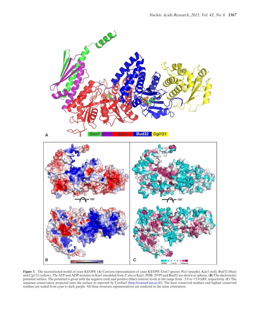
web server (45). The model for the complete yeast KEOPScomplex is represented in Figure 3A. Like archaeal KEOPS,yeast KEOPS forms a linear arrangement of subunits, withGon7 at one end and Cgi121 at the other. The active site ofKae1, centered on the conserved metal cluster and contain-ing the ATP binding site, is at the bottom of a crater whosewalls are made of Kae1, Bud32, Pcc1 and Gon7.
The crystal structures of archaeal Pcc1 alone and of thePcc1/Kae1 complex showed that Pcc1 forms homodimers(17). It was therefore suggested that the intact archaeal com-plex would form a dimer of heterotetramers for which Pcc1would function as the dimerization unit (SupplementaryFigure S1) (9). The structure of the Gon7/Pcc1 dimer showsthat Pcc1 occupies the same interface as that used for Pcc1homodimer formation (Figure 1B and C). We thereforeset out to determine the quaternary structure of the com-plete yeast KEOPS complex. Recombinant yeast KEOPScomplex was purified after high yield expression from apolycistronic construct in E. coli cells. We first determinedthe molecular mass in solution of the recombinant yeastKEOPS complex by SEC-MALLS. The extracted molecu-lar weight was 109.7 ± 5 kDa (Figure 4A), which is close tothe theoretical molecular weight of the pentameric KEOPScomplex (117.7 kDa), suggesting yeast KEOPS forms ahetero-pentamer in solution. In order to confirm the stoi-chiometry and to evaluate our model of the complex, we in-vestigated the intact KEOPS complex in solution by SAXS
coupled to gel filtration. The molar mass extracted fromthe scattering curve corresponding to the top of the elutionpeak is equal to 122 ± 10 kD (Supplementary Table S2), ingood agreement with the SEC-MALLS results. We evalu-ated our above-mentioned linear model of the yeast KEOPScomplex against the SAXS data. The program BUNCH(49) was used for modeling the missing parts of the model(mainly in the Gon7 subunit) as chains of dummy residueswhose C-alpha positions are separated by 3.8 A. Recon-structing the backbone and adding the side chains using theprogram PD2 (50) then completed the model. The scatter-ing amplitude curves calculated on the final model by usingthe program CRYSOL (51) is in good agreement with theexperimental curve (χ = 1.25) (Figure 4B). These data val-idate the quaternary structure and the molecular architec-ture of our model of yeast KEOPS.
It was shown that the five KEOPS subunits are requiredfor the biosynthesis of tRNA t6A (6,8) and that Kae1 trans-fers the threonylcarbamoyl moiety from TCA onto sub-strate tRNA (6,9). In our model, the catalytic center ofKae1 contacts the C-terminal region of Bud32 and Pcc1and Gon7 surround the active site entrance. The bottom ofthe crater formed by Gon7-Pcc1-Kae1-Bud32 has a posi-tively charged and conserved surface (Figures 3 and 5). Af-ter refinement of the Bud32-Cgi121 structure we observedresidual electron density for four sulphate ions. In a pre-viously determined structure of P. abyssi Kae1 we identi-fied three tungstate ions (12) after soaking the crystals in atungstate solution for phasing the diffraction data. Sulfateand tungstate ions often occupy the positions of DNA- orRNA-backbone phosphates in apo-crystal structures of nu-cleic acid binding proteins. We positioned these sulfate andtungstate ions onto our model of the yeast KEOPS com-plex in Figure 5. One sulfate ion occupies an ATP phos-phate position in the active site of Bud32 and two othersare bound at the end of its C-terminal helix, close to the en-trance of the active site of Kae1. When the structure of P.abyssi Kae1 is superposed onto the modeled yeast ternarycomplex, two of the tungstate ions are found close to theC-terminal of Bud32. The presence of these ions and thesurrounding positive surface patch suggest that this regionof the complex could be involved in the binding of nu-cleic acids. We generated a model for the KEOPS–tRNA(PDB: 3L0U) complex by rigid global docking with Patch-Dock (53), specifying that A37 of tRNA directly contactsHis141/His145/Asp344. The resulting model orients the an-ticodon stem loop of tRNA toward the active site of Kae1and shows that all the subunits except Cgi121 of KEOPS arepotentially involved in the binding of tRNA (Figure 5). Werealize that tRNA probably undergoes important structuralrearrangements during the modification reaction, preclud-ing more rigorous modeling.
DISCUSSION
Experimental data obtained from archaeal and eukaryotict6A biosynthesis systems led to the conclusion that theKEOPS complex catalyzes the transfer of the threonylcar-bamoyl moiety from the unstable TCA intermediate ontothe A37 base of acceptor tRNA (7–9). The active sites ofthe t6A biosynthesis systems are located on the totally con-
Nucleic Acids Research, 2015, Vol. 43, No. 6 3367
Figure 3. The reconstituted model of yeast KEOPS. (A) Cartoon representation of yeast KEOPS: Gon7 (green), Pcc1 (purple), Kae1 (red), Bud32 (blue)and Cgi121 (yellow). The ATP and ADP moieties in Kae1 (modeled from P. abyssi Kae1, PDB: 2IVP) and Bud32 are shown as spheres. (B) The electrostaticpotential surface. The potential is given with the negative (red) and positive (blue) contour levels in the range from –5.0 to +5.0 kBT, respectively. (C) Thesequence conservation projected onto the surface as reported by ConSurf (http://consurf.tau.ac.il/). The least conserved residues and highest conservedresidues are scaled from cyan to dark purple. All three structure representations are rendered in the same orientation.

3368 Nucleic Acids Research, 2015, Vol. 43, No. 6
Figure 4. The determination of the quaternary structure of yeast KEOPS in solution. (A) The gel filtration profile and molecular weight extracted bySEC-MALLS. (B) The fitting of the yeast KEOPS model (red line) to the SAXS experimental curves (black dots). The missing regions of Gon7 were addedby modeling with BUNCH program and represented as green spheres.

Nucleic Acids Research, 2015, Vol. 43, No. 6 3369
Figure 5. Docking model for KEOPS–tRNA complex by PatchDock. The A37 of tRNA and active site (H141, H145 and D344) of Kae1 are highlightedand denoted. The orientation is the same as for Figure 3. We have also represented the sulfate ions that were present in the yeast Bud32-Cgi121 structure(green spheres) and the tungstate ions present in the P. abyssi Kae1 structure (yellow spheres taken from PDB: 2IVO) were superposed onto the yeast Kae1model.
3370 Nucleic Acids Research, 2015, Vol. 43, No. 6
served metal cluster in the Kae1/Qri7/YgjD proteins. How-ever, the role of the other subunits (Bud32, Pcc1, Cgi121,Gon7) remains largely unknown. Analysis of the archaealKEOPS complex showed that Pcc1, Bud32 and Kae1 arethe minimal protein set for the in vitro t6A biosynthesisbut that Cgi121 increases the efficiency of the reaction (9).Bud32 was shown to possess both ATPase and kinase ac-tivities (9,17). Active site mutations of Bud32 (D161A/R inyeast and D127R in archaea) affect kinase activity (16,48),t6A biosynthesis activity (6,9) and ATPase activity (9). Thetransfer of the threonylcarbamoyl moiety from TCA ontoA37 of tRNA does not, in principle, require ATP hydrolysisnor the kinase activity of Bud32. This was clearly demon-strated by the study of the mitochondrial Qri7 enzyme,which is active without assistance of other proteins, form-ing together with Sua5 the minimal t6A biosynthesis sys-tem. Though AMPPNP was present in the crystallizationconditions, no nucleotide was bound in Bud32 in the struc-tures of archaeal Bud32 (16,17), which was explained byits disordered N-terminus and P-loop region. ATP is firmlybound in the yeast Bud32/Cgi121 complex interacting ina canonical manner with the well-structured P-loop. Kae1and Cgi121 seem to have antagonistic effects on the ATPaseactivity of Bud32. Bud32 in complex with Cgi121 was alsoshown to have autophosphorylation activity (54), whichwas inhibited by increasing concentrations of Kae1 (16).
Bud32 belongs to the RIO-type family of atypical smallkinases that lack the kinase activation loop (55). Rio1 andRio2 are involved in the maturation of the ribosome. Itwas recently shown that Rio2 does not only function as akinase but that it exhibits robust ATPase activity in vitro(56). Interestingly, the energy released by ATP hydrolysisis required for dissociation of Rio2 from the ribosome, anecessary step in pre-40S particle maturation. The crys-tal structure of Chaetomium thermophilum Rio2 containeda phospho-aspartate intermediate typically found in P-type ATPases but unusual for protein kinases. Phosphoryl-transfer from ATP to Asp257 in Rio2’s active site and subse-quent hydrolysis of the aspartylphosphate was proposed tobe a trigger to power 40S subunit biogenesis (56). Asp257in C. thermophilum Rio2 is conserved in the Bud32 activesite (corresponding residues are Asp142 in P. abyssi andAsp182 in S. cerevisiae) suggesting that also Bud32 couldfunction as a P-type ATPase during the t6A synthesis reac-tion. We hypothesize that Bud32 might regulate the inter-action of the KEOPS complex with substrate tRNA. Thepositioning of the Bud32 subunit within our model of theKEOPS complex supports this hypothesis. The presence ofthe C-terminal helix of Bud32 near the active site of Kae1offers an opportunity to couple ATP hydrolysis by Bud32and tRNA binding by the KEOPS complex. The conservedand very positively charged C-terminus of Bud32 lies at theentrance of the active site of Kae1 (Figure 3B and Supple-mentary Figure S4). We therefore expect that Bud32 mightinterfere with the binding of the bulky tRNA substrate inthe Kae1 active site. ATPases often undergo conformationaltransitions between the ATP and ADP bound states. ATPhydrolysis could influence the relative position of the N-terminal and C-terminal lobes of Bud32 and hence change,for instance, the position of the C-terminal helix, which inturn may modify its interactions with the substrate tRNA
during t6A biosynthesis. Many RNA-binding processes areregulated by nucleotide triphophosphate hydrolysis. For in-stance, the elongation factor EF-Tu undergoes a conforma-tional change upon GTP hydrolysis that dissociates its com-plex with aminacyl-tRNA during peptide bond formationon the ribosome (57).
Mao et al. used the structure of archaealKae1/Bud32/Cgi121 complex to study the functionaleffects of complex disrupting mutations in yeast (17).Interface mutations (L172E/A176R) that interrupt inter-action between Cgi121 and Bud32 displayed slow growthphenotype, analogous to the CGI121 gene deletion mutant.The present structure of the yeast Bud32/Cgi121 complexconfirms that the interfacial residues targeted in Mao’sstudy make crucial interactions in the yeast complex andhence these functional data are in full agreement with ourstructure and model.
Gon7, the fifth fungus-specific KEOPS subunit, was pre-dicted to be largely unstructured (Supplementary FigureS2A), which we confirmed by NMR measurements (Sup-plementary Figure S2B). We showed here that it forms atight complex with Pcc1, which functions as a dimeriza-tion unit of the archaeal KEOPS complex. Also the humanKEOPS complex was reported to form a dimer of heterote-tramers (58). Upon complex formation with Pcc1, abouthalf of Gon7 acquires a well-defined structure and formsa continuous �-sheet structure with Pcc1. The binding ofGon7 to Pcc1 is incompatible with Pcc1 homodimer forma-tion. Pcc1 can therefore not function as a dimerization unitin the yeast KEOPS complex, which we experimentally con-firmed by Sec-MALLS and SAXS data (Figure 4).
Our model of the complete yeast KEOPS, which is com-patible with our SAXS data (Figures 3 and 4), has all sub-units aligned with Gon7 on one end and Cgi121 at theother. Our model shows that the region of the KEOPS com-plex around the ATP binding site of Kae1 is very positivelycharged (Figure 3B) and compatible with tRNA binding.Residues from Bud32, Pcc1 and Kae1 contribute to thispositive surface patch, and the three subunits might be in-volved in tRNA binding (Figure 5). It should be mentionedthat Qri7 forms homodimers and that the homodimer mim-ics the archaeal Pcc1/Kae1 and bacterial YgjD/YeaZ het-erodimers. Disruption of the Qri7 homodimer results in lossof t6A activity. The functional necessity of this higher or-der structure for t6A activity is not yet clear, but we hy-pothesize that it is important for tRNA binding. The roleof the fungi-specific Gon7 remains intriguing. We showedhere that it imposes a different quaternary structure of theKEOPS complex adding another degree of complexity tothe t6A mechanism.
CONCLUSION
Based on the present crystal structures of Pcc1/Gon7and Bud32/Cgi121. We propose a linear heteropentamericstructure of the complete yeast KEOPS complex that iscompatible with SAXS data. Our KEOPS model displays aconserved and positively charged surface formed by Bud32,Kae1 and Pcc1, suggesting they form the binding site fortRNA during t6A biosynthesis. Further experimental inves-tigation on the assembly of the KEOPS and tRNA binding

Nucleic Acids Research, 2015, Vol. 43, No. 6 3371
is needed to unravel its mechanism of tRNA t6A biosynthe-sis and clarify its relationship with telomere elongation andtranscriptional regulation.
ACCESSION NUMBERS
4WWA, 4WW5, 4WW9, 4WW7, 4WX8 and 4WXA.
SUPPLEMENTARY DATA
Supplementary Data are available at NAR Online.
ACKNOWLEDGEMENTS
We thank Charlotte Saint-Andre for technical assistance inprotein preparation; Andy Thompson (SOLEIL, St Aubin)for his assistance with the SAD data collection and EwenLescop (ICSN, Gif s/Yvette) for the NMR contribution.
FUNDING
The French Infrastructure for Integrated Structural Bi-ology (FRISBI) [ANR-10-INSB-05-01]; the ‘Lidex-BIG’Project Funding [IDEX Paris-Saclay ANR-11-IDEX-0003-02]; 2009 CSC Ph.D. Fellowship [to W.Z.].Conflict of interest statement. None declared.
REFERENCES1. Takemura,S., Murakami,M. and Miyazaki,M. (1969) The primary
structure of isoleucine transfer ribonucleic acid from Torulopsis utilis.Complete digestion with ribonucleases and construction of model ofits secondary structure. J. Biochem., 65, 553–566.
2. Parthasarathy,R., Ohrt,J.M. and Chheda,G.B. (1974) Conformationand possible role of hypermodified nucleosides adjacent to 3′-end ofanticodon in tRNA: N-(purin-6-ylcarbamoyl)-L-threonine riboside.Biochem. Biophys. Res. Commun., 60, 211–218.
3. Murphy,F.V.T., Ramakrishnan,V., Malkiewicz,A. and Agris,P.F.(2004) The role of modifications in codon discrimination bytRNA(Lys)UUU. Nat. Struct. Mol. Biol., 11, 1186–1191.
4. El Yacoubi,B., Hatin,I., Deutsch,C., Kahveci,T., Rousset,J.P.,Iwata-Reuyl,D., Murzin,A.G. and de Crecy-Lagard,V. (2011) A rolefor the universal Kae1/Qri7/YgjD (COG0533) family in tRNAmodification. EMBO J., 30, 882–893.
5. Daugeron,M.C., Lenstra,T.L., Frizzarin,M., El Yacoubi,B., Liu,X.,Baudin-Baillieu,A., Lijnzaad,P., Decourty,L., Saveanu,C.,Jacquier,A. et al. (2011) Gcn4 misregulation reveals a direct role forthe evolutionary conserved EKC/KEOPS in the t6A modification oftRNAs. Nucleic Acids Res., 39, 6148–6160.
6. Srinivasan,M., Mehta,P., Yu,Y., Prugar,E., Koonin,E.V., Karzai,A.W.and Sternglanz,R. (2011) The highly conserved KEOPS/EKCcomplex is essential for a universal tRNA modification, t6A. EMBOJ., 30, 873–881.
7. Perrochia,L., Crozat,E., Hecker,A., Zhang,W., Bareille,J., Collinet,B.,van Tilbeurgh,H., Forterre,P. and Basta,T. (2013) In vitrobiosynthesis of a universal t6A tRNA modification in Archaea andEukarya. Nucleic Acids Res., 41, 1953–1964.
8. Wan,L.C., Mao,D.Y., Neculai,D., Strecker,J., Chiovitti,D.,Kurinov,I., Poda,G., Thevakumaran,N., Yuan,F., Szilard,R.K. et al.(2013) Reconstitution and characterization of eukaryoticN6-threonylcarbamoylation of tRNA using a minimal enzymesystem. Nucleic Acids Res., 41, 6332–6346.
9. Perrochia,L., Guetta,D., Hecker,A., Forterre,P. and Basta,T. (2013)Functional assignment of KEOPS/EKC complex subunits in thebiosynthesis of the universal t6A tRNA modification. Nucleic AcidsRes., 41, 9484–9499.
10. Deutsch,C., El Yacoubi,B., de Crecy-Lagard,V. and Iwata-Reuyl,D.(2012) The biosynthesis of threonylcarbamoyl adenosine (t6A), auniversal tRNA nucleoside. J. Biol. Chem., 287, 13666–13673.
11. Zhang,W., Collinet,B., Perrochia,L., Durand,D. and vanTilbeurgh,H. (2015) The ATP-mediated formation of theYgjD–YeaZ–YjeE complex is required for the biosynthesis of tRNAt6A in Escherichia coli. Nucleic Acids Res., 43, 1804–1817.
12. Hecker,A., Leulliot,N., Gadelle,D., Graille,M., Justome,A.,Dorlet,P., Brochier,C., Quevillon-Cheruel,S., Le Cam,E., vanTilbeurgh,H. et al. (2007) An archaeal orthologue of the universalprotein Kae1 is an iron metalloprotein which exhibits atypicalDNA-binding properties and apurinic-endonuclease activity in vitro.Nucleic Acids Res., 35, 6042–6051.
13. Nichols,C.E., Lamb,H.K., Thompson,P., Omari,K.E., Lockyer,M.,Charles,I., Hawkins,A.R. and Stammers,D.K. (2013) Crystalstructure of the dimer of two essential Salmonella typhimuriumproteins, YgjD & YeaZ and calorimetric evidence for the formationof a ternary YgjD-YeaZ-YjeE complex. Protein Sci., 22, 628–640.
14. Buss,K.A., Cooper,D.R., Ingram-Smith,C., Ferry,J.G., Sanders,D.A.and Hasson,M.S. (2001) Urkinase: structure of acetate kinase, amember of the ASKHA superfamily of phosphotransferases. J.Bacteriol., 183, 680–686.
15. Hecker,H., Graille,M., Madec,E, Gadelle,D., Lecam,E., vanTilbeurgh,H. and Forterre,P.2009)Theuniversal Kae1 protein and theassociated Bud32 kinase (PRPK), a mysteriousprotein coupleprobably essential for genome maintenance in Archaea andEukarya.Biochem Soc Trans., 37, 29–35.
16. Hecker,A., Lopreiato,R., Graille,M., Collinet,B., Forterre,P.,Libri,D. and van Tilbeurgh,H. (2008) Structure of the archaealKae1/Bud32 fusion protein MJ1130: a model for the eukaryoticEKC/KEOPS subcomplex. EMBO J., 27, 2340–2351.
17. Mao,D.Y., Neculai,D., Downey,M., Orlicky,S., Haffani,Y.Z.,Ceccarelli,D.F., Ho,J.S., Szilard,R.K., Zhang,W., Ho,C.S. et al. (2008)Atomic structure of the KEOPS complex: an ancient proteinkinase-containing molecular machine. Mol. Cell, 32, 259–275.
18. LaRonde-LeBlanc,N. and Wlodawer,A. (2005) A family portrait ofthe RIO kinases. J. Biol. Chem., 280, 37297–37300.
19. Downey,M., Houlsworth,R., Maringele,L., Rollie,A., Brehme,M.,Galicia,S., Guillard,S., Partington,M., Zubko,M.K., Krogan,N.J.et al. (2006) A genome-wide screen identifies the evolutionarilyconserved KEOPS complex as a telomere regulator. Cell, 124,1155–1168.
20. Garay-Arroyo,A., Colmenero-Flores,J.M., Garciarrubio,A. andCovarrubias,A.A. (2000) Highly hydrophilic proteins in prokaryotesand eukaryotes are common during conditions of water deficit. J.Biol. Chem., 275, 5668–5674.
21. Corbacho,I., Olivero,I. and Hernandez,L.M. (2005) A genome-widescreen for Saccharomyces cerevisiae nonessential genes involved inmannosyl phosphate transfer to mannoprotein-linkedoligosaccharides. Fungal Genet. Biol., 42, 773–790.
22. Pennock,E., Buckley,K. and Lundblad,V. (2001) Cdc13 deliversseparate complexes to the telomere for end protection and replication.Cell, 104, 387–396.
23. Martin,V., Du,L.L., Rozenzhak,S. and Russell,P. (2007) Protection oftelomeres by a conserved Stn1-Ten1 complex. Proc. Natl. Acad. Sci.U.S.A., 104, 14038–14043.
24. Kisseleva-Romanova,E., Lopreiato,R., Baudin-Baillieu,A.,Rousselle,J.C., Ilan,L., Hofmann,K., Namane,A., Mann,C. andLibri,D. (2006) Yeast homolog of a cancer-testis antigen defines anew transcription complex. EMBO J., 25, 3576–3585.
25. Juneau,K., Palm,C., Miranda,M. and Davis,R.W. (2007)High-density yeast-tiling array reveals previously undiscoveredintrons and extensive regulation of meiotic splicing. Proc. Natl. Acad.Sci. U.S.A., 104, 1522–1527.
26. Collinet,B., Friberg,A., Brooks,M.A., van den Elzen,T., Henriot,V.,Dziembowski,A., Graille,M., Durand,D., Leulliot,N., Saint Andre,C.et al. (2011) Strategies for the structural analysis of multi-proteincomplexes: lessons from the 3D-Repertoire project. J. Struct. Biol.,175, 147–158.
27. Kabsch,W. (1993) Automatic processing of rotation diffraction datafrom crystals of initially unknown symmetry and cell constants. J.Appl. Crystallogr., 26, 795–800.
28. Schneider,T.R. and Sheldrick,G.M. (2002) Substructure solution withSHELXD. Acta Crystallogr. D Biol. Crystallogr., 58, 1772–1779.
29. Bricogne,G., Vonrhein,C., Flensburg,C., Schiltz,M. and Paciorek,W.(2003) Generation, representation and flow of phase information in

3372 Nucleic Acids Research, 2015, Vol. 43, No. 6
structure determination: recent developments in and around SHARP2.0. Acta Crystallogr. D Biol. Crystallogr., 59, 2023–2030.
30. Murshudov,G.N., Vagin,A.A. and Dodson,E.J. (1997) Refinement ofmacromolecular structures by the maximum-likelihood method. ActaCrystallogr. D Biol. Crystallogr., 53, 240–255.
31. Perrakis,A., Morris,R. and Lamzin,V.S. (1999) Automated proteinmodel building combined with iterative structure refinement. Nat.Struct. Biol., 6, 458–463.
32. Adams,P.D., Grosse-Kunstleve,R.W., Hung,L.W., Ioerger,T.R.,McCoy,A.J., Moriarty,N.W., Read,R.J., Sacchettini,J.C., Sauter,N.K.and Terwilliger,T.C. (2002) PHENIX: building new software forautomated crystallographic structure determination. ActaCrystallogr. D Biol. Crystallogr., 58, 1948–1954.
33. Vagin,A. and Teplyakov,A. (2000) An approach to multi-copy searchin molecular replacement. Acta Crystallogr. D Biol. Crystallogr., 56,1622–1624.
34. Terwilliger,T.C., Grosse-Kunstleve,R.W., Afonine,P.V.,Moriarty,N.W., Zwart,P.H., Hung,L.W., Read,R.J. and Adams,P.D.(2008) Iterative model building, structure refinement and densitymodification with the PHENIX AutoBuild wizard. Acta Crystallogr.D Biol. Crystallogr., 64, 61–69.
35. Murshudov,G.N., Skubak,P., Lebedev,A.A., Pannu,N.S.,Steiner,R.A., Nicholls,R.A., Winn,M.D., Long,F. and Vagin,A.A.(2011) REFMAC5 for the refinement of macromolecular crystalstructures. Acta Crystallogr. D Biol. Crystallogr., 67, 355–367.
36. Emsley,P. and Cowtan,K. (2004) Coot: model-building tools formolecular graphics. Acta Crystallogr. D Biol. Crystallogr., 60,2126–2132.
37. Kabsch,W. (2010) XDS. Acta Crystallogr. D Biol. Crystallogr., 66,125–132.
38. Sheldrick,G.M. (2010) Experimental phasing with SHELXC/D/E:combining chain tracing with density modification. Acta Crystallogr.D Biol. Crystallogr., 66, 479–485.
39. McCoy,A.J., Grosse-Kunstleve,R.W., Adams,P.D., Winn,M.D.,Storoni,L.C. and Read,R.J. (2007) Phaser crystallographic software.J. Appl. Crystallogr., 40, 658–674.
40. Cowtan,K. (2010) Recent developments in classical densitymodification. Acta Crystallogr. D Biol. Crystallogr., 66, 470–478.
41. Winn,M.D., Ballard,C.C., Cowtan,K.D., Dodson,E.J., Emsley,P.,Evans,P.R., Keegan,R.M., Krissinel,E.B., Leslie,A.G., McCoy,A.et al. (2011) Overview of the CCP4 suite and current developments.Acta Crystallogr. D Biol. Crystallogr., 67, 235–242.
42. Vagin,A. and Teplyakov,A. (2010) Molecular replacement withMOLREP. Acta Crystallogr. D Biol. Crystallogr., 66, 22–25.
43. Konarev,P.V., Petoukhov,M.V., Volkov,V.V. and Svergun,D.I. (2006)ATSAS 2.1, a program package for small-angle scattering dataanalysis. J. Appl. Crystallogr., 39, 277–286.
44. Konarev,P.V., Volkov,V.V., Sokolova,A.V., Koch,M.H.J. andSvergun,D.I. (2003) PRIMUS: a Windows PC-based system forsmall-angle scattering data analysis. J. Appl. Crystallogr., 36,1277–1282.
45. Kelley,L.A. and Sternberg,M.J. (2009) Protein structure prediction onthe Web: a case study using the Phyre server. Nat. Protoc., 4, 363–371.
46. David,G. and Perez,J. (2009) Combined sampler robot andhigh-performance liquid chromatography: a fully automated systemfor biological small-angle X-ray scattering experiments at theSynchrotron SOLEIL SWING beamline. J. Appl. Crystallogr., 42,892–900.
47. Svergun,D.I., Petoukhov,M.V. and Koch,M.H. (2001) Determinationof domain structure of proteins from X-ray solution scattering.Biophys. J., 80, 2946–2953.
48. Facchin,S., Lopreiato,R., Stocchetto,S., Arrigoni,G., Cesaro,L.,Marin,O., Carignani,G. and Pinna,L.A. (2002) Structure-functionanalysis of yeast piD261/Bud32, an atypical protein kinase essentialfor normal cell life. Biochem. J., 364, 457–463.
49. Petoukhov,M.V. and Svergun,D.I. (2005) Global rigid body modelingof macromolecular complexes against small-angle scattering data.Biophys. J., 89, 1237–1250.
50. Moore,B.L., Kelley,L.A., Barber,J., Murray,J.W. andMacDonald,J.T. (2013) High-quality protein backbonereconstruction from alpha carbons using Gaussian mixture models. J.Comput. Chem., 34, 1881–1889.
51. Svergun,D., Barberato,C. and Koch,M.H.J. (1995) CRYSOL––aprogram to evaluate X-ray solution scattering of biologicalmacromolecules from atomic coordinates. J. Appl. Crystallogr., 28,768–773.
52. Hecker,A., Leulliot,N., Gadelle,D., Graille,M., Justome,A.,Dorlet,P., Brochier,C., Quevillon-Cheruel,S., Le Cam,E., vanTilbeurgh,H. et al. (2007) An archaeal orthologue of the universalprotein Kae1 is an iron metalloprotein which exhibits atypicalDNA-binding properties and apurinic-endonuclease activity in vitro.Nucleic Acids Res., 35, 6042–6051.
53. Schneidman-Duhovny,D., Inbar,Y., Nussinov,R. and Wolfson,H.J.(2005) PatchDock and SymmDock: servers for rigid and symmetricdocking. Nucleic Acids Res., 33, W363–W367.
54. Miyoshi,A., Kito,K., Aramoto,T., Abe,Y., Kobayashi,N. andUeda,N. (2003) Identification of CGI-121, a novel PRPK(p53-related protein kinase)-binding protein. Biochem. Biophys. Res.Commun., 303, 399–405.
55. LaRonde-LeBlanc,N. and Wlodawer,A. (2005) The RIO kinases: anatypical protein kinase family required for ribosome biogenesis andcell cycle progression. Biochim. Biophys. Acta, 1754, 14–24.
56. Ferreira-Cerca,S., Sagar,V., Schafer,T., Diop,M., Wesseling,A.M.,Lu,H., Chai,E., Hurt,E. and LaRonde-LeBlanc,N. (2012)ATPase-dependent role of the atypical kinase Rio2 on the evolvingpre-40S ribosomal subunit. Nat. Struct. Mol. Biol., 19, 1316–1323.
57. Nilsson,J. and Nissen,P. (2005) Elongation factors on the ribosome.Curr. Opin. Struct. Biol., 15, 349–354.
58. Costessi,A., Mahrour,N., Sharma,V., Stunnenberg,R., Stoel,M.A.,Tijchon,E., Conaway,J.W., Conaway,R.C. and Stunnenberg,H.G.(2012) The human EKC/KEOPS complex is recruited to Cullin2ubiquitin ligases by the human tumour antigen PRAME. PLoS One,7, e42822.