crystalstructureofmonomericphotosystemiifrom ... · the molar extinction coefficient for chla...
TRANSCRIPT
Crystal Structure of Monomeric Photosystem II fromThermosynechococcus elongatus at 3.6-A Resolution*□S
Received for publication, March 26, 2010, and in revised form, May 12, 2010 Published, JBC Papers in Press, June 17, 2010, DOI 10.1074/jbc.M110.127589
Matthias Broser‡, Azat Gabdulkhakov§1, Jan Kern‡2, Albert Guskov§3, Frank Muh‡, Wolfram Saenger§4,and Athina Zouni‡5
From the ‡Institute of Chemistry, Max Volmer Laboratory of Biophysical Chemistry, Technische Universitat Berlin,Strasse des 17 Juni 135, D-10623 Berlin and the §Institute of Chemistry and Biochemistry/Crystallography, Freie Universitat Berlin,Takustrasse 6, D-14195 Berlin, Germany
The membrane-embedded photosystem II core complex(PSIIcc) uses light energy to oxidize water in photosynthesis.Information about the spatial structure of PSIIcc obtained fromx-ray crystallography was so far derived from homodimericPSIIcc of thermophilic cyanobacteria. Here, we report the firstcrystallization and structural analysis of themonomeric form ofPSIIcc with high oxygen evolution capacity, isolated from Ther-mosynechococcus elongatus. The crystals belong to the spacegroup C2221, contain one monomer per asymmetric unit, anddiffract to a resolution of 3.6 A. The x-ray diffraction pattern ofthe PSIIcc-monomer crystals exhibit less anisotropy (depen-dence of resolution on crystal orientation) compared with crystalsof dimeric PSIIcc, and the packing of themoleculeswithin the unitcell is different. In the monomer, 19 protein subunits, 35 chloro-phylls, two pheophytins, the non-heme iron, the primary plasto-quinone QA, two heme groups, 11 �-carotenes, 22 lipids, sevendetergentmolecules, and theMn4Ca cluster of thewater oxidizingcomplex could be assigned analogous to the dimer. Based on thenewstructural information, the roles of lipids andprotein subunitsin dimer formation of PSIIcc are discussed.Due to the lack of non-crystallographic symmetry and the orientation of the membranenormal of PSIIcc perpendicular (�87°) to the crystallographicb-axis, further information about the structure of theMn4Ca clus-ter is expected to become available from orientation-dependentspectroscopy on this new crystal form.
The primary processes of oxygenic photosynthesis takesplace in the thylakoid membranes of plants, green algae, and
cyanobacteria and involve the cooperation of several protein-cofactor complexes. Among these complexes, only photosys-tems I (PSI)6 and II (PSII) are able to perform light-inducedcharge separation necessary to convert photon energy into abiochemically amenable form.The photosystem II core complex (PSIIcc) is a light-driven
water-plastoquinone-oxidoreductase, which is characterizedby the unique property of abstracting electrons from watermolecules, the primary electron source in oxygenic photosyn-thesis (1–3). Several x-ray crystal structures of homodimericPSIIcc from thermophilic cyanobacteria have been publishedwith resolutions ranging from 3.8 to 2.9 Å (4–9). The mono-mers in the dimer are related by a non-crystallographicC2 rota-tion axis. Each monomer contains 17 membrane-intrinsic andthree membrane-extrinsic protein subunits, the latter beinglocated at the lumenal side of the membrane. The redox-activecofactors are harbored by the heterodimeric protein matrixformed by subunits D1 (PsbA) and D2 (PsbD) and are arrangedin two pseudo-C2 symmetric branches.This entity, referred to as reaction center (RC), consists of
four chlorophyll a (Chla) molecules, two pheophytins a(PheoD1, PheoD2), two plastoquinones (PQ) QA and QB withone non-heme iron located in between, two redox activetyrosines (YZ and YD), and the water oxidizing complex(WOC). Whereas the tightly bound QA acts as electron trans-mitter, the mobile QB is the substrate of the quinone reductasepart of PSIIcc. An additional plastoquinone molecule (QC) ofunresolved function has been located next to the QB bindingsite in the recent crystal structure of dimeric PSIIcc at 2.9-Åresolution. In the RC, light-induced charge separation takesplace leading to the oxidation of PD1 to the cation radical PD1 .�.The electron is transferred via PheoD1 and QA to QB. PD1 .� hasan exceptionally strong oxidizing power (1.25 eV (10)) and isable to abstract electrons via YZ from the WOC, a hetero-nuclearMn4Ca cluster located at the lumenal side of PSII. Afterthe accumulation of four redox equivalents in the so-calledS-states (S0, S1, …, S4) of the WOC (11), water is oxidized tomolecular oxygen.The crystals of dimeric PSIIcc belong to the orthorhombic
space group P212121 and diffract to a maximal resolution of
* This work was supported by the Deutsche Forschungsgemeinschaft withinthe framework of Sfb 498 (projects A4 and C7) and the Deutsche Forschungs-gemeinschaft-Cluster of Excellence “UniCat” (project B1) coordinated bythe Technische Universitat Berlin.
The atomic coordinates and structure factors (code 3KZI) have been deposited inthe Protein Data Bank, Research Collaboratory for Structural Bioinformatics,Rutgers University, New Brunswick, NJ (http://www.rcsb.org/).
□S The on-line version of this article (available at http://www.jbc.org) containssupplemental Figs. S1–S6 and Tables S1 and S2.
1 Present address: Institute of Protein Research, Russian Academy of Sciences,142290 Pushchino, Moscow region, Russia.
2 Present address: Physical Biosciences Division, Lawrence Berkeley NationalLaboratory, Berkeley, CA 94720.
3 Present address: Biomedical Structural Biology, Nanyang Technological Uni-versity, IMCB, 61 Biopolis Dr., 138673 Singapore.
4 To whom correspondence may be addressed. Tel.: 49-30-838-53412; Fax:49-30-838-56702; E-mail: [email protected].
5 To whom correspondence may be addressed. Tel.: 49-30-314-25650; Fax:49-30-314-21122; E-mail: [email protected].
6 The abbreviations used are: PS, photosystem; PSIIcc, photosystem II corecomplex; RC, reaction center; DDM, n-dodecyl-�-D-maltoside; MDGD,monogalactosyldiacylglycerol; DGDG, digalactosyldiacylglycerol; SQDG,sulfoquinovosyldiacylglycerol; Chl, chlorophyll; Pheo, pheophytin; PQ,plastoquinone; WOC, water oxidizing complex.
THE JOURNAL OF BIOLOGICAL CHEMISTRY VOL. 285, NO. 34, pp. 26255–26262, August 20, 2010© 2010 by The American Society for Biochemistry and Molecular Biology, Inc. Printed in the U.S.A.
AUGUST 20, 2010 • VOLUME 285 • NUMBER 34 JOURNAL OF BIOLOGICAL CHEMISTRY 26255
by guest on February 6, 2019http://w
ww
.jbc.org/D
ownloaded from

�2.9 Å (8). Their x-ray diffraction pattern is highly anisotropic(the attained resolution depends on the orientation of the crys-tal in the x-ray beam), which limits the useful resolution range(12, 13). This feature seems to originate from the packing of thedimeric complexes in the unit cell, but no other crystal formhasbeen found for cyanobacterial PSIIcc so far.In addition to dimeric PSIIcc, a monomeric form with high
oxygen evolution capacity has been prepared (14–16). It istherefore attractive to crystallize the PSIIcc monomer inattempting to achieve a more suitable crystal packing for x-raystructure analysis. In earlier work, the monomer was found toaggregate in solution (15). Therefore, it was considered to beinappropriate for crystallization.We succeeded in developing anovel method for purifying an intact monomeric PSIIcc thatcrystallizes in a different form. The new crystals enabled us toobtain the first x-ray structuralmodel of the PSIIccmonomer at3.6-Å resolution. Based on these data, we address several openquestions concerning the role of protein subunits and lipids forthe oligomeric state of PSIIcc and the assembly/disassembly ofPSIIcc during the repair cycle of photodamaged subunit D1(17–19). We also discuss the possibility of improving thestructural analysis of the WOC by orientation-dependentspectroscopy.
EXPERIMENTAL PROCEDURES
Protein Purification—The initial purification steps of PSIIccfollowed the preparation protocol published in (14) with slightmodifications. After two consecutive chromatography steps,the fraction containing the PSIIcc monomer was concentratedto�5mMChla and loaded onto a third column (diameter of 15mm, length of 410 mm, ToyoPearl DEAE 650S, Tosoh Bio-science) pre-equilibrated with 20 mM MES-NaOH (pH 5.0), 20mM CaCl2, 0.5 M betaine monohydrate, 0.02% (w/v) n-dodecyl-�-D-maltoside (DDM). After washing at a constant salt concen-tration for 6 cv with a flow rate of 3 ml/min, monomeric PSIIccwas eluted in a linear salt gradient (MgSO4, 0–50mM, 8 cv). Thefraction was concentrated in Amicon stirring cells using a Mil-lipore Biomax 100membrane (Millipore,MA). For further con-centration to volumes �1 ml and to change the buffer condi-tions after ion exchange chromatography, the sample waswashed three times with 10 mM MES-NaOH (pH 6.0), 5 mM
CaCl2, 5 mM MgCl2, 0.02% (w/v) DDM using Sartorius UltraFree 100 concentrators in a centrifuge at 3000� g at 4 °C. Sam-ples were concentrated to 3 mM Chla and either directly usedfor crystallization or stored in liquid nitrogen.Crystallization—A broad crystallization screen was set up
using a robot and the sitting drop vapor diffusion method andyielded conditions that were further optimized. Crystals weregrown using the microbatch method by mixing the proteinsolution (3 mM Chla corresponding to �25 mg/ml of protein)with the same volume of precipitant solution. Between 4 and 10�l of the finally obtained solution (containing 1.5 mM Chla,�19% (w/v) PEG 400, 0.1 M PIPES, pH 7.0, 0.2 M CaCl2, 0.01%(w/v) DDM) was placed either in a 96-well plate (IMP@CT,Greiner-Bio-one, Germany) or in the middle of a Teflon tube(inner diameter 1 mm; YCM Europe) and closed with sealingtape or sealing plaster. The crystals grew in 3 to 6 days at 18 °Cin the dark and were directly flash-cooled in a nitrogen gas
stream at 100 K after soaking with 28% (w/v) PEG 400 to pro-vide cryoprotection.Crystallographic Data Collection and Analysis—The data set
was collected at the European Synchrotron Radiation Facility(ESRF, beam line ID 29), integrated and scaled with XDS (20).The structure of the PSIIcc monomer was resolved by themolecular replacement method with the PHASER program(21) using one monomeric part of the 2.9-Å resolution struc-ture of homodimeric PSIIcc (8) (PDB entry 3BZ1) as searchmodel. Model rebuilding and refinement were done usingCOOT (22) and the CNS 1.2 package (23), respectively. Thestructure was refined with the rigid-body procedure for theprotein part of the complex and the annealing procedure forthe cofactors. The final model of PSIIcc monomer showsR/Rfree factors of 0.297/0.308, with rootmean square deviationsfrom ideal geometry of 0.013 Å for bond lengths and 2.1° forbond angles.Analytical Ion Exchange and Gel Permeation Chromatogra-
phy—For analytical anion exchange chromatography, a smallcolumn (5 mm diameter, 200 mm length, Toyopearl DEAE 650S)was used connected to an Akta FPLC system (AKTApurifier,Amersham Biosciences) with simultaneous detection at 205,280, and 680nm.Gel permeation chromatography experimentswere conducted as described in Ref. 15, with simultaneousdetection at 222, 280, and 680 nm.Dynamic Light Scattering—Dynamic light scattering was
performed using a DynaPro Titan instrument with a tunablelaser diode at 833 nm wavelength (Wyatt Technology Corpo-ration, Santa Barbara, CA). The protein was dissolved in 100mM PIPES (pH 7.0), 10 mM CaCl2, 0.5 M betaine monohydrate,0.03% (w/v) DDM and filtered through a Millex sterile filter(0.22 �m pore size) into a 100-�l cuvette. To obtain the diffu-sion coefficient DZ, autocorrelation functions of 50 measure-ments were averaged and analyzed using the instrumental soft-ware (Dynamics 6.9.2.9, Wyatt Technology Corporation, SantaBarbara, CA).Spectroscopic Quantitation of Carotenes—Pigments of redis-
solved crystals of monomeric and dimeric PSIIcc were ex-tracted in 80% (v/v) aqueous acetone and spectrawere recordedin the wavelength region from 800 to 400 nm, normalized to664 nm, and the absorbance difference was calculated. By usingthe molar extinction coefficient for Chla (76,800 M�1 cm�1 at664 nm (24)) and �-carotene (144,000 M�1 cm�1 at 454 nm(25)) a difference of 1.3 � 0.2 �-carotene/36 Chla betweenmonomeric and dimeric PSIIcc was calculated (assuming thatthe absorption of pheophytins at 664 nm is reduced by �50%compared with Chla).Mass Spectrometry and SDS-PAGE—MALDI-TOFMS anal-
ysis was conducted using an Ultraflex II Spectrometer (BrukerDaltonics, Germany) in the linear mode using sinapinic acid asmatrix. SDS-PAGEwas performed with a Phast System (Amer-sham Biosciences) using precast HD-SDS gels. Gels were runand silver stained following the protocol of the manufacturer.Oxygen Evolution Activity Assay—Oxygen evolution of PSII
samples were measured at room temperature using a homebuilt Clark-type electrode (26). The excitation was performedeither with saturating continuous white light from a tungstenlamp passed through a heat filter or with repetitive 1-Hz flashes
Crystal Structure of Monomeric Photosystem II
26256 JOURNAL OF BIOLOGICAL CHEMISTRY VOLUME 285 • NUMBER 34 • AUGUST 20, 2010
by guest on February 6, 2019http://w
ww
.jbc.org/D
ownloaded from
from a xenon flash lamp. The sample was diluted to 20–50 �M
Chla in a buffer containing 20mMMES-NaOH (pH5.0- 6.0), 20mM CaCl2, and 10 mM MgCl2. Artificial electron acceptorsadded were either 2 mM 2,6-dichloro-p-benzoquinone for con-tinuous excitation or 2 mM K3[Fe(CN)6] and 0.4 mM phenyl-p-benzoquinone for single flash excitations. The electrode wascalibrated using air-saturated and nitrogen-saturated water atatmospheric pressure.
RESULTS
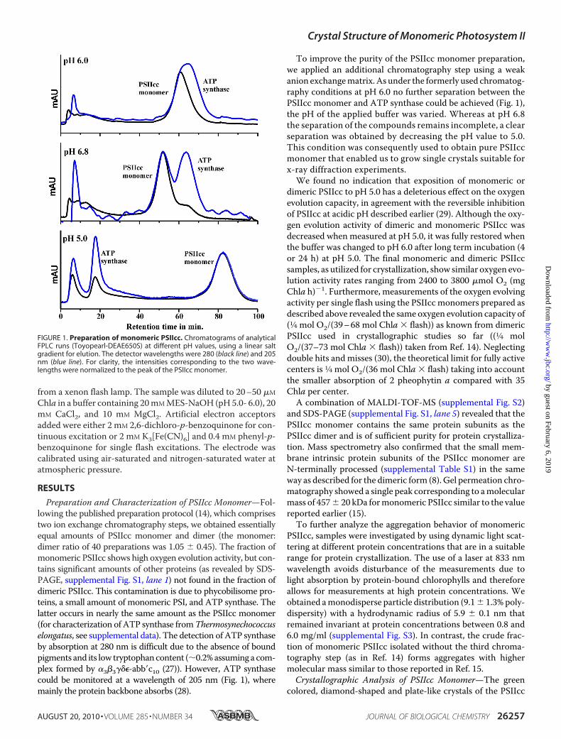
Preparation and Characterization of PSIIcc Monomer—Fol-lowing the published preparation protocol (14), which comprisestwo ion exchange chromatography steps, we obtained essentiallyequal amounts of PSIIcc monomer and dimer (the monomer:dimer ratio of 40 preparations was 1.05 � 0.45). The fraction ofmonomeric PSIIcc shows high oxygen evolution activity, but con-tains significant amounts of other proteins (as revealed by SDS-PAGE, supplemental Fig. S1, lane 1) not found in the fraction ofdimeric PSIIcc. This contamination is due to phycobilisome pro-teins, a small amount of monomeric PSI, and ATP synthase. Thelatter occurs in nearly the same amount as the PSIIcc monomer(for characterization of ATP synthase fromThermosynechococcuselongatus, see supplemental data). The detection of ATP synthaseby absorption at 280 nm is difficult due to the absence of boundpigments and its low tryptophancontent (�0.2%assuming a com-plex formed by �3�3���-abb�c10 (27)). However, ATP synthasecould be monitored at a wavelength of 205 nm (Fig. 1), wheremainly the protein backbone absorbs (28).
To improve the purity of the PSIIcc monomer preparation,we applied an additional chromatography step using a weakanion exchangematrix. As under the formerly used chromatog-raphy conditions at pH 6.0 no further separation between thePSIIcc monomer and ATP synthase could be achieved (Fig. 1),the pH of the applied buffer was varied. Whereas at pH 6.8the separation of the compounds remains incomplete, a clearseparation was obtained by decreasing the pH value to 5.0.This condition was consequently used to obtain pure PSIIccmonomer that enabled us to grow single crystals suitable forx-ray diffraction experiments.We found no indication that exposition of monomeric or
dimeric PSIIcc to pH 5.0 has a deleterious effect on the oxygenevolution capacity, in agreement with the reversible inhibitionof PSIIcc at acidic pH described earlier (29). Although the oxy-gen evolution activity of dimeric and monomeric PSIIcc wasdecreased when measured at pH 5.0, it was fully restored whenthe buffer was changed to pH 6.0 after long term incubation (4or 24 h) at pH 5.0. The final monomeric and dimeric PSIIccsamples, as utilized for crystallization, show similar oxygen evo-lution activity rates ranging from 2400 to 3800 �mol O2 (mgChla h)�1. Furthermore, measurements of the oxygen evolvingactivity per single flash using the PSIIcc monomers prepared asdescribed above revealed the same oxygen evolution capacity of(1⁄4 mol O2/(39–68 mol Chla � flash)) as known from dimericPSIIcc used in crystallographic studies so far ((1⁄4 molO2/(37–73 mol Chla � flash)) taken from Ref. 14). Neglectingdouble hits and misses (30), the theoretical limit for fully activecenters is 1⁄4 mol O2/(36 mol Chla � flash) taking into accountthe smaller absorption of 2 pheophytin a compared with 35Chla per center.
A combination of MALDI-TOF-MS (supplemental Fig. S2)and SDS-PAGE (supplemental Fig. S1, lane 5) revealed that thePSIIcc monomer contains the same protein subunits as thePSIIcc dimer and is of sufficient purity for protein crystalliza-tion. Mass spectrometry also confirmed that the small mem-brane intrinsic protein subunits of the PSIIcc monomer areN-terminally processed (supplemental Table S1) in the sameway as described for the dimeric form (8). Gel permeation chro-matography showed a single peak corresponding to amolecularmass of 457� 20 kDa formonomeric PSIIcc similar to the valuereported earlier (15).To further analyze the aggregation behavior of monomeric
PSIIcc, samples were investigated by using dynamic light scat-tering at different protein concentrations that are in a suitablerange for protein crystallization. The use of a laser at 833 nmwavelength avoids disturbance of the measurements due tolight absorption by protein-bound chlorophylls and thereforeallows for measurements at high protein concentrations. Weobtained amonodisperse particle distribution (9.1� 1.3% poly-dispersity) with a hydrodynamic radius of 5.9 � 0.1 nm thatremained invariant at protein concentrations between 0.8 and6.0 mg/ml (supplemental Fig. S3). In contrast, the crude frac-tion of monomeric PSIIcc isolated without the third chroma-tography step (as in Ref. 14) forms aggregates with highermolecular mass similar to those reported in Ref. 15.Crystallographic Analysis of PSIIcc Monomer—The green
colored, diamond-shaped and plate-like crystals of the PSIIcc
FIGURE 1. Preparation of monomeric PSIIcc. Chromatograms of analyticalFPLC runs (Toyopearl-DEAE650S) at different pH values, using a linear saltgradient for elution. The detector wavelengths were 280 (black line) and 205nm (blue line). For clarity, the intensities corresponding to the two wave-lengths were normalized to the peak of the PSIIcc monomer.
Crystal Structure of Monomeric Photosystem II
AUGUST 20, 2010 • VOLUME 285 • NUMBER 34 JOURNAL OF BIOLOGICAL CHEMISTRY 26257
by guest on February 6, 2019http://w
ww
.jbc.org/D
ownloaded from
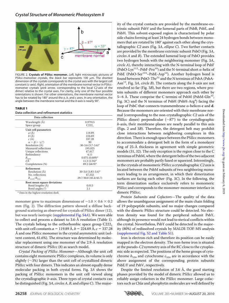
monomer grew to maximum dimensions of �1.0 � 0.6 � 0.2mm (Fig. 2). The diffraction pattern showed a diffuse back-ground scattering as observed for crystals of PSIIcc dimer (12),but was nearly isotropic (supplemental Fig. S4A). We were ableto collect and process a dataset to 3.6-Å resolution (Table 1).The crystals belong to the orthorhombic space group C2221with unit cell constants a � 119.89 Å, b � 224.69 Å, c � 337.28Å and one PSIIcc monomer in the crystal asymmetric unit (sol-vent content, 61.6%). The structure was determined by molec-ular replacement using one monomer of the 2.9-Å resolutionstructure of dimeric PSIIcc (8) as search model.Crystal Packing of PSIIcc Monomer—Although the unit cell
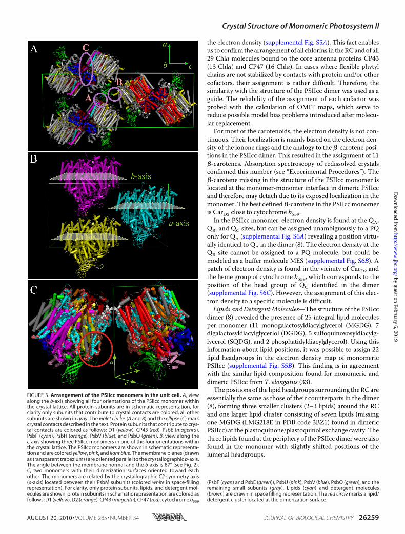
contains eight monomeric PSIIcc complexes, its volume is onlyslightly (�3%) larger than the unit cell of crystallized dimericPSIIcc with four dimers. This indicates an almost equally densemolecular packing in both crystal forms. Fig. 3A shows thepacking of PSIIcc monomers in the unit cell viewed alongthe crystallographic b-axis. Three types of crystal contacts canbe distinguished (Fig. 3A, circles A,B, and ellipse C). Themajor-
ity of the crystal contacts are provided by the membrane-ex-trinsic subunit PsbV and the lumenal parts of PsbB, PsbE, andPsbH. This solvent-exposed region is characterized by polarside chains forming at least 24 hydrogen bonds betweenmono-mers that are rotated by 180° against each other along the crys-tallographic C2 axes (Fig. 3A, ellipse C). Two further contactsare provided by themembrane extrinsic subunit PsbO (Fig. 3A,circles A and B). The extended lumenal loop of PsbO providestwo hydrogen bonds with the neighboring monomer (Fig. 3A,circle A), thereby interacting with the N-terminal loop of PsbF(PsbO-Lys112–PsbF-Pro10) and the N-terminal short �-helix ofPsbE (PsbO-Ser115–PsbE-Asp12). Another hydrogen bond isfound between PsbO-Thr51 and theN terminus of PsbA (PsbA-Asn12, Fig. 3A, circle B). The contacts along the b-axis are notresolved so far (Fig. 3B), but there are two regions, where pro-tein subunits of different monomers approach each other by5–6 Å. These comprise the C termini of PsbM (PsbM-Gln33,Fig. 3C) and the N terminus of PsbH (PsbH-Arg4) facing theloop of PsbC that connects transmembrane �-helices c and d.Because the monomers are oriented with their membrane nor-mal (corresponding to the non-crystallographic C2 axis of thePSIIcc dimer) perpendicular (�87°) to the crystallographicb-axis, their membrane planes are nearly parallel to this axis(Figs. 2 and 3B). Therefore, the detergent belt may prohibitclose interactions between neighboring complexes in thisdirection. There is enough space between the PSIIccmonomersto accommodate a detergent belt in the form of a monolayerring of 25-Å thickness in agreement with simple geometricmodels (31, 32). The only exception is the region close to the Cterminus of PsbM,where the detergent belts of the two adjacentmonomers are probably partly fused or squeezed. Interestingly,in the crystals ofmonomeric PSIIcc a crystallographicC2 axis islocated between the PsbM subunits of two neighboring mono-mers leading to an arrangement, in which their dimerizationsurfaces are facing each other (Fig. 3C). In the following, theterm dimerization surface exclusively refers to monomericPSIIcc and corresponds to themonomer-monomer interface indimeric PSIIcc.Protein Subunits and Cofactors—The quality of the data
allows the unambiguous assignment of the main chain foldingof 19 polypeptide subunits, and no major changes comparedwith the dimeric PSIIcc structure could be detected. No elec-tron density was found for the peripheral subunit PsbY,although its presence would not lead to sterical conflicts withinthe crystal. Nevertheless, PsbY could be detected in the major-ity (80%) of redissolved crystals by MALDI-TOF-MS analysis(supplemental Fig. S2 and Table S1).Iron is electron-rich and therefore its position can be easily
mapped in the electron density. The non-heme iron is situatedat the pseudo-C2 symmetry axis of the RC close to the cytoplas-mic side as expected. The positions of the heme groups of cyto-chrome b559 and cytochrome c550 are in accordance with theabove assignment of the corresponding protein subunitsPsbE/F and PsbV, respectively.Despite the limited resolution of 3.6 Å, the good starting
phases provided by the model of dimeric PSIIcc allowed us toreliably assign cofactors in the PSIIcc monomer. Large cofac-tors such as Chla and pheophytinmolecules are well defined by
FIGURE 2. Crystals of PSIIcc monomer. Left, light microscopic pictures ofPSIIcc-monomer crystals, the black bar represents 100 �m. The shortestdimension of the crystals corresponds to the crystal axis with the largest cellconstant (c-axis). Right, orientation of the membrane normal vector in PSIIcc-monomer crystals (pink arrow, corresponding to the local C2-axis of thedimer) relative to the crystal axes. For clarity, only one of the four possibleorientations is shown. For other orientations, the membrane normal vectorhas to be rotated by 180° around the a, b, and c axes. In any orientation, theangle between the membrane normal and the b-axis is nearly 90°.
TABLE 1Data collection and refinement statistics
Data collection
Wavelength (Å) 0.97915Space group C2221Unit cell parametersa (Å) 119.89b (Å) 224.69c (Å) 337.28� � � � � (°) 90
Resolution (Å) 30-3.6 (3.7-3.6)aMeasured reflections 195.025Unique reflections 47,417Redundancy 4.11Rsym 0.075 (0.689)aI/� 11.3 (2.34)aCompleteness (%) 89.2 (68.9)a
RefinementResolution (Å) 30-3.6 (3.83-3.6)aNo. reflections 47,332Rwork/Rfree 29.7/30.8 (45.4/45.7)
Root mean square deviationsBond lengths (Å) 0.013Bond angles (°) 2.1
a Data in the highest resolution shell.
Crystal Structure of Monomeric Photosystem II
26258 JOURNAL OF BIOLOGICAL CHEMISTRY VOLUME 285 • NUMBER 34 • AUGUST 20, 2010
by guest on February 6, 2019http://w
ww
.jbc.org/D
ownloaded from
the electron density (supplemental Fig. S5A). This fact enablesus to confirm the arrangement of all chlorins in theRCandof all29 Chla molecules bound to the core antenna proteins CP43(13 Chla) and CP47 (16 Chla). In cases where flexible phytylchains are not stabilized by contacts with protein and/or othercofactors, their assignment is rather difficult. Therefore, thesimilarity with the structure of the PSIIcc dimer was used as aguide. The reliability of the assignment of each cofactor wasprobed with the calculation of OMIT maps, which serve toreduce possible model bias problems introduced after molecu-lar replacement.For most of the carotenoids, the electron density is not con-
tinuous. Their localization is mainly based on the electron den-sity of the ionone rings and the analogy to the �-carotene posi-tions in the PSIIcc dimer. This resulted in the assignment of 11�-carotenes. Absorption spectroscopy of redissolved crystalsconfirmed this number (see “Experimental Procedures”). The�-carotene missing in the structure of the PSIIcc monomer islocated at the monomer-monomer interface in dimeric PSIIccand therefore may detach due to its exposed localization in themonomer. The best defined �-carotene in the PSIIccmonomeris CarD2 close to cytochrome b559.
In the PSIIcc monomer, electron density is found at the QA,QB, and QC sites, but can be assigned unambiguously to a PQonly for QA (supplemental Fig. S6A) revealing a position virtu-ally identical to QA in the dimer (8). The electron density at theQB site cannot be assigned to a PQ molecule, but could bemodeled as a buffer molecule MES (supplemental Fig. S6B). Apatch of electron density is found in the vicinity of CarD2 andthe heme group of cytochrome b559, which corresponds to theposition of the head group of QC identified in the dimer(supplemental Fig. S6C). However, the assignment of this elec-tron density to a specific molecule is difficult.Lipids and Detergent Molecules—The structure of the PSIIcc
dimer (8) revealed the presence of 25 integral lipid moleculesper monomer (11 monogalactosyldiacylglycerol (MGDG), 7digalactosyldiacylglycerlol (DGDG), 5 sulfoquinovosyldiacylg-lycerol (SQDG), and 2 phosphatidyldiacylglycerol). Using thisinformation about lipid positions, it was possible to assign 22lipid headgroups in the electron density map of monomericPSIIcc (supplemental Fig. S5B). This finding is in agreementwith the similar lipid composition found for monomeric anddimeric PSIIcc from T. elongatus (33).The positions of the lipid headgroups surrounding theRCare
essentially the same as those of their counterparts in the dimer(8), forming three smaller clusters (2–3 lipids) around the RCand one larger lipid cluster consisting of seven lipids (missingone MGDG (LMG218E in PDB code 3BZ1) found in dimericPSIIcc) at the plastoquinone/plastoquinol exchange cavity. Thethree lipids found at the periphery of the PSIIcc dimerwere alsofound in the monomer with slightly shifted positions of thelumenal headgroups.
FIGURE 3. Arrangement of the PSIIcc monomers in the unit cell. A, viewalong the b-axis showing all four orientations of the PSIIcc monomer withinthe crystal lattice. All protein subunits are in schematic representation, forclarity only subunits that contribute to crystal contacts are colored, all othersubunits are shown in gray. The violet circles (A and B) and the ellipse (C) markcrystal contacts described in the text. Protein subunits that contribute to crys-tal contacts are colored as follows: D1 (yellow), CP43 (red), PsbE (magenta),PsbF (cyan), PsbH (orange), PsbV (blue), and PsbO (green). B, view along thec-axis showing three PSIIcc monomers in one of the four orientations withinthe crystal lattice. The PSIIcc monomers are shown in schematic representa-tion and are colored yellow, pink, and light blue. The membrane planes (drawnas transparent trapeziums) are oriented parallel to the crystallographic b-axis.The angle between the membrane normal and the b-axis is 87° (see Fig. 2).C, two monomers with their dimerization surfaces oriented toward eachother. The monomers are related by the crystallographic C2-symmetry axis(a-axis) located between their PsbM subunits (colored white in space-fillingrepresentation). For clarity, only protein subunits, lipids, and detergent mol-ecules are shown; protein subunits in schematic representation are colored asfollows: D1 (yellow), D2 (orange), CP43 (magenta), CP47 (red), cytochrome b559
(PsbF (cyan) and PsbE (green)), PsbU (pink), PsbV (blue), PsbO (green), and theremaining small subunits (gray). Lipids (cyan) and detergent molecules(brown) are drawn in space filling representation. The red circle marks a lipid/detergent cluster located at the dimerization surface.
Crystal Structure of Monomeric Photosystem II
AUGUST 20, 2010 • VOLUME 285 • NUMBER 34 JOURNAL OF BIOLOGICAL CHEMISTRY 26259
by guest on February 6, 2019http://w
ww
.jbc.org/D
ownloaded from

In dimeric PSIIcc, seven pairs of lipids are located at themonomer-monomer interface due to the non-crystallographicC2 symmetry (Fig. 4B). At the dimerization surface of thePSIIcc monomer, five lipids were found: two lipids pointing tothe cytoplasmic side (SQDG 1 and MGDG 2, Fig. 4A, fornomenclature, see supplemental Table S2) and three to thelumenal side (MGDG3 andDGDG4 and 5).Whereas the head-groups of the lipids oriented toward the cytoplasm are at thesamepositions as in the dimer, the headgroups of the remaininglipids are found to be slightly shifted. Furthermore, the electrondensity suggests that oneMGDG found in the dimer (MGDG5)is replaced by DGDG (DGDG 5) or DDM in the monomer (Fig.4A). By analyzing the lipid positions in the PSIIccmonomer, wefound 10 lipids, including DGDG 4 and DGDG 5 located at thedimerization surface, which follow the pseudo-C2 symmetry ofthe RC.Seven detergent molecules (DDM) per monomer could be
assigned in the PSIIcc dimer. Three of them are located atthe periphery and four at the monomer-monomer interface.We resolved seven DDM molecules in the PSIIcc monomer:three at the periphery and four at the dimerization surface. Twoof the three peripheral DDMmolecules are at similar positionsas in Ref. 8, but the headgroup of the third is shifted by about 7Å and contributes to crystal contacts. Two of the DDMassigned at the dimerization surface (DDM 6 and 7) are nearly
at the same position as described for dimeric PSIIcc. Two fur-therDDM(DDM8and 9)were located at newpositions in closevicinity to DGDG 4. The headgroup of the latter is rotated andforms polar contacts (Fig. 3C, red circle, supplemental Fig. S5C,and Fig. 4A) with DDM 8. The positions of DGDG 4 and DDM8 and 9 would interfere with subunit CP47 from the secondmonomer in the dimer.Mn4Ca Cluster—In the PSIIcc monomer, electron density
arising from the metal ions of the Mn4Ca cluster was found inthe same position as reported for the PSIIcc dimer (8). In agree-ment with EPRmeasurements on the PSIIcc monomer from T.elongatus (16), we assume that the structure of the cluster isessentially the same in monomeric and dimeric PSIIcc. Due tothe limited resolution, the assignment of a chloride ion in thevicinity of the Mn4Ca cluster was not possible.
DISCUSSION
In this study, we present the first structure of a monomericPSIIcc with high oxygen evolution capacity. The results showthat in the PSIIcc monomer 19 of the 20 subunits are arrangedidentically to the corresponding subunits in the dimer.Although PsbY was found to be present in most of the redis-solved crystals examined byMALDI-TOF-MS, we obtained noelectron density for this subunit in the actual dataset. This find-ing may be attributed to variant, substoichiometric occupancy
FIGURE 4. Schematic representation of PSIIcc monomer (A) and one monomer of PSIIcc dimer (B) looking at the dimerization surface (or the monomer-monomer interface). For clarity we show only the four major membrane intrinsic subunits (D1, yellow; D2, orange; CP43, violet; CP47, red), the small subunitsat the dimerization surface (PsbT, PsbM, and PsbL), the three extrinsic subunits (PsbO, green; PsbU, light blue; PsbV, gray), and the non-heme iron cofactor (bluesphere). Lipids and detergent molecules are shown as indicated (SQDG, MGDG, DGDG, and DDM) and numbered as in the main text. A and B, black filledpictograms represent molecules found in both (PSIIcc monomer and homodimer) structures. Molecules (or changed headgroups) found only in the monomer(A) are shown in red. In B, the white pictograms show molecules present only in the model of PSIIcc dimer. The pictograms are contoured black or blue; indicatingthat molecules are assigned to one (contoured black) or the other (blue, also indicated by prime) monomer of the homodimer. For molecules present in bothA and B the same numbers were taken as defined in supplemental Table S2. Lipids and DDM only found in homodimer are numbered according to the PDB file3BZ1 as in supplemental Table S2.
Crystal Structure of Monomeric Photosystem II
26260 JOURNAL OF BIOLOGICAL CHEMISTRY VOLUME 285 • NUMBER 34 • AUGUST 20, 2010
by guest on February 6, 2019http://w
ww
.jbc.org/D
ownloaded from
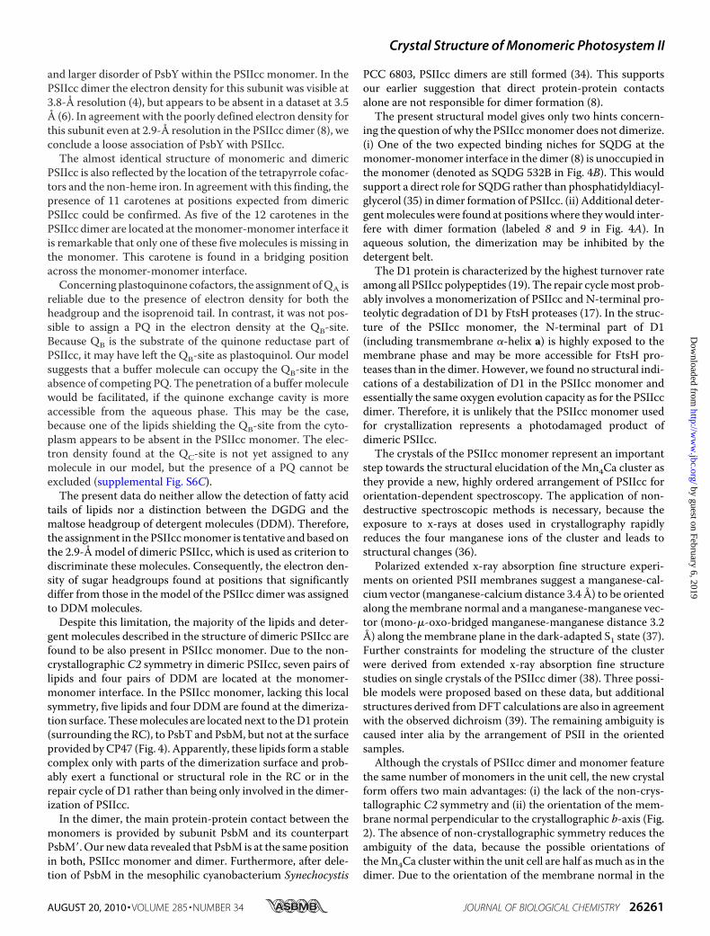
and larger disorder of PsbY within the PSIIcc monomer. In thePSIIcc dimer the electron density for this subunit was visible at3.8-Å resolution (4), but appears to be absent in a dataset at 3.5Å (6). In agreement with the poorly defined electron density forthis subunit even at 2.9-Å resolution in the PSIIcc dimer (8), weconclude a loose association of PsbY with PSIIcc.The almost identical structure of monomeric and dimeric
PSIIcc is also reflected by the location of the tetrapyrrole cofac-tors and the non-heme iron. In agreement with this finding, thepresence of 11 carotenes at positions expected from dimericPSIIcc could be confirmed. As five of the 12 carotenes in thePSIIcc dimer are located at themonomer-monomer interface itis remarkable that only one of these five molecules is missing inthe monomer. This carotene is found in a bridging positionacross the monomer-monomer interface.Concerning plastoquinone cofactors, the assignment ofQA is
reliable due to the presence of electron density for both theheadgroup and the isoprenoid tail. In contrast, it was not pos-sible to assign a PQ in the electron density at the QB-site.Because QB is the substrate of the quinone reductase part ofPSIIcc, it may have left the QB-site as plastoquinol. Our modelsuggests that a buffer molecule can occupy the QB-site in theabsence of competing PQ. The penetration of a buffermoleculewould be facilitated, if the quinone exchange cavity is moreaccessible from the aqueous phase. This may be the case,because one of the lipids shielding the QB-site from the cyto-plasm appears to be absent in the PSIIcc monomer. The elec-tron density found at the QC-site is not yet assigned to anymolecule in our model, but the presence of a PQ cannot beexcluded (supplemental Fig. S6C).The present data do neither allow the detection of fatty acid
tails of lipids nor a distinction between the DGDG and themaltose headgroup of detergent molecules (DDM). Therefore,the assignment in the PSIIccmonomer is tentative andbased onthe 2.9-Åmodel of dimeric PSIIcc, which is used as criterion todiscriminate these molecules. Consequently, the electron den-sity of sugar headgroups found at positions that significantlydiffer from those in themodel of the PSIIcc dimer was assignedto DDMmolecules.Despite this limitation, the majority of the lipids and deter-
gent molecules described in the structure of dimeric PSIIcc arefound to be also present in PSIIcc monomer. Due to the non-crystallographic C2 symmetry in dimeric PSIIcc, seven pairs oflipids and four pairs of DDM are located at the monomer-monomer interface. In the PSIIcc monomer, lacking this localsymmetry, five lipids and four DDM are found at the dimeriza-tion surface. Thesemolecules are located next to theD1 protein(surrounding the RC), to PsbT and PsbM, but not at the surfaceprovided byCP47 (Fig. 4). Apparently, these lipids form a stablecomplex only with parts of the dimerization surface and prob-ably exert a functional or structural role in the RC or in therepair cycle of D1 rather than being only involved in the dimer-ization of PSIIcc.In the dimer, the main protein-protein contact between the
monomers is provided by subunit PsbM and its counterpartPsbM�. Our newdata revealed that PsbM is at the same positionin both, PSIIcc monomer and dimer. Furthermore, after dele-tion of PsbM in the mesophilic cyanobacterium Synechocystis
PCC 6803, PSIIcc dimers are still formed (34). This supportsour earlier suggestion that direct protein-protein contactsalone are not responsible for dimer formation (8).The present structural model gives only two hints concern-
ing the question of why the PSIIccmonomer does not dimerize.(i) One of the two expected binding niches for SQDG at themonomer-monomer interface in the dimer (8) is unoccupied inthe monomer (denoted as SQDG 532B in Fig. 4B). This wouldsupport a direct role for SQDG rather than phosphatidyldiacyl-glycerol (35) in dimer formation of PSIIcc. (ii) Additional deter-gentmolecules were found at positionswhere theywould inter-fere with dimer formation (labeled 8 and 9 in Fig. 4A). Inaqueous solution, the dimerization may be inhibited by thedetergent belt.The D1 protein is characterized by the highest turnover rate
among all PSIIcc polypeptides (19). The repair cyclemost prob-ably involves a monomerization of PSIIcc and N-terminal pro-teolytic degradation of D1 by FtsH proteases (17). In the struc-ture of the PSIIcc monomer, the N-terminal part of D1(including transmembrane �-helix a) is highly exposed to themembrane phase and may be more accessible for FtsH pro-teases than in the dimer. However, we found no structural indi-cations of a destabilization of D1 in the PSIIcc monomer andessentially the same oxygen evolution capacity as for the PSIIccdimer. Therefore, it is unlikely that the PSIIcc monomer usedfor crystallization represents a photodamaged product ofdimeric PSIIcc.The crystals of the PSIIcc monomer represent an important
step towards the structural elucidation of the Mn4Ca cluster asthey provide a new, highly ordered arrangement of PSIIcc fororientation-dependent spectroscopy. The application of non-destructive spectroscopic methods is necessary, because theexposure to x-rays at doses used in crystallography rapidlyreduces the four manganese ions of the cluster and leads tostructural changes (36).Polarized extended x-ray absorption fine structure experi-
ments on oriented PSII membranes suggest a manganese-cal-cium vector (manganese-calcium distance 3.4 Å) to be orientedalong themembrane normal and amanganese-manganese vec-tor (mono-�-oxo-bridged manganese-manganese distance 3.2Å) along the membrane plane in the dark-adapted S1 state (37).Further constraints for modeling the structure of the clusterwere derived from extended x-ray absorption fine structurestudies on single crystals of the PSIIcc dimer (38). Three possi-ble models were proposed based on these data, but additionalstructures derived fromDFT calculations are also in agreementwith the observed dichroism (39). The remaining ambiguity iscaused inter alia by the arrangement of PSII in the orientedsamples.Although the crystals of PSIIcc dimer and monomer feature
the same number of monomers in the unit cell, the new crystalform offers two main advantages: (i) the lack of the non-crys-tallographic C2 symmetry and (ii) the orientation of the mem-brane normal perpendicular to the crystallographic b-axis (Fig.2). The absence of non-crystallographic symmetry reduces theambiguity of the data, because the possible orientations oftheMn4Ca cluster within the unit cell are half as much as in thedimer. Due to the orientation of the membrane normal in the
Crystal Structure of Monomeric Photosystem II
AUGUST 20, 2010 • VOLUME 285 • NUMBER 34 JOURNAL OF BIOLOGICAL CHEMISTRY 26261
by guest on February 6, 2019http://w
ww
.jbc.org/D
ownloaded from
new crystal form, a better discrimination is possible betweenabsorber-backscatter vectors oriented parallel and perpendic-ular to the membrane plane. Therefore, it is expected that a fullset of polarized extended x-ray absorption fine structure spec-tra along the crystal axes will significantly expand the availablestructural information about theMn4Ca cluster, especially withrespect to the manganese-calcium interaction. With theseadditional constraints, a selection between the currently dis-cussed models may become feasible. These experiments are inprogress. The absence of non-crystallographic symmetry incrystals of monomeric PSIIcc will also reduce the complexity ofthe spectra derived from electron paramagnetic resonancespectroscopy on the paramagnetic S2-state of the Mn4Ca clus-ter in single crystals (40, 41) and thus facilitate the assignmentsof spectral features.Besides the still limited information concerning the Mn4Ca
cluster, a higher resolved overall structure of PSIIcc is indispen-sable to clarify many questions still open in the present struc-tural models. Therefore, further optimization of the diffractionquality of PSIIcc crystals is crucial. To achieve this goal, the newcrystal form features the important advantage of significantlylower anisotropy in the diffraction pattern comparedwith crys-tals of dimeric PSIIcc.Moreover, an improvement of the crystalstructure of monomeric PSIIcc is also a prerequisite to eluci-date the origin of the different oligomerization states of PSIIcc,which may help to clarify the mechanism of D1 exchange.
Acknowledgments—We acknowledge beamtime for x-ray data collec-tion at synchrotrons ESRF (Grenoble) and SLS (Villigen) and compe-tent support. We thank Drs. K. Sauer, J. Yano, V. K. Yachandra, andG. Renger for fruitful discussions and D. DiFiore for skillful technicalassistance.
REFERENCES1. Wydrzynski, T. J., and Satoh, K. (2005) Photosystem II: The Light-driven
Water: Plastoquinone Oxidoreductase, Springer, Dordrecht2. Kern, J., and Renger, G. (2007) Photosynth. Res. 94, 183–2023. Renger, G., and Renger, T. (2008) Photosynth. Res. 98, 53–804. Zouni, A., Witt, H. T., Kern, J., Fromme, P., Krauss, N., Saenger, W., and
Orth, P. (2001) Nature 409, 739–7435. Kamiya,N., and Shen, J. R. (2003)Proc. Natl. Acad. Sci. U.S.A. 100, 98–1036. Ferreira, K. N., Iverson, T. M., Maghlaoui, K., Barber, J., and Iwata, S.
(2004) Science 303, 1831–18387. Loll, B., Kern, J., Saenger, W., Zouni, A., and Biesiadka, J. (2005) Nature
438, 1040–10448. Guskov, A., Kern, J., Gabdulkhakov, A., Broser,M., Zouni, A., and Saenger,
W. (2009) Nat. Struct. Mol. Biol. 16, 334–3429. Kawakami, K., Umena, Y., Kamiya, N., and Shen, J. R. (2009) Proc. Natl.
Acad. Sci. U.S.A. 106, 8567–857210. Rappaport, F., and Diner, B. A. (2008) Coord. Chem. Rev. 252, 259–27211. Kok, B., Forbush, B., and McGloin, M. (1970) Photochem. Photobiol. 11,
457–47512. Loll, B. (2005) Photosystem II from the Cyanobacterium Thermosynecho-
coccus elongatus at 3.2-Å Resolution. Ph.D. thesis, Freie Universitat Berlin,Berlin, Germany
13. Kern, J., Loll, B., Zouni, A., Saenger, W., Irrgang, K. D., and Biesiadka, J.(2005) Photosynth. Res. 84, 153–159
14. Kern, J., Loll, B., Luneberg, C., DiFiore, D., Biesiadka, J., Irrgang, K. D., andZouni, A. (2005) Biochim. Biophys. Acta 1706, 147–157
15. Zouni, A., Kern, J., Frank, J., Hellweg, T., Behlke, J., Saenger, W., andIrrgang, K. D. (2005) Biochemistry 44, 4572–4581
16. Mamedov, F., Nowaczyk, M. M., Thapper, A., Rogner, M., and Styring, S.(2007) Biochemistry 46, 5542–5551
17. Komenda, J., Tichy, M., Prasil, O., Knoppova, J., Kuvikova, S., de Vries, R.,and Nixon, P. J. (2007) Plant Cell 19, 2839–2854
18. Komenda, J., Nickelsen, J., Tichy, M., Prasil, O., Eichacker, L. A., andNixon, P. J. (2008) J. Biol. Chem. 283, 22390–22399
19. Kato, Y., and Sakamoto, W. (2009) J. Biochem. 146, 463–46920. Kabsch, W. (1993) J. Appl. Crystallogr. 26, 795–80021. McCoy, A. J., Grosse-Kunstleve, R. W., Adams, P. D., Winn, M. D., Sto-
roni, L. C., and Read, R. J. (2007) J. Appl. Crystallogr. 40, 658–67422. Emsley, P., and Cowtan, K. (2004)Acta Crystallogr. D Biol. Crystallogr. 60,
2126–213223. Brunger, A. T., Adams, P. D., Clore, G. M., DeLano, W. L., Gros, P.,
Grosse-Kunstleve, R.W., Jiang, J. S., Kuszewski, J., Nilges,M., Pannu,N. S.,Read, R. J., Rice, L. M., Simonson, T., and Warren, G. L. (1998) ActaCrystallogr. D Biol. Crystallogr. 54, 905–921
24. Porra, R. J., Thompson, W. A., and Kriedemann, P. E. (1989) Biochim.Biophys. Acta 975, 384–394
25. Lichtenthaler, H. K. (1987)Methods Enzymol. 148, 350–38226. Clark, L. C. J. (1956) ASAIO J. 2, 41–4827. Nakamura, Y., Kaneko, T., Sato, S., Ikeuchi, M., Katoh, H., Sasamoto, S.,
Watanabe, A., Iriguchi, M., Kawashima, K., Kimura, T., Kishida, Y.,Kiyokawa, C., Kohara, M., Matsumoto, M., Matsuno, A., Nakazaki, N.,Shimpo, S., Sugimoto,M., Takeuchi, C., Yamada,M., andTabata, S. (2002)DNA Res. 9, 135–148
28. Scopes, R. K. (1974) Anal. Biochem. 59, 277–28229. Schlodder, E., and Meyer, B. (1987) Biochim. Biophys. Acta 890, 23–3130. Renger, G., and Hanssum, B. (2009) Photosynth. Res. 102, 487–49831. Muh, F., and Zouni, A. (2008) Biochim. Biophys. Acta 1778, 2298–230732. Roth, M., Arnoux, B., Ducruix, A., and Reiss-Husson, F. (1991) Biochem-
istry 30, 9403–941333. Loll, B., Kern, J., Saenger, W., Zouni, A., and Biesiadka, J. (2007) Biochim.
Biophys. Acta 1767, 509–51934. Bentley, F. K., Luo, H., Dilbeck, P., Burnap, R. L., and Eaton-Rye, J. J. (2008)
Biochemistry 47, 11637–1164635. Kruse, O., Hankamer, B., Konczak, C., Gerle, C., Morris, E., Radunz, A.,
Schmid, G. H., and Barber, J. (2000) J. Biol. Chem. 275, 6509–651436. Yano, J., Kern, J., Irrgang, K. D., Latimer, M. J., Bergmann, U., Glatzel, P.,
Pushkar, Y., Biesiadka, J., Loll, B., Sauer, K., Messinger, J., Zouni, A., andYachandra, V. K. (2005) Proc. Natl. Acad. Sci. U.S.A. 102, 12047–12052
37. Pushkar, Y., Yano, J., Glatzel, P., Messinger, J., Lewis, A., Sauer, K., Berg-mann, U., and Yachandra, V. (2007) J. Biol. Chem. 282, 7198–7208
38. Yano, J., Kern, J., Sauer, K., Latimer,M. J., Pushkar, Y., Biesiadka, J., Loll, B.,Saenger, W., Messinger, J., Zouni, A., and Yachandra, V. K. (2006) Science314, 821–825
39. Sproviero, E. M., Gascon, J. A., McEvoy, J. P., Brudvig, G. W., and Batista,V. S. (2008) J. Am. Chem. Soc. 130, 6728–6730
40. Teutloff, C., Pudollek, S., Kessen, S., Broser, M., Zouni, A., and Bittl, R.(2009) Phys. Chem. Chem. Phys. 11, 6715–6726
41. Matsuoka, H., Furukawa, K., Kato, T.,Mino, H., Shen, J. R., andKawamori,A. (2006) J. Phys. Chem. B 110, 13242–13247
Crystal Structure of Monomeric Photosystem II
26262 JOURNAL OF BIOLOGICAL CHEMISTRY VOLUME 285 • NUMBER 34 • AUGUST 20, 2010
by guest on February 6, 2019http://w
ww
.jbc.org/D
ownloaded from
Saenger and Athina ZouniMatthias Broser, Azat Gabdulkhakov, Jan Kern, Albert Guskov, Frank Müh, Wolfram
at 3.6-Å ResolutionelongatusThermosynechococcusCrystal Structure of Monomeric Photosystem II from
doi: 10.1074/jbc.M110.127589 originally published online June 17, 20102010, 285:26255-26262.J. Biol. Chem.
10.1074/jbc.M110.127589Access the most updated version of this article at doi:
Alerts:
When a correction for this article is posted•
When this article is cited•
to choose from all of JBC's e-mail alertsClick here
Supplemental material:
http://www.jbc.org/content/suppl/2010/06/16/M110.127589.DC1
http://www.jbc.org/content/285/34/26255.full.html#ref-list-1
This article cites 39 references, 9 of which can be accessed free at
by guest on February 6, 2019http://w
ww
.jbc.org/D
ownloaded from