csi for ecology - education fund
TRANSCRIPT

CSI for Ecology
Critical Science Investigation
Gwen Foote, Science Teacher
Nautilus Middle School
4301 N Michigan Ave
Miami Beach, FL
305-532-3481
305-532-8906
Need for and Goals of the Program
“All children have a natural curiosity about the world around them.”
For information concerning IMPACT II opportunities, Adapter and Disseminator grants, please contact:
The Education Fund 305-892-5099, Ext. 18 E-mail: [email protected] Web site: www.educationfund.org
1
Table of Contents
Page
Goals and Objectives …………………………………………………3
Sunshine State Standards …………………………………………….5
Course Outline/Overview…………………………………………….6
Lesson Plans
Lesson 1…Taxonomy………………………………………...7
Lesson 2…Diversity and Classification………………………8
Lesson 3…Owl Pellets and prey examination………………..16
Lesson 4…Invertebrates………………...……………………17
Lesson 5…Segmented Worms……………………………….19
Lesson 6…Grasshoppers-Insects…………………………….23
Lesson 7….Fun with Microscopes …………………………..24
Lesson 8…Amphibians & Frog Dissection…………………..26
Resource List…………………………………………………………28
Adapter Application - The Education Fund
2
CSI for Ecology
Critical Science Investigation
Goals and Objectives:
This program fills a need, critical in concrete stages of development in science
that broadens understanding and applied knowledge essential in ecology and biology.
This program introduces general structures of species, investigates organs and systems,
physiology, and interdependence of the major organs in various species of the animal
kingdom. Students acquire and retain an in-depth understanding of the diversity of life
and relationships through basic anatomy and physiology analyzing comparative anatomy,
function, evolution. Students in every domain of science should have the opportunity to
use scientific inquiry and develop the ability to think and act in ways associated with
inquiry, including using appropriate tools and techniques to gather data. Inquiry is the
thread that binds science courses. Teachers give students opportunities to engage in and
reflect on natural phenomena through inquiry (observing, organizing, experiment/
laboratory and communicating). This is the heart of science.
Students want to learn about subjects they personally connect with or have prior
knowledge. Active learning lessons, through inquiry, encourage integration into students’
daily lives and focused interest. CSI scenarios connect science with concepts of personal
physical investigation in explanations. CSI and medical shows are very popular. Ecology
is key emphasis in global media. Computer interactive use biological and scientific
investigation through clinical laboratory procedures cultivating needed recruitment in this
professional field. National Geographic investigations through dissections /necropsies of
sharks, showed the brain to be in a “Y” shape to conduct messages with all 6 senses.
Sharks can reproduce asexually when a two sexed copulation is not accessible.
Hands on investigations of species are explained through form and function,
classifying and organizing orders. Impact of this investigation may justify reclassification
of a species or discovery of a new or similar one, which is happening now (i.e. Marlin
and billfish species, various marine animal research)
3
Florida Sunshine State Standards
SC.F: Processes of Life
SC.F.1.3: The student describes patterns of structure and function in living things. SC.F.1.3.2: The student knows structural basis of most organisms is the cell and most organisms are single cells, while some, including humans, are multicellular. SC.F.1.3.2.1: knows the structures of cells, function and ways these mirror the structure and function of multicellular organisms. SC.F.1.3.2.2: understands cells of unicellular organisms are similar to those of multicellular organisms. SC.F.1.3.3: The student knows that in multicellular organisms cells grow and divide to make more cells in order to form and repair various organs and tissues. SC.F.1.3.5: The student explains how the life functions of organisms are related to what occurs within the cell. SC.F.1.3.5.1: understands that the diversity of cell structure permits a diversity of functions for the organism.
SC.F.1.3.6: The student knows that the cells with similar functions have similar structures, whereas those with different structures have different functions. SC.F.1.3.6.1: knows that the cells with similar functions have similar structures, whereas those with different structures have different functions. SC.F.1.3.6.2: uses tools to identify and compare cell structures (for example, microscope, hand lenses, bioscopes). SC.F.2.3: The student understands the process and importance of genetic diversity. SC.F.2.3.1: The student knows the patterns and advantages of sexual and asexual reproduction in plants and animals. SC.F.2.3.3: The student knows that generally organisms in a population live long enough to reproduce because they have survival characteristics. SC.F.2.3.3.1: knows ways organisms are adapted to their environment. SC.F.2.3.3.2: understands that species have characteristics that enable their populations to cycle within varying periods of time (minutes to hundreds of years).
SC.G: How Living Things Interact with Their Environment
SC.G.2.3.3: The student knows that a brief change in the limited resources of an ecosystem may alter the size of a population or the average size of individual organisms and that long-term change may result in the elimination of animal populations SC.G.2.3.3.1: understands that changes in the environment cause changes in populations.
SC.G.2.3.4: The student understands that humans are a part of an ecosystem and their activities may deliberately or inadvertently alter the equilibrium in ecosystems. SC.G.2.3.4.1: extends and refines knowledge of ways that human activities may deliberately or inadvertently alter the equilibrium in the ecosystem.
4
Course Outline/Overview
This program introduces students to classification by exploring anatomical
comparisons of vertebrates with functional relationships of organs and changing
environments in which vertebrates have adapted. Role playing as “Young Interning
Scientists”, students participate by investigating scientifically oriented questions, with
priority to evidence, developing and evaluating forensic analysis. Students work in small
cooperative learning groups participating in scientific inquiry activities. They research
several species of animals to see understand relationships to each other and their
ecological biomes. Through hands on “Critical Science Investigations”, young interns
become familiar with proper clinical laboratory technique and understand how animals
impact each other and are impacted by their habitats, based on evidential findings,
interpretation of comparative anatomy, classification, and organization of Animal
Kingdom. Cooperative groups examine species and relationships.
Laboratory dissection activities emphasize participation and achievements,
developing a sense of relationship between evidence and explanation. Science
methodology of organization, order, and classification of the Animal Kingdom species
through investigation of form and function of preserved organisms provide examination
of evidence through necropsies. Investigation of internal anatomy allows guided inquiry
through organ identification and function in physiology. Students create graphic
organizers to illustrate species’ systems, to identify purposes and relationships.
Students learn to classify specimens in Kingdom, Phylum, Class, Order, Family,
Genus, and Species based on form, function, and physical characteristics. Learning
through inquiry activities develops skills and proficiency with critical thinking. An
“inside view of animals” combines information for evidence and practical technique.
Laboratory exercises provide direct observation, analysis, written explanations, and/or
5
dissections. Activities using multiple intelligences include visual aids through interactive
technology, preliminary lessons in classification through songs, poems, and alliteration,
hands on examinations of preserved specimens, and discussions of categorization with
naturalist descriptions of characteristics. Small learning groups communicate and explain
their procedures and evidential findings. Group presentations each represent a Phylum
and Class, in sharing evidence of examinations.
Optimizing Learning Students retain (if they focus attention on the lesson at hand): -10% of what they read -26% of what they hear -30% of what they see -50% of what they see and hear -70% of what they say and -90% of what they say and do. If we want to optimize learning, it helps to learn in a multi-sensory environment.
Anticipated Project Results: Students learn to reflect on natural phenomena through inquiry (observing,
organizing, experiment/ laboratory and communicating). Active learning lessons, through
inquiry, encourage integration of students’ daily lives and focus on interest of ecology.
Preferred computer activities investigate biology through interactive clinical laboratory
procedures. The classic comparative anatomy lessons integrate current research, with
hands on analysis of species through form and function. Vertebrate groups are organized
phylogenetically. Necropsy with hands on inquiry activities of investigation of order,
classification, and adaptation of organisms in the animal kingdom. Laboratory projects
develop inquiry skills and conceptual understanding.
6

Lesson 1: Tree of Life
http://tolweb.org/tree/
Through video presentations and interactive virtual dissections on computer,
classes learned about taxonomy, nomenclature, and the vocabulary that is used in
classification. We sang a song with memory techniques.
King Philip Came Over For Good Spaghetti
Kingdom Phylum Class Order Family Genus Species This strategy is practiced for the duration of the CSI project. This helps classify animals
studied and students learn how names relate to form and function. Students learn the
correlation between Latin, Spanish, and definition. Students learn about anatomy and
how animals propagate their species.
7
Lesson 2: Taxonomy
Animal Diversity: Form and Function
Ways to organize animal diversity
What is a tissue? A tissue is a group of cells working together for a particular function.
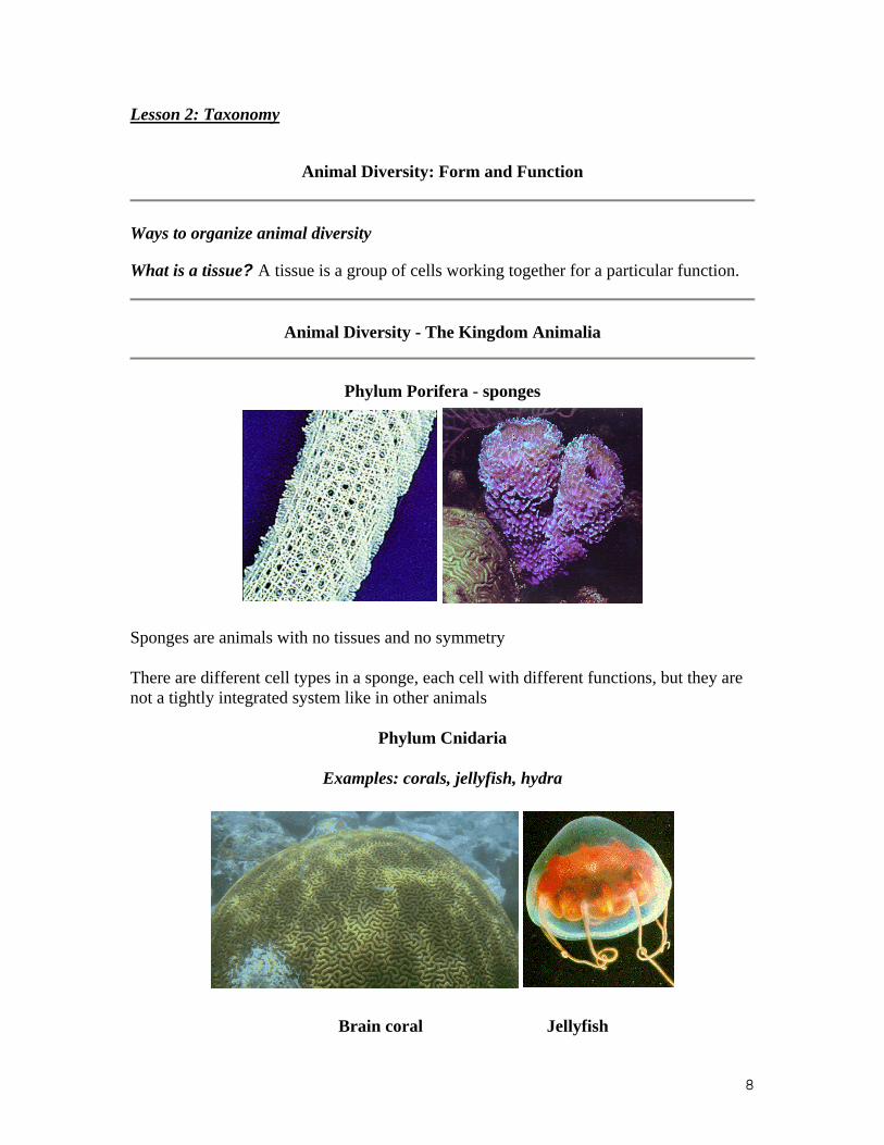
Animal Diversity - The Kingdom Animalia
Phylum Porifera - sponges
Sponges are animals with no tissues and no symmetry
There are different cell types in a sponge, each cell with different functions, but they are not a tightly integrated system like in other animals
Phylum Cnidaria
Examples: corals, jellyfish, hydra
Brain coral Jellyfish
8
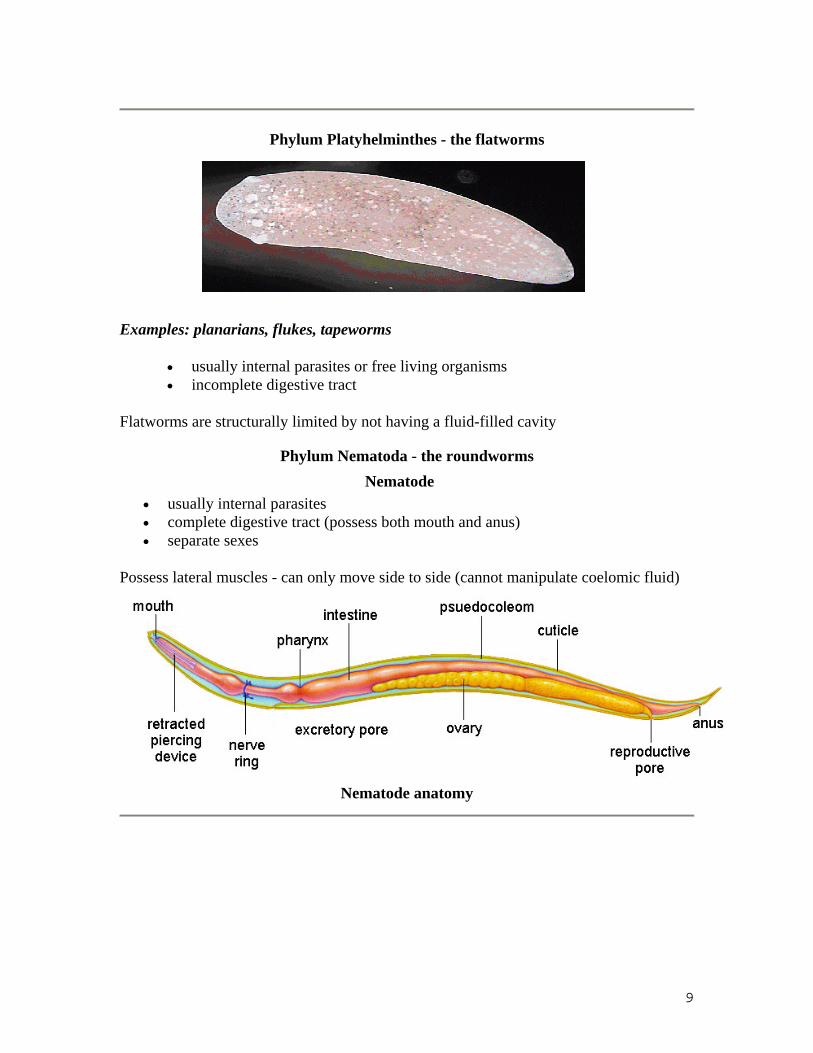
Phylum Platyhelminthes - the flatworms
Examples: planarians, flukes, tapeworms
• usually internal parasites or free living organisms • incomplete digestive tract
Flatworms are structurally limited by not having a fluid-filled cavity
Phylum Nematoda - the roundworms Nematode
• usually internal parasites • complete digestive tract (possess both mouth and anus) • separate sexes
Possess lateral muscles - can only move side to side (cannot manipulate coelomic fluid)
Nematode anatomy
9
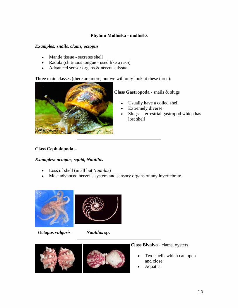
Phylum Molluska - mollusks
Examples: snails, clams, octopus
• Mantle tissue - secretes shell • Radula (chitinous tongue - used like a rasp) • Advanced sensor organs & nervous tissue
Three main classes (there are more, but we will only look at these three):
Class Gastropoda - snails & slugs
• Usually have a coiled shell • Extremely diverse • Slugs = terrestrial gastropod which has
lost shell
Class Cephalopoda –
Examples: octopus, squid, Nautilus
• Loss of shell (in all but Nautilus) • Most advanced nervous system and sensory organs of any invertebrate
Octapus vulgaris Nautilus sp.
Class Bivalva - clams, oysters
• Two shells which can open and close
• Aquatic
10

Phylum Annelida - segmented worms
Examples: earthworms, leeches, polychetes
polychete worm
• Ventral nerve cord • Two types of muscles - longitudinal & circular support
Full utilization of coelom in movement
Phylum Arthropoda - arthropods
Examples: scorpions, spiders, insects
• Hardened chitinous exoskeleton • Specialized segmentation • Jointed appendages • Specialized respiratory organs (fully terrestrial) • Division of labor in life cycle • usually used for delivering venom or other toxins to prey
Examples: scorpions, spiders, horseshoe crabs, ticks
Tarantula sp. Scorpion
Examples: millipedes, centipedes, lobsters, insects
11

•
12

Class Crustacea - aquatic, all have gills o aquatic o all have gills (even terrestrial ones like the rolly-polly)
o o Examples: crabs, lobster, barnacles, pillbugs, hermit crab, shrimp
• Class Insecta - most diverse group on the earth
Examples: ants, bees, butterflies, silverfish, roaches
o Body parts in three segments o Most have wings & advanced sensory organs o Advanced communication, social structure
13
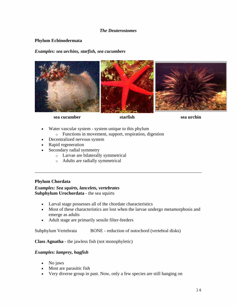
The Deuterostomes
Phylum Echinodermata
Examples: sea urchins, starfish, sea cucumbers
sea cucumber starfish sea urchin
• Water vascular system - system unique to this phylum o Functions in movement, support, respiration, digestion
• Decentralized nervous system • Rapid regeneration • Secondary radial symmetry
o Larvae are bilaterally symmetrical o Adults are radially symmetrical
Phylum Chordata Examples: Sea squirts, lancelets, vertebrates Subphylum Urochordata - the sea squirts
• Larval stage possesses all of the chordate characteristics • Most of these characteristics are lost when the larvae undergo metamorphosis and
emerge as adults • Adult stage are primarily sessile filter-feeders
Subphylum Vertebrata BONE - reduction of notochord (vertebral disks)
Class Agnatha - the jawless fish (not monophyletic)
Examples: lamprey, hagfish
• No jaws • Most are parasitic fish • Very diverse group in past. Now, only a few species are still hanging on
14
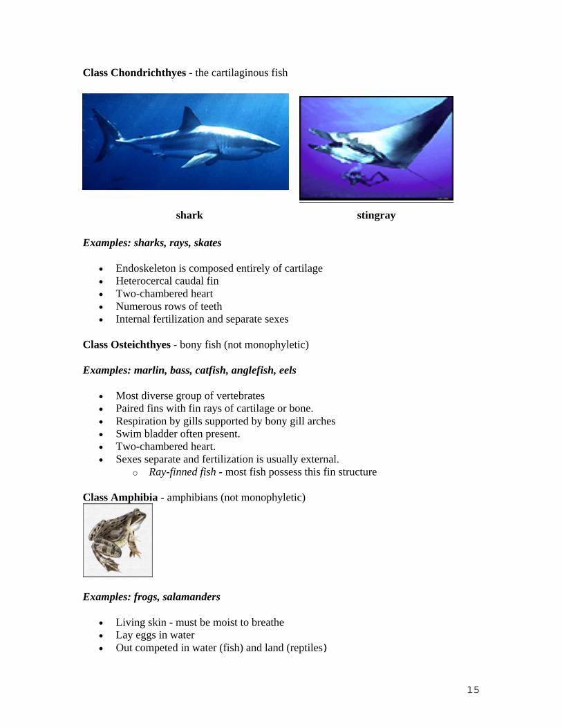
Class Chondrichthyes - the cartilaginous fish
shark stingray
Examples: sharks, rays, skates
• Endoskeleton is composed entirely of cartilage • Heterocercal caudal fin • Two-chambered heart • Numerous rows of teeth • Internal fertilization and separate sexes
Class Osteichthyes - bony fish (not monophyletic)
Examples: marlin, bass, catfish, anglefish, eels
• Most diverse group of vertebrates • Paired fins with fin rays of cartilage or bone. • Respiration by gills supported by bony gill arches • Swim bladder often present. • Two-chambered heart. • Sexes separate and fertilization is usually external.
o Ray-finned fish - most fish possess this fin structure
Class Amphibia - amphibians (not monophyletic)
Examples: frogs, salamanders
• Living skin - must be moist to breathe • Lay eggs in water • Out competed in water (fish) and land (reptiles)
15
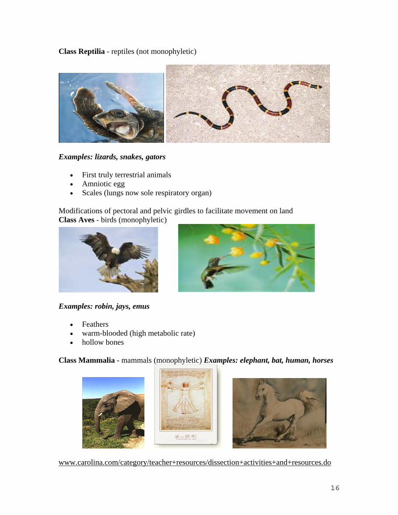
Class Reptilia - reptiles (not monophyletic)
Examples: lizards, snakes, gators
• First truly terrestrial animals • Amniotic egg • Scales (lungs now sole respiratory organ)
Modifications of pectoral and pelvic girdles to facilitate movement on land Class Aves - birds (monophyletic)
Examples: robin, jays, emus
• Feathers • warm-blooded (high metabolic rate) • hollow bones
Class Mammalia - mammals (monophyletic) Examples: elephant, bat, human, horses
www.carolina.com/category/teacher+resources/dissection+activities+and+resources.do
16
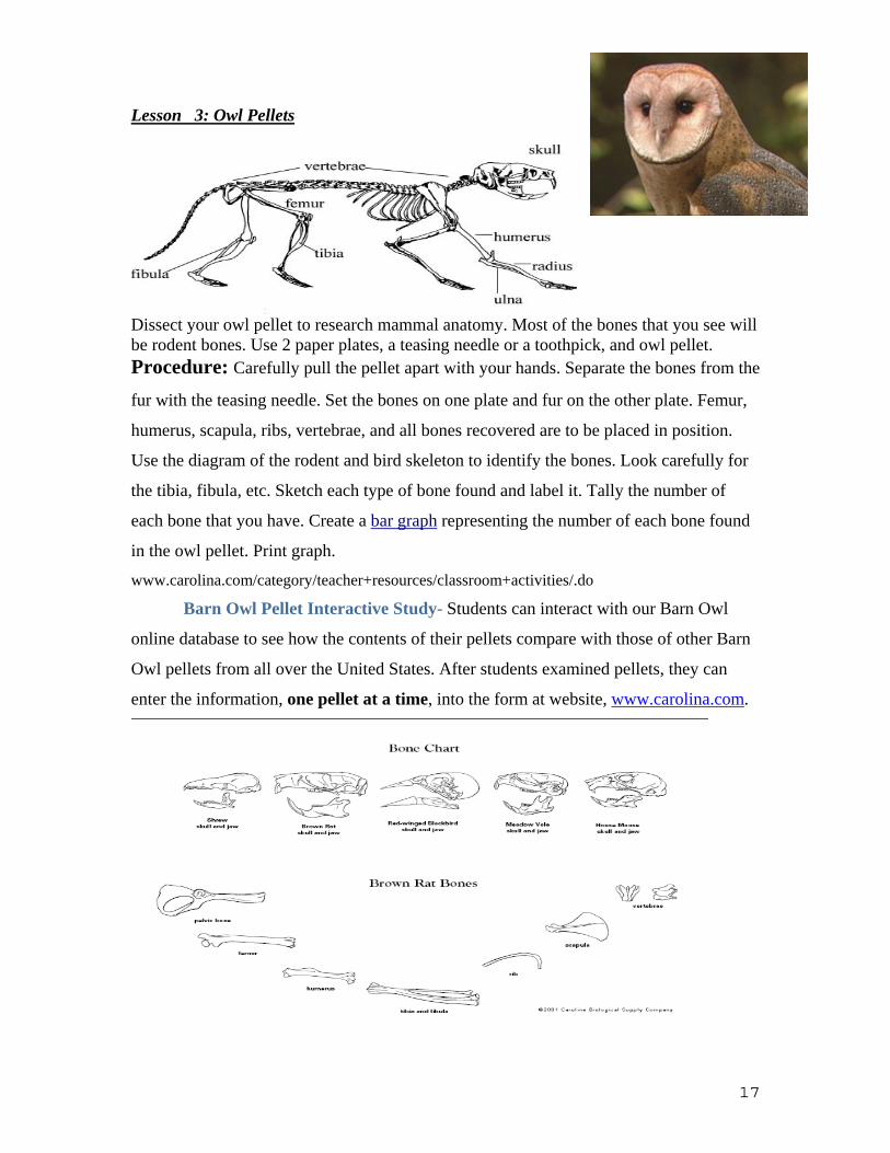
Lesson 3: Owl Pellets
Dissect your owl pellet to research mammal anatomy. Most of the bones that you see will be rodent bones. Use 2 paper plates, a teasing needle or a toothpick, and owl pellet. Procedure: Carefully pull the pellet apart with your hands. Separate the bones from the
fur with the teasing needle. Set the bones on one plate and fur on the other plate. Femur,
humerus, scapula, ribs, vertebrae, and all bones recovered are to be placed in position.
Use the diagram of the rodent and bird skeleton to identify the bones. Look carefully for
the tibia, fibula, etc. Sketch each type of bone found and label it. Tally the number of
each bone that you have. Create a bar graph representing the number of each bone found
in the owl pellet. Print graph.
www.carolina.com/category/teacher+resources/classroom+activities/.do
Barn Owl Pellet Interactive Study- Students can interact with our Barn Owl
online database to see how the contents of their pellets compare with those of other Barn
Owl pellets from all over the United States. After students examined pellets, they can
enter the information, one pellet at a time, into the form at website, www.carolina.com.
17
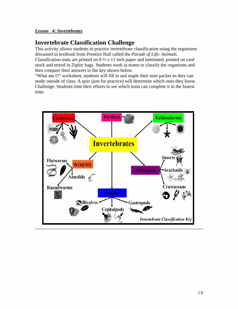
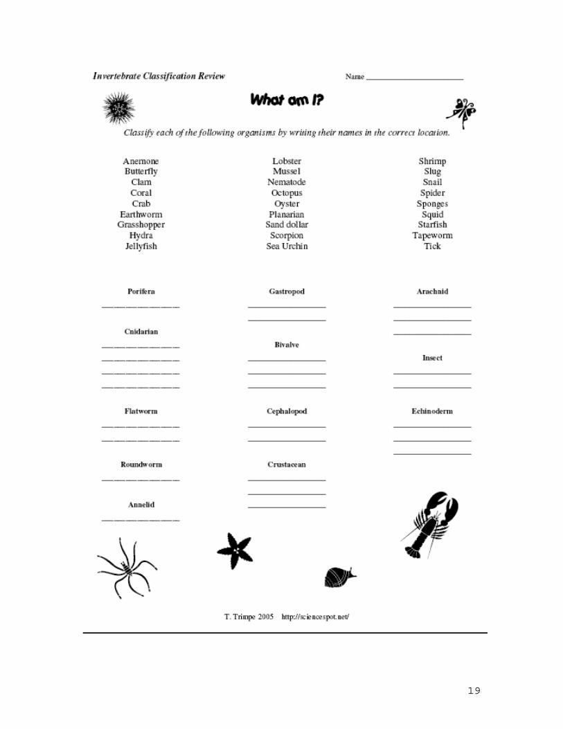
Lesson 4: Invertebrates
Invertebrate Classification Challenge This activity allows students to practice invertebrate classification using the organisms discussed in textbook from Prentice Hall called the Parade of Life: Animals. Classification mats are printed on 8 ½ x 11 inch paper and laminated, printed on card stock and stored in Ziploc bags. Students work in teams to classify the organisms and then compare their answers to the key shown below. “What am I?” worksheet, students will fill in and staple their note packet so they can study outside of class. A quiz (just for practice) will determine which ones they know. Challenge: Students time their efforts to see which team can complete it in the fastest time.
18
19
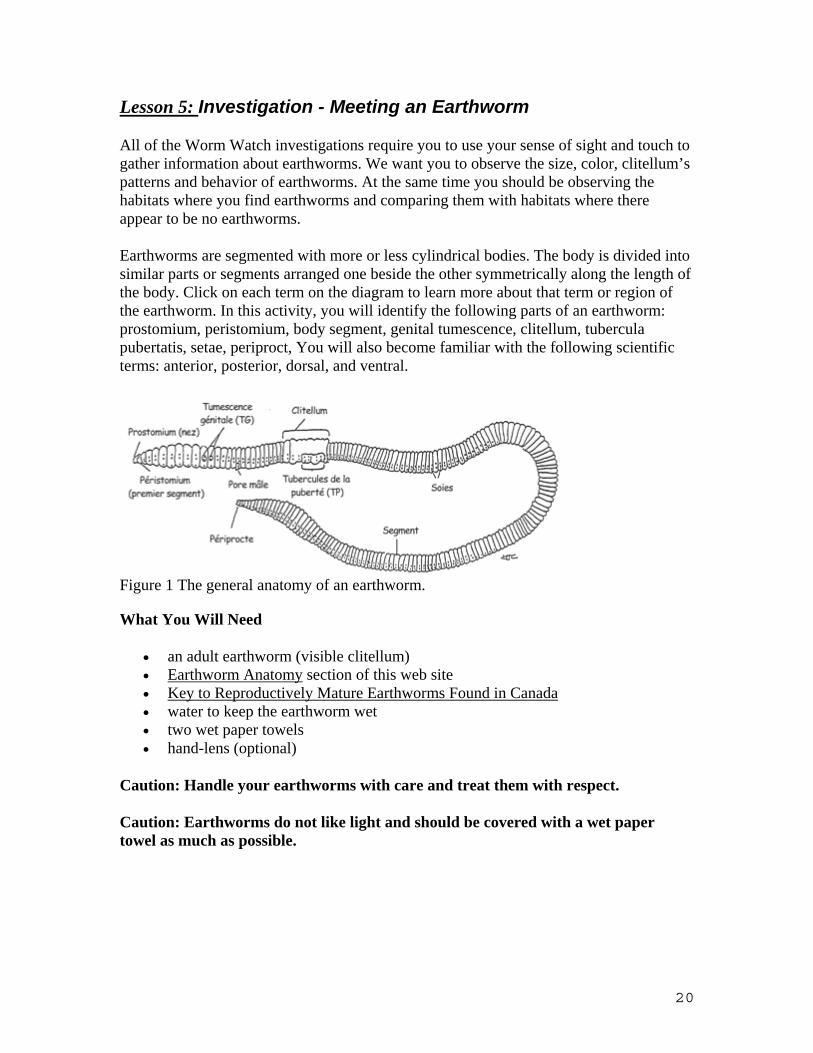
Lesson 5: Investigation - Meeting an Earthworm
All of the Worm Watch investigations require you to use your sense of sight and touch to gather information about earthworms. We want you to observe the size, color, clitellum’s patterns and behavior of earthworms. At the same time you should be observing the habitats where you find earthworms and comparing them with habitats where there appear to be no earthworms.
Earthworms are segmented with more or less cylindrical bodies. The body is divided into similar parts or segments arranged one beside the other symmetrically along the length of the body. Click on each term on the diagram to learn more about that term or region of the earthworm. In this activity, you will identify the following parts of an earthworm: prostomium, peristomium, body segment, genital tumescence, clitellum, tubercula pubertatis, setae, periproct, You will also become familiar with the following scientific terms: anterior, posterior, dorsal, and ventral.
Figure 1 The general anatomy of an earthworm.
What You Will Need
• an adult earthworm (visible clitellum) • Earthworm Anatomy section of this web site • Key to Reproductively Mature Earthworms Found in Canada • water to keep the earthworm wet • two wet paper towels • hand-lens (optional)
Caution: Handle your earthworms with care and treat them with respect.
Caution: Earthworms do not like light and should be covered with a wet paper towel as much as possible.
20
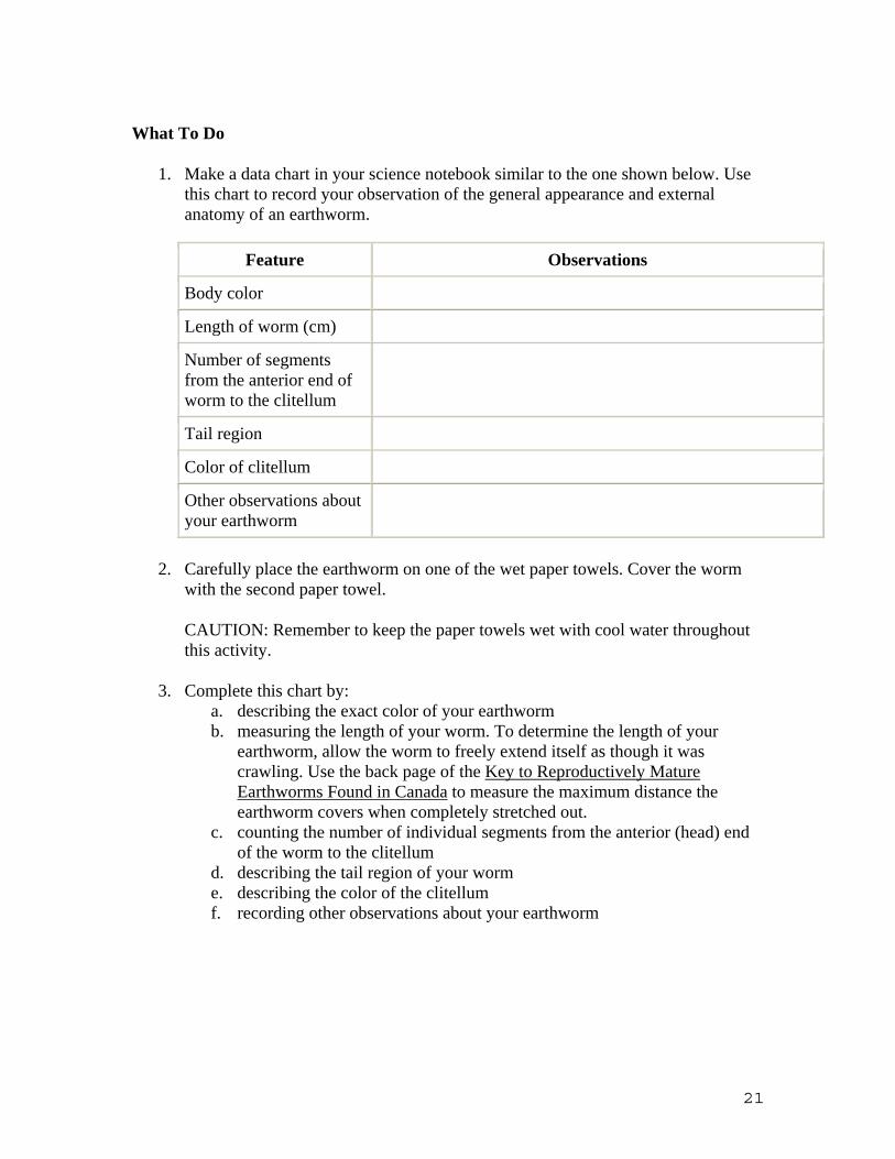
What To Do
1. Make a data chart in your science notebook similar to the one shown below. Use this chart to record your observation of the general appearance and external anatomy of an earthworm.
Feature Observations
Body color
Length of worm (cm)
Number of segments from the anterior end of worm to the clitellum
Tail region
Color of clitellum
Other observations about your earthworm
2. Carefully place the earthworm on one of the wet paper towels. Cover the worm with the second paper towel.
CAUTION: Remember to keep the paper towels wet with cool water throughout this activity.
3. Complete this chart by: a. describing the exact color of your earthworm b. measuring the length of your worm. To determine the length of your
earthworm, allow the worm to freely extend itself as though it was crawling. Use the back page of the Key to Reproductively Mature Earthworms Found in Canada to measure the maximum distance the earthworm covers when completely stretched out.
c. counting the number of individual segments from the anterior (head) end of the worm to the clitellum
d. describing the tail region of your worm e. describing the color of the clitellum f. recording other observations about your earthworm
21

4. Make a second data chart with the following headings:
Name Of Part Description and Sketch of the Part
Location of Part
Function of Part
peristomium
Peristomium- 1st body segment.
Anterior end of the worm on its dorsal surface
Peristomium contains the mouth.
Prostomium
genital tumescence (GT)
Clitellum
tubercula pubertatis (TP)
Setae
Segment
periproct
5. Complete this chart by referring to the diagram, the Earthworm Anatomy section of the Worm Watch web site, and the living worm. The first one has been done for you.
a. Locate each part of the worm. b. Describe the location using terms such as anterior, posterior, dorsal, and
ventral. c. Use the Earthworm Anatomy handout to help you summarize the function
of each part of the earthworm.
6. Return your worm to its container or its natural habitat when you are finished.
CAUTION: Do not put "store-bought" worms into your garden. It is important not to introduce new earthworm species into soil. As a challenge activity write-down all the exotic insects, plants, fish, birds and animals that have been introduced to your area and describe how they have affected the landscape, other species, and human activities.
7. Wash your hands with warm water and soap as soon as you have completed the investigation.
22
Key Terms - prostomium, peristomium, body segment, genital tumescences (GT), clitellum, tubercula pubertatis (TP), setae, periproct , anterior, posterior, dorsal, and ventral
What Did You Discover
1. Why is it important to observe and describe the details of an earthworm? 2. What part of the earthworm is only found on adult worms? 3. Make a scientific drawing of your earthworm. Pay particular attention to the:
• color of the earthworm • number of segments between the anterior end of the worm and clitellum • length of the specimen • genital tumescences (GT) - make sure to draw the exact pattern of the GT
on your earthworm • clitellum- pay careful attention to the size, shape, and color of clitellum • tubercula pubertatis (TP)- make sure to draw the exact shape of the TP and
their location on the clitellum setae
Welcome to the School Programs Area. The following resources are available: Student Investigations:
• Introduction to Earthworm Science • Investigation 1 - Meeting an Earthworm • Investigation 2 - Identifying an Earthworm • Investigation 3 - National Earthworm Survey
Lesson Plans for Teachers:
• Lesson Plan 1- Meeting an Earthworm • Lesson Plan 2 - Identifying an Earthworm • Lesson Plan 3 - National Earthworm Survey
If you are looking for more activities and information, check out the Discover Soil Activities area and the Resources area.
http://www.naturewatch.ca/english/wormwatch/index.html
23
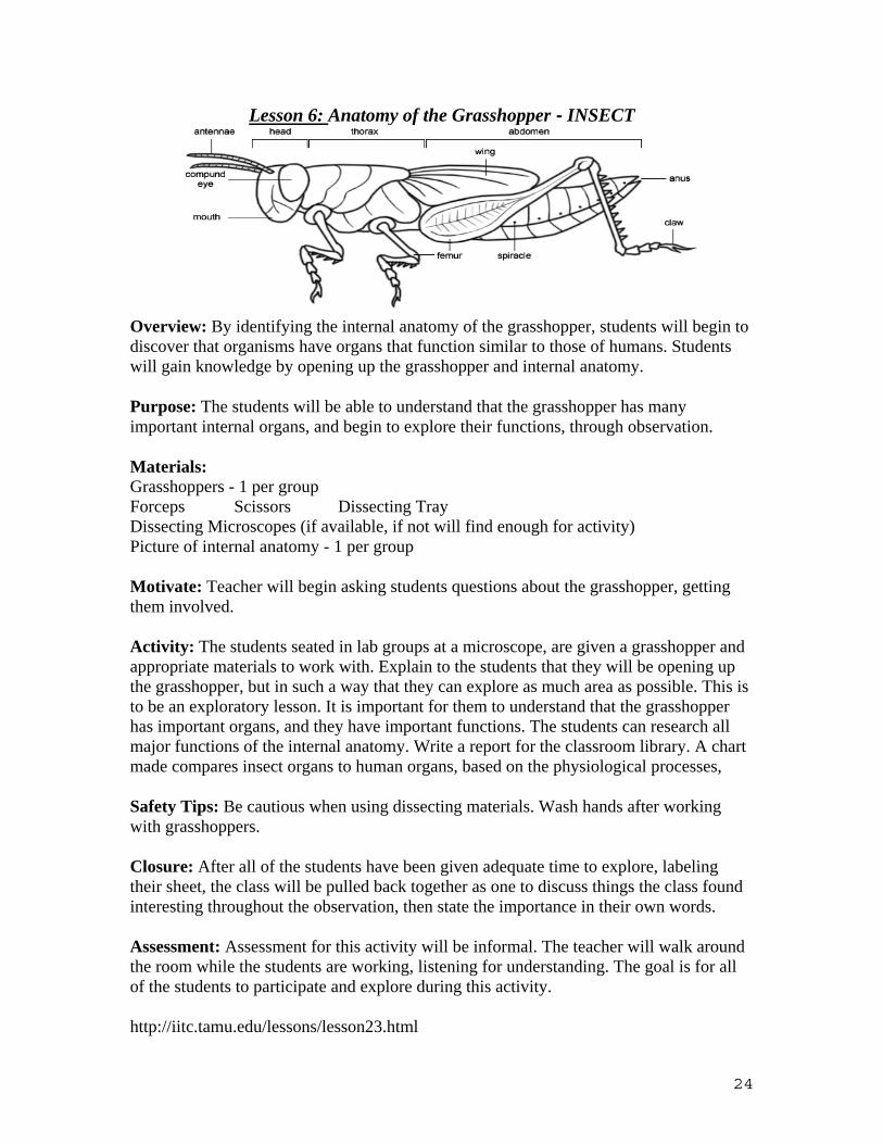
Lesson 6: Anatomy of the Grasshopper - INSECT
Overview: By identifying the internal anatomy of the grasshopper, students will begin to discover that organisms have organs that function similar to those of humans. Students will gain knowledge by opening up the grasshopper and internal anatomy.
Purpose: The students will be able to understand that the grasshopper has many important internal organs, and begin to explore their functions, through observation.
Materials: Grasshoppers - 1 per group Forceps Scissors Dissecting Tray Dissecting Microscopes (if available, if not will find enough for activity) Picture of internal anatomy - 1 per group
Motivate: Teacher will begin asking students questions about the grasshopper, getting them involved.
Activity: The students seated in lab groups at a microscope, are given a grasshopper and appropriate materials to work with. Explain to the students that they will be opening up the grasshopper, but in such a way that they can explore as much area as possible. This is to be an exploratory lesson. It is important for them to understand that the grasshopper has important organs, and they have important functions. The students can research all major functions of the internal anatomy. Write a report for the classroom library. A chart made compares insect organs to human organs, based on the physiological processes,
Safety Tips: Be cautious when using dissecting materials. Wash hands after working with grasshoppers.
Closure: After all of the students have been given adequate time to explore, labeling their sheet, the class will be pulled back together as one to discuss things the class found interesting throughout the observation, then state the importance in their own words.
Assessment: Assessment for this activity will be informal. The teacher will walk around the room while the students are working, listening for understanding. The goal is for all of the students to participate and explore during this activity.
http://iitc.tamu.edu/lessons/lesson23.html
24
Lesson 7: Learn to use your Microscope
Grade level(s): K-8 Subject: Life sciences Topic: Using a microscope in the classroom Estimated class time: 60 minutes Objective: To help students improve their skills in making slides and using a microscope. Materials Qty Product description 1 Fun with Your Microscope (Book) 1 Student Pack Glass Microscope Slides 1 Glass Cover slips
1 Getting to Know Your Microscope Set
www.carolina.com
Review the fundamental parts of a microscope and basic microscope operation. Review slide making techniques by preparing slides for students to view and use as examples of proper technique.
Lesson outline
1. Question and review a. Ask students to identify the fundamental parts of a microscope. b. Explain how the compound microscope differs from the dissecting
microscope c. Ask students how much they already know about microscope operation
and slide preparation.
25
2. Introduce background information a. Explain proper microscope care and operation. b. Explain why compound microscopes and dissecting microscopes are used
for different purposes. c. Explain the special techniques used to prepare specimens and create slides
for microscopic study. d. Explain pertinent lab safety issues for slide preparation and microscopy.
3. Guided practice a. Guide students through hands-on preparation of different types of
microscope slides. b. Guide students as they view the slides they have prepared. Have them
record their observations in writing and sketches. 4. Independent practice/homework
Have students collect specimens at home, such as dust samples (from different areas of their houses), hair samples, or fingerprints, and bring them to school for study.
5. Wrap-up Ask your students what they learned and how the different techniques can have practical applications, e.g., in forensics or medical research.
Assessment Students will:
• Write about and sketch what they observed. (Note: If some students had difficulty viewing their specimens, ask them to explain what they think went wrong and how they can correct it the next time.)
• Describe procedures for preparing samples of other items they may wish to view under the microscope.
Cooperative learning ideas
• Have students compare and contrast their findings with those of their classmates. • Have students work together to explain their findings and how they can improve
their technique.
Cross-curriculum ideas
• Math: Young students count the number of specimens viewed on a slide, calculate magnification power, and learn about the metric system (e.g., the millimeter and micron). Older students can use the microscope’s mechanical stage to estimate the area of the viewing field at different magnifications and estimate the size of organisms viewed.
• Language: Students write a paragraph describing what they observed with the microscope.
• Art: Students sketch what they observed with the microscope.
26
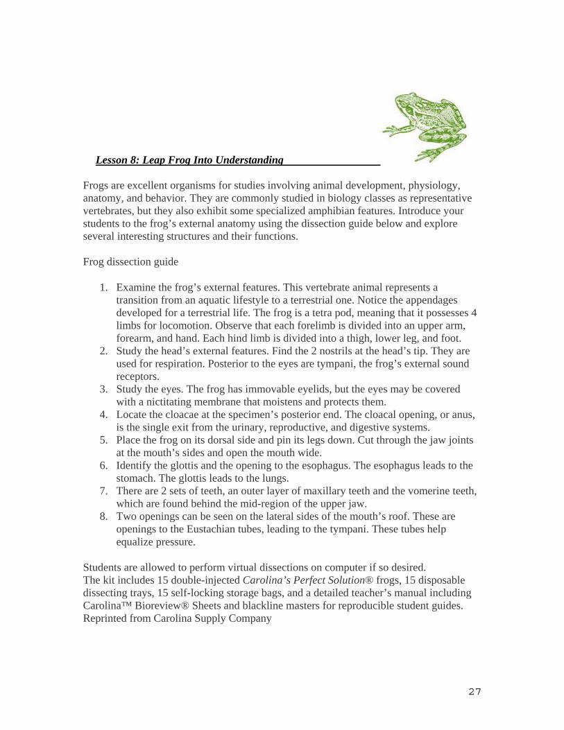
Lesson 8: Leap Frog Into Understanding
Frogs are excellent organisms for studies involving animal development, physiology, anatomy, and behavior. They are commonly studied in biology classes as representative vertebrates, but they also exhibit some specialized amphibian features. Introduce your students to the frog’s external anatomy using the dissection guide below and explore several interesting structures and their functions. Frog dissection guide
1. Examine the frog’s external features. This vertebrate animal represents a transition from an aquatic lifestyle to a terrestrial one. Notice the appendages developed for a terrestrial life. The frog is a tetra pod, meaning that it possesses 4 limbs for locomotion. Observe that each forelimb is divided into an upper arm, forearm, and hand. Each hind limb is divided into a thigh, lower leg, and foot.
2. Study the head’s external features. Find the 2 nostrils at the head’s tip. They are used for respiration. Posterior to the eyes are tympani, the frog’s external sound receptors.
3. Study the eyes. The frog has immovable eyelids, but the eyes may be covered with a nictitating membrane that moistens and protects them.
4. Locate the cloacae at the specimen’s posterior end. The cloacal opening, or anus, is the single exit from the urinary, reproductive, and digestive systems.
5. Place the frog on its dorsal side and pin its legs down. Cut through the jaw joints at the mouth’s sides and open the mouth wide.
6. Identify the glottis and the opening to the esophagus. The esophagus leads to the stomach. The glottis leads to the lungs.
7. There are 2 sets of teeth, an outer layer of maxillary teeth and the vomerine teeth, which are found behind the mid-region of the upper jaw.
8. Two openings can be seen on the lateral sides of the mouth’s roof. These are openings to the Eustachian tubes, leading to the tympani. These tubes help equalize pressure.
Students are allowed to perform virtual dissections on computer if so desired. The kit includes 15 double-injected Carolina’s Perfect Solution® frogs, 15 disposable dissecting trays, 15 self-locking storage bags, and a detailed teacher’s manual including Carolina™ Bioreview® Sheets and blackline masters for reproducible student guides. Reprinted from Carolina Supply Company
27
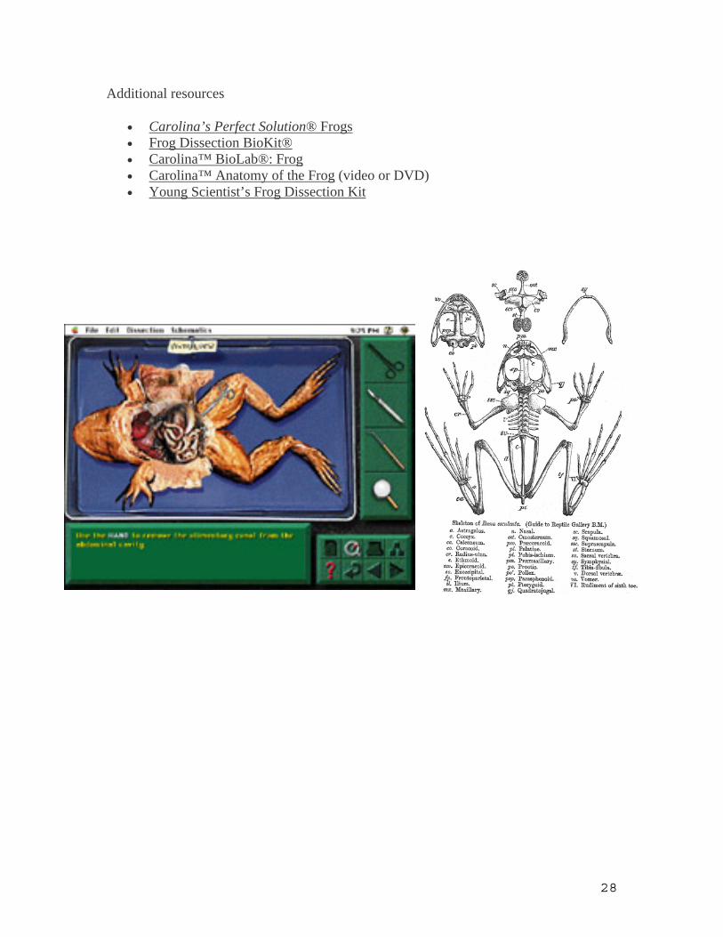
Additional resources
• Carolina’s Perfect Solution® Frogs • Frog Dissection BioKit® • Carolina™ BioLab®: Frog • Carolina™ Anatomy of the Frog (video or DVD) • Young Scientist’s Frog Dissection Kit
28
Resource List including:
Supplemental materials (suppliers)
Carolina Supplies
Ward Science Company
Parade of Life: Animals. Prentice Hall http://yucky.discovery.com/flash/worm/
www.carolina.com/category/teacher+resources/dissection+activities+and+resources.do
http://www.carolina.com/category/teacher+resources/classroom+activities.do?page=all
http://wardsci.com/category.asp_Q_c_E_807_A_Animals
http://froguts.com/flash_content/index.html
http://www.nwf.org/frogwatchUSA
www.allaboutfrogs.com
www.whitman.edu/biology/vpd/main.html
www.ebioinfogen.com/cat.htm
www.biolabsoftware.com
http://tolweb.org/tree/ Tree of life Websit http://www.naturewatch.ca/english/wormwatch/index.html
http://iitc.tamu.edu/lessons/lesson23.html
29