current and emerging techniques for diagnostic mutation detection · 9 2 current and emerging...
TRANSCRIPT

9
2
Current and Emerging Techniques for Diagnostic Mutation Detection
An Overview of Methods for Mutation Detection
Claire F. Taylor and Graham R. Taylor
1. Mutation Detection: An IntroductionThis chapter provides a broad overview of the range of mutation detection
techniques that are now available. For the purposes of this chapter, a mutation can be defined as a sequence
change in a test sample compared with the sequence of a reference standard.This definition implies nothing about the phenotypic consequences (e.g., path-ogenicity) of a mutation. A polymorphism may be defined as a mutation thatoccurs in a substantial proportion (>1%) of a population and is tacitly assumedto be non-pathogenic, although the true pathogenicity may be unknown. Apolymorphism has also been defined as a Mendelian trait that exists in the pop-ulation, with the frequency of the more rare of the two alleles greater than 1–2%(1). If we accept that DNA sequence is a Mendelian trait, then the two defini-tions of polymorphism are the same.
The detection of a single base change in the human genome requires asignal�background ratio of 1�6 × 10⁹—a formidable task. To achieve suchselectivity in the field of electronics would require amplification and noisereduction, and it is no surprise that analogous processes are found in molecu-lar genetics—for example, amplification by the polymerase chain reaction(PCR) and noise reduction by the stringent annealing of probes and primers.
Mutation detection techniques can be divided into techniques that test forknown mutations (genotyping) and those that scan for any mutation in a par-
From: Methods in Molecular Medicine, vol. 92: Molecular Diagnosis of Genetic Diseases, Second EditionEdited by: R. Elles and R. Mountford © Humana Press Inc., Totowa, NJ
CH02,9-44,36pgs 8/11/03 12:29 PM Page 9
ticular target region (mutation scanning). Broader aspects of mutation detec-tion include identification of gene dosage alterations, gross re-arrangements,and methylation. There are several well-known genotyping and scanning meth-ods in routine diagnostic use. Many of these are covered in detail in this volumeand elsewhere (1,2). This chapter focuses primarily on recent modifications,development, and evaluation of these techniques.
The primary considerations in any approach to mutation detection aresensitivity (the proportion of mutations that can be detected) and specificity(the proportion of false-positives). The cost per genotype and throughput arealso important factors in service delivery. It is often difficult to evaluatethese features accurately from the published scientific literature—presumably,one of the reasons why the Human Genome Organisation Mutation Detec-tion training courses (http://www.leeds.ac.uk/cmgs/leedsdna/science/hugo/index.html) and workshops (http://www.mutations2001.bled.si/) haveproven so popular.
2. GenotypingBecause sequence changes can abolish or create cleavage sites for the wide
range of commercially available restriction endonucleases (REs), RE poly-morphisms were the first tools used for genetic mapping and diagnosis, in com-bination with Southern blotting of genomic DNA (3,4). Although there are stillsome applications—for example, mapping large deletions or rearrangements,for which Southern blotting is the best method—the polymerase chain reaction(PCR) is now the method of choice for routine genotyping (5).
2.1. Genotype Analysis Using the PCR
The analysis of restriction fragment length polymorphism (RFLP) is now pri-marily of historical interest as a first choice for genotyping, although it is arobust method. Amplicons are generated to flank a polymorphic RE site andsubjected to digestion, and the presence or absence of the site can be determinedby agarose gel electrophoresis of the digested amplicon and visualization usingethidium bromide staining and ultraviolet (UV) illumination. Artificial restric-tion sites can be produced by incorporating modifications into one of theamplimers to increase the range of polymorphisms that can be examined. Therequirement to hold stocks of a range of different REs, and two-step genotyp-ing process (amplification followed by digestion) does not lend itself to eitherrapid or high-throughput genotyping. Gains in throughput can be achievedusing high-density gels such as the microtiter array diagonal gel electrophore-sis (MADGE) format (6).
10 Taylor and Taylor
CH02,9-44,36pgs 8/11/03 12:29 PM Page 10

2.2. Genotyping for Linkage Analysis
2.2.1. Microsatellite Analysis
Linkage mapping has been accelerated by the description of microsatellites(7). Microsatellite repeats are mono-, di-, tri-, or tetranucleotide repeats that dis-play polymorphism with respect to the length of the repeat. The origin of thislength polymorphism is believed to be “strand slippage” during replication. Onestrand may form a short hairpin and produce a copy of different length in thedaughter strand. The PCR gained widespread usage when microsatellites werefirst described, and they were ideally suited for analysis by designing PCRprimers (amplimers) that bind to unique sequences flanking the microsatelliterepeat motif. Amplicons must be sized to within the resolution of the repeat motifto provide a genotype. This requires sizing fragments of approx 100–300 base-pairs in length with an accuracy of ±1 basepair. The most accurate way to dothis is to use some form of automated fragment analysis equipment such as thecommercially available automated DNA sequencers. Using capillary arrays andmultiple sample loading, genotyping throughputs of up to 500 per hour can beachieved, a total far beyond the current requirements of diagnostic laboratories.
2.2.2. SNPs
Recently, interest has returned to single-nucleotide polymorphisms (SNPs),of which RE polymorphisms are a subset. SNPs are di-allelic (although in prin-ciple there is no reason why a particular base could not be substituted by morethan one alternative), and thus less informative individually than microsatel-lites, but far more abundant (8). The human genome may contain millions ofSNPs, yet they are probably more abundant in noncoding regions of the genome.Several efforts are now underway to produce genomic SNP maps (9).Genotyping will then aim to type sets of SNPs, or possibly SNP haplotypes,since it is now becoming clear that recombination preserves blocks of haplo-types (linkage disequilibrium) over substantial physical distances (10). Themain appeal of SNPs is the prospect of high-throughput automated analysisusing array or “chip” technology (11); however, a variety of generic mutationdetection techniques can be adapted for SNP detection.
2.2.3. Amplification Refractory Mutation System (ARMS)
ARMS is a modification of conventional PCR in which one of the amplimersis designed to have the polymorphic base in the template at its 3′ position (12).Taq polymerase is unable to extend from a mismatched base, and thus the gen-eration of a PCR product occurs only if the 3′ base in the primer matches thetemplate. The technique can be multiplexed to type up to 20 SNPs simultane-
Diagnostic Mutation Detection 11
CH02,9-44,36pgs 8/11/03 12:29 PM Page 11

ously. In practice, an additional mismatch at the 3′ minus 3 nucleotide positionis required to destabilize primer binding for a stronger assay. A weakness ofthe standard ARMS approach is that two tubes (wild-type and mutant) arerequired for a full genotype. However, by modifying the primer length (or flu-orescent label if using fluorescent analysis), it is possible to generate differentproducts from each allele. ARMS is a low-cost approach that can use standardlaboratory equipment. Higher throughput can be achieved by using closed-tubeassay systems or adaptation of high-throughput gel formats such as the MADGEsystem (13).
2.2.4. Minisequencing
Minisequencing, also referred to as single-nucleotide primer extension (14)and genetic bit analysis (15), determines the base immediately 3′ to a primerby extending the primer by one base only (16). Base extension can be moni-tored by gel electrophoresis (17), and commercial kits are available to run theseassays on DNA sequencers—for example, “SnapShot” from AppliedBiosystems (ABI). As with most genotyping assays, if the variant is present asa minority species (for example, in a tumor or a germinal mosaic), the relia-bility of the assay declines, although increased sensitivity of detection by pre-treatment of a mixed population containing H-ras codon 61 mutants has beenreported (18) using the MutEx assay (19). High-throughput and solid-phaseadaptations have also been described (20). Readily adaptable to microtiter (21),high-performance liquid chromatography (HPLC) (22), and array (23) and massspectroscopy (24) formats, it has considerable potential as a high-throughputsystem. Although the original report (16) described detection from genomicDNA without amplification, all subsequent reports have used PCR amplifica-tion to prepare primer extension templates. In highly parallel systems, the abil-ity to amplify templates becomes rate-limiting. Minisequencing is thus aflexible method that can operate using fairly basic equipment, or can be adaptedto highly automated systems.
2.2.5. TaqMan and Molecular Beacons
TaqMan is a closed-system assay that can be adapted for gene dosage as wellas genotyping. Single-nucleotide differences are detected in PCR products bythe sequence-specific hybridization of a probe that contains both a fluorochromeand a quencher. When hybridized, the quencher molecule is cleaved, and thebound fluorochrome can be detected by a fluorescence assay. Since it is possi-ble to have different colored fluorochromes, the probes can be differentiallylabeled, allowing both alleles of an SNP to be typed in the same tube (25).Molecular beacons are hairpin-shaped structures that contain a fluorochromeand a quencher on the 5′ and 3′ ends. In free solution, the fluorochrome is
12 Taylor and Taylor
CH02,9-44,36pgs 8/11/03 12:29 PM Page 12

quenched, but upon hybridization to a specific target the hairpin opens and themolecule becomes fluorescent. These molecules can be used in a closed systemfor allelic discrimination of PCR products (26). Both assays can be read in real-time or end-point formats, using fluorescent plate or tube readers. The“Scorpion” assay is an interesting development that combines an amplificationprimer and beacon-like detection component in the same molecule to enablereal-time genotyping (27). The LightCycler system (Idaho Technology, IdahoFalls, ID) uses fluorescence resonance energy transfer (FRET) to perform real-time PCR and genotyping using two oligonucleotides, one carrying an energyacceptor and the other an emitter. The oligonucleotides hybridize in tandem onthe template. The first dye (fluorescein) is excited by the LightCycler’s LED(Light Emitting Diode) filtered light source, and emits green fluorescent lightat a slightly longer wavelength. When the two dyes are in close proximity, theemitted energy excites the LC Red 640 attached to the second hybridizationprobe that subsequently emits red fluorescent light at an even longer wave-length. The LightCycler is set to detect the longer wavelength (640 nm) light.Energy transfer is highly dependent on the spacing between the two dye mol-ecules. Only when the molecules are in close proximity (a distance between 1and 5 nucleotides) is the energy transferred at high efficiency, and fluorescenceis proportional to the amount of bound primers.
Once suitable oligonucleotides are designed, the genotyping of a sample isstraightforward. The instrument is programmed to amplify the DNA and to per-form a melting curve analysis. A perfect match has a higher melting tempera-ture than a mismatch. In this way, the LightCycler directly genotypes a sampleafter amplification with no additional handling. With dual-color detection, it ispossible to simultaneously genotype two different mutations in one PCR run.Although the LightCycler uses a rather idiosyncratic arrangement of sealedglass capillaries, other closed-system plate readers for 96- or 384-well platesand automated plate loading are commercially available. The choice of systemwill probably depend to a large extent on the cost of consumables.
2.2.6. Ligation
The specificity of DNA ligase for perfectly matched double-stranded DNA,particularly thermostable ligase (28,29), has been exploited as a genotyping toolfor the ligase chain reaction and the ligase amplification reaction (30). Ingenetic testing, ligase reactions have been more widely used to genotype PCRproducts rather than to perform the amplifcation reaction directly—for exam-ple, in the development of an assay to genotype 31 pathogenic variants in thecystic fibrosis gene ABCC7 (31).Two sets of oligonucleotide probes can be lig-ated only if they are hybridized to a perfectly matched template, the oligonu-cleotide ligation assay (OLA). This has been adapted to produce a dual-color
Diagnostic Mutation Detection 13
CH02,9-44,36pgs 8/11/03 12:29 PM Page 13

microtiter readout (32) and gel-based systems (33) in which the ligation prod-ucts are distinguished by fragment color and mobility, enabling automatedgenotype readout. Ligation systems can also be modified to perform microsatel-lite genotyping (34). This adaptation has the potential to be developed into anarray-based system for microsatellite analysis. Ligation has also been adaptedto seal nicked circular probes producing “padlock probes” that can be thenamplified by rolling circle replication (35–37).
2.2.7. Pyrosequencing
Pyrosequencing is a non-electrophoretic real-time DNA sequencing methodthat uses a unique approach to read small runs of bases (38). The luciferase-luciferin light release is a detection signal for nucleotide incorporation intotarget DNA. This method can be adapted for automated high-throughput oper-ation, and has the advantage of typing bases that flank the SNP to confirm thatthe correct target is being analyzed (39). Pyrosequencing of the human p53 geneusing a nested multiplex PCR method for amplification of exons 5–8 has beendescribed, reporting accurate detection of p53 mutations and allele distribution(40). If the current length of sequence limitation can be overcome, pyrose-quencing has considerable potential as a highly automatable sequencing tool.
2.2.8. Invader
Invader technology uses a Flap Endonuclease (FEN) for allele discriminationand a universal FRET reporter system. A study by Mein et al. (41) genotypedthree hundred and eighty-four individuals across a panel of 36 SNPs and oneinsertion-deletion (indel) polymorphism with Invader assays using a PCR prod-uct as a template. The average failure rate of 2.3% was mainly associated withPCR failure, and the typing was 99.2% accurate when compared with genotypesthat were generated with established techniques. Semi-automated data inter-pretation allows the generation of approx 25,000 genotypes per person per week,10-fold greater than gel-based SNP typing and microsatellite typing. Using an“Invader squared” method, Factor V Leiden genotyping has been achieved ongenomic DNA samples without prior amplification (42), although most assaysin routine use now rely on the PCR to generate templates for genotyping.
2.2.9. Hybridization
Allele-specific hybridization (ASH) was one of the early methods ofgenotyping (43), originally using genomic DNA as template, later with PCR-amplified DNA. By carefully controlling the stringency of hybridization,18- to 22-mer probes can discriminate between single base substitutions oftarget. This technique is still used, and forms the basis of some commercial testkits for cystic fibrosis (44) and Human Leukocyte Antigen (HLA) typing (45).
14 Taylor and Taylor
CH02,9-44,36pgs 8/11/03 12:29 PM Page 14

Real-time hybridization analysis (dynamic allele-specific hybridization[DASH]) makes the assay more robust, since the denaturation of probe andtarget can be monitored over a range of temperatures (46). Hybridization canalso be monitored by surface plasmon resonance, enabling optical biosensorsto perform automated genotyping (47,48). Using this procedure (49,50), it waspossible to perform real-time monitoring of hybridization between target single-stranded PCR products, enabling a one-step, non-radioactive protocol to per-form cystic fibrosis diagnosis.
2.2.10. Arrays
The idea of using arrays for high-throughput genotyping has been in existencefor many years. Early arrays were two-dimensional spots of DNA targets onnylon or nitrocellulose membranes, and the method of detection was ASH (51).This method still has value, and recent improvements in the oligonucleotide-binding capacity of membranes (52) could extend this further. However DNAarrays typically refer to glass, plastic, or silicon supports with either oligonu-cleotide or cloned DNA attached by adhesion or covalent linkage. Arrays thatare mechanically deposited onto a glass microscope slide have feature sizes ofapprox 200 microns, and are scanned at 5–20-micron resolution. Such arrayscan carry 10–15,000 features. Affymetrix manufacture high-density arrays bya proprietary photochemical oligonucleotide synthesis method that can resultin a small (10-µ) feature size, enabling a large number of 20–24-base oligonu-cleotide probes to be packed into a small area (53). Although these arrays havehad the most success in gene-expression studies, they have not yet producedthe anticipated breakthrough in DNA sequencing (or “resequencing”) (54) ormutation scanning, although their use has been reported in ABCC7 (55), mito-chondrial (56), and BRCA1 (57) mutation detection. The reason for the lim-ited use of the Affymetrix system for mutation detection thus far lies in itslimited sensitivity. Di-deoxy sequencing of the p53 gene in 100 primary humanlung cancers by cycle sequencing was compared with sequence analysis byusing the p53 GeneChip assay (58). The GeneChip assay detected 46 of 52 mis-sense mutations (88%), but 0 of 5 frameshift mutations. The specificity ofdirect sequencing and of the p53 GeneChip assay in detecting p53 mutationswere 100% and 98%, respectively. Although more mutations were detected inp53 by manual sequencing than by use of a p53 gene chip, direct sequencingand the p53 GeneChip were not infallible at p53 mutation detection. In anotherstudy (59), reported a 92% sensitivity for the detection of p53 mutations in aseries of 108 ovarian tumors, less than might be expected from a current muta-tion scanning tool such as denaturing high-performance liquid chromatogra-phy (DHPLC). Hybridization may not be the best way to exploit arrayed DNAfor mutation detection. Several recent studies have indicated that the use of
Diagnostic Mutation Detection 15
CH02,9-44,36pgs 8/11/03 12:29 PM Page 15

primer extension (15,23) or ligation (60) can improve the specificity of muta-tion detection on arrays. With mechanically prepared arrays, this is not diffi-cult to set up, as the oligonucleotide can be arrayed with the 3′ end (the substratefor primer extension) free, and the 5′ end anchored. However, in light-directedoligonucleotide synthesis, the 3′ end of the probe is anchored to the solid sup-port. Although this is not a problem for ligation reactions, it does mean thatdirect primer extensions for the arrayed oligonucleotide are not possible. Thisproblem can be circumvented by conducting the primer extension reaction insolution and then capturing the reaction products by means of 5′ tags on thesubstrate with complementary tags on the array (61–63). “Zip-code address-able” arrays provide a generic solution for genotyping, as primer sets can becustom-designed to work on standard chips. The same design principle can beapplied to “liquid-phase arrays,” which are latex microbeads that can be sortedusing a fluorescence-activated cell sorter (FACS). By addressing each bead witha different tag, up to 96 primer extension reactions can be monitored in a singletube (64,65). The same principle can also be applied to provide templates forultra-rapid mass-spectroscopic genotyping, which is likely to be the method ofchoice for ultra high-throughput genotyping (24). Here, primer extension prod-ucts are simply weighed to determine the nucleotide added. Commercial sys-tems are available that include primer design software, sets of validated SNPs,and high-throughput genotype analysis software.
3. DosageAlthough methods for the detection of point mutations and small insertions or
deletions in DNA are well-established, the detection of larger (>100 bp) genomicduplications or deletions can be more difficult. Most mutation scanning methodsuse PCR as a first step, but the subsequent analyses are usually qualitative ratherthan quantitative. Gene dosage methods based on PCR must be absolutely quan-titative (i.e., they should report molar quantities of starting material) or semi-quantitative (i.e., they should report gene dosage relative to an internal standard).Without some method of quantitation, heterozygous deletions may be overlooked,and may therefore not be fully evaluated. Gene dosage methods can provide theadditional benefit of reporting allele drop-out in the PCR.
Large genomic duplications and deletions have been recognized as patho-genic mutations for many years—for example in alpha-thalassemia (66,67),Duchenne and Becker Muscular Dystrophies (68) and more recently in famil-ial breast cancer (69), and hereditary non-polyposis colorectal cancer (HNPCC)(70,71). Based on the May 2000 Human Gene Mutation Database, deletions andduplications represented 5.5% of reported mutations (72). Because many muta-
16 Taylor and Taylor
CH02,9-44,36pgs 8/11/03 12:29 PM Page 16
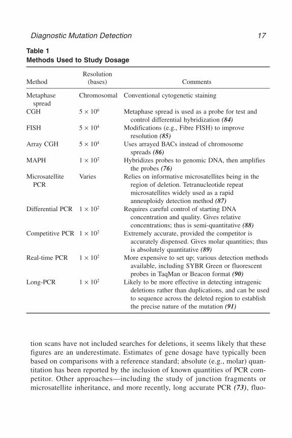
tion scans have not included searches for deletions, it seems likely that thesefigures are an underestimate. Estimates of gene dosage have typically beenbased on comparisons with a reference standard; absolute (e.g., molar) quan-titation has been reported by the inclusion of known quantities of PCR com-petitor. Other approaches—including the study of junction fragments ormicrosatellite inheritance, and more recently, long accurate PCR (73), fluo-
Diagnostic Mutation Detection 17
Table 1Methods Used to Study Dosage
ResolutionMethod (bases) Comments
Metaphase Chromosomal Conventional cytogenetic stainingspread
CGH 5 × 10⁶ Metaphase spread is used as a probe for test and control differential hybridization (84)
FISH 5 × 10⁴ Modifications (e.g., Fibre FISH) to improve resolution (85)
Array CGH 5 × 10⁴ Uses arrayed BACs instead of chromosome spreads (86)
MAPH 1 × 10² Hybridizes probes to genomic DNA, then amplifies the probes (76)
Microsatellite Varies Relies on informative microsatellites being in the PCR region of deletion. Tetranucleotide repeat
microsatellites widely used as a rapidanneuploidy detection method (87)
Differential PCR 1 × 10² Requires careful control of starting DNAconcentration and quality. Gives relative concentrations; thus is semi-quantitative (88)
Competitive PCR 1 × 10² Extremely accurate, provided the competitor is accurately dispensed. Gives molar quantities; thus is absolutely quantitative (89)
Real-time PCR 1 × 10² More expensive to set up; various detection methods available, including SYBR Green or fluorescent probes in TaqMan or Beacon format (90)
Long-PCR 1 × 10² Likely to be more effective in detecting intragenic deletions rather than duplications, and can be usedto sequence across the deleted region to establish the precise nature of the mutation (91)
CH02,9-44,36pgs 8/11/03 12:29 PM Page 17

rescence in situ hybridization (FISH) (74,75), multiplex amplifiable probehybridization (MAPH) (76), comparative genomic hybridization (CGH)(77–79), and array-CGH (80)—have also been employed. In some cases, knowl-edge of the gene (or exon) dosage may not be sufficient to establish the path-ogenic consequences of a genotype. For example, in spinal muscular atrophy,in which gene duplications and unstable regions of the genome can complicatethe issue (81). Although reciprocal translocations would escape detection bysimple dosage techniques, robust low-cost dosage methods may find utility inrapid screening for supernumerary chromosomes (82,83).
Techniques for detecting gene-dosage alterations can be broadly dividedinto three types: cytogenetic, solid-phase hybridization, or PCR amplification.
4. Methods for Studying DNA MethylationIn the human genome, DNA methylation is found in the form of 5-methyl
cytosine, located almost exclusively within CpG dinucleotides (for a recentreview, see 92). Perturbations of the normal pattern of methylation are associ-ated with disorders of imprinting and X-chromosome inactivation and alsowith oncogenesis, and can be considered to be epigenetic mutations.
A number of methods for the study of the pattern of cytosine methylation atspecific loci have been described (93,94), all depending on one of three mech-anisms to discriminate between methylated and unmethylated cytosines:
• differential cleavage by methylation-sensitive restriction enzymes• differential cleavage by chemicals• differential reactivity with sodium bisulphite
4.1. Differential Cleavage by Methylation-Sensitive Restriction Enzymes
Restriction endonucleases that are unable to cleave DNA when their restric-tion sites contain 5-methyl cytosine have long been recognized as a tool for thestudy of cytosine methylation (95). Assays that utilize methylation-dependentrestriction enzymes are a more recent advance. Digestion and thus methylationare monitored either by Southern blot or by PCR using primers flanking therestriction site (96,97). These methods are relatively simple and widely used,despite a number of drawbacks that include the confinement of analysis tocytosine residues within restriction sites and the possibility of misleading resultsas a result of partial digestion or PCR failure.
4.2. Differential Cleavage by Chemicals
In the Maxam-Gilbert sequencing protocol, hydrazine is used to cleave DNAat cytosine and thymine residues (98). 5-methyl cytosine is resistant to
18 Taylor and Taylor
CH02,9-44,36pgs 8/11/03 12:29 PM Page 18

hydrazine cleavage, and appears as a gap on a Maxam-Gilbert genomic sequenc-ing ladder (99). The original protocol was time-consuming and required largequantities of DNA; later developments such as ligation-mediated PCR (100)addressed a number of these problems. Despite these improvements, the pres-ence of 5-methyl cytosine still must be inferred from the absence of a band,although a protocol allowing the positive display of methylated residues usingpermanganate has been described (101).
4.3. Differential Reactivity with Sodium Bisulfite
Upon reaction with bisulfite, cytosine is deaminated to uracil, whereas5-methyl cytosine is not reactive. During a subsequent PCR, uracil residues areamplified as thymine, and 5-methyl cytosine is amplified as cytosine. Thissequence conversion provides positive identification of methylated residues inthe starting sample (102). Direct sequencing of PCR products yields a strand-specific average of the methylation pattern in the starting population of mole-cules. For information about the methylation pattern of individual molecules,cloning of the PCR products prior to sequencing is required.
Bisulfite modification can lead to the methylation-dependent creation ofnovel restriction sites or retention of existing sites. PCR followed by restric-tion digestion provides a rapid method for screening specific CpG sites, whichdoes not rely on an absence of cleavage to detect methylation and can also beused as a quantitative assay (103). Quantification of the level of methylation atspecific CpG sites can also be done by a single-nucleotide primer extensionassay (MS-SnuPE) (104).
Methylation-specific PCR (MSP) (105) uses PCR primers designed toamplify bisulfite-modified DNA, which can differentiate between methylatedand umethylated DNA. MSP is extremely sensitive, and can detect the presenceof methylated DNA at levels as low as 1/1000 (105). A single-tube method forthe detection of methylation at 15q11-q13 for the diagnosis of Prader-Willi syn-drome and Angelman syndrome (106) is widely used in diagnostic laborato-ries. A real-time methylation-sensitive PCR has been described, which can beused to quantify methylation (107).
5. Mutation Scanning MethodsMutation scanning is the search for novel sequence variants within a defined
DNA fragment. Numerous methods that exploit different physical, chemical,and biological consequences of DNA sequence variation have been developedto facilitate mutation scanning. The ideal mutation scanning method has beencharacterized as one that would screen kilobase lengths of DNA with 100% sen-sitivity and specificity, and would completely define the mutation (108). Itwould be a simple, single-step, non-electrophoretic protocol with high through-
Diagnostic Mutation Detection 19
CH02,9-44,36pgs 8/11/03 12:29 PM Page 19
put and low cost, requiring no complex equipment and no harmful reagents.Cost and data-analysis time continue to be major barriers to meeting the demandfor genetic testing, and no current method satisfies all of these criteria.
Most scanning methods do not identify the precise nature of the change tothe DNA sequence, although some indicate the location of the mutation withinthe fragment analyzed. Consequently, the majority of methods are used as afirst-round screen to identify those samples that contain mutations, and thesesamples are subsequently sequenced to define the mutations.
Several factors influence the choice of scanning method:
5.1. Mutation Detection Sensitivity
In the clinical diagnostic setting, sensitivity should be as close to 100% asreasonably practicable. Mutation scanning for other purposes such as candidategene analysis may be able to tolerate a trade-off between a reduction in sensi-tivity and an increase in throughput. In practice, it is unlikely that any singletechnique will detect 100% of mutations. An awareness of the limitations ofthe technique selected is essential. Factors that influence sensitivity includefragment resolution, reactivity of any enzymes or chemicals used, and templatefeatures such as sequence (e.g., G + C content), length, and secondary struc-ture. The measurement of sensitivity is empirical: the literature is replete withexamples of non-blinded studies or studies using small series, from which it isdifficult to draw general conclusions about assay performance.
In a prescreening method, low specificity (large numbers of false-positives)may generate excessive downstream analysis and reduce the advantage of pre-screening. Some regions of interest may be highly polymorphic, and generatemany samples that require further analysis. Although there have been claimsthat common polymorphisms generate “characteristic” mobility shifts—forexample, in DMPLC HPLC analysis—these claims should be treated with cau-tion in a diagnostic setting.
5.2. Suitability for Proposed Sample Type
Current diagnostic practice is largely restricted to genomic DNA samplesextracted from peripheral blood lymphocytes. Future developments are likelyto include increasing analysis of DNA extracted from tumor samples, whichpresents a number of problems that are not encountered when studying germlineDNA. In germline samples, mutations can be present at 0% (homozygous orhemizygous wild-type), 50% (heterozygous) or 100% (homozygous or hem-izygous mutation) of the total DNA, depending on zygosity, unless mosaicismis present. In tumor samples, the mutation can be present at any proportion ofthe total DNA because of factors that include loss of heterozygosity, contami-
20 Taylor and Taylor
CH02,9-44,36pgs 8/11/03 12:29 PM Page 20

nation of the tumor with surrounding wild-type material, and variable propor-tions of mutant cells in the tumor. Some methods such as DHPLC are betterable to detect mutations that are present as a minor fraction in the sample (109).Many methods are dependent on the generation of heteroduplex DNA for thedetection of mutations: depending on whether the expected mutations are likelyto be homozygous, hemizygous, or heterozygous it may or may not be neces-sary to add 50% wild-type DNA to the samples.
5.3. Suitability for Predicted Mutation Type
Some of the methods described here have limitations on the types of muta-tions they can detect. For instance, DHPLC cannot reliably detect homozygousmutations; heteroduplex analysis (HA) detects insertions/deletions with higherefficiency than substitutions, and the protein trucation test (PTT) detects onlypolypeptide-chain-terminating mutations.
When the nature of mutation is unknown, a detection method that is unbi-ased toward any type of mutation should be used. For conditions/genes inwhich a single type of mutation predominates, it may be more appropriate toselect a method designed to detect only that type of mutation.
5.4. Features of the DNA Sequence Analyzed
Knowledge of the presence of common polymorphisms in the fragment tobe analyzed may also affect the choice of method. With the exception of thescanning methods that unambiguously identify the mutation present, in mostcases the available information will only indicate that a mutation is present orabsent. Some methods—for instance, DHPLC and fluorescent single strand con-formation polymorphism (FSSCP)—may produce a mutation profile, which,at least superficially, appears characteristic for the mutation (110,111), butthere is evidence to suggest that this may be unreliable (112,113). Thus, it isusually necessary to sequence all samples showing a change from the wild-typepattern. Thus, in the presence of a common polymorphism, a large proportionof samples may require analysis by both a scanning method and DNA sequenc-ing and in these cases DNA sequencing alone may be a more suitable choice.
5.5. Health and Safety Considerations
Both legislation and good practice require that, as much as reasonably prac-ticable, when alternative techniques are available, the safer option should bechosen. Non-radioactive detection methods are thus preferable to radioactivedetection, and methods that avoid the use of toxic chemicals are preferable tothose methods that are dependent on the use of toxic chemicals.
Diagnostic Mutation Detection 21
CH02,9-44,36pgs 8/11/03 12:29 PM Page 21

5.6. Expected Requirements for Sample Throughput
As the expected throughput increases, it becomes necessary to increaseautomation, decrease analysis time and complexity, decrease the number ofmanipulations, and increase the level of multiplexing (reviewed in 114).
5.7. Capital Equipment Costs and Ongoing Running Costs
DHPLC, microarrays, and any technique that requires fluorescent labelingand detection requires a significant investment in equipment before the tech-nique can be established in a laboratory.
5.8. Requirement for Post-PCR Manipulation
It is usually advantageous to minimize the number of post-PCR manipula-tions for several reasons. The more stages involved in an assay, the greater thelikelihood for operator error. Complex techniques are usually low-throughputand less amenable to automation. Additionally, a requirement for post-PCRreactions will result in an increase in the cost per genotype.
Although there are many different mutation scanning methods, most can befitted into one of four categories: physical methods (which depend upon thepresence of a mutation changing the physical properties of the DNA molecule),cleavage methods (which identify the presence of a mutation by the differen-tial cleavage of wild-type and mutant DNA), and methods that detect the con-sequence of mutation in a protein molecule or a functional assay. Finally, directsequencing can itself be used to detect mutations.
6. Physical MethodsFor physical methods, the practical result of sequence variation is a differ-
ential physical property of wild-type vs mutant DNA—for example, gel mobil-ity or homoduplex stability. Although physical methods typically require littlepost-PCR manipulation and can be performed in a low-technology format usingroutine laboratory equipment, throughput and sensitivity have been enhancedby the utilization of fluorescent labeling and automated detection.
6.1. Single-Strand Conformation Polymorphism (SSCP)
Single-stranded DNA in non-denaturing solution folds in a sequence-specificmanner. A change in the DNA sequence causes a change in the folded struc-ture, which in turn alters the mobility of the conformer on a non-denaturing gel(115). The sensitivity reported for SSCP ranges between 35% and 100%,although the majority of studies detected more than 80% of mutations. Multipleconditions of analysis can be used to increase the sensitivity (116,117). Onemajor limitation for SSCP is fragment size: a study by Sheffield et al. (118)
22 Taylor and Taylor
CH02,9-44,36pgs 8/11/03 12:29 PM Page 22

reported that sensitivity varied dramatically with fragment size, and that theoptimum size was as little as approx 150 bp. Three hundred bp is generallyregarded as the upper limit for fragment size (119). Utilization of fluorescenceand capillary electrophoresis (CE) technology has resulted in higher sensitiv-ities in blinded trials, and may allow high-sensitivity detection in larger frag-ments (120–122).
Dideoxy-fingerprinting (ddF) is an interesting variant of the SSCP method,in which chain-terminated products are analyzed by SSCP, resulting in increasedsensitivity but a rather complex image to analyze (123). Very high sensitivityhas been reported using ddF on a high throughput CE system (124).
6.2. Heteroduplex Analysis (HA) and Conformation-Sensitive Gel Electrophoresis (CSGE)
On electrophoresis in a non-denaturing gel, heteroduplexes have retardedmobility compared to homoduplexes (125). The technique was first describedfor insertion/deletion mutations, but can also be applied to single-base mis-matches (126). HA has been successfully applied to fragments of >1 kb in size,although evidence suggests that mutation detection efficiency may be reducedin larger fragments (127). Like SSCP, HA is a very simple technique, requir-ing no DNA labeling or specialized equipment, and the two techniques can berun together on a single gel (128).
Conformation-sensitive gel electrophoresis (CSGE) is a variant of the HAmethod, employing mildly denaturing gel conditions (129). For fragments inthe size range of 200–800 bp, sensitivity of 88% has been detected, and areduction in the maximum size of the fragment has been associated with anincrease in the detection rate to 100% (130). Mutations within 50 bp of the endof a fragment are not detected, presumably because the distortion of the duplexis not great enough to generate a significant mobility shift (129). Recent devel-opments in CSGE include the application of fluorescent labeling and detection(131,132) and capillary electrophoresis (133).
6.3. Denaturing Gradient Gel Electrophoresis (DGGE)
In DGGE (134), duplex DNA is electrophoresed through a gradient ofincreasing denaturant concentration. At a characteristic point in this gradient,the duplex will become partially denatured, and electrophoretic mobility willbe retarded as a result. Stacking forces make DNA denaturation highly sensi-tive to nucleotide sequence: a single nucleotide substitution significantly altersthe melting properties and hence the mobility in DGGE. Separation of differ-ent homoduplex molecules can be achieved by DGGE, although separation ofhomo- and heteroduplex DNA is far greater. A major constraint on DGGE isthat mutations can only be detected in the lowest melting domain of the frag-
Diagnostic Mutation Detection 23
CH02,9-44,36pgs 8/11/03 12:29 PM Page 23

ment because complete denaturation of the molecule retards the mobility suf-ficiently that no separation of mutant and wild-type molecules occurs. To ensurethat the region of interest forms the lowest melting domain, a GC clamp of20–40 bp is usually added to one end of the fragment to be analyzed (135). Thesensitivity of DGGE is in the range of 95–100% (136) for fragments of up to500 bp.
In classical DGGE, separation is achieved by electrophoresis through a poly-acrylamide gel containing a chemical denaturant gradient. Variations on theprinciple of DGGE include temperature-gradient gel electrophoresis (137) andconstant denaturant gel electrophoresis (CDGE) (138). CDGE has been adaptedto a fluorescent CE format (139).
The principal disadvantages of DGGE are a relatively low-throughput, com-plex primer design to include GC clamps in the optimum position and main-tain the fragment to be scanned as a single melting domain, and a requirementfor extensive optimization for each analysis. Yet its high sensitivity has madeit a relatively popular technique within the diagnostic setting.
A temperature-gradient capillary electrophoresis technique that works on thesame principle as DGGE has recently been described (140). No prior labelingof the sample is required, and the technique is fully automated for high through-put. 5/5 mutations were tested in a proof of principle, although a full evalua-tion of the mutation detection efficiency has not yet been made.
6.4. Denaturing High-Performance Liquid Chromatography (DHPLC)
DHPLC (141), also known as temperature-modulated heteroduplex analy-sis (TMHA), exploits the differential melting properties of homo- and hetero-duplex DNA in order to detect mutations in a manner that has some similaritiesto DGGE. Differential retention on a chromatography column under conditionsof partial thermal denaturation is the physical principle behind DHPLC. Despiteits recent introduction, DHPLC has become very popular, and is widely usedfor both research and diagnostic applications.
Many studies have examined the sensitivity and specificity of DHPLC, andit is clear from these studies that DHPLC is a highly sensitive (91–100% detec-tion) and specific technique (see 122,142–144), although analysis at multipletemperatures may be required for maximum detection (111). The principaladvantages of DHPLC are its high sensitivity and high throughput, coupled withminimal post-PCR manipulation and no requirement for sample labeling,although a modification to utilize fluorescent detection has been described(145). Disadvantages include the high capital equipment cost and the need topredict a precise temperature for analysis of each fragment, although theoret-ical prediction from the DNA sequence is possible (142).
24 Taylor and Taylor
CH02,9-44,36pgs 8/11/03 12:29 PM Page 24
6.5. Carbodiimide Modification
Carbodiimide modifies G- and T-bases that are not base-paired. Its use formutation detection in mobility shift and primer extension assays has beendescribed (146,147) although the method is not widely used.
7. Cleavage MethodsCleavage methods are able to scan larger fragments than most of the physi-
cal techniques, and to identify the location of the mutation in the fragment. Formost of the cleavage techniques, a single assay condition is applicable to theanalysis of all fragments, whereas many of the physical assays require specificoptimization for each different fragment analyzed. Cleavage techniques wereoriginally devised for radioactive labeling, polyacrylamide gel electrophoresis(PAGE), and autoradiography, and can still be used in this format although non-radioactive and/or fluorescent versions of most methods have been described.None of the cleavage methods are now widely used, probably because of theconsiderable amount of post-PCR manipulation required to generate data.
7.1. Chemical Cleavage of Mismatch (CCM)
Mismatched C- and T-bases can be chemically modified by hydroxylamineand osmium tetroxide, and the modified duplex cleaved at the site of the mod-ification (148). The sample to be tested is mixed with a labeled wild-type probeto generate heteroduplexes. For maximum detection, both possible heterodu-plexes should be investigated, as modification is restricted to mismatchedC- and T-residues. Cleavage products are separated by electrophoresis, with thesize of the cleaved product providing the approximate location of the mutation.CCM has an extremely high mutation detection rate of essentially 100% (149),although the failure to detect T:G mismatches in some sequence contexts hasbeen reported (150,151). CCM is applicable to DNA fragments of 1 kb orlonger. However, it has suffered from the disadvantages of being highly labo-rious and requiring radioactive labeling and highly toxic chemicals for DNAmodification, although more recent adaptations to the protocol have addressedmany of these problems (152–155).
7.2. Enzyme Cleavage of Mismatch (EMC)
The resolvase T4 endonuclease VII introduces double-stranded breaks intoduplex DNA at the site of single-base mismatches and small loops (156). Thisactivity is used for mutation detection in the enzyme cleavage of mismatch assay(EMC) (157,158), also developed commercially as Enzyme MismatchDetection (EMD). T7 endonuclease I has also been tested in EMC assays (159).
Although T4 endonuclease VII shows variable reactivity with different typesof mismatches and loop and is also dependent on sequence context, the muta-
Diagnostic Mutation Detection 25
CH02,9-44,36pgs 8/11/03 12:29 PM Page 25

tion detection rate of EMC is high—in the range of 91-100% (160,161). LikeCCM, EMC performs well on fragments of over 1 kb. One drawback of EMCis nonspecific background cleavage, which can complicate interpretation andmay obscure genuine results.
More recently, the use of a plant endonuclease, CEL I, in a similar type ofassay has been reported (162,163). Initial results were promising, and suggestedthat compared to T4 endonuclease VII, CEL I has more even activity with dif-ferent mismatches and less nonspecific activity. A high-throughput mutationscreening assay utilizing CEL I has recently been described (164). It seems thatthus far, the ideal mismatch-cleavage enzyme has not been identified, althoughrecently a thermostable endonuclease V has been described that may havepotential (165). Any enzymatic system must be competitive against inceasinglyfacile physicochemical methods and direct sequencing iteslf.
7.3. Ribonuclease Mismatch Cleavage
Ribonuclease mismatch cleavage was the first of the mismatch cleavagetechniques to be developed. It relies on the ability of RNase A and other RNasesto cleave RNA�RNA and RNA�DNA duplexes at or near single-base mis-matches (166,167). Different mismatches are cleaved with differing efficiency(168) with sequence context perhaps accounting for at least part of this vari-ability; small insertions and deletions are also detected (169). Detection ratesare typically in the range of 60–90% (170). Like the other mismatch cleavagetechniques, RNase cleavage is able to analyze fragments of up to 1 kb ormore (170). The major disadvantage of RNase cleavage is the requirement tosynthesize RNA in vitro. The non-isotopic (NIRCA) format devised by Goldricket al. has the advantage of requiring no specialised equipment, and is availablein commercial kit form and clinical diagnostic applications have been described(171,172).
7.4. Base Excision Sequence Scanning (BESS)
Two versions of the BESS technique (also referred to as glycosylase-mediated mutation detection) exist: BESS-T and BESS-G. In the BESS-Treaction, the incorporation of deoxyuridine during PCR, followed by a reac-tion with uracil N-glycosylase and endonuclease IV, which respectively removethe uracil base and cleave the deoxyribose-phosphate backbone at the abasicsite results in the generation of a series of nested DNA fragments, essentiallysimilar to a T-sequencing ladder (173). The presence of a mutation is detectedas a change to the band pattern in the wild-type, and in this respect is essen-tially the same as orphan peak analysis. A BESS-G protocol, analogous toBESS-T, uses proprietary reagents to generate a G ladder (174). Both reactionsmust be carried out to be able to detect all possible single-base substitutions.
26 Taylor and Taylor
CH02,9-44,36pgs 8/11/03 12:29 PM Page 26

The original protocol used radioactive labeling, and modification to use flu-orescent labeling has been described (174). In most cases, BESS not only iden-tifies the presence and location of a mutation, but also defines the nature of thechange to the sequence.
7.5. Cleavage Fragment-Length Polymorphism (CFLP)
Cleavase I is a proprietary structure-specific endonuclease that cleavessingle-stranded DNA at sites of secondary structure to produce a characteris-tic pattern of bands for any fragment. Mutations in the DNA fragment result ina change to the band pattern (175,176). Reported mutation detection rates are92–100% (177) in fragments of up to 550 bp, with indications that fragmentsof up to 1 kb can be analyzed.
BESS/GMPD and Cleavase do not require the prior generation of heterodu-plex DNA, and as a result are independent of sample zygosity. LikeBESS/GMPD, Cleavase generates a complex band pattern, and its interpreta-tion is not necessarily straightforward.
7.6. MutS
The E. coli MutS protein binds to mismatched DNA (178). This property hasbeen exploited in both a gel shift assay (179) and an exonuclease protectionassay (19). The latter method reports the position of the mutation, although thesensitivity of the assay has not been established over a large range of samples.Solid-phase immobilized MutS has also been used to detect mutations by bind-ing to nitrocellulose filters (180) or magnetic capture.
8. Sequencing MethodsThere are two basic sequencing formats in current use: sequencing using
dideoxynucleotide chain terminators (181) and the less widely used chemicalcleavage method (98). Alternative methods do exist, but sequencing byhybridization (182) has yet to deliver large-scale sequencing; pyrosequencingis making some progress (40) and resequencing by mass spectroscopy requiresfurther improvements of fragment cleavage protocols (24).
Assuming perfect data quality, the Sanger method provides absolute infor-mation about the position and nature of a sequence change. It is universallyapplied in mutation detection for defining mutations identified by scanning tech-niques, and is generally regarded as the “gold standard” to which other tech-niques are compared. Sequencing is also widely used as a primary mutationscreening technique, which probably reflects the easy commercial availabilityof the technology together with familiarity with the technique.
The requirements of the human genome project have prompted technologi-cal development so that sequencing is now a high-throughput, high-accuracy
Diagnostic Mutation Detection 27
CH02,9-44,36pgs 8/11/03 12:29 PM Page 27
technique. The finished human genome sequence has accuracy of 99.99% (183).However, to achieve this, each base has been sequenced on average at least 8–10times, a depth of coverage not generally used for mutation screening.
Few objective analyses of the mutation detection sensitivity of sequencinghave been carried out, partly because of the inherent difficulty in determiningthe false-negative rate. Several studies have shown that mutation detection ratescan be substantially less than 100% (11,58,184,185) and that factors includingsequencing chemistry, the nature of the samples analyzed, the depth of cover-age and the method of data analysis undoubtedly influence the sensitivity.
For sequencing, as for any method, failure to detect a mutation can occurbecause the mutation does not generate a difference between wild-type andmutant data, or because the method of data analysis fails to detect a differencethat is present. DNA sequencing generates a more significant burden for dataanalysis than most other scanning methods, because sequencing with both for-ward and reverse primers, which would be regarded as the minimum accept-able standard for diagnostic work, generates two pieces of data per basepairanalyzed, whereas most other techniques generate one or a few pieces of dataper fragment analyzed. There are two ways to analyze DNA sequence data:either by visual inspection, which is the only method available for manual gels,and often also used for fluoresecent electropherograms. The alternative, whichis to use software such as PolyPhred (184) or TraceDiff (186), is only availablefor automated fluorescent sequencing, and is still dependent on good-qualityraw data.
Comparative sequence analysis (CSA) (187), a development of orphan peakanalysis (188) is an alternative method of analyzing the products, making adirect comparison of mutant and wild-type sequencing data without the use ofbase-calling software. Although sensitivity is high and mutations are definedas well as identified, the limitations that apply to sequencing also apply to CSA.
Sequencing of heterozygotes by matrix-assisted laser desorption/ionizationtime-of-flight mass spectrometry (MALDI TOF MS) has been demonstrated(189). This technique—which is fast, accurate, and fully automated—hastremendous potential for mutation scanning, although current technical limi-tations on read length must be overcome.
The use of high-density oligonucleotide microarrays for mutation scanningis an application of sequencing by hybridization, which in principle can screenkilobase lengths of DNA for novel mutations with near 100% sensitivity (190).The principle has been tested for the HIV protease, BRCA1, p53, and ATMgenes, among others (57,191,192). Sensitivity is in the range of 91–99% and isgreater for homozygous than for heterozygous changes. Detection of insertionor deletion mutations, especially at repeated sequences remains problematic.
28 Taylor and Taylor
CH02,9-44,36pgs 8/11/03 12:29 PM Page 28

9. Protein MethodsA fourth group of methods are those that detect sequence variation at the pro-
tein level, either as functional assays or by examining the protein productdirectly. As a group, these methods are characterised by being highly labor-intensive, with low throughput. However, these disadvantages are offset by theability to screen large fragments of DNA in a single reaction and obtaining infor-mation about the biological consequences of the mutation.
9.1. The Protein Truncation Test
The protein truncation test (PTT), also known as the in vitro protein synthesisassay (193,194) detects mutations which result in premature truncation of trans-lation. Labeled protein synthesized in vitro is analysed by sodium dodecyl sul-fate-polyacrylamide gel electrophoresis (SDS-PAGE), with the presence of atruncating mutation indicated by a change in size of the protein compared to awild-type control. Sensitivity for truncating mutations is high (reviewed in195) with most false-negative results because of mutations at the ends of thefragment. Fragment size for PTT analysis is typically in the range of 1–1.5 kb:for the majority of genes, PTT analysis requires cDNA or large exons as a start-ing material. The biggest advantage of PTT is that only mutations with a func-tional consequence, such as truncating mutations, are identified. A yeast in vivoassay for truncating mutations, with the ability to screen fragments of up to3.5 kb has also been described (196).
9.2. Functional Assays
A small number of assays that directly test protein function from a clonedDNA sequence have been described (197–199). Successful applications of func-tional assays have been described (see 185, 200). However, applications for func-tional assays are limited, not least because of the paucity of information aboutthe molecular function of many disease-associated proteins. A functional assaycan only exist when the function of the protein is known; functional protein canbe expressed in vitro or in vivo and a quantifiable assay designed. Many pro-teins have multiple functional domains: an assay which tests onefunction does not necessarily test all the functions of the protein; furthermore,functional assays only test nucleotide function at the protein level: nucleotidechanges may also have effects on function at the RNA level (see 201).
10. Summary and Future DevelopmentsTo summarize: there are many varied methods available for scanning for
unknown mutations, and it is not necessarily a simple matter to select an appro-priate method for any individual mutation screening task. The very existence
Diagnostic Mutation Detection 29
CH02,9-44,36pgs 8/11/03 12:29 PM Page 29

of such a wide selection of different methods in itself implies that there is nosingle ideal method: there may be better or worse choices for the task at hand,but there is rarely a right or wrong answer.
For the period January–June 2001 a survey was made of the method used forinitial scanning for novel mutations in papers published in the journals NatureGenetics, the American Journal of Human Genetics, the Journal of MedicalGenetics, Human Molecular Genetics, and Human Mutation. All papers thatdescribe mutation scanning and which specified the technique employed wereincluded, regardless of study size or purpose. When more than one method wasused for primary screening, all methods were counted. In total, 185 reports weresurveyed.
At present, no mutation scanning method is entirely satisfactory, or meetseven current diagnostic demands. Recent trends include adapting existing meth-ods to automated processes using automated data collection and robotic samplehandling.
Microarray sequencing, which now exists in a variety of formats, is poten-tially a tremendously powerful technique. It is capable of far higher through-put than any other, and may be the only technique that can match the demandsfor sequence variation data generated as a consequence of the completion ofthe human genome sequence. However, whether the arrays will be read by massspectroscopy, fluorescence, or some other technique remains to be established.These techniques must compete with microfabricated alternatives to estab-
30 Taylor and Taylor
Table 2Methods Currently Used for PrimaryMutation Scanning
Method Number of times reported
SSCP 158Heteroduplex 115CSGE 118DGGE 113DHPLC 116PTT 118Sequencing 109GMPD 111RNA-SSCP 111DdF 111Functional assay 111
CH02,9-44,36pgs 8/11/03 12:29 PM Page 30

lished electric field separation technologies (202). Improvements to the sensi-tivity of mutation detection will inevitably push the burden of genetic diagnosticwork into data analysis, and also sample preparation. The probable increase innumbers and types of mutation identified is a potentially valuable resource, notonly for the clinical insights concerning genotype and phenotype relationships,but also as part of the ongoing process to document human genome sequencevariation. In this regard, it is important that standard nomenclature (203,204)and databases (72,205) are developed to maximize these benefits.
References1. Vogel, F. and Motulsky, A. G. (1986) Human Genetics. Springer-Verlag, Berlin.2. Taylor, G. R. (1997) Laboratory Methods for the Detection of Mutations and
Polymorphisms in DNA, CRC Press, Boca Raton, FL.3. Kan, Y. W. and Dozy, A. M. (1978) Antenatal diagnosis of sickle-cell anaemia by
D.N.A. analysis of amniotic-fluid cells. Lancet 2, 910–912.4. Kan,Y. W., et al. (1975) Deletion of alpha-globin genes in haemoglobin-H disease
demonstrates multiple alpha-globin structural loci. Nature 255, 255–256.5. Mullis, K., et al. (1975) Specific enzymatic amplification of DNA in vitro: the poly-
merase chain reaction. Cold Spring Harbor Symp. Quant. Biol. 51, 263–273.6. Day, I. N. and Humphries, S. E. (1994) Electrophoresis for genotyping: microtiter
array diagonal gel electrophoresis on horizontal polyacrylamide gels, hydrolink,or agarose. Anal. Biochem. 222, 389–395.
7. Weber, J. L. and May, P. E. (1989) Abundant class of human DNA polymorphismswhich can be typed using the polymerase chain reaction. Am. J. Hum. Genet. 44,388–396.
8. Nickerson, D. A., et al. (1992) Identification of clusters of biallelic polymorphicsequence-tagged sites (Pstss) that generate highly informative and automatablemarkers for genetic-linkage mapping. Genomics 12, 377–387.
9. Sachidanandam, R., et al. (2001) A map of human genome sequence variation con-taining 1.42 million single nucleotide polymorphisms. Nature 409, 928–933.
10. Reich, D. E., et al. (2001) Linkage disequilibrium in the human genome. Nature411, 199–204.
11. Wang, D. G., et al. (1998) Large-scale identification, mapping, and genotyping ofsingle-nucleotide polymorphisms in the human genome. Science 280, 1077–1082.
12. Newton, C. R., et al. (1989) Analysis of any point mutation in DNA—The ampli-fication refractory mutation system (ARMS). Nucleic Acids Res. 17, 2503–2516.
13. O’Dell, S. D., Gaunt, T. R., and Day, I. N. (2000) SNP genotyping by combina-tion of 192-well MADGE, ARMS and computerized gel image analysis.Biotechniques 29, 500–506.
14. Krook, A., Stratton, I. M., and O’Rahilly, S. (1992) Rapid and simultaneous detec-tion of multiple mutations by pooled and multiplex single nucleotide primer exten-sion: application to the study of insulin-responsive glucose transporter and insulin
Diagnostic Mutation Detection 31
CH02,9-44,36pgs 8/11/03 12:29 PM Page 31
receptor mutations in non-insulin-dependent diabetes. Hum. Mol. Genet. 1,391–395.
15. Head, S. R., et al. (1997) Nested genetic bit analysis (N-GBA) for mutation detec-tion in the p53 tumor suppressor gene. Nucleic Acids Res. 25, 5065–5071.
16. Sokolov, B. P. (1990) Primer extension technique for the detection of singlenucleotide in genomic DNA. Nucleic Acids Res. 18, 3671.
17. Piggee, C. A., Muth, J., Carrilho, E., and Karger, B. L. (1997) Capillary elec-trophoresis for the detection of known point mutations by single-nucleotide primerextension and laser-induced fluorescence detection. J. Chromatogr. A 781,367–375.
18. Parsons, B. L. and Heflich, R. H. (1998) Detection of basepair substitution muta-tion at a frequency of 1 × 10(–7) by combining two genotypic selection methods,MutEx enrichment and allele-specific competitive blocker PCR. Environ. Mol.Mutagen. 32, 200–211.
19. Ellis, L. A., Taylor, G. R., Banks, R., and Baumberg, S. (1994) MutS binding pro-tects heteroduplex DNA from exonuclease digestion in- vitro—A simple methodfor detecting mutations. Nucleic Acids Res. 22, 2710–2711.
20. Pastinen, T., Partanen, J., and Syvanen, A. C. (1996) Multiplex, fluorescent, solid-phase minisequencing for efficient screening of DNA sequence variation. Clin.Chem. 42, 1391–1397.
21. Syvanen, A. C. and Landegren, U. (1994) Detection of point mutations by solid-phase methods. Hum. Mutat. 3, 172–179.
22. Kosaki, K., et al. (2001) Fluorescence-based DHPLC for allelic quantification bysingle- nucleotide primer extension. J. Biochem. Biophys. Methods 47, 111–119.
23. Metspalu, A., et al. (1997) Arrayed primer extension (APEX) for mutation detec-tion using gene specific DNA chips. Am. J. Hum. Genet. 61, 1301.
24. Buetow, K. H., et al. (2001) High-throughput development and characterization ofa genomewide collection of gene-based single nucleotide polymorphism markersby chip- based matrix-assisted laser desorption/ionization time-of-flight massspectrometry. Proc. Natl. Acad. Sci. USA 98, 581–584.
25. Livak, K. J. (1999) Allelic discrimination using fluorogenic probes and the5′ nuclease assay. Genetic Analysis 14, 143–149.
26. Tyagi, S., Bratu, D. P., and Kramer, F. R. (1998) Multicolor molecular beacons forallele discrimination. Nat. Biotechnol. 16, 49–53.
27. Thelwell, N., Millington, S., Solinas, A., Booth, J., and Brown, T. (2000) Mode ofaction and application of scorpion primers to mutation detection. Nucleic Acids Res.28, 3752–3761.
28. Tong, J., Cao, W. G., and Barany, F. (1999) Biochemical properties of a highfidelity DNA ligase from Thermus species AK16D. Nucleic Acids Res. 27,788–794.
29. Pritchard, C. E. and Southern, E. M. (1997) Effects of base mismatches on join-ing of short oligodeoxynucleotides by DNA ligases. Nucleic Acids Res. 25,3403–3407.
32 Taylor and Taylor
CH02,9-44,36pgs 8/11/03 12:29 PM Page 32

30. Barany, F. (1991) Genetic disease detection and DNA amplification using clonedthermostable ligase. Proc. Natl. Acad. Sci. USA 88, 189–193.
31. Brinson, E. C., et al. (1997) Introduction to PCR/OLA/SCS, a multiplex DNA test,and its application to cystic fibrosis [published erratum appears in Genet. Test.1998;2(4):385]. Genet. Test. 1, 61–68.
32. Samiotaki, M., Kwiatkowski, M., Parik, J., and Landegren, U. (1994) Dual-colordetection of DNA sequence variants by ligase-mediated analysis. Genomics 20,238–242.
33. Gasparini, P., et al. (1999) Analysis of 31 CFTR mutations by polymerase chainreaction/oligonucleotide ligation assay in a pilot screening of 4476 newborns forcystic fibrosis. J. Med. Screen. 6, 67–69.
34. Zirvi, M., et al. (1999) Ligase-based detection of mononucleotide repeat sequences.Nucleic Acids Res. 27, e40.
35. Baner, J., Nilsson, M., Mendel-Hartvig, M., and Landegren, U. (1998) Signalamplification of padlock probes by rolling circle replication. Nucleic Acids Res.26, 5073–5078.
36. Lizardi, P. M., et al. (1998) Mutation detection and single-molecule counting usingisothermal rolling-circle amplification. Nat. Genet. 19, 225–232.
37. Thomas, D. C., Nardone, G. A., and Randall, S. K. (1999) Amplification ofpadlock probes for DNA diagnostics by cascade rolling circle amplification or thepolymerase chain reaction. Arch. Pathol. Lab Med. 123, 1170–1176.
38. Ronaghi, M., Nygren, M., Lundeberg, J., and Nyren, P. (1999) Analyses ofsecondary structures in DNA by pyrosequencing. Anal. Biochem. 267, 65–71.
39. Nordstrom, T., et al. (2000) Direct analysis of single-nucleotide polymorphism ondouble-stranded DNA by pyrosequencing. Biotechnol. Appl. Biochem. 31,107–112.
40. Garcia, C. A., et al. (2000) Mutation detection by pyrosequencing: sequencing ofexons 5-8 of the p53 tumor suppressor gene. Gene 253, 249–257.
41. Mein, C. A., et al. (2000) Evaluation of single nucleotide polymorphism typ-ing with invader on PCR amplicons and its automation. Genome Res. 10,330–343.
42. Ryan, D., Nuccie, B., and Arvan, D. (1999) Non-PCR-dependent detection of thefactor V Leiden mutation from genomic DNA using a homogeneous invadermicrotiter plate assay. Molecular Diagnosis 4, 135–144.
43. Conner, B. J., et al. (1983) Detection of sickle cell beta S-globin allele byhybridization with synthetic oligonucleotides. Proc. Natl. Acad. Sci. USA 80,278–282.
44. Heim, R. A., Sugarman, E. A., and Allitto, B. A. (2001) Improved detection of cysticfibrosis mutations in the heterogeneous U.S. population using an expanded, pan-ethnic mutation panel. Genet. Med. 3, 168–176.
45. Blair, A., Bugawan, T. L., and Erlich, H. A. (1997) PCR-based DNA typing for theHLA-C locus using an immobilized oligonucleotide probe array in the line blotformat. Hum. Immunol. 55, 144.
Diagnostic Mutation Detection 33
CH02,9-44,36pgs 8/11/03 12:29 PM Page 33

46. Howell, W. M., Jobs, M., Gyllensten, U., and Brookes, A. J. (1999) Dynamicallele-specific hybridization. a new method for scoring single nucleotide poly-morphisms. Nat. Biotechnol. 17, 87–88.
47. Gotoh, M., et al. (1997) Rapid method for detection of point mutations using mis-match binding protein (MutS) and an optical biosensor. Genetic Analysis 14,47–50.
48. Gotoh, M., Hasegawa, Y., Shinohara, Y., Shimizu, M., and Tosu, M. (1995) A newapproach to determine the effect of mismatches on kinetic parameters in DNAhybridization using an optical biosensor. DNA Res. 2, 285–293.
49. Feriotto, G., et al. (2001) Biosensor technology for real-time detection of the cysticfibrosis W1282X mutation in CFTR. Hum. Mutat. 18, 70–81.
50. Feriotto, G., Lucci, M., Bianchi, N., Mischiati, C., and Gambari, R. (1999)Detection of the deltaF508 (F508del) mutation of the cystic fibrosis gene by sur-face plasmon resonance and biosensor technology. Hum. Mutat. 13, 390–400.
51. Bugawan, T. L., Begovich, A. B., and Erlich, H. A. (1990) Rapid HLA-DPB typingusing enzymatically amplified DNA and nonradioactive sequence-specific oligonu-cleotide probes. Immunogenetics 32, 231–241.
52. Brown, T. J. and Anthony, R. M. (2000) The addition of low numbers of 3′ thyminebases can be used to improve the hybridization signal of oligonucleotides for usewithin arrays on nylon supports. J. Microbiol. Methods 42, 203–207.
53. Lipshutz, R. J., Fodor, S. P., Gingeras, T. R., and Lockhart, D. J. (1999) High den-sity synthetic oligonucleotide arrays. Nat. Genet. 21, 20–24.
54. Fodor, S. P. (1997) DNA sequencing—Massively parallel genomics. Science 277,393.
55. Cronin, M. T., et al. (1996) Cystic-fibrosis mutation detection by hybridization tolight-generated dna-probe arrays. Hum. Mutat. 7, 244–255.
56. Chee, M., et al. (1996) Accessing genetic information with high-density dna arrays.Science 274, 610–614.
57. Hacia, J. G., Brody, L. C., Chee, M. S., Fodor, S. P., and Collins, F. S. (1996)Detection of heterozygous mutations in BRCA1 using high density oligonucleotidearrays and two-colour fluorescence analysis [see comments]. Nat. Genet. 14,441–447.
58. Ahrendt, S. A., et al. (1999) Rapid p53 sequence analysis in primary lung cancerusing an oligonucleotide probe array. Proc. Natl. Acad. Sci. USA 96, 7382–7387.
59. Wen, W. H., et al. (2000) Comparison of TP53 mutations identified by oligonu-cleotide microarray and conventional DNA sequence analysis. Cancer Res. 60,2716–2722.
60. Favis, R., et al. (2000) Universal DNA array detection of small insertions and dele-tions in BRCA1 and BRCA2. Nat. Biotechnol. 18, 561–564.
61. Ben Dor, A., Karp, R., Schwikowski, B., and Yakhini, Z. (2000) Universal DNAtag systems: a combinatorial design scheme. J. Comput. Biol. 7, 503–519.
62. Hirschhorn, J. N., et al. (2000) SBE-TAGS: an array-based method for efficientsingle-nucleotide polymorphism genotyping. Proc. Natl. Acad. Sci. USA 97,12,164–12,169.
34 Taylor and Taylor
CH02,9-44,36pgs 8/11/03 12:29 PM Page 34

63. Fan, J. B., et al. (2000) Parallel genotyping of human SNPs using generichigh-density oligonucleotide tag arrays. Genome Res. 10, 853–860.
64. Ye, F., et al. (2001) Fluorescent microsphere-based readout technology formultiplexed human single nucleotide polymorphism analysis and bacterialidentification. Hum. Mutat. 17, 305–316.
65. Armstrong, B., Stewart, M., and Mazumder, A. (2000) Suspension arrays for highthroughput, multiplexed single nucleotide polymorphism genotyping. Cytometry40, 102–108.
66. Mathew, C. G., Rousseau, J., Rees, J. S., and Harley, E. H. (1983) The molecular basisof alpha thalassaemia in a South African population. Br. J. Haematol. 55, 103–111.
67. Borriello, F., Weinberg, D. S., and Mutter, G. L. (1994) Evaluation of genedeletions by quantitative polymerase chain-reaction—experience with the alpha-thalassemia model. Diagnostic Molecular Pathology 3, 246–254.
68. Kunkel, L. M. (1986) Analysis of deletions in DNA from patients with Becker andDuchenne muscular dystrophy. Nature 322, 73–77.
69. Puget, N., et al. (1999) Screening for germ-line rearrangements and regulatorymutations in BRCA1 led to the identification of four new deletions. CancerResearch 59, 455–461.
70. Mauillon, J. L., et al. (1996) Identification of novel germline hMLH1 mutationsincluding a 22 kb Alu-mediated deletion in patients with familial colorectal cancer.Cancer Res. 56, 5728–5733.
71. Wijnen, J., et al. (1998) MSH2 genomic deletions are a frequent cause of HNPCC.Nat. Genet. 20, 326–328.
72. Krawczak, M. and Cooper, D. N. (1997) The Human Gene Mutation Database.Trends Genet. 13, 121–122.
73. CoulterMackie, M. B., Applegarth, D. A., Toone, J. R., and Gagnier, L. (1998) Aprotocol for detection of mitochondrial DNA deletions: Characterization of a noveldeletion. Clin. Biochem. 31, 627–632.
74. Voskova-Goldman, A., Peier, A., Caskey, C. T., Richards, C. S., and Shaffer, L. G.(1997) DMD-specific FISH probes are diagnostically useful in the detection offemale carriers of DMD gene deletions. Neurology 48, 1633–1638.
75. Worley, K. C., et al. (1995) Rapid molecular cytogenetic analysis of x-chromosomalmicrodeletions—fluorescence in-situ hybridization (Fish) For complex glycerolkinase-deficiency. Am. J. Med. Genet. 57, 615–619.
76. Armour, J. A. L., Sismani, C., Patsalis, P. C., and Cross, G. (2000) Measurementof locus copy number by hybridisation with amplifiable probes. Nucleic Acids Res.28, 605–609.
77. Bentz, M., Plesch, A., Stilgenbauer, S., Dohner, H., and Lichter, P. (1998) Minimalsizes of deletions detected by comparative genomic hybridization. GenesChromosomes Cancer 21, 172–175.
78. Kraus, J., et al. (1997) High-resolution comparative hybridization to combed DNAfibers. Hum. Genet. 99, 374–380.
79. Pinkel, D., et al. (1998) High resolution analysis of DNA copy number variationusing comparative genomic hybridization to microarrays. Nat. Genet. 20, 207–211.
Diagnostic Mutation Detection 35
CH02,9-44,36pgs 8/11/03 12:29 PM Page 35
80. Pollack, J. R., et al. (1999) Genome-wide analysis of DNA copy-number changesusing cDNA microarrays. Nat. Genet. 23, 41–46.
81. Parsons, D. W., et al. (1998) Diagnosis of spinal muscular atrophy in an SMN non-deletion patient using a quantitative PCR screen and mutation analysis. J. Med.Genet. 35, 674–676.
82. Celi, F. S., et al. (1994) Determination of gene dosage by a quantitative adaptationof the polymerase chain-reaction (Gd-pcr)—Rapid detection of deletions andduplications of gene-sequences. Genomics 21, 304–310.
83. Gelfi, C., Cossu, G., Carta, P., Serra, M., and Righetti, P. G. (1995) Gene dosagein capillary electrophoresis: Pre-natal diagnosis of Down’s syndrome.J Chromatogr. 718, 405–412.
84. Kallioniemi, O. P. (1996) Comparative genetic hybridization gaining in popular-ity. Trends Genet. 12, 237–238.
85. Ligon, A. H., Kashork, C. D., Richards, C. S., and Shaffer, L. G. (2000)Identification of female carriers for Duchenne and Becker muscular dystrophiesusing a FISH-based approach. Eur. J. Hum. Genet. 8, 293–298.
86. Wolf, S., et al. (1999) Direct visual resolution of gene copy number in the humanphotopigment gene array. Investig. Ophthalmol. Vis. Sci. 40, 1585–1589.
87. Pertl, B., et al. (1999) Rapid detection of chromosome aneuploidies by quantita-tive fluorescence PCR: first application on 247 chorionic villus samples. J. Med.Genet. 36, 300–303.
88. Yau, S. C., Bobrow, M., Mathew, C. G., and Abbs, S. J. (1996) Accuratediagnosis of carriers of deletions and duplications in Duchenne/Becker musculardystrophy by fluorescent dosage analysis. J. Med. Genet. 33, 550–558.
89. Roetger, A., Brandt, B., and Barnekow, A. (1997) Competitive-differentialpolymerase chain reaction for gene dosage estimation of erbB-1 (egfr), erbB-2, anderbB-3 oncogenes. DNA Cell Biol. 16, 443–448.
90. Heid, C. A., Stevens, J., Livak, K. J., and Williams, P. M. (1996) Real time quan-titative PCR. Genome Res. 6, 986–994.
91. Barnes, W. M. (1994) PCR amplification of up to 35-kb DNA with high fidelityand high yield from lambda bacteriophage templates. Proc. Natl. Acad. Sci. USA91, 2216–2220.
92. Robertson, K. D. and Wolffe, A. P. (2000) DNA methylation in health and disease.Nat. Rev. Genet. 1, 11–19.
93. Clark, S. J., Harrison, J., Paul, C. L., and Frommer, M. (1994) High sensitivitymapping of methylated cytosines. Nucleic Acids Res. 22, 2990–2997.
94. Oakeley, E. J. (1999) DNA methylation analysis: a review of current methodolo-gies. Pharmacol. Ther. 84, 389–400.
95. Bird, A. P. and Southern, E. M. (1978) Use of restriction enzymes to studyeukaryotic DNA methylation: I. The methylation pattern in ribosomal DNA fromXenopus laevis. J. Mol. Biol. 118, 27–47.
96. Singer-Sam, J., LeBon, J. M., Tanguay, R. L., and Riggs, A. D. (1990 A quantita-tive HpaII-PCR assay to measure methylation of DNA from a small number of cells.Nucleic Acids Res. 18, 687.
36 Taylor and Taylor
CH02,9-44,36pgs 8/11/03 12:29 PM Page 36

97. Chotai, K. A. and Payne, S. J. (1998) A rapid, PCR based test for differentialmolecular diagnosis of Prader—Willi and Angelman syndromes. J. Med. Genet.35, 472–475.
98. Maxam, A. M. and Gilbert, W. (1977) A new method for sequencing DNA. Proc.Natl. Acad. Sci. USA 74, 560–564.
99. Church, G. M. and Gilbert, W. (1984) Genomic sequencing. Proc. Natl. Acad.Sci. USA 81, 1991–1995.
100. Pfeifer, G. P., Steigerwald, S. D., Mueller, P. R., Wold, B., and Riggs, A. D. (1989)Genomic sequencing and methylation analysis by ligation mediated PCR. Science246, 810–813.
101. Fritzsche, E., Hayatsu, H., Igloi, G. L., Iida, S., and Kossel, H. (1987) The useof permanganate as a sequencing reagent for identification of 5-methylcytosineresidues in DNA. Nucleic Acids Res. 15, 5517–5528.
102. Frommer, M., et al. (1992) A genomic sequencing protocol that yields a positivedisplay of 5-methylcytosine residues in individual DNA strands. Proc. Natl.Acad. Sci. USA 89, 1827–1831.
103. Sadri, R. and Hornsby, P. J. (1996) Rapid analysis of DNA methylation using newrestriction enzyme sites created by bisulfite modification. Nucleic Acids Res. 24,5058–5059.
104. Gonzalgo, M. L. and Jones, P. A. (1997) Rapid quantitation of methylation dif-ferences at specific sites using methylation-sensitive single nucleotide primerextension (Ms-SNuPE). Nucleic Acids Res. 25, 2529–2531.
105. Herman, J. G., Graff, J. R., Myohanen, S., Nelkin, B. D., and Baylin, S. B. (1996)Methylation-specific PCR: a novel PCR assay for methylation status of CpGislands. Proc. Natl. Acad. Sci. USA 93, 9821–9826.
106. Zeschnigk, M., Lich, C., Buiting, K., Doerfler, W., and Horsthemke, B. (1997)A single-tube PCR test for the diagnosis of Angelman and Prader-Willi syn-drome based on allelic methylation differences at the SNRPN locus. Eur. J. Hum.Genet. 5, 94–98.
107. Muller-Tidow, C., et al. (2001) Analyses of the genomic methylation status of thehuman cyclin A1 promoter by a novel real-time PCR-based methodology. FEBSLett. 490, 75–78.
108. Cotton, R. G. H. (1997) Slowly but surely towards better scanning for mutations.Trends Genet. 13, 43–46.
109. Jones, A. C., Sampson, J. R., Hoogendoorn, B., Cohen, D., and Cheadle, J. P.(2000) Application and evaluation of denaturing HPLC for molecular geneticanalysis in tuberous sclerosis. Hum. Genet. 106, 663–668.
110. Nickerson, M. L., Warren, M. B., Zbar, B., amd Schmidt, L. S. (2001)Random mutagenesis-PCR to introduce alterations into defined DNAsequences for validation of SNP and mutation detection methods. Hum. Mutat.17, 210–219.
111. Arnold, N., et al. (1999) A highly sensitive, fast, and economical technique formutation analysis in hereditary breast and ovarian cancers. Hum. Mutat. 14,333–339.
Diagnostic Mutation Detection 37
CH02,9-44,36pgs 8/11/03 12:29 PM Page 37
112. Dobson-Stone, C., et al. (2000) Comparison of fluorescent single-strand confor-mation polymorphism analysis and denaturing high-performance liquid chro-matography for detection of EXT1 and EXT2 mutations in hereditary multipleexostoses. Eur. J. Hum. Genet. 8, 24–32.
113. Klein, W., et al. (2000) The IL-10 gene is not involved in the predisposition toinflammatory bowel disease. Electrophoresis 21, 3578–3582.
114. Mir, K. U. and Southern, E. M. (1999) Determining the influence of structure onhybridization using oligonucleotide arrays. Nat. Biotechnol. 17, 788–792.
115. Orita, M., Iwahana, H., Kanazawa, H., Hayashi, K., and Sekiya, T. (1989)Detection of polymorphisms of human DNA by gel electrophoresis as single-strand conformation polymorphisms. Proc. Natl. Acad. Sci. USA 86, 2766–2770.
116. Ravnik-Glavac, M., Glavac, D., and Dean, M. (1994) Sensitivity of single-strandconformation polymorphism and heteroduplex method for mutation detection inthe cystic fibrosis gene. Hum. Mol. Genet. 3, 801–807.
117. Vidal-Puig, A. and Moller, D. E. (1994) Comparative sensitivity of alternativesingle-strand conformation polymorphism (SSCP) methods. Biotechniques 17,490–492, 494, 496.
118. Sheffield, V. C., et al. (1993) The sensitivity of single-strand conformation poly-morphism analysis for the detection of single base substitutions. Genomics 16,325–332.
119. Hayashi, K. and Yandell, D. W. (1993) How sensitive is PCR-SSCP? Hum. Mutat.2, 338–346.
120. Ellison, J. S. (1996) Fluorescence-based mutation detection. Single-strandconformation polymorphism analysis (F-SSCP). Mol. Biotechnol. 5, 17–31.
121. Larsen, L., Christiansen, M., Vuust, J., and Andersen, P. S. (1999) High-through-put single-tranded conformation polymorphism analysis by automated capillaryelectrophoresis: Robust multiplex analysis and pattern based identification ofallelic variants. Hum. Mutat. 13, 318–327.
122. Ellis, L. A., Taylor, C. F., and Taylor, G. R. (2000) A Comparison of FluorescentSSCP and Denaturing HPLC for High Throughput Mutation Scanning. Hum.Mutat. 15, 556–564.
123. Liu, Q., Feng, J., and Sommer, S. S. (1996) Bi-directional dideoxy fingerprint-ing (Bi-ddF): a rapid method for quantitative detection of mutations in genomicregions of 300–600 bp. Hum. Mol. Genet. 5, 107–114.
124. Larsen, L. A., et al. (2001) Automated mutation screening using dideoxy finger-printing and capillary array electrophoresis. Hum. Mutat. 18, 451–457.
125. Nagamine, C. M., Chan, K., and Lau, Y. F. (1989) A PCR artifact: generation ofheteroduplexes. Am. J. Hum. Genet. 45, 337–339.
126. White, M. B., Carvalho, M., Derse, D., O’Brien, S. J., and Dean, M. (1992)Detecting single base substitutions as heteroduplex polymorphisms. Genomics12, 301–306.
127. Boyd, M., Lanyon, W. G., and Connor, J. M. (1993) Screening for molecularpathologies in Lesch-Nyhan syndrome. Hum. Mutat. 2, 127–130.
38 Taylor and Taylor
CH02,9-44,36pgs 8/11/03 12:29 PM Page 38

128. Spritz, R. A. and Holmes, S. A. (1993) Polymerase chain reaction detection of anovel human KIT (mast/stem cell growth factor receptor) gene polymorphism bysingle-strand conformation polymorphism analysis or by SmaI or BstNI cleav-age. Hum. Genet. 92, 208–209.
129. Ganguly, A., Rock, M. J., and Prockop, D. J. (1993) Conformation-sensitive gelelectrophoresis for rapid detection of single-base differences in double-strandedPCR products and DNA fragments: evidence for solvent-induced bends in DNAheteroduplexes. Proc. Natl. Acad. Sci. USA 90, 10,325–10,329.
130. Korkko, J., Annunen, S., Pihlajamaa, T., Prockop, D. J., and Ala-Kokko, L. (1998)Conformation sensitive gel electrophoresis for simple and accurate detection ofmutations: Comparison with denaturing gradient gel electrophoresis andnucleotide sequencing. Proc. Natl. Acad. Sci. USA 95, 1681–1685.
131. Ganguly, T., Dhulipala, R., Godmilow, L., and Ganguly, A. (1998) High through-put fluorescence-based conformation-sensitive gel electrophoresis (F-CSGE)identifies six unique BRCA2 mutations and an overall low incidence of BRCA2mutations in high-risk BRCA1-negative breast cancer families. Hum. Genet. 102,549–556.
132. Edwards, S. M., Kote-Jarai, Z., Hamoudi, R., and Eeles, R. A. (2001) Animproved high throughput heteroduplex mutation detection system for screeningBRCA2 mutations-fluorescent mutation detection (F-MD). Hum. Mutat. 17,220–232.
133. Rozycka, M., Collins, N., Stratton, M. R., and Wooster, R. (2000) Rapid detec-tion of DNA sequence variants by conformation-sensitive capillary elec-trophoresis. Genomics 70, 34–40.
134. Fischer, S. G. and Lerman, L. S. (1983) DNA fragments differing by singlebase-pair substitutions are separated in denaturing gradient gels: correspondencewith melting theory. Proc. Natl. Acad. Sci. USA 80, 1579–1583.
135. Myers, R. M., Fischer, S. G., Maniatis, T., and Lerman, L. S. (1985) Modificationof the melting properties of duplex DNA by attachment of a GC-richDNA-sequence as determined by denaturing gradient gel-electrophoresis. NucleicAcids Res. 13, 3111–3129.
136. Gejman, P. V., Cao, Q., Guedj, F., and Sommer, S. (1998) The sensitivity ofdenaturing gradient gel electrophoresis: a blinded analysis. Mutat. Res. 382,109–114.
137. Henco, K., Harders, J., Wiese, U., and Riesner, D. (1994) Temperature gradientgel electrophoresis (TGGE) for the detection of polymorphic DNA and RNA.Methods Mol. Biol. 31, 211–228.
138. Hovig, E., Smith-Sorensen, B., Brogger, A., and Borrensen, A. L. (1991) Constantdenaturant gel electrophoresis, a modification of denaturing gradient gel elec-trophoresis, in mutation detection. Mutat. Res. 263, 61.
139. Khrapko, K., et al. (1994) Constant denaturant capillary electrophoresis (CDCE):a high resolution approach to mutational analysis. Nucleic Acids Res. 22,364–369.
Diagnostic Mutation Detection 39
CH02,9-44,36pgs 8/11/03 12:29 PM Page 39

140. Gao, Q. F. and Yeung, E. S. (2000) High-throughput detection of unknown muta-tions by using multiplexed capillary electrophoresis with poly(vinylpyrrolidone)solution. Analyt. Chem. 72, 2499–2506.
141. Underhill, P. A., Jin, L., Zemans, R., Oefner, P. J., and Cavalli-Sforza, L. L.(1996) A pre-Columbian Y chromosome-specific transition and its implicationsfor human evolutionary history. Proc. Natl. Acad. Sci. USA 93, 196–200.
142. Jones, A. C., et al. (1999) Optimal temperature selection for mutation detectionby denaturing HPLC and comparison to single-stranded conformation polymor-phism and heteroduplex analysis. Clin. Chem. 45, 1133–1140.
143. O’Donovan, M. C., et al. (1998) Blind analysis of denaturing high-performanceliquid chromatography as a tool for mutation detection. Genomics 52, 44–49.
144. Liu, W. G., Smith, D. I., Rechtzigel, K. J., Thibodeau, S. N., and James, C. D.(1998) Denaturing high performance liquid chromatography (DHPLC) used inthe detection of germline and somatic mutations. Nucleic Acids Res. 26,1396–1400.
145. Hecker, K. H., Taylor, P. D., and Gjerde, D. T. (1999) Mutation detection bydenaturing DNA chromatography using fluorescently labeled polymerase chainreaction products. Anal. Biochem. 272, 156–164.
146. Novack, D. F., Casna, N. J., Fischer, S. G., and Ford, J. P. (1986) Detectionof single base-pair mismatches in dna by chemical modification followed byelectrophoresis in 15-percent polyacrylamide-gel. Proc. Natl. Acad. Sci. USA 83,586–590.
147. Ganguly, A. and Prockop, D. J. (1990) Detection of single-base mutations by reac-tion of DNA heteroduplexes with a water-soluble carbodiimide followed byprimer extension: application to products from the polymerase chain reaction.Nucleic Acids Res. 18, 3933–3939.
148. Cotton, R. G. H., Rodrigues, N. R., and Campbell, R. D. (1988) Reactivity of cyto-sine and thymine in single base-pair mismatches with hydroxylamine and osmiumtetroxide and its application to the study of mutations. Proc. Natl. Acad. Sci. USA85, 4397–4401.
149. Condie, A., et al. (1993) Detection of point mutations in the P53 gene—comparison of single-strand conformation polymorphism, constant denaturantgel-electrophoresis, and hydroxylamine and osmium-tetroxide techniques. Hum.Mutat. 2, 58–66.
150. Forrest, S. M., Dahl, H. H., Howells, D. W., Dianzani, I., and Cotton, R. G.(1991) Mutation detection in phenylketonuria by using chemical cleavage ofmismatch: importance of using probes from both normal and patient samples. Am.J. Hum. Genet. 49, 175–183.
151. Hull, J., Shackleton, S., and Harris, A. (1994) Analysis of mutations and alter-native splicing patterns in the CFTR gene using mRNA derived from nasal epithe-lial cells. Hum. Mol. Genet. 3, 1141–1146.
152. Verpy, E., Biasotto, M., Meo, T., and Tosi, M. (1994) Efficient detection of pointmutations on color-coded strands of target DNA. Proc. Natl. Acad. Sci. USA 91,1873–1877.
40 Taylor and Taylor
CH02,9-44,36pgs 8/11/03 12:29 PM Page 40

153. Rowley, G., Saad, S., Gianelli, F., amd Green, P. M. (1995) Ultrarapid mutationdetection by multiplex, solid-phase chemical cleavage. Genomics 30, 574–582.
154. Hansen, L. L., Justesen, J., and Kruse, T. A. (1996) Sensitive and fast mutationdetection by solid phase chemical cleavage. Hum. Mutat. 7, 256–263.
155. Roberts, E., Deeble, V. J., Woods, C. G., and Taylor, G. R. (1997) Potassium per-manganate and tetraethylammonium chloride are a safe and effective substitutefor osmium tetroxide in solid-phase fluorescent chemical cleavage of mismatch.Nucleic Acids Res. 25, 3377–3378.
156. Solaro, P. C., Birkenkamp, K., Pfeiffer, P., and Kemper, B. (1993) Endo-nuclease VII of phage T4 triggers mismatch correction in vitro. J. Mol. Biol. 230,868–877.
157. Youil, R., Kemper, B. W., and Cotton, R. G. H. (1995) Screening for mutationsby enzyme mismatch cleavage with T4 endonuclease-7. Proc. Natl. Acad. Sci.USA 92, 87–91.
158. Giunta, C., et al. (1996) Rapid diagnosis of germline p53 mutation using theenzyme mismatch cleavage method. Diagnostic Molecular Pathology 5, 265–270.
159. Mashal, R. D., Koontz, J., and Sklar, J. (1995) Detection of mutations by cleav-age of DNA heteroduplexes with bacteriophage resolvases. Nat. Genet. 9,177–183.
160. DelTito, B. J., et al. (1998) Automated fluorescent analysis procedure for enzy-matic mutation detection. Clin. Chem. 44, 731–739.
161. Otway, R., Tetlow, N., Hornby, J., and Kohonen-Corish, M. (2000) Evaluation ofenzymatic mutation detectiontrade mark in hereditary nonpolyposis colorectalcancer. Hum. Mutat. 16, 61–67.
162. Oleykowski, C. A., Bronson, M. C., Godwin, A. K., and Yeung, A. T. (1998)Mutation detection using a novel plant endonuclease. Nucleic Acids Res. 26,4597–4602.
163. Kulinski, J., Besack, D., Oleykowski, C. A., Godwin, A. K., and Yeung, A. T.(2000) CEL I enzymatic mutation detection assay. Biotechniques 29, 44–46, 48.
164. Colbert, T., et al. High-throughput screening for induced point mutations. PlantPhysiol. 126, 480–484.
165. Huang, J., Lu, J., Barany, F., and Cao, W. (2001) Multiple cleavage activities ofendonuclease V from Thermotoga maritima: recognition and strand nickingmechanism. Biochemistry 40, 8738–8748.
166. Myers, R. M., Larin, Z., and Maniatis, T. (1985) Detection of single base substi-tutions by ribonuclease cleavage at mismatches in RNA-DNA duplexes. Science230, 1242–1246.
167. Winter, E., Yamamoto, F., Almoguera, C., and Perucho, M. (1985) A method todetect and characterize point mutations in transcribed genes—amplification andoverexpression of the mutant c-ki-ras allele in human tumor cells. Proc. Natl.Acad. Sci. USA 82, 7575–7579.
168. Lopez-Galindez, C., et al. (1988) Analysis of genetic variability and mapping ofpoint mutations in influenza virus by the RNase A mismatch cleavage method.Proc. Natl. Acad. Sci. USA 85, 3522–3526.
Diagnostic Mutation Detection 41
CH02,9-44,36pgs 8/11/03 12:29 PM Page 41

169. Miki, Y., et al. (1992) Disruption of the APC gene by a retrotransposal insertionof L1 sequence in a colon cancer. Cancer Res. 52, 643–645.
170. Goldrick, M. M., et al. (1996) NIRCA™—A rapid robust method for screeningfor unknown point mutations. Biotechniques 21, 106–112.
171. Prescott, J., (1999) Cleavage of double-stranded copy RNA by RNase 1 andRNase T1 provides a robust means to detect p53 gene mutations in clinical spec-imens. 20, 1149–1161.
172. Faudoa, R., Xue, Z., Lee, F., Baser, M. E., and Hung, G. (2000) Detection of novelNF2 mutations by an RNA mismatch cleavage method. Hum. Mutat. 15, 474–478.
173. Hawkins, G. A. and Hoffman, L. M. (1997) Mutation detection and DNA fin-gerprinting at single nucleotide resolution using base excision sequence scanning(BESS). Clin. Chem. 43, 23.
174. Hawkins, G. A. and Hoffman, L. M. (1999) Rapid DNA mutation identificationand fingerprinting using base excision sequence scanning. Electrophoresis 20,1171–1176.
175. Brow, M. A., et al. (1996) Differentiation of bacterial 16S rRNA genes and inter-genic regions and Mycobacterium tuberculosis katG genes by structure-specificendonuclease cleavage. J. Clin. Microbiol. 34, 3129–3137.
176. Marshall, D. J., et al. (1997) Determination of hepatitis C virus genotypes in theUnited States by Cleavase Fragment Length Polymorphism analysis. J. Clin.Microbiol. 35, 3156–3162.
177. Rossetti, S., Englisch, S., Bresin, E., Pignatti, P. F., and Turco, A. E. (1997)Detection of mutations in human genes by a new rapid method: cleavage frag-ment length polymorphism analysis (CFLPA). Molecular and Cellular Probes11, 155–160.
178. Jiricny, J., Su, S. S., Wood, S. G., and Modrich, P. (1988) Mismatch-containingoligonucleotide duplexes bound by the escherichia-coli muts-encoded protein.Nucleic Acids Res. 16, 7843–7853.
179. Lishanski, A., Ostrander, E. A., and Rine, J. (1994) Mutation detection bymismatch binding-protein, MUTS, in amplified DNA—application to thecystic-fibrosis gene. Proc. Natl. Acad. Sci. USA 91, 2674–2678.
180. Wagner, R., Debbie, P., and Radman, M. (1995) Mutation detection usingimmobilized mismatch binding-protein (MUTS). Nucleic Acids Res. 23,3944–3948.
181. Sanger, F., Nicklen, S., and Coulson, A. R. (1977) DNA sequencing with chain-terminating inhibitors. Proc. Natl. Acad. Sci. USA 74, 5463–5467.
182. Parinov, S., et al. (1996) DNA sequencing by hybridization to microchip octa-and decanucleotides extended by stacked pentanucleotides. Nucleic Acids Res.24, 2998–3004.
183. Lander, E. S., et al. (2001) Initial sequencing and analysis of the human genome.Nature 409, 860–921.
184. Nickerson, D. A., Tobe, V. O., and Taylor, S. L. (1997) PolyPhred: automating thedetection and genotyping of single nucleotide substitutions using fluorescence-based resequencing. Nucleic Acids Res. 25, 2745–2751.
42 Taylor and Taylor
CH02,9-44,36pgs 8/11/03 12:29 PM Page 42

185. Duddy, P. M., Hanby, A. M., Barnes, D. M., and Camplejohn, R. S. (2000)Improving the detection of p53 mutations in breast cancer by use of the FASAY,a functional assay. J. Mol. Diagn. 2, 139–144.
186. Bonfield, J. K., Rada, C., and Staden, R. (1998) Automated detection of pointmutations using fluorescent sequence trace subtraction. Nucleic Acids Res. 26,3404–3409.
187. Mattocks, C., Tarpey, P., Bobrow, M., and Whittaker, J. (2000) Comparativesequence analysis (CSA): a new sequence-based method for the identification andcharacterization of mutations in DNA. Hum. Mutat. 16, 437–443.
188. Hattori, M., Shibata, A., Yoshioka, K., and Sakaki, Y. (1993) Orphan peak analy-sis: a novel method for detection of point mutations using an automated fluores-cence DNA sequencer. Genomics 15, 415–417.
189. Fu, D. J., et al. (1998) Sequencing exons 5 to 8 of the p53 gene by MALDI-TOFmass spectrometry. Nat. Biotechnol. 16, 381–384.
190. Ginot, F. (1997) Oligonucleotide micro-arrays for identification of unknownmutations: how far from reality? Hum. Mutat. 10, 1–10.
191. Kozal, M. J., et al. (1996) Extensive polymorphisms observed in HIV-1 clade Bprotease gene using high-density oligonucleotide arrays. Nat. Med. 2, 753–759.
192. Wikman, F. P., et al. (2000) Evaluation of the performance of a p53 sequencingmicroarray chip using 140 previously sequenced bladder tumor samples. Clin.Chem. 46, 1555–1561.
193. Roest, P. A., Roberts, R. G., Sugino, S., van Ommen, G. J., and Den Dunnen,J. T. (1993) Protein truncation test (PTT) for rapid detection of translation-terminating mutations. Hum. Mol. Genet. 2, 1719–1721.
194. Powell, S. M., et al. (1993) Molecular diagnosis of familial adenomatouspolyposis. N. Engl. J. Med. 329, 1982–1987.
195. Den Dunnen, J. T. and van Ommen, G. J. (1999) The protein truncation test:A review. Hum. Mutat. 14, 95–102.
196. Ishioka, C., et al. (1997) Detection of heterozygous truncating mutations in theBRCA1 and APC genes by using a rapid screening assay in yeast. Proc. Natl.Acad. Sci. USA 94, 2449–2453.
197. Flaman, J. M., et al. (1995) A simple p53 functional assay for screening cell lines,blood, and tumors. Proc. Natl. Acad. Sci. USA 92, 3963–3967.
198. Kruger, W. D. and Cox, D. R. (1995) A yeast assay for functional detection ofmutations in the human cystathionine beta-synthase gene. Hum. Mol. Genet. 4,1155–1161.
199. Schwartz, H., Alvares, C. P., White, M. B., and Fields, S. (1998) Mutation detec-tion by a two-hybrid assay. Hum. Mol. Genet. 7, 1029–1032.
200. Bougeard, G., et al. (2001) Detection of 11 germline inactivating TP53 mutationsand absence of TP63 and HCHK2 mutations in 17 French families withLi-Fraumeni or Li-Fraumeni-like syndrome. J. Med. Genet. 38, 253–257.
201. Davies, R. C., Bratt, E., and Hastie, N. D. (2000) Did nucleotides or amino acidsdrive evolutionary conservation of the WT1 +/–KTS alternative splice? Hum. Mol.Genet. 9, 1177–1183.
Diagnostic Mutation Detection 43
CH02,9-44,36pgs 8/11/03 12:29 PM Page 43

202. Xue, G., Pang, H. M., and Yeung, E. S. (2001) On-line nanoliter cycle sequenc-ing reaction with capillary zone electrophoresis purification for DNA sequenc-ing. J. Chromatogr. A 914, 245–256.
203. Dunnen, J. T. and Antonarakis, S. E. (2000) Mutation nomenclature extensionsand suggestions to describe complex mutations: a discussion. Hum. Mutat. 15,7–12.
204. Beutler, E., McKusick, V. A., Motulsky, A. G., Scriver, C. R., and Hutchinson, F.(1996) Mutation nomenclature: nicknames, systematic names, and uniqueidentifiers. Hum. Mutat. 8, 203–206.
205. Auerbach, A. D. (2000) 8th International HUGO-Mutation Database InitiativeMeeting, April 9, 2000, Vancouver, Canada. Hum. Mutat. 16, 265–268.
44 Taylor and Taylor
CH02,9-44,36pgs 8/11/03 12:29 PM Page 44

http://www.springer.com/978-0-89603-932-2