cxr simple by drraghu ram
TRANSCRIPT
11
Chest Chest Radiography Radiography InterpretationInterpretation
Dr. Raghu Ram Dr. Raghu Ram Uppalapati Uppalapati
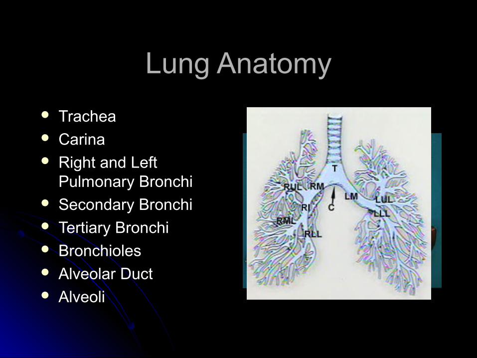
Lung AnatomyLung Anatomy
TracheaTrachea CarinaCarina Right and Left Right and Left
Pulmonary BronchiPulmonary Bronchi Secondary BronchiSecondary Bronchi Tertiary BronchiTertiary Bronchi BronchiolesBronchioles Alveolar DuctAlveolar Duct AlveoliAlveoli
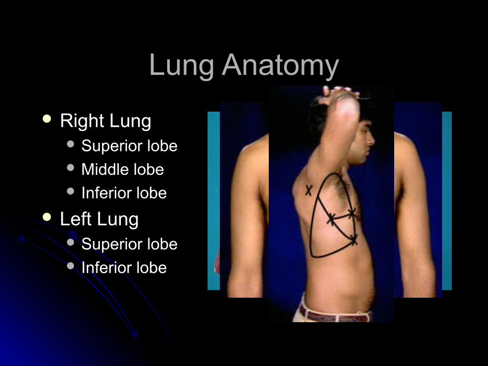
Lung AnatomyLung Anatomy
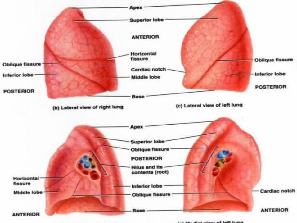
Right LungRight Lung Superior lobeSuperior lobe Middle lobeMiddle lobe Inferior lobeInferior lobe
Left LungLeft Lung Superior lobeSuperior lobe Inferior lobeInferior lobe
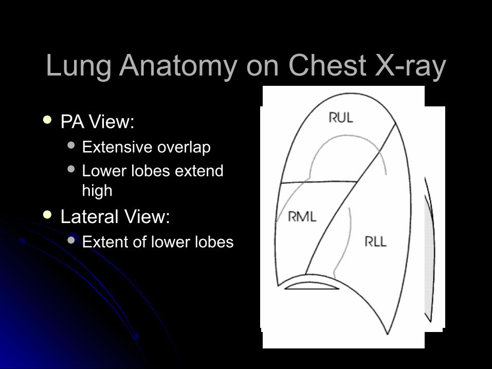
Lung Anatomy on Chest X-rayLung Anatomy on Chest X-ray
PA View:PA View: Extensive overlapExtensive overlap Lower lobes extend Lower lobes extend
highhigh
Lateral View:Lateral View: Extent of lower lobesExtent of lower lobes
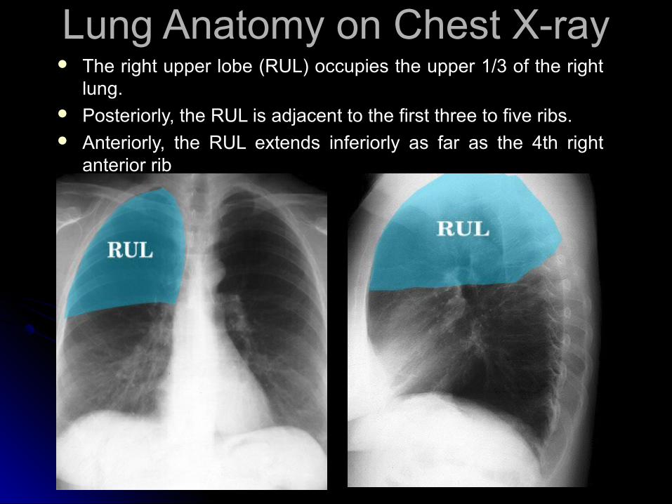
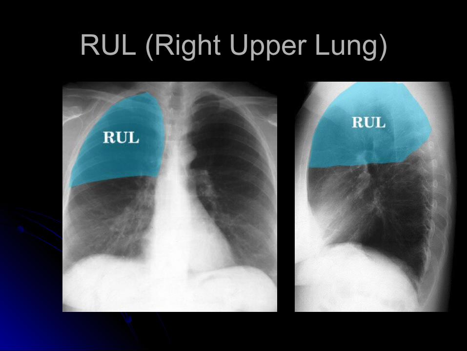
Lung Anatomy on Chest X-rayLung Anatomy on Chest X-ray The right upper lobe (RUL) occupies the upper 1/3 of the right The right upper lobe (RUL) occupies the upper 1/3 of the right
lung. lung. Posteriorly, the RUL is adjacent to the first three to five ribs. Posteriorly, the RUL is adjacent to the first three to five ribs. Anteriorly, the RUL extends inferiorly as far as the 4th right Anteriorly, the RUL extends inferiorly as far as the 4th right
anterior ribanterior rib
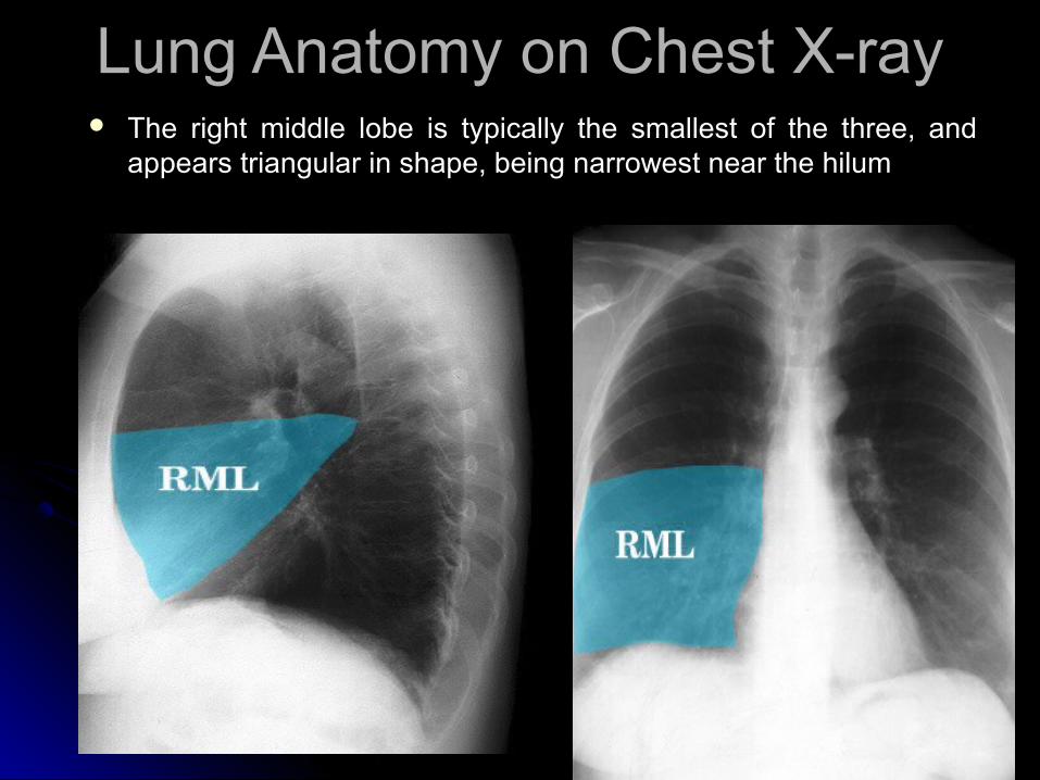
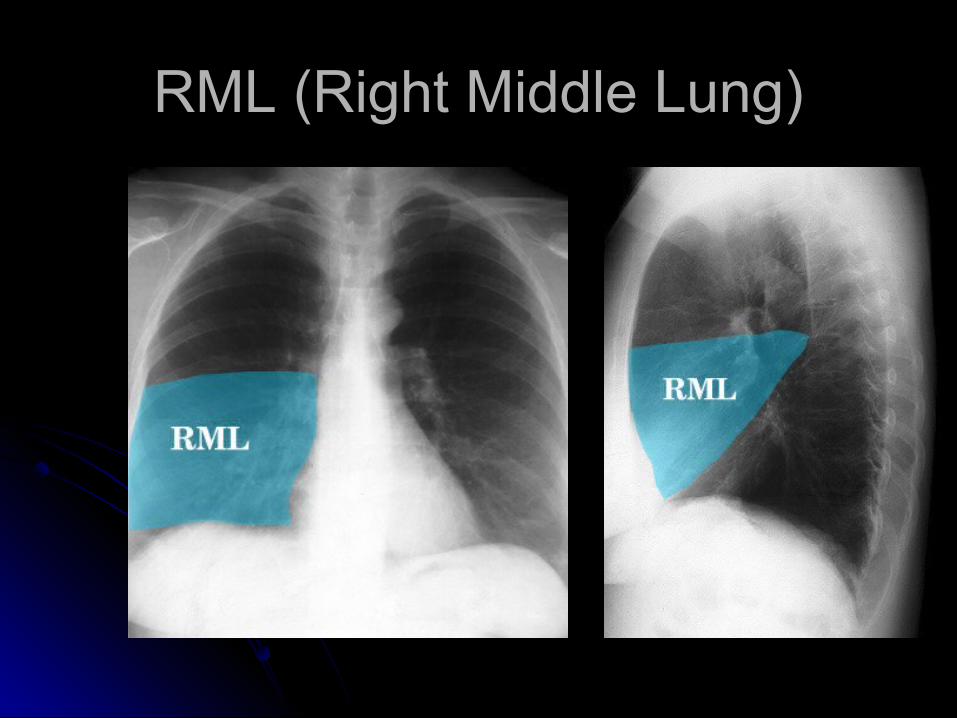
Lung Anatomy on Chest X-rayLung Anatomy on Chest X-ray The right middle lobe is typically the smallest of the three, and The right middle lobe is typically the smallest of the three, and
appears triangular in shape, being narrowest near the hilumappears triangular in shape, being narrowest near the hilum
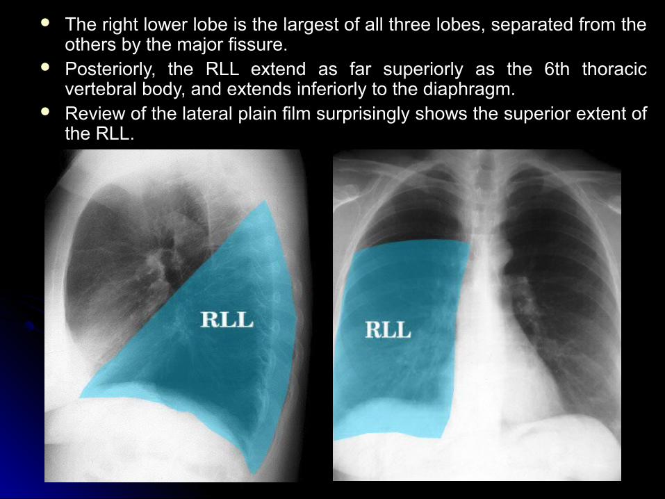
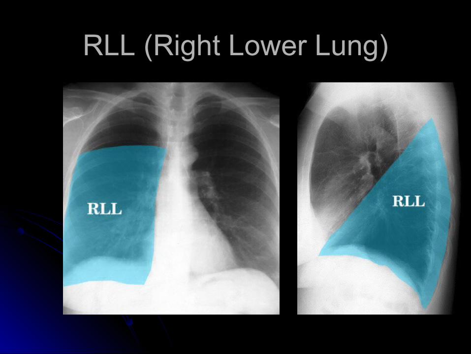
The right lower lobe is the largest of all three lobes, separated from the The right lower lobe is the largest of all three lobes, separated from the others by the major fissure.others by the major fissure.
Posteriorly, the RLL extend as far superiorly as the 6th thoracic Posteriorly, the RLL extend as far superiorly as the 6th thoracic vertebral body, and extends inferiorly to the diaphragm. vertebral body, and extends inferiorly to the diaphragm.
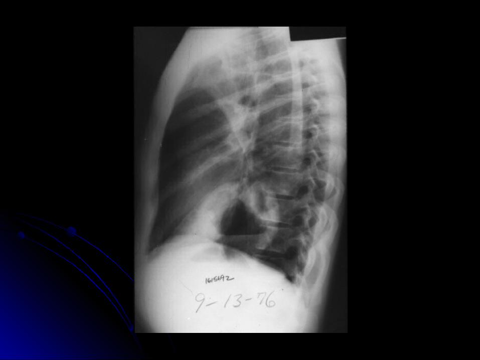
Review of the lateral plain film surprisingly shows the superior extent of Review of the lateral plain film surprisingly shows the superior extent of the RLL.the RLL.
Lung Anatomy on Chest X-rayLung Anatomy on Chest X-ray
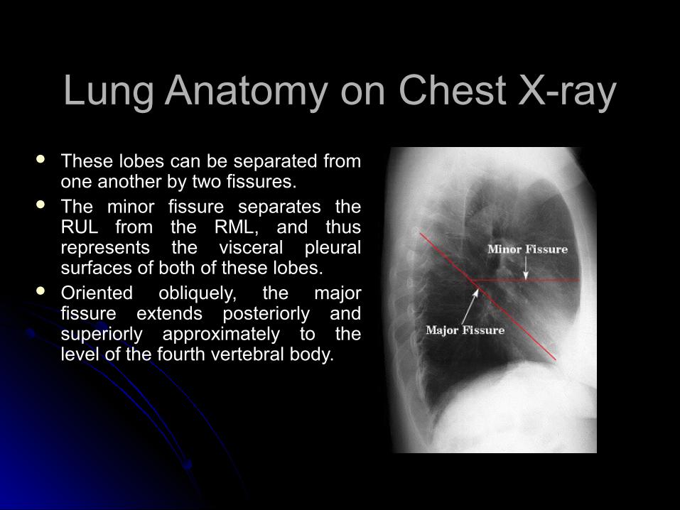
These lobes can be separated from These lobes can be separated from one another by two fissures.one another by two fissures.
The minor fissure separates the The minor fissure separates the RUL from the RML, and thus RUL from the RML, and thus represents the visceral pleural represents the visceral pleural surfaces of both of these lobes.surfaces of both of these lobes.
Oriented obliquely, the major Oriented obliquely, the major fissure extends posteriorly and fissure extends posteriorly and superiorly approximately to the superiorly approximately to the level of the fourth vertebral body. level of the fourth vertebral body.
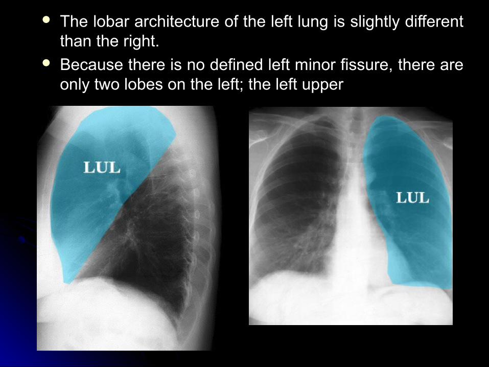
The lobar architecture of the left lung is slightly different The lobar architecture of the left lung is slightly different than the right.than the right.
Because there is no defined left minor fissure, there are Because there is no defined left minor fissure, there are only two lobes on the left; the left upper only two lobes on the left; the left upper
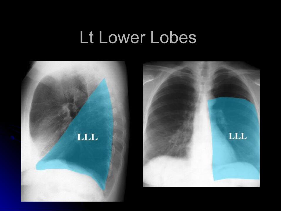
Lt Lower Lobes Lt Lower Lobes
Left lower lobesLeft lower lobes
Lung Anatomy on Chest X-rayLung Anatomy on Chest X-ray
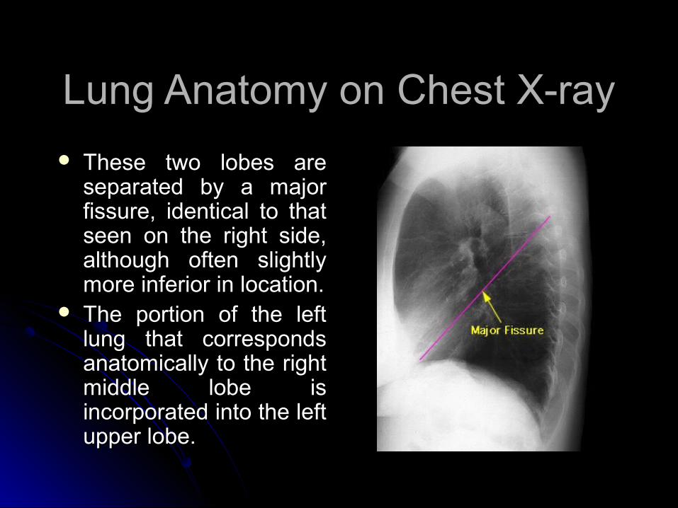
These two lobes are These two lobes are separated by a major separated by a major fissure, identical to that fissure, identical to that seen on the right side, seen on the right side, although often slightly although often slightly more inferior in location.more inferior in location.
The portion of the left The portion of the left lung that corresponds lung that corresponds anatomically to the right anatomically to the right middle lobe is middle lobe is incorporated into the left incorporated into the left upper lobe.upper lobe.
RUL (Right Upper Lung)RUL (Right Upper Lung)
RML (Right Middle Lung)RML (Right Middle Lung)
RLL (Right Lower Lung)RLL (Right Lower Lung)
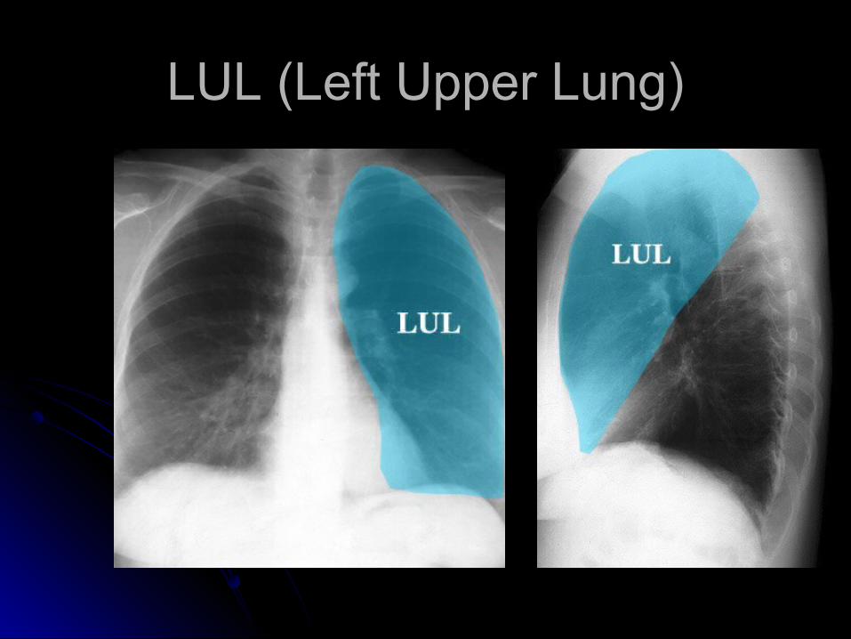
LUL (Left Upper Lung)LUL (Left Upper Lung)
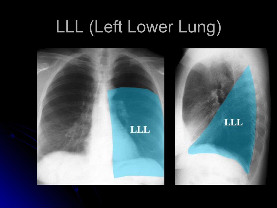
LLL (Left Lower Lung)LLL (Left Lower Lung)
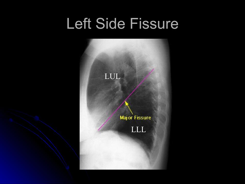
Left Side FissureLeft Side Fissure
LUL
LLL
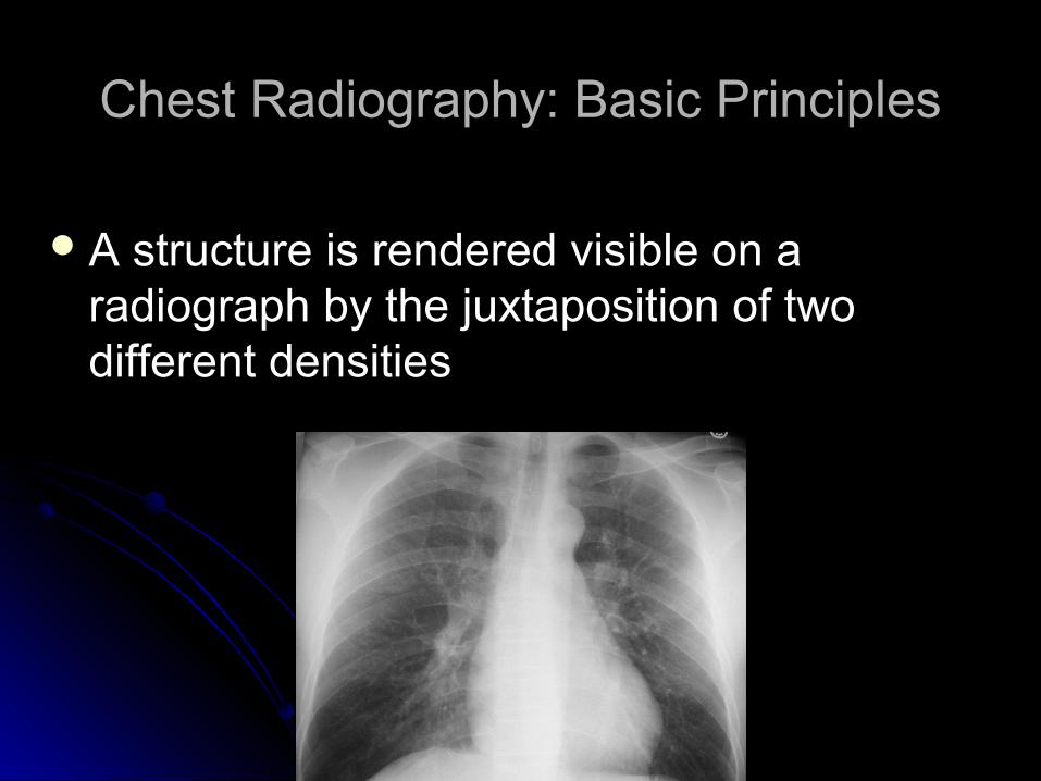
A structure is rendered visible on a A structure is rendered visible on a radiograph by the juxtaposition of two radiograph by the juxtaposition of two different densitiesdifferent densities
Chest Radiography: Basic PrinciplesChest Radiography: Basic Principles
Silhouette SignSilhouette Sign
Loss of the expected interface normally Loss of the expected interface normally created by juxtaposition of two structures created by juxtaposition of two structures of different densityof different density
No boundary can be seen between two No boundary can be seen between two structures of similar densitystructures of similar density
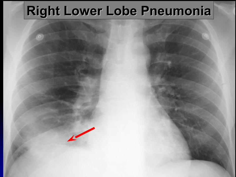
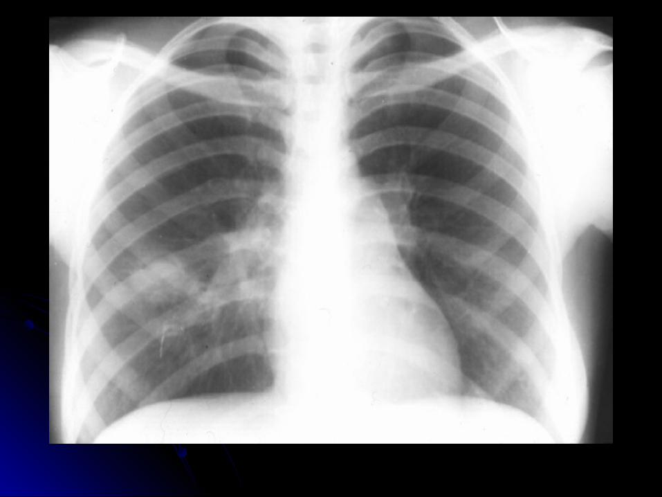
Right Lower Lobe PneumoniaRight Lower Lobe Pneumonia
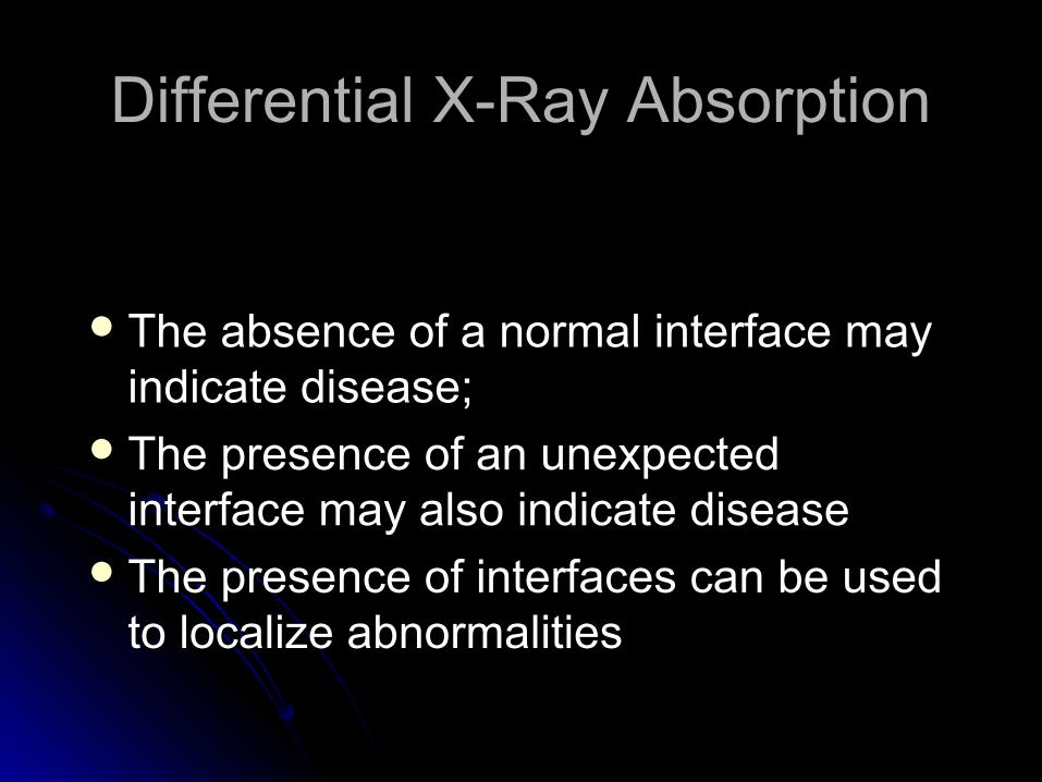
Differential X-Ray AbsorptionDifferential X-Ray Absorption
The absence of a normal interface may The absence of a normal interface may indicate disease; indicate disease;
The presence of an unexpected The presence of an unexpected interface may also indicate diseaseinterface may also indicate disease
The presence of interfaces can be used The presence of interfaces can be used to localize abnormalitiesto localize abnormalities
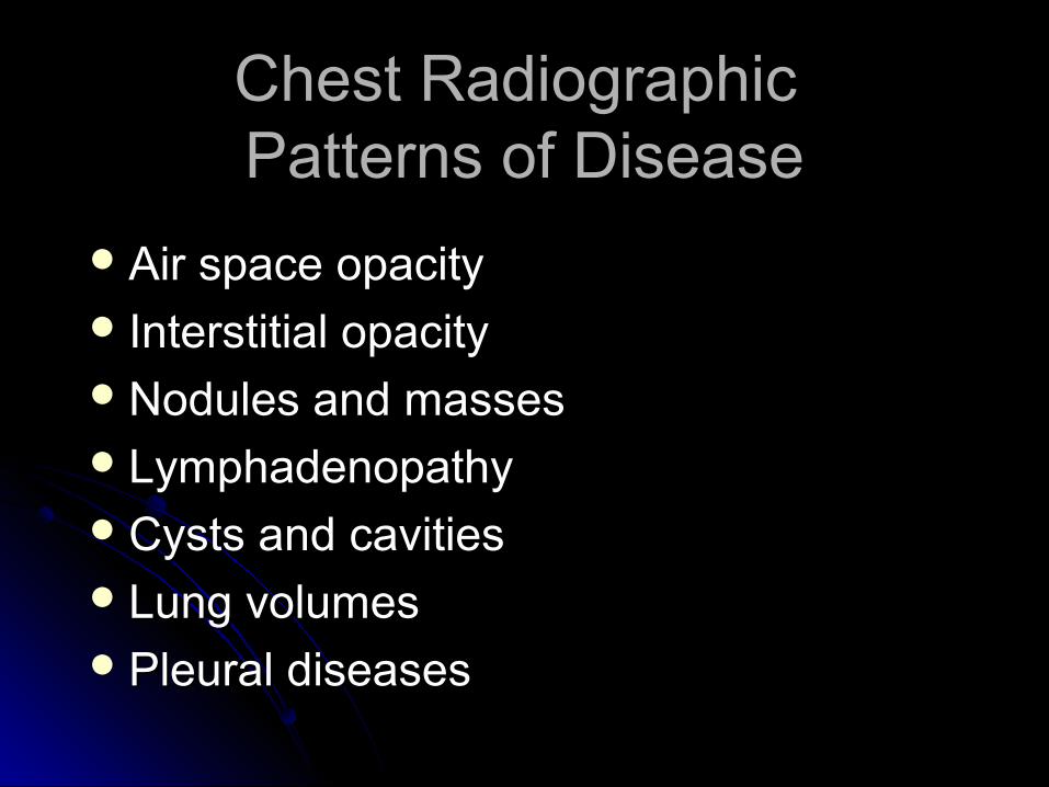
Chest Radiographic Chest Radiographic Patterns of DiseasePatterns of Disease
Air space opacityAir space opacity Interstitial opacityInterstitial opacityNodules and massesNodules and massesLymphadenopathyLymphadenopathyCysts and cavitiesCysts and cavitiesLung volumesLung volumesPleural diseasesPleural diseases
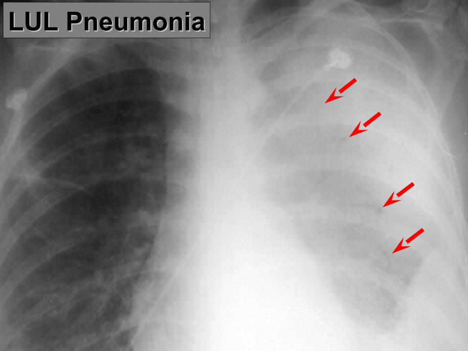
LUL PneumoniaLUL Pneumonia
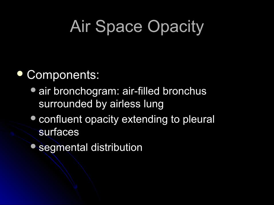
Air Space OpacityAir Space Opacity
Components:Components:air bronchogram: air-filled bronchus air bronchogram: air-filled bronchus
surrounded by airless lungsurrounded by airless lungconfluent opacity extending to pleural confluent opacity extending to pleural
surfacessurfacessegmental distributionsegmental distribution
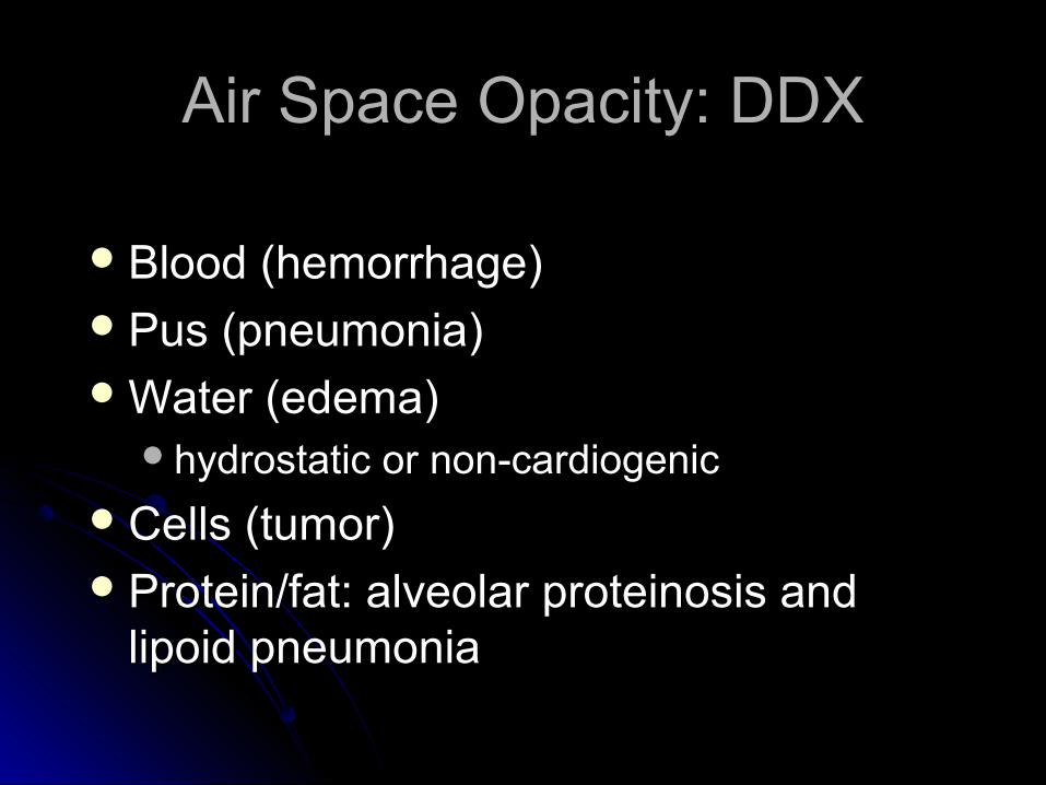
Air Space Opacity: DDXAir Space Opacity: DDX
Blood (hemorrhage)Blood (hemorrhage)Pus (pneumonia)Pus (pneumonia)Water (edema)Water (edema)
hydrostatic or non-cardiogenichydrostatic or non-cardiogenic
Cells (tumor)Cells (tumor)Protein/fat: alveolar proteinosis and Protein/fat: alveolar proteinosis and
lipoid pneumonia lipoid pneumonia
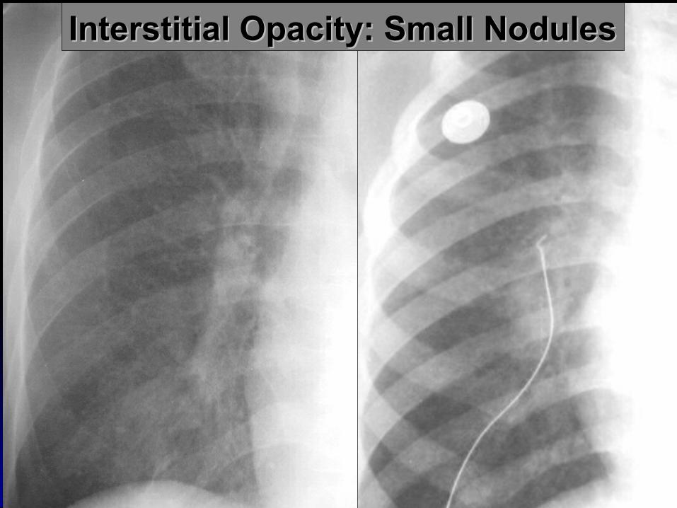
Interstitial Opacity: Small NodulesInterstitial Opacity: Small Nodules

Interstitial Opacity: Interstitial Opacity: LinesLines
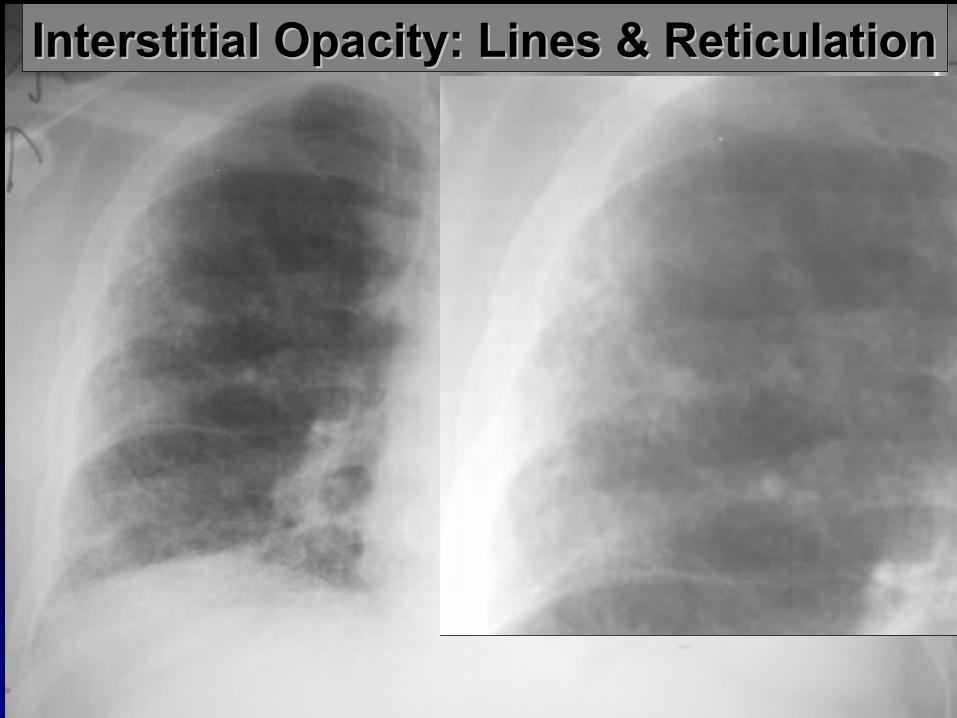
Interstitial Opacity: Lines & ReticulationInterstitial Opacity: Lines & Reticulation
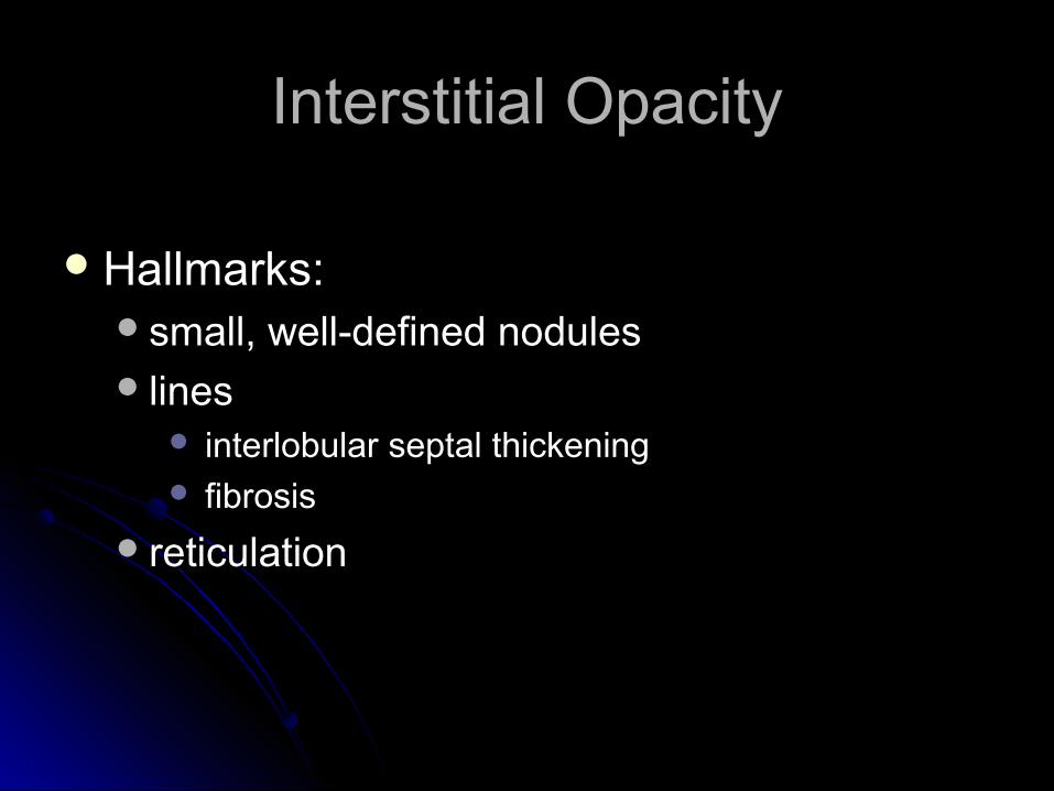
Interstitial OpacityInterstitial Opacity
Hallmarks:Hallmarks:small, well-defined nodulessmall, well-defined nodules lines lines
interlobular septal thickeninginterlobular septal thickening fibrosisfibrosis
reticulationreticulation
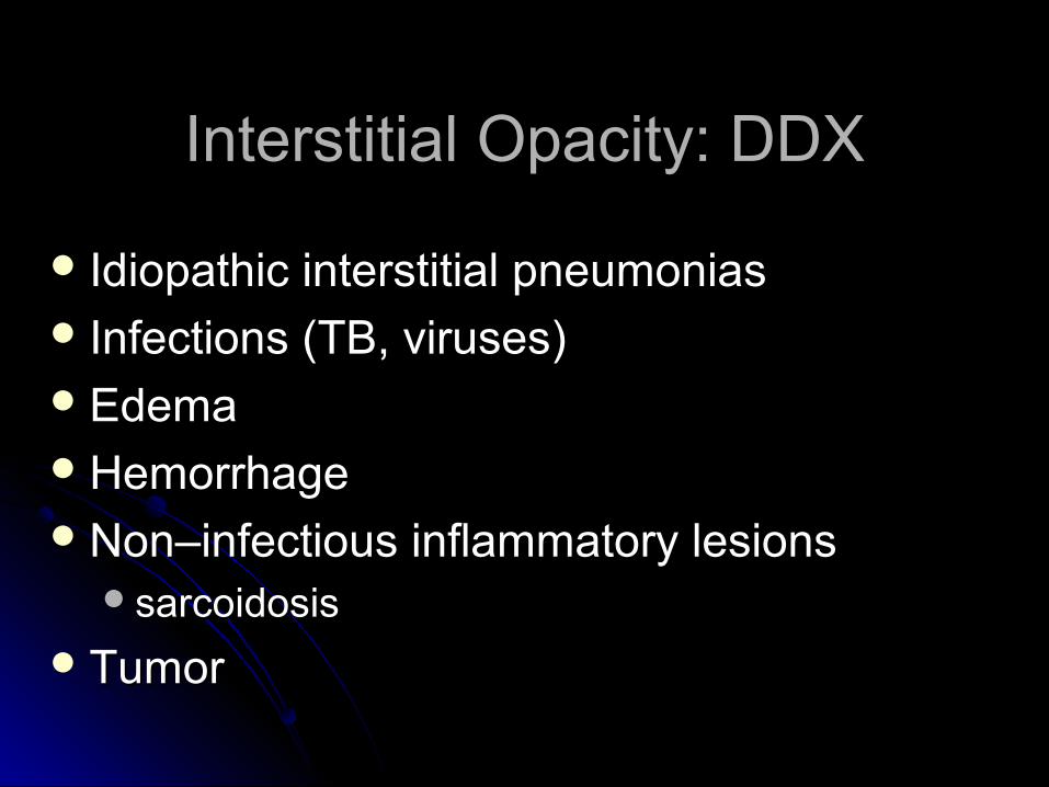
Interstitial Opacity: DDXInterstitial Opacity: DDX
Idiopathic interstitial pneumoniasIdiopathic interstitial pneumonias Infections (TB, viruses)Infections (TB, viruses)EdemaEdemaHemorrhageHemorrhageNon–infectious inflammatory lesionsNon–infectious inflammatory lesions
sarcoidosissarcoidosis
TumorTumor
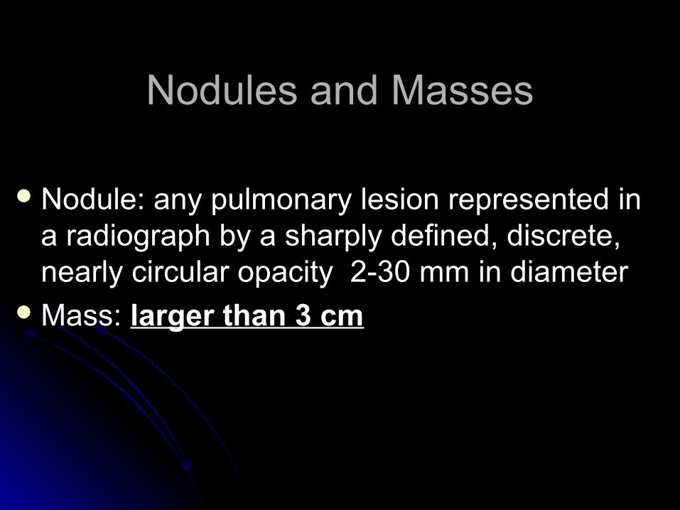
Nodules and MassesNodules and Masses
Nodule: any pulmonary lesion represented in Nodule: any pulmonary lesion represented in a radiograph by a sharply defined, discrete, a radiograph by a sharply defined, discrete, nearly circular opacity 2-30 mm in diameternearly circular opacity 2-30 mm in diameter
Mass: Mass: larger than 3 cmlarger than 3 cm
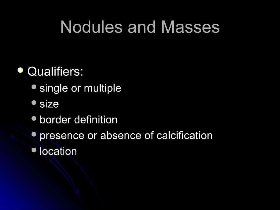
Nodules and MassesNodules and Masses
Qualifiers:Qualifiers:single or multiplesingle or multiplesizesizeborder definitionborder definitionpresence or absence of calcificationpresence or absence of calcification locationlocation
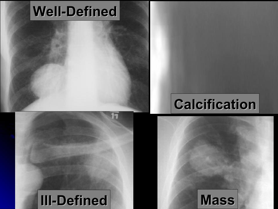
MassMass
CalcificationCalcification
Well-DefinedWell-Defined
Ill-DefinedIll-Defined
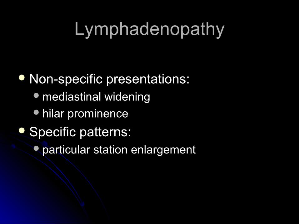
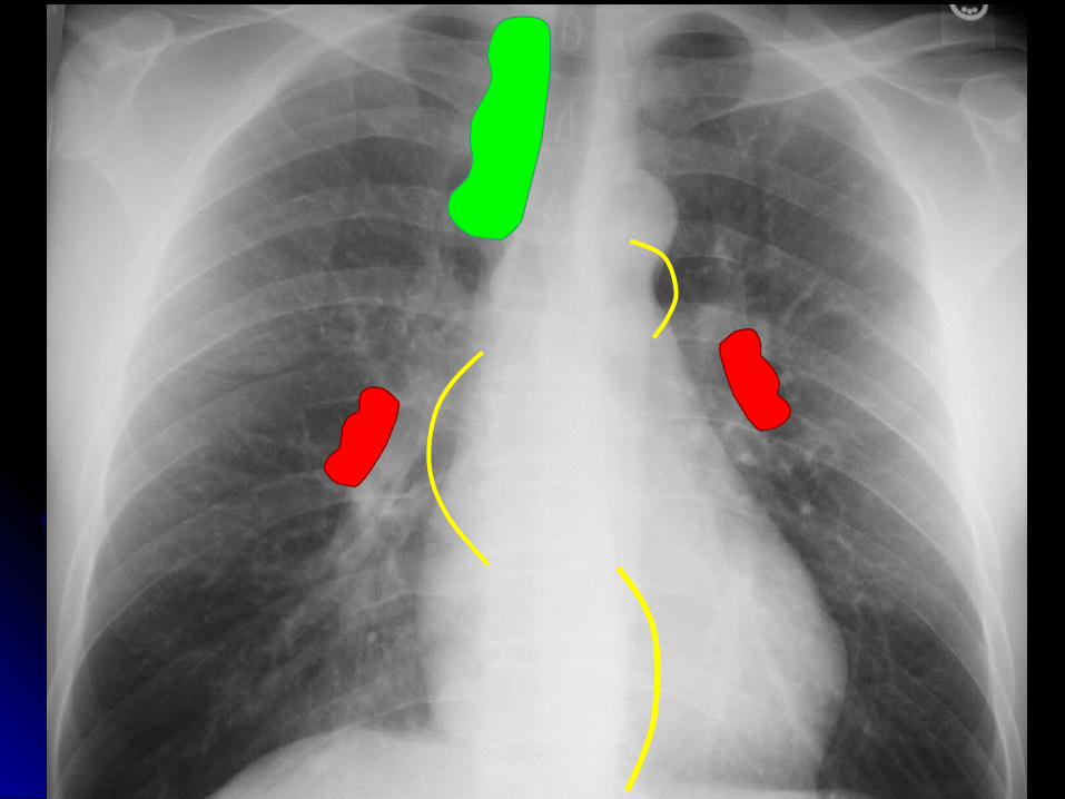
LymphadenopathyLymphadenopathy
Non-specific presentations:Non-specific presentations:mediastinal wideningmediastinal wideninghilar prominencehilar prominence
Specific patterns:Specific patterns:particular station enlargementparticular station enlargement
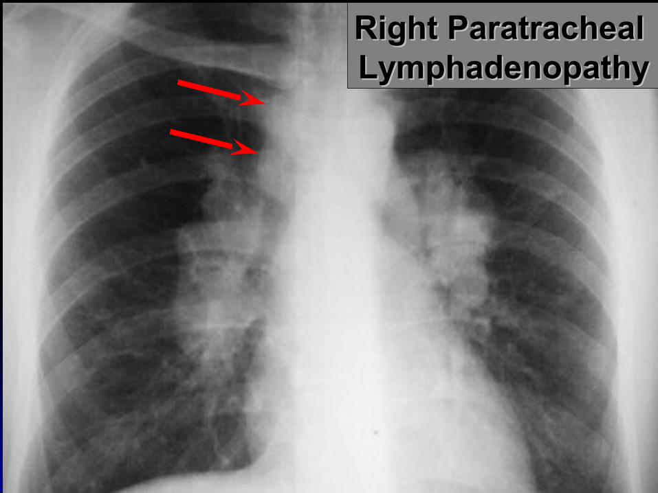
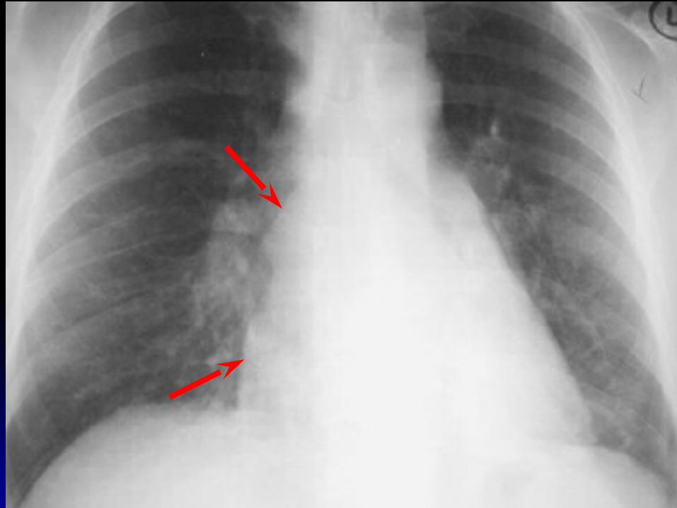
Right Paratracheal Right Paratracheal LymphadenopathyLymphadenopathy
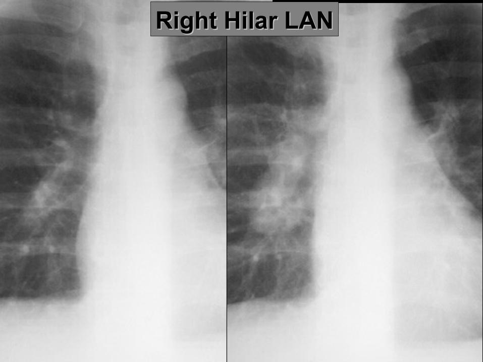
Right Hilar LANRight Hilar LAN
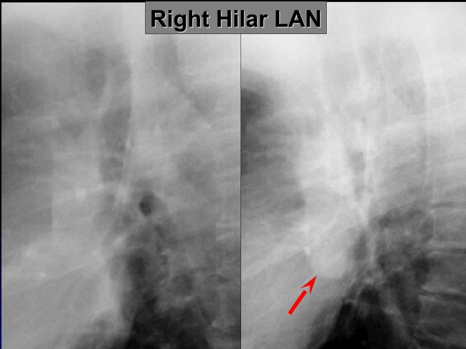
Right Hilar LANRight Hilar LAN

Left Hilar LANLeft Hilar LAN
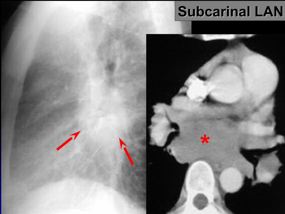
Subcarinal LANSubcarinal LAN
*
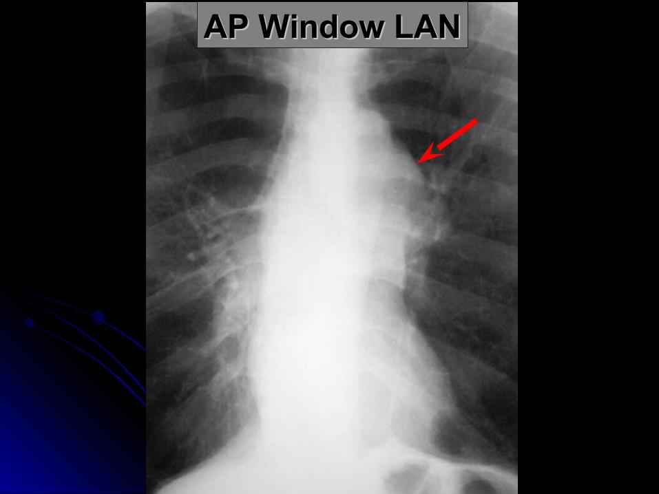
AP Window LANAP Window LAN
Cysts & CavitiesCysts & Cavities
CystCyst: abnormal pulmonary parenchymal : abnormal pulmonary parenchymal space, not containing lung but filled with air space, not containing lung but filled with air and/or fluid, congenital or acquired, with a and/or fluid, congenital or acquired, with a wall thickness greater than 1 mmwall thickness greater than 1 mm
epithelial lining often presentepithelial lining often present
Cysts & CavitiesCysts & Cavities
CavityCavity: Abnormal pulmonary : Abnormal pulmonary parenchymal space, not containing lung but parenchymal space, not containing lung but filled with air and/or fluid, caused by tissue filled with air and/or fluid, caused by tissue necrosis, with a definitive wall greater than necrosis, with a definitive wall greater than 1 mm in thickness and comprised of 1 mm in thickness and comprised of inflammatory and/or neoplastic elementsinflammatory and/or neoplastic elements
Cysts & CavitiesCysts & Cavities
Characterize:Characterize:wall thickness at thickest portionwall thickness at thickest portion inner lininginner liningpresence/absence of air/fluid levelpresence/absence of air/fluid levelnumber and locationnumber and location
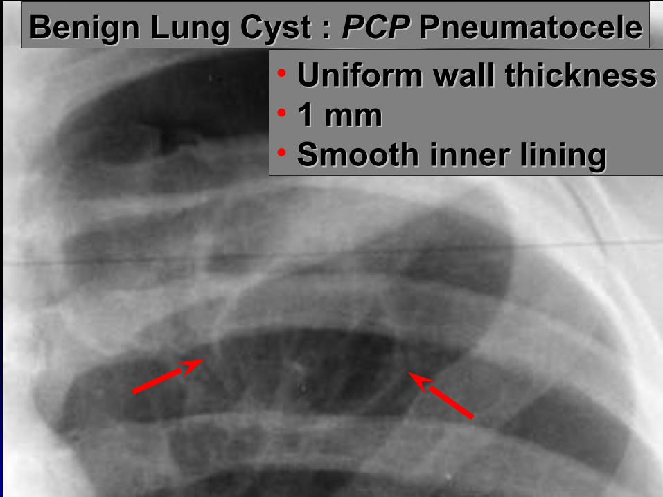
Benign Lung Cyst : Benign Lung Cyst : PCPPCP Pneumatocele Pneumatocele
• Uniform wall thicknessUniform wall thickness• 1 mm1 mm• Smooth inner liningSmooth inner lining
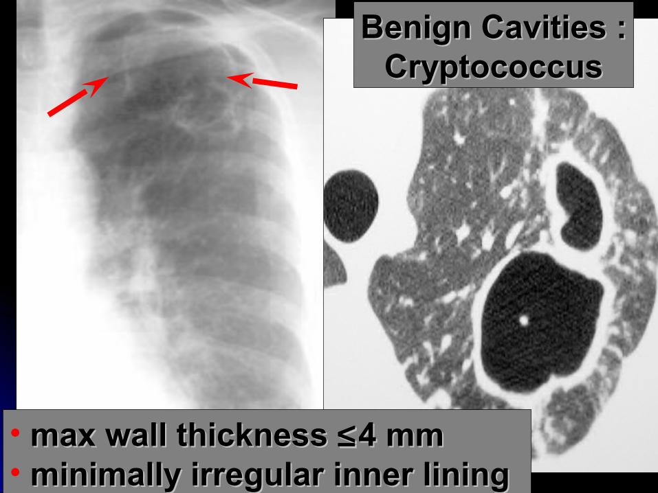
Benign Cavities :Benign Cavities :CryptococcusCryptococcus
• max wall thickness max wall thickness ≤≤4 mm4 mm• minimally irregular inner liningminimally irregular inner lining
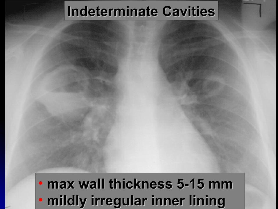
Indeterminate CavitiesIndeterminate Cavities
• max wall thickness 5-15 mmmax wall thickness 5-15 mm• mildly irregular inner liningmildly irregular inner lining
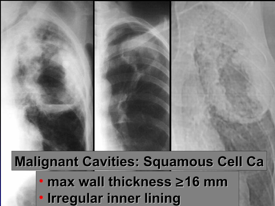
Malignant Cavities: Squamous Cell CaMalignant Cavities: Squamous Cell Ca
• max wall thickness max wall thickness ≥≥16 mm16 mm• Irregular inner liningIrregular inner lining
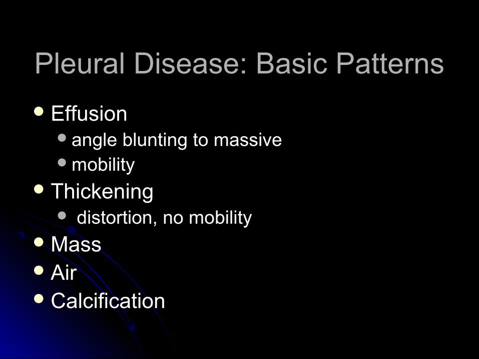
Pleural Disease: Basic PatternsPleural Disease: Basic Patterns
EffusionEffusionangle blunting to massiveangle blunting to massivemobilitymobility
ThickeningThickening distortion, no mobilitydistortion, no mobility
MassMassAirAirCalcificationCalcification
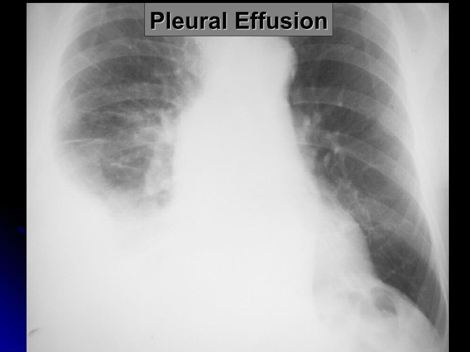
Pleural EffusionPleural Effusion
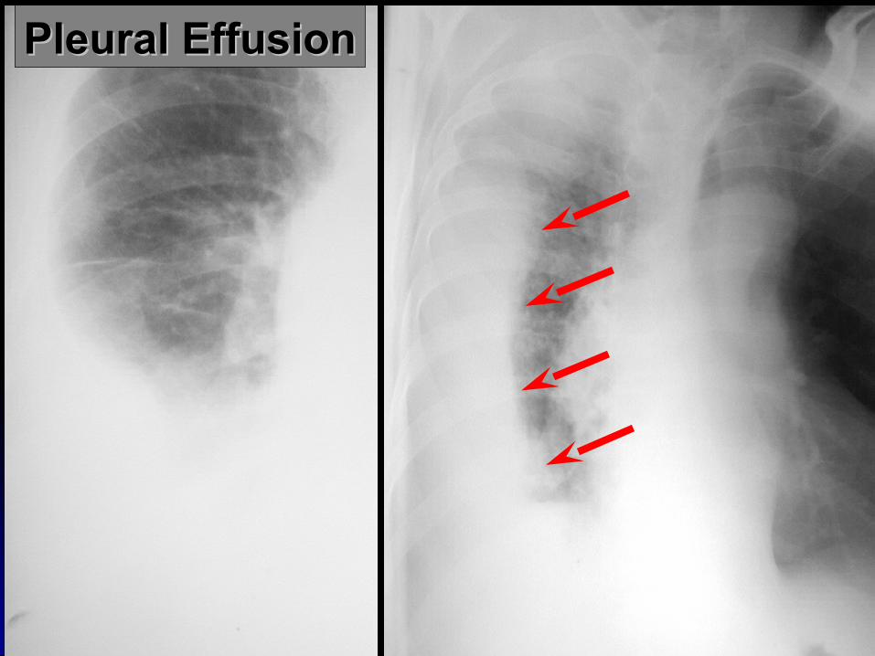
Pleural EffusionPleural Effusion
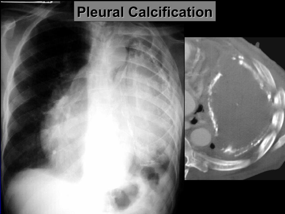
Pleural CalcificationPleural Calcification


SOME INTERESTING SOME INTERESTING X-RAYS & DISCUSSIO NX-RAYS & DISCUSSIO N
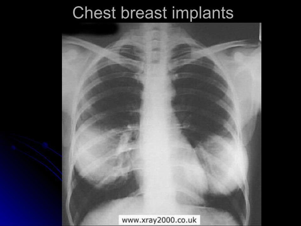
Chest breast implantsChest breast implants
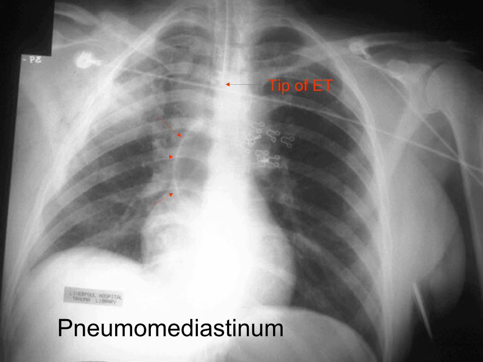
Tip of ET
Pneumomediastinum

wide wide mediastinummediastinum
obliteration of obliteration of aortic knobaortic knob
Rt mainstem Rt mainstem shift up and shift up and rightright
NG deviate NG deviate to rightto right
pleural cappleural cap
Major Vessel Injury
Potential X ray findings
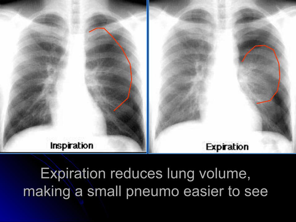
Expiration reduces lung volume, Expiration reduces lung volume, making a small pneumo easier to seemaking a small pneumo easier to see

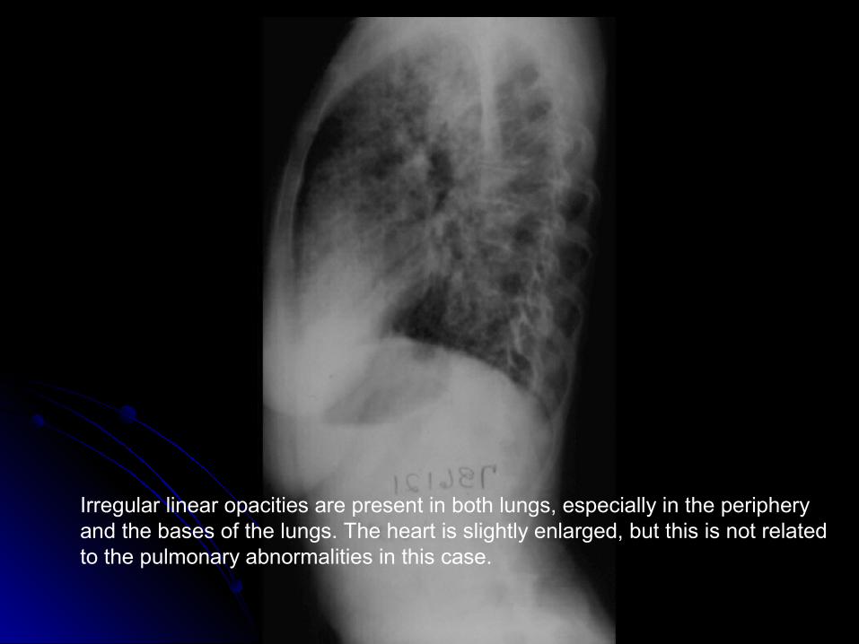
Irregular linear opacities are present in both lungs, especially in the periphery and the bases of the lungs. The heart is slightly enlarged, but this is not related to the pulmonary abnormalities in this case.
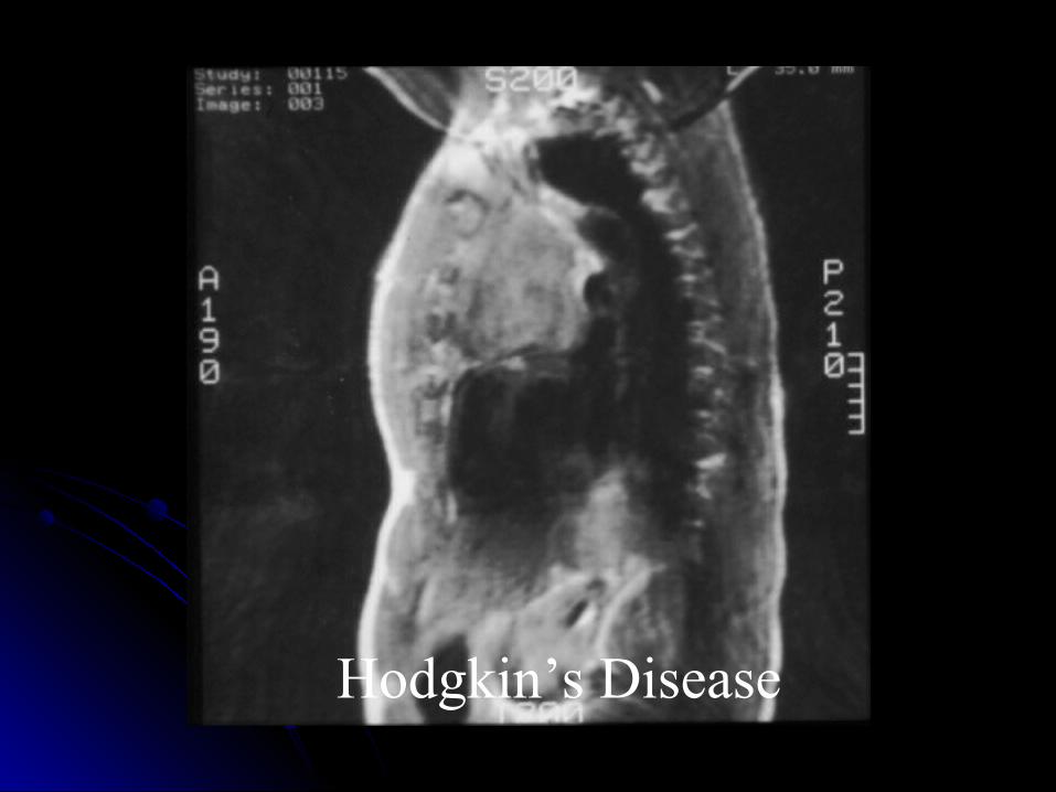
Hodgkin’s Disease
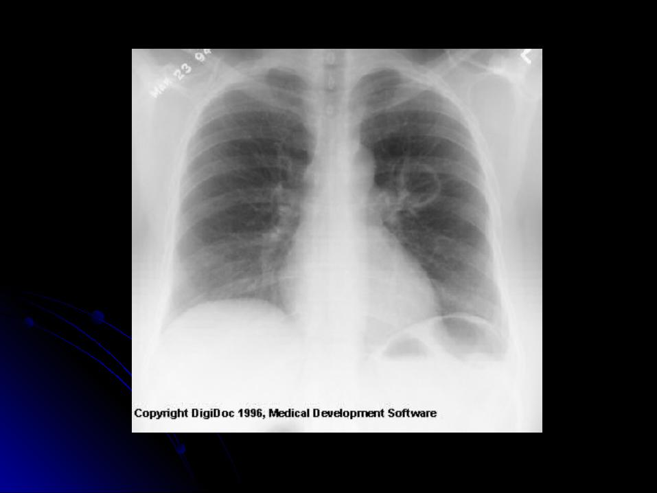
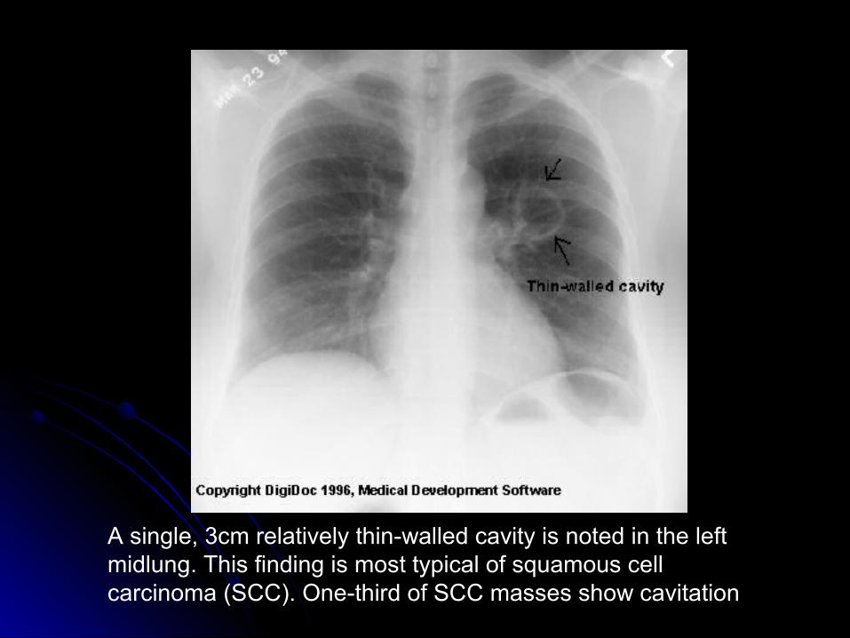
A single, 3cm relatively thin-walled cavity is noted in the left midlung. This finding is most typical of squamous cell carcinoma (SCC). One-third of SCC masses show cavitation
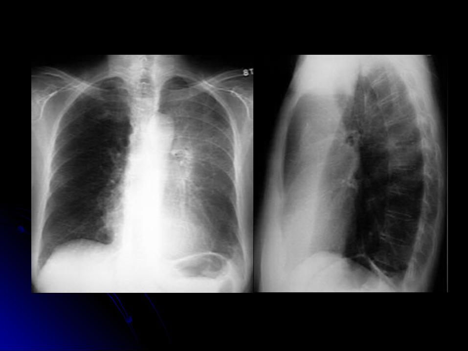
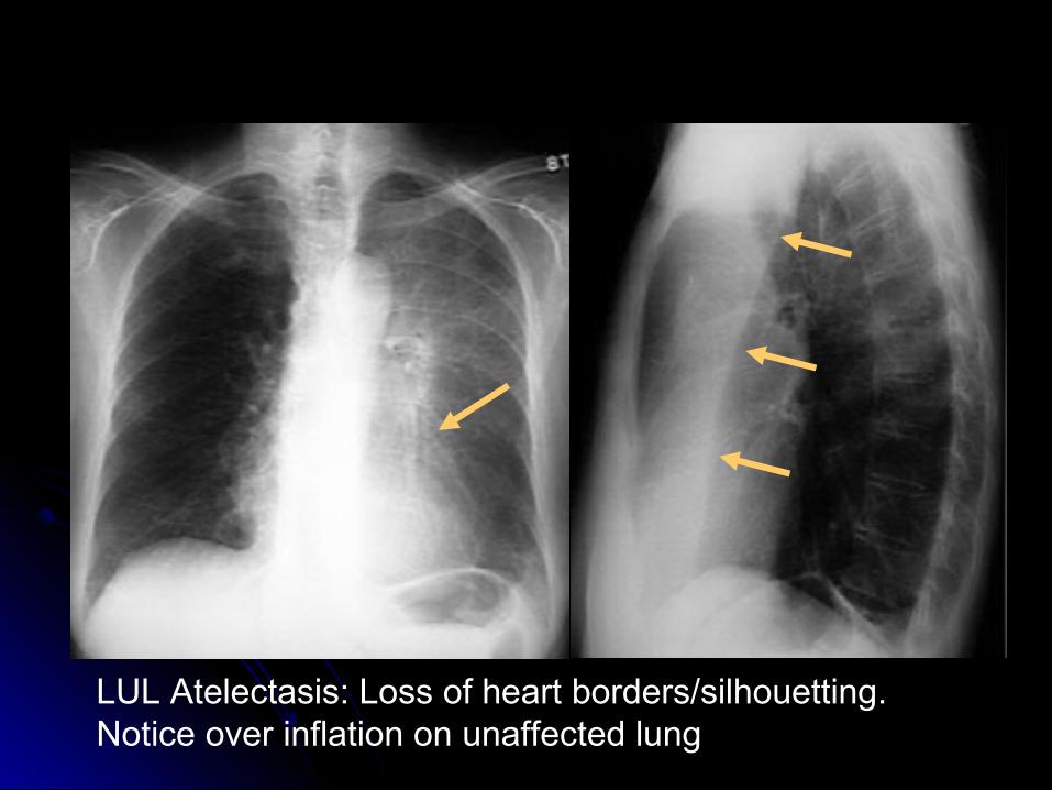
LUL Atelectasis: Loss of heart borders/silhouetting. Notice over inflation on unaffected lung
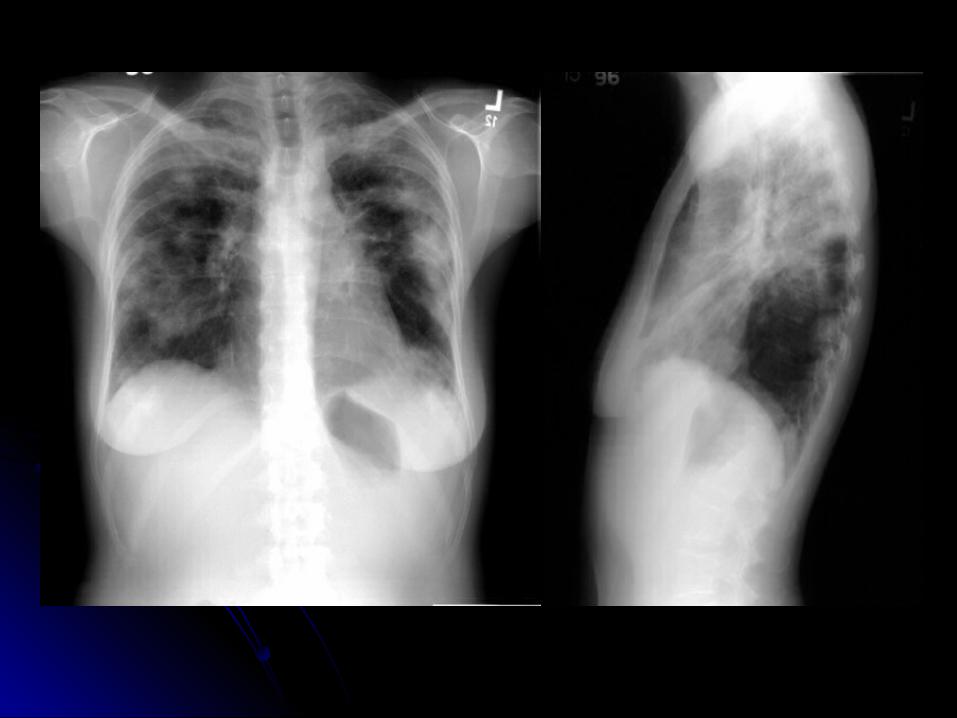
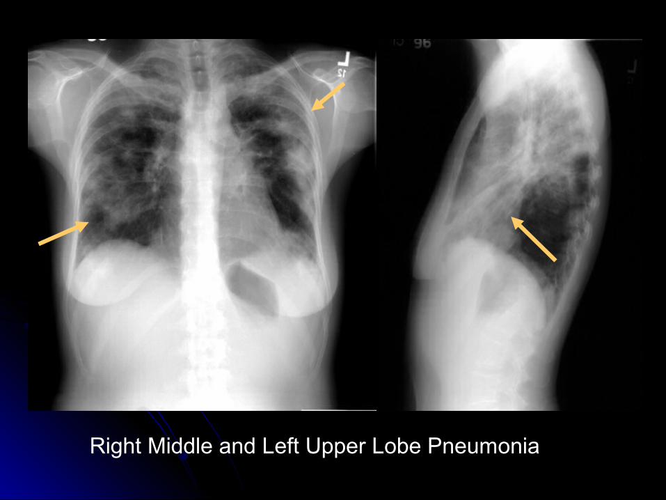
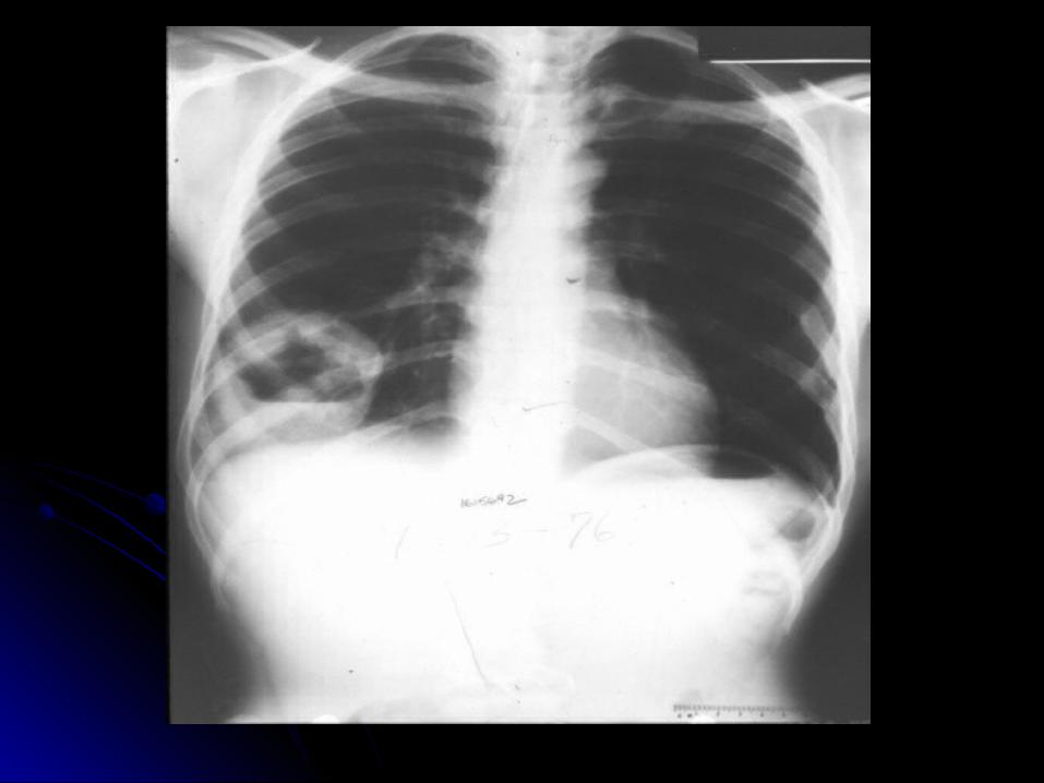
Right Middle and Left Upper Lobe Pneumonia
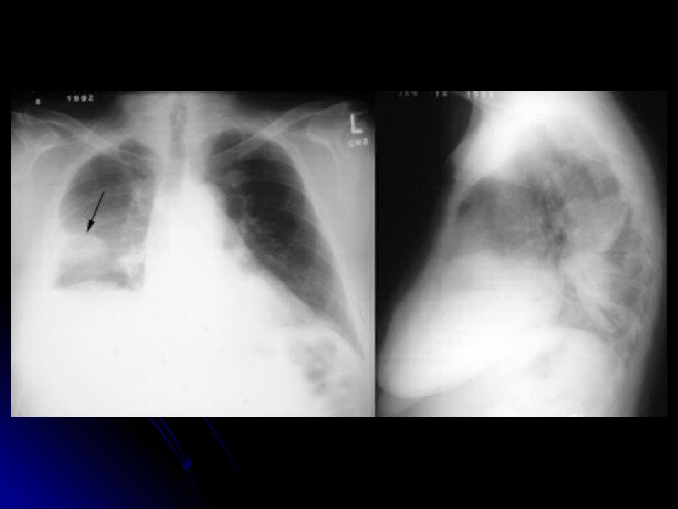
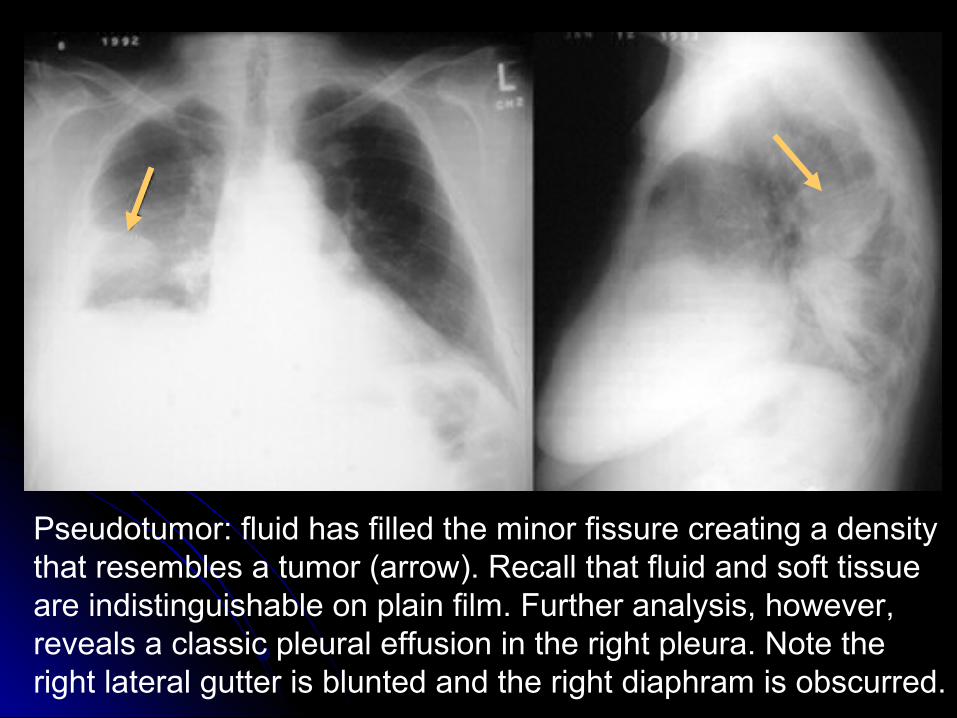
Pseudotumor: fluid has filled the minor fissure creating a density that resembles a tumor (arrow). Recall that fluid and soft tissue are indistinguishable on plain film. Further analysis, however, reveals a classic pleural effusion in the right pleura. Note the right lateral gutter is blunted and the right diaphram is obscurred.

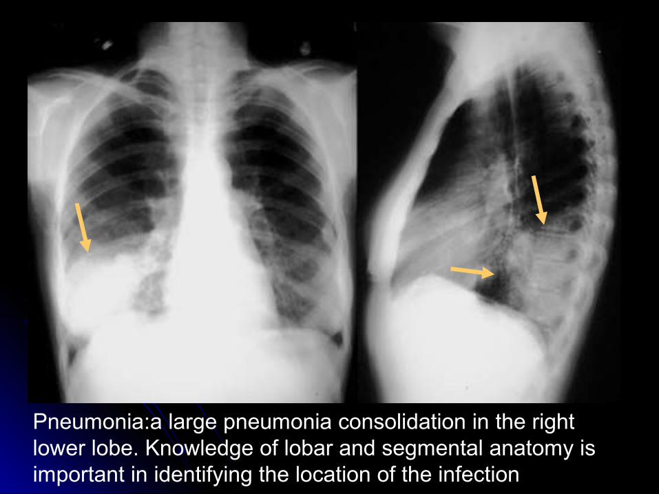
Pneumonia:a large pneumonia consolidation in the right lower lobe. Knowledge of lobar and segmental anatomy is important in identifying the location of the infection
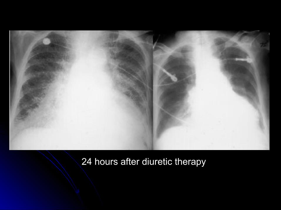
24 hours after diuretic therapy
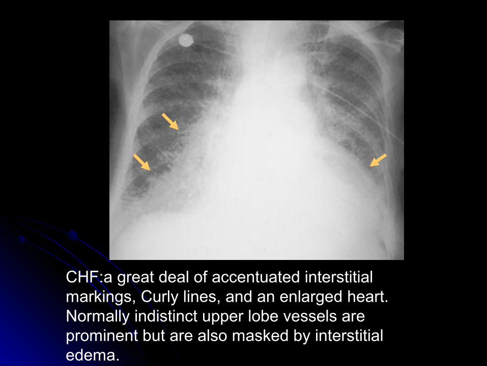
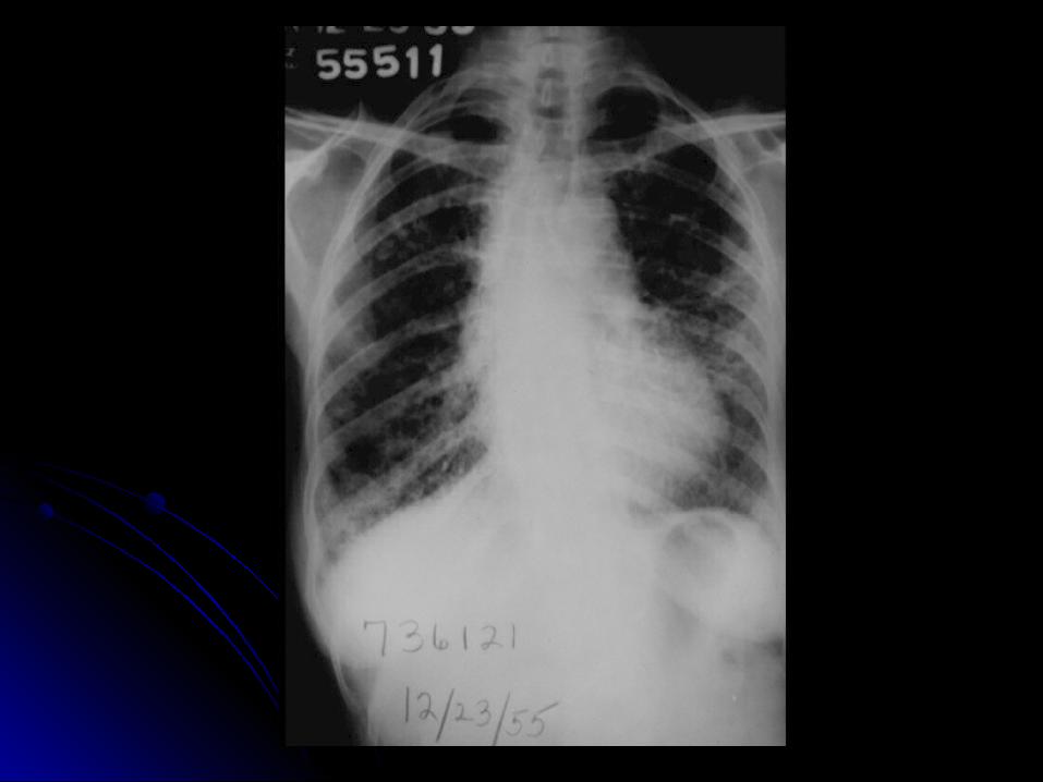
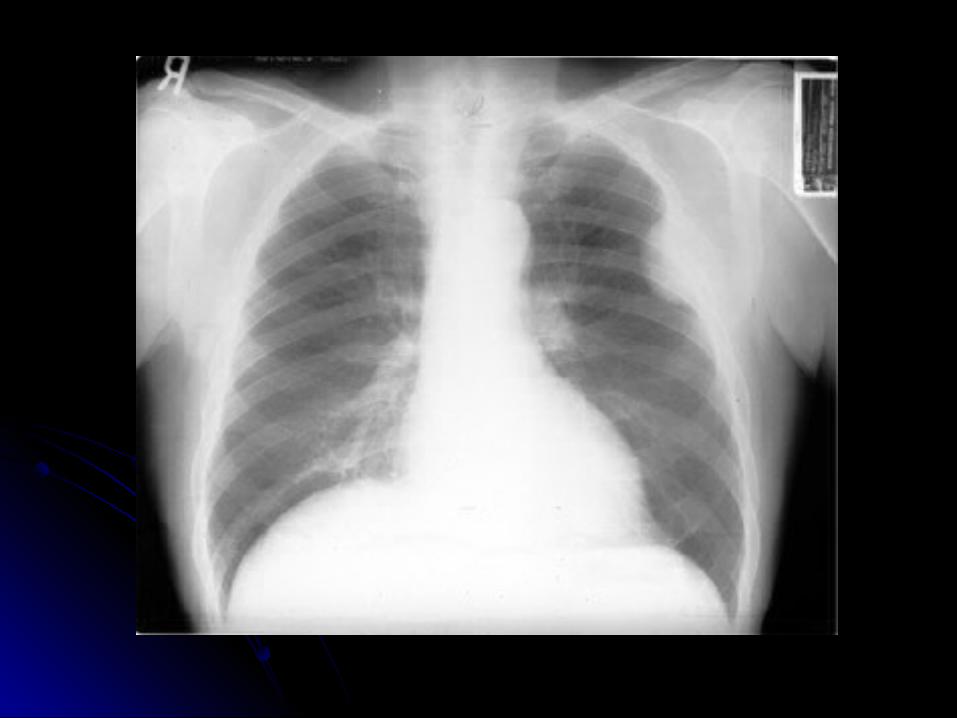
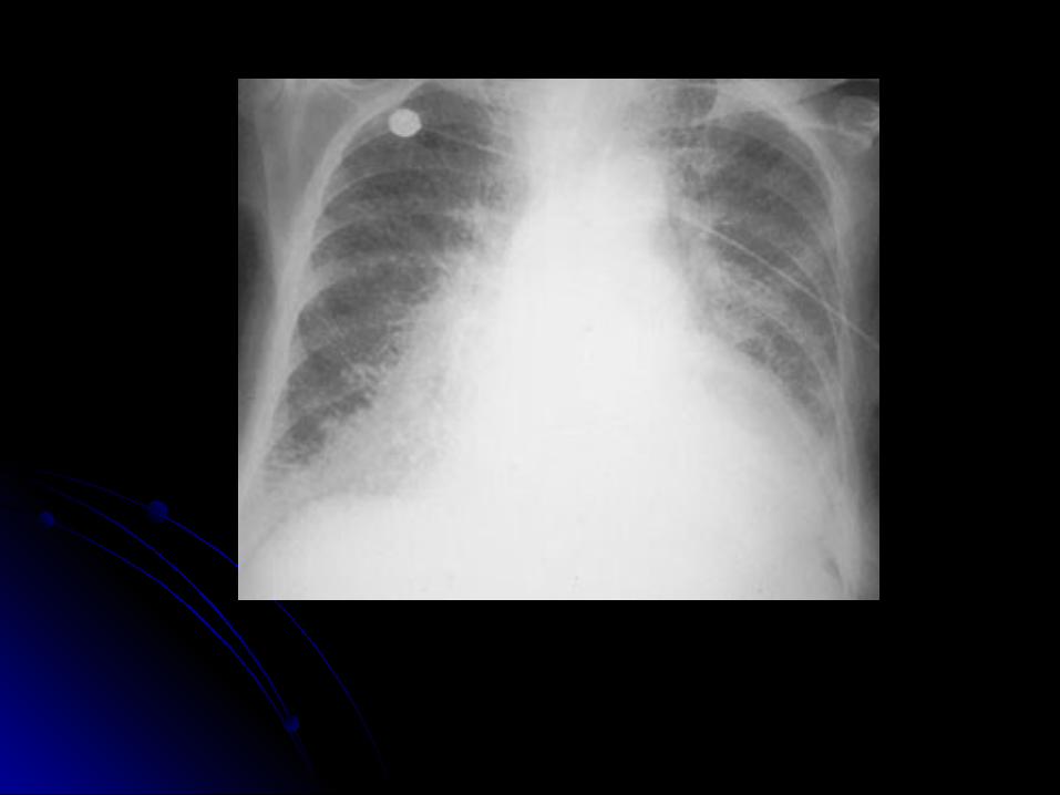
CHF:a great deal of accentuated interstitial markings, Curly lines, and an enlarged heart. Normally indistinct upper lobe vessels are prominent but are also masked by interstitial edema.