cyanobacterial, algal and fungal biofilm on sandstone...
TRANSCRIPT

33 (1): 101-105 (2009)
© 2009 Institute of Botany and Botanical Garden Jevremovac, Belgrade
UDK 582.232+582.26/.28](497.113)
Original Scientific Paper
Received: 30 April 2009 Revision accepted: 30 June 2009
✳correspondence: [email protected]
AbstrAct Biofilm on sandstone substrata of Eiffel´s Lock in Bečej contains complex consortia of algae, cyanobacteria and fungi. Filamentous cyanobacteria (Nostoc sp, Leptolyngbia sp., Stigonema ocellatum) and green algae (Desmococcus olivaceus and Haemaotococcus pluvialis) formed dense mucous layer with characteristic coloration of substrata. Melanized fungal structures (hyphae, chlamydospores and conidia) were intertwined with cyanobacteria and algae formed biofilm. Dominant fungal genera were Alternaria, Aureobasidium, Bipolaris, Cladosporium, Drechslera, Epicoccum, belongs to Dematiaceous hyphomycetes. Biofilm constituents’ interaction results in the bioweathering of the sandstone substrata through mechanical penetration, acid corrosion and production of secondary mycogenic biominerals. Modern concept of conservation of cultural and historic monuments implements multidisciplinary process and collaboration of art and science.
Key words: micromycetes, photosynthetic organisms, biofilm, biodeterioration, cultural heritage
cyanobacterial, algal and fungal biofilm on sandstone substrata of Eiffel´s Lock in bečej (serbia)
Milica Ljaljević Grbić1✳, Gordana Subakov-Simić1, Jelena Krizmanić1 and Veselin Lađić2
1 Institute of Botany and Garden Jevremovac, Faculty of Biology, University of Belgrade, Takovska 43, 11000 Belgrade, Serbia
2 Garnet, Beogradska 22, 11253 Belgrade, Serbia
IntroductIon
It is well known that living organisms will eventually colonize stone surfaces exposed to the environment, especially by algae and fungi. This colonization depends on many different conditions and on the characteristics and origin of the stone substrata. The importance of microbial biofilms in the deterioration and degradation of historic and modern building exteriors, a process commonly referred to as “weathering”, is well-recognized (Gaylarde et al. 1999, Gaylarde & Morton 2002).
In historic objects, all nutritional components have been subject to ageing, desiccation, oxidation and other chemical transformations in the course of the art object becoming with time a historic one. On such material,
microorganisms can get only the most basic nourishment and accidental moisture supply. That is why the microorganisms settling on historic objects are a selected group of oligotrophs able to develop there. The descending strains are already adapted to living on a given group of objects, and destroying them (Strzelezyk 2004).
Biofilms of different stone materials contains complex consortia of algae, cyanobacteria, heterotrophic bacteria, fungi, lichens, protozoa and a variety of small animals, mosses and plants. The growth of phototrophic microorganism, algae and cyanobacteria, on the external surfaces of stones can cause discoloration and physico-chemical deterioration. Cyanobacteria like oxygenic, phototrophic bacteria that can colonies stone in monuments and produce aesthetic changes due to stains, colored
102 33 (1)
biofilms and incrustations. Their tolerance to desiccation, water stress and varying light intensities helps to explain their frequent occurrence on stone surfaces. Fungi, algae and cyanobacteria are particularly adapted to survive UV exposure and repeated drying and rehydration (Gaylard et al. 2005, Potts 1994, Yancey et al. 1982) and might be expected to be the major groups in biofilms on exposed surfaces. Many of cyanobacteria, actinomycetes and fungi produce pigments, leading to the typical discoloration seen on poorly maintained buildings, in addition to their degradation (Ortega-Calvo et al. 1991, Ortega-Morales et al. 2000). The waste products of algae and bacteria (or dead cells of these organisms), decaying leaves, and bird droppings can provide such food sources for fungi (Kumar & Kumar 1999). High temperatures, low humidity, high incidence of UV light and low nutrients require fungi that have evolved special survival skills. Fungi contain melanin, microsporines, carotenoids and probably other pigments that help protect them from UV light. Fungi interact with natural rock systems, soil and buildings – carbonates (limestone, dolomite, marble), sandstones, gypsum and granites – under a broad variety of environmental conditions. This interaction results in the bio-weathering of the mineral substrates, in the formation of secondary bio-minerals on the attacked substrates.
MAthErIAL And MEthods
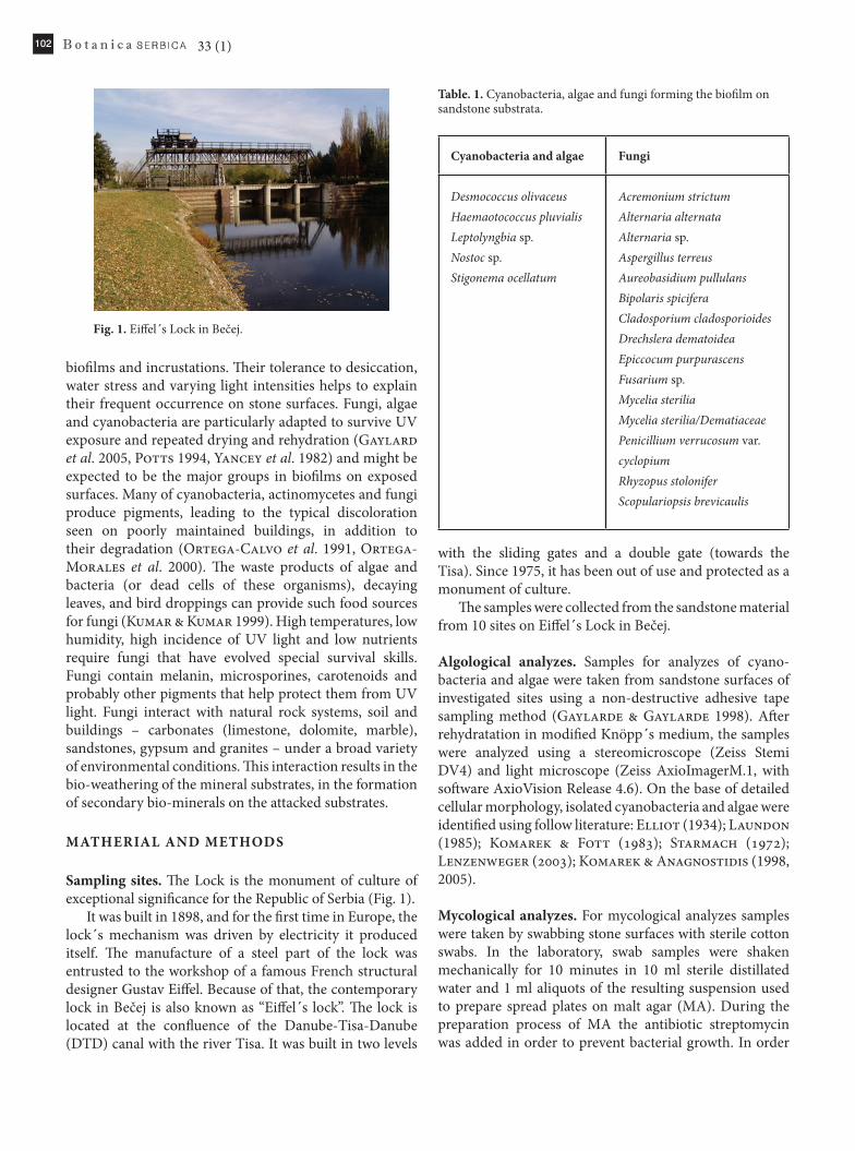
sampling sites. The Lock is the monument of culture of exceptional significance for the Republic of Serbia (Fig. 1).
It was built in 1898, and for the first time in Europe, the lock´s mechanism was driven by electricity it produced itself. The manufacture of a steel part of the lock was entrusted to the workshop of a famous French structural designer Gustav Eiffel. Because of that, the contemporary lock in Bečej is also known as “Eiffel´s lock”. The lock is located at the confluence of the Danube-Tisa-Danube (DTD) canal with the river Tisa. It was built in two levels
with the sliding gates and a double gate (towards the Tisa). Since 1975, it has been out of use and protected as a monument of culture.
The samples were collected from the sandstone material from 10 sites on Eiffel´s Lock in Bečej.
Algological analyzes. Samples for analyzes of cyano-bacteria and algae were taken from sandstone surfaces of investigated sites using a non-destructive adhesive tape sampling method (Gaylarde & Gaylarde 1998). After rehydratation in modified Knöpp´s medium, the samples were analyzed using a stereomicroscope (Zeiss Stemi DV4) and light microscope (Zeiss AxioImagerM.1, with software AxioVision Release 4.6). On the base of detailed cellular morphology, isolated cyanobacteria and algae were identified using follow literature: Elliot (1934); Laundon (1985); Komarek & Fott (1983); Starmach (1972); Lenzenweger (2003); Komarek & Anagnostidis (1998, 2005).
Mycological analyzes. For mycological analyzes samples were taken by swabbing stone surfaces with sterile cotton swabs. In the laboratory, swab samples were shaken mechanically for 10 minutes in 10 ml sterile distillated water and 1 ml aliquots of the resulting suspension used to prepare spread plates on malt agar (MA). During the preparation process of MA the antibiotic streptomycin was added in order to prevent bacterial growth. In order
Fig. 1. Eiffel´s Lock in Bečej.
table. 1. Cyanobacteria, algae and fungi forming the biofilm on sandstone substrata.
cyanobacteria and algae Fungi
Desmococcus olivaceusHaemaotococcus pluvialisLeptolyngbia sp.Nostoc sp.Stigonema ocellatum
Acremonium strictumAlternaria alternataAlternaria sp.Aspergillus terreusAureobasidium pullulansBipolaris spiciferaCladosporium cladosporioidesDrechslera dematoideaEpiccocum purpurascensFusarium sp.Mycelia steriliaMycelia sterilia/DematiaceaePenicillium verrucosum var. cyclopiumRhyzopus stoloniferScopulariopsis brevicaulis
103Milica Ljaljević Grbić et al.: Cyanobacterial, algal and fungal biofilm on sandstone substrata of Eiffel´s Lock in Bečej (Serbia)
to isolate as wide range of micromycetes as possible the samples were plated in triplicate. The plates were incubated on 22 ± 2º C and read after 72 to 120 hours. Fungal colonies formed on the medium were identified based on the both macroscopic and microscopic characteristics of each isolated colony using identification keys (Ainsworth et al. 1973; Ellis & Ellis 1997; Pitt 1979; Rapper & Fennel 1965). Slides were analyzed using a light microscope (Zeiss AxioImagerM.1, with software AxioVision Release 4.6).
For both algological and mycological analyzes slides were mounted in glycerol and stained in Lactophenol Cotton Blue.
rEsuLts And dIscussIon
Biofilm-forming organisms were identified to the species or genus level, based on macro and micromorphological characteristics.
Photosynthetic organisms detected. Analyses have shown that cyanobacterial and algal colonization occurred on more sites of monument. Biofilms containing photosynthetic organisms were consisting of five taxa of cyanobacteria and green algae (Tab. 1).
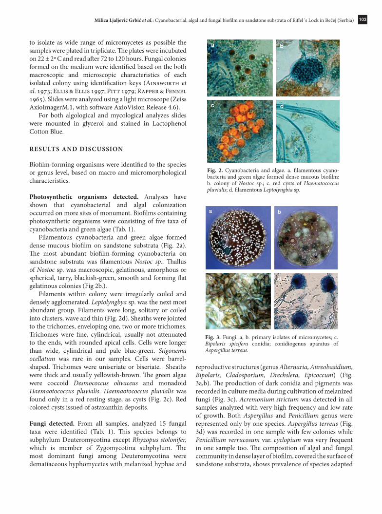
Filamentous cyanobacteria and green algae formed dense mucous biofilm on sandstone substrata (Fig. 2a). The most abundant biofilm-forming cyanobacteria on sandstone substrata was filamentous Nostoc sp.. Thallus of Nostoc sp. was macroscopic, gelatinous, amorphous or spherical, tarry, blackish-green, smooth and forming flat gelatinous colonies (Fig 2b.).
Filaments within colony were irregularly coiled and densely agglomerated. Leptolyngbya sp. was the next most abundant group. Filaments were long, solitary or coiled into clusters, wave and thin (Fig. 2d). Sheaths were jointed to the trichomes, enveloping one, two or more trichomes. Trichomes were fine, cylindrical, usually not attenuated to the ends, with rounded apical cells. Cells were longer than wide, cylindrical and pale blue-green. Stigonema ocellatum was rare in our samples. Cells were barrel-shaped. Trichomes were uniseriate or biseriate. Sheaths were thick and usually yellowish-brown. The green algae were coccoid Desmococcus olivaceus and monadoid Haemaotococcus pluvialis. Haemaotococcus pluvialis was found only in a red resting stage, as cysts (Fig. 2c). Red colored cysts issued of astaxanthin deposits.
Fungi detected. From all samples, analyzed 15 fungal taxa were identified (Tab. 1). This species belongs to subphylum Deuteromycotina except Rhyzopus stolonifer, which is member of Zygomycotina subphylum. The most dominant fungi among Deuteromycotina were dematiaceous hyphomycetes with melanized hyphae and
reproductive structures (genus Alternaria, Aureobasidium, Bipolaris, Cladosporium, Drechslera, Epicoccum) (Fig. 3a,b). The production of dark conidia and pigments was recorded in culture media during cultivation of melanized fungi (Fig. 3c). Acremonium strictum was detected in all samples analyzed with very high frequency and low rate of growth. Both Aspergillus and Penicillium genus were represented only by one species. Aspergillus terreus (Fig. 3d) was recorded in one sample with few colonies while Penicillium verrucosum var. cyclopium was very frequent in one sample too. The composition of algal and fungal community in dense layer of biofilm, covered the surface of sandstone substrata, shows prevalence of species adapted
Fig. 2. Cyanobacteria and algae. a. filamentous cyano-bacteria and green algae formed dense mucous biofilm; b. colony of Nostoc sp.; c. red cysts of Haematococcus pluvialis; d. filamentous Leptolyngbia sp.
Fig. 3. Fungi. a, b. primary isolates of micromycetes; c. Bipolaris spicifera conidia; conidiogenus aparatus of Aspergillus terreus.

104 33 (1)
on desiccation and UV exposure. Carbonate sandstone investigated is more receptive for fungal and algal colonization because of high porosity and rough surface in contrast of stone with compact structure (Florian 2004). The cyanobacteria are generally better adapted to withstand such adverse conditions, producing protective pigments within their cells or sheaths (Gaylarde & Gaylarde 2000). Red cysts are more resistant to photoinhibition than green cysts, strongly indicating a photoprotective role for astaxanthin. Other studies support the major role of astaxanthin accumulation in Haematococcus as being a form of protection against high light and oxygen radicals (Kobayashi 1992). It is known that Haematococcus pluvialis can cause red discoloration of marble (May 2003).
The microfungi identified, especially melanized hyphomycetes from genus Alternaria, Cladosporium, Drechslera, Epicoccum, causes discoloration, as well as mechanical exfoliation of building stone material analyzed through mechanical hyphae penetration and production of different pigments and organic acids (Milanesi et al. 2005). Alcaliphylous species Scopulariopsis brevicaulis and Acremonium strictum are degradable for carbonate stone material. Bipolaris spicifera is Dematiaceous hyphomycetes known as human pathogenic species (Buzina et al. 2003). Microbial biofilms modify the capillary water uptake of the porous sandstone material investigated, causing alterations in the water-vapor diffusion. Surface active compounds in the biofilm provoke a decrease of the pore water tension changing the specific moisture relationship of stone and protecting microorganisms against water loss and desiccation and favoring subsequent microbial contamination and their bio-corrosive activity (Warscheid 1996). Besides balancing humidity changes, the biofilm protects the stone microbiota from extreme temperatures as well as toxic impact by salt and heavy metal accumulation. This may explain the resistance of some microorganisms to biocidal treatments (Gaylarde & Morton 1999).
concLusIon
“What is needed is an intelligent choice of the habitat to be studied, a habitat that is not only important and amenable to study, but exciting to visit. There is no reason why the microbial ecologist cannot carry on important studies in interesting and exciting locations” (Brock TD. 1987, Urzi & Realini 1998).
rEFErEncEs
Ainsworth GC, Sparrow FK & Sussman SA. 1973. The Fungi. Volume IVA, Taxonomic Review with Keys: Ascomycetes and Fungi Imperfecti. Acad. Press, New York and London.
Brock TD. 1987. The study of the microorganisms in situ: progress and problems. In: Fletcher M, Gray TRG & Jones JG (eds.), Ecology of Microbial Communities, pp. 1-17, Society for General Microbiology, Cambridge.
Buzina W, Braun H, Schimpl K & Stammberger H. 2003. Bipolaris spicifera causes fungus balls of the sinuses and triggers polypoid chronic chinosinusitis in an immun-ocompetent patient. J. Clin. Microbiol. 41: 4885-4887.
Elliot AM. 1934. Morphology and life history of Haematococcus pluvialis. Arch. Protistenk. 82: 250-272.
Ellis MB & Ellis PJ. 1997. Microfungi on Land Plants. An Identification Handbook. The Rich. Publ. Co. Ltd.
Florian M-LE. 2004. Fungal facts. Solving fungal problems in heritage collections. Archetype publications.
Gaylarde CC & Gaylarde PM. 2005. A comparative study of the major microbial biomass of biofilms on exteriors of buildings in Europe and Latin America. Int. Biodeter. Biodegr. 55: 131-139.
Gaylarde CC & Morton LHG. 1999. Deteriogenic biofilms on buildings and their control: A review. Biofoulnig 14(1): 59-74.
Gaylarde CC & Morton LHG. 2002. Biodeterioration of minerals. In: Bitton (ed.), Encyclopedia of Environmental Microbiology, pp. 516-527, Wiley, NY.
Gaylarde PM & Gaylarde CC. 1998. A rapid method for the detection of algae and cyanobacteria on the external surfaces of buildings. In: Gaylarde CC, Barbosa TCP & Gabilan NH (Eds.), Biodegradation and Biodeterioration in Latin America—Third Latin American Biodegradation & Biodeterioration Symposium, 1998. The British Phycological Society, UK, Paper No 37.
Gaylarde PM & Gaylarde CC. 1999. Colonization seq-uence of phototrophs on painted walls in Latin America. Int. Biodeter. Biodegr. 44: 168.
Gaylarde PM & Gaylarde CC. 2000. Algae and cyano-bacteria on painted buildings in Latin America. Int Biodeter. Biodegr. 46: 93-97.
Kobayashi M, Kakizono T, Nishio N & Shiro N. 1992. Effects of light intensity, light quality, and illumination cycle on astaxanthin fomation in green alga, Haematococcus pluvialis. J. Ferm. Bioeng. 74 (1): 61-63.
Komarek J & Anagnostidis K. 1998. Cyanoprokaryota. 1. Teil: Chroococcales. In: Ettl H., Gärtner G, Heynig H & Mollenhauer D (eds), Süßwasserflora von Mitteleuropa. Band 19/1, pp. 1-548, Spektrum Akademischer Verlag, Berlin.

105Milica Ljaljević Grbić et al.: Cyanobacterial, algal and fungal biofilm on sandstone substrata of Eiffel´s Lock in Bečej (Serbia)
Komarek J & Anagnostidis K. 2005. Cyanoprokaryota. 2. Teil: Oscillatoriales. In: Büdel B., Gärtner G, Krienitz L & Schagerl M (eds), Süßwasserflora von Mitteleuropa. Band 19/2, pp. 1-759, Spektrum Akademischer Verlag, Berlin.
Komarek J & Fott B. 1983. Das phytoplankton des Susswassers. Band XVI. In: Huber-Pestalozzi C (ed.), Die Binnengewässer 7. Teil, 1. Halfte, pp. 1-1044, Schweizerbart’sche Verlagbuchhandlung, Stuttgart.
Kumar R & Kumar AV. 1999. Biodeterioration of stone in tropical environments. An overview. The Getty Conservation Institute, USA.
Laundon JR. 1985. Desmococcus olivaceus. The name of the common subaerial Green algae. Taxon 34 (4): 671-672.
Lenzenweger R. 2003. Dezmidiaceenflora von Osterreich. Teil 4. In: Kies L & Schnetter R (eds.), Bibliotheca Phycologica, Band 111, pp. 1-87, J. Cramer, Berlin.
May E. 2003. Microbes on building stone – for good or ill? Culture 24 (2): 5-8.
Milanesi C, Baldi F, Vignani R, Ciampolini F, Faleri C & Cresti M. 2005. Fungal deterioration of medieval wall fresco determined by analysing small fragments containing copper. Int. Biodeter. Biodegr. 57 (1): 7-13.
Ortega-Calvo JJ, Hernandez-Marine H & Saiz-Jimenez C. 1991. Biodeterioration of building stones by cyanobacteria. Int. Biodeter. 28: 165-186.
Ortega-Morales O, Guezennec J, Hernandez-Duque G, Gaylarde CC & Gaylarde PM. 2000. Phototrophic biofilms on ancient Mayan buildings in Yucatan, Mexico. Curr. Microbiol. 40: 81-85.
Pitt JI. 1979. The Genus Penicillium and Its Teleomorphic State Eupenicillium and Talaromyces. Acad. Press NY.
Potts M. 1994. Desiccation tolerance of prokaryotes. Microbiol. Rev. 58: 755-805.
Raper BK & Fennel DI. 1965. The Genus Aspergillus. The Williams & Wilkins Co., Baltimore.
Starmach K. 1972. Chlorophyta III. In: Starmach K & Sieminska J (eds.), Flora Slodkowodna Polski 10, pp. 1-750, Panstwowe Wydawnictwo Naukowe. Warszawa.
Strzelezyk A. 2004. Observations on aesthetic and structural changes induced in Polish historic objects by microorganisms. Int. Biodeter. Biodegr. 53: 151-156.
Urzi C & Realini M. 1998. Colour changes of Noto’s Calcareous sandstone as related with its colonization by microorganisms. Int. Biodeter. Biodegr. 42: 45-54.
Warscheid T. 1996. Impacts of microbial biofilms in the deterioration of inorganic building materials and their relevance for the conservation practice. Int. Z. Bauinstandsetzen 2 (6): 493-504.
Yancey PH, Clark ME, Hand SC, Bowlus RD & Somero GN. 1982. Living with water stress: evolution of osmolyte systems. Science 217: 1214-1222.
rEzIME
Biofilm kamena peščara istraživane prevodnice sadrži kompleks cijanobakterija, algi i gljiva. Končaste cijanobakterije (Nostoc sp, Leptolyngbia sp., Stigonema ocellatum) i zelene alge (Desmococcus olivaceus i
Haemaotococcus pluvialis) obrazuju gust sluzavi sloj uz karakterističnu obojenost podloge. Melanizovane gljivične strukture (hife, hlamidospore i konidije) su isprepletane sa cijanobakterijama i algama formirajući biofilm. Najbrojniji su bili pripadnici rodova Alternaria, Aureobasidium, Bipolaris, Cladosporium, Drechslera, Epicoccum iz grupe Dematiaceae. Interakcije organizama biofilma rezultiraju propadanjem površine kamena peščara putem mehaničkog prodiranja, biokorozijom i produkcijom sekundarnih bio-minerala. Savremeni koncept konzervacije kulturno-istorijskih objekata podrazumeva multidisciplinarni pristup i povezanost nauke i umetnosti.
Ključne reči: mikromicete, fotosintetski organizmi, biofilm, biodeterioracija, kulturna baština.
cijanobakterije, alge i gljive u biofilmu na kamenu peščaru Ajfelove prevodnice kod bečeja (srbija)
Milica Ljaljević Grbić, Gordana Subakov-Simić, Jelena Krizmanić i Veselin Lađić