cyclin d1, id1 and emt in breast cancer - biomed central
TRANSCRIPT

RESEARCH ARTICLE Open Access
Cyclin D1, Id1 and EMT in breast cancerNicholas P Tobin1,2, Andrew H Sims3, Katja L Lundgren1, Sophie Lehn1,4 and Göran Landberg1,4*
Abstract
Background: Cyclin D1 is a well-characterised cell cycle regulator with established oncogenic capabilities. Despitethese properties, studies report contrasting links to tumour aggressiveness. It has previously been shown thatsilencing cyclin D1 increases the migratory capacity of MDA-MB-231 breast cancer cells with concomitant increasein ‘inhibitor of differentiation 1’ (ID1) gene expression. Id1 is known to be associated with more invasive features ofcancer and with the epithelial-mesenchymal transition (EMT). Here, we sought to determine if the increase in cellmotility following cyclin D1 silencing was mediated by Id1 and enhanced EMT-features. To further substantiatethese findings we aimed to delineate the link between CCND1, ID1 and EMT, as well as clinical properties inprimary breast cancer.
Methods: Protein and gene expression of ID1, CCND1 and EMT markers were determined in MDA-MB-231 andZR75 cells by western blot and qPCR. Cell migration and promoter occupancy were monitored by transwell andChIP assays, respectively. Gene expression was analysed from publicly available datasets.
Results: The increase in cell migration following cyclin D1 silencing in MDA-MB-231 cells was abolished by Id1siRNA treatment and we observed cyclin D1 occupancy of the Id1 promoter region. Moreover, ID1 and SNAI2 geneexpression was increased following cyclin D1 knock-down, an effect reversed with Id1 siRNA treatment. Similarmigratory and SNAI2 increases were noted for the ER-positive ZR75-1 cell line, but in an Id1-independent manner.In a meta-analysis of 1107 breast cancer samples, CCND1low/ID1high tumours displayed increased expression of EMTmarkers and were associated with reduced recurrence free survival. Finally, a greater percentage of CCND1low/ID1high tumours were found in the EMT-like ‘claudin-low’ subtype of breast cancer than in other subtypes.
Conclusions: These results indicate that increased migration of MDA-MB-231 cells following cyclin D1 silencingcan be mediated by Id1 and is linked to an increase in EMT markers. Moreover, we have confirmed a relationshipbetween cyclin D1, Id1 and EMT in primary breast cancer, supporting our in vitro findings that low cyclin D1expression can be linked to aggressive features in subgroups of breast cancer.
Keywords: Cyclin D1, Id1, EMT, breast cancer, migration, recurrence-free survival, claudin-low
BackgroundCyclin D1 along with its binding partners CDK 4/6 par-tially mediate G1 to S-phase transition of the cell cyclethrough phosphorylation and inactivation of retinoblas-toma (Rb) protein with subsequent release of E2F tran-scription factors [1-3]. The oncogenic activities of theprotein have been addressed in numerous studies, [4-7]and many human cancers including breast, colon, andprostate, overexpress cyclin D1 [8-10]. More recently, a
number of cyclin D1 studies in breast cancer havefocused on functions that are not directly related to cellcycle maintenance. Cyclin D1 can modulate the activityof transcription factors and histone deacetylase [11], itcan activate oestrogen receptor in the absence of oestro-gen [12], and it can bind to the upstream regulatoryregion of the diverse Notch1 gene [13]. Previous workby our group revealed a novel induction of breast cancercell migration after cyclin D1 silencing, which mayaccount for a worse clinical outcome for patients withlow expression of the protein [14]. Of the genes upregu-lated following this silencing, Inhibitor of differentiation1 (Id1), a basic helix-loop helix (bHLH) family member,represents a potential candidate modulating the effect ofcyclin D1 on cell migration.
* Correspondence: [email protected] Breast Cancer Research Unit, School of Cancer, EnablingSciences and Technology, University of Manchester, Manchester AcademicHealth Science Centre, Paterson Institute for Cancer Research, The ChristieNHS Foundation Trust, Wilmslow Road, Manchester, M20 4BX, UKFull list of author information is available at the end of the article
Tobin et al. BMC Cancer 2011, 11:417http://www.biomedcentral.com/1471-2407/11/417
© 2011 Tobin et al; licensee BioMed Central Ltd. This is an Open Access article distributed under the terms of the Creative CommonsAttribution License (http://creativecommons.org/licenses/by/2.0), which permits unrestricted use, distribution, and reproduction inany medium, provided the original work is properly cited.

The four Id proteins (termed 1-4) represent the classV subgroup of the bHLH family, however in contrast toother bHLH transcription factors (that modulate geneexpression though dimerization and DNA binding ofcanonical E-box promoter regions in target genes [15]),the Id proteins lack a DNA binding domain and insteadbind to other bHLH family monomers, negatively regu-lating their activity [16]. Id1 has been associated withbreast cancer progression in a number of studies. ID1promoter regulation is lost in aggressive breast cancercells [17], Id1 is associated with induction of cell prolif-eration and invasion [18], and stable antisense targetingof Id1 represses an aggressive and metastatic phenotypein mammary epithelial cells [19]. Recent data has alsorevealed that cyclin D1 binds to the ID1 promoterregion in the mammary gland, and negatively regulatesits transcription in mouse retina [13]. Given the role ofId1 in cell invasion and metastasis, it represents a strongcandidate for driving breast cancer cell migration fol-lowing cyclin D1 silencing.Increased motility and invasiveness are inherent prop-
erties of a mesenchymal phenotype [20], and the processwhereby a non-motile epithelial cell procures these traitsis termed epithelial to mesenchymal transition (EMT).Recently, a role for EMT in the process of cancer metas-tasis has been postulated, and direct evidence of EMThas been demonstrated in a mouse mammary tumourmodel [21]. A number of distinct changes occur duringthe transition to a mesenchymal phenotype, most nota-bly the down-regulation of epithelial markers such as E-cadherin, and an upregulation of mesenchymal markersincluding Snail, Slug, vimentin, Twist and fibronectin[22]. In addition, a number of phenotypic changes occurincluding loss of cell polarity and tight junction regula-tion, accompanied by cytoskeletal changes [23,24] andenhanced cell migration/invasion [25]. Id1 has pre-viously been implicated with EMT both directly,through suppression of E-cadherin and zonula occlu-dins-1 (ZO-1), in human kidney cells [26] and indirectly,through loss of Krueppel-like factor 17 (KLF17) inbreast cancer cells [27]. As such, we wished to clarifywhether the increase in cell migration following cyclinD1 silencing was due to an Id1-dependent increase inEMT markers.In this study, we demonstrate that silencing Id1 pre-
vents the cyclin D1 mediated increase in MDA-MB-231breast cancer cell migration. We have identified that anincrease in SNAI2 mRNA expression following cyclinD1 silencing is abolished in cyclin D1/Id1 doubleknock-down cells. A meta-analysis of primary breasttumours revealed significant associations betweenCCND1, ID1, CDH1 (E-cadherin) and recurrence-freesurvival. CCND1 and ID1 gene expression was also cor-related with EMT-associated genes including, VIM,
SNAI1, SNAI2, and TWIST1. Finally, the recently estab-lished claudin-low subtype of breast cancer, which isenriched in EMT markers, was found to have a four-fold greater proportion of CCND1low/ID1high tumourscompared to other breast cancer subtypes.
MethodsCell cultureThe human breast cancer cell lines MDA-MB-231 andZR75-1 (ATCC, Int., Manassas, VA) were maintained inRPMI 1640 medium supplemented with 10% fetal calfserum (FCS), sodium pyruvate (1 mM) and 1 × PEST(streptomycin 90 μg/ml, penicillin 90 IU/ml). Cells weremaintained in a humidified atmosphere of 5% CO2/95%air at 37°C.
siRNA and vector transfections7.5 × 105 cells were seeded in a 10 cm (47.16 cm2) cul-ture dish with PEST-free serum-containing media (SM)for 24 h. The media was subsequently removed andPEST-free serum-free media (SFM) added along with 1ml siRNA solution (OptiMEM, Gibco, Lipofectamine2000, Invitrogen Life Technologies, Carlsbad, CA) givinga final concentration of 40 nM oligonucleotides. 5 hafter transfection, SFM was replaced with SM and cellswere allowed to grow for 20 h before harvesting formigration assay or western blot. ON-TARGETplusSMARTpool siRNA targeting cyclin D1, Id1 or Slug(Dharmacon RNA Technologies, Lafayette, CO) wereincluded as standard experimental protocol. A non-tar-geting pool was used as negative control. For vectorexperiments, cells were treated as above with the follow-ing exceptions: seeding density was 11 × 105 cells in a47.16 cm2 culture dish and 1.5 μg of Id1 vector pCMV-SPORT6 or control vector pCMV6 was used (InvitrogenLife Technologies, Carlsbad, CA).
Western blottingWestern blot was performed as previously described[28] with the following antibodies: anti-cyclin D1 (1:500,DCS-6, DAKO, Denmark), anti- Id1 (1:400, C-20, SantaCruz), and anti-Actin (1:1000, I-19, Santa Cruz, CA,USA) Proteins were visualized with horseradish peroxi-dase conjugated secondary antibodies using theenhanced chemiluminescence detection system (Amer-sham Pharmacia Biotech, Little Chalfont, UK).
Migration assaysCell migration was routinely carried out in 8 μm-porepolycarbonate membrane Transwell chambers with adiameter of 6.5 mm (Corning, Inc. Corning, NY). Themembranes were incubated in 150 μl serum-free RPMI1640 for an initial equilibrium period of 1 h. Cells wereresuspended in serum-free medium (1 × 10⁶ cells/ml)
Tobin et al. BMC Cancer 2011, 11:417http://www.biomedcentral.com/1471-2407/11/417
Page 2 of 14
and 100,000 cells were added to each migration cham-ber. The chambers were placed into wells containing600 μl 10% FCS medium and cells were allowed tomigrate for 4 h after siRNA or vector transfections.Cells remaining in the chamber were removed with acotton swab and the migrated cells situated on thelower side of membranes were fixed for 15 min in PBScontaining 4% paraformaldehyde. Membranes were cutand mounted on glass slides for DAPI staining andcounted using a fluorescent microscope (cells in three10X-magnification fields representing the compositionof the membrane were counted). Assays were performedin triplicate with two migration membranes for everytreatment, and the total number of migrated cells wasused for graphical and statistical purposes.
qPCR and Chromatin immunoprecipitation assay (ChIP)Total RNA was isolated using an RNeasy Plus Kit (Qia-gen, West Sussex, UK). RNA was eluted and quantifiedusing a Nanodrop spectrometer (ThermoScientific, Lei-cestershire, UK). The reverse transcription step was per-formed using the TaqMan Reverse TranscriptionReagent Kit (Applied Biosystems, CA, USA) accordingto manufacturer’s guidelines. TaqMan real time PCRwas designed using the Universal Probe Library (RocheDiagnostics, West Sussex, UK). Primers and sequencescan be found in Additional File 1. RT-PCR was per-formed with 5 ng template cDNA using Taqman MasterMix (Applied Biosystems, CA, USA) and an ABI prism7900 HT sequence detection system (Applied Biosys-tems, CA, USA). ChIP assay was carried out using theMAGnify™ Chromatin Immunoprecipitation System(Invitrogen Life Technologies, Carlsbad, CA). 3 μg ofanti-cyclin d1 antibody (DCS-6, DAKO, Denmark) wasused to pull down cyclin D1, with subsequent detectionof Id1 using SimpleChIP™ Human Id1 Promoter Pri-mers (Cell Signalling Technology, Danvers, MA) andMrg1 as positive control (Eurofins Laboratories Ltd.,Manchester, UK). Promoter occupancy was calculatedbased on the ratio of ChIP to input control.
Microarray analysisGene expression analysis of cyclin D1 silenced cells wasdescribed previously [14]. All data is MIAME compliantand raw data has been deposited at the NCBI Geo data-base (Accession number GSE27260). A meta-analysis ofsix Affymetrix gene expression datasets comprising atotal of 1107 primary human breast cancers was per-formed as previously described [29]. Clinicopathologicalfeatures were retrieved from the original studies. Thefollow up endpoints for the Chin et al., Pawitan et al.and Sotoriou et al. datasets were recurrence-free survi-val, whereas for Desmedt et al., Ivshina et al. and Wanget al. datasets it was disease-free survival. The gene
expression datasets of Herschkowitz et al. [30] and Neveet al. [31] were used to compare expression of ID1,CCND1 and EMT-related genes across the breast cancersubtypes, including claudin-low as determined by thestudy and breast cell lines respectively.
Statistical methodsTo examine the statistical significance of the differencesseen in the Boyden migration and qPCR a two-tailedstudent’s t-test was employed, assuming unequal var-iance. Unless noted otherwise, the standard error of themean is stated. Statistical analyses were performed usingSPSS software (version 17.0, SPSS, Chicago, IL, USA).For examination of the statistical significance of associa-tions between CCND1, ID1 and other categorical vari-ables, Spearman’s rank-order correlation coefficient,Kruskal-Wallis and Wilcoxon/Mann-Whitney tests wereused as indicated in figure legends. To study recur-rence-free survival the Kaplan-Meier method wasemployed and to compare recurrence-free survivalamong different quartiles the log-rank test was used.For claudin-low subtype comparison a Chi2 test wasemployed.
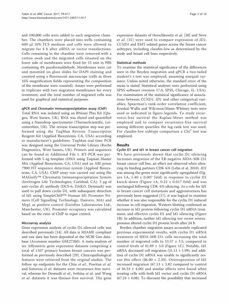
ResultsCyclin D1 and Id1 in breast cancer cell migrationWe have previously shown that cyclin D1 silencingincreases migration of the ER-negative MDA-MB-231breast cancer cell line, an effect not observed when silen-cing its binding partners CDK 4/6 (Lehn et al. 2010). ID1was among the genes most significantly upregulated (Fig-ure 1A, 1.40 ± 0.007 fold) in response to cyclin D1knock-down (Figure 1A, 0.22 ± 0.017 fold) and wasunchanged following CDK 4/6 silencing. As a role for Id1in breast cancer cell metastasis and aggressiveness haspreviously been suggested [17], it was logical to examinewhether it was also responsible for the cyclin D1 inducedincrease in cell migration. Western blotting confirmed anincrease in Id1 protein following cyclin D1 siRNA treat-ment, and effective cyclin D1 and Id1 silencing (Figure1B). In addition, neither Id1 silencing nor vector overex-pression altered cyclin D1 protein levels after 24 h.Boyden chamber migration assays accurately replicated
previous experimental results, with cyclin D1 siRNAtreatment of MDA-MB-231 cells increasing the totalnumber of migrated cells to 53.57 ± 3.5, compared tocontrol levels of 41.89 ± 3.0 (Figure 1C). Notably, Id1siRNA decreased cell migration (31.13 ± 1.99), and addi-tion of cyclin D1 siRNA was unable to significantly res-cue this effect (46.00 ± 2.34). Overexpression of Id1increased migration (67.13 ± 3.82 compared to controlof 36.33 ± 4.00) and similar effects were found whentreating cells with both Id1 vector and cyclin D1 siRNA(67.24 ± 6.08). To discount the possibility that increased
Tobin et al. BMC Cancer 2011, 11:417http://www.biomedcentral.com/1471-2407/11/417
Page 3 of 14
siRNA concentration may have a negative impact onmigration in the cyclin D1 and Id1 siRNA treated cells,we assessed single and double concentrations of siRNAin control cells and found no significant difference incell migration (data not shown). To determine if Id1could be a transcriptional target of cyclin D1 in MDA-
MB-231 cells, we performed a ChIP assay, and demon-strated that cyclin D1 occupancy in the Id1 promoterwas significantly higher (1.30% of input) in cyclin D1pull-down than in a negative mouse IgG control, andhigher still than the positive control Mrg1 (Bienvenu etal. 2010) (Figure 1D).
Figure 1 Effect of cyclin D1 and Id1 on breast cancer cell protein expression and migration. Actively cycling MDA-MB-231 and ZR75-1cells were monitored 20 h post-transfection with the indicated siRNA (cyclin D1/CDK4/6/Id1) or vector (Id1) for changes in gene or proteinexpression, and migration. Blots are representative, and plots are mean values from at least three independent experiments. Error bars representstandard deviation. MDA-MB-231 cells: (A) Microarray analysis. Left panel: CCND1 gene expression, right panel: ID1 gene expression (B) Westernblot for cyclin d1, Id1 and Actin protein, (C) Cell migration as measured by Boyden chamber assay, dots indicate total number of migrated cells.(D) ChIP assay for Id1 promoter region following cyclin D1 pull down. ZR75-1: (E) Western blot for cyclin d1 and Actin protein (F) Cellmigration- Boyden chamber assay. ***P ≥ 0.001, **P ≥ 0.01, *P ≥ 0.05 vs. control, two-tailed student’s t-test.
Tobin et al. BMC Cancer 2011, 11:417http://www.biomedcentral.com/1471-2407/11/417
Page 4 of 14

We next examined whether cyclin D1 silencing couldeffect migration in an ER-positive breast cancer cell linewith similar cyclin D1 levels to MDA-MB-231 cells.siRNA treatment against cyclin D1 reduced its proteinlevels (Figure 1E) and also significantly increased migra-tion (39.71 ± 5.04 compared to control of 20.14 ± 2.66,Figure 1F) of ZR75-1 cells. However, given the extre-mely low protein expression levels of Id1 in ZR75-1cells, it is unlikely that the increase in migration ismediated through Id1 in this cell line. In addition to theinteraction we have demonstrated between cyclin D1and Id1, other regulators of Id1 have been previouslyidentified. TGF-beta [32], KLF17 [33] and Src [34] areall known to interact and influence Id1 expression.Thus, levels of Id1 protein in ZR75-1 cells may reflectinteractions with other transcriptional regulators. Todirectly address this, we examined TGF-beta (a knowninducer of Id1 [32]) gene expression in a range of breastcancer cell lines and noted high levels in MDA-MB-231cells relative to ZR75-1 cells (Additional file 2). Impor-tantly, none of the aforementioned transcripts werealtered in our expression array data in response to cyclinD1 silencing and are hence unlikely to contribute to themigratory effect we have observed.Together, these data indicate that the increase in Id1
following cyclin D1 silencing in MDA-MB-231 cells isresponsible for their enhanced migratory capacity, butthat this does not appear to be the only mechanism bywhich cyclin D1 can induce cell migration. Mountingevidence has indicated the occurrence of an EMT-likephenotype in migratory breast cancer cells [35,36].Given this evidence we wished to determine whetherthe Id1 induced increase in migration following cyclinD1 silencing may be mediated through enhanced fea-tures of EMT.
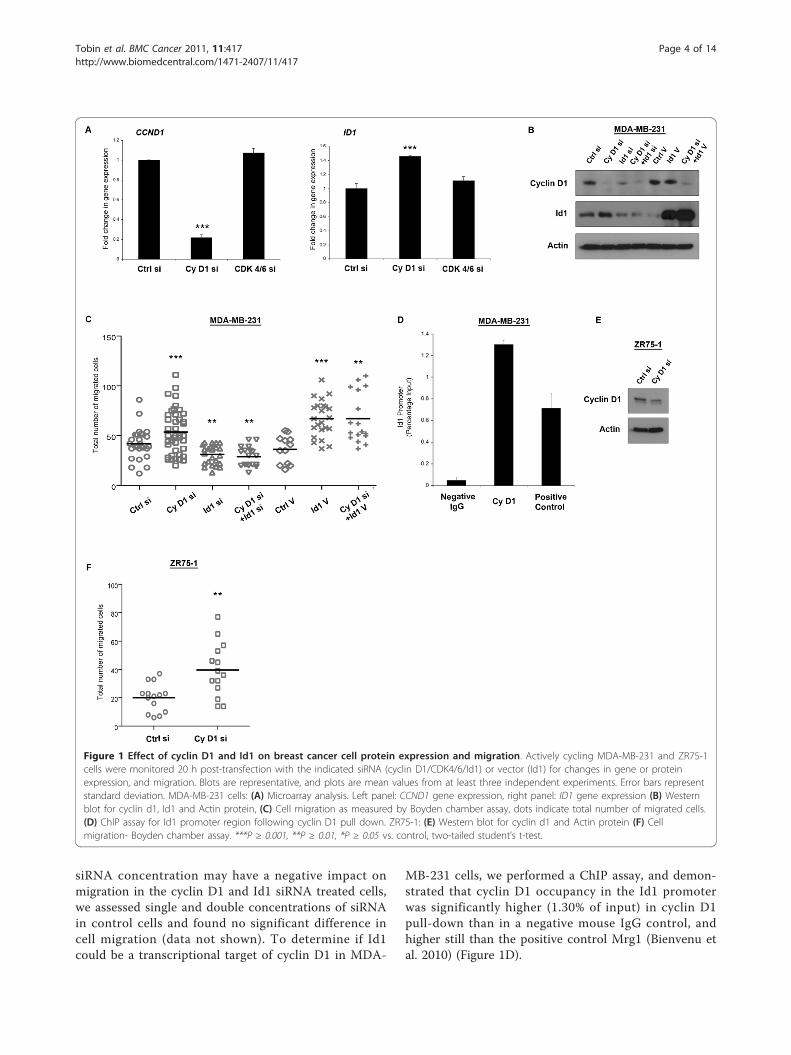
Cyclin D1 silencing in MDA-MB-231 cells increases EMTgene expression in an Id1 dependent mannerExamination of EMT-related genes in the microarrayanalysis of MDA-MB-231 cells showed significantincreases in SNAI2 (1.19 ± 0.03 fold), CDH11 (OB-Cad-herin, 1.18 ± 0.03 fold), and TWIST1 (1.13 ± 0.05 fold),following cyclin D1 silencing. A modest increase inSNAI2 (1.06 ± 0.01 fold) expression was noted afterCDK4/6 silencing, but neither siRNA treatment had aneffect on SNAI1 or VIM expression (Figure 2A).Using siRNA against cyclin D1 and Id1 we confirmed
significantly decreased levels of CCND1 by qPCR, andfound that Id1 siRNA had no significant impact onCCND1 expression (Figure 2B) after 24 h. Increased ID1levels (1.58 ± 0.09 fold) were noted following cyclin D1silencing (Figure 2C) and the effect of Id1 siRNA onID1 expression was reduced when combined with cyclinD1 siRNA (0.18 ± 0.01 vs. 0.28 ± 0.06 fold respectively,
P = 0.019). As noted in our microarray data, cyclin D1silencing increased SNAI2 levels, a result validated byqPCR analysis (1.41 ± 0.1 fold). This increase wasreversed when cyclin D1 was silenced in combinationwith Id1 (0.73 ± 0.13 fold of control, Figure 2D). Id1overexpression increased SNAI2 levels (1.34 ± 0.22 fold),an effect greatly enhanced when cyclin D1 was alsosilenced (2.39 ± 0.64 fold). Notably, silencing of cyclinD1 was unable to increase MDA-MB-231 cell migrationwhen Slug was also silenced (Additional File 3). We alsoobserved an increase in SNAI2 expression followingcyclin D1 silencing (Figure 2E) in ZR75-1 cells (1.34 ±0.05 fold, Figure 2F).These results suggest a novel effect whereby cyclin D1
silencing enhances a mesenchymal phenotype in MDA-MB-231 and ZR75-1 cells. In order to further validateour hypothesis, we next examined gene expression datafrom a large cohort of breast cancer patients.
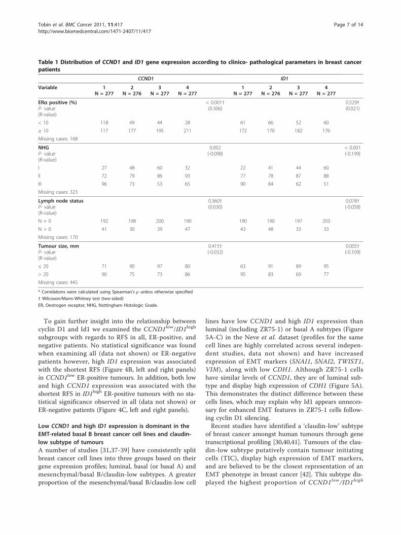
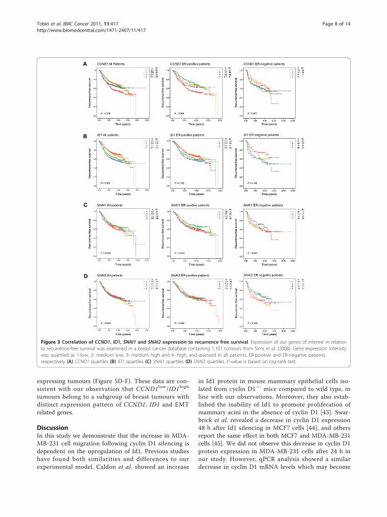
CCND1 and ID1 expression are correlated toclinicopathological parameters and predict recurrence riskin breast cancerTo investigate the relationship between CCND1 and ID1expression in primary breast tumours we used a pre-viously published meta-analysis consisting of six groupsof tumours on Affymetrix arrays totaling 1 107 samples.Due to the large number of patients and spread of geneexpression values we quartiled each gene, giving us thefollowing subgroups- 1 (low expression), 2 (low-med-ium), 3 (medium-high) and 4 (high). Initial examinationof clinicopathological parameters revealed that ID1 wasnegatively correlated to tumour grade (p < 0.001), andsize (p = 0.005). CCND1 expression was associated withER-positive breast cancers (p < 0.001), and lower histo-logical grade (p = 0.002) (Table 1). Neither CCND1 norID1 provided independent prognostic information in aCox multivariate analysis (data not shown).Next, we determined how these quartiles related to
recurrence-free survival (RFS) in the combined datasets.In all patients, and particularly in the subgroup of ER-positive patients, high expression of CCND1 was asso-ciated with the shortest RFS (p = 0.049 and p = 0.006,respectively, Figure 3A, left and middle panels, log-ranktest). This effect was not observed in the ER-negativesubgroup (Figure 3A, right panel). Conversely, low ID1expression was associated with the shortest RFS in allpatients (p < 0.001, Figure 3B, left panel), but not in theER-positive and negative subgroups (Figure 3B, middleand right panels). The levels of EMT-related genes,SNAI1 (Figure 3C), SNAI2 (Figure 3D), VIM or TWIST(Additional File 4B and 4C) were not of significant prog-nostic value. However, CDH1 (E-cadherin) significantlypredicted RFS in all and ER-positive patients (AdditionalFile 4A, left and middle panels).
Tobin et al. BMC Cancer 2011, 11:417http://www.biomedcentral.com/1471-2407/11/417
Page 5 of 14
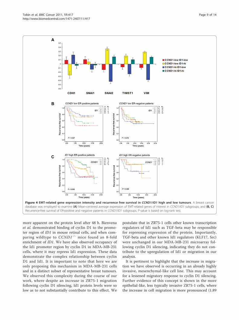
Low CCND1 and high ID1 expressing tumours showincreased EMT-related gene expression and predict risk ofrecurrence in breast tumoursAs our in vitro experiments indicated that CCND1low/ID1high breast cancer cells exhibit increased invasion andexpression of the SNAI2 gene, and our survival analysisindicated that low CCND1 and high ID1 expression canpredict RFS in breast cancer patients; we examined allfour combinations of CCND1low/high and ID1 low/high
gene expression in relation to well-characterised EMTgenes in all patients of the same tumour material. The
highest expression of SNAI2, TWIST1, VIM and lowestexpression of CDH1 was found in the CCND1low/ID1high
subgroup of tumours (Figure 4A, yellow bars). Furtherweight was added to this analysis when examining theCCND1low/high/ID1low subgroups of tumours (Figure 4A,red and blue bars, respectively). These tumours encom-pass the lowest expression of SNAI2, TWIST1, VIM andhighest expression of CDH1. This suggests, as ourMDA-MB-231 in vitro experiments demonstrated, thatcyclin D1 is unable to influence the induction of EMTin the absence of Id1.
Figure 2 Effect of cyclin d1 and Id1 on EMT markers. MDA-MB-231 cells were monitored 20 h post-transfection with the indicated siRNA(cyclin D1/CDK4/6/Id1) or vector (Id1) for changes in EMT-related gene expression by microarray analysis. Additionally, MDA-MB-231 and ZR75-1gene expression was examined by qPCR assay. Plots are mean values from at least three independent experiments Error bars represent standarddeviation. (A) Microarray analysis of SNAI1, SNAI2, CDH11, TWIST1 and VIM gene expression. (B-D) CCND1, ID1 and SNAI2 in MDA-MB-231 cells. (E,F) qPCR analysis of CCND1 and SNAI2 in ZR75-1 cells. ***P ≥ 0.001, **P ≥ 0.01, *P ≥ 0.05 vs. control, two-tailed student’s t-test.
Tobin et al. BMC Cancer 2011, 11:417http://www.biomedcentral.com/1471-2407/11/417
Page 6 of 14
To gain further insight into the relationship betweencyclin D1 and Id1 we examined the CCND1low/ID1high
subgroups with regards to RFS in all, ER-positive, andnegative patients. No statistical significance was foundwhen examining all (data not shown) or ER-negativepatients however, high ID1 expression was associatedwith the shortest RFS (Figure 4B, left and right panels)in CCND1low ER-positive tumours. In addition, both lowand high CCND1 expression was associated with theshortest RFS in ID1high ER-positive tumours with no sta-tistical significance observed in all (data not shown) orER-negative patients (Figure 4C, left and right panels).
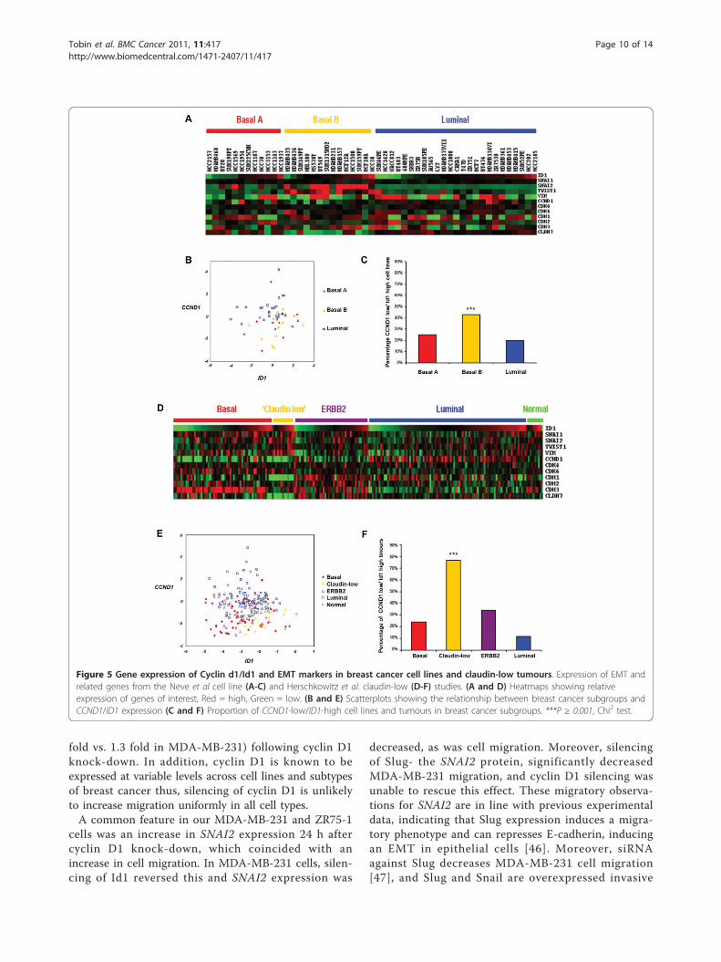
Low CCND1 and high ID1 expression is dominant in theEMT-related basal B breast cancer cell lines and claudin-low subtype of tumoursA number of studies [31,37-39] have consistently splitbreast cancer cell lines into three groups based on theirgene expression profiles; luminal, basal (or basal A) andmesenchymal/basal B/claudin-low subtypes. A greaterproportion of the mesenchymal/basal B/claudin-low cell
lines have low CCND1 and high ID1 expression thanluminal (including ZR75-1) or basal A subtypes (Figure5A-C) in the Neve et al. dataset (profiles for the samecell lines are highly correlated across several indepen-dent studies, data not shown) and have increasedexpression of EMT markers (SNAI1, SNAI2, TWIST1,VIM), along with low CDH1. Although ZR75-1 cellshave similar levels of CCND1, they are of luminal sub-type and display high expression of CDH1 (Figure 5A).This demonstrates the distinct difference between thesecells lines, which may explain why Id1 appears unneces-sary for enhanced EMT features in ZR75-1 cells follow-ing cyclin D1 silencing.Recent studies have identified a ‘claudin-low’ subtype
of breast cancer amongst human tumours through genetranscriptional profiling [30,40,41]. Tumours of the clau-din-low subtype putatively contain tumour initiatingcells (TIC), display high expression of EMT markers,and are believed to be the closest representation of anEMT phenotype in breast cancer [42]. This subtype dis-played the highest proportion of CCND1low/ID1high
Table 1 Distribution of CCND1 and ID1 gene expression according to clinico- pathological parameters in breast cancerpatients
CCND1 ID1
Variable 1N = 277
2N = 276
3N = 277
4N = 277
1N = 277
2N = 276
3N = 277
4N = 277
ERa positive (%)P- value(R-value)
< 0.001†(0.306)
0.529†(0.021)
< 10 118 49 44 28 61 66 52 60
≥ 10 117 177 195 211 172 170 182 176
Missing cases: 168
NHGP- value(R-value)
0.002(-0.098)
< 0.001(-0.199)
I 27 48 60 32 22 41 44 60
II 72 79 86 93 77 78 87 88
III 96 73 53 65 90 84 62 51
Missing cases: 323
Lymph node statusP- value(R-value)
0.360†(0.030)
0.078†(-0.058)
N = 0 192 198 200 190 190 190 197 203
N > 0 41 30 39 47 43 48 33 33
Missing cases: 170
Tumour size, mmP- value(R-value)
0.415†(-0.032)
0.005†(-0.109)
≤ 20 71 90 97 80 63 91 89 95
> 20 90 75 73 86 95 83 69 77
Missing cases: 445
* Correlations were calculated using Spearman’s r unless otherwise specified
† Wilcoxon/Mann-Whitney test (two-sided)
ER, Oestrogen receptor; NHG, Nottingham Histologic Grade.
Tobin et al. BMC Cancer 2011, 11:417http://www.biomedcentral.com/1471-2407/11/417
Page 7 of 14
expressing tumours (Figure 5D-F). These data are con-sistent with our observation that CCND1low/ID1high
tumours belong to a subgroup of breast tumours withdistinct expression pattern of CCND1, ID1 and EMTrelated genes.
DiscussionIn this study we demonstrate that the increase in MDA-MB-231 cell migration following cyclin D1 silencing isdependent on the upregulation of Id1. Previous studieshave found both similarities and differences to ourexperimental model. Caldon et al. showed an increase
in Id1 protein in mouse mammary epithelial cells iso-lated from cyclin D1-/- mice compared to wild type, inline with our observations. Moreover, they also estab-lished the inability of Id1 to promote proliferation ofmammary acini in the absence of cyclin D1 [43]. Swar-brick et al. revealed a decrease in cyclin D1 expression48 h after Id1 silencing in MCF7 cells [44], and othersreport the same effect in both MCF7 and MDA-MB-231cells [45]. We did not observe this decrease in cyclin D1protein expression in MDA-MB-231 cells after 24 h inour study. However, qPCR analysis showed a similardecrease in cyclin D1 mRNA levels which may become
Figure 3 Correlation of CCND1, ID1, SNAI1 and SNAI2 expression to recurrence free survival. Expression of our genes of interest in relationto recurrence-free survival was examined in a breast cancer database containing 1,107 tumours from Sims et al. (2008). Gene expression intensitywas quartiled as 1-low, 2- medium low, 3- medium high and 4- high, and assessed in all patients, ER-positive and ER-negative patients,respectively (A) CCND1 quartiles (B) ID1 quartiles (C) SNAI1 quartiles (D) SNAI2 quartiles. P-value is based on log-rank test.
Tobin et al. BMC Cancer 2011, 11:417http://www.biomedcentral.com/1471-2407/11/417
Page 8 of 14
more apparent on the protein level after 48 h. Bienvenuet al. demonstrated binding of cyclin D1 to the promo-ter region of ID1 in mouse retinal cells, and when com-paring wildtype to CCND1-/- mice found an 8-foldenrichment of ID1. We have also observed occupancy ofthe Id1 promoter region by cyclin D1 in MDA-MB-231cells, where it may repress Id1 expression. These datademonstrate the complex relationship between cyclinD1 and Id1. It is important to note that here we areonly proposing this mechanism in MDA-MB-231 cellsand in a distinct subset of representative breast tumours.We observed this complexity during the course of ourwork, where despite an increase in ZR75-1 migrationfollowing cyclin D1 silencing, Id1 protein levels were solow as to not substantially contribute to this effect. We
postulate that in ZR75-1 cells other known transcriptionregulators of Id1 such as TGF-beta may be responsiblefor repressing expression of the protein. Importantly,TGF-beta and other known Id1 regulators (KLF17, Src)were unchanged in our MDA-MB-231 microarray fol-lowing cyclin D1 silencing, indicating they do not con-tribute to the upregulation of Id1 or migration in ouranalysis.It is pertinent to highlight that the increase in migra-
tion we have observed is occurring in an already highlyinvasive, mesenchymal-like cell line. This may accountfor a lessened migratory response to cyclin D1 silencing.Further evidence of this concept is shown in the moreepithelial-like, less typically invasive ZR75-1 cells, wherethe increase in cell migration is more pronounced (1.89
Figure 4 EMT-related gene expression intensity and recurrence free survival in CCND1/ID1 high and low tumours. A breast cancerdatabase was employed to examine (A) Mean-centered average expression of EMT-related genes of interest in CCND1/ID1 subgroups, and (B, C)Recurrence-free survival of ER-positive and negative patients in CCND1/ID1 subgroups. P-value is based on log-rank test.
Tobin et al. BMC Cancer 2011, 11:417http://www.biomedcentral.com/1471-2407/11/417
Page 9 of 14
fold vs. 1.3 fold in MDA-MB-231) following cyclin D1knock-down. In addition, cyclin D1 is known to beexpressed at variable levels across cell lines and subtypesof breast cancer thus, silencing of cyclin D1 is unlikelyto increase migration uniformly in all cell types.A common feature in our MDA-MB-231 and ZR75-1
cells was an increase in SNAI2 expression 24 h aftercyclin D1 knock-down, which coincided with anincrease in cell migration. In MDA-MB-231 cells, silen-cing of Id1 reversed this and SNAI2 expression was
decreased, as was cell migration. Moreover, silencingof Slug- the SNAI2 protein, significantly decreasedMDA-MB-231 migration, and cyclin D1 silencing wasunable to rescue this effect. These migratory observa-tions for SNAI2 are in line with previous experimentaldata, indicating that Slug expression induces a migra-tory phenotype and can represses E-cadherin, inducingan EMT in epithelial cells [46]. Moreover, siRNAagainst Slug decreases MDA-MB-231 cell migration[47], and Slug and Snail are overexpressed invasive
Figure 5 Gene expression of Cyclin d1/Id1 and EMT markers in breast cancer cell lines and claudin-low tumours. Expression of EMT andrelated genes from the Neve et al cell line (A-C) and Herschkowitz et al. claudin-low (D-F) studies. (A and D) Heatmaps showing relativeexpression of genes of interest, Red = high, Green = low. (B and E) Scatterplots showing the relationship between breast cancer subgroups andCCND1/ID1 expression (C and F) Proportion of CCND1-low/ID1-high cell lines and tumours in breast cancer subgroups. ***P ≥ 0.001, Chi2 test.
Tobin et al. BMC Cancer 2011, 11:417http://www.biomedcentral.com/1471-2407/11/417
Page 10 of 14
ductal carcinoma [48]- a form of breast cancer hall-marked by cell migration. In our experimental model,Slug would appear a likely candidate mediating theobserved migratory effects, however it is entirely plau-sible that it does so in conjunction with other EMTfactors. We also found statistically significant changesin TWIST1 and CDH11 (the positive EMT-regulatoralso known as OB-cadherin) following cyclin D1 silen-cing, both of which have been implicated withenhanced cell motility [49,50]. The changes in ourEMT markers are in the order of 1.13 to 1.19 fold ofcontrol by expression array analysis (Figure 2A). Wenote that these figures are more meaningful whentaken in the context of the most increased gene in ourexpression array, which was only upregulated 1.8 fold[14]. As may be expected from treatment with siRNA,many more genes were downregulated in the arrayanalysis than upregulated, again highlighting theimportance of the increases in our mesenchymal mar-kers. It is likely that all of these factors work in con-cert to promote a migratory and EMT-like phenotype,and that small gains in expression of a number ofEMT genes can contribute to a greater overall effect.The relationship between cyclin D1 expression and
patient outcome remains a controversial area, with stu-dies reporting both positive and negative associations.CCND1 gene amplification has been related to poor dis-ease outcome in ER-positive patients [51,52], but otherscorrelate cyclin D1 protein expression with both better[53,54] and worse [55] prognosis. It has been proposedthat subgroup analysis with small numbers of patients[56] and splice variants of the gene have contributed tothese contrasting results. In agreement with others [57],we found an association between high CCND1 expres-sion and poor prognosis (Figure 3A). However, whenexamining ID1 high tumours, both the highest and low-est expression quartiles of CCND1 were correlated toreduced RFS/DFS but only in the ER-positive subgroup(Figure 4C). A similar trend was noted for ID1, where inall patients low expression of the gene was associatedwith a shortest RFS (Figure 3B), but in the CCND1 lowER-positive subgroup of tumours, a positive correlationwas found (Figure 4B).Whilst this may appear contrasting to our in vitro
data, we reason that cyclin D1 low, ER-positive tumoursbest represent our cell line model. We chose two celllines (MDA-MB-231 and ZR75-1) based on their highexpression of cyclin D1 (regardless of oestrogen receptorstatus). We then reduced these high levels using siRNAand noted an increase in cell migration and EMT mar-kers. As ER-negative tumours are consistently cyclin D1low, these are less representative our in vitro experi-ments. ER-positive tumours however are typically cyclinD1 high, thus by choosing tumours that are cyclin D1
low in this subgroup, we are more correctly mimickingour in vitro setting, where expression of cyclin D1 mayhave been lost. This yields the interesting observationthat ER-positive tumours with low cyclin D1 appear tobehave similarly to ER-negative tumours with regards totheir relationship to EMT markers and the claudin-lowsubtype. Thus, should ER-positive tumours that havelost expression of cyclin D1 be considered more ER-negative-like? Whilst the answer to this question is farbeyond the scope of this study, what is clear is that theeffect we are observing is centred on loss of cyclin D1and not on the oestrogen receptor status of our testingmaterial.Interestingly, the CCND1low/ID1high and CCND1high/
ID1high tumours both displayed increased expression ofEMT-related genes (Figure 4A, yellow and green barsrespectively). This suggests that in the context of thesesubgroups, ID1 is vital for increased EMT gene expres-sion and when CCND1 is low it enhances the EMTphenotype.We did not observe any meaningful impact of EMT
genes in individual Kaplin-meier analysis on patient sur-vival in our dataset. There has been an explosion ofEMT related data in recent years in the breast cancerfield. Central to many of these publications has been theability of EMT to putatively enhance stem cell-relatedfeatures and promote the metastatic process [58,59]. Ofparticular note, the idea of cells that have undergoneEMT residing at the leading edge of an invasive tumourand promoting metastasis at the tumour- stroma inter-face has garnered much attention [60]. This hypothesismay be one explanation as to why EMT markers suchas SNAI1, SNAI2, TWIST1 and VIM do not show anyprognostic significance in our model- if the cells thathave undergone EMT reside at the leading edge of thetumour, strong expression of their genes could easily belost amongst the entirety of the tumour body. In thesecircumstances, any strong links to prognosis would alsobe diluted.A second, more straightforward explanation as to why
we have not observed prognostic significance of EMT-related genes centers upon a keystone principal. Upre-gulation of one EMT gene, e.g. SNAI1, is not enough toinduce a transition to mesenchymal phenotype. This issupported by the board range of expression values ofEMT genes across all breast cancer tumours and sub-types in our study (Figure 5D). Induction of EMTrequires a reduction in CDH1 expression and upregula-tion of the potent SNAI1, SNAI2 and TWIST1 genes(amongst others). In order to examine the effect ofEMT in our cohort, we would have to combine alltumours with these gene properties- giving us a ‘clau-din-low’ subgroup. Unfortunately, we have too few casesin our claudin-low dataset to give any relevant
Tobin et al. BMC Cancer 2011, 11:417http://www.biomedcentral.com/1471-2407/11/417
Page 11 of 14
prognostic information. In order to explore this furthera cohort consisting of a large representation of claudin-low tumours, preferably with micro-dissection of thetumour-stroma interface would be required.Much like CCND1, some controversy surrounds
expression patterns of ID1, and despite numerous linksto invasion and migration in breast cancer [43,44] somegroups report an absence of the protein in the normalmammary gland [61]. Perk et al. assessed Id1 proteinexpression in mammary carcinomas [62] and foundnuclear expression of Id1 in a rare subtype of breastcancer, metaplastic mammary tumours. Metaplastic can-cers have a unique genetic profile that is notably, mostclosely related to the claudin-low subtype of breast can-cer [41,63,64] and are very poorly differentiated. Giventhe poor outcome associated with metaplastic cancer, itmay indicate why high ID1 expression in CCND1 lowtumours gave the shortest RFS.Adding further weight to our analysis, we found the
greatest proportion of CCND1low/ID1high cell lines andtumours in the claudin-low subgroup, which have apoor prognosis [65], associations with EMT and che-motherapy resistance [66] and has stem-cell tumourinitiating features [42]. A number of these properties arereflected in both the cell lines and patient material usedwithin this study, potentially indicating a central role forcyclin D1 and Id1 in this subgroup.
ConclusionsThe increase in MDA-MB-231 migration we haveobserved following cyclin D1 silencing is dependent onan upregulation of Id1 and induction of a moremesenchymal phenotype. Patients with CCND1low/ID1high tumours have a shorter RFS and we have showna link between CCND1low/ID1high tumours and the clau-din-low subgroup of breast cancer.
Additional material
Additional file 1: qPCR primers. Sequences of primers used in thisstudy.
Additional file 2: TGF-b gene expression in breast cancer cell lines.The dataset from Neve et al. was employed to examine TGF-b geneexpression in breast cancer cell lines. The bar at the bottom of the figurerepresents the subtype of each cell line. Blue = luminal, Orange = BasalA, Red = Basal B. Cell lines of interest are highlighted with a blackrectangle, and are ZR75-1 and MDA-MB-231 cell.
Additional file 3: Cyclin D1 silencing does not increase MDA-MB-231 cell migration in the absence of Slug. Actively cycling MDA-MB-231 cells were monitored 20 h post-transfection with the indicated siRNA(cyclin D1/slug) for changes in cell migration and gene expression. Errorbars represent standard deviation. (A) Cell migration as measured byBoyden chamber assay (B) qPCR analysis of slug expression. ***P ≥ 0.001,**P ≥ 0.01, *P ≥ 0.05 vs. control, two-tailed student’s t-test.
Additional file 4: Correlation of CDH1, VIM and TWIST1 expressionto recurrence free survival. Expression of our genes of interest inrelation to recurrence free survival was examined in a breast cancer
meta-analysis. (A) CDH1 quartiles (B) VIM quartiles (C) TWIST1 quartiles. P-value is based on log-rank test.
Acknowledgements and fundingWe wish to thank Prof. Anthony Howell for contributions to the editingprocess. This study was supported by grants from the Swedish CancerSociety, the Swedish Research Council, the Knut and Alice WallenbergFoundation, Malmö University Hospital Research and Cancer Funds, LundUniversity Research Funds, South Swedish and South-East Swedish BreastCancer groups and Breakthrough Breast Cancer Unit, Manchester, UK. AHS isalso grateful for funding from Breakthrough Breast Cancer. The funders hadno role in study design, data collection and analysis, decision to publish, orpreparation of the manuscript.
Author details1Breakthrough Breast Cancer Research Unit, School of Cancer, EnablingSciences and Technology, University of Manchester, Manchester AcademicHealth Science Centre, Paterson Institute for Cancer Research, The ChristieNHS Foundation Trust, Wilmslow Road, Manchester, M20 4BX, UK. 2CancerCenter Karolinska, Karolinska Institute and University Hospital, Stockholm, S-17176, Sweden. 3Applied Bioinformatics of Cancer, Breakthrough BreastCancer Research Unit, Edinburgh Cancer Research Centre, Institute ofGenetics and Molecular Medicine, Crewe Road South Edinburgh, EH4 2XR,UK. 4Department of Laboratory Medicine, Center for Molecular Pathology,Lund University, Malmö University Hospital, Malmö, SE-205 02, Sweden.
Authors’ contributionsNT performed the experimental work and the statistical analyses as well asdrafting the manuscript. KL helped with the experimental work, andstatistical analysis as well as drafting the manuscript. SL performed migrationassays and western blots. AHS performed the statistical analyses and thebioinformatics related experimental work, as well as drafting the manuscript.GL was the principal investigator of the study and participated in the studydesign and interpretation of the data and helped to draft the manuscript.All authors read and approved the final manuscript.
Competing interestsThe authors declare that they have no competing interests.
Received: 4 April 2011 Accepted: 28 September 2011Published: 28 September 2011
References1. Kato J, Matsushime H, Hiebert SW, Ewen ME, Sherr CJ: Direct binding of
cyclin D to the retinoblastoma gene product (pRb) and pRbphosphorylation by the cyclin D-dependent kinase CDK4. Genes Dev1993, 7(3):331-342.
2. Lundberg AS, Weinberg RA: Functional inactivation of the retinoblastomaprotein requires sequential modification by at least two distinct cyclin-cdk complexes. Mol Cell Biol 1998, 18(2):753-761.
3. Weinberg RA: The retinoblastoma protein and cell cycle control. Cell1995, 81(3):323-330.
4. Jiang W, Kahn SM, Zhou P, Zhang YJ, Cacace AM, Infante AS, Doi S,Santella RM, Weinstein IB: Overexpression of cyclin D1 in rat fibroblastscauses abnormalities in growth control, cell cycle progression and geneexpression. Oncogene 1993, 8(12):3447-3457.
5. Lammie GA, Fantl V, Smith R, Schuuring E, Brookes S, Michalides R,Dickson C, Arnold A, Peters G: D11S287, a putative oncogene onchromosome 11q13, is amplified and expressed in squamous cell andmammary carcinomas and linked to BCL-1. Oncogene 1991, 6(3):439-444.
6. Wang TC, Cardiff RD, Zukerberg L, Lees E, Arnold A, Schmidt EV: Mammaryhyperplasia and carcinoma in MMTV-cyclin D1 transgenic mice. Nature1994, 369(6482):669-671.
7. Zhou P, Jiang W, Zhang YJ, Kahn SM, Schieren I, Santella RM, Weinstein IB:Antisense to cyclin D1 inhibits growth and reverses the transformedphenotype of human esophageal cancer cells. Oncogene 1995,11(3):571-580.
Tobin et al. BMC Cancer 2011, 11:417http://www.biomedcentral.com/1471-2407/11/417
Page 12 of 14
8. Jares P, Rey MJ, Fernandez PL, Campo E, Nadal A, Munoz M, Mallofre C,Muntane J, Nayach I, Estape J, et al: Cyclin D1 and retinoblastoma geneexpression in human breast carcinoma: correlation with tumourproliferation and oestrogen receptor status. J Pathol 1997, 182(2):160-166.
9. Sutter T, Doi S, Carnevale KA, Arber N, Weinstein IB: Expression of cyclinsD1 and E in human colon adenocarcinomas. J Med 1997, 28(5-6):285-309.
10. Drobnjak M, Osman I, Scher HI, Fazzari M, Cordon-Cardo C: Overexpressionof cyclin D1 is associated with metastatic prostate cancer to bone. ClinCancer Res 2000, 6(5):1891-1895.
11. Coqueret O: Linking cyclins to transcriptional control. Gene 2002, 299(1-2):35-55.
12. Zwijsen RM, Wientjens E, Klompmaker R, van der Sman J, Bernards R,Michalides RJ: CDK-independent activation of estrogen receptor by cyclinD1. Cell 1997, 88(3):405-415.
13. Bienvenu F, Jirawatnotai S, Elias JE, Meyer CA, Mizeracka K, Marson A,Frampton GM, Cole MF, Odom DT, Odajima J, et al: Transcriptional role ofcyclin D1 in development revealed by a genetic-proteomic screen.Nature 2010, 463(7279):374-378.
14. Lehn S, Tobin NP, Berglund P, Nilsson K, Sims AH, Jirstrom K, Harkonen P,Lamb R, Landberg G: Downregulation of the Oncogene Cyclin D1Increases Migratory Capacity in Breast Cancer and Is Linked toUnfavorable Prognostic Features. Am J Pathol 2010.
15. Ephrussi A, Church GM, Tonegawa S, Gilbert W: B lineage–specificinteractions of an immunoglobulin enhancer with cellular factors invivo. Science 1985, 227(4683):134-140.
16. Benezra R, Davis RL, Lockshon D, Turner DL, Weintraub H: The protein Id: anegative regulator of helix-loop-helix DNA binding proteins. Cell 1990,61(1):49-59.
17. Singh J, Murata K, Itahana Y, Desprez PY: Constitutive expression of the Id-1 promoter in human metastatic breast cancer cells is linked with theloss of NF-1/Rb/HDAC-1 transcription repressor complex. Oncogene 2002,21(12):1812-1822.
18. Lin CQ, Singh J, Murata K, Itahana Y, Parrinello S, Liang SH, Gillett CE,Campisi J, Desprez PY: A role for Id-1 in the aggressive phenotype andsteroid hormone response of human breast cancer cells. Cancer Res 2000,60(5):1332-1340.
19. Fong S, Itahana Y, Sumida T, Singh J, Coppe JP, Liu Y, Richards PC,Bennington JL, Lee NM, Debs RJ, et al: Id-1 as a molecular target intherapy for breast cancer cell invasion and metastasis. Proc Natl Acad SciUSA 2003, 100(23):13543-13548.
20. Hay ED: The mesenchymal cell, its role in the embryo, and theremarkable signaling mechanisms that create it. Dev Dyn 2005,233(3):706-720.
21. Trimboli AJ, Fukino K, de Bruin A, Wei G, Shen L, Tanner SM, Creasap N,Rosol TJ, Robinson ML, Eng C, et al: Direct evidence for epithelial-mesenchymal transitions in breast cancer. Cancer Res 2008, 68(3):937-945.
22. Micalizzi DS, Farabaugh SM, Ford HL: Epithelial-mesenchymal transition incancer: parallels between normal development and tumor progression. JMammary Gland Biol Neoplasia 2010, 15(2):117-134.
23. Ikenouchi J, Matsuda M, Furuse M, Tsukita S: Regulation of tight junctionsduring the epithelium-mesenchyme transition: direct repression of thegene expression of claudins/occludin by Snail. J Cell Sci 2003, 116(Pt10):1959-1967.
24. Ozdamar B, Bose R, Barrios-Rodiles M, Wang HR, Zhang Y, Wrana JL:Regulation of the polarity protein Par6 by TGFbeta receptors controlsepithelial cell plasticity. Science 2005, 307(5715):1603-1609.
25. Hajra KM, Chen DY, Fearon ER: The SLUG zinc-finger protein represses E-cadherin in breast cancer. Cancer Res 2002, 62(6):1613-1618.
26. Li Y, Yang J, Luo JH, Dedhar S, Liu Y: Tubular epithelial celldedifferentiation is driven by the helix-loop-helix transcriptionalinhibitor Id1. J Am Soc Nephrol 2007, 18(2):449-460.
27. Gumireddy K, Li A, Gimotty PA, Klein-Szanto AJ, Showe LC, Katsaros D,Coukos G, Zhang L, Huang Q: KLF17 is a negative regulator of epithelial-mesenchymal transition and metastasis in breast cancer. Nat Cell Biol2009, 11(11):1297-1304.
28. Berglund P, Stighall M, Jirstrom K, Borgquist S, Sjolander A, Hedenfalk I,Landberg G: Cyclin E overexpression obstructs infiltrative behavior inbreast cancer: a novel role reflected in the growth pattern of medullarybreast cancers. Cancer Res 2005, 65(21):9727-9734.
29. Sims AH, Smethurst GJ, Hey Y, Okoniewski MJ, Pepper SD, Howell A,Miller CJ, Clarke RB: The removal of multiplicative, systematic bias allows
integration of breast cancer gene expression datasets - improving meta-analysis and prediction of prognosis. BMC Med Genomics 2008, 1:42.
30. Herschkowitz JI, Simin K, Weigman VJ, Mikaelian I, Usary J, Hu Z,Rasmussen KE, Jones LP, Assefnia S, Chandrasekharan S, et al: Identificationof conserved gene expression features between murine mammarycarcinoma models and human breast tumors. Genome Biol 2007, 8(5):R76.
31. Neve RM, Chin K, Fridlyand J, Yeh J, Baehner FL, Fevr T, Clark L, Bayani N,Coppe JP, Tong F, et al: A collection of breast cancer cell lines for thestudy of functionally distinct cancer subtypes. Cancer Cell 2006,10(6):515-527.
32. Liang YY, Brunicardi FC, Lin X: Smad3 mediates immediate early inductionof Id1 by TGF-beta. Cell research 2009, 19(1):140-148.
33. Gumireddy K, Li A, Gimotty PA, Klein-Szanto AJ, Showe LC, Katsaros D,Coukos G, Zhang L, Huang Q: KLF17 is a negative regulator of epithelial-mesenchymal transition and metastasis in breast cancer. Nature cellbiology 2009, 11(11):1297-1304.
34. Gautschi O, Tepper CG, Purnell PR, Izumiya Y, Evans CP, Green TP,Desprez PY, Lara PN, Gandara DR, Mack PC, et al: Regulation of Id1expression by SRC: implications for targeting of the bonemorphogenetic protein pathway in cancer. Cancer research 2008,68(7):2250-2258.
35. Fu J, Qin L, He T, Qin J, Hong J, Wong J, Liao L, Xu J: The TWIST/Mi2/NuRDprotein complex and its essential role in cancer metastasis. Cell Res 2010.
36. Gilles C, Polette M, Zahm JM, Tournier JM, Volders L, Foidart JM,Birembaut P: Vimentin contributes to human mammary epithelial cellmigration. J Cell Sci 1999, 112(Pt 24):4615-4625.
37. Charafe-Jauffret E, Ginestier C, Iovino F, Wicinski J, Cervera N, Finetti P,Hur MH, Diebel ME, Monville F, Dutcher J, et al: Breast cancer cell linescontain functional cancer stem cells with metastatic capacity and adistinct molecular signature. Cancer Res 2009, 69(4):1302-1313.
38. Kao J, Salari K, Bocanegra M, Choi YL, Girard L, Gandhi J, Kwei KA,Hernandez-Boussard T, Wang P, Gazdar AF, et al: Molecular profiling ofbreast cancer cell lines defines relevant tumor models and provides aresource for cancer gene discovery. PLoS One 2009, 4(7):e6146.
39. Lapuk A, Marr H, Jakkula L, Pedro H, Bhattacharya S, Purdom E, Hu Z,Simpson K, Pachter L, Durinck S, et al: Exon-level microarray analysesidentify alternative splicing programs in breast cancer. Mol Cancer Res2010, 8(7):961-974.
40. Creighton CJ, Chang JC, Rosen JM: Epithelial-mesenchymal transition(EMT) in tumor-initiating cells and its clinical implications in breastcancer. J Mammary Gland Biol Neoplasia 2010, 15(2):253-260.
41. Prat A, Parker JS, Karginova O, Fan C, Livasy C, Herschkowitz JI, He X,Perou CM: Phenotypic and molecular characterization of the claudin-lowintrinsic subtype of breast cancer. Breast Cancer Res 2010, 12(5):R68.
42. Creighton CJ, Li X, Landis M, Dixon JM, Neumeister VM, Sjolund A,Rimm DL, Wong H, Rodriguez A, Herschkowitz JI, et al: Residual breastcancers after conventional therapy display mesenchymal as well astumor-initiating features. Proc Natl Acad Sci USA 2009,106(33):13820-13825.
43. Caldon CE, Swarbrick A, Lee CS, Sutherland RL, Musgrove EA: The helix-loop-helix protein Id1 requires cyclin D1 to promote the proliferation ofmammary epithelial cell acini. Cancer Res 2008, 68(8):3026-3036.
44. Swarbrick A, Akerfeldt MC, Lee CS, Sergio CM, Caldon CE, Hunter LJ,Sutherland RL, Musgrove EA: Regulation of cyclin expression and cellcycle progression in breast epithelial cells by the helix-loop-helix proteinId1. Oncogene 2005, 24(3):381-389.
45. Lee JY, Kang MB, Jang SH, Qian T, Kim HJ, Kim CH, Kim Y, Kong G: Id-1activates Akt-mediated Wnt signaling and p27(Kip1) phosphorylationthrough PTEN inhibition. Oncogene 2009, 28(6):824-831.
46. Chen H, Zhu G, Li Y, Padia RN, Dong Z, Pan ZK, Liu K, Huang S:Extracellular signal-regulated kinase signaling pathway regulates breastcancer cell migration by maintaining slug expression. Cancer Res 2009,69(24):9228-9235.
47. Bolos V, Peinado H, Perez-Moreno MA, Fraga MF, Esteller M, Cano A: Thetranscription factor Slug represses E-cadherin expression and inducesepithelial to mesenchymal transitions: a comparison with Snail and E47repressors. J Cell Sci 2003, 116(Pt 3):499-511.
48. Come C, Magnino F, Bibeau F, De Santa Barbara P, Becker KF, Theillet C,Savagner P: Snail and slug play distinct roles during breast carcinomaprogression. Clin Cancer Res 2006, 12(18):5395-5402.
Tobin et al. BMC Cancer 2011, 11:417http://www.biomedcentral.com/1471-2407/11/417
Page 13 of 14
49. Mironchik Y, Winnard PT Jr, Vesuna F, Kato Y, Wildes F, Pathak AP,Kominsky S, Artemov D, Bhujwalla Z, Van Diest P, et al: Twistoverexpression induces in vivo angiogenesis and correlates withchromosomal instability in breast cancer. Cancer Res 2005,65(23):10801-10809.
50. Nakajima G, Patino-Garcia A, Bruheim S, Xi Y, San Julian M, Lecanda F,Sierrasesumaga L, Muller C, Fodstad O, Ju J: CDH11 expression isassociated with survival in patients with osteosarcoma. Cancer GenomicsProteomics 2008, 5(1):37-42.
51. Berns EM, Foekens JA, van Staveren IL, van Putten WL, de Koning HY,Portengen H, Klijn JG: Oncogene amplification and prognosis in breastcancer: relationship with systemic treatment. Gene 1995, 159(1):11-18.
52. Bieche I, Olivi M, Nogues C, Vidaud M, Lidereau R: Prognostic value ofCCND1 gene status in sporadic breast tumours, as determined by real-time quantitative PCR assays. Br J Cancer 2002, 86(4):580-586.
53. Gillett C, Smith P, Gregory W, Richards M, Millis R, Peters G, Barnes D: CyclinD1 and prognosis in human breast cancer. Int J Cancer 1996, 69(2):92-99.
54. Hwang TS, Han HS, Hong YC, Lee HJ, Paik NS: Prognostic value ofcombined analysis of cyclin D1 and estrogen receptor status in breastcancer patients. Pathol Int 2003, 53(2):74-80.
55. Han S, Park K, Bae BN, Kim KH, Kim HJ, Kim YD, Kim HY: Cyclin D1expression and patient outcome after tamoxifen therapy in estrogenreceptor positive metastatic breast cancer. Oncol Rep 2003, 10(1):141-144.
56. Taneja P, Maglic D, Kai F, Zhu S, Kendig RD, Fry EA, Inoue K: Classical andNovel Prognostic Markers for Breast Cancer and their ClinicalSignificance. Clin Med Insights Oncol 2010, 4:15-34.
57. Kenny FS, Hui R, Musgrove EA, Gee JM, Blamey RW, Nicholson RI,Sutherland RL, Robertson JF: Overexpression of cyclin D1 messenger RNApredicts for poor prognosis in estrogen receptor-positive breast cancer.Clin Cancer Res 1999, 5(8):2069-2076.
58. Mimeault M, Batra SK: Functions of tumorigenic and migrating cancerprogenitor cells in cancer progression and metastasis and theirtherapeutic implications. Cancer Metastasis Rev 2007, 26(1):203-214.
59. Mimeault M, Batra SK: Interplay of distinct growth factors duringepithelial mesenchymal transition of cancer progenitor cells andmolecular targeting as novel cancer therapies. Ann Oncol 2007,18(10):1605-1619.
60. Blick T, Widodo E, Hugo H, Waltham M, Lenburg ME, Neve RM,Thompson EW: Epithelial mesenchymal transition traits in human breastcancer cell lines. Clin Exp Metastasis 2008, 25(6):629-642.
61. Uehara N, Chou YC, Galvez JJ, de-Candia P, Cardiff RD, Benezra R,Shyamala G: Id-1 is not expressed in the luminal epithelial cells ofmammary glands. Breast Cancer Res 2003, 5(2):R25-29.
62. Perk J, Gil-Bazo I, Chin Y, de Candia P, Chen JJ, Zhao Y, Chao S, Cheong W,Ke Y, Al-Ahmadie H, et al: Reassessment of id1 protein expression inhuman mammary, prostate, and bladder cancers using a monospecificrabbit monoclonal anti-id1 antibody. Cancer Res 2006, 66(22):10870-10877.
63. Hennessy BT, Gonzalez-Angulo AM, Stemke-Hale K, Gilcrease MZ,Krishnamurthy S, Lee JS, Fridlyand J, Sahin A, Agarwal R, Joy C, et al:Characterization of a naturally occurring breast cancer subset enrichedin epithelial-to-mesenchymal transition and stem cell characteristics.Cancer Res 2009, 69(10):4116-4124.
64. Weigelt B, Kreike B, Reis-Filho JS: Metaplastic breast carcinomas are basal-like breast cancers: a genomic profiling analysis. Breast Cancer Res Treat2009, 117(2):273-280.
65. Luini A, Aguilar M, Gatti G, Fasani R, Botteri E, Brito JA, Maisonneuve P,Vento AR, Viale G: Metaplastic carcinoma of the breast, an unusualdisease with worse prognosis: the experience of the European Instituteof Oncology and review of the literature. Breast Cancer Res Treat 2007,101(3):349-353.
66. Farmer P, Bonnefoi H, Anderle P, Cameron D, Wirapati P, Becette V,Andre S, Piccart M, Campone M, Brain E, et al: A stroma-related genesignature predicts resistance to neoadjuvant chemotherapy in breastcancer. Nat Med 2009, 15(1):68-74.
Pre-publication historyThe pre-publication history for this paper can be accessed here:http://www.biomedcentral.com/1471-2407/11/417/prepub
doi:10.1186/1471-2407-11-417Cite this article as: Tobin et al.: Cyclin D1, Id1 and EMT in breast cancer.BMC Cancer 2011 11:417.
Submit your next manuscript to BioMed Centraland take full advantage of:
• Convenient online submission
• Thorough peer review
• No space constraints or color figure charges
• Immediate publication on acceptance
• Inclusion in PubMed, CAS, Scopus and Google Scholar
• Research which is freely available for redistribution
Submit your manuscript at www.biomedcentral.com/submit
Tobin et al. BMC Cancer 2011, 11:417http://www.biomedcentral.com/1471-2407/11/417
Page 14 of 14