cytomorphologic analysis of adenoid cystic carcinoma of the breast: one case laura chiapparini;...
TRANSCRIPT
CYTOMORPHOLOGIC ANALYSIS OF ADENOID
CYSTIC CARCINOMA OF THE BREAST: one case
Laura Chiapparini; Chiara Scacchi;Mara Lusiardi; Clementina Di Tonno.
Unit of Diagnostic Cytology
2
INTRODUCTION
• Adenoid cystic carcinoma (ACC) is a rare form of adenocarcinoma that usually arises in the salivary glands.
• ACC of the breast represents less than 1% of malignant breast tumors and its prognosis is usually favorable.
• With the current work we present a case of ACC of the breast diagnosed in our Institute, the relative cytologic picture and the problems in the differential diagnosis.
3
THE CASE
• A 60 year old woman.
• Clinical diagnosis: painful lesion of almost 2 cm, in the inferior external quadrant of the left breast.
• Ultrasound diagnosis: nodule of almost 1.5 cm, discretely vascularized at color-doppler, compatible with a heteroproductive lesion.
• The lesion was fine-needle aspirated.
4
MATERIALS AND METHODS
The material obtained by fine-needle aspiration was used to prepare:
• 2 air-dried slides, subsequently stained with May-Grünwald Giemsa (MGG)
• 3 slides were fixed in 95% alcohol, 2 of which stained with Hematoxylin & Eosin (H&E) and 1 following Papanicolaou (PAP)
• 5 preparations in monolayer, processed with the ThinPrep (TP) method, 1 of which was stained with PAP and 4 utilized for immunocytochemical studies.
5
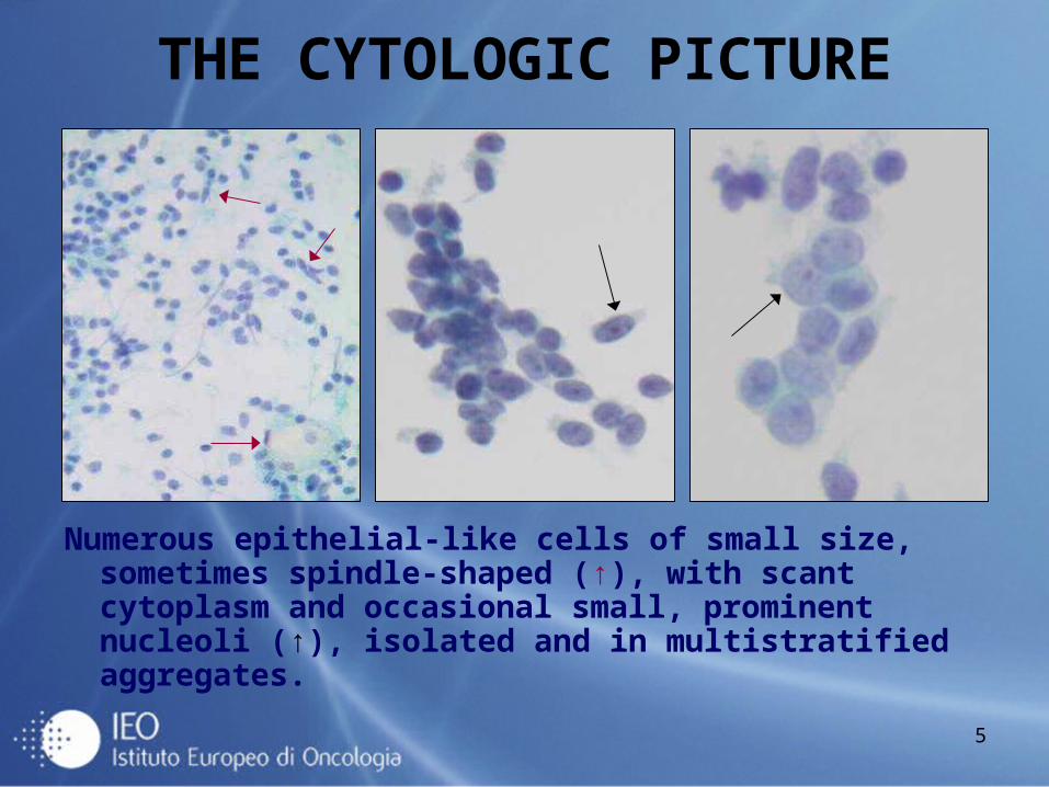
THE CYTOLOGIC PICTURE
Numerous epithelial-like cells of small size, sometimes spindle-shaped (↑), with scant cytoplasm and occasional small, prominent nucleoli (↑), isolated and in multistratified aggregates.
6
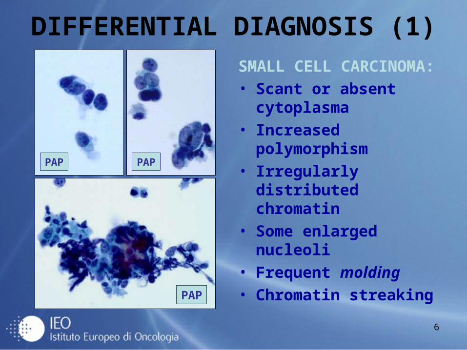
DIFFERENTIAL DIAGNOSIS (1)
SMALL CELL CARCINOMA:• Scant or absent
cytoplasma• Increased polymorphism • Irregularly distributed
chromatin • Some enlarged nucleoli • Frequent molding• Chromatin streaking
PAP
PAP PAP
7
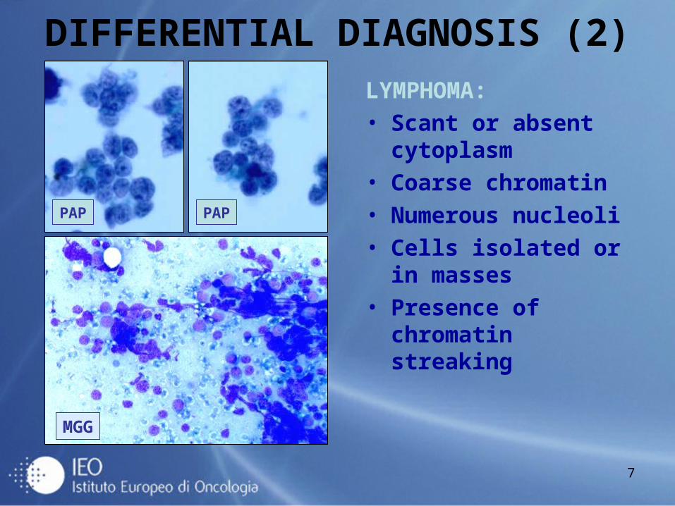
DIFFERENTIAL DIAGNOSIS (2)
LYMPHOMA:• Scant or absent
cytoplasm• Coarse chromatin • Numerous nucleoli• Cells isolated or in
masses• Presence of chromatin
streaking
MGG
PAP PAP
8
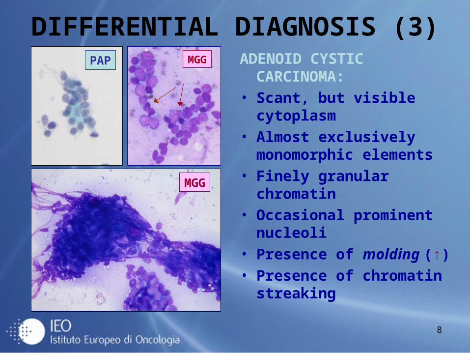
DIFFERENTIAL DIAGNOSIS (3)ADENOID CYSTIC
CARCINOMA:• Scant, but visible
cytoplasm• Almost exclusively
monomorphic elements• Finely granular chromatin• Occasional prominent
nucleoli • Presence of molding (↑)• Presence of chromatin
streaking
MGG
MGGPAP
9
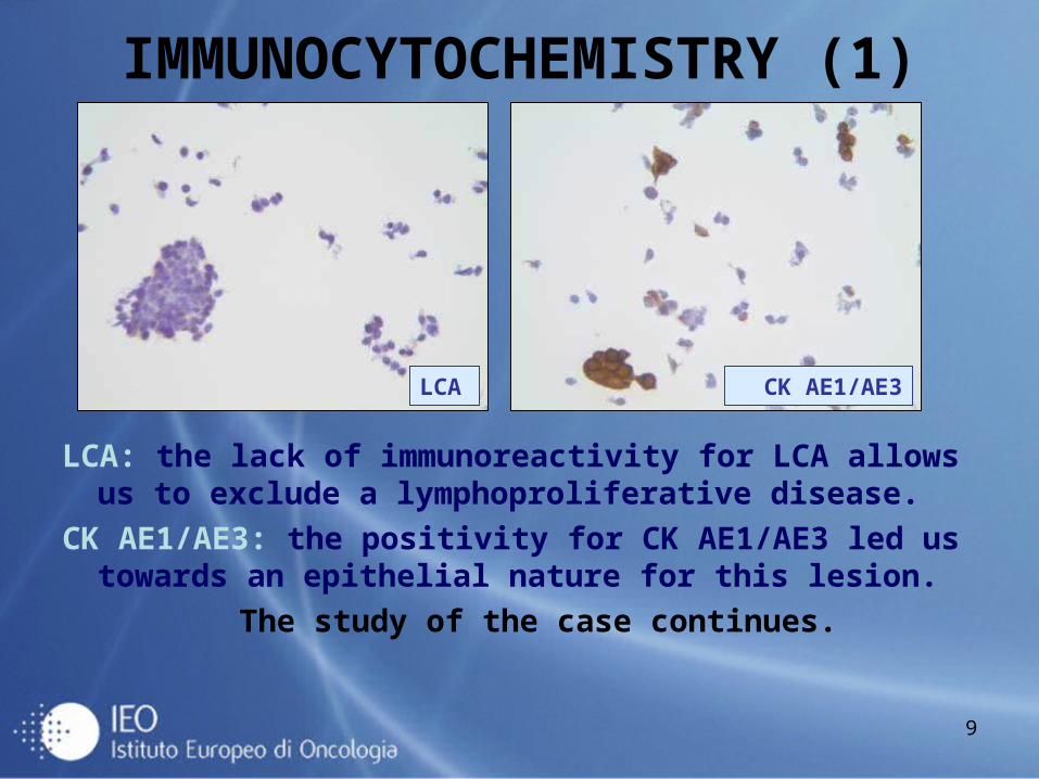
IMMUNOCYTOCHEMISTRY (1)
LCA: the lack of immunoreactivity for LCA allows us to exclude a lymphoproliferative disease.
CK AE1/AE3: the positivity for CK AE1/AE3 led us towards an epithelial nature for this lesion.
The study of the case continues.
LCA CK AE1/AE3
10
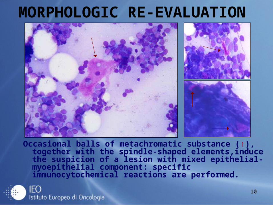
MORPHOLOGIC RE-EVALUATION
Occasional balls of metachromatic substance (↑), together with the spindle-shaped elements,induce the suspicion of a lesion with mixed epithelial-myoepithelial component: specific immunocytochemical reactions are performed.
11
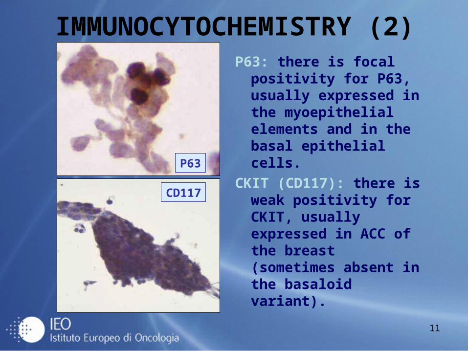
IMMUNOCYTOCHEMISTRY (2)P63: there is focal
positivity for P63, usually expressed in the myoepithelial elements and in the basal epithelial cells.
CKIT (CD117): there is weak positivity for CKIT, usually expressed in ACC of the breast (sometimes absent in the basaloid variant).
P63
CD117
12
OUR DIAGNOSIS
Presence of malignant tumor cells, epithelial-like and spindle, of small size, with scant or absent cytoplasm and with occasional prominent nucleoli, isolated or in large masses, sometimes with the phenomenon of molding, often associated with chromatin streaking.(C5 according to the European Guide-lines-1997).
13
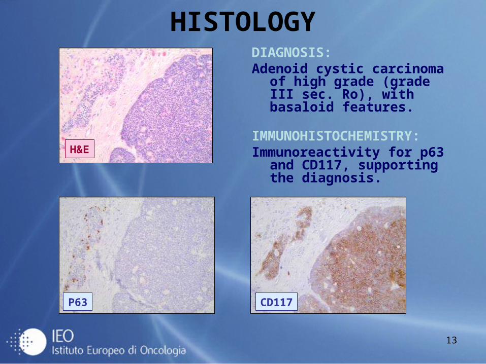
HISTOLOGYDIAGNOSIS:Adenoid cystic carcinoma of
high grade (grade III sec. Ro), with basaloid features.
IMMUNOHISTOCHEMISTRY:Immunoreactivity for p63 and
CD117, supporting the diagnosis.
H&E
P63 CD117
14
CONCLUSIONS• ACC of the breast is a rare neoplasm (0.1-1% of all
malignant breast tumors).• The cytologic picture of well-differentiated ACC is
characterized by amorphous, hyaline material associated with a biphasic cellular component.
• In our case, the scarse hyaline material and the predominance of small-sized epithelial cells, often fragile or in masses with the phenomenon of molding, rendered difficult the differential diagnosis with other rare breast neoplasms of less favorable prognosis, such as small cell carcinoma (2-5%) and lymphoma (0.05-0.5%) .
• A careful analysis of the cytologic picture led us to suspect the presence of an epithelial-myoepithelial lesion, whose definitive classification was made on the surgical specimen.
15
BIBLIOGRAPHY• Mastropasqua MG, Maiorano E, Pruneri G, et al.
Immunoreactivity for c-kit and p63 as an adjunct in the diagnosis of adenoid cystic carcinoma of the breast. Mod Pathol 2005; 18: 1277-1282.
• Law YM, Quek ST et al. Adenoid cystic carcinoma of the breast. Singapore Med J 2009; 50: 8-11.
• Alis H, Yigitbas H, Kapan S, et al. Multifocal adenoid cystic carcinoma of the breast: an unusual presentation. Can j Surg, Vol.51, No. 2, April 2008.
• Kasagawa T, Suzuki M, Doki T, et al. Two cases of adenoid cystic carcinoma: preoperative cytological findings were useful in determining treatment strategy. Breast Cancer 2006; 13: 112-116.