cytoplsmic bodi m lar and me sized neurons. we present, is por alog
TRANSCRIPT
CYTOPLASMIC INCLUSIONS IN GANGUON CELLSASSOCIATED WriT PARKINSONLAN STATES
A NFUROCULAR CHANGE STUDIED IN 53 CASES AND 206 CoNwoLs *Im IL Lwm, M.D.
From tkisesc Albwt R.swdAlugegoS1tesh Cfroc Dimsad 9h Depwext of Pathology, Stae of New Yor,
Dousate Melol Caff,srhy, N.Y.
Among the less well known forms of neuroceul r tthe casi occurrence of discrete, nonganuar, po to ccytoplsmic bodi m lar andme sized neurons. We present,they are usuay in te pgmented cells of thenigra and other "n n "in ng in cells of the braminstemi. Sinc Lews2 d in92 of these structuresm the axsa ticuis and the dorsal vagl n s, they have beensubjected to only infreunt and itermittent ive tio ToTr&iakoff8 must go the cedit for describi th bodies i their mostcommon site of occurrence, the antia nigra, and the dem ationof a possible reationship to paralysis agitans. Hassler' stated tat hehad never sem the bodies in the sile bra, while Kla ques-tioned thmeir specificity in dithi parins Grf d andBosanquetinm presented the first adequat derplion of theLew body i English i it as quite speific for paralysisagtans when it occurred m a t with of ceraincell groups of he sntia nigralThe _pr study has the twofoldobjective of de inin the rea iD of these bodies to the Iousforms of park im and of arifying furtl the natue of thecyoamc alerton.The terms `paralysis aitans" and "idiopac parinism" w
be used mtng y is por Alog with'parkinim " as defined below, they are nmplying aspecific etiologic factor or lack of knowledge t f. Parinnstates" and parnism" (tout moying adjectve) are em-ploed as synonymous i ms referrig to a c al slla-tion without reference to eti or pa lesi
SEE ON OF CASESGroup A
This group cone d of 53 pats with parinsnism of all typesa at had had coete ne examination during the periods*nPe,nteIi part at the Fhfty-fourth AnlM of the American Auociatiee
of Patbolos and Bacte sts OQd, bio lA 6, z9S8&Reed for pbAn Fdbuay ISO 19S9.
1117
1948 to 1957 at the Jewish ChroncDisase Hospital and 1954 to 1957at the Kings County Hopital. Patients xhiiting other extrapyra-midal syndromes were excluded. Subdivision into 3 caegoides bydetailed review of the clinical records, acoding to the criteria estab-lished, was carried out indep tly of the nion for Lewybodies.
Posteweplsaiti Parkinsonism (9 cases). Twenty-two cases in groupA wer considered dinically to be ofphl tic orig, but only 9of these met the cnteria dted below. No patient was retained i thiscategory in the abnce of the following features, considered validind tions of en litis: (i) The esode occurred during the periodI9I8 to 1930. (2) At least 3 of thefolowing observations were specifi-cally recorded: (a) fever; (b) s e ; (c) d banceof sleeppattern; (d) oculogyric cses; (e) other oculomotor sgns, e.g., his-tory of diplopia, etc.; and (f) other evidence of diffuse central nervoussystem disorder.
In addition to the above, at least 3 of the following criteria wererequired: (a) oculogyric crises sbsequent to the acute episode;(b) evidence of other oculomotor damage of nontransient nature, per-sisting beyond or developing after the is; (c) persistentsleep diu ; (d) onset of parkinn symptoms (at least twoof the following: tremor, rigidity, masklike fes, ness, eyfatiguabilit, weakness of voice) within io years of the onset of theencephalitIc episode; (e) rapid pro ion early in the course, withdominance of rigidity within 5 years of onset; (f) sialorrhea; (g) ap-pearance of tics subsequent to the episode of encephalitis. Age ofonset was not considered a crterion smice is constituted one of thevariables under study.
Uxdassified Parkinsomax States (I5 cases). This subdivision in-duded the I3 cases id to be postencephalitic on clinicalgronds but which could not be included in the potenephaltic groupas previously define TWO addial cases with the parkinsoianstate, in which an ad ae histoy was lacing or m which demisefolowed too rapidly for adequate cification, were also included.
Paralysis Agitaxs (29 cases). This subgroup consisted of thosepatetsmi whom the onset of parkisnm wa insidious, progressionwas gradual, initial manifestations were often llid, but eventualinvolvement of all exeiI was recorded. All the patients wereconsidered by the cinica observers to have the idiopathic type ofdisorder, i.e., true paalysi agitans.
xII8 LrpxN Vol. 35, No. 6

De,P INSONIN STATE
Group B, ControlsThis group consisted of 206 consecutive adults who had had com-
plete necrops e nons in 1957. These remained after detaiedindependent review of the dinil ds had elimiated all possblecases of park i or those m which information was insuietto alow d evaluation.Matcked Cotrols (Io6 cases). AU,mnbers of group B were
listed in numerical order of necops numbers with indications of age,sex, and race, but without reference to or kno of the pnceof Iey bodies. E ch case of parkino was n in order andmatched with the first two listed cases of the same age, sex, and race inthe control group. In those intances in which was not possible,the case was held until succeeding cases were mathd he unmatchedbalances were then itymatced h an allowable age rangeof +I-, then + 2, and finally + 3 years. In no instance was the re-quirement for corresponding race or sex relaxd, and it was un
sary to exceed the 3-year range.
TADT IAge and Dwatiox of Mxen ix 53 e of Prkinxox States
Me am Mm d. f m
Puki.samiau sain At oix At dm e Total
P Nfephaic 294 ± 3.6t 50.5 ± 2.9 28.5 ± 3.1
Iithic so6 3.9 65.51+3 10.3 14Uncdassfied 414 3.3 6z.6± 22 2I.6 2.5
ANp momastates 46.9 ±1I 6I.6 ±I2 45 + 14 22.0 2.8 I6.6 ±.3
* msanc in wich angion it toa- bodies we-re cunaecdt TIe val givaeare tb etic mean tbotheadard error.
Unmached Coxtrols. The contrcasesremang foflowing thematching procedure were divided into two groups usig the mean ageof the parins state group as the arbitrary point of divison(6i.6 years); 41 cases we younger and 59 we oder than this age.
METHODSMorphologic Ixwestigatiox
Sections from all case, study and control, were screened for thepresence of Lewy bodies in a single slide, 5 to 7 pL i thickness, andstained with hematoxylin and eo When the slide repreented onlya midbrai hemition, 2 such were screened. On each lide an area
Nov.-Dec, z959Q III9

outlined with India ink, encompassing the entire substantia nigra anda broad adjacent zone, was subjected to a systematic cell-by-cell ex-amination at a magnification of 44o to 66o times. Sections of cortex,thalm us, caudate nucleus, putamen, globus pallidus, pons (icludinglocus caeruleus), cerebellum (roof nuclei as well as cortical structures),and medulla (including dorsal vagal nucleus and inferior olive) wereexamined in an identic manner in I4 selected cases of parkinsonianstates and in 4 of the controls in which the bodies were encountered onexmination of the suantia a.The criteria established for the Lewy body were: (a) intracyto-
plasmic location within a cell identifiable as a neuron (to be considereda neuron, a cell was required to ontain either a typical neuronalnucleus, or identifiable Nissl substance, and preferably both; the pres-ence of '%euronelanin" pigment alone was not considered sfficient);(b) anuaxity; (c) polychroatoplia; and (d) either a sur-rounding halo or clear zone or a central density visible under phasecontrast microscopy. Adherence to all criteria was required in orderto exclude such structures as extruded "hypertrophic nucleoli" (Wolf-Orton "indusions"7), as well as conglomerated eosinophlilic "proto-melanin" granules. An arbitrary grading stem was employed asfolows: If i to 5 Lewy bodies were present in the entire section or in 2hemisctions, the section was rated as I+; 6 to 20 bodies, as 2+; andgreater than 20, as 3+.The sections in both the parkinsonian and control series were taken
from various levels, but the large majority from both groups repre-sented the caudal half of the extent of the substantia nigra. Followingscreening, systematic at a magnification of ioo or I50 timeswas performed to form an impression as to the relative number ofpigmented cells.
Histockemical InvestigatioxThe following staining procedures were carried out simultaneously
on selected sections of all categories of cases in which bodies were en-countered: Periodic add-Schiff; Feulgen'; methyl green-pyronin(2 methods9'0); azure B11; aci-dine orange fluorescencel2 at pH 4.8,both with and without ribonudeaseU; io per cent perchloric13 and 5per cent trichloroacetic acid14 pretreatment; tests for lead15 and ironl;chromotrope aniline blue'; trichrmeM on zenkerized secidons; SudanIV in isopropyl alcohol; Sudan black B in propylene glycol; phosphin3R fluorescence'7; and acid-fast stains.18
1120 TLIPKIN Vol. 35, No. 6

PARKINSONIAN STATE
REsuLTS
The relationships beween the par n states on the one handand the ages at onset, at death, and the duration of ifiness, on theother, are smmaz in Table I. There were 29 males and 24 femalesin the group with parkinian states. The mean brain weights (± thestandard errors) in gams were: for all patients with parkinsianstates, I,234 ± 27; for males, 1,285 + 26; for fles, I,145 ± 30;for the patients with the postencephatic variety, IJ38 + 62; and forthe ets with idiopathic disorder, 1,238 + 26.Encephalomlacia was noted in 7 cases, 5 of which were in the
idiopathic group and one each in the u fied and postee icgroups; none showed involv t of the s tia nigra Grossatrophy of the globus paflidus was observed in 9 of 25 cases (36 percent) where tis structure was described in ent detaiL Conclu-sive m OSCOPic evidence of pallidal atrophy or degeeration wasnoted inmo of 34 cases (88 per cent). No valid quantitative estimateof the degree of either atherosclerosis or of arteriolar sclerosis waspossible.
In 45 of the 53 cases (85 per cent) of parinnm, specific men-tion was made of the gross a a of the substantigra and inall of these some degree of pigment loss was described However, noquantItae criterion regarding this change could be tablished fromthe d ions of several different prosectors.
In the matched control group, 21 CaSeS (19.8 per cent) exhibitedgrossly evident or microscopically significant lesions, but were withoutcinically recorded signs or symptoms during life which could beascribed to the central nervous system. ThirtY-five cases (33 per cent)were without morphologic or cinical evidence of central nervous sys-tem involvement while 42 cases (39.6 per cent) exhibited both asignifict cerebral lesion and clinically manifest evidence of neuro-logic disorder. In 8 cases there were no morphologic lesions, but someclinical evidence suggestive of neurologic dysfunction was mifestThe commonest lesion encoutered in the control group was en-
cehonlacia. This was presnt 44 cases (41.5 per cent) or in69.8 per cent of all cases with significant morphologic alteration in thebrain or cord. Other conditions present were su v meningitis,tuberculous meningitis, bran tumors (astrocytomas, grades II and IV,and meningioma), brain cs and, in a sine instance, cryptococcalmeningitis.
Nov.-ec. zgSp 1121
In none of the cases in either of the control groups was any specificmention made of the ite at of the globus pallidusor depigmentation of the substantia nigra.
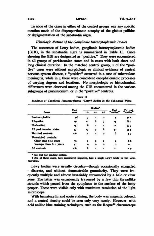
Histogic Nature of the Gangfioxic Ixtracytopasmxic BodisThe occurence of Lewy bodies, lionic tacytoaic bodies
(GIB), in the substantia nigra is summaized in Table II. Casesshowing the GIB are deat as positive hey were encoumteredin all groups of prkin n states and im cases with both short andlong clinial durationI the matced control group, 2 of the "pos'tive" cases were t morphologic or clinical evidence of centralnervous system disease, I positive" occrred in a case of tuberculousmeingitis, while in 3 there we coident e al c procesesOf varying degrees and locations. No morphoogic or eicaldifferences re observed among the GIB encutered in the varioussubgroups of parkinsonism, or in the "positive" controls.
Tia. II1sc~kuc of Gaugluc lx1raoo%aic (Levy) Bodes ix the Substetis N-p
Total TOa iW emtGRt, C I+ 2+ 3+
Postencphalitic 9t 3 I 0 4 44.4Idiopathic 29 12 6 7 25 86.2Undainihed IS 8 2 I II 73.3ARp I s 53 23 9 8 40 75.5Matw tr I 06 4 2 0 6 S.?mtded -oIrlo:Oder than6ys 9 2 0 2 4 6.8Youngr tlhn 6i years 41 o o 0 0 0
Allcontro 206 6 2 2 I0 4.9
* See txt for grvang sytemt One of te c hu,ee Iered ae,hd a ne Ly body in the loan
Lewy bodies were usually circular-though ccasi d_-dicrete, and without demonstable ganularity. They were fre-quently multiple and alnost ivariably srn by a halo or rzone. The latter was occasonay traversed by a few tin eadkestrands which passed from the cytopasm to the surface of the bodyproper. Thee were visible only with maimm resolution of the lightmiLcroscope.With hmatoxylin and eon staining, the body was magenta colored,
and a central density could be seen only very rarely. However, withacid aniline blue staining tiques, such as the Roque"' chromotrope
Vol. 3_5, No. 6I1122 NIPI

PARKINSONIAN STATE
aniline blue, the bodies stained a uniform blue and fairly frequentlyexhibited a well defined bright red staining etral density. Differ-ential sfaining of the core was also observed with the Masson techniquewhere the core was stained red. The cores were demonstatd in allvarieties of the parkinsian state as wel as in the "positive" controls.The bodis did not take the periodic acid-Schiff or Feulgen stains.The Mallory lead and Perls iron stains were negative.
Azure B stained these bodies uniformly a slightly deeper blue thanthe very pale blue of the surrounding cytoplasm, and much less in-tensely than the remaining Nissl substance. Tlis slight blue stainingdid not occur when preceded by tratment with ribouclease, trichloro-acetc or peclonc ads. With the methyl green-pyronin e,the GIB were stained a faint red, while with the acidine orange pro-cedure, they were pale red or pink when observed with ultravioletlight exctation The latter staining reaction did not occur in sectionspretreted with trichloroacetic and perchloric adds.
In every instance where a nudeus was observable in a neuron con-taining a GIB, the former structure was eccentric and most frequentlylocated at the pole of the c d etrically opposite the Ley body(Fig. 3 and 4). Pyknosis was uncomon, and frank karyorrhexiswas not encountered. Occasionally, a b clete neumon was se tocontain the body (Fig. 8). Multiple refractile eosnophlic intranu rstructures (Wolf-Orton bodies), although frequent enough m the vicin-ity of GIB-contaning cels, were observed to occur in the same cell asthe GIB in only 3 instances.
In Lybodyontaining neurons, the Nisl substance was some-times absent, but more often was reduced to a thin, deeply basophilicperipheral rim (Figs. 2 to 4), while the major part of the cytoplasmwas uniformly and finely granular and only very weakly basophilic.When, as usual, these cells contained pigment, the number of "neuro-melanin" granules frequently appeared to be less than that in adjacentcells. There was also a distinct tendency for the remaining pigmentgranules to cluster around the GIB and, by contrast, to accentuate thehalo, forming an almost semicircular ttern (Figs. 3 and 4). Cyto-plasmic vacuolation was rarel encountered (Fig. 6). Still more rarely,vacuolation coincided in spac with the halo. This was noted mostfrequently when phase microscopic examination or acid aniline bluestaining revealed a particularly prominent or dense central core (Fig.7). ExcePt in those instance where the degenerative cnges theneurons were particularly advanced, satellitosis was not observed inrelation to neurons containing GIB.No objective graing system could be established with respect to the
Nov.-De- z95!9 I1123

degree of cell loss. Although in afew cases of idiopathic parkinson- .}* W
a 0 "0 uoism, appreciable cell loss was not Sdemustrated, in most of them this Q S
ocurred to a variable degree. In all 0-a %" 0 0 nof the cases of tic o 0o o opa.rknsoism the paucity of neuronsinthe was minme-in X Ps~US nigra Was ime._ HeO+i
diately apparent and was accopan- . _ _ied by an icreasei galcells. The 0 0 0occurrence of GIB in areas other a W
i i B- r g n^i _^ stathesubsata igrasswnmmar- -.O%.O%.
ized in Tablem. 0000
DISCUSSION
It is fairly ally hed that OV O O O O
the substana nigrais more orlessaffected in all parkin states ai.(with the exception of the rare in-_stance of so-caled " nigral" t 0 0 0 0parkinsonism). Greenfield"' conid- - . 8ered" pa.rasisagi s.. .a 3L.zsystematic degenerationof aspial a o a0 0 0 0 P
type affecting a neuronal system b , 6whose nodal point is the substatia . 0
nigra."Hassns"sdemorstration of 0 0_greater involvement in the 8poencticf m,dl K s4 d 1t fom ||ssle9desciption of preferential zones of ; 91 0
cel destruction which he considered j 0characteristic of rtain forms of ;_parkiin , while firming the %.'O5universalty of mv mentofthis . ! 0 2pigmented structwe, n eith- Z .eer instancebe used asmorpologic xcriteria M t classificat oof anindvivdual case. Since the work of .xTretriakoff,3 all authors specificaly -- 4. .conceredwith theGIBand its rela- |otionship to parkin an states have A i nagreed on its occurence m the sub- E7 ,stantianigra in cases of paralysis
LIE}EN Vol. 35, No. 6I1124
PARKINSONIAN STATE
agitans. Its appearance in other regions in this disease is also notdisputed; for ple, Beheim-Schwarzbach demonstrated "Massonpositive vacuoles" in the locus caeruleus.2'
However, the present observations in cases of postencephalitic park-insonism are at variance with the conclusions of earlier authors, withthe major exception of Klaue.5 It would appear (Table II) that one ismore liely to encounter a Lewy body in a section of subtantia nigrain a case of idiopathic than in postencephalitic parkinoism Sincethe work of Beheim-Schwarzbach and the present investigation indi-cate the occurrence of these bodies, albeit infrequently, in patientswithout parkinsonism, it is necessay to exmne the possiblity thattheir appearance in postencephalitic parkinsonism may be due to merechance. Statistical analysis of the incdence data shows that this is notthe case (P<o.ooi).
In examples of the postencephalitic variety, the sparse occurrenceof Lewy bodies may be due to the greater severity of involvement ofthe substantia nigra in this condition. This has been described byHassin, and is supported by observations in the present study as indi-cated by more uniform and severe neuronal loss, accompanied by glialcell icrease. The infrequent apparance of GIB may also be relatedto factors which are readily apparent in Table I. The patients with thepstecephalitic type of disorder, although they developed the diseaseat an earlier age and succumbed at a younger age than those with theidiopathic parkinsonian state, have had a very significantly greaterduration of illn. In this connection, the well known progression ofthe signs and symptoms of all forms of parkinsonism (initially morerapid in the postencephalitic variety) should be considered along withthe data in Table II concerning the relation between the incidence ofLewy bodies and the duration of the disease. A "positive" case (meanduration, I4.5 + 4 years) is more likely to be of shorter overall dura-tion thanis a"negative" one(22.0 + 2.8years). If it is accepted thatduring the course of any form of parkinsoism there is more or lessgradual but progressive degenerative loss of neurons, it is then possiblethat in the postencephalitic form, as a result of the greater duration,fewer neus exist which are capable of harboring these bodies.Scnning the substantia nigra in the control series did not reveal
appreciable differences with respect to the number of neurons presentin "positive" as compared to "negative" cases.The histochemical procedures employed in this study contnbuted
little to the erization of the bodies. The amount of ribonucleicacd must be very smal indeed since the staining reactions for it wereso very weakly positive. The negative Feulgen reaction would appear
Nov.-Dec@, z959 1125

to eliminate the possibility that deoxyribonucleic acid was present indetectable amounts. The negative periodic acid-Schiff reaction indi-cated the absnce of complex carbohydrates. Moreover, no fat, lead,or iron could be demonstrted. Thus, by e sion, it seems likely thatthese bodies are of protein nature.The resemblance of the Lewy body to that seen in Pick's disease is
quite stiking. Greenfield and B et found that the only demon-trable difference was the e of a central core, a sowhat tenuousdistinction which is not further carified by more recent studies relyingalmost eclusively on silver 2mpregnations.2 The differentiation fromthe bodies observed in myoclonus epilepsy ts primaril on theirmetachromatic prrties n the latter condition, firstd na byLafora and Glueck.~2The appeance of the GIB is somewhat recent of the Negri body, but it does not ehibit the basophiTic partidesor the Feulgen-poitive internal struture d rt by Moulton."Other cyto c io , such as those of cytomegalic inclondisease and the Guarnieri body of m , are his mially andmophologically quite distinct2.The observations of the neuon which ontains the Lwy body con-
tribute to anun t of this strcture. In no case was a GIBfoumd in an otherwise normal neur Evidence that the neuron hadundergone some form of degeneration was indicated by eccentricity ofthe nucleus, less commonly encountered nuclear abnomalities, mar-gination and loss of Nissl substance, ap t partial loss of pand caracteristic arrangement of its remnts, and the occurrence ofcytopasmic vacuolation. In instances in which multiple GIB urredwithin the same cell, it was not unusual for some of the bodies to hibitvariations suggestive of a progressive change. These observations, con-sidered in conjunction with the data related to incidence and the histo-chemical features, suggest that te bodies rep t a nonsecific formof degenerative alteration.A final consideration bearing on this point is the preferential occur-
rence of the bodies in pigmented neurons. Thea of pigmenta-tion in the substanti nigra is phylogenically' as well as ontogenically"a relatively late event. While there is no avaiable information as tothe function of the pigment in these cls, it is rtain that at one timeor another in the early portion of the life span of the neuron, this sub-stance must be formed. It is uncertain whether or not the process offormation is continuous, but it is not uncommon to observe in other-wise intact neurons in the substantia nigra of normal individuals,collections of granules of the same size and shape as the "neuromelaVin"granules which are unpigmented and eosinophilic. They are similar if
I1126 LIPEN Vol. 35, No. 6

Now.-Dec 59 P9N STATE 1127
not identil to those dcribed many years ago by Marins as anearly manifestation of the process of Amtion. In any case thepigmented ganglion cells of the subtantia nigra must be consideredeither to be experencng or to have experienced an additional metabolicdemand not made upon other neurons of the ntral nervous systemwith the excepton of those of the locus aeruleus and the dorsal vaglnucleus. These regions, perhaps not coincidentally, are the next mostfrequent s of occurrnc of the Lewy body.
SUMARY
Brnssue from 53 patients suffering from parkinsoian st and206 pats without parkinsism, serving as controls, was systemati-calgy m in relation to the occurrence of ganlionic itacyto-plaic bodies. These dIstInive structures were found in thesubstantia nigra of 75 per cent of all cases with parkin n states(44 per cent of the cases with the potencephalitic variety and 86 percent of those with the idiopathic varety), and i 4.9 per cent of thecontrol series. Histochemial, morphologic and statistical data arepresented, indicating the probability that these bodies represent a formof nspeciic ne llular degerat, posibly of abiotrophicnature.
REFERENCESz. Lewy, F. H. Pmalysis Agitans L Patholoch Anomi In: Hndbuch
der Neurobgic. dowsky, IL (ed.). Julius S r, Bin, 1912, pp.92933.
2. Lwy, F. EL Ced by Greenfidd and sauet3. Trfiakoff, C. Contribuon i raude de l'anatomie p i d us niger
des ng avec q s d ns ives i honie destroubles du tomn mucire et de a maadic de Panson. Thkse de Pais,1919, I124 pp.
4. Hassler L Zur Pathologie der Paalysis agitans und des posenzeDhalitischenPalki-soismus J. Psychol. Newol., LeiPzig, 1938, 48, 387-476.
5. Klaue, R. Padknsonischc Krankheit (parlysis ns) und -itishr rkins s. Versuch er klinisch-aatomiSdn Differentialdiagnose. Arch. f. PsyCi., 1940, III, 25I-321.
6. Greefied J. C., and Ba , F. D. Ihe brain-stem lesions inkinsJ. Nerol. Nerosug. & PsYckia., 1953, i6, 213-226.
7. Wolf, A, and Orton, S. T. The occ e of intranuclear incsons in humannerve cells in a varety of diseases. BxU. Newol. Ist., New York, 1932,2, 194209.
8. Uie, R. D. Hi oic Tecic and Practical H my. TheB hakston Co., New York, 1954, (a) I56; (b) 132-133; (C) 243-244;(d) 35I-352.
9. Kurnick, N. B. Htistological aining with methyl-green-pyronin. StakTechuol., 1952, 27, 233-242.

1128 UPKIN Vol. 35, No. 6
Io. Jordan, B. HL, and Baker, J. R. A simple promethyl green tehnique.Quart. J. Micr. Sc., 1955, 96, 177-179.
IS. Brachet, J. The use of basic dyes and ribonudase for the cytochemial detec-tion of iboncic acid. Quart. J. Micr. Sc., 1953, 94, 1-10.
12. Armstrng, J. A. hemical diffentiation of nuclic acids by means ofnduced fore Ezper. CeU Res., I9S6, iI, 640-643.
13. Koenig, HL, and Stahecker, HL Futher studies on the difftial eractioof nucleic acids from ma lian nerve cells with prchloric acid. (Abstractand discussion.) J. Nat. Caxcer Ixst., I95I-I952, 12, 237-240.
14. Hims, KI B.; Rii, R.; Hoffman, J.; Pollster, A. W., and Post, J. Cyto-plasmic ribonlc acid in the human li celL A. M. A. Arch. Path., I9,58 345-353.
I5. Mallory, F. B. Patholgical Technique. W. B. Saunders Co., Philadelpia,1938, p. 143.
i6. Roque, A. L o aniline blue method of s Mallory bodiesof Lanec's Cir Lb. Ie., 1953, 2, I5-21.
17. Popper, H. Histogic d tion of vitamin A in human orpns nder maland nIdr ic conditions. Arch. Path., '94', 31, 766-802.
I8. Wolf, A., and Pappeheime, A. K Ocrremnce and iributio of acid-fastpigment in the centrl nervous J. Newopath. & Ezper. NewroL,1945, 4, 402-4o6.
I9. Greenfied J. G. The Pathology of Parkinson's Disease. In: James Prnson.A Bicentenary Volume of Papers Dealing with inson's Diase, Incor-porating the l "Essay on the Sha Palsy." Critchley, KL (ed.)Macmillan & Co., Ldon and NeW York, 1955, p. 239.
20. Hasin, G. B. Hioathology of the Periphral and Central Nervous Sygems.Paul B. Hoeber, Inc-, New York, 1940, pp. 386-387.
21. Beheim-Schwarbach, D. Uber Zellib-verindmgen im Nucleus c ulbei sonian-Symptomen. J. NerC-. & Mext. Dis., 1952, II6, 6I9-631.
22. S enbeg, L The histoltic ructure of the "incluion bodies" of theneurns in Pick's disease J. Newropa. & Rxper. Necw., 1958, 17, 346-35I.
23. Lafora, G. R., and Ghek, B. Beitrag zur olie der ischenEpilepsie. Ztsckr. f. d. ges. Neuol. u. Psyciat, Ig9I, 6, I-I4.
24. M outn, J. K A histochmical study of the Negri body of rabie Am. J.Pat., 1954, 30, 533-543-
25. Dyckman, J., and BeIlamy, J. Histochelical stues in c alic inclusiondisease A. M. A. Arck. Path., 1953, 56, 360-363.
26. Wolman, M. Pathoic findings in hmorgic sma (Pupura variolosa.)Report of a casem with special refereuce to Feulgen's reaction in tissue. Am.J. Cli. Path., 195I, 21, 1127-1138.
27. Adler, A. Melanin p t i the centl nervo m of vert tes.J. ComP. NeroL, 1939, 70, 3I5-330.
28. Foley, J. K, and Baxter, D. Observations on the morphology of the cells ofthe lcus coeruieus and ania nigra in infants. (Abstract and discus-sion.) J. Neuropatt. & Ezper. Newol., 1956, 15, 219-221.
29. Marmnesco, G. La Celhue Nerveuse. Pais, Igo. Cited by Tr3tiakoffisSincere apption is creyed for the rnagnat advice and cooperation of
Dr S. KLAronson, B. W. Volk and P. J. Fi d Mr. Herbert Fpdher peadthe Otom His aid and the ch of Glr Ros and
i ldrd Ambi are gafull ackno
PARKNSONIA STATE
[IM f
NOV-kc. t95f9 1129

Vol. 3S., No.6
LEGENDS FOR FIGURESEetwher Mned lustrations wer jprepared from scions stained with
winaoynad f BS
FeG X. Ganlon ce cotinin a sine typica, wedmed Lewy body. Tleunelets is at the bwer pole of the ce, out of the plne of focus. Post-aicepimitic p 1rkinsm. X 980
FM. 2. Neuro containing a hrga toughedknse Ley body. Tke bal is kswell drfined. Postencebhalitic pakinsaniun Substantia nigra. X 200
F.3. Two werons, bth s m palmic i c boswhich vary in and tIhe de of d r of suou losNote the e ra m of r i Nisd sds ane ad the fan grbut hise feate appancoe of the cytoplsm. Parlysis agitans.Substantia X 980
FoG 4. Two nwurons in the lo. c-aeruleus Lewy bodie with de-grees of tiatio Palysis X
I1130 IPMEN

PARKINSONIAN STATE
2
3
Nov.-Dcc, I939 I113 I
I
4

Vol. 35, No. 6
FIG. 5. Neurons undergoing degenerative change containing 3 distinct GIB, ofvarying degrees of "mturationP" The larest bodv has an unusually wide haloand shows a somewhat variable density. Two glial cells are in the immeiateVicinity. ParalYSiS agitanS SUbstantia nigra. X I200.
FIG. 6. Ganglio cell with a Lrge cytoplasmic vacuole and 3 dense Lewy bodies.Note absence of "neuromelanin" pigment. Paralysis agitans. Pons. X i200.
FIG. 7. Neuron, from dorsal vagal nucleu, with maly eccentric el Ilnuckus (N). ITe large GIB (edges indicated by arrows) shows a deeplychromatropophilic (red) central density. The cytoplasmic vacuolation en-croaches upon the halo. Note the granui of the cytoplasm. Paralysisagitans Roque's chromotrope 2R-aniline blue stain. X 1200.
FIG. 8. A binucleate neuron (N) with GIB showing "frayed" irular edges(arrows) and cytlsmkic vacuolation encroaching upon the halo. Parablysagitans. Substantia nigra. ChroMOtrope 2R-anilie bhle. X I200.
I1132 LIEPKIN

PARKINSONIAN STATE
5
1'
_j).
Orr,
OWN,}
,I;.
B
Nov.-Decc, i95!9 I1133
6