dctbp mediates transcriptional repression by knirps, kru¨ ppel and
TRANSCRIPT
The EMBO Journal Vol.17 No.23 pp.7009–7020, 1998
dCtBP mediates transcriptional repression by Knirps,Kruppel and Snail in the Drosophila embryo
Yutaka Nibu, Hailan Zhang, Ewa Bajor1,Scott Barolo2, Stephen Small1 andMichael Levine3
Department of Molecular and Cellular Biology, Division of Genetics,401 Barker Hall, University of California, Berkeley, CA 94720,1Department of Biology, 1009 Main Building, 100 Washington SquareEast, New York University, New York, NY 10003-6688 and2Department of Biology, Bonner Hall, 9500 Gilman Drive, UCSD,La Jolla, CA 92093-0347, USA3Corresponding authore-mail: [email protected]
The pre-cellular Drosophila embryo contains 10well characterized sequence-specific transcriptionalrepressors, which represent a broad spectrum of DNA-binding proteins. Previous studies have shown that twoof the repressors, Hairy and Dorsal, recruit a commonco-repressor protein, Groucho. Here we present evi-dence that three different repressors, Knirps, Kruppeland Snail, recruit a different co-repressor, dCtBP.Mutant embryos containing diminished levels of mater-nal dCtBP products exhibit both segmentation anddorsoventral patterning defects, which can be attrib-uted to loss of Kruppel, Knirps and Snail activity. Incontrast, the Dorsal and Hairy repressors retain atleast some activity indCtBP mutant embryos. dCtBPinteracts with Kru ppel, Knirps and Snail through arelated sequence motif, PXDLSXK/H. This motif isessential for the repression activity of these proteins intransgenic embryos. We propose that dCtBP representsa major form of transcriptional repression in develop-ment, and that the Groucho and dCtBP co-repressorsmediate separate pathways of repression.Keywords: CtBP/Drosophila/embryo/Knirps/Kru¨ppel/Snail
Introduction
Transcriptional repressors establish localized stripes, bandsand tissue-specific patterns of gene expression in the pre-cellularDrosophilaembryo (e.g. Rivera-Pomar and Ja¨ckle,1996; Dubnicoffet al., 1997; Jiminezet al., 1997; Nibuet al., 1998; Poortingaet al., 1998). Patterning of boththe anteroposterior and dorsoventral axes depends onbroadly distributed activators and localized sequence-specific repressors. For example, the maternal Dorsalgradient can activaterhomboidin both ventral and lateralregions of early embryos, but the Snail repressor keeps itoff in the ventral mesoderm (Ipet al., 1992). Similarly,the maternal Bicoid gradient can activate theevestripe 2enhancer in a broad anterior domain, but the Giant andKruppel repressors restrict the pattern within sharp stripeborders (Smallet al., 1991, 1992).
© Oxford University Press 7009
Recent studies have identified two putative co-repressorsin the early embryo, Groucho (Paroushet al., 1994) anddCtBP (Nibuet al., 1998; Poortingaet al., 1998). Bothproteins are encoded by maternally expressed genes, areubiquitously distributed throughout the early embryo andare brought to the DNA template through interactions withsequence-specific regulatory factors. Groucho mediatestranscriptional repression by Dorsal and Hairy. Dorsal isinherently an activator, but can recruit the Groucho co-repressor when it interacts with specific DNA-bindingproteins located within the silencer elements of thezenand dpp genes (Dubnicoffet al., 1997). Hairy repressespair-rule genes, such asftz and runt, in early embryos,and later is involved in neurogenesis (e.g. Jimenezet al.,1996). These functions of Hairy have been shown todepend on a specific sequence motif, WRPW, which isimportant for interactions with Groucho (Fisheret al.,1996). The removal of maternal Groucho products resultsin complex patterning defects in mutant embryos, includingdisruptions in both segmentation and dorsoventral pat-terning (Paroushet al., 1994; Dubnicoffet al., 1997).
Recent studies have identified a second putative co-repressor in the early embryo, dCtBP (Nibuet al., 1998;Poortingaet al., 1998), which is theDrosophilahomologof the mammalian CtBP protein (e.g. Schaeperet al.,1995; Turner and Crossley, 1998). CtBP attenuates tran-scriptional activation by the adenovirus E1A protein; itbinds E1A through a specific sequence motif located nearthe C-terminus of E1A, P-DLS-K (Schaeperet al., 1995;Sollerbrantet al., 1996). This motif is conserved in twounrelated repressors in theDrosophilaembryo, Snail andKnirps (Nibu et al., 1998). Gene dosage assays areconsistent with the occurrence of interactions betweenKnirps and dCtBPin vivo. Moreover, the P-DLS-K motifwas shown to be important for the repression activity ofa Gal4–Knirps fusion protein in transgenic embryos (Nibuet al., 1998). The functional significance of the P-DLS-Kmotif in the Snail repressor currently is unknown. TheHairy repressor contains a divergent sequence, P-SLV-K,which raises the possibility that Hairy-mediated repressiondepends on both Groucho and dCtBP (Poortingaet al.,1998).
In the present study, we analyze the expression of anumber of target genes, both authentic and synthetic, indCtBPmutant embryos to obtain evidence that the dCtBPco-repressor is essential for Snail function. Evidence isalso presented that a third sequence-specific repressor inthe early embryo, Kru¨ppel, depends on dCtBP. The C-terminal repression domain of Kru¨ppel contains a sequence(P-DLS-H) that is related to the P-DLS-K motif in E1A,Knirps and Snail; mutations in this sequence disrupt therepression activity of a Gal4–Kru¨ppel fusion protein intransgenic embryos. Dorsal and Hairy retain at least somerepression activity indCtBPmutants, suggesting that they
Y.Nibu et al.
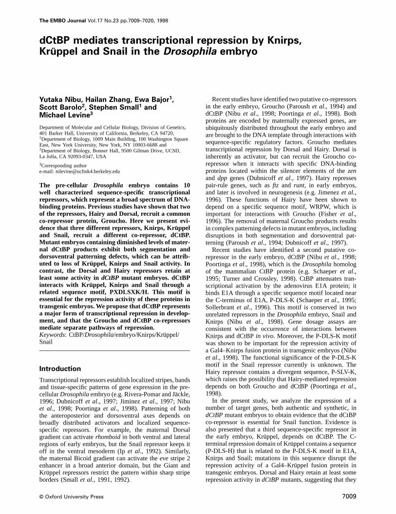
Fig. 1. Altered patterns of segmentation gene expression indCtBPmutants. Embryos were obtained fromdCtBPgermline mosaics and hybridizedwith digoxigenin-labeled antisense RNA probes. The staining patterns were visualized by histochemical staining, and the embryos are oriented withanterior to the left and dorsal up (except K and L). (A andB) Kruppel expression pattern in wild-type (A) anddCtBPmutant (B) pre-cellularembryos. Strong staining is observed in central regions, and a weak pattern is detected in head regions. As shown previously (Poortingaet al., 1998),the Kruppel expression pattern is essentially normal in mutant embryos. (C andD) knirps expression pattern in wild-type (C) anddCtBPmutant (D)pre-cellular embryos. Staining is detected in both a band in the presumptive abdomen and an extended patch in antero-ventral regions. Theknirpspattern is essentially normal in the mutant embryos (Poortingaet al., 1998). (E andF) giant expression pattern in wild-type (E) anddCtBPmutant(F) pre-cellular embryos.giant is expressed in both anterior and posterior regions. The posterior band is expanded in mutant embryos (see brackets).A similar alteration ingiant expression is observed inKruppel mutants (Kraut and Levine, 1991). (G andH) eveexpression pattern in wild-type (G)anddCtBPmutant (H) cellularized embryos. There is a severe disruption in theevepattern, with a loss of stripes 4–6 and a fusion of stripes 2 and 3.This altered pattern combines features seen inknirps– andKruppel– mutants (see I and J). (I ) eveexpression pattern inknirps– embryo. There is aloss of stripes 4, 5 and 6. (J) eveexpression pattern inKruppel– embryo. Stripes 213 and 4–6 are fused into two broad bands. (K ) evestripe 2expression in a wild-type, gastrulating embryo. AlacZ reporter gene driven by the minimal, 480 bp stripe 2 enhancer was visualized with alacZantisense RNA probe. (L ) Same as (K) except that the stripe 2 reporter gene was crossed into a mutant embryo derived from adCtBPgermlineclone. There is a posterior expansion of the stripe 2 pattern, which suggests a loss of Kru¨ppel-mediated repression.
do not require dCtBP as a co-repressor. It would appearthat the bulk of the patterning defects observed indCtBPmutants can be attributed to the loss of Knirps, Kru¨ppeland Snail activity. We suggest that Groucho and dCtBPmediate separate pathways of transcriptional repression.
Results
Maternally encoded dCtBP products are distributed uni-formly throughout early embryos (Nibuet al., 1998;Poortingaet al., 1998), and mutants derived fromdCtBPgermline clones exhibit altered patterns of segmentation,gene expression and severe patterning defects (Poortingaet al., 1998). These mutants also possess disruptions indorsoventral patterning. As a first step towards identifyingthe repressors that might require dCtBP as a co-repressor,we have analyzed the expression of a number of authenticand synthetic marker genes in mutant embryos derivedfrom germline clones, which are homozygous for a P-induced mutation indCtBP (see Materials and methods).
The role of dCtBP in segmentationIn situ hybridization assays suggest that the mutantembryos exhibit a severe reduction in, but not completeelimination of,dCtBPexpression (data not shown).dCtBPmutant embryos exhibit essentially normal patterns ofKruppel (Figure 1B; compare with A) andknirps (Figure1D; compare with C) expression (see Poortingaet al.,1998). These results suggest that the Hunchback (Hb)repressor does not require dCtBP as a co-repressor, since
7010
Hb is important for establishing the anterior borders ofboth theKruppel and knirps expression patterns (Struhlet al., 1992).
There is a substantial expansion of the posteriorgiantexpression pattern indCtBPmutants (Figure 1F; comparewith E). A similar expansion was observed inKruppel–
mutants (Kraut and Levine, 1991), thereby raising thepossibility that Kruppel-mediated repression depends ondCtBP. Further evidence stems from the analysis ofeve.
As shown previously (Poortingaet al., 1998), there isa severe disruption of theeveexpression pattern indCtBPmutants (Figure 1H; compare with G). The altered patterncombines aspects of bothknirps (Figure 1I) andKruppel(Figure 1J) mutants (Fraschet al., 1987). As in the caseof knirps– embryos, dCtBP mutants exhibit a severereduction in eve stripes 4–6. LikeKruppel– embryos,dCtBP mutants display fusions of stripes 2 and 3. Thislatter phenotype might result, in part, from a breakdownin the Kruppel-mediated repression of theeve stripe 2enhancer (Stanojevicet al., 1991). To test this idea, aneve–lacZtransgene containing the 480 bp minimal stripe2 enhancer (Smallet al., 1992) was crossed intodCtBPmutant embryos (Figure 1L; compare with 1K). Expressiondirected by the transgene was detected byin situhybridiza-tion using alacZantisense RNA probe. IndCtBPmutants,the posterior stripe 2 border expands into central regions,similar to that observed inKruppel mutants (or whenthe Kruppel-binding sites in the stripe 2 enhancer aremutagenized; see Stanojevicet al., 1991). These resultssuggest that both Knirps and Kru¨ppel require dCtBP to
dCtBP is a co-repressor of Knirps, Kruppel and Snail
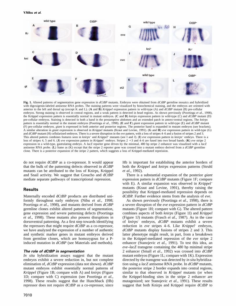
Fig. 2. Altered patterns of dorsoventral patterning genes indCtBPmutants. Embryos are oriented with anterior to the left and stained afterin situhybridization with different digoxigenin-labeled antisense RNA probes. (A andB) rhomboidexpression pattern in wild-type (A) anddCtBPmutant(B) pre-cellular embryos.rhomboidis normally expressed in two lateral stripes along the length of the embryo (A), but is derepressed in ventralregions indCtBPmutants (B). This staining pattern is similar to that observed insnail– mutants (C). (C) rhomboidexpression pattern in asnail–/snail– homozygote. Strong staining is observed in both lateral and ventral regions. (D) snail expression in adCtBPmutant pre-cellularembryo. Staining is observed in ventral regions that normally invaginate to form the mesoderm. This staining pattern is similar to that observed inwild-type embryos and suggests that the derepression of therhomboidstaining pattern seen indCtBPmutants (B) is not due to a loss insnailexpression but, rather, results from a loss in Snail repressor function. (E andF) sim expression pattern in wild-type (E) anddCtBPmutant (F) pre-cellular embryos.sim is normally expressed in two lateral lines that coincide with the presumptive mesectoderm (E), but there is a severederepression in the pattern indCtBPmutants (F). This alteration in thesim pattern probably results from a loss in Snail repressor activity (see Kasaiet al., 1992).
function as repressors. In contrast, neither Hb nor Giantappear to require dCtBP; the latter repressor is requiredfor establishing the anterior stripe 2 border (Smallet al.,1991, 1992), which is normal indCtBP mutants (seeFigure 1L).
Dorsoventral patterningThe Snail repression domain contains both a conservedcopy of the P-DLS-K motif, as well as the slightlydivergent sequence, P-DLS-R. Mutations in the formersequence attenuate the binding of Snail to a GST–dCtBPfusion protein (Nibuet al., 1998). As a first step towardsdetermining whether Snail requires dCtBP to mediatetranscriptional repressionin vivo, the expression patternsof different Snail target genes were examined indCtBPmutant embryos.
rhomboidis expressed in lateral stripes that help specifyventral regions of the neurogenic ectoderm (Figure 2A;see Bieret al., 1990; Ipet al., 1992). It is repressed inthe ventral mesoderm by Snail, and insnail– mutants thereis a severe derepression of therhomboidstaining pattern(Figure 2C; compare with A). A similar disruption of thepattern is observed indCtBP– embryos (Figure 2B). Thisis not due to the loss of Snail products, sincesnail
7011
expression appears to be essentially normal indCtBPmutants (Figure 2D). Thus, it would appear that Snailrepressor activity depends on dCtBP.
Snail represses a number of neurogenic genes in theventral mesoderm. Among these issingle minded(sim),which specifies the mesectoderm at the ventral midline ofadvanced-stage embryos (Nambuet al., 1991; Kasaiet al.,1992). sim initially is expressed in ventrolateral lines(Figure 2E) that coincide with the ventral-most cells ofthe presumptive neurogenic ectoderm. IndCtBPmutants,there is a severe derepression of thesim staining pattern(Figure 2F), again suggesting that the Snail repressorrequires dCtBP as a co-repressorin vivo.
Synthetic transgenesThe preceding studies suggest that Kru¨ppel, Knirps andSnail requiredCtBP1 gene activity in the early embryo.More definitive evidence was obtained by analyzing theexpression of synthetic transgenes indCtBP mutantembryos (Figure 3). Each of the transgenes contains amodified form of the 700 bprhomboid lateral stripeenhancer (NEE), which lacks the four native Snailrepressor sites (Ipet al., 1992). The enhancer directsequally strong expression in both lateral and ventral

Y.Nibu et al.
regions due to the loss of the Snail sites. As shownpreviously (Grayet al., 1994), two synthetic Snail sitespositioned within 50 bp of the NEE activators restorerepression in ventral regions, so that the reporter gene isexpressed in lateral stripes, similar to the endogenouspattern (Figure 3A). However, the same transgene exhibitsa derepressed staining pattern indCtBPmutants, indicatinga loss of Snail-mediated repression (Figure 3B). In theseexperiments, the modified enhancer was placed betweentwo marker genes,white and lacZ, and transgene expres-sion was monitored with awhite hybridization probe.
To assess the importance of dCtBP in Knirps-mediatedrepression, a different version of the enhancer was ana-lyzed that contains synthetic Knirps-binding sites in placeof the Snail sites (see diagram below, Figure 3C and D).In wild-type embryos, Knirps represses the modified NEEso that thewhite expression pattern includes a gap in thepresumptive abdomen where there are high levels of theKnirps repressor (arrowhead, Figure 3C). This gap is lost
7012
in dCtBP mutants (Figure 3D), similar to the situationobserved inknirps– embryos (Arnostiet al., 1996). Theseresults suggest that dCtBP is required for Knirps-mediatedrepression of the modified NEE.
Insertion of Kruppel-binding sites in the NEE resultsin a central gap in thewhite expression pattern (Figure3E). This gap is lost indCtBPmutants (Figure 3F), whichsuggests that dCtBP is also required for Kru¨ppel-mediatedrepression. Further evidence for this possibility stems fromin vitro binding assays (Figure 3G). In these experiments,a full-length Kruppel protein was labeled with [35S]me-thionine by in vitro translation, and mixed with a GST–dCtBP fusion protein. The wild-type protein binds toGST–dCtBP, but not to a GST control protein. Aminoacid substitutions in the Kru¨ppel P-DLS-H motif abolishdCtBP binding (Figure 3G).
dCtBP is not essential for Dorsal or HairyrepressionDorsal and Hairy require Groucho to mediate transcrip-tional repression (Paroushet al., 1994; Dubnicoffet al.,1997). Hairy also contains a weak dCtBP interactionmotif, P-SLV-K, and it has been suggested that Hairy-mediated repression depends on both Groucho and dCtBP(Poortingaet al., 1998). To investigate this possibility, asynthetic Hairy target gene was analyzed indCtBPmutants(Figure 4). The target gene contains a modified NEE withtwo synthetic Hairy repressor sites (Barolo and Levine,1997). In wild-type embryos, the enhancer directs apair-rule pattern of expression (Figure 4C) due to inter-
Fig. 3. Expression patterns of synthetic reporter genes indCtBPmutants. DifferentlacZ–whitereporter genes were introduced intodCtBPmutant embryos and stained afterin situ hybridization with awhite antisense RNA probe. Cellularized embryos are oriented withanterior to the left and dorsal up. (A andB) white staining patterns ina wild-type (A) anddCtBPmutant embryo (B). The reporter genecontains a modifiedrhomboidlateral stripe enhancer (NEE) that lacksthe four native Snail-binding sites, but contains two synthetic sitesflanking the four Dorsal activator sites (see diagram beneath theembryos). The synthetic sites mediate repression in ventral regions bySnail (arrowhead), so thatwhite staining is restricted to lateral stripes(A). There is a severe derepression of the staining pattern indCtBPmutants (B, arrowhead), suggesting the loss of Snail repressor activity.(C andD) white staining patterns in a wild-type (C) anddCtBPmutant (D) embryo. The modified NEE contains two synthetic Knirpssites in place of the Snail sites (see diagram). Normally, the enhanceris repressed in the presumptive abdomen by Knirps (C; arrowhead).This repression is lost indCtBPmutants (D, arrowhead), whichsuggests a loss of Knirps repressor function. (E andF) white stainingpattern in a wild-type (E) anddCtBPmutant (F) embryo. Themodified NEE contains two synthetic Kru¨ppel-binding sites in place ofKnirps or Snail sites (see diagram). This enhancer directs a stainingpattern with a broad gap in central regions in wild-type embryos(arrowhead, E). This gap coincides with regions containing high levelsof the Kruppel repressor (see Figure 1). The gap is lost indCtBPmutants (F, arrowhead), suggesting a loss of Kru¨ppel repressorfunction. (G) GST pull-down assays. A full-length Kru¨ppel proteinwas labeled with [35S]methionine byin vitro translation (lane 1). Itwas incubated with a full-length GST–dCtBP fusion protein producedin bacteria, and the bound protein was recovered on glutathione–Sepharose 4B beads, and fractionated on an SDS–polyacrylamide gel.Kruppel does not bind the GST moiety (lane 2), but selectivelyinteracts with the GST–dCtBP fusion protein (lane 3). For comparison,lane 1 contains 10% of the total amount of the35S-labeled Kru¨ppelprotein used in the binding reaction. Three amino acid substitutions inthe P-DLS-H motif (PEDLSMH to AAALSMH) eliminate binding ofthe Kruppel protein to the GST–dCtBP fusion protein (lane 6;compare with lane 3). The binding assays were done essentially asdescribed in Nibuet al. (1998).

dCtBP is a co-repressor of Knirps, Kruppel and Snail
Fig. 4. Hairy and Dorsal mediate repression indCtBPmutants. Embryos were hybridized with various digoxigenin-labeled antisense RNA probesand are oriented with anterior to the left and dorsal up. (A andB) hairy expression pattern in wild-type (A) anddCtBPmutant (B) cellularizedembryos.hairy is normally expressed in seven stripes and a small patch near the head (A). There is a severe disruption in the pattern indCtBPmutants (B), similar to the alteredevepattern (see Figure 1H). There is a loss ofhairy stripes 4–6 and a fusion of stripes 2 and 3 (B).(C andD) white staining pattern of a reporter gene containing a modified NEE. The enhancer contains two synthetic Hairy repressor sites in place ofthe Snail, Knirps and Kru¨ppel sites used in Figure 3. The Hairy sites result in the periodic repression of thewhite staining pattern; the sites ofrepression coincide with thehairy stripes (C; compare with A). Thewhite staining pattern is altered indCtBPmutants (D), although there isrepression in regions of residualhairy expression (arrowheads, D; compare with B). This suggests that Hairy can continue to function as a repressorin dCtBP mutants. (E andF) zenexpression pattern in a wild-type (E) anddCtBPmutant (F) pre-cellular embryo.zencan be activated throughoutearly embryos, but is normally repressed in ventral and lateral regions by the maternal Dorsal nuclear gradient. This repression depends on Dorsal–Groucho interactions (Dubnicoffet al., 1997). The broad dorsal on/ventral offzenexpression pattern is not altered indCtBPmutants (F), suggestingthat Dorsal-mediated repression does not depend on dCtBP.
stripe repression by Hairy (Figure 4A). The same synthetictransgene directs an altered pattern of expression indCtBPmutants (Figure 4D), whereby there are only three sitesof repression rather than seven. These sites coincide withthe abnormalhairy pattern observed indCtBP mutants(Figure 4B), which results from disruptions inKruppeland knirps activity. Instead of sevenhairy stripes, thereare only two stripes and a broad band, similar to theabnormaleve pattern (see Figure 1H). It would appearthat the residual Hairy products continue to repress themodified NEE, although the repression may not be asrobust as that observed in wild-type embryos.
The Dorsal protein requiresgroucho1 gene activity torepress the expression ofdpp and zen (Dubnicoff et al.,1997). To determine whether Dorsal-mediated repressionalso depends ondCtBP1 activity, zen expression wasanalyzed indCtBP mutants (Figure 4F). There is noobvious change in thezenpattern as compared with wild-type embryos (Figure 4E). In both cases,zenexhibits abroad dorsal on/ventral off pattern, suggesting that thematernal Dorsal gradient can represszen in ventral andlateral regions in both wild-type and mutant embryos.
7013
In summary, the genetic analysis ofdCtBP mutantssuggests that Kru¨ppel, Knirps and Snail depend ondCtBP1
activity, while Hairy and Dorsal continue to function asrepressors in the absence of the dCtBP co-repressor.However, it is possible that the full repression activityof Hairy depends on both Groucho and dCtBP (seeDiscussion).
The P-DLS-K/H motif is essential for repression bySnail, Knirps and KruppelPrevious studies have shown that a Gal4–Knirps fusionprotein containing the C-terminal third of the Knirpsprotein (amino acid residues 255–429) can repress amodified eve stripe 2–lacZ reporter gene in transgenicembryos (Nibuet al., 1998). The fusion protein containsthe Knirps P-DLS-K motif, and mutations in this sequence(PMDLSMK to AAAASMK) inactivate its repressionactivity. These results suggest that dCtBP is an importantcomponent of Knirps-mediated repression, but do notexclude the possibility that additional sequences in Knirpsare also important for repression.
To address this issue of sufficiency, the function of the

Y.Nibu et al.
Fig. 5. The P-DLS-K motif is essential for Knirps-mediatedrepression. Cellularizing embryos were hybridized with mixtures of adigoxigenin-labeledknirps antisense RNA (red) and a fluorescein-labeledeveantisense RNA (black). They are oriented with anterior tothe left and dorsal up. (A) Double staining pattern in a wild-typeembryo.eveis expressed in a series of seven stripes, whileknirps isexpressed at the anterior pole and antero-ventral regions, as well as ina broad posterior band which encompassesevestripes 4 and 5.(B) Same as (A) except that the embryo contains a transgene with thefull-length knirps coding region placed under the control of theevestripe 2 enhancer. As shown previously (Kosman and Small, 1997),the ectopicknirps stripe leads to the repression ofevestripe 3.(C) Same as (B) except that theknirps coding region was mutagenizedto disrupt the P-DLS-K motif (PMDLSMK to AAAASMK). Theectopicknirps stripe does not cause an obvious change in theevepattern; in particular, stripe 3 expression is normal. Transgenic strainsthat express higher levels of the mutagenized protein exhibit weakalterations in the stripe 3 pattern, suggesting that the mutant Knirpsprotein retains weak repressor activity (data not shown).
P-DLS-K motif was examined in the context of the full-length, wild-type protein (Figure 5). Knirps is normallyexpressed in two domains, one anterior toeve stripe 1(Figure 5A) and the other in the presumptive abdomen,spanningevestripes 4, 5 and 6. The posterior border ofstripe 3 is thought to depend on repression by Knirps(Smallet al., 1996). As shown previously, ectopic expres-sion of knirps with the eve stripe 2 enhancer results inthe loss of stripe 3 expression (Figure 5B) and dominantlethality (Kosman and Small, 1997). It has been suggestedthat the endogenous stripe 3 pattern is repressed by thediffusion of ectopic Knirps products from stripe 2 (Kosmanand Small, 1997). A mutant form of Knirps that lacks the
7014
Fig. 6. The P-DLS-K and P-DLS-R motifs are essential for Snail-mediated repression. Cellularizing embryos were hybridized withdigoxigenin-labeledsnail or rhomboidantisense RNA probes and areoriented with anterior to the left and dorsal up. (A) snail expressionpattern in a transgenic embryo that contains thesnail coding regionunder the control of theevestripe 2 enhancer.snail expression isobserved both in ventral regions (the endogenous pattern) and in anectopic stripe 2 pattern. (B) rhomboidexpression pattern in thetransgenic strain shown in (A). The ectopicsnail stripe creates a gap(see arrow) in therhomboidlateral stripes, but not in the aminoserosapattern present in the dorsal-most regions of the embryo. This result isconsistent with the notion that Snail is a direct repressor of therhomboid NEE. (C) Same as (B) except that thesnail coding regionwas mutagenized to disrupt both the P-DLS-K and related P-DLS-Rmotifs. The ectopic Snail stripe does not alter therhomboidpattern,suggesting that the P-DLS-K and P-DLS-R motifs are essential forSnail-mediated repression. Similar results were obtained with a mutantSnail protein lacking only the P-DLS-K motif (H.Zhang, data notshown).
P-DLS-K motif does not repress stripe 3 expression(Figure 5C). The mutant protein is identical to nativeKnirps except for four changes in the P-DLS-K motif(PMDLSMK to AAAASMK). The mutant protein isexpressed at the same levels as the wild-type protein(Figure 5C; compare with B, and data not shown), butdoes not mediate efficient repression. Moreover, while theectopic expression of the wild-type Knirps protein resultsin embryonic lethality, transgenic strains that misexpresssimilar levels of the mutant protein are fully viable(E.Bajor and S.Small, unpublished observations). These

dCtBP is a co-repressor of Knirps, Kruppel and Snail
Fig. 7. The P-DLS-H motif is essential for Kru¨ppel-mediated repression. Embryos expressing either wild-type or mutant forms of a Gal4–Kru¨ppelfusion protein were hybridized with alacZ antisense RNA probe, and are oriented with anterior to the left. (A and D) Summary of expressionvectors and reporter genes used to analyze the Kru¨ppel repressor. A Gal4–Kru¨ppel fusion protein was expressed either in ventral regions of earlyembryos using thetwist promoter region (A) or central regions using theKruppel promoter (Kr 2.5) region (D). Two different reporter genes wereused to monitor the activities of the fusion protein. A stripe 2–stripe 3 reporter gene contains Gal4-binding sites (U) near the distal stripe 2 enhancer.This reporter gene normally exhibits stripes oflacZ expression in both dorsal and ventral regions (see diagram in A). The other reporter genecontains a modifiedrhomboidNEE containing four Gal4-binding sites (U) placed upstream of thetwist proximal enhancer (PE). This reporter genenormally exhibits both lateral lines oflacZ expression and a broad band of staining in the ventral mesoderm (see diagram in D). (B) Expression ofthe stripe 2–stripe 3lacZ reporter gene in a transgenic embryo that contains thetwist–Gal4/Kruppel vector. The Gal4–Kru¨ppel fusion proteincontains the C-terminal 101 amino acid residues from Kru¨ppel. It binds to the Gal4 sites in the distal stripe 2 enhancer and represses the stripe 2pattern in ventral regions (arrowhead). The stripe 3 enhancer is located nearly 1 kb downstream of the Gal4-binding sites and is not affected by thefusion protein. (C) Same as (B) except that theKruppel coding sequence was mutagenized to disrupt the P-DLS-H motif. The mutant Gal4–Kru¨ppelfusion protein does not repress the stripe 2 pattern (arrowhead). (E) Expression of the NEE-PElacZ reporter gene in a transgenic embryo thatcontains theKruppel–Gal4/Kruppel expression vector. The fusion protein is expressed in central regions of the embryo, binds to the Gal4 sites in theNEE, and represses the NEE-driven lateral lines (open arrowheads). Thetwist PE is located nearly 400 bp downstream of the Gal4 sites in themodified NEE and is not affected by the fusion protein. (F) Same as (E) except that the Gal4–Kru¨ppel fusion protein contains point mutations in theP-DLS-H motif. This mutant protein does not repress the modified NEE in central regions (arrowheads).
results suggest that P-DLS-K represents the primaryrepression motif in the Knirps protein, although highlevels of the mutant protein cause weak and variabledisruptions in the stripe 3 pattern (data not shown).
Similar assays were used to assess the significance ofthe P-DLS-K and P-DLS-R motifs in the Snail repressor(Figure 6). Theeve stripe 2 enhancer was used to mis-expresssnail in transgenic embryos (Figure 6A).snail isnormally expressed in ventral regions where it helpsestablish the limits of the presumptive mesoderm byrepressing various target genes such asrhomboid (seeFigure 2). The ectopicsnail stripe results in an abnormalrhomboidpattern (Figure 6B) that contains a gap in thevicinity of evestripe 2 (arrow, Figure 6B; compare withA). This observation suggests that ectopic Snail products
7015
bind to the endogenousrhomboid NEE and repress itstranscription. Point mutations in the P-DLS-K and P-DLS-R motifs eliminate the repression activity of an otherwisenormal stripe 2–snail transgene (Figure 6C). The mutantsnail RNA is expressed at levels comparable with thewild-type RNA (data not shown). Additional studiesindicate that mutations in the P-DLS-K motif alone, withP-DLS-R intact, result in only weak repression of therhomboidpattern (H.Zhang, data not shown).
Tissue culture assays identified a repression domain ina C-terminal region of Kru¨ppel (Hanna-Roseet al., 1997).To assess the significance of the P-DLS-H motif containedin this domain, we analyzed the activities of a Gal4–Kruppel fusion protein that contains the C-terminal 101amino acids residues from Kru¨ppel. The chimeric coding

Y.Nibu et al.
Fig. 8. dCtBP can mediate transcriptional repression when tethered to DNA. (A–D) Diagrams of the expression vector andlacZ reporter gene usedin this analysis. The reporter gene (A) containslacZ driven by a modifiedrhomboidNEE that contains three Gal4-binding sites (UAS), three Twist(t) activator sites and three Dorsal (dl) activator sites. This reporter gene is normally activated in the ventral mesoderm (see diagram in C). A Gal4–dCtBP fusion protein containing the full-length dCtBP sequence was expressed in central regions of transgenic embryos using theKruppel promoterregion (see diagram in D). (E) lacZ staining pattern obtained with the reporter gene in a wild-type embryo. Uniform staining is observed throughoutthe ventral mesoderm. (F) Same as (E) except that thelacZ reporter gene was crossed into a transgenic strain carrying the Kru¨ppel–Gal4/dCtBPexpression vector. The Gal4–dCtBP fusion protein mediates efficient repression of the reporter gene in central regions (arrowhead).
sequence was expressed in ventral regions of transgenicembryos under the control of thetwist promoter region(Figure 7A). A lacZ reporter gene was introduced intoembryos expressing this fusion protein (Figure 7B). Itcontains theeve stripe 2 and stripe 3 enhancers, andnormally exhibits equally intense expression in both dorsaland ventral regions (data not shown). The distal stripe 2enhancer contains two tandem Gal4-binding sites (UAS),and when thelacZ reporter gene is crossed into embryosexpressing thetwist–gal4/Kruppel vector, it is repressedin the ventral mesoderm (arrowhead, Figure 7B). Theintroduction of just three amino acid substitutions in theP-DLS-H motif (PEDLSMH to AAALSMH) eliminatesthe repression activity of the Kru¨ppel fusion protein (Figure7C). The same substitutions also eliminate interactionsbetween Kru¨ppel and dCtBPin vitro (see Figure 3G).
Similar results were obtained when the Gal4–Kru¨ppelfusion protein was expressed in central regions of trans-genic embryos using the Kru¨ppel promoter region (Figure7D). ThelacZ reporter gene used to assess the activity ofthis expression vector contains a modifiedrhomboidlateralstripe enhancer placed upstream of the proximal enhancerfrom the twist promoter. Normally, the reporter gene isexpressed in lateral lines (mediated by the modifiedrhomboid enhancer) and the ventral mesoderm (twistenhancer). However, there is a gap in the lateral lineswhen the reporter gene is crossed into embryos expressingthe Kruppel–gal4/Kruppel expression vector (Figure 7E,arrowheads). This gap results from the binding of theGal4–Kruppel fusion protein to UAS sites in the distalrhomboidenhancer. The gap is lost with a mutant fusionprotein containing amino acid substitutions in the P-
7016
DLS-H motif, thereby indicating the importance of thismotif in vivo.
dCtBP functions as a co-repressor in vivoWe previously presented evidence that dCtBP mediatesweak repression in transgenic embryos when it is tetheredto DNA via the Gal4 DNA-binding domain (Nibuet al.,1998). There are multiple dCtBP transcripts, and the onethat was used encodes a ‘short form’ of the proteincomposed of 383 amino acid residues. The longest dCtBPcoding region that has been characterized specifies avariant protein composed of 450–500 amino acid residues.The short and long versions have slightly differentC-terminal sequences, and the long form also contains aninsert of three amino acid residues in the N-terminal halfof the protein. The following experiment was done todetermine whether the full-length dCtBP protein mediatesmore efficient repression than the short form.
The full-length dCtBP-coding sequence was expressedas a Gal4 fusion protein and placed under the controlof the 2.5 kb Kruppel promoter region, which directsexpression in a broad central band in pre-cellular embryos(see Figure 1A and Figure 8D summary). A modifiedrhomboidNEE was used to monitor the activities of theGal4–dCtBP fusion protein (described by Gray and Levine,1996). This enhancer lacks Snail repressor sites andcontains three UAS sequences (Figure 8A). Normally, theenhancer directs expression in ventral regions encom-passing the presumptive mesoderm (Figure 8E). However,when crossed into transgenic embryos expressing theGal4–dCtBP fusion protein, it is repressed in centralregions (Figure 8F, arrowheads). These results suggest

dCtBP is a co-repressor of Knirps, Kruppel and Snail
Table I. Summary of repressors in the pre-cellular embryo
Family Co-repressor Range of action References
I. AP axisGap repressors
Huckebein zinc finger ? ? Bronneret al. (1992)Tailless nuclear receptor ? ? Pignoniet al. (1990)Hunchback zinc finger ? short? Smallet al. (1993)Kruppel zinc finger dCtBP short Gray and Levine (1996)Knirps nuclear receptor dCtBP short Arnostiet al. (1996)Giant bZIP ? short? Smallet al. (1993)
Pair-rule (primary)Hairy bHLH Groucho (dCtBP) long Barolo and Levine (1997)Eve homeodomain ? ? Fraschet al. (1987)
II. DV axisDorsal rel Groucho long Caiet al. (1996)Snail zinc finger dCtBP short Grayet al. (1994)
that the full-length dCtBP protein corresponds to a bonafide co-repressor.
Discussion
We have presented evidence that dCtBP functions as a co-repressor for three different sequence-specific repressors inthe early embryo, Knirps, Snail and Kru¨ppel. A relatedsequence motif is important for the binding of dCtBP: P-DLS-K (Knirps and Snail) and P-DLS-H (Kru¨ppel). Theanalysis of various target genes, both authentic and syn-thetic, indCtBPmutant embryos suggests that the dCtBPco-repressor is less critical for the activities of the Dorsaland Hairy repressors, which have been shown to interactwith the Groucho co-repressor. We propose that dCtBPand Groucho mediate separate pathways of transcriptionalrepression.
Modes of repressionTable I lists 10 different well characterized repressors inthe pre-cellular embryo. These represent a broad samplingof different DNA-binding proteins, including members ofthe Rel, homeodomain, zinc finger, bHLH, bZIP andnuclear receptor families. We have shown that three ofthese 10 repressors, Kru¨ppel, Knirps and Snail, interactwith the dCtBP co-repressor. However, it would appearthat most or all of the remaining repressors do not requiredCtBP (summarized in Table I). For example, the factthat the anterior border ofevestripe 2 is normal indCtBPmutants suggests that the Giant repressor can functionin the absence of dCtBP. Similar arguments apply toHunchback and Dorsal. Evidence that Hairy does notrequire dCtBP stems from the analysis of a modifiedrhomboidlateral stripe enhancer containing synthetic Hairyrepressor sites (Figure 4), although it is possible that fullrepression activity depends on both Groucho and dCtBP.It is possible that the Huckebein repressor does not requiredCtBP since thesnail expression pattern is repressed fromthe posterior pole indCtBPmutants (data not shown); thisrepression has been shown to be mediated by Huckebein(Reuter and Leptin, 1994). Similarly, the Tailless repressormay not require dCtBP since theevestripe 3/7 enhancerexhibits a normal stripe 7 pattern of expression indCtBPmutants (data not shown); stripe 7 is lost intaillessmutants(Frasch and Levine, 1987; Steingrimssonet al., 1991).
As discussed previously, at least two of the repressors,
7017
Dorsal and Hairy, interact with the Groucho co-repressor.Due to the severe patterning defects observed ingrouchomutant embryos (Paroushet al., 1994), it is difficult toassess which of the remaining pre-cellular repressors alsorequire the Groucho co-repressor. Removal of maternalGroucho products leads to the derepression of thetaillessexpression pattern, and a corresponding suppression ofKruppel and knirps expression in central and abdominalregions (Paroushet al., 1997). Despite these severe pat-terning defects, it would appear that the Snail repressordoes not require Groucho sincerhomboidexhibits normalstripes of expression ingrouchomutants (Dubnicoffet al.,1997). We suggest that Groucho and dCtBP mediatedifferent pathways of transcriptional repression. Hairy andDorsal interact with Groucho but may not require dCtBPto mediate transcriptional repressionin vivo. Conversely,at least one of the repressors that interacts with dCtBP,Snail, does not appear to require Groucho. We note,however, that a previous study identified dCtBP throughthe use of Hairy protein sequences in yeast two-hybridassays (Poortingaet al., 1998). It is therefore possiblethat dCtBP normally works together with Groucho tomediate transcriptional repression by Hairy.
As many as half of the pre-cellular repressors do notrequire either Groucho or dCtBP (see Table I). It isconceivable that some of the remaining repressors interactwith co-repressor complexes containing histonedeacetylases, which appear to represent a major mechanismof transcriptional repression in mammalian systems(reviewed by Pazin and Kadonaga, 1997). For example,the bHLH Mad–Max complex interacts with a Rpd3histone deacetylase via interactions with the Sin3 adaptorprotein, while nuclear receptor proteins such as the thyroidhormone receptor interact with histone deacetylases viaN-Cor/Smrt proteins. Recent binding assays raise thepossibility that mammalian Groucho proteins interact withhistone H3 (Palapartiet al., 1997), while mammalian CtBPinteracts with the HDAC1 histone deacetylase (Sundqvistet al., 1998). We note, however, that there is currently nogenetic evidence for these interactionsin vivo.
The mechanism by which dCtBP mediates transcrip-tional repression is unknown. However, the current studyprovides evidence against a previously proposed mechan-ism for Kruppel (Saueret al., 1995). We have shown thatKruppel activity is lost indCtBP mutants (see Figures 1and 3), and that the C-terminal region of the protein

Y.Nibu et al.
contains an essential P-DLS-H repression motif (see Figure7). Moreover, preliminary studies suggest that ectopicexpression of the native Kru¨ppel protein causes patterningdefects in early embryos, which are reversed when the P-DLS-H motif is mutagenized (Y.Nibu, unpublishedresults). These results strongly suggest that Kru¨ppel-mediated repression depends on the recruitment of thedCtBP co-repressor. The earlier study provided evidencethat repression depends on direct interactions of Kru¨ppelwith theβ-subunit of the TFIIE general transcription factor(Saueret al., 1995). It is conceivable that this mechanismof repression is employed in other tissues at later stagesin theDrosophilalife cycle, although we note that a recentstudy provides strong evidence that a mammalian Kru¨ppel-like protein also employs a CtBP co-repressor (Turnerand Crossley, 1998).
Short-range versus long-range repressionPrevious studies suggest that repressors can be classifiedwith regard to their range of action (Caiet al., 1996;Barolo and Levine, 1997). Short-range repressors functionover distances of,100 bp to quench either upstreamactivators or core components of the transcription complex(Arnosti et al., 1996; Gray and Levine, 1996). In contrast,long-range repressors can inhibit the transcription complexover distances of.1 kb. For example, the ventral silencerelement (VRE) from thezenpromoter region keepszenexpression off in ventral and lateral regions in responseto the maternal Dorsal gradient (Caiet al., 1996). TheVRE can repress the ventral expression of the heterologouseve stripe 2 enhancer even when it is positioned nearly5 kb from the enhancer and target promoter (Caiet al.,1996). It has been suggested that short-range repressioncan account for the autonomous activities of differentenhancers contained within a complex, modular promoterregion. A repressor bound to one enhancer does notinterfere with the activators contained within a neighboringenhancer. For example, the binding of Kru¨ppel to theevestripe 2 enhancer (to form the posterior border) does notinterfere with the expression of the stripe 3 enhancer(Small et al., 1993).
Five of the 10 pre-cellular repressors have been exam-ined with regard to range of action (see Table I). Two ofthe five, Dorsal and Hairy, function as long-rangerepressors (Caiet al., 1996; Barolo and Levine, 1997),while the remaining three, Kru¨ppel, Knirps and Snail,work over short distances (Grayet al., 1994; Arnostiet al., 1996; Gray and Levine, 1996). Thus, it is conceivablethat the Groucho co-repressor mediates long-range repres-sion, while dCtBP mediates short-range repression. Theremay be additional mechanisms of short-range repression.For example, Giant and Hunchback are likely to functionas short-range repressors since they interact with specificenhancers in the modularevepromoter region. However,as discussed above, neither protein appears to requiredCtBP to mediate transcriptional repression. It currentlyis unclear whether Tailless, Hu¨ckebein and Eve functionas short-range or long-range repressors.
Materials and methods
P-transformation vectors and in situ hybridizationThe P-element transformation vectors were injected intoyw embryos,as described previously (e.g. Smallet al., 1992; Kosman and Small,
7018
1997). Both theknirps andsnail coding regions were placed under thecontrol of theevestripe 2 enhancer, which involved the use of a modifiedpCasPeR transformation vector containing two tandem copies of anaugmented stripe 2 enhancer and an frt–stop–frt cassette (Kosman andSmall, 1997). The stripe 2–knirps and stripe 2–snail vectors wereintroduced into males that express the yeast Flp recombinase under thecontrol of a sperm-specific tubulin promoter (Kosman and Small, 1997).This results in the rearrangement of the expression vectors to produceactive Knirps and Snail proteins in F1 embryos. The stripe 2–knirpsexpression vector previously was shown to repress the expression ofevestripe 3 (Kosman and Small, 1997). Additional transgenic strains wereobtained that contain point mutations in the P-DLS-K dCtBP-bindingmotif in theknirpscoding region (Figure 5). The same stripe 2 expressionvector was used to misexpresssnail (Figure 6). Both the full-length,wild-type coding region, and a mutant derivative containing substitutionsin the P-DLS-K and P-DLS-R motifs, were inserted into the stripe 2pCasPeR vector.
Two differentgal4 expression vectors were used in this study, KREGand TWIG (Figures 7 and 8). The KREG vector is a derivative ofpCaSpeR-AUG-bgal (Thummelet al., 1988). It contains a 2.5 kbEcoRIfragment from theKruppel promoter region (Ipet al., 1991) placedupstream of theevepromoter, which extends from –42 bp upstream ofthe eve transcription start site and extends through codon 22 of thecoding region. The resultingKruppel–evefragment was fused via aBamHI site to a fragment containing codons 1–93 of thegal4 codingsequence, followed by a polylinker from the pSCTEV gal93-LF0-stopplasmid (Seipelet al., 1992). A BamHI–XbaI fragment containing thelacZ gene was excised from the vector and replaced withKpnI–XbaIfragments containing eitherKruppel C-terminal sequences or the longform of dCtBP. These latter fragments were fused in-frame with thegal41–93 sequence in the transformation vector. The TWIG P-expressionvector is described in Arnostiet al. (1996), where it is called ‘pTwiggy’.
Several differentlacZ–whitereporter genes were used in this study.The modified rhomboid NEE derivatives containing synthetic Snail-,Knirps- or Kruppel-binding sites (Figure 3) are described in Grayet al.(1994), Gray and Levine (1996) and Arnsotiet al. (1996). Specifically,lab stocks G8.7, G8.8 and G8.11 were used for the NEE.sna reportergene (Figure 3A and B; Gray and Levine, 1996). Stocks A45 and A46were used for the NEE.kni reporter gene (Figure 3C and D; Arnostiet al., 1996). Stocks G5.3, G5.5 and G5.8 were used for the NEE.Krreporter gene (Figure 3E and F; Gray and Levine, 1996). Theevestripe2–lacZ reporter gene used in Figure 1K and L corresponds to laboratorystocks E278, E280, E282, E283 and E286 (see Smallet al., 1992). Themodified NEE containing Hairy-binding sites (Figure 4) is described inBarolo and Levine (1997); this experiment involved the use of laboratorystock BR2.10. The stripe 2–stripe 3 reporter gene used in Figure 7 wasprepared by removing theevestripe 2/UAS sequence from the stripe 2–lacZ reporter gene described in Arnostiet al. (1996). It was used toreplace the stripe 2 enhancer contained in the stripe 2–stripe 3 reportergene described in Smallet al. (1993); the distal stripe 2/UAS sequencewas separated from the proximal stripe 3–lacZ sequence with a 750 bpspacer DNA from the GFP reporter gene (see Barolo and Levine, 1997).Laboratory stocks 2UG3-1, 2UG3-2 and 2UG3-5 were used for theexperiments shown in Figure 7B and C.
The NEE-2XPE reporter gene used in Figure 7E and F was preparedwith the 520 bptwi 2XPE DNA fragment described by Jianget al.(1993), and a modified 300 bprhomboidNEE, which contains four Gal4UAS recognition sequences interspersed among the Dorsal and bHLHactivator sites (see below). These fragments were inserted into thepolylinker of the C4PLZ transformation vector (Wharton and Crews,1993). A 340 bp CAT spacer DNA (Barolo and Levine, 1997) wasinserted as aDraI fragment between the distal NEE and the proximal2XPE regulatory sequences. Laboratory stocks RUCPT-3, RUCPT-5 andRUCPT-8 were used for these experiments.
A rhomboid NEE containing three UAS sites was used for theexperiments shown in Figure 8; laboratory stocks G18.2, G18.3 andG18.10 were used. This NEE–lacZ reporter gene is described by Grayand Levine (1996).
Embryos were collected from different wild-type, transgenic andmutant strains, and then fixed and hybridized as described by Jianget al.(1991). Fixed embryos were hybridized with a variety of differentdigoxigenin-labeled antisense RNA probes. TheKruppel, knirps, giantandevecDNAs that were used to produce these probes are described inKraut and Levine (1991), Smallet al. (1992) and Arnostiet al. (1996).The rhomboid, snail and sim cDNAs are described in Kosmanet al.(1991), Ip et al. (1992) and Gonzalez-Crespo and Levine (1993). Thezen probe is described in Caiet al. (1996), and thelacZ and white
dCtBP is a co-repressor of Knirps, Kruppel and Snail
antisense RNA probes used to detect the expression of the differentsynthetic reporter genes are described in Gray and Levine (1996). Thedouble-staining experiment shown in Figure 5 involved the use of biotin-labeled and fluorescein-labeled probes, as described in Kosman andSmall (1997).
Recombinant DNAsTheevestripe 2–knirpsexpression vector used for the experiment shownin Figure 5B is identical to the one described in Kosman and Small(1997). A mutant form ofknirps, lacking the P-DLS-K motif, wasprepared by PCR mutagenesis. AClaI–XbaI fragment containing codons255–429 was annealed with the following four oligonucleotides: 1,ACTGGTACCATCGATGTCTGCCTGGAGG; 2, TAAGCGGCCGCG-GCTCCTTCTTGAGCGGAAACGGTGG; 3, ATTGCGGCCGCTAGC-ATGAAGACCTCGCGGAGC; 4, AATTCTAGATTAGACACACACG-AATATTCCCC. The underlined nucleotides in primers 2 and 3 indicatesubstitutions that create a uniqueNotI site within the dCtBP-bindingmotif. This converts the normal motif, PMDLSMK, into AAAASMK.This mutant form of Knirps fails to bind a GST–dCtBP fusion proteinin vitro (Y.Nibu, unpublished results). The mutagenizedClaI–XbaI knirpscDNA fragment was used to replace the normal sequence within thenormal stripe 2–knirps expression vector (Figure 5C).
Theevestripe 2–snail expression vector contains the full-lengthsnailcoding region (Figure 6B). It was mutagenized to disrupt both the P-DLS-K and P-DLS-R motifs. This mutagenesis involved the use of thefollowing mutagenic oligonucleotides: 1. GTTTGCCCAGGATCA-GGCGCAGGCTGCAGCCCTGAAACGGGGTCGC; and 2, GAAACC-GGTTCCGAGGCAGAGGCTGCTGCAGTGCGAAATGACATC. Theunderlined nucleotides in oligonucleotide 1 convert the PQDLSLK motifinto AQAAALK, while the changes in oligonucleotide 2 convert thePEDLSVR motif into AEAAAVR. A DNA fragment containing thismutagenizedsnail sequence was used to replace the correspondingregion of the wild-type sequence to produce the mutant stripe 2–snailP-expression vector used in the experiment shown in Figure 6C.
The C-terminal region of the Kru¨ppel-coding sequence, codons 402–502, was cloned into the KREG and TWIG expression vectors (Figure7), as follows. An 800 bpSalI–EcoRI fragment was isolated from a full-length Kruppel cDNA. This fragment contains codons 402–502 of theprotein-coding region as well as the entire 39 untranslated region (39UTR). It was cloned into theXhoI–EcoRI sites of a modified pBluescriptSK(1) plasmid that containsNcoI–NdeI–KpnI–XhoI sites at the originalKpnI–ApaI–XhoI sites. Codons 402–502 were isolated on aKpnI–XbaIfragment and inserted into the uniqueKpnI andXbaI sites of the KREGand TWIG P-expression vectors, which places the Kru¨ppel-codingsequences in-frame with codons 1–93 ofgal4.
A mutant form of Kruppel was used in the binding assay shown inFigure 3G and in thein vivo repression assays shown in Figure 7. Afull-length, 2.1 kb Kruppel cDNA was cloned into thePstI–EcoRI sitesof the pBluescript SK(1) plasmid (Stratagene, La Jolla, CA). Thisplasmid was used as a template to produce the35S-labeled Kru¨ppelprotein used in Figure 3G. A mutant form of this protein, Kru¨ppel∆PED(Figure 3G), was created by site-directed mutagenesis using the followingmutagenic oligonucleotide: TGCATGCTCAAAGCGGCCGCCTCGGT-TTGCTC. The underlined nucleotides indicate changes that convert thedCtBP-binding motif present in the C-terminal region of Kru¨ppel,PEDLSMH, into the mutant sequence AAALSMH. These three aminoacid substitutions eliminate binding of an otherwise normal Kru¨ppelprotein to the GST–dCtBP fusion protein (Figure 3G). This mutagenizedKruppel cDNA was digested withSalI and the 39 Sal–EcoRI fragmentwas inserted into the modified pBluescript plasmid, and inserted intothe KREG and TWIG P-expression vectors as aKpnI–XbaI site, asdescribed above.
A full-length, 2.6 kb dCtBP cDNA was isolated as aBglII–BglIIfragment. It specifies a putative protein composed of 458 amino acidresidues. The cDNA also contains a 100 bp 59 UTR, a 1 kb 39 UTRand polylinker sequences from the pACTDrosophilatwo-hybrid library(see Nibuet al., 1998). The cDNA was inserted into theSmaI site ofthe pBluescript II KS(1) plasmid after insertion of aBglII linkersequence. A DNA fragment containing codons 1–13 was created withthe following oligonucleotides: CATGGACAAAAATCTGATGATGCC-GAAGCGTTCGCGCAT and CGATGCGCGAACGCTTCGGCATCAT-CAGATTTTTGTCCATGGTAC. The underlined nucleotides indicate theinitiating methionine in the coding sequence. The resulting double-stranded DNA fragment contains a cohesive 59 sequence for the uniqueKpnI site in the KREG expression vector. The 39 sequence contains anauthenticClaI site within the dCtBP-coding region. The remainder of
7019
the dCtBP-coding region was cloned as aClaI–XbaI fragment into theKREG vector.
Fly stocksThe following strain was used to producedCtBP germline clones:FRT82B-P1590/TM3, Sb (kindly provided by Norbert Perrimon). ThisdCtBP mutant is identical to the l(3)03463 used in our previous paper(Nibu et al., 1998). Males heterozygous for the dominantovoD allele(stock #2149; see Chou and Perrimon, 1996) were mated with femalescarrying the yeast Flp recombinase gene under the control of thehsp70promoter (stock #1970; Bloomington stock center). Double heterozygousmales carrying ovoD and the Flp recombinase were mated with thedCtBP P1590 stock. Embryos were collected for 24 h, aged another24 h and then heat-shocked on three successive days at 37°C for 3 h.The heat-shocked larvae were grown to adults, and virgin females wereselected and mated with either FRT82B-P1590 males oryw malescarrying a lacZ reporter gene. Embryos were collected from thisfinal mating, fixed and hybridized with various digoxigenin-labeledRNA probes.
Null mutations in Kruppel, knirps and snail were used in theexperiments shown in Figures 1 and 2. Two differentKruppel alleleswere used,Kr1 and Kr9. The Kr9 mutant was used in the experimentshown in Figure 1J; similar results were obtained withKr1. The sna18
andkni9 alleles were used for the experiments shown in Figures 2C and1I, respectively. In all cases, embryos were collected from the balancedstocks, fixed and hybridized. Thesna18 strain was obtained from theBloomington stock center (#2311), while theKr1, Kr9 and kni9 strainswere obtained from the UmeaDrosophila stock center in Sweden(#41510, #39752 and #Z844, respectively).
Acknowledgements
We thank Norbert Perrimon, and the Bloomington and Umea stockcenters for providing fly strains. We also thank Jumin Zhou, HilaryAshe, John Cowden and Sumio Ohtsuki for hybridization probes. Weare grateful to Mattias Mannervik for advice on generating germlinemosaics and GST pull-down assay, and Cindy Kong for technicalassistance. Y.N. is a fellow of the Japan Society for the Promotion ofScience (JSPS). This work was funded by grants from the NIH (GM46638) to M.L. and NSF (IBN-95135) to S.S.
References
Arnosti,D.N., Gray,S., Barolo,S., Zhou,J. and Levine,M. (1996) The gapprotein knirps mediates both quenching and direct repression in theDrosophilaembryo.EMBO J., 15, 3659–3666.
Barolo,S. and Levine,M. (1997)hairy mediates dominant repression inthe Drosophilaembryo.EMBO J., 16, 2883–2891.
Bier,E., Jan,L.Y. and Jan,Y.N. (1990)rhomboid, a gene required fordorsoventral axis establishment and peripheral nervous systemdevelopment inDrosophila melanogaster. Genes Dev., 4, 190–203.
Bronner,G., Chu-LaGraff,Q., Doe,C.Q., Cohen,B., Weigel,D., Taubert,H.and Jaeckle,H. (1994) Sp1/egr-like zinc-finger protein required forendoderm specification and germ-layer formation inDrosophila.Nature, 369, 664–668.
Cai,H.N., Arnosti,D.N. and Levine,M. (1996) Long-range repression inthe Drosophilaembryo.Proc. Natl Acad. Sci. USA, 93, 9309–9314.
Chou,T.-B. and Perrimon,N. (1996) The autosomal FLP-DFS techniquefor generating germline mosacis inDrosophila melanogaster. Genetics,144, 1673–1679.
Dubnicoff,T., Valentine,S.A., Chen,G., Shi,T., Lengyel,J.A., Paroush,Z.and Courey,A.J. (1997) Conversion of dorsal from an activator toa repressor by the global corepressor Groucho.Genes Dev., 11,2952–2957.
Fisher,A.L., Ohsako,S. and Caudy,M. (1996) The WRPW motif of thehairy-related basic helix–loop–helix repressor proteins acts as a 4-amino-acid transcription repression and protein–protein interactiondomain.Mol. Cell. Biol., 16, 2670–2677.
Frasch,M., Hoey,T., Rushlow,C., Doyle,H. and Levine,M. (1987)Characterization and localization of the even-skipped protein ofDrosophila. EMBO J., 6, 749–759.
Gonzalez-Crespo,S. and Levine,M. (1993) Interactions between dorsaland helix–loop–helix proteins initiate the differentiation of theembryonic mesoderm and neuroectoderm inDrosophila. Genes Dev.,7, 1703–1713.

Y.Nibu et al.
Gray,S. and Levine,M. (1996) Short-range transcriptional repressorsmediate both quenching and direct repression within complex loci inDrosophila. Genes Dev., 10, 700–710.
Gray,S., Szymanski,P. and Levine,M. (1994) Short-range repressionpermits multiple enhancers to function autonomously within a complexpromoter.Genes Dev., 8, 1829–1838.
Hanna-Rose,W., Licht,J.D. and Hansen,U. (1997) Two evolutionarilyconserved repression domains in theDrosophilaKruppel protein differin activator specificity.Mol. Cell. Biol., 17, 4820–4829.
Ip,Y.T., Kraut,R., Levine,M. and Rushlow,C.A. (1991) The dorsalmorphogen is a sequence-specific DNA-binding protein that interactswith a long-range repression element inDrosophila. Cell, 64, 439–446.
Ip,Y.T., Park,R.E., Kosman,D., Bier,E. and Levine,M. (1992) The dorsalgradient morphogen regulates stripes of rhomboid expression in thepresumptive neuroectoderm of theDrosophila embryo.Genes Dev.,6, 1728–1739.
Jiang,J., Kosman,D., Ip,Y.T. and Levine,M. (1991) The dorsal morphogengradient regulates the mesoderm determinant twist in earlyDrosophilaembryos.Genes Dev., 5, 1881–1891.
Jiang,J., Cai,H., Zhou,Q. and Levine,M. (1993) Conversion of a dorsal-dependent silencer into an enhancer: evidence for dorsal corepressors.EMBO J., 12, 3201–3209.
Jimenez,G., Pinchin,S.M. and Ish-Horowicz,D. (1996)In vivointeractions of theDrosophilaHairy and Runt transcriptional repressorswith target promoters.EMBO J., 15, 7088–7098.
Jimenez,G., Paroush,Z. and Ish-Horowicz,D. (1997) Groucho acts as acorepressor for a subset of negative regulators, including Hairy andEngrailed.Genes Dev., 11, 3072–3082.
Kasai,Y., Nambu,J.R., Lieberman,P.M. and Crews,S.T. (1992) Dorsal–ventral patterning inDrosophila: DNA binding of snail protein to thesingle-minded gene.Proc. Natl Acad. Sci. USA, 89, 3414–3418.
Kosman,D. and Small,S. (1997) Concentration-dependent patterning byan ectopic expression domain of theDrosophila gap geneknirps.Development, 124, 1343–1354.
Kosman,D., Ip,Y.T., Levine,M. and Arora,K. (1991) Establishment ofthe mesoderm–neuroectoderm boundary in theDrosophila embryo.Science, 254, 118–122.
Kraut,R. and Levine,M. (1991) Mutually repressive interactions betweenthe gap genesgiant and Kruppel define middle body regions of theDrosophilaembryo. Development, 111, 611–621.
Nambu,J.R., Lewis,J.O., Wharton,K.A.,Jr and Crews,S.T. (1991). TheDrosophila single-minded gene encodes a helix–loop–helix proteinthat acts as a master regulator of CNS midline development. Cell, 67,1157–1167.
Nibu,Y., Zhang,H. and Levine,M. (1998) Interaction of short-rangerepressors withDrosophila CtBP in the embryo. Science, 280, 101–104.
Palaparti,A., Baratz,A. and Stifani,S. (1997) The Groucho/transducin-like enhancer of split transcriptional repressors interact with thegenetically defined amino-terminal silencing domain of histone H3.J. Biol. Chem., 272, 26604–26610.
Paroush,Z., Finley,R.L.,Jr, Kidd,T., Wainwright,S.M., Ingham,P.W.,Brent,R. and Ish-Horowicz,D. (1994) Groucho is required forDrosophila neurogenesis, segmentation and sex determination andinteracts directly with hairy-related bHLH proteins. Cell, 79, 805–815.
Paroush,Z., Wainwright,S.M. and Ish-Horowicz,D. (1997) Torsosignalling regulates terminal patterning inDrosophilaby antagonisingGroucho-mediated repression. Development, 124, 3827–3834.
Pazin,M.J. and Kadonaga,J.T. (1997) What’s up and down with histonedeacetylation and transcription?Cell, 89, 325–328.
Pignoni,F., Baldarelli,R.M., Steingrimsson,E., Diaz,R.J., Patapoutian,A.,Merriam,J.R. and Lengyel,J.A. (1990) TheDrosophila genetaillessis expressed at the embryonic termini and is a member of the steroidreceptor superfamily. Cell, 62, 151–163.
Poortinga,G., Watanabe,M. and Parkhurst,S.M. (1998)DrosophilaCtBP:a Hairy-interacting protein required for embryonic segmentation andhairy-mediated transcriptional repression.EMBO J., 17, 2067–2078.
Reuter,R. and Leptin,M. (1994) Interacting functions of snail, twist andhuckebein during the early development of germ layers inDrosophila.Development, 120, 1137–1150.
Rivera-Pomar,R. and Jaeckle,H. (1996a) From gradients to stripes inDrosophila embryogenesis: filling in the gaps.Trends Genet., 12,478–483.
Rusch,J. and Levine,M. (1994) Regulation of the dorsal morphogen bythe Toll and torso signaling pathways: a receptor tyrosine kinaseselectively masks transcriptional repression.Genes Dev., 8, 1247–1257.
7020
Sauer,F., Fondell,J.D., Ohkuma,Y., Roeder,R.G. and Jaeckle,H. (1995)Control of transcription by Kru¨ppel through interactions with TFIIBand TFIIE-beta. Nature, 375, 162–164.
Schaeper,U., Boyd,J.M., Verma,S., Uhlmann,E., Subramanian,T. andChinnadurai,G. (1995) Molecular cloning and characterization of acellular phosphoprotein that interacts with a conserved C-terminaldomain of adenovirus E1A involved in negative modulation ofoncogenic transformation.Proc. Natl Acad. Sci. USA, 92, 10467–10471.
Seipel,K., Georgiev,O. and Schaffner,W. (1992) Different activationdomains stimulate transcription from remote (‘enhancer’) and proximal(‘promoter’) positions.EMBO J., 11, 4961–4968.
Small,S., Kraut,R., Hoey,T., Warrior,R. and Levine,M. (1991)Transcriptional regulation of a pair-rule stripe inDrosophila. GenesDev., 5, 827–839.
Small,S., Blair,A. and Levine,M. (1992) Regulation of even-skippedstripe 2 in theDrosophilaembryo.EMBO J., 11, 4047–4057.
Small,S., Arnosti,D.N. and Levine,M. (1993) Spacing ensuresautonomous expression of different stripe enhancers in the even-skipped promoter. Development, 119, 762–772.
Small,S., Blair,A. and Levine,M. (1996) Regulation of two pair-rulestripes by a single enhancer in theDrosophila embryo.Dev. Biol.,175, 314–324.
Sollerbrant,K., Chinnadurai,G. and Svensson,C. (1996) The CtBP bindingdomain in the adenovirus E1A protein controls CR1-dependenttransactivation.Nucleic Acids Res., 24, 2578–2584.
Stanojevic,D., Small,S. and Levine,M. (1991) Regulation of asegmentation stripe by overlapping activators and repressors in theDrosophilaembryo. Science, 254, 1385–1387.
Steingrimsson,E., Pignoni,F., Liaw,G.J. and Lengyel,J.A. (1991) Dualrole of the Drosophila pattern genetailless in embryonic termini.Science, 254, 418–421.
Struhl,G., Johnston,P. and Lawrence,P.A. (1992) Control ofDrosophilabody pattern by the hunchback morphogen gradient. Cell, 69, 237–249.
Sundqvist,A., Sollerbrant,K. and Svensson,C. (1998) The carboxy-terminal region of adenovirus E1A activates transcription throughtargeting of a C-terminal binding protein–histone deacetylase complex.FEBS Lett., 429, 183–188.
Thummel,C.S., Boulet,A.M. and Lipshitz,H.D. (1988) Vectors forDrosophila P-element-mediated transformation and tissue culturetransfection.Gene, 74, 445–456.
Turner,J. and Crossley,M. (1998). Cloning and characterization ofmCtBP2, a co-repressor that associates with basic Kru¨ppel-like factorand other mammalian transcriptional regulators.EMBO J., 17,5129–5140.
Wharton,K.A.,Jr and Crews,S.T. (1993) CNS midline enhancers of theDrosophilaslit and Toll genes.Mech. Dev., 40, 141–154.
Received September 21, 1998; accepted October 11, 1998