december 2006, no. 44 - esrf
TRANSCRIPT

ContentsEditorial
3 ESRF supports art and archaeology
Feature news: art and science4—5 Why do the red walls of Pompeii go black?6—7 X-rays help to unravel brush strokes
9—10 Cosmetics and paints share the same recipes11—12 Aztec pigment proves to be ahead of its time
13 Different minerals dictate colour of glass14—15 Bliss group helps to crack Da Vinci’s code
Feature news17 Fossilized embryo structure gets clearer18 We have been listening to our readers
Interview19—20 What’s it like being a PhD student at the
ESRF?
The machine22—24 Filling patterns start with a fine compromise
Selected scientific highlights28—29 New developments at ID20
30 Soft condensed matter group
User’s view25—26 Art historian crosses over into science
Visiting a beamline31 BM28: a British XMaS creates an international
magnetic attraction
Gallery of events32 ESRF researchers share their science32 ESRF hosts the latest three-way meeting 33 Event remembers the work of Paolo Carra34 Hercules goes from strength to strength
ESRF NEWSLETTER N°44 • DECEMBER 2006
The ESRF and Institute of Physics Publishing have formed apartnership to produce ESRF Newsletter. Institute of Physics
Publishing produces ESRF Newsletter under contract. The ESRFprovides editorial content and retains editorial control, while
production, advertising sales, printing and distribution arehandled by Institute of Physics Publishing. Institute of PhysicsPublishing is an experienced STM publisher and produces a
portfolio of magazines, including Physics World and CERN Courier.
Erratum: In the May issue of the ESRF Newsletter, in the featureentitled “Stardust may hold secrets of solar system” (pp10–11),the caption to the picture at top left should have read: “Beamteams. The team from University of Frankfurt, Ghent University andthe University of Antwerp (left) did experiments using ID13.
Editor: Montserrat Capellas EspunyEuropean Synchrotron Radiation Facility, BP220, F-38043 Grenoble cedex; tel: +33 476 88 26 63; e-mail: [email protected]
Editorial committee: Nick Brookes, DominiqueCornuéjols, Pascal Elleaume, Andreas Freund, AxelKaprolat, Sine Larsen, Sean McSweeney, RoselynMason, Till Metzger, Manuel Rodríguez , FrancescoSette and Bill Stirling
ESRF Newsletter is produced for the ESRF by Institute ofPhysics Publishing Ltd, Dirac House, Temple Back,Bristol BS1 6BE, UK; tel: +44 (0)117 929 7481; e-mail: [email protected]; Web: iop.org
Publisher: Jo NicholasContract manager: Kerry HopkinsProduction: Kate BoothbyTechnical Illustrator: Alison ToveyDisplay advertisement manager: Edward JostAdvertisement production: Rachel SermonProduct manager: Angela Gage
ISSN 1011-9310
©2006 ESRF
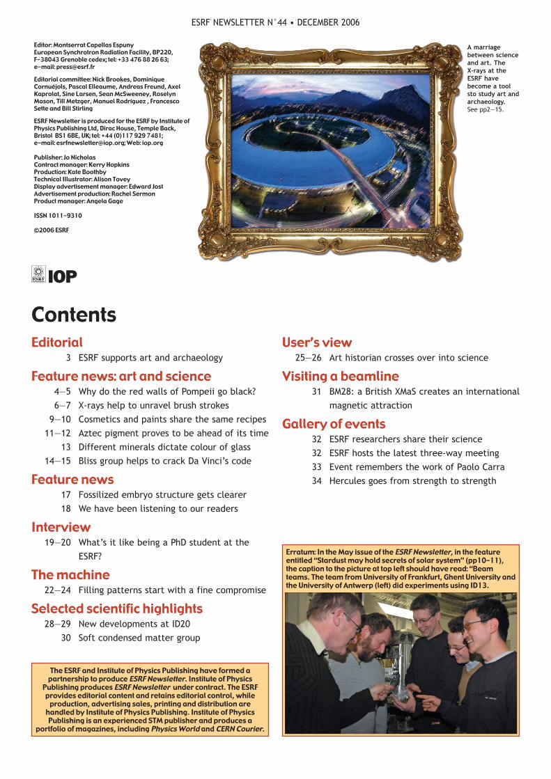
A marriagebetween scienceand art. TheX-rays at theESRF havebecome a toolsto study art andarchaeology.See pp2—15.
This issue is dedicated to the
chemical and physical
analysis of museum objects
at the ESRF. Whatever the technical
and methodological approach,
questions commonly tackled by
archaeologists can be separated into
two main concerns: a better
understanding of the past and an
intelligent prediction of the future.
Regarding the past, analyses are
intended to reveal as much as
possible about the secrets of
artefacts. What are the objects
made of? Which ingredients were
used in their fabrication? How were
they made (the extraction of the
materials, purification, synthesis,
and so on)? Where do the
ingredients and the objects come
from? When and by whom were they
made (with the particular purpose
of authentication)?
Regarding the future, a great deal
of research is being carried out to
improve methods of restoration and
conservation to preserve objects for
as long as possible. Furthermore,
deeper knowledge of ancient
techniques can sometimes provide
new ideas for the development of
future technologies.
Much information can be obtained
without the use of synchrotron
radiation, such as from macroscopic
observations (the shape of an
object, the place where it was
discovered, etc). However,
synchrotron radiation can be used
to provide an insight into the
chemical and/or structural
compositions of samples.
Such analyses present a number of
difficulties. First, the artefacts are
precious, so sampling is usually
forbidden or must remain limited in
number and size. Second, they are
often made of complex mixtures of
organic and mineral materials, so a
single method is generally not
enough to identify all of the
ingredients. In addition, they are
rather heterogeneous at the
micrometre scale, which requires
discriminative techniques, such as
high-resolution imaging.
The benefits of SRThe use of synchrotron radiation in
this field of research has a number
of advantages:
● the relatively non-invasive
methods don’t require sample
destruction because they are based
on light—matter interactions (from
infrared to X-rays);
● the abundance of information that
can be gathered (from the atomic
to the structural level);
● the quality of data, thanks to the
high beam brightness, allowing
researchers to obtain data rapidly
and from a small amount of matter;
● the beam-spot size, in particular
for micro-imaging, which enables
selective analysis.
All of these attributes explain the
growing interest in using the ESRF in
this field, as exemplified by the
creation of a dedicated review
committee, Environmental and
Cultural Heritage Matters (EC), in
2005, and by the increasing number
of proposals in this area.
The ESRF and some users are
carrying out various studies on
“hard matter” — ancient metals,
and the decoration of jades or
bronzes — and “soft matter” —
paper, textiles and wood (ESRF
Newsletter 42, December 2005) and
paint, for which questions relative
to past and future are tackled. The
ESRF is also involved in analysing
the remains of historical figures like
Napoleon and Agnès Sorel (ESRF
Newsletter 41, July 2005).
In this issue, recent results are
presented in various related fields.
Some of them involve improving our
understanding of the past (e.g.
ancient techniques of painting on
pp6, 9, 11 and 14 and glass
manufacturing on p13); others are
intended to give insight into the
future by improving our knowledge
of degradation and restoration (e.g.
the blackening of pigment on p4,
the interaction between lead
pigments and binders in oil
paintings on p9, and the
degradation of fibres in ancient
Chinese silk on p8). The common
aim here is to see the invisible. ●
ESRF NEWSLETTER N°44 • DECEMBER 2006
ESRF SUPPORTS ART AND ARCHAEOLOGY
3
EditorialGuest editors: Marine Cotte, postdoctoral researcher, and Jean Susini, scientist in charge of ID21 and ID22

On 24 August AD79, Vesuvius erupted, burying
nearby towns under pumice and ash. The Villa
Sora in Torre del Greco remained entombed
until 20 years ago, when excavation works revealed its
hidden secrets. In the remains of the house the frescoes
featured the distinctive red often found in Pompeii and
Herculaneum. However, this has since turned black in
many places in a rapid degradation that’s not well
understood scientifically.
Scientists have wondered for many years why the red
in Pompeii’s walls, a dye that is made from cinnabar
(HgS), turns black. In 1BC, Vitruvius, in his treatise De
Architectura, mentioned the problem, which, at the
time, could be prevented by applying a type of varnish
based on “punic wax”. The causes and mechanisms that
are responsible for cinnabar discoloration still remain a
mystery. Consequently, conservators are unable to avoid
the deterioration. The most common explanation of the
phenomenon is that exposure to sunlight transforms it
into metacinnabar, which is black. A Franco-Italian team
of researchers has now used the ESRF synchrotron light
to study four samples to test this theory.
No longer seeing redThe scientists found that the chemical composition in
the affected samples was different from that of
metacinnabar, which indicated that some important
chemical reactions had taken place. On the one hand,
cinnabar had reacted with chlorine, which led to the
formation of grey chlorine mercury compounds. The
chlorine came from the sea and possibly punic wax (the
wax that was used in the frescoes). On the other hand,
the sulphation of calcite resulted in the black coating
on the paintings’ surface.
The scientists investigated a cross-section of one of the
samples to map the depth of alteration in the painting.
Why have the characteristic red walls of Pompeii, revealed by excavations of the city buried by the eruption
of Vesuvius, turned black over the last two decades? ESRF’s synchrotron offers new insight into this process.
ESRF NEWSLETTER N°44 • DECEMBER 2006
WHY DO THE RED WALLS OF POMPEII GO BLACK?
Feature news
4
Turning black. A wall painted red in the remains of Pompeii.
A new approach. A visible light microscopy image (left) and
the atomic distribution of chlorine (right) in one of the
samples with moderate alterations (obtained using μ-XRF with
an exciting beam of 3.9 keV).
Visible light image (a) and the distribution of reduced (b) and oxidized (c) sulphurs on the most degraded wall sample (8 ×7mm).
Amer
ican
Che
mic
al S
ocie
ty
Mar
io P
agan
a
Amer
ican
Che
mic
al S
ocie
ty

They realized that this layer was only around 5μm thick
and that beneath it the cinnabar remained intact.
So what makes the red turn black so quickly? “The
chemical distribution of the samples is not stable,
which means that atmospheric conditions probably play
a role in this change of colour,” explains Marine Cotte,
the first author of the team’s paper. “The sun surely
influences this process, but the rain may possibly do so
too.” Atmospheric contamination or bacterial activities
can also contribute to sulphation mechanisms.
Important research“The research carried out at the ESRF has an
extraordinary importance not only for the conservation
of wall paintings in the Villa Sora but in general for the
preservation of Roman wall paintings discovered in the
most important Roman archeological sites (e.g. Pompeii
and Herculaneum),” says Corrado Gratziu, who is
professor emeritus in geology with a specialization in
petrology of sedimentary rocks at the University of Pisa,
and part of the team.
The experiments carried out at the ESRF needed high
chemical sensitivity, low detection limits and high
lateral resolution of synchrotron X-ray
microspectroscopy. The researchers did the experiments
on the X-ray microscopy beamline ID21 by combining
micro-X-ray fluorescence mapping and micro-X-ray
absorption spectroscopy. The former detected the
chlorine and sulphur presence while the latter
identified their speciation (i.e. the way they are
bonded to other atoms).
The research is still far from complete: “The next step
is to examine more samples — not only from frescoes in
the archaeological site but also from those in museums.
In this way we will be able to compare the results and
better establish the causes for their degradation,”
explains Cotte.
UNESCO made Pompeii a World Heritage Site in 1997,
and it is the most visited archaeological site in Europe,
according to the Italian ministry of foreign affairs. ●
MC
ReferenceM Cotte et al. (in press) Blackening of Pompeian
Cinnabar paintings: X-ray micro-spectroscopy analysis,
Analytical Chemistry.
ESRF NEWSLETTER N°44 • DECEMBER 2006
5
ESRF NEWSLETTER N°44 • DECEMBER 2006
6
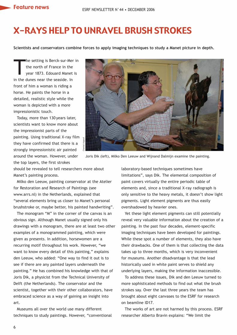
The setting is Berck-sur-Mer in
the north of France in the
year 1873. Edouard Manet is
in the dunes near the seaside. In
front of him a woman is riding a
horse. He paints the horse in a
detailed, realistic style while the
woman is depicted with a more
impressionistic touch.
Today, more than 130years later,
scientists want to know more about
the impressionist parts of the
painting. Using traditional X-ray film
they have confirmed that there is a
strongly impressionistic air painted
around the woman. However, under
the top layers, the first strokes
should be revealed to tell researchers more about
Manet’s painting process.
Milko den Leeuw, painting conservator at the Atelier
for Restoration and Research of Paintings (see
www.arrs.nl) in the Netherlands, explained that
“several elements bring us closer to Manet’s personal
brushstroke or, maybe better, his painted handwriting”.
The monogram “M” in the corner of the canvas is an
obvious sign. Although Manet usually signed only his
drawings with a monogram, there are at least two other
examples of a monogrammed painting, which were
given as presents. In addition, horsewomen are a
recurring motif throughout his work. However, “we
want to know every detail of this painting,” explains
den Leeuw, who added: “One way to find it out is to
see if there are any painted layers underneath the
painting.” He has combined his knowledge with that of
Joris Dik, a physicist from the Technical University of
Delft (the Netherlands). The conservator and the
scientist, together with their other collaborators, have
embraced science as a way of gaining an insight into
art.
Museums all over the world use many different
techniques to study paintings. However, “conventional
laboratory-based techniques sometimes have
limitations”, says Dik. The elemental composition of
paint covers virtually the entire periodic table of
elements and, since a traditional X-ray radiograph is
only sensitive to the heavy metals, it doesn’t show light
pigments. Light element pigments are thus easily
overshadowed by heavier ones.
Yet these light element pigments can still potentially
reveal very valuable information about the creation of a
painting. In the past four decades, element-specific
imaging techniques have been developed for paintings.
While these spot a number of elements, they also have
their drawbacks. One of them is that collecting the data
takes up to three months, which is very inconvenient
for museums. Another disadvantage is that the lead
historically used in white paint serves to shield any
underlying layers, making the information inaccessible.
To address these issues, Dik and den Leeuw turned to
more sophisticated methods to find out what the brush
strokes say. Over the last three years the team has
brought about eight canvases to the ESRF for research
on beamline ID17.
The works of art are not harmed by this process. ESRF
researcher Alberto Bravin explains: “We limit the
Scientists and conservators combine forces to apply imaging techniques to study a Manet picture in depth.
Joris Dik (left), Milko Den Leeuw and Wijnand Dalmijn examine the painting.
X-RAYS HELP TO UNRAVEL BRUSH STROKES
Feature news
Chan
tal
Argo
ud

ESRF NEWSLETTER N°44 • DECEMBER 2006
7
exposure time of the X-rays so that we don’t cause any
damage to the paintings.”
The new research on paintings at the ESRF is based on
the K-edge imaging technique. This involves taking two
images at two different energies and bracketing the
threshold (K-edge) energy of the element to be
investigated. This energy is element-specific. The pair
of images — acquired using a monochromatic X-ray
beam — are logarithmically subtracted to yield two
complementary images: one maps the specific element
while the other maps the background.
With the painting facing the invisible X-rays,
researchers take scans of the canvas in strips of 150mm
and assemble them. Each scan takes a minute.
In the Manet painting the researchers examined the
distribution of lead, mercury and barium, a pigment
lengthener of 19th- and 20th-century paint. Thanks to
the imaging technique, the researchers identified an
underlayer in the painting where barium allowed them
to see a modified version of the scene. In this hidden
sketch the shadow of the horse is situated at a higher
level and facing in a different direction from that in the
final image. In addition, several other barium stripes
indicate a history of undocumented restorations.
The researchers also noticed in the barium underlayer
the presence of a first sketch that was linked to a
watercolour by Manet that is part of the collection at
the Brooklyn Museum of Art in New York.
“Manet apparently used the more nebulous
relationships of the colours of the underpainting to help
to establish the tonalities that he sought for the final
effect. It makes sense that below the top layer one
could find a thin and freely painted underlayer,” says
Anne Coffin Hanson, a Manet expert at the University of
Yale, in her book Manet and the Modern Tradition.
The ESRF’s X-rays gave the scientists new clues about
the painting. Milko den Leeuw will include the
conclusions of the investigations in an upcoming
publication about the painting in 2007. “The unravelled
brushstrokes from totally different pigments could only
be seen by synchrotron radiation," says den Leeuw. ●
MC
ReferenceKrug K, Dik J, et al. 2006 Visualization of pigment
distributions in paintings using synchrotron K-edge
imaging Applied Physics A 83(2) 247— 251.
Want toplace anadvert
in
ESRFNewsletter
?If so,
contact
Ed Jost:

ESRF NEWSLETTER N°44 • DECEMBER 2006
8
Scientists from Germany, the UK and the ID13
beamline have recently studied ancient Chinese
silk fabrics found in the Famen Si (Doorway
Temple), one of the most important Buddhist
sanctuaries. Their results reveal for the first time how
fabric degradation is related to the structure of the
individual fibres. This may open new doors for the
preservation of ancient silk artefacts.
Some 1300years ago the emperors of the T’ang
dynasty (AD618—907) offered silk fabrics as part of a
gift to the Famen Si. Today these fabrics, along with
many others, continue their slow decay. Their
preservation and restoration is the aim of a bilateral
German-Chinese project. Many factors have contributed
to their decay throughout the centuries, including light,
humidity and microbes. However, not much was known
about how this occurs and how to prevent it, until now.
The researchers from the ESRF, the Fachhochschule
Köln (Germany), the Römisch-Germanisches
Zentralmuseum in Mainz (Germany) and the Textile
Conservation Centre in Southampton (UK) used scanning
synchrotron radiation microdiffraction, with a 1 μm
beam, to investigate three pieces of fabric at different
stages of decay. As the beam is only one-tenth the
diameter of a silkworm’s thread, the internal structure
of individual fibres could be probed.
The results show that the onset of fabric degradation
can be related to the loss of an amorphous fraction of
protein chains in the fibres. This breakdown in the silk’s
hierarchical structure makes it fragile and prone to
becoming disordered. Consequently, cleavage can occur
in the remaining nanofibrils, leading to fibre- and
fabric-scale disintegration.
The key to preserving fabric may thus be to protect
the fibres’ nanofibrillar morphology. The team suggests
using an artificial polymer matrix to replace the missing
protein and retard degradation. This molecular-scale
scaffolding may ensure that artefacts from our cultural
heritage can be appreciated centuries from now. ●
MC
ReferencesGreiff S et al. 2004 Scientific analysis of ancient and
historic textiles: informing preservation, display and
interpretation AHRC Research Centre for Textile
Conservation and Textile Studies First Annual
Conference (Archetype Publications, London).
Hermes A C et al. 2006 Characterizing the decay of
ancient Chinese silk fabrics by microbeam synchrotron
radiation diffraction Biomacromolecules 7 777—783.
ID13 has helped scientists to investigate the decay of ancient Chinese silk found in a Buddhist sanctuary.
Top left: The pagoda of the
Famen Si, which was heavily
affected by rain then pulled
down in 1987. During its
dismantling a vault was
discovered that contained
rich treasures, including the
silk garments that have
been studied at the ESRF.
Centre: Optical microscopy
images of a current plain-
weave silk fabric (Fm) and
three archaeological plain-
weave silk fabrics (F1—F3)
from the Famen Si site,
classified according to the
increasing visibility of their
decay. Bottom: A very well
preserved silk gown from
the Famen Si site.
Fam
en T
empl
e M
useu
m (
Chin
a)Fa
men
Tem
ple
Mus
eum
(Ch
ina)
Amer
ican
Che
mic
al S
ocie
ty
UNRAVELLING THE THREAD OF FIBRES’ DECAY
Feature news
Ancient pharmaceutical recipes, as well as
archaeological findings from Egyptian burial
sites, show that lead salts were used in
cosmetics and medicines in a mixture that included fat,
which stuck them together in lead soap. These soaps
are also present in some oil canvases and sometimes
produce protrusions that damage the painting. Two
teams of French scientists (one team with
pharmacologists; the other with painting curators) have
reproduced these mixtures, following ancient recipes,
and have monitored their kinetics.
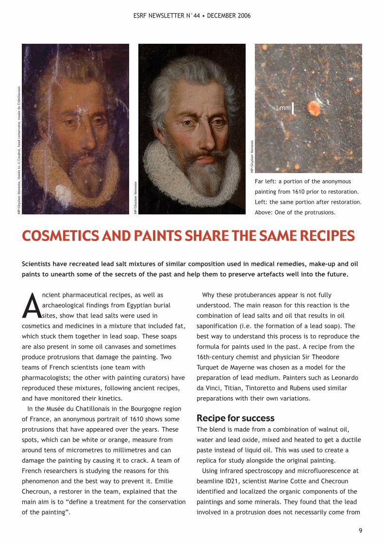
In the Musée du Chatillonais in the Bourgogne region
of France, an anonymous portrait of 1610 shows some
protrusions that have appeared over the years. These
spots, which can be white or orange, measure from
around tens of micrometres to millimetres and can
damage the painting by causing it to crack. A team of
French researchers is studying the reasons for this
phenomenon and the best way to prevent it. Emilie
Checroun, a restorer in the team, explained that the
main aim is to “define a treatment for the conservation
of the painting”.
Why these protuberances appear is not fully
understood. The main reason for this reaction is the
combination of lead salts and oil that results in oil
saponification (i.e. the formation of a lead soap). The
best way to understand this process is to reproduce the
formula for paints used in the past. A recipe from the
16th-century chemist and physician Sir Theodore
Turquet de Mayerne was chosen as a model for the
preparation of lead medium. Painters such as Leonardo
da Vinci, Titian, Tintoretto and Rubens used similar
preparations with their own variations.
Recipe for successThe blend is made from a combination of walnut oil,
water and lead oxide, mixed and heated to get a ductile
paste instead of liquid oil. This was used to create a
replica for study alongside the original painting.
Using infrared spectroscopy and microfluorescence at
beamline ID21, scientist Marine Cotte and Checroun
identified and localized the organic components of the
paintings and some minerals. They found that the lead
involved in a protrusion does not necessarily come from
Scientists have recreated lead salt mixtures of similar composition used in medical remedies, make-up and oil
paints to unearth some of the secrets of the past and help them to preserve artefacts well into the future.
Far left: a portion of the anonymous
painting from 1610 prior to restoration.
Left: the same portion after restoration.
Above: One of the protrusions.
INP/
Ghy
slai
n Va
nnes
te
INP/
Ghy
slai
n Va
nnes
te
INP/
Ghy
slai
n Va
nnes
te,
than
ks t
o J
LCo
udro
t, h
ead
cons
erva
tor,
mus
ée d
u Ch
atill
onna
isESRF NEWSLETTER N°44 • DECEMBER 2006
9
COSMETICS AND PAINTS SHARE THE SAME RECIPES
pigment but more likely from the siccative agent added
to oil to accelerate its drying. In their modern
reconstruction they observed that crystallized lead soap
aggregates can be formed after only one month and
that, contrary to what is sometimes found, protrusion
formation doesn’t necessarily require centuries.
As far as preservation is concerned, the researchers
found that water and heating accelerate saponification,
so cold and dry conditions are best to keep this process
to a minimum.
This study has changed scientists’ attitude to the
protrusions. “They have always been considered a
mechanical problem more than a chemical one. We
thought that they were coming from bad-quality white
lead coming up to the surface of the painting because of
the age and the settling of the oil,” says Checroun. The
English and Dutch carried out research into these
protuberances and showed that lead soap created this
effect. “We confirm that the French phenomenon of
protrusion has the same explanation,” she adds.
Studying make-upLead substances are also abundant in make-up powders
from ancient Egypt. In 2001 Philippe Walter at the
Centre de Recherche et de Restauration des Musées de
France (C2RMF) and colleagues at CNRS Grenoble and
ESRF, in collaboration with l’Oréal, studied some recipes
for Egyptian cosmetics. They noticed the presence of
two lead ingredients (laurionite and phosgenite), which
are both very rare in nature.
This led scientists to conclude that Egyptians
manufactured artificial lead-based compounds and
added them to the cosmetic product. This revealed that
wet chemistry was already being practised in around
2000BC. In addition, some cosmetics were found to be
a mixture of these complex minerals, sometimes
including fat.
Amazingly, recipes for the treatment of skin and eye
diseases written on papyrus from 1500BC describe
preparations based on heated mixtures of oil and lead
salts. These formulations are equivalent to lead
plasters, and pastes often used from Antiquity up to the
20th century.
Researching historical cosmetics and pigments in
parallel can prove interesting and fruitful because these
compounds have many similarities — after all, applying
make-up is essentially face-painting! ●
MC
ReferencesCotte M (in press) Kinetics of oil saponification by lead
salts in ancient preparations of pharmaceutical lead
plasters and painting lead mediums Talanta (available
online 18 April).
Martinetto P 2001 Synchrotron X-ray micro-beam studies
of ancient Egyptian make up Nucl. Instr. and Meth. B
181 744—748.
ESRF NEWSLETTER N°44 • DECEMBER 2006Feature news
10
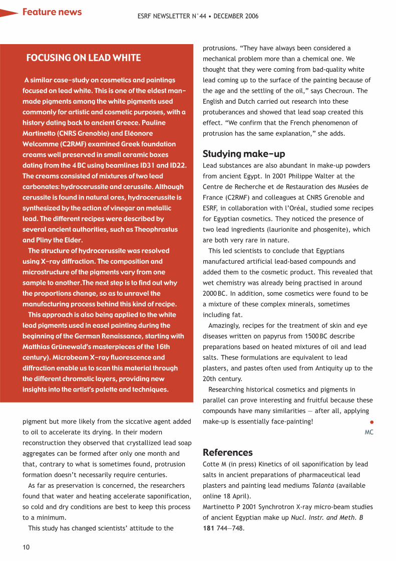
FOCUSING ON LEAD WHITE
A similar case-study on cosmetics and paintingsfocused on lead white. This is one of the eldest man-made pigments among the white pigments usedcommonly for artistic and cosmetic purposes, with ahistory dating back to ancient Greece. PaulineMartinetto (CNRS Grenoble) and EléonoreWelcomme (C2RMF) examined Greek foundationcreams well preserved in small ceramic boxesdating from the 4 BC using beamlines ID31 and ID22.The creams consisted of mixtures of two leadcarbonates: hydrocerussite and cerussite. Althoughcerussite is found in natural ores, hydrocerussite issynthesized by the action of vinegar on metalliclead. The different recipes were described byseveral ancient authorities, such as Theophrastusand Pliny the Elder.
The structure of hydrocerussite was resolvedusing X-ray diffraction. The composition andmicrostructure of the pigments vary from onesample to another.The next step is to find out whythe proportions change, so as to unravel themanufacturing process behind this kind of recipe.
This approach is also being applied to the whitelead pigments used in easel painting during thebeginning of the German Renaissance, starting withMatthias Grünewald’s masterpieces of the 16thcentury). Microbeam X-ray fluorescence anddiffraction enable us to scan this material throughthe different chromatic layers, providing newinsights into the artist’s palette and techniques.

ESRF NEWSLETTER N°44 • DECEMBER 2006
11
Mineral blue pigments were very expensive in
ancient times. The alternatives to mineral
pigments were organic colorants, which were
altered by external conditions. As a consequence there
isn’t much blue in frescoes from the Middle Ages and
the Renaissance. However, the Maya civilization
achieved what contemporary and previous civilizations
couldn’t: they synthesized a long-lasting blue pigment
from an organic colorant. The pigment’s amazing
stability has led researchers to study it thoroughly,
especially with regard to the creation of new materials.
The colorants used in antiquity were organic
molecules extracted from plants and animals. They
were not suitable for use in artworks because they are
not stable for many years. They fade with light, age
and exposure to pollutants, and some of them even
react with other chemicals that are used in artwork.
By contrast, pigments that are made from minerals
are very stable, which makes them preferable for many
artistic works such as murals, oil paintings and
polychromatic pottery.
A pigment with longevityThe Maya succeeded in “mineralizing” indigo — the
most common blue colorant known to many ancient
civilizations. They embedded it in a particular clay
mineral called palygorskite. This combination results in
a beautiful turquoise-blue with an amazing resistance to
acids and biodegradation through the centuries. The
pigment is present mostly in Mexico, but it has also
been found in Guatemala, Belize and Cuba.
Researchers at the ESRF, the CNRS (Grenoble) and the
AZTEC PIGMENT PROVES TO BE AHEAD OF ITS TIME
How did the Maya civilization create a beautiful, long-lasting turquoise-blue pigment when no-one else could?
Pottery figurine from the Great Temple, Mexico City,
representing Tláloc, the Aztec god of rain. Images of Tláloc
are commonly decorated in blue.
The west wall, room 1 of the Mayan site Bonampak, from
8AD, exhibiting different blue and green hues.
C Re
yes-
Vale
rio
C Re
yes-
Vale
rio

ESRF NEWSLETTER N°44 • DECEMBER 2006
12
Instituto Nacional de Antropología e Historia (Mexico)
have studied this pigment in some depth. Their research
has three aims: “We want to study its composition in
archaeological samples, since it can enlighten us about
the origin of the materials used, trade routes and
manufacturing technology,” explains Manuel Sanchez
del Río, a physicist at ESRF. “Second, the chemical
interaction between indigo and clay, which is
responsible for its resistance, is still not fully
understood. Finally, their research is also looking to
develop new composites that incorporate organic
molecules in inorganic materials.”
The team has synthesized several Maya blue samples
using palygorskite clay, known by the Mayas as “white
earth”, and also sepiolite, a clay with a similar channel-
like structure. Much more resistant pigments are
obtained from the palygorskite substrate than from the
sepiolite. The scientists have studied archaeological
samples using microdiffraction and microfluorescence at
ID18F and ID22. Also, XAFS experiments were carried
out at ID26 and powder diffraction at BM25A.
The research into the Maya blue will answer questions
about the past, and may also be useful for the future.
EricDooryhée and co-workers (CNRS Grenoble) have
been trying to mimic the properties of this pigment
using other classes of nanoporous material, in the
search for new organomineral composites with potential
uses in optics and optotronics. They recently succeeded
in incorporating and stabilizing the indigo dye in
ordered aluminosilicates. By controlling and changing
the synthesis parameters, the scientists expect to
obtain stable and non-toxic Maya-like hybrid colorants.
It is hoped that this work will enable researchers to
unravel the secrets of the Maya blue in order to
understand its history better and to use this knowledge
in future applications. ●
MC
ReferencesM Sánchez del Río et al. 2004 Microanalysis study of
archaeological mural samples containing Maya blue
pigment Spectrochimica Acta B 59 1619—1625.
M Sánchez del Río et al. 2005 Fe K-edge XANES of Maya
blue pigment Nucl. Instr. and Meth. B 338 55—60.
M Sánchez del Río et al. 2006 Synthesis and acid resistance
of Maya blue pigment Archaeometry 48 115—130.
Feature news
e2v scientific instruments

DIFFERENT MINERALS DICTATE COLOUR OF GLASS
Glassy matrices can exhibit different colours
depending on the oxidation state and the
electronic configuration of the metal ions that
are contained in them. For example, iron at high
concentration can produce a green coloration.
To minimize this problem in transparent artefacts,
artisans from around the first millennium BC added
substances to neutralize the colorant effects of the
iron. Before the Roman period, antimony was the main
bleaching agent, while from the 2BC manganese was
more frequently used. As far as opaque vitreous
materials are concerned, the colour and opaque effects
were obtained by means of many different substances,
depending on the age of the material and the desired
effects. The colour and opacity of red glass, in
particular, are frequently due to the presence of tiny
metallic copper particles.
Over the last four years, to improve our
understanding of how ancient glasses were
manufactured and the origin of their colours, scientists
from Italy and the ESRF have been studying different
artwork from Sicily and Pompeii. They use X-ray
absorption spectroscopy (XAS) to find out the oxidation
state of iron and manganese in a number of transparent
glass samples of archaeological interest, characterized
by different colours (from green to pale brown to
uncoloured), and of copper in a series of opaque red
tesserae. “The advantages of using XAS are that it is
non-destructive, it allows virtually any size and type of
sample, and it is applicable to most of the elements of
interest,” explains Francesco D’Acapito, scientist in
charge of the Collaborating Research Group (CRG)
beamline GILDA (BM08).
Thanks to XAS, scientists can track the presence of
monovalent copper cations incorporated into the glass
matrix of two opaque red artefacts, accompanied by
copper nanoclusters. In other examples, the results
show that the final artwork underwent intentional
discoloration during production.
In another part of this research the team focused on
the formation of the lustre on ceramics. In several
glazes of shards belonging to 10th- and 13th-century
pottery from Iran, researchers noticed that the
presence of copper and silver ions in the glaze confirms
that lustre formation results from a copper- and silver-
alkali ion exchange, followed by nucleation and growth
of metal nanoparticles. Previous research into
Renaissance pottery gave very similar results (ESRF
Newsletter 38, December 2003). ●
MC
ReferencesR Arletti et al. (in press) Roman coloured and opaque
glass: a chemical and spectroscopic study Applied
Physics A.
S Quartieri et al. 2002 Fe and Mn K-edge XANES study of
ancient Roman glasses Eur. J. Mineral. 14 749—756.
S Quartieri et al. 2005 The ancient glass production of
the medieval Val Gargassa glasshouse: Fe and Mn XANES
study J. Non-Cryst. Solids 351 3013—3022.
S Padovani et al. 2006 XAFS study of copper and silver
nanoparticles in glazes of medieval middle-east
lusterware (10th—13th century) Appl. Phys. A 83 521—
528.
Scientists from Italy and the ESRF are studying how
the elements in glass can determine its colour.
A sample of ancient mosaic tesserae from the Museo
Archeologico Regionale Eoliano.
ESRF NEWSLETTER N°44 • DECEMBER 2006
13
Asse
ssor
ato
Regi
onal
e Al
Ben
i Cu
ltur
ali,
Am
bien
tali
E Al
la P
ubbl
ica
Istr
uzio
ne D
ella
Reg
ione
Sic
ilian
a
ESRF NEWSLETTER N°44 • DECEMBER 2006
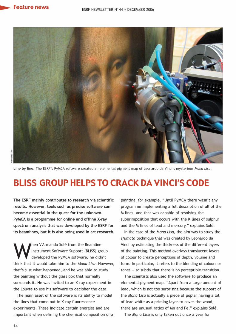
When VArmando Solé from the Beamline
Instrument Software Support (BLISS) group
developed the PyMCA software, he didn’t
think that it would take him to the Mona Lisa. However,
that’s just what happened, and he was able to study
the painting without the glass box that normally
surrounds it. He was invited to an X-ray experiment in
the Louvre to use his software to decipher the data.
The main asset of the software is its ability to model
the lines that come out in X-ray fluorescence
experiments. These indicate certain energies and are
important when defining the chemical composition of a
painting, for example. “Until PyMCA there wasn’t any
programme implementing a full description of all of the
M lines, and that was capable of resolving the
superimposition that occurs with the K lines of sulphur
and the M lines of lead and mercury,” explains Solé.
In the case of the Mona Lisa, the aim was to study the
sfumato technique that was created by Leonardo da
Vinci by estimating the thickness of the different layers
of the painting. This method overlays translucent layers
of colour to create perceptions of depth, volume and
form. In particular, it refers to the blending of colours or
tones — so subtly that there is no perceptible transition.
The scientists also used the software to produce an
elemental pigment map. “Apart from a large amount of
lead, which is not too surprising because the support of
the Mona Lisa is actually a piece of poplar having a lot
of lead white as a priming layer to cover the wood,
there are unusual ratios of Mn and Fe,” explains Solé.
The Mona Lisa is only taken out once a year for
The ESRF mainly contributes to research via scientific
results. However, tools such as precise software can
become essential in the quest for the unknown.
PyMCA is a programme for online and offline X-ray
spectrum analysis that was developed by the ESRF for
its beamlines, but it is also being used in art research.
Line by line. The ESRF’s PyMCA software created an elemental pigment map of Leonardo da Vinci’s mysterious Mona Lisa.
VAr
man
do S
olé
BLISS GROUP HELPS TO CRACK DA VINCI’S CODE
Feature news
14
ESRF NEWSLETTER N°44 • DECEMBER 2006
15
ESRF NEWSLETTER N°44 • DECEMBER 2006
conservation studies. The team from the Centre de
Recherche et de Restauration des Musées de France
(C2RMF) used an X-ray tube that was optimized at low
energies and they had only three hours to experiment.
The same team from C2RMF, led by Philippe Walter,
had already used the software to study techniques used
by Matthias Grünewald, a painter in the 15th and 16th
centuries. Thanks to the experiments at the ESRF
beamlines ID21 and ID22, and to PyMCA, the team
created an elemental map of the painting’s cross-
section. Characterization of the pigments pointed out
various minerals for green and black. Results showed
small particles of antimony correlated with sulphur,
which led to the identification of stibnite — an
extremely rare black pigment.
Despite being created only in late 2004, PyMCA has
already crossed borders. It is used not only by the
C2RMF researchers but also at other synchrotron
sources, such as DESY (Germany), CHESS and SSRL (US),
Elettra (Italy) and X-ray tube-based laboratories
elsewhere in the world. ●
MC
Our state of the artOur state of the art14kV14kV PES nowPES now
Boast a 200,000Boast a 200,000resolving power!resolving power!
Launched ProductsSub-meV PES analyser: MBS A-1Super intense He lamp: MBS L-1
Spin detector: MUSPINGas jet device: MBS JD-01Gas cell: MBS GC-01 etc.
for Detailed information Contact Us;Contact Us;
Seminariegatan 29B, SE-752 28,Uppsala, SWEDEN
Tel: +46 18 290960 Fax: +46 18 [email protected]
BLISSFUL SOFTWAREEncyclopaedia Britannica defines software as“instructions that tell a computer what to do. Softwarecomprises the entire set of programs, procedures,and routines associated with the operation of acomputer system.” The ESRF’s Beamline InstrumentSoftware Support (BLISS), comprising 17 computerengineers and physicists, is developing this essentialtool for computerized experiments. It created thesoftware to control experiments remotely. Datavisualization and scientific analysis tools complete thecontrol by allowing scientists to evaluate the quality ofthe experiment immediately afterwards. “Ourmission is to assist researchers with theirexperiments; we are at their service,” explainsVicente Rey, head of BLISS.
The programs don’t have a commercial use andscientists can request or download them for free. Theyare flexible, easy to configure and versatile, so theycan adapt to different beamlines.
Want toplace anadvert in
ESRFNewsletter
?If so,
contact
Ed Jost:
Photonic ScienceX-Ray, Gamma & Neutron Imaging SystemsInput sizes up to 150mm
Resolution – 4x2.6k pixels –single module
Stackable taper/CCD moduleswith combined single image– 1x3 or 2x3
In-vacuum version or vacuuminterfaced with ConFlat® flange
16-bit 10MHz or 12-bit fast readout
No need to shutter camera or beam
Sequences of fast images are possible,and exposures down to milliseconds
UK Tel: +44 (0) 1580 881199 Fax: +44 (0) 1580 880910 www.photonic-science.co.ukFrance Tel: +33 476 93 57 20 Fax: +33 476 93 57 22 email: [email protected]
X–Ray Imager VHR 3x133 million pixels
Neutron Camera
X–Ray Imager VHR

FOSSILIZED EMBRYO STRUCTURE GETS CLEARER
The evidence of the first animals on Earth goes
back some 700million years. However, the origin
of complex animals that have symmetry in a
central plane (bilateria) has remained a point of
speculation until now. Researchers from China, the US
and France have found that complex embryonic
development equivalent to that of some modern
bilateria existed 40million years earlier than thought
(i.e. 580million years ago).
In addition to the scientists’ studies with a scanning
electron microscope, they came to the ESRF to use the
powerful X-rays of ID19 to investigate non-destructively
the internal structures of fossilized embryos from south-
west China. They demonstrated that the cellular
cleavage pattern in the fossil embryos bears a striking
resemblance to the pattern of modern polar lobe-
forming embryos.
The polar lobe is a structure observed in many
molluscs, such as the mud snail, and in a few annelids.
It is a symmetry-breaking process that occurs at the
early stage of embryonic development and leads to
blastomeres (embryonic cells) of unequal sizes. This
process leads to particular embryonic structures
(trefoil, J-shaped and five-lobed) that are unique to the
early development of polar lobe-forming embryos. The
researchers have identified typical structures from
phosphate deposits that are 580million years old.
Important evidenceThese findings provide new evidence pushing the arrival
of the first bilaterian animals back to as early as
40million years before the Cambrian period. The
beginning of the Cambrian (540million years ago) is
known as the Cambrian explosion because that’s when
most of the major groups of animals appear in the fossil
record. This breakthrough implies that a complex
embryonic development comparable to that of modern
molluscs existed much earlier than scientists thought.
The team examined the samples taken from the
Precambrian rocks in Weng’an, China, using synchrotron
radiation microtomography at the ESRF and the National
Synchrotron Radiation Research Centre in Taiwan.
Thanks to the 3D data collected on the ID19 beamline,
they revealed the typical internal structures linked to
the polar-lobe formation process, which validated their
interpretation of these fossils. “Taking into account the
size of the samples [250—500μm] and their
mineralization pattern, only microtomography with
submicrometric resolution and phase contrast could
have revealed the internal structures in detail with
their 3D organization,” says Paul Tafforeau, a
paleontologist at the ESRF and an author of the paper.
The team is continuing its investigation into the
history of living beings by studying other fossils from
the Precambrian rocks. ●
MC
ReferenceChen et al. 2006 Phosphatized polar lobe-forming
embryos from the Precambrian of southwest China
Science 312 1644—1646.
A team of researchers has used ID19 to explore the
structure of fossilized embryos from south-west China.
Embryonic exploration. Virtual cross-section of a fossilized
embryo around 580million years old. Different colours
correspond to each of the three embryonic cells (blastomeres).
The blue blastomere is twice the volume of the others, which
strengthens the polar-lobe formation interpretation.
Li G
ang
and
Paul
Taf
fore
au
250μm
ESRF NEWSLETTER N°44 • DECEMBER 2006
17
Feature news

ESRF NEWSLETTER N°44 • DECEMBER 2006
The editor of the ESRF Newsletter and Institute of
Physics Publishing carried out a survey of its
readers last July to find out what they thought of
the publication. Overall the feedback was very positive,
but there is always room for improvement and the
suggestions that readers had were very welcome.
The newsletter is read primarily by the scientific
community, especially users and potential users of the
synchrotron facility. At present each issue is read by
more than 10000 people from all over the world. There
were 331 respondents to the survey (3.3% of the total
readership). The profile of these individuals reflects
that of a typical ESRF user: male, aged 35—54, holding
a PhD, and working as a scientist or a lecturer in a
university or research-council laboratory.
Most of the survey results relating to the profile of the
readers were unsurprising. Nevertheless, some aspects
where there is still potential to improve did stand out.
For example, only 16% of readers are younger than 35,
while only 13.8% are PhD students or postdoctoral
researchers. This suggests that more effort should be
made to attract younger scientists. Targeting this group
more aggressively will also make them more aware of
how the ESRF can benefit their research.
Good news is that the newsletter’s readership has
increased by 22% during the last two years. This is
partly owing to a collaborative effort by Institute of
Physics Publishing and the ESRF to enlarge the
distribution list. This includes sending copies of the
magazine to relevant conferences and new synchrotron
sources. In this way new users can be targeted directly
while existing users can be kept abreast of the latest
developments at the ESRF.
The survey also revealed which of the newsletter’s
contents are most popular with the readership. The
three favourite sections (in order) are the Scientific
Highlights, the Feature News and the Scientific Articles.
The popularity of the Scientific Highlights section
probably reflects its diversity, because it covers a
variety of scientific interests.
Two of the sections that are read the most have a
style and structure that is typical of a scientific
publication in a specialized journal. The exception to
this is the Feature News, which is written in a more
journalistic style and is enjoyed by most of the readers.
The survey also revealed that the least-read articles
in the newsletter include the Machine section, the
Gallery of Events and the Interviews (both of ESRF staff
and users). Around 30% of the readers who responded to
the survey never (or rarely) read these items.
All respondents were entered into a draw for a $200
Amazon voucher. The lucky winner was Geert Silversmit
from Ghent University. ●
MC
WE HAVE BEEN LISTENING TO OUR READERS
Earlier this year survey of our readers revealed that there’s still some way to go to serve their needs better.
Reader satisfaction. Most of the feedback from the newsletter’s readers is very positive, but there are lessons to be learned.
Chan
tal
Argo
ud
18
Feature news
How much have you learned aboutyour subject since starting your PhD?Roberta Poloni The learning process for me has been a
curve that goes up all of the time. At the beginning I
had to do a lot of studying about the subject of my
thesis, which is not something that I was familiar with.
Later I could focus on the techniques.
Yvonne Gründer I have learned a lot about the
beamline and my subject. I just had my first results,
but we have to repeat the experiment to confirm them.
Guillaume Potdevin This is our first job, one could say.
Therefore the number of different concepts that you
learn in the first years is very important. The
international nature of the ESRF has made it very easy
for me to adapt to the system, because I like meeting
and mixing with people from new cultures a great deal.
How much have you learned about thescience at the ESRF in general?RP I think it’s difficult to understand everything that
goes on at the ESRF. Seminars are a good source of
information, but if it is not in my field I don’t
understand the details of the research.
GP In most of the seminars and talks that I attend, I
probably understand 50% of the content having been at
the ESRF for two years. Even if I’ve studied the subject,
I can’t always understand everything. In a lot of cases
you actually need to be a specialist in the particular
topic to understand it properly
They arrive at the ESRF straight from university and
are faced with the challenge of working in a scientific
institute. They take the first steps of their career at
the ESRF and the synchrotron offers them the chance
to carry out a PhD surrounded by researchers from
different disciplines who can help them with their
work. They also learn about the science being done
by other students, thanks to the annual Students’
Day. This is an excellent springboard to promote their
work here. There are 33 full-time PhD students as
well as many others who carry out part of their
research at the facility. Three of them — Yvonne
Gründer, Roberta Poloni and Guillaume Potdevin —
talk freely about their experiences at the ESRF.
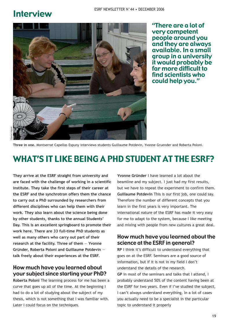
Three in one. Montserrat Capellas Espuny interviews students Guillaume Potdevin, Yvonne Gruender and Roberta Poloni.
WHAT’S IT LIKE BEING A PHD STUDENT AT THE ESRF?
ESRF NEWSLETTER N°44 • DECEMBER 2006
19
Chan
tal
Argo
ud
“There are a lot ofvery competentpeople around youand they are alwaysavailable. In a smallgroup in a universityit would probably befar more difficult tofind scientists whocould help you.”
Interview
ESRF NEWSLETTER N°44 • DECEMBER 2006
20
YG There should be specialized seminars and more
general presentations that we could follow more easily.
What are the benefits of doing a PhD atthe ESRF rather than somewhere else?YG The ESRF is a great place to meet a lot of people.
For starters the people on my beamline (ID32) are very
helpful. Then you get to know a lot of users by going
for lunch and having informal discussions when they are
on the beamline. This can be an opportunity for future
collaborations.
RP There are a lot of very competent people around
you and they are always available. In a small group in a
university it would probably be far more difficult to find
scientists who could help you. An important benefit of
being at the ESRF is that you can get beamtime more
easily, such as when there are buffer days.
GP Additionally, the ESRF provides a lot of material and
expertise in many domains. This makes it easier to get
both information and help when your subject deals with
a wide variety of technical aspects.
Do you feel that you’re well supportedby your supervisor?YG I really appreciate the level of freedom at the ESRF
that allows me to work at my own pace. I like to be
independent and not have someone looking over my
shoulder at what I’m doing all of the time. At the same
time we have group meetings every week, so I really
feel integrated into the group.
GP There are quite a few PhD students who claim that
they are not supported enough. Supervisors at the ESRF
are always available, but they are not very proactive.
You often have to learn things the hard way. However,
there is always someone to give you a hand when you
really need it.
RP: When I carry out experiments I get help from
colleagues from my university and also my beamlines
(BM29 and ID24). On a day-to-day basis they are also
able to help if I have any questions.
What do you envisage for your careerfollowing the ESRF?GP I wouldn’t mind working in industry or even
changing my subject. I like what I do, but I think it’s
dangerous to stay in the same place for a long time.
RP I would definitely like to stay in academia because I
enjoy studying and handling data more than working on
the beamline constantly. I definitely envisage a
postdoctoral position but I don't know where.
YG I still don't know what I’ll do after my thesis but for
the moment I like working in academia.
As well as adjusting to a workenvironment you’re living far fromhome. Do you enjoy life in Grenoble?YG I come from Berlin and for me it is amazing that it
only takes me 10minutes by bike to get to work. I only
miss German bread and beer!
GP Grenoble is great because it is close to the
mountains and has many cultural activities.
RP The lifestyle is definitely different from the Italian
lifestyle. Moreover, with the mountains surrounding us
and the little nightlife of the town, I think I’ve learned
to live more for the days than for the nights. ●
MC
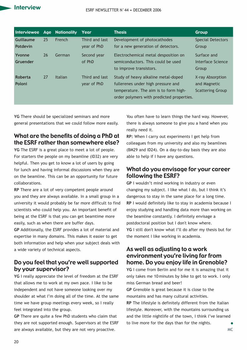
Guillaume 25 French Third and last Development of photocathodes Special Detectors
Potdevin year of PhD for a new generation of detectors. Group
Yvonne 26 German Second year Electrochemical metal desposition on Surface and
Gruender of PhD semiconductors. This could be used Interface Science
to improve transistors. Group
Roberta 27 Italian Third and last Study of heavy alkaline metal-doped X-ray Absorption
Poloni year of PhD fullerenes under high pressure and and Magnetic
temperature. The aim is to form high- Scattering Group
order polymers with predicted properties.
Interviewee Age Nationality Year Thesis Group
Interview
ESRF
We Highlight Science
European Sychrotron Radiation Facility
Physics and Chemistry, Life Sciences and Medicine, Earth and Environmental Sciences, Surface and Materials Sciences.
The European Synchrotron Radiation Facility (ESRF) is Europe’s most powerful light source. The ESRF offers you an exciting opportunity to work with international teams using synchrotron light in Grenoble, in the heart of the French Alps.
Have a look at our vacancies at www.esrf.frContact us at [email protected]
European Synchrotron Radiation FacilityESRF, BP 220, F-38043 Grenoble Cedex 9, FRANCE, Tel.+33 476 88 20 00 www.esrf.fr
Scientists - Post doctoral fellows - PhDstudents - Engineers - Technicians - Administrative staffists - Pos g e

ESRF NEWSLETTER N°44 • DECEMBER 2006
The ESRF has developed new filling patterns for its
beamlines by taking account of the parameters of the
experiments as well as the limits of the hardware.
The ESRF has the specificity to deliver various
filling patterns from one week to another,
following a mode schedule that is determined for
a period of six months. In fact the injector complex
allows a variety of filling patterns in the storage ring
(1—992 bunches of electrons) with the possibility of
filling one or more bunches with a greater number of
electrons. The maximum number of bunches is 992
because the radio frequency (RF) cavities of the storage
ring make particles cross the accelerating gap when the
voltage compensates exactly for the average energy loss
per turn owing to synchrotron radiation. Consequently,
the 352.2MHz RF, which corresponds to a multiple
h=992 of the particle revolution frequency (ring
circumference 844m), groups electrons with a
periodicity of 2.8ns.
The choice of filling pattern results from a fine
compromise between user requirements and the
hardware and tuning capabilities of the accelerator. To
understand it better, let’s first look more closely at the
different requirements of the users, which vary
depending on the type of experiment. Most experiments
require a stable high-brightness beam, with a filling
pattern that is compatible with the saturation of the
detector. The time-structure user community can be
divided into two groups: those who need a pure pulsed
beam at a high repetition rate and those who use the
short pulses at a low repetition rate.
On the machine side, user requirements can be
translated into stability, brightness, bunch length,
energy spread, peak brightness and purity.
The stability of the beamline optics during an
experiment is essentially determined by the current
variation between two refills. The longer the beam
lifetime, the more stable the beam. The contribution of
filling pattern to the beam lifetime is mainly governed
by the higher probability of a collision between
particles when electron density increases in a bunch.
This contribution to the lifetime, called the Touschek
effect, is dominant in single bunch but also has a
significant impact in all other modes of operation.
The brightness in terms of filling pattern is limited by
the heat load capacity of some machine components.
Front-end absorbers and dipole crotch absorbers limit
the current to 300mA with the present machine
configuration. Nevertheless, the beam power deposited
in the RF fingers (which are used to connect various
pieces of the vacuum chamber) restrains the current to
a much lower value with few high-current bunches. The
vertical emittance, which is also a key parameter
governing the brightness, is strongly affected by
transverse instabilities caused by ion trapping when all
bunches are filled. This effect, which improves with
vacuum conditioning of the ring, is always present in
uniform filling. This beam blow-up disappears as soon as
there is a gap of more than 10% in the multibunch
filling pattern. One should also note that, in a few
bunch modes, the vertical emittance is voluntarily
increased from 20pmrad up to 60pmrad to increase
the lifetime. The energy spread, which is constant at
less than 4.5mA per bunch, starts to increase to more
than this value. This effect makes the increase of the
undulator spectral brightness less than linear versus
current. One should also note that, with proper
calibration of beam-position monitors, positional
stability should not depend on the filling pattern.
Regarding the bunch length, increasing the number of
electrons in a bunch induces a stronger interaction with
the impedance of the surrounding vacuum chamber,
which leads to bunch lengthening. The bunch length,
which is 20ps rms at 0.2mA per bunch (200mA uniform
22
uniform 45%
hybrid 24×8 8%
16 bunch 23%
4×10 mA 6%
2× filling 18%13
Figure 1. Repartition of filling modes at the ESRF in 2006.
The machine
FILLING PATTERNS START WITH A FINE COMPROMISE

filling), reaches 60ps at 10mA per bunch.
It has previously been possible to deliver intensities of
up to 20mA in single bunch. However, this is now
limited to a peak brightness of 10mA in operation owing
to the increasing number of low gap chambers that are
installed in the insertion device straight sections, which
increases the impedance of the ring.
The purity and extreme contrast requested in pulsed
modes between filled and unfilled buckets (<10—9) is
obtained after the cleaning process done in the storage
ring at each refill.
Given the machine potential and limitations, five
filling patterns are delivered routinely to users (as shown
in figure 1 and with the parameters given in the table):
Uniform (200mA) With a maximum of
992bunches, this filling pattern, which has
the lowest current per bunch (0.2mA), gives
the longest lifetime of 80h at 200mA. However, owing
to ion-trapping instabilities, the lowest vertical
emittance is limited to 30pmrad in this mode. The
maximum current, at present limited to 200mA owing
to longitudinal beam instabilities that are induced by
the RF cavities (HOM), is routinely raised to 250mA
during machine-dedicated time. This will be increased
to 300mA by using the longitudinal multibunch
feedback, which is being developed. Ion instabilities
could possibly be limited using a transverse multibunch
feedback, which is also being developed.
2 × 1—3 (200mA) With approximately 2 × 352
bunches, the current per bunch is slightly
more than in uniform, reducing the lifetime
to 65h at 200mA. The presence of two empty gaps
totally removes the beam-ion instabilities and
consequently gives a minimum vertical emittance of
around 25pm. The two-bunch train is used by the
pulsed-beam community, besides being useful for tuning
the time parameters of the beamlines.
Hybrid (200 mA) This filling pattern is
organized into 24 groups of eight
consecutive bunches at 1 mA per bunch with
a single bunch of 4 mA in a gap. The maximum current
is 200 mA owing to the heat load that is induced in the
RF fingers. Cleaning allows the pulsed-beam
community to use this filling pattern. A fast chopper
can isolate the single bunch and use it in pump and
probe experiments with a time resolution given by the
X-ray pulse length.
16 bunch (90 mA) With 16 equidistant
bunches the maximum current is limited to
100mA owing to the heat load induced in
the RF fingers. The high current per bunch (5.6mA)
reduces the lifetime to 12h, even with a blow-up of the
vertical emittance. This pattern is ideal for the pure
pulsed-beam users (e.g. nuclear resonance scattering).
ESRF NEWSLETTER N°44 • DECEMBER 2006
23
uniform 2 × 1/3 7/8 hybrid 16 bunch 4 bunchsingle single
Repartition of (%) 45 18 — 0 — — 8 — 23 6
Modes (2006) (h) 2365 946 — 0 — — 420 — 1209 315
Refill current (mA) 200 200 200 — 2 196 — 4 90 40
Number of bunches 992 704 868 — 1 24 × 8 — 1 16 4
Number of refills a day 2 2 — 2 — — 2 — 4 6
Current decay between 25 35 — — — 60 — 2 40 20
refills (mA)
Average current (mA) 185 180 — — — 160 — 3.7 70 30
Lifetime at refill current (h) 80 65 72 — 15 30 — 7 12 6
H emittance (nm rad) 4 4 4.2 — — 4.2 — — 4.5 4.5
V remittance (pm rad) 30 25 22 — — 60 — — 60 60
Energy spread (×10—3) 1 1 1 — 1 1 — 1 1.2 1.5
Bunch length (ps) 20 20 20 — 35 25 — 40 50 60
Current, lifetime, bunch length and energy spread in various representative filling modes used at the ESRF

ESRF NEWSLETTER N°44 • DECEMBER 2006
In addition the time spacing of 176ns is long enough for
a chopper to isolate a single X-ray pulse.
4 bunch (40mA) With four equidistant
bunches the maximum current is limited to
45mA by transverse beam instabilities.
Owing to the very high current per bunch (10mA), the
lifetime is reduced to 6h, even with an artificial blow-
up of the vertical emittance. This pattern is targeted at
Laue experiments on proteins that require an intense
flash of X-rays in a single shot. The pulse spacing is as
long as 705ns and single pulses of X-rays are readily
isolated by a chopper.
The simultaneous demand for time structure and high
intensity has led to the design of a new multibunch filling
pattern (figure 2). With chromaticities identical to present
multibunch modes, the lifetime is around 72h at 200mA.
The presence of a gap, one-eighth of the circumference,
excludes beam-ion instabilities, thus maintaining the
vertical emittance at a value as low as 22pmrad. The
width of the gap is reduced to the minimum value
required by the most demanding beamline to minimize
the current by bunch and maximize the lifetime. A single
bunch of 2mA, placed in the middle of the gap, is
delivered with a contrast ratio of 10—9 between filled and
unfilled bunches. To optimize the use of this mode the
first and last bunch of the train are filled at 1mA. This
new mode, which has been tested by a few beamlines
during machine-dedicated time, is ready for operation.
With improvement, the multibunch feedback could be
operational in this configuration and consequently the
maximum current could be increased to 300mA.
Topping up: a way to improve stability?Implementing particle injection with front ends open in
2003 has greatly improved the stability of the beamline
optics, with continuous availability of the X-ray beam.
Thanks to the long lifetime in multibunch (close to 80h),
only two short refills are done each day. Between each
refill the beam is delivered for 12h without any stability
perturbation and with a smooth current variation of 15%.
By contrast the larger current variation during decay
makes topping-up (or more frequent injection) the most
interesting in time-structure modes.
The cleaning process done in the storage ring
currently blows up the beam for 30 s at each refill,
making it incompatible with more frequent injections. A
process to clean the injected beam in the booster is
under development. The extreme purity required by the
most demanding beamline makes this process very
challenging for routine operation. With a refill every
5min, the stability will improve immediately. However,
with the present configuration of the lattice, beam-
position deviation induced in the storage ring by the
injection magnets, such as kickers, makes the beam
unusable during injection, imposing gating in the data
acquisition on beamlines. Some work is required to
minimize this. Topping-up in time-structure modes will
be implemented when the cleaning process in the
injector becomes operational. ●
J-LREVOL AND LHARDY
ESRF OPERATION MANAGERS.
Figure 2. A high-brightness filling pattern with time structure.
2 mA
1 mA1 mA
1 turn
6262
200 mA in 868 bunches
The machine
The ESRF Newsletter brings you the latest news about scientificand technical developments at the ESRF. Join your fellow scientists and research project managers in 56 other countries and subscribe to the ESRF Newsletter today.
Keep up to date with the latest progress in your field.
If you would like to apply for a free subscription, e-mail your name and address to [email protected].
S U B S C R I B E F O R F R E E T O D A Y
24

ESRF NEWSLETTER N°44 • DECEMBER 2006
25
Joris Dik is possibly the most “artistic” user of the
ESRF. He holds an MA in art history and it wasn’t
until his PhD that he came into contact with
science. He then started focusing on the authentication
and conservation of paintings, teaming up with the
restorer Milko den Leeuw. He has worked for auction
houses such as Christie’s, but he now devotes his time
to his students at the Technical University of Delft,
where he teaches materials science in art and
archaeology. He is proof that science and art are not
incompatible, and he and Den Leeuw use the ESRF
regularly for their investigations.
Why did you change your career pathso drastically?It wasn’t really a change in career. In my family there
are some painting restorers: my grandfather and my
uncle both made a living out of it. When I left school I
was determined to become one, but then I realized that
I had “two left hands”. You need manual dexterity to
carry out restorations yourself and I didn’t have any. I
then became interested in the materials and the
techniques of painting.
How did you get into the scientificaspects of art?I was given the chance of a one-year traineeship in the
research department of the Getty Museum in Los
Angeles. It was a fantastic year when I learned a lot
about examining art. Then I went back to Holland,
where I met a chemistry professor, Henk Schenk, who
was crazy enough to hire me as a PhD student.
How difficult was it to do a PhD inchemistry?It was hard. I didn’t have any knowledge of the theory
so I had to study a lot. I found out that there is a nice
interface between science and arts. During my PhD I
used the ESRF several times. In the end, everything
turned out well — after five years I got my PhD.
User’s view
The appliance of science to art. Joris Dik (centre) surrounded by his colleagues
during a visit to carry out an experiment at the ESRF.
“In the researchdepartment of theGetty Museum...Ilearned a lot aboutexamining art. Then Iwent back to Holland,where I met achemistry professor,Henk Schenk, whowas crazy enough tohire me as a PhDstudent.”
ART HISTORIAN CROSSES OVER INTO SCIENCE

What is the main aim of yourresearch?I focus on conservation studies. Specifically, I work on
cadmium pigments and how they lose their colour
through the centuries. The aim is to understand the
degradation mechanism so that we can stop the damage
before it is too late. I am working in collaboration with
museums on paintings from Van Gogh at the moment.
How important is the use ofsynchrotron light in artistic research?Synchrotron radiation is very useful for certain
experiments, but you should only use it when it is
necessary. There are two different paths of analytical
possibilities at the ESRF that can be of interest for art
researchers: the analysis of microsamples, and the non-
destructive analysis of entire objects, such as bones and
paintings. At the moment my team has a research
project for visualization studies on ID17. In general
there’s a lot of research on art going on at the ESRF and
there is even a review committee focused only on
proposals for experiments about art. However, there are
still things to be done.
How could research into art develop atthe ESRF?Personally, I would be in favour of further measures to
help with the transportation of objects for non-
destructive analysis. Obviously the safe acclimatization
of objects is important, but perhaps the community
could also think about setting up a sort of travel and
insurance fund, because these are costs that usually
cannot be paid for from running budgets. It might be an
idea to request European funding for this with the help
of the ESRF. After all, in the EU we share not only the
synchrotron facility but also our cultural heritage.
Could you develop new techniques foryour research at the ESRF?Not really. I think we are too small a community to
demand instrumental developments. Nevertheless, we
should be opportunistic and make the most of the new
possibilities that come up.
What is the state of the research intoart carried out at other synchrotrons?The ESRF is one of the leaders in this field. SRS, BESSY,
SOLEIL and CHESS at Cornell are also quite active in this
field. However, it’s worth pointing out that neutron
sources are also a good way to study art and are
complementary to synchrotron sources.
Do you feel closer to science or to arttoday?I definitely feel closer to science, but I would like to be
nearer to art. I am very happy with what I am doing,
but I can’t help missing art. ●
MC
NAPLES YELLOW FOR DATINGJoris Dik did his thesis on Naples yellow, a pigmentthat has existed for thousands of years. AncientEgyptians used it, but it fell into obscurity until itreappeared in AD 1000. Slowly its use spread towestern Europe from the Middle East in the 16ththrough to the 18th century. The pigment’s complexphase composition changed throughout the years,and Dik’s aim was to determine these different
compositions.His researchcould proveuseful fordatingpurposes.When he cameto the ESRF forthe first time,Dik carried outX-raydiffractionexperimentson ID11 and
BM01. He says that, as a result of his work, “the earlytype of pigment, in the 16th century, was very pureand contained lead and antimonite. However, as itspread north, it was adulterated.” The pigment wascommonly called Naples yellow because peoplebelieved that it came from a volcanic mineral fromVesuvius, although this was eventually proved to bea misconception.
ESRF NEWSLETTER N°44 • DECEMBER 2006
26
User’s view

• international news coverage of pioneering
developments
•features by some of the world’s leading physicists
•the latest vacancies in industry and academia
•online resources and events calendar
physicsweb.org
Keep up to date – subscribe to our FREEnewsalert service
27

Combining resonant X-ray scattering (RXS) techniques
and extreme experimental conditions, such as low
temperatures, high pressures and high magnetic fields,
opens new possibilities of investigating structural,
magnetic and anomalous scattering properties of strongly
correlated electron systems. Careful variation of the
experimental environment conditions in which a material
is placed while making observations helps to unravel the
ground-state properties of these complex materials and
their enigmatic modifications. Of particular importance
in this regard are low temperatures, high magnetic fields
and high pressures, which are pioneered at the ID20
Magnetic Scattering Beamline at the ESRF.
Electronic degrees of freedom promote structural
modifications, transport effects and magnetic
phenomena such as spontaneous long-range magnetic
order, magnetoresistivity and magnetoelectricity. Many
transition metal oxides exhibit interesting long-range
ordered electronic states, such as ferroelectrics, colossal
magnetoresistive manganites and cuprate
superconductors. Even though magnetic ordering is
common, it is rare to find the coexistence of multiple
long-range orderings. Materials possessing more than one
“ferroic” order (which include long-range magnetism,
ferroelectricity and elastic distortion) are called
multiferroics, and this class of compound comprises
exceptional materials, for which the electric polarization
can be controlled by a magnetic field, or for which the
magnetic polarization can be controlled by an electric
field. As well as being of fundamental scientific interest,
such materials have great potential as an alternative
route to spintronic devices, and they open the possibility
of a number of technological applications (Fiebig 2005).
RXS is a site-, element- and shell-selective technique
that allows the exploration of the anisotropic properties
of atomic scattering tensors. It represents a unique
method of characterizing the microscopic atomic
interactions in solids from the electronic point of view.
RXS is based on enhancing the X-ray scattering cross-
section close to an absorption edge, such as the K edges
of transition metals, the L2,3 edges of rare earths and
the M4,5 edges of actinides. The resonant process
involves promoting core electrons to empty
intermediate states in the vicinity of the Fermi level,
and their subsequent decay accompanied by the
emission of an elastic photon, which reflects the
Scientific highlights
LPaolasini,1 SBWilkins,1 CMazzoli,1 BDetlefs,2 VScagnoli,1 AFondacaro,1 FdeBergevin,1 CDetlefs,1 PBernard,1
GPeppelin,1 DMannix,3 DMcMorrow,4 ABoothroyd5
1 ESRF, 2 ITU, Karlsruhe, Germany, 3 XMaS, CRG, ESRF, 4 University College London, UK, 5 Oxford University, UK
NEW DEVELOPMENTS AT ID20Resonant X-ray scattering under extreme conditions:investigating the physics of multiple-ordered states
28
Figure 1. Superconducting 10T split-pair magnet (Oxford
Instruments) on the a-magnetic six-circle diffractometer
(Huber GmbH) in the ID20 second experimental hutch.
ESRF NEWSLETTER N°44 • DECEMBER 2006

ESRF NEWSLETTER N°44 • DECEMBER 2006
29
electronic state of the magnetically ordered species.
Moreover, the resonant cross-section has a
characteristic polarization dependence, which can be
exploited to obtain further information about the
magnetic-moment direction and the space group of the
ordered state.
The recently commissioned high-field magnet facility
on ID20 opens new avenues of research in materials
with complex and competing order parameters,
including multiferroics.
A superconducting split-pair 10T magnet, which can
reach the basic temperature of 1.8K and is
accommodated on a dedicated six-circle diffractometer,
is designed to support heavy loads and built from non-
magnetic materials (figure 1). An in-vacuum
polarization analyser assembly is mounted on the
detector arm and can rotate about the X-ray beam that
is scattered by the sample to make the full linear
polarization analysis. The custom design of the magnet
allows RXS at low energy (down to 3.5 keV) with wide
scattering angles and low absorbing beryllium windows.
In one of the first experiments, this facility
investigated the phase diagram of the multiferroic
compound TbMnO3. Previous bulk measurements
established that in zero applied magnetic field the
ferroelectric polarization, P, aligns along the c-axis, as
in the phase labelled FE (P||c) in figure 2. Meanwhile
above a critical threshold of applied magnetic field, P
flops to be along the a-axis, FE (P||a) (Kimura et al.
2003). However, these measurements obviously don’t
provide a microscopic atomic-scale description of
changes to the structural and magnetic correlations.
Scattering data, particularly RXS with its element and
electron shell sensitivities, provides such information.
The experiment performed at both the Tb L edges and
Mn K edge with the new magnet facility allowed us to
study the phase diagram in great detail (Mannix et al.).
For example, for all of the phases identified by the bulk
studies, we studied the underlying chemical and
magnetic structures.
As a result the different commensurate (C) and
incommensurate (IC) phases can be studied in detail,
and the contribution to the X-ray diffraction signals
from Tb of Mn electrons can be distinguished, as well as
the corresponding magnetic and electronic modulations
in each phase. This allowed us to establish, among
other things, that an IC to C (lock-in) transition drives
the flop of the polarization along the a-axis.
ConclusionsCombining extreme conditions, such as high pressures,
low temperatures and high magnetic fields, with the
RXS technique opens new possibilities to investigate and
single out structural, magnetic and anomalous
scattering. New and exciting fields of research, such as
orbitally ordered systems, multiferroics, actinides and
charge-ordered systems, can benefit from these unique
sample environment conditions.
Planned improvements to the focusing properties and
stability of the beamline optics will allow extensive use
of these combined techniques in the future.
ReferencesMFiebig 2005 Revival of the magnetoelectric effect
J Phys D 38 R123.
TKimura et al. 2003 Magnetic control of ferroelectric
polarization Nature 426 55.
DMannix et al. in prep.
Figure 2. Left: magnetic field dependence of RXS at TbL3 edge
of satellite reflections (5k0) at 2K (top) and the corresponding
H—T phase diagram (bottom). Right: Evolution of the intensity
(top) and the wavevector (bottom), measured in reciprocal
lattice units (rlu), of the satellite reflection (5k0) as a function
of the temperature, under an applied magnetic field of 8T.
500
250
0
0.28
0.27
0.26
0.25
wa
veve
cto
r (r
lu)
10 20 30 40100 2 4 6 8
magnetic field (T) temperature (K)
50
40
30
20
10
0
0.5
0.4
0.3
0.2
wa
veve
cto
r (r
lu)
tem
pe
ratu
re (
K)
ICI AFM, PE
PM, PE
C AFM, FE (P||a)
IC2 AFM, FE (P||c)
TMnN2
TTbN3
TMnN1
cou
nts
(s)
C AFMFE (P||a)
IC2 AFMFE (P||c)
IC1 AFMPE
(5K0) B=8TTb L3 π–σ
(5K0) T=2KTb L3 π–σ
ESRF NEWSLETTER N°44 • DECEMBER 2006
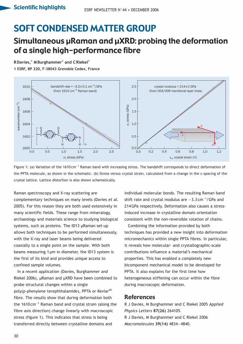
ESRF NEWSLETTER N°44 • DECEMBER 2006
30
Raman spectroscopy and X-ray scattering are
complementary techniques on many levels (Davies et al.
2005). For this reason they are both used extensively in
many scientific fields. These range from mineralogy,
archaeology and materials science to studying biological
systems, such as proteins. The ID13 μRaman set-up
allows both techniques to be performed simultaneously,
with the X-ray and laser beams being delivered
coaxially to a single point on the sample. With both
beams measuring 1μm in diameter, the ID13 system is
the first of its kind and provides unique access to
confined sample volumes.
In a recent application (Davies, Burghammer and
Riekel 2006), μRaman and μXRD have been combined to
probe structural changes within a single
poly(p-phenylene terephthalamide), PPTA or Kevlar49
fibre. The results show that during deformation both
the 1610cm—1 Raman band and crystal strain (along the
fibre axis direction) change linearly with macroscopic
stress (figure 1). This indicates that stress is being
transferred directly between crystalline domains and
individual molecular bonds. The resulting Raman band
shift rate and crystal modulus are —3.3 cm—1/GPa and
214GPa respectively. Deformation also causes a stress-
induced increase in crystalline domain orientation
consistent with the non-reversible rotation of chains.
Combining the information provided by both
techniques has provided a new insight into deformation
micromechanics within single PPTA fibres. In particular,
it reveals how molecular- and crystallographic-scale
contributions influence a material’s mechanical
properties. This has enabled a completely new
bicomponent mechanical model to be developed for
PPTA. It also explains for the first time how
heterogeneous stiffening can occur within the fibre
during macroscopic deformation.
ReferencesR J Davies, M Burghammer and C Riekel 2005 Applied
Physics Letters 87(26) 264105.
R J Davies, M Burghammer and C Riekel 2006
Macromolecules 39(14) 4834—4840.
SOFT CONDENSED MATTER GROUPSimultaneous μRaman and μXRD: probing the deformationof a single high-performance fibre
Figure 1: (a) Variation of the 1610cm—1 Raman band with increasing stress. The bandshift corresponds to direct deformation of
the PPTA molecule, as shown in the schematic. (b) Stress versus crystal strain, calculated from a change in the c-spacing of the
crystal lattice. Lattice distortion is also shown schematically.
1610
1608
1606
1604
1602
16000.0 0.5 1.0 1.5 2.0 2.5
wa
ven
um
be
r (c
m–1)
C
O||
C
O||
N N||
H H n
bandshift rate = –3.3± 0.1 cm–1/GPa
(from 1610 cm–1 Raman band)
2.5
2.0
1.5
1.0
0.5
0.00.0 0.2
crystal modulus = 214 ± 2 GPa
(from 004/006 meridional layer lines)
b
a
c
σ, st
ress
(G
Pa
)
0.4 0.6 0.8 1.0 1.2
σ, stress (GPa) εc, crystal strain (%)
RDavies,1 MBurghammer1 and CRiekel1
1 ESRF, BP 220, F-38043 Grenoble Cedex, France
Scientific highlights
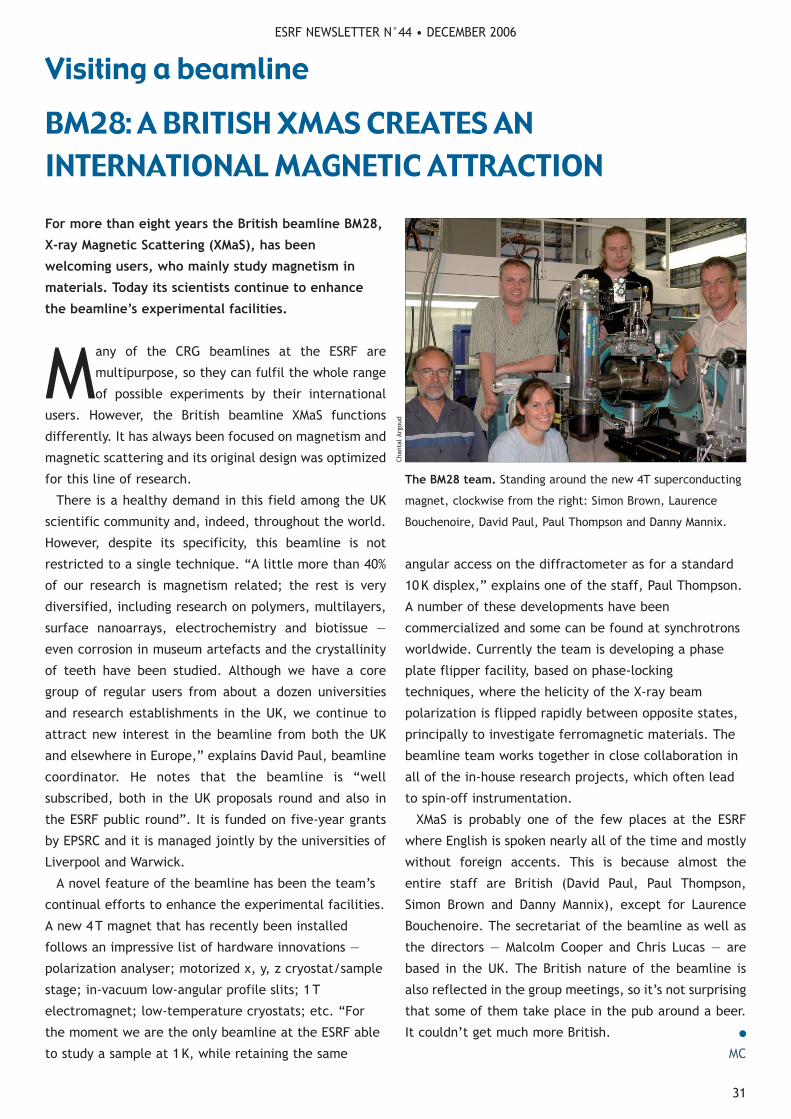
ESRF NEWSLETTER N°44 • DECEMBER 2006
31
Visiting a beamline
Many of the CRG beamlines at the ESRF are
multipurpose, so they can fulfil the whole range
of possible experiments by their international
users. However, the British beamline XMaS functions
differently. It has always been focused on magnetism and
magnetic scattering and its original design was optimized
for this line of research.
There is a healthy demand in this field among the UK
scientific community and, indeed, throughout the world.
However, despite its specificity, this beamline is not
restricted to a single technique. “A little more than 40%
of our research is magnetism related; the rest is very
diversified, including research on polymers, multilayers,
surface nanoarrays, electrochemistry and biotissue —
even corrosion in museum artefacts and the crystallinity
of teeth have been studied. Although we have a core
group of regular users from about a dozen universities
and research establishments in the UK, we continue to
attract new interest in the beamline from both the UK
and elsewhere in Europe,” explains David Paul, beamline
coordinator. He notes that the beamline is “well
subscribed, both in the UK proposals round and also in
the ESRF public round”. It is funded on five-year grants
by EPSRC and it is managed jointly by the universities of
Liverpool and Warwick.
A novel feature of the beamline has been the team’s
continual efforts to enhance the experimental facilities.
A new 4T magnet that has recently been installed
follows an impressive list of hardware innovations —
polarization analyser; motorized x, y, z cryostat/sample
stage; in-vacuum low-angular profile slits; 1T
electromagnet; low-temperature cryostats; etc. “For
the moment we are the only beamline at the ESRF able
to study a sample at 1K, while retaining the same
angular access on the diffractometer as for a standard
10K displex,” explains one of the staff, Paul Thompson.
A number of these developments have been
commercialized and some can be found at synchrotrons
worldwide. Currently the team is developing a phase
plate flipper facility, based on phase-locking
techniques, where the helicity of the X-ray beam
polarization is flipped rapidly between opposite states,
principally to investigate ferromagnetic materials. The
beamline team works together in close collaboration in
all of the in-house research projects, which often lead
to spin-off instrumentation.
XMaS is probably one of the few places at the ESRF
where English is spoken nearly all of the time and mostly
without foreign accents. This is because almost the
entire staff are British (David Paul, Paul Thompson,
Simon Brown and Danny Mannix), except for Laurence
Bouchenoire. The secretariat of the beamline as well as
the directors — Malcolm Cooper and Chris Lucas — are
based in the UK. The British nature of the beamline is
also reflected in the group meetings, so it’s not surprising
that some of them take place in the pub around a beer.
It couldn’t get much more British. ●
MC
The BM28 team. Standing around the new 4T superconducting
magnet, clockwise from the right: Simon Brown, Laurence
Bouchenoire, David Paul, Paul Thompson and Danny Mannix.
For more than eight years the British beamline BM28,
X-ray Magnetic Scattering (XMaS), has been
welcoming users, who mainly study magnetism in
materials. Today its scientists continue to enhance
the beamline’s experimental facilities.
BM28: A BRITISH XMAS CREATES AN INTERNATIONAL MAGNETIC ATTRACTION
Chan
tal
Argo
ud

ESRF NEWSLETTER N°44 • DECEMBER 2006
32
The science at the ESRF covers such a range of
disciplines that it can be difficult for individual
scientists to know what goes on there outside
their own field. This is the reason why the ESRF
Experiments Division organizes biannual Science Days.
These events are also intended to stimulate discussions
between scientists. This year the Science Day took
place in Autrans on 7 and 8 June and had a very young
atmosphere and, out of the 145 participants, 43% were
PhD students or postdoctoral researchers.
This year the structure of the event was slightly
modified: the chairs of the different sessions were
scientists from different groups than the speakers. “We
wanted to remind speakers that they should address
everybody and to make the audience feel free to ask
questions, even if they are basic,” explains Andy Fitch,
one of the organizers. In this way the audience could
see Nick Brookes, head of the X-ray Absorption and
Magnetic Scattering Group, introducing biology talks,
which are far away from his field. The new formula
“was quite successful, and worked in some talks better
than in others”, says Fitch. According to Blanca Detlefs
from ID20, “since the event is only two days long, the
oral presentations should be more general so that
everybody can follow them”.
Four students were awarded a poster prize:
Guillaume Potdevin, Adeline Buffet, Guillaume Morard
and Filippo Bencivenga. Their efforts won them a
bottle of champagne. “We decided on four prizes in
recognition of the very high general standard of the
students' work and its presentation in the posters; it's
important that students know that their efforts are
appreciated,” explains Fitch. ●
MC
Lively discussions. Coffee breaks were a good opportunity to
discuss recent scientific achievements with colleagues.
Gallery of events
ESRF RESEARCHERSSHARE THEIR SCIENCE
ESRF HOSTS THE LATEST THREE-WAY MEETING
The biannual Science Days take on a slightly different
format and prove to be just as successful as before.
Last summer the ESRF opened its doors to staff from the three biggest synchrotron facilities in the world.
Every 18months the ESRF, the APS or SPring8 hosts
a meeting to discuss scientific and technical
issues of common interest. This year the ESRF
welcomed 31 participants in the Three-Way Meeting on
19 and 21 June. The event started with the 4th X-ray
Optics Workshop. The agenda included talks on the
aging of accelerator components; micro- and
nanofocusing; high-energy insertion devices; extreme
conditions; the accelerator complex; and recent
highlights and future developments of X-ray imaging and
synchrotron-radiation instrumentation. ●
MC
All together. The Three-Way Meeting held this year brought
together more than 30 people from three different continents.
Chan
tal
Argo
udM
onts
erra
t Ca
pella
s
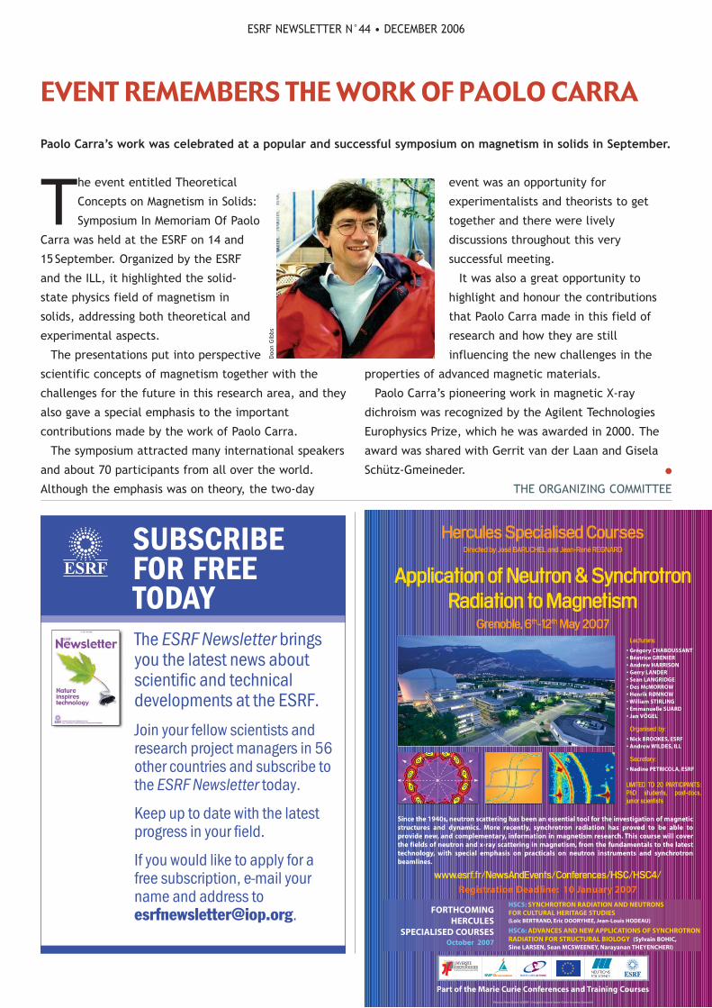
The event entitled Theoretical
Concepts on Magnetism in Solids:
Symposium In Memoriam Of Paolo
Carra was held at the ESRF on 14 and
15September. Organized by the ESRF
and the ILL, it highlighted the solid-
state physics field of magnetism in
solids, addressing both theoretical and
experimental aspects.
The presentations put into perspective
scientific concepts of magnetism together with the
challenges for the future in this research area, and they
also gave a special emphasis to the important
contributions made by the work of Paolo Carra.
The symposium attracted many international speakers
and about 70 participants from all over the world.
Although the emphasis was on theory, the two-day
event was an opportunity for
experimentalists and theorists to get
together and there were lively
discussions throughout this very
successful meeting.
It was also a great opportunity to
highlight and honour the contributions
that Paolo Carra made in this field of
research and how they are still
influencing the new challenges in the
properties of advanced magnetic materials.
Paolo Carra’s pioneering work in magnetic X-ray
dichroism was recognized by the Agilent Technologies
Europhysics Prize, which he was awarded in 2000. The
award was shared with Gerrit van der Laan and Gisela
Schütz-Gmeineder. ●
THE ORGANIZING COMMITTEE
ESRF NEWSLETTER N°44 • DECEMBER 2006
Paolo Carra’s work was celebrated at a popular and successful symposium on magnetism in solids in September.
EVENT REMEMBERS THE WORK OF PAOLO CARRA
Part of the Marie Curie Conferences and Training Courses
FORTHCOMINGHERCULES
SPECIALISED COURSES October 2007
www.esrf.fr/NewsAndEvents/Conferences/HSC/HSC4/
Registration Deadline: 10 January 2007
LIMITED TO 20 PARTICIPANTS:PhD students, post-docs,junior scientists
Hercules Specialised CoursesDirected by José BARUCHEL and Jean-René REGNARD
Application of Neutron & SynchrotronRadiation to Magnetism
Grenoble, 6th-12th May 2007 Lecturers:
• Grégory CHABOUSSANT• Béatrice GRENIER• Andrew HARRISON• Gerry LANDER• Sean LANGRIDGE• Des McMORROW• Henrik RØNNOW• William STIRLING• Emmanuelle SUARD• Jan VÖGEL
Organised by:
• Nick BROOKES, ESRF• Andrew WILDES, ILL
Secretary:
• Nadine PETRICOLA, ESRF
Photos: Peter Ginter & ESRF • Design & layout: Anne-Claire lecomte, Grenoble
Since the 1940s, neutron scattering has been an essential tool for the investigation of magneticstructures and dynamics. More recently, synchrotron radiation has proved to be able toprovide new, and complementary, information in magnetism research. This course will coverthe fields of neutron and x-ray scattering in magnetism, from the fundamentals to the latesttechnology, with special emphasis on practicals on neutron instruments and synchrotronbeamlines.
HSC5: SYNCHROTRON RADIATION AND NEUTRONSFOR CULTURAL HERITAGE STUDIES(Loïc BERTRAND, Eric DOORYHEE, Jean-Louis HODEAU)
HSC6: ADVANCES AND NEW APPLICATIONS OF SYNCHROTRONRADIATION FOR STRUCTURAL BIOLOGY (Sylvain BOHIC,Sine LARSEN, Sean MCSWEENEY, Narayanan THEYENCHERI)
The ESRF Newsletter bringsyou the latest news aboutscientific and technicaldevelopments at the ESRF.
Join your fellow scientists andresearch project managers in 56other countries and subscribe tothe ESRF Newsletter today.
Keep up to date with the latestprogress in your field.
If you would like to apply for afree subscription, e-mail yourname and address [email protected].
SUBSCRIBEFOR FREE TODAY
Doo
n G
ibbs

ESRF NEWSLETTER N°44 • DECEMBER 2006
34
Hercules is stronger than ever. This spring the
Higher European Research Course for Users of
Large Experimental Systems, organized by the
Institut National de Polytechnique de Grenoble (INPG)
and Joseph Fourier University (UJF), started a series of
one-week specialized courses on synchrotron radiation.
These are composed of lectures and practicals aimed at
PhD students and postdoctoral scientists. They are
supported by the European Commission. The Hercules
programme’s main feature is a five-week European
course in March on large instruments, which focuses on
both synchrotron and neutron radiation.
The first Hercules specialized course in 2006, on
21—26 May, covered X-ray imaging techniques based on
absorption, phase contrast, diffraction and
fluorescence. One of the participants said: “[The
course] enables us to broaden our knowledge — we get
to learn different techniques that we didn’t know about
before coming.”
Even though the programme is limited to 20
participants, the lectures are open to anyone who is
interested in the subject. For instance, Paul Tafforeau,
a palaeontologist working at the ESRF, listens to the
lecture courses to learn more about the techniques that
allow him to scan his findings everyday. “I like to know
the theoretical part of my practice,” he says.
From the 77 applications to attend the course, the
organizers selected the candidates who would profit the
most from this experience and who showed definite
motivation. The students came from all over Europe.
One of the organizers and speakers, José Baruchel,
commented: “Participants get together and can create
for themselves a network of acquaintances for the
future.” However, the delegates are not the only ones
to benefit from the course. “Hercules also represents
an enrichment for the speakers, who learn from their
colleagues,” adds Baruchel.
At the time of going to press there were two new
specialized courses planned for the end of the year: one
on structural genomics and the other on surfaces,
interfaces and nanostructures. Registration for the
courses for 2007 is now open.
For more information, visit www.esrf.fr/
NewsAndEvents/Conferences/HSC/. ●
ANNE BENHAMOU
Hercules now offers a new set of one-week courses
that are already proving popular with delegates.
The first ones. The group of researchers who benefited from the first Hercules specialized course on synchrotron radiation.
Poster session. Participants had the opportunity to explain
some of their research to lecturers and other delegates.
HERCULES GOES FROM STRENGTH TO STRENGTH
Gallery of eventsCh
anta
l Ar
goud
Mon
tser
rat
Cape
llas


New vacuum
components
catalogue
Creatingvacuumsciencesolutions
Presentingour newcatalogue...
United KingdomTel:+44 (0)870 4287646e-mail: [email protected]
FranceTel: +33 (0)437 65 17 50e-mail: [email protected]
GermanyTel: +49 (0)2305 947 508e-mail: [email protected]
Our new catalogue –containing over 600 pagesof innovative products to meet the needs of scientists throughoutEurope, will be available in June.Reserve your copy now by visiting our website:www.caburn.co.uk or contact your local
technical sales office.