deletions associated with stabilization of the top1 …...deletions associated with stabilization of...
TRANSCRIPT

Deletions associated with stabilization of the Top1cleavage complex in yeast are products of thenonhomologous end-joining pathwayJang-Eun Choa,1 and Sue Jinks-Robertsona,2
aDepartment of Molecular Genetics and Microbiology, Duke University Medical Center, Durham, NC 27710
Contributed by Sue Jinks-Robertson, September 20, 2019 (sent for review August 14, 2019; reviewed by Hannah L. Klein and Sergei M. Mirkin)
Topoisomerase I (Top1) resolves supercoils by nicking one DNAstrand and facilitating religation after torsional stress has beenrelieved. During its reaction cycle, Top1 forms a covalent cleavagecomplex (Top1cc) with the nicked DNA, and this intermediate canbe converted into a toxic double-strand break (DSB) during DNAreplication. We previously reported that Top1cc trapping in yeastincreases DSB-independent, short deletions at tandemly repeatedsequences. In the current study, we report a type of DSB-dependent mutation associated with Top1cc stabilization: largedeletions (median size, ∼100 bp) with little or no homology atdeletion junctions. Genetic analyses demonstrated that Top1cc-dependent large deletions are products of the nonhomologousend-joining (NHEJ) pathway and require Top1cc removal fromDNA ends. Furthermore, these events accumulated in quiescentcells, suggesting that the causative DSBs may arise outside thecontext of replication. We propose a model in which the ends ofdifferent, Top1-associated DSBs are joined via NHEJ, which resultsin deletion of the intervening sequence. These findings have im-portant implications for understanding the mutagenic effects ofchemotherapeutic drugs that stabilize the Top1cc.
topoisomerase I | nonhomologous end joining | double-strand break | yeast
Topoisomerase I (Top1) resolves DNA torsional stress by re-moving supercoils that accumulate during transcription,
replication, and chromatin remodeling (1). Top1 is a type IB en-zyme that uses an active site tyrosine to cleave one DNA strand.This generates a transient Top1 cleavage complex (Top1cc) inwhich the enzyme is covalently attached to the 3′ end of the nick,leaving a 5′-OH on the other side. Controlled rotation of thestrand downstream of the Top1cc relaxes DNA, and subsequentattack of the 3′-phosphotyrosyl bond by the 5′-OH restores strandcontinuity and releases the enzyme. Top1 is the sole target of thechemotherapeutic drug camptothecin (CPT), which stabilizes theTop1cc by reversibly inhibiting the religation step (2–4). The en-counter of a stabilized cleavage complex with the replication ma-chinery converts the Top1-generated nick into a toxic double-strand break (DSB) that can trigger subsequent cell death. CPTderivatives irinotecan and topotecan thus preferentially kill rapidlydividing cells and are currently used in the clinic to treat tumorsthat include small-cell lung carcinoma, colorectal cancer, andovarian cancer (5). In Saccharomyces cerevisiae, DSBs are pre-dominantly repaired by homologous recombination and defectsin this pathway greatly sensitize cells to the toxic effects of CPT(6, 7). The alternative nonhomologous end-joining (NHEJ)pathway is relatively inefficient and only becomes important inthe absence of homologous recombination (8).In addition to the critical role that Top1 plays during normal
DNA metabolic processes, it also can be a source of spontaneousmutagenesis in yeast. This activity was initially discovered duringexamination of the elevated mutagenesis that accompanies veryhigh levels of transcription (9, 10). Top1 was found to be re-sponsible for a distinctive mutation signature composed of thedeletion of a repeat unit within a small number of 2- to 5-bp
tandem repeats. Because the rate of these events is not affectedby loss of homologous recombination or NHEJ, the relevant in-termediate is a nick rather than a DSB. Furthermore, Top1-dependent deletions occur at distinct hot spots and most are atpositions where a single ribonucleotide is embedded in DNA (11,12). Ribonucleotide-dependent events reflect sequential cleavageof the same DNA strand by Top1, followed by enzyme-mediatedreligation across the resulting gap (13–15). There are events,however, that are ribonucleotide independent and are specificallyelevated under conditions where the Top1cc accumulates, sug-gesting a second mechanism of deletion formation (11). Twoconditions are known to increase Top1cc accumulation in yeast:expression of the mutant Top1-T722A protein, which has reducedreligation activity compared to the wild-type (WT) enzyme (16,17), or treatment of WT cells with CPT.While CPT treatment increases small deletions in highly active
yeast genes (18), earlier experiments in mammalian cells dem-onstrated that most forward mutations associated with topotecanor CPT treatment were larger-scale deletions and/or rearrange-ments (19, 20). In the current study, we report that either ex-pression of Top1-T722A or treatment of WT haploid yeast cellswith CPT likewise generates large deletions in a frameshift-reversion assay. The median deletion size was ∼100 bp, although
Significance
The separation of DNA strands during replication and tran-scription creates torsional stress (supercoiling) that requiresresolution by topoisomerases. Topoisomerase 1 (Top1) removessupercoils by transiently nicking one DNA strand, which allowsits rotation around the intact, complementary strand. Stabiliza-tion of the covalent Top1–DNA cleavage intermediate results inpotentially toxic DNA damage during replication, making thisenzyme an attractive target of chemotherapeutic drugs. We findthat genetic or chemical Top1 stabilization gives rise to largedeletions that require the nonhomologous end-joining pathwayand most likely reflect the joining of nonadjacent ends. Fur-thermore, the relevant Top1-mediated damage accumulates innondividing cells, suggesting a potential mechanism for geneticchange that occurs outside the context of replication.
Author contributions: J.-E.C. and S.J.-R. designed research; J.-E.C. performed research;J.-E.C. and S.J.-R. analyzed data; and J.-E.C. and S.J.-R. wrote the paper.
Reviewers: H.L.K., New York University School of Medicine; and S.M.M., Tufts University.
Competing interest: S.J.-R. and H.L.K. are coauthors on a 2019 review.
This open access article is distributed under Creative Commons Attribution-NonCommercial-NoDerivatives License 4.0 (CC BY-NC-ND).
Data deposition: Python script was deposited at Github (https://github.com/muthalpy/MHfinderatDelJct.git).1Present address: Lineberger Comprehensive Cancer Center, University of North Carolinaat Chapel Hill, Chapel Hill, NC 27599.
2To whom correspondence may be addressed. Email: [email protected].
This article contains supporting information online at www.pnas.org/lookup/suppl/doi:10.1073/pnas.1914081116/-/DCSupplemental.
www.pnas.org/cgi/doi/10.1073/pnas.1914081116 PNAS Latest Articles | 1 of 9
GEN
ETICS
Dow
nloa
ded
by g
uest
on
Apr
il 12
, 202
0

deletions as large as 4 kb were detected. Genetic analyses dem-onstrated that large deletions require removal of the Top1cc fromDNA ends and that they are products of NHEJ. Furthermore,deletions accumulated in quiescent cells, suggesting that for-mation of the initiating DSBs does not necessarily require DNAreplication. We propose a model in which the accumulation oftrapped Top1 generates multiple DSBs, with subsequent NHEJ-mediated joining of 2 different breaks resulting in deletion of theintervening sequence.
ResultsThe mutagenic potential of the Top1cc was examined byexpressing the Top1-T722A protein, which is a well-establishedCPT mimic (16), in a haploid yeast strain. Prior studies using aplasmid-encoded top1-T722A allele fused to the CUP1 promoterdemonstrated synthetic lethality with genes involved in homolo-gous recombination (21), consistent with conversion of the sta-bilized Top1cc into a toxic DSB. For our studies, the genomictop1-T722A allele was fused to a galactose-inducible promoter(pGAL1) for regulated expression. Top1cc-induced mutationswere monitored using the lys2ΔBglNR +4 frameshift allele, thereversion of which requires a net −1 mutation (22). Compensa-tory mutations are generally limited to a 150-bp reversion window
defined by stop codons in the alternative reading frames. Aminoacids in this region are functionally dispensable, which allows alarge variety of compensatory mutations to be detected (23).Furthermore, the reversion window for the lys2ΔBglNR allelelacks a tandem-repeat hot spot for the Top1-dependent smalldeletions previously associated with transcription (9), which elim-inates the occurrence of these events. Finally, because Top1 istargeted to transcribed regions through interaction with thephosphorylated C-terminal domain of elongating RNA polymer-ase II (24, 25), the promoter of the chromosomal lys2ΔBglNRreporter was replaced with the highly active pTET promoter (Fig.1A). Following the selection of Lys+ revertants, compensatorychanges were determined by sequencing the reversion windowfollowing amplification of a ∼900-bp fragment.
Top1-T722A Expression Is Associated with Large Deletions. In initialexperiments, cells were grown in synthetic complete mediumsupplemented with 2% galactose (SG) for 3 d prior to selectiveplating on synthetic dextrose medium deficient in lysine (SD-lys).In a top1Δ or pGAL-TOP1 strain, the reversion rate of the pTET-lys2ΔBglNR allele was 2–3 × 10⁻⁸ (Fig. 2A and SI Appendix, TableS1). The reversion rate in the strain containing the pGAL-top1-T722A allele was elevated ∼8-fold, and very strikingly, 80% (35/44)of revertants contained a large deletion (Fig. 1B and SI Appendix,
A
pTET lys2
Lys2F14 Lys2ATG
kanMXpTEFTEF terminator
~4.1 kb deletion
LYSWINF
ATG 700 bpreversion
window (150 bp)
LYSWINR
B
Fig. 1. Molecular features of large deletions associated with Top1-T722A expression. (A) Elements in the 4 kb upstream of the LYS2 ORF (pTEF-kanMX and pTET)and the first 1 kb of the lys2ΔBglNR allele are shown. The red box corresponds to the first 700 bp of the 4.2-kb LYS2 ORF; the white box indicates the reversionwindow for the lys2ΔBglNR allele. LYSWINF and LYSWINR were used to amplify the reversion window; when a product was not obtained, forward primersLys2ATG and Lys2F14 were used in conjunction with LYSWINR. Brackets designate regions where the deletion endpoints for the ∼4.1-kb deletions were located.Upstream endpoints were confined to kanMXmarker, while downstream endpoints resided within or downstream of the reversionwindow. (B) Deletions confinedto LYS2 in WT cells grown for 3 d in SG. Black bars represent the length of individual deletion events; a number to the left of a bar indicates the number of times agiven deletionwas observed. The size of each deletion and the sequence of the endpoint homology is to the right of each black bar. The size and endpoint data forall deletions are visually presented in SI Appendix, Figs. S1 and S2, and the precise endpoints are in SI Appendix, Table S2.
2 of 9 | www.pnas.org/cgi/doi/10.1073/pnas.1914081116 Cho and Jinks-Robertson
Dow
nloa
ded
by g
uest
on
Apr
il 12
, 202
0

Table S2 for deletion coordinates). We used 5 bp as the cutoff fordefining a large deletion as this was the maximum size previouslyassociated with Top1 activity in a CAN1 forward mutation assay(9). The median large-deletion size was 115 bp, and the fusionjunctions either contained no homology or only a small amount ofmicrohomology. The largest deletion spanned the reversion win-dow and removed 532 bp of the LYS2 coding sequence, althoughmost deletions were confined to or did not extend far outsidethis window.In contrast to consistent amplification of genomic DNA from
Lys+ colonies in other studies, ∼10% (3/44) of revertants failedto yield a PCR product. Additional analyses demonstrated thatthese contained a ∼4-kb deletion that began in or downstream ofthe reversion window and terminated in a kanMX marker thatwas part of the pTET cassette. Although the fusion endpointswere variable, all generated a functional protein that was missingthe Lys2 N terminus and presumably was expressed from theTEF promoter (see Fig. 1A and SI Appendix, Fig. S1, for a visualsummary of all ∼4-kb deletions, and SI Appendix, Table S2, for
the corresponding deletion coordinates). Because both deletionclasses had similar genetic requirements, they are consideredjointly in the analyses that follow.
Large Deletions Require the NHEJ Pathway. The presence of little orno homology at large-deletion junctions suggested that they werelikely the result of NHEJ-mediated repair of Top1-associatedDSBs. This was confirmed by deleting YKU70, which encodes asubunit of the Ku (Ku70–Ku80) end-protection complex that isrequired for NHEJ. In the yku70Δ background, the Lys+ rate wasreduced 3.3-fold, and there was only one large deletion among the35 revertants sequenced; this corresponds to an estimated 100-foldreduction in the large-deletion rate (Fig. 2A). Exo1 participates inthe removal of the 5′ ends of DSBs, and we examined whether itsactivity was relevant to large deletions in general and/or the 4-kbclass of large deletions in particular. The rate of large deletionswas elevated 3.8-fold in an exo1Δ background (Fig. 2A and SIAppendix, Fig. S2 and Tables S1 and S2), however, demonstratingthat Exo1 suppresses these events. This result, together with thestrong dependence on Ku, is most consistent with a model in whichtop1-T722A–dependent large deletions reflect the joining of non-adjacent, unresected ends.Prior studies demonstrated synthetic lethality between the top1-
T722A allele and deletion ofMRE11 (21), which encodes a subunitof the multifunctional MRX (Mre11–Rad50–Xrs2; MRE11–RAD50–NBS1 or MRN in mammals) complex (26). In the contextof MRX, the nuclease activity of Mre11 is required to removecovalently attached Spo11 following meiotic DSB formation (27)and to process damaged/blocked DNA ends during mitosis (28).Because loss of Mre11 nuclease activity confers weaker CPTsensitivity than loss of the entire protein (29), we examinedwhether the nuclease activity was required for survival and/orlarge-deletion formation in the pGAL-top1-T722A background. Incontrast toMRE11 deletion, themre11-D56N nuclease-dead allele(27) was compatible with the top1-T722A allele, and its presencewas associated with a 6.8-fold increase in the large-deletion rate(Fig. 2A). This increase is consistent with single-molecule studiesdemonstrating that human MRN releases Ku from DNA ends(30), which precludes NHEJ. Although our mre11-D56N top1-T722A strain was viable, synthetic lethality between top1-T722Aand the nuclease-deficient mre11-H125L/D126V allele was pre-viously reported (31). This discrepancy could reflect strain back-ground and/or allele-specific differences.Top1 cleavage at a single ribonucleotide embedded in duplex
DNA can lead to direct DSB formation in vitro and in vivo (32).Whether Top1-T722A cleavage at ribonucleotides is a majorcontributor to large deletions in the current system was exam-ined by expressing Top1-T722A in a background with an ele-vated genomic burden of ribonucleotides (rnh201Δ pol2-M644Gbackground; ref. 33). The total Lys+ reversion frequency waselevated 2.3-fold in an rnh201Δ pol2-M644G background, butthis reflected a specific increase in 4-bp deletions (19/43 versus2/44 in WT; P < 0.001), consistent with the sequential-cleavagemechanism Top1-generated small deletions. Although thesedata exclude ribonucleotide cleavage as a major source of largedeletions, we note that there was a proportional increase in the∼4-kb events (3/38 in WT versus 7/19 in the rnh201Δ pol2-M644G mutant; P = 0.03 with Bonferroni correction).
Large Deletions Are Time as Well as Growth Dependent. The mea-surement of mutation rates by fluctuation analysis requires thatindependent cultures contain the same total number of cells andassumes that events occur during nonselective growth. In ex-periments with pGAL-top1-T722A strains, however, we foundthat continued incubation of saturated cultures was associatedwith the accumulation of Lys+ revertants. To further investigatethis, aliquots of cultures were plated as soon as cells ceasedgrowing (day 3 [D3]), and the remaining cells were then
0
1
10
100
1000
Mut
atio
nra
te o
r fre
quen
cy (1
0-7 )
A
Large delsOther
B
Lys+
colo
nies
per 1
09ce
lls
0
20
40
60
80
100
120
140
160
D3 D5/6 D8/9 D3 D5/6 D8/9
syl-GSsyl-DS
Large delsOther
Fig. 2. Top-T722A expression is associated with large deletions. (A) Rate orfrequency of large deletions in pGAL-top1-T722A cells grown in synthetic ga-lactose (SG) medium. D3 and D9/10 data reflect the selective plating ofWT cellsafter 3 and 9 or 10 d of nonselective growth, respectively. All other strains weregrown for 3 d prior to selective plating. The total Lys+ rate or frequency with95% confidence intervals is plotted; frequencies are given for WT D3 and WTD9/10 data and rates for the others. Filled blue areas represent the frequencyof large deletions. (B) Time-dependent accumulation of Lys+ colonies on SD-lysand SG-lys plates. The total number of viable cells plated on SD-lys and SG-lysmedia was 9.7 × 10⁸ and 16.8 × 10⁸, respectively. Filled blue areas are theproportion of large deletions in the corresponding spectra. Primary data for Aand B are in SI Appendix, Tables S1 and S3, respectively.
Cho and Jinks-Robertson PNAS Latest Articles | 3 of 9
GEN
ETICS
Dow
nloa
ded
by g
uest
on
Apr
il 12
, 202
0
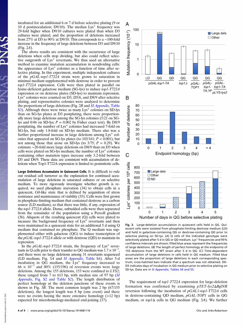
incubated for an additional 6 or 7 d before selective plating (9 or10 d postinoculation; D9/10). The median Lys+ frequency was29-fold higher when D9/10 cultures were plated than when D3cultures were plated, and the proportion of deletions increasedfrom 27% at D3 to 90% at D9/10. This corresponds to a ∼100-foldincrease in the frequency of large deletions between D3 and D9/10(Fig. 2A).The above results are consistent with the occurrence of large
deletions when cells stop dividing, but also could reflect selec-tive outgrowth of Lys+ revertants. We thus used an alternativemethod to examine mutation accumulation in nondividing cells:the appearance of Lys+ colonies as a function of time after se-lective plating. In this experiment, multiple independent culturesof the pGAL-top1-T722A strain were grown to saturation inminimal medium supplemented with dextrose in order to preventtop1-T722A expression. Cells were then plated in parallel onlysine-deficient galactose medium (SG-lys) to induce top1-T722Aexpression or on dextrose plates (SD-lys) to maintain repression.Lys+ colonies were counted on D3, D5/6, and D8/9 after selectiveplating, and representative colonies were analyzed to determinethe proportions of large deletions (Fig. 2B and SI Appendix, TableS3). Although there were twice as many Lys+ colonies on SD-lysthan on SG-lys plates at D3 postplating, there were proportion-ally more large deletions among the SG-lys colonies (5/21 on SG-lys and 0/46 on SD-lys; P = 0.002 by Fisher exact test). By D8/9postplating, the number of Lys+ colonies had increased 7-fold onSG-lys, but only 1.8-fold on SD-lys medium. There also was afurther proportional increase in large deletions among Lys+ col-onies that appeared on SG-lys plates (to 103/161; P < 0.001), butnot among those that arose on SD-lys (to 3/75; P = 0.29). Weestimate ∼20-fold more large deletions on D8/9 than on D3 whencells were plated on SG-lys medium; the number of Lys+ coloniescontaining other mutation types increase only 3.2-fold betweenD3 and D8/9. These data are consistent with accumulation of de-letions when Top1-T722A expression is limited to postmitotic cells.
Large Deletions Accumulate in Quiescent Cells. It is difficult to ruleout residual cell turnover as the explanation for continued accu-mulation of large deletions in saturated cultures or on selectivemedium. To more rigorously investigate whether growth is re-quired, we used phosphate starvation (34) to obtain cells in aquiescent, G0-like state that is defined by acquisition of stressresistance and maintenance of viability (35). Cells were first grownin phosphate-limiting medium that contained dextrose as a carbonsource (LD medium), so that there was little, if any, expression ofthe top1-T722A allele. Dense, unbudded cells were then separatedfrom the remainder of the population using a Percoll gradient(36). Aliquots of the resulting quiescent (Q) cells were plated tomeasure the background frequency of Lys+ revertants and thenwere maintained in a quiescent state for an additional 5 d using Qmedium that contained no phosphate. The Q medium was sup-plemented either with galactose (QG) to induce transcription ofthe pGAL-top1-T722A allele or with dextrose (QD) to maintain itsrepression.In the pGAL-top1-T722A strain, the frequency of Lys+ rever-
tants in Q cells prior to their transfer to QGmedium was 1.7 × 10⁻⁷,and there were no large deletions among 31 revertants sequenced(LD medium; Fig. 3A and SI Appendix, Table S4). After 5-dincubation in QG medium, the Lys+ frequency increased to66 × 10−7 and 80% (155/196) of reversion events were largedeletions. Among the 155 deletions, 153 were confined to LYS2;these ranged from 7 to 613 bp, with median size of 97 bp (SIAppendix, Fig. S2 and Table S2). The length distribution ofperfect homology at the deletion junctions of these events isshown in Fig. 3B. The most common length was 2 bp (67/155deletions), the longest length was 8 bp (one event) and therewere no events having the more extensive homology (>12 bp)expected for microhomology-mediated end-joining (37).
The requirement of top1-T722A expression for large-deletionformation was confirmed by examining pTET-lys2ΔBglNRreversion following the incubation of pGAL1-top1-T722A cellsin dextrose-containing QD medium, pGAL-TOP1 cells in QGmedium, or top1Δ cells in QG medium (Fig. 3A). We further
0100200300400500600700800
0 1 2 3 4 5
Mut
atio
n fre
quen
cy (1
0-7 )
A
Lys+
colo
nies
per
108
cells
C
Large delsOtherND
Number of days in QG before selective plating
0
1
10
100Large delsOther
pGAL-TOP1
LD QG QD QG QG QG QGpGAL-top1-TA top1 pGAL-
top1-TA,YF
pGAL-top1-TA
pLYS-lys2
Num
ber o
f del
etio
ns
Endpoint homology (bp)
01020304050607080
0 1 2 3 4 > 4
B
Fig. 3. Large-deletion accumulation in quiescent cells held in QG. Qui-escent cells were isolated from phosphate-limiting dextrose medium (LD)and held in galactose-containing QG or dextrose-containing QD prior toselective plating on SD-lys. (A) Q cells of the indicated genotype wereselectively plated after 5 d in QG or QD medium. Lys+ frequencies and 95%confidence intervals are shown; filled blue areas represent the frequenciesof large deletions. (B) The length of perfect homology at the endpoints of155 deletions from the WT strain after 5 d in QG. (C ) Time-dependentaccumulation of large deletions in cells held in QG medium. Filled blueareas are the proportion of large deletions in each corresponding spec-trum; cross-hatched bars indicate that a spectrum was not obtained. D0–D5 indicates days of incubation in QG medium prior to selective plating onSD-lys. Data are in SI Appendix, Tables S4 and S5.
4 of 9 | www.pnas.org/cgi/doi/10.1073/pnas.1914081116 Cho and Jinks-Robertson
Dow
nloa
ded
by g
uest
on
Apr
il 12
, 202
0

confirmed a requirement for Top1-T722A catalytic activity bymutating the active-site tyrosine (pGAL1-top1-T722A,Y727F allele)as well as the stimulatory effect of high transcription using a strainin which the lys2ΔBglNR allele was under the control of the en-dogenous LYS2 promoter. Finally, the time dependence ofLys+ colony accumulation was examined by incubating quiescentcells for 1, 2, 3, 4, or 5 d in QG medium prior to selective plating.The accumulation of revertants was roughly linear with time (Fig.3C and SI Appendix, Table S5). Although we hypothesize thatthe entire deletion process occurs in Q cells, microscopic ex-amination after selective plating revealed that most cells un-derwent 1 to 2 divisions. It thus is possible that only the primarylesion associated with Top1-T722A expression accumulatesduring quiescence, with the remaining steps in deletion for-mation occurring after selective plating.
Genetic Control of Large Deletions in Quiescent Cells. The NHEJdependence of large deletions that arose during growth in SGmedium was recapitulated in Percoll-isolated Q cells incubated for5 d in QG medium. In a yku70Δ background, the large-deletionfrequency was reduced several orders of magnitude (Fig. 4) and asimilar decrease was observed in a dnl4Δ background that lackedthe dedicated NHEJ ligase, DNA ligase 4 (38). Finally, we exam-ined the role of DNA polymerase 4 (Pol4), an X-family polymerasethat is important during end/gap filling during error-prone NHEJ(39). In contrast to the complete dependence of large deletions onKu and Dnl4, their frequency was reduced only ∼3.5-fold in a pol4Δbackground. This is consistent either with the direct joining of bluntends, which requires no DNA synthesis, and/or the participation ofalternative DNA polymerases in end/gap filling (40, 41).Before top1-T722A associated ends can be joined by NHEJ,
the 3′-linked Top1cc must be removed. Tdp1 is a tyrosyl-DNAphosphodiesterase that releases peptides covalently linked to 5′or 3′ DNA ends, including those that remain after Top1cc pro-teolysis (42). In quiescent cells, TDP1 loss was associated with onlya 2.8-fold reduction in large deletions (Fig. 4), indicating in-volvement of additional end-processing proteins/complexes. Wss1,which is the functional homolog of the mammalian SPRTN pro-tein (43), is a metalloprotease that degrades the protein compo-nent of DNA–protein cross-links and acts in parallel with Tdp1 toprocess potentially lethal Top1ccs in yeast (44). Compared to WT,there was a 2.3-fold reduction in the large-deletion frequency
in the wss1Δ background, which is similar to the reduction ob-served in the tdp1Δ background. In a tdp1Δ wss1Δ double-mutantbackground, however, the frequency of large deletions was reducedseveral orders of magnitude (Fig. 4), which is consistent withfunctional redundancy in Top1cc removal.In growing cells, deletion of EXO1 was associated with an ∼4-
fold increase in the large-deletion rate (Fig. 2A). By contrast,there was no change in the large-deletion frequency when exo1ΔQ cells were examined (Fig. 4). This is consistent with the cellcycle regulation of end resection, and its expected inefficiencyoutside of S phase (45). Alternatively, there may be little Exo1that persists or is expressed in Q cells. Finally, the effect of themre11-D56N nuclease-dead allele was markedly different ingrowing versus Q cells. Whereas there was an ∼7-fold increase inthe large-deletion rate in mre11-D56N growing cells, there wasno significant change in the deletion frequency when Q cellswere examined (Fig. 4). This difference likely reflects the cellcycle-dependent activation of Mre11 nuclease activity (46, 47).
Deletion of MRE11 or MUS81 Is Not Lethal in Q Cells Expressing Top1-T722A. An advantage of limiting expression of Top1-T722A toquiescent cells is the potential to bypass the very slow growth and/orsynthetic lethality observed in some null backgrounds. Syntheticlethality between top1-T722A and MRE11 or MUS81 deletion waspreviously reported (21) and was confirmed in our system. Neitheran mre11Δ nor a mus81Δ strain containing the pGAL-top1-T722Aallele was able to grow in SG medium, while both grew in SDmedium. The replication-specific synthetic lethality is assumed toreflect an inability to process large numbers of replication forks thatstall or collapse at Top1ccs. When Top1-T722A expression waslimited to quiescent cells, the WT,mre11Δ, andmus81Δ strains hadsimilar viability decreases (20%, 33%, and 10%, respectively) after5 d in QG medium. In the mre11Δ background, the Lys+ frequencyat day 5 was reduced ∼25-fold, and there were no large deletionsamong 71 revertants sequenced. The requirement for MRE11 isconsistent with the essential role of MRX in NHEJ (48, 49). Mus81is part of the Mus81–Mms4 structure-specific nuclease (50, 51), andin the mus81Δ background, there was a 4-fold reduction in thelarge-deletion frequency (Fig. 4).
CPT Treatment Induces Large Deletions in pGAL-TOP1 Quiescent Cells.To examine whether treating TOP1 cells with CPT inducedlarge-deletion formation, we incubated Q cells containing a pGAL-TOP1 allele for 5 d in QG medium supplemented with 100 μMCPT dissolved in DMSO or with an equal volume of DMSO.There was no toxicity associated with CPT treatment in QG me-dium, consistent with the quiescent state of cells. Although thetotal Lys+ reversion frequency was only 2-fold higher in the pres-ence of CPT, there was a dramatic change in the mutation spec-trum upon CPT addition (SI Appendix, Tables S2 and S4). In theDMSO control, only a single large deletion was detected among41 revertants analyzed. By contrast, there were 32 large deletionsamong 90 Lys+ colonies analyzed following incubation in thepresence of CPT (P < 0.001).
DiscussionStabilization of the covalent Top1cc leads to potentially toxic DSBformation during replication and in yeast, to elevated levels ofhomologous recombination (7, 52). An additional collateral effectis the accumulation of small 2- to 5-bp deletions that involve onlynicked DNA and are DSB independent (18). In the current study,we used haploid yeast strains to more broadly examine mutageniceffects associated with stabilization of the Top1cc either byexpressing the Top1-T722A protein or by adding CPT to WT cells.Regardless of the mechanism of Top1cc stabilization, NHEJ-dependent large deletions with little or no junction homology werethe predominant outcome. Because of the selective system used,these deletions fell into 2 distinct classes: deletions up to ∼600 bp
Mutation frequency in Q cells (10-7)
0 20 40 60 80 100 120
mus81
mre11
mre11-D56N
exo1
tdp1 wss1
wss1
tdp1
pol4
dnl4
yku70
WT Large dels
Other
Gen
otyp
e
Fig. 4. Genetic requirements of Top1-T722A–associated deletions in qui-escent cells. Quiescent cells were isolated from phosphate-limiting dex-trose medium (LD) and held in QG medium for 5 d prior to selective platingon SD-lys. Lys+ frequencies and 95% confidence intervals are shown; filledblue areas are the frequencies of large deletions in each spectrum. Dataare in SI Appendix, Table S4.
Cho and Jinks-Robertson PNAS Latest Articles | 5 of 9
GEN
ETICS
Dow
nloa
ded
by g
uest
on
Apr
il 12
, 202
0

(median size, ∼100 bp) that were confined to LYS2, and 3.2- to4.1-kb deletions that deleted the 5′ end of LYS2 and fusedthe remainder of the gene to the upstream kanMX gene. Thedispensability of the N terminus of the Lys2 protein as well assequence downstream of the theoretical window for compensa-tory changes was unexpected and further extends the flexibilityof LYS2-based frameshift-reversion reporters. Given the re-quirement for the creation of a functional protein, the currentanalyses likely detected only a subset of deletions and their up-per size limit remains unknown.NHEJ assays in yeast typically measure the error-prone repair
of a defined chromosomal DSB or the circularization of a line-arized plasmid following transformation (53). In the absence of arepair template for homologous recombination, Ku-dependentNHEJ occurs efficiently. The ends are directly joined in the caseof plasmid-based assays or are only slightly modified by deletion/insertion of a few base pairs to prevent reiterative cleavage inchromosomal assays. The absolute Ku dependence of the Top1-dependent deletions and the repressive effect of the Exo1 exo-nuclease on these events is inconsistent with significant loss ofsequence from the ends of a single DSB. We suggest instead thatthe events observed here reflect the NHEJ-mediated joining ofends from 2 independent DSBs. These genetic requirements, aswell as the requirement for Top1 processing by Tdp1/Wss1, alsoexclude an alternative mechanism in which Top1 directly gen-erates a deletion intermediate on only one DNA strand. This canoccur if a given Top1cc is attacked by the 5′-OH generated by aseparate aborted cleavage–ligation reaction (Fig. 5A and refs. 54and 55). We note that a similar Top1-only mechanism involvingsister chromatids would be expected to generate duplications aswell as deletions and duplications were not detected (see below).Our initial assumption was that the relevant DSBs were most
likely generated by the encounter of 2 distinct Top1ccs with in-dependent replication forks. There are several reasons why wedisfavor this particular mechanism. First, the accumulation of
time-dependent deletions in saturated cultures, on solid mediumafter selective plating, and in isolated quiescent cells is suggestiveof replication-independent DSB formation. Because cells undergo 1to 2 divisions after selective plating, however, it remains possiblethat only the relevant Top1ccs (i.e., stabilized nicks) arise in non-dividing cells and that these are subsequently converted to DSBsupon selective plating. Second, the effects of exo1Δ and the mre11-D56N allele were different in dividing versus quiescent cells. Ad-ditionally, deletion of theMRE11 orMUS81 gene, which is lethal ina top1-T722A background, was tolerated when top1-T722A expres-sion was limited to quiescent cells. Finally, as illustrated in Fig. 5B, ifthe encounter of 2 separate Top1ccs by converging replication forksbreaks both sister chromatids, duplications as well as deletions areexpected. Although duplications should be compatible with Lys2function, they strikingly were absent in the current analysis. Wenote, however, that if both replication-associated breaks are on thesame sister chromatid (not shown), only deletions would occur. Inthis context, however, repair via the much more efficient recombi-nation pathway would be expected to exclude frequent NHEJ.If Top1-dependent DSBs are not replication-associated, then
how do they arise? In vitro, DSBs can be directly generated byTop1 if one DNA strand contains a ribonucleotide (32). The firststrand is broken when Top1 incises at the ribonucleotide and isreleased, leaving behind a nick flanked by a 2′,3′-cyclic phosphateand a 5′-OH. Top1 incision of complementary strand nearby re-sults in a DSB with the enzyme linked to the 3′ end. Although theenhanced accumulation of DSBs in a genetic background thatelevates ribonucleotides levels in DNA indicates that this alsooccurs in vivo (32), elevating the level of ribonucleotides had noeffect on the large-deletion frequency in our system. As an alter-native replication-independent mechanism, we suggest that therelevant DSBs can arise either when Top1 incises both DNAstrands in close proximity or when Top1 nicks DNA when there isa preexisting nick on the complementary strand (Fig. 5C). Thiswould be followed by Top1cc removal by Tdp1/Wss1 and
OR
Tdp1Wss1
(Mus81)
KuMre11
Dnl4(Pol4)
Ku-dependentdeletion
B
C
Duplication
A
Tdp1Wss1
(Mus81)
AND
Ku-independentdeletion
Fig. 5. Mechanisms for large-deletion formation. Deletions are initiated by 2 trapped Top1ccs (yellow lollipops); arrowheads and black circles indicate 3′-OH and5′-OH ends, respectively. Red lines correspond to newly replicated DNA strands. (A) Top1ccs are trapped on the same DNA strand and the attack (blue arrow) of thephosphotyrosine bond of the upstream Top1cc by the 5′-OH of the second Top1cc reseals the top strand and deletes the intervening DNA segment. Because Top1facilitates the ligation, NHEJ is not required. (B) Top1ccs are on complementary strands, and both sister chromatids are broken when encountered by convergingreplication forks. NHEJ-mediated ligation to the fragment from the sister chromatid generates either a deletion or duplication of the region flanked by the nicks. (C)DSBs are created when Top1 cleaves opposite random or Top1-generated nicks. The fragment between the DSBs is lost and NHEJ ligates the broken ends followingTop1cc removal. Proteins involved in large-deletion formation in Q cells are indicated at the relevant step. Those that play a relatively minor role are in parentheses.
6 of 9 | www.pnas.org/cgi/doi/10.1073/pnas.1914081116 Cho and Jinks-Robertson
Dow
nloa
ded
by g
uest
on
Apr
il 12
, 202
0

engagement of Ku for subsequent NHEJ. Although we consid-ered the large deletions that were confined to LYS2 and thosethat created a fusion protein jointly, when data from all geneticbackgrounds were summed, there was significantly more of the4-kb fusion class detected when cells were grown for 3 d in SGmedium than when top1-T722A expression was limited to quies-cent cells (21/132 and 14/543, respectively, in SI Appendix, TableS2; P < 0.0001). It is possible that this difference reflects a shiftfrom a mostly replication-dependent mechanism to a mostlyreplication-independent mechanism.NHEJ plays only a very minor role during DSB repair in yeast,
and large deletions similar to those reported here were not asso-ciated with CPT treatment or Top1-T722A expression in a diploidstrain (52). Although NHEJ is only a minor DSB repair pathway inyeast, it plays a prominent role during DSB repair in mammaliancells (56). We suggest that the Top1cc-dependent deletions de-scribed here are likely relevant to uncharacterized rearrangementsobserved 20 y ago when selecting forward mutations in mamma-lian cells treated with a Top1 poison (19, 20). These may havebeen replication dependent in a context where NHEJ is efficient,while being largely relegated to nondividing cells in yeast. SimilarTop1-initiated events may provide a source of genetic change inevolving cancer cells, especially following patient treatment with aTop1 poison. In addition, a similar mechanism that is primarilytranscription driven may be a potential source of genetic insta-bility in dormant tumor cells (57) and in postmitotic tissues (58).Although the deletion size was limited to ∼4 kb because of theselective system used here, larger deletions or rearrangementswithin/between chromosomes are expected and could be de-tected using an appropriate selective system.
Materials and MethodsStrain Constructions. SI Appendix, Table S6 contains a list of the strains usedin the experiments reported. All growth was at 30 °C, and 2 independentisolates of each genetic background were used to generate data. Strains weremaintained as glycerol stocks at −80 °C, and all were derived from YPH45[MATa ura3-52 ade2-101oc lys2-801am trp1Δ1], a strain congenic with S288C (59)by transformation or by mating with an isogenic strain to combine desiredgenetic markers. Derivatives were made by transformation or through thecrossing of appropriate YPH45 derivatives of opposite mating type. The MATαversion of YPH45 was obtained through a mating-type switch using a pGAL-HOplasmid. In all strains, the endogenous LYS2 locus on chromosome II was de-leted, and the full-length gene was inserted on chromosome III near ARS306 inthe orientation in which replication and transcription forks move in the samedirection (60). The transplanted allele was either under control of the LYS2promoter or regulated by a 2-component, doxycycline-repressible pTET-offsystem (61). The lys2ΔBglNR allele (22) was derived by mutating homopoly-mer runs >3 N in the reversion window of the lys2ΔBgl allele, an allele with a4-bp insertion generated by filling in a unique BglII restriction site within LYS2(23). The delitto perfetto method, which involves insertion of a selectablecassette and its subsequent replacement with a DNA fragment of interest bycounterselection (62), was used to introduce the lys2ΔBglNR allele from XhoI/AflII-digested pSR701 (22).
To construct the pGAL-TOP1 allele, the promoter of TOP1was replacedwithpGAL1 via one-step allele replacement using a TRP1:pGAL1 cassette ampli-fied from plasmid pFA6a-TRP1-PGAL1 (63). Delitto perfetto was then usedto introduce the top1-T722A or top1-T722A,Y727F allele as part of a smallduplex fragment obtained by annealing complementary oligonucleotides.The mre11-D56N allele was introduced by 2-step allele replacement usingSphI-digested pSM444 (64). The TOP1, DNL4, YKU70, POL4, EXO1, MRE11,MUS81, TDP1, and WSS1 genes were deleted by 1-step allele replacementusing a PCR-generated cassette amplified from a plasmid containing an ap-propriate selective marker. The loxP-TRP1-loxP disruption cassette was am-plified from pSR954, which was constructed by replacing the kanMX4 gene ofpUG6 (65) with TRP1. The loxP-URA3Kl-loxP cassette was amplified frompUG72 (66) and the natMX4 marker from pAG25 (67). The loxP-hph-loxPcassette was amplified from pSR955, which was constructed by inserting aBglII/SacI fragment containing hphMX4 (67) into BglII/SacI-digested pUG6 (65).
Mutation Rate and Frequency Measurements in Galactose Medium. In-dependent cultures were started by inoculating single colonies from YEPD
plates (1% yeast extract, 2% Bacto-peptone, 250 μg/mL adenine hemisulfatesupplemented with 2% dextrose) into synthetic complete medium (6.7 g/L yeastnitrogen base plus 1.4 g of a mix of all amino acids, uracil, and adenine) sup-plemented with 2% galactose (SG). Following 3 d of growth, cells were washedwith H2O, and appropriate dilutions were plated onto synthetic complete dex-trose medium lacking lysine (SD-lys) and YEPD to determine the total number ofLys+ revertants and the average number of cells, respectively, in each culture.Colonies were counted 3 d after plating. Mutation rates were calculated using themethod of the median (68), and 95% confidence intervals were determined asdescribed previously (69). Alternatively, the mean Lys+ frequency was calculatedand the SEM was used to derive 95% confidence intervals. The rate or frequencyof large deletions was estimated by multiplying the rate or frequency ofLys+ colonies by the proportion of large deletions in the corresponding spectrum.
To examine the accumulation of Lys+ revertants in saturated cultures, SGcultures were inoculated to a concentration of 20,000 cells per mL. Cellswere counted daily using a hemocytometer and reached saturation 3 to 4 dafter inoculation. To determine Lys+ reversion frequencies before and aftercultures reached saturation, aliquots of each culture were removed 3 d and 9or 10 d after inoculation and plated selectively and nonselectively. Followingthe calculation of the mutation frequency in each culture, mean frequencies,95% confidence intervals, and large-deletion frequencies were determinedas described above. Data are in SI Appendix, Table S1.
Time-Dependent Accumulation of Large Deletions on SD-Lys and SG-Lys Plates.Independent cultures were grown for 4 d in SD medium to suppress pGAL-top1-T722A expression. Cells were washed, and appropriate dilutions wereplated on YEPD plates to determine the number of viable cells in each cul-ture. The remaining cells were plated in parallel on SD-lys and SG-lys plates,and Lys+ colony appearance was monitored over a 9-d period. On D3, D5/6,or D8/9, newly appearing Lys+ colonies were counted and DNA from rep-resentative colonies was isolated and analyzed to obtain a correspondingspectrum. Data are provided in SI Appendix, Table S3.
Mutation Frequencies in Quiescent Cells. Five to 10 colonies from YPD plateswere inoculated into 250 or 500 mL of phosphate-limiting L medium (0.07%phosphate-free yeast nitrogen base, 10 μg/mL KH2PO4, 0.1% KCl, 0.5%NH4SO4,2% dextrose, 1.4 g/L complete amino acids mix) and incubated with shaking for7 to 8 d. Cells were collected by centrifugation, washed with H2O, resuspendedin 1 mL of 50 mM Tris·HCl, pH 7.9, and sonicated for 3 min using an IsonicDigital Ultrasonic Cleaner. Percoll gradients were made by mixing 1 part 1.5 MNaCl with 9 parts Percoll (density 1.130 g/mL; GE Healthcare), and 25 mL weredistributed into 30-mL glass conical tubes. Tubes were centrifuged for 15 min at13,800 rpm at 20 °C in a fixed-angle Sorvall SS-34 rotor. Cells were loaded ontoPercoll gradients and centrifuged for 1 h at 400 rcf in a swinging bucket rotor(Eppendorf centrifuge 5810R) at 20 °C. After centrifugation, quiescent cellswere taken from the bottom fraction, washed with H2O, and counted in ahemocytometer. Appropriate dilutions were plated on YEPD to determine vi-ability and on SD-lys to determine the number of preexisting revertants fol-lowing incubation in L medium. Cells were then pelleted and resuspended inphosphate-free Q medium (0.07% phosphate-free yeast nitrogen base, 0.1%KCl, 0.5% NH4SO4, 1.4 g/L complete amino acids mix) containing either 2%dextrose or 2% galactose (QD and QG, respectively). Following incubation ofQG or QD cultures on a roller drum for the appropriate time period, cells werewashed and appropriate dilutions were plated on YEPD and SD-lys in order todetermine total viable cells and Lys+ revertants, respectively, in each culture.Colonies were counted 3 d postplating, and mean Lys+ frequencies, 95% con-fidence intervals, and large-deletion frequencies were determined as describedabove. Data from Q-medium experiments are in SI Appendix, Tables S4 and S5.
Mutation Spectra. For SG liquid cultures, only a single colony was selectedfrom each culture to ensure independence. For SG-lys plate experiments andQ-media experiments in which cells were not dividing, multiple Lys+ colonieswere analyzed from a single plate/culture. Following genomic DNA isolation,the lys2 reversion window was amplified using forward primer LYSWINF (5-GCCTCATGATAGTTTTTCTAACAAATACG) or Lys2ATG (5′-GACTAACGAAAA-GGTCTGGATAG) with reverse primer LYSWINR (5′-CCCATCACACATACCA-TCAAATCCAC). If the initial PCR failed, a product was obtained using primerLys2F14 (5′-CACAGTTTTAGCGAGG) and LYSWINR. PCR products were sequencedusing the forward or reverse primer used for amplification. Sequencing wasdone by the Duke University DNA Analysis Facility, Eurofins MWG Operon, orEton Bioscience. Sequences were analyzed using DNASTAR Seqman Pro, Snap-Gene Viewer, BBedit, and Python. Deletions confined to LYS2 ORF were alignedusing DNASTAR Seqman Pro and pasted into BBedit to save as a raw txt file forPython analysis. A Python script (70) was written to export CVS files containingdeletion start sites, sizes, junction identities, and the number of occurrences of
Cho and Jinks-Robertson PNAS Latest Articles | 7 of 9
GEN
ETICS
Dow
nloa
ded
by g
uest
on
Apr
il 12
, 202
0

each deletion. Python also was used to generate JPG files of individual deletionspectra. The 3.2- to 4.1-kb deletions were analyzed using Seqman Pro andSnapGene Viewer; deletion endpoints, sizes, and junction identities weremanually determined and plotted using Python. Coordinates of the deletionsdetected in different genetic backgrounds and/or media types are given in SIAppendix, Table S2. Corresponding graphical summaries of 3.2- to 4.1-kb de-letions and deletions confined to the lys2ΔBlgNR reversion window are pre-sented in SI Appendix, Figs. S1 and S2, respectively.
Data Availability. All data discussed in the paper are in the SI Appendix.
ACKNOWLEDGMENTS. We thank Alex Neil and Sergei Mirkin for sharingtheir procedure for isolating quiescent cells following phosphate starvation;Karen O’Connell and Muthana Al Abo for their help in writing Pythonscripts; and Tom Petes for comments on the manuscript. This work wassupported by a grant to S.J.-R. from the National Institutes of Health (R35GM203587).
1. Y. Pommier, Y. Sun, S. N. Huang, J. L. Nitiss, Roles of eukaryotic topoisomerases intranscription, replication and genomic stability. Nat. Rev. Mol. Cell Biol. 17, 703–721(2016).
2. Y. Fan, J. N. Weinstein, K. W. Kohn, L. M. Shi, Y. Pommier, Molecular modeling studiesof the DNA-topoisomerase I ternary cleavable complex with camptothecin. J. Med.Chem. 41, 2216–2226 (1998).
3. Y. H. Hsiang, R. Hertzberg, S. Hecht, L. F. Liu, Camptothecin induces protein-linkedDNA breaks via mammalian DNA topoisomerase I. J. Biol. Chem. 260, 14873–14878(1985).
4. M. R. Redinbo, L. Stewart, P. Kuhn, J. J. Champoux, W. G. Hol, Crystal structures ofhuman topoisomerase I in covalent and noncovalent complexes with DNA. Science279, 1504–1513 (1998).
5. Y. Pommier, Drugging topoisomerases: Lessons and challenges. ACS Chem. Biol. 8, 82–95 (2013).
6. W. K. Eng, L. Faucette, R. K. Johnson, R. Sternglanz, Evidence that DNA topoisomeraseI is necessary for the cytotoxic effects of camptothecin. Mol. Pharmacol. 34, 755–760(1988).
7. J. Nitiss, J. C. Wang, DNA topoisomerase-targeting antitumor drugs can be studied inyeast. Proc. Natl. Acad. Sci. U.S.A. 85, 7501–7505 (1988).
8. S. J. Boulton, S. P. Jackson, Saccharomyces cerevisiae Ku70 potentiates illegitimateDNA double-strand break repair and serves as a barrier to error-prone DNA repairpathways. EMBO J. 15, 5093–5103 (1996).
9. M. J. Lippert et al., Role for topoisomerase 1 in transcription-associated mutagenesisin yeast. Proc. Natl. Acad. Sci. U.S.A. 108, 698–703 (2011).
10. T. Takahashi, G. Burguiere-Slezak, P. A. Van der Kemp, S. Boiteux, Topoisomerase 1provokes the formation of short deletions in repeated sequences upon high tran-scription in Saccharomyces cerevisiae. Proc. Natl. Acad. Sci. U.S.A. 108, 692–697 (2011).
11. J. E. Cho, N. Kim, Y. C. Li, S. Jinks-Robertson, Two distinct mechanisms of topoisomerase1-dependent mutagenesis in yeast. DNA Repair (Amst.) 12, 205–211 (2013).
12. N. Kim et al., Mutagenic processing of ribonucleotides in DNA by yeast topoisomeraseI. Science 332, 1561–1564 (2011).
13. J. E. Cho et al., Parallel analysis of ribonucleotide-dependent deletions produced byyeast Top1 in vitro and in vivo. Nucleic Acids Res. 44, 7714–7721 (2016).
14. S. Y. Huang, S. Ghosh, Y. Pommier, Topoisomerase I alone is sufficient to produceshort DNA deletions and can also reverse nicks at ribonucleotide sites. J. Biol. Chem.290, 14068–14076 (2015).
15. J. L. Sparks, P. M. Burgers, Error-free and mutagenic processing of topoisomerase1-provoked damage at genomic ribonucleotides. EMBO J. 34, 1259–1269 (2015).
16. W. C. Colley, M. van der Merwe, J. R. Vance, A. B. Burgin, Jr, M. A. Bjornsti, Substitutionof conserved residues within the active site alters the cleavage religation equilibrium ofDNA topoisomerase I. J. Biol. Chem. 279, 54069–54078 (2004).
17. M. D. Megonigal, J. Fertala, M. A. Bjornsti, Alterations in the catalytic activity of yeastDNA topoisomerase I result in cell cycle arrest and cell death. J. Biol. Chem. 272,12801–12808 (1997).
18. R. Sloan, S. N. Huang, Y. Pommier, S. Jinks-Robertson, Effects of camptothecin orTOP1 overexpression on genetic stability in Saccharomyces cerevisiae. DNA Repair(Amst.) 59, 69–75 (2017).
19. E. Balestrieri, R. Zanier, F. Degrassi, Molecular characterisation of camptothecin-induced mutations at the hprt locus in Chinese hamster cells. Mutat. Res. 476, 63–69 (2001).
20. H. Hashimoto, S. Chatterjee, N. A. Berger, Mutagenic activity of topoisomerase I in-hibitors. Clin. Cancer Res. 1, 369–376 (1995).
21. R. J. Reid et al., Selective ploidy ablation, a high-throughput plasmid transfer pro-tocol, identifies new genes affecting topoisomerase I-induced DNA damage. GenomeRes. 21, 477–486 (2011).
22. K. Lehner, S. V. Mudrak, B. K. Minesinger, S. Jinks-Robertson, Frameshift mutagenesis:The roles of primer-template misalignment and the nonhomologous end-joiningpathway in Saccharomyces cerevisiae. Genetics 190, 501–510 (2012).
23. C. N. Greene, S. Jinks-Robertson, Frameshift intermediates in homopolymer runs are re-moved efficiently by yeast mismatch repair proteins.Mol. Cell. Biol. 17, 2844–2850 (1997).
24. H. P. Phatnani, J. C. Jones, A. L. Greenleaf, Expanding the functional repertoire of CTDkinase I and RNA polymerase II: Novel phosphoCTD-associating proteins in the yeastproteome. Biochemistry 43, 15702–15719 (2004).
25. J. Wu, H. P. Phatnani, T. S. Hsieh, A. L. Greenleaf, The phosphoCTD-interacting do-main of topoisomerase I. Biochem. Biophys. Res. Commun. 397, 117–119 (2010).
26. T. T. Paull, 20 years of Mre11 biology: No end in sight. Mol. Cell 71, 419–427 (2018).27. S. Moreau, J. R. Ferguson, L. S. Symington, The nuclease activity of Mre11 is required
for meiosis but not for mating type switching, end joining, or telomere maintenance.Mol. Cell. Biol. 19, 556–566 (1999).
28. E. P. Mimitou, L. S. Symington, Ku prevents Exo1 and Sgs1-dependent resection ofDNA ends in the absence of a functional MRX complex or Sae2. EMBO J. 29, 3358–3369 (2010).
29. N. K. Hamilton, N. Maizels, MRE11 function in response to topoisomerase poisons isindependent of its function in double-strand break repair in Saccharomyces cerevisiae.PLoS One 5, e15387 (2010).
30. L. R. Myler et al., Single-molecule imaging reveals how Mre11-Rad50-Nbs1 initiatesDNA break repair. Mol. Cell 67, 891–898.e4 (2017).
31. S. S. Foster, A. Balestrini, J. H. Petrini, Functional interplay of the Mre11 nuclease andKu in the response to replication-associated DNA damage. Mol. Cell. Biol. 31, 4379–4389 (2011).
32. S. N. Huang, J. S. Williams, M. E. Arana, T. A. Kunkel, Y. Pommier, TopoisomeraseI-mediated cleavage at unrepaired ribonucleotides generates DNA double-strandbreaks. EMBO J. 36, 361–373 (2017).
33. S. A. Nick McElhinny et al., Genome instability due to ribonucleotide incorporationinto DNA. Nat. Chem. Biol. 6, 774–781 (2010).
34. M. M. Klosinska, C. A. Crutchfield, P. H. Bradley, J. D. Rabinowitz, J. R. Broach, Yeastcells can access distinct quiescent states. Genes Dev. 25, 336–349 (2011).
35. J. V. Gray et al., “Sleeping beauty”: Quiescence in Saccharomyces cerevisiae. Micro-biol. Mol. Biol. Rev. 68, 187–206 (2004).
36. C. Allen et al., Isolation of quiescent and nonquiescent cells from yeast stationary-phase cultures. J. Cell Biol. 174, 89–100 (2006).
37. D. D. Villarreal et al., Microhomology directs diverse DNA break repair pathways andchromosomal translocations. PLoS Genet. 8, e1003026 (2012).
38. S. H. Teo, S. P. Jackson, Identification of Saccharomyces cerevisiae DNA ligase IV: In-volvement in DNA double-strand break repair. EMBO J. 16, 4788–4795 (1997).
39. J. M. Daley, R. L. Laan, A. Suresh, T. E. Wilson, DNA joint dependence of pol X familypolymerase action in nonhomologous end joining. J. Biol. Chem. 280, 29030–29037 (2005).
40. C. Y. Chan, A. Galli, R. H. Schiestl, Pol3 is involved in nonhomologous end-joining inSaccharomyces cerevisiae. DNA Repair (Amst.) 7, 1531–1541 (2008).
41. S. F. Tseng, A. Gabriel, S. C. Teng, Proofreading activity of DNA polymerase Pol2mediates 3′-end processing during nonhomologous end joining in yeast. PLoS Genet.4, e1000060 (2008).
42. J. J. Pouliot, K. C. Yao, C. A. Robertson, H. A. Nash, Yeast gene for a Tyr-DNA phos-phodiesterase that repairs topoisomerase I complexes. Science 286, 552–555 (1999).
43. J. Fielden, A. Ruggiano, M. Popovi�c, K. Ramadan, DNA protein crosslink proteolysisrepair: From yeast to premature ageing and cancer in humans. DNA Repair (Amst.) 71,198–204 (2018).
44. J. Stingele, M. S. Schwarz, N. Bloemeke, P. G. Wolf, S. Jentsch, A DNA-dependentprotease involved in DNA-protein crosslink repair. Cell 158, 327–338 (2014).
45. L. S. Symington, End resection at double-strand breaks: Mechanism and regulation.Cold Spring Harb. Perspect. Biol. 6, a016436 (2014).
46. P. Huertas, F. Cortés-Ledesma, A. A. Sartori, A. Aguilera, S. P. Jackson, CDK targets Sae2 tocontrol DNA-end resection and homologous recombination. Nature 455, 689–692 (2008).
47. E. Cannavo et al., Regulatory control of DNA end resection by Sae2 phosphorylation.Nat. Commun. 9, 4016 (2018).
48. S. J. Boulton, S. P. Jackson, Components of the Ku-dependent non-homologous end-joining pathway are involved in telomeric length maintenance and telomeric silenc-ing. EMBO J. 17, 1819–1828 (1998).
49. J. K. Moore, J. E. Haber, Cell cycle and genetic requirements of two pathways ofnonhomologous end-joining repair of double-strand breaks in Saccharomyces cer-evisiae. Mol. Cell. Biol. 16, 2164–2173 (1996).
50. S. A. Bastin-Shanower, W. M. Fricke, J. R. Mullen, S. J. Brill, The mechanism of Mus81-Mms4 cleavage site selection distinguishes it from the homologous endonucleaseRad1-Rad10. Mol. Cell. Biol. 23, 3487–3496 (2003).
51. K. T. Ehmsen, W. D. Heyer, Saccharomyces cerevisiae Mus81-Mms4 is a catalytic, DNAstructure-selective endonuclease. Nucleic Acids Res. 36, 2182–2195 (2008).
52. S. L. Andersen, R. S. Sloan, T. D. Petes, S. Jinks-Robertson, Genome-destabilizing ef-fects associated with top1 loss or accumulation of top1 cleavage complexes in yeast.PLoS Genet. 11, e1005098 (2015).
53. J. M. Daley, P. L. Palmbos, D. Wu, T. E. Wilson, Nonhomologous end joining in yeast.Annu. Rev. Genet. 39, 431–451 (2005).
54. K. A. Henningfeld, S. M. Hecht, A model for topoisomerase I-mediated insertions anddeletions with duplex DNA substrates containing branches, nicks, and gaps. Bio-chemistry 34, 6120–6129 (1995).
55. N. Kim, S. Jinks-Robertson, The Top1 paradox: Friend and foe of the eukaryotic ge-nome. DNA Repair (Amst.) 56, 33–41 (2017).
56. E. M. Kass, M. Jasin, Collaboration and competition between DNA double-strandbreak repair pathways. FEBS Lett. 584, 3703–3708 (2010).
57. S. Kleffel, T. Schatton, Tumor dormancy and cancer stem cells: Two sides of the samecoin? Adv. Exp. Med. Biol. 734, 145–179 (2013).
58. P. J. McKinnon, Topoisomerases and the regulation of neural function. Nat. Rev.Neurosci. 17, 673–679 (2016).
59. R. S. Sikorski, P. Hieter, A system of shuttle vectors and yeast host strains designed forefficient manipulation of DNA in Saccharomyces cerevisiae. Genetics 122, 19–27(1989).
8 of 9 | www.pnas.org/cgi/doi/10.1073/pnas.1914081116 Cho and Jinks-Robertson
Dow
nloa
ded
by g
uest
on
Apr
il 12
, 202
0

60. N. Kim, A. L. Abdulovic, R. Gealy, M. J. Lippert, S. Jinks-Robertson, Transcription-associ-ated mutagenesis in yeast is directly proportional to the level of gene expression andinfluenced by the direction of DNA replication. DNA Repair (Amst.) 6, 1285–1296 (2007).
61. G. Bellí, E. Garí, M. Aldea, E. Herrero, Functional analysis of yeast essential genes usinga promoter-substitution cassette and the tetracycline-regulatable dual expressionsystem. Yeast 14, 1127–1138 (1998).
62. F. Storici, M. A. Resnick, The delitto perfetto approach to in vivo site-directed mu-tagenesis and chromosome rearrangements with synthetic oligonucleotides in yeast.Methods Enzymol. 409, 329–345 (2006).
63. M. S. Longtine et al., Additional modules for versatile and economical PCR-based genedeletion and modification in Saccharomyces cerevisiae. Yeast 14, 953–961 (1998).
64. B. Llorente, L. S. Symington, The Mre11 nuclease is not required for 5′ to 3′ resectionat multiple HO-induced double-strand breaks. Mol. Cell. Biol. 24, 9682–9694 (2004).
65. U. Güldener, S. Heck, T. Fielder, J. Beinhauer, J. H. Hegemann, A new efficient gene dis-ruption cassette for repeated use in budding yeast. Nucleic Acids Res. 24, 2519–2524 (1996).
66. U. Gueldener, J. Heinisch, G. J. Koehler, D. Voss, J. H. Hegemann, A second set of loxPmarker cassettes for Cre-mediated multiple gene knockouts in budding yeast. NucleicAcids Res. 30, e23 (2002).
67. A. L. Goldstein, J. H. McCusker, Three new dominant drug resistance cassettes forgene disruption in Saccharomyces cerevisiae. Yeast 15, 1541–1553 (1999).
68. D. E. Lea, C. A. Coulson, The distribution of the numbers of mutants in bacterialpopulations. J. Genet. 49, 264–285 (1949).
69. R. M. Spell, S. Jinks-Robertson, Determination of mitotic recombination rates byfluctuation analysis in Saccharomyces cerevisiae. Methods Mol. Biol. 262, 3–12 (2004).
70. J.-E. Cho, M. Al Abo, MHfinderatDelJct. Github. https://github.com/muthalpy/MHfinderatDelJct.git. Deposited 10 October 2019.
Cho and Jinks-Robertson PNAS Latest Articles | 9 of 9
GEN
ETICS
Dow
nloa
ded
by g
uest
on
Apr
il 12
, 202
0